Reversing the Resistance Phenotype of the Biomphalaria glabrata Snail Host Schistosoma mansoni...
14
Reversing the Resistance Phenotype of the Biomphalaria glabrata Snail Host Schistosoma mansoni Infection by Temperature Modulation Wannaporn Ittiprasert, Matty Knight* Biomedical Research Institute, Rockville, Maryland, United States of America Abstract Biomphalaria glabrata snails that display either resistant or susceptible phenotypes to the parasitic trematode, Schistosoma mansoni provide an invaluable resource towards elucidating the molecular basis of the snail-host/schistosome relationship. Previously, we showed that induction of stress genes either after heat-shock or parasite infection was a major feature distinguishing juvenile susceptible snails from their resistant counterparts. In order to examine this apparent association between heat stress and snail susceptibility, we investigated the effect of temperature modulation in the resistant snail stock, BS-90. Here, we show that, incubated for up to 4 hrs at 32uC prior to infection, these resistant snails became susceptible to infection, i.e. shedding cercariae at 5 weeks post exposure (PE) while unstressed resistant snails, as expected, remained resistant. This suggests that susceptibility to infection by this resistant snail phenotype is temperature-sensitive (ts). Additionally, resistant snails treated with the Hsp 90 specific inhibitor, geldanamycin (GA) after heat stress, were no longer susceptible to infection, retaining their resistant phenotype. Consistently, susceptible snail phenotypes treated with 100 mM GA before parasite exposure also remained uninfected. These results provide direct evidence for the induction of stress genes (heat shock proteins; Hsp 70, Hsp 90 and the reverse transcriptase [RT] domain of the nimbus non-LTR retrotransposon) in B. glabrata susceptibility to S. mansoni infection and characterize the resistant BS-90 snails as a temperature-sensitive phenotype. This study of reversing snail susceptibility phenotypes to S. mansoni provides an opportunity to directly track molecular pathway(s) that underlie the B. glabrata snail’s ability to either sustain or destroy the S. mansoni parasite. Citation: Ittiprasert W, Knight M (2012) Reversing the Resistance Phenotype of the Biomphalaria glabrata Snail Host Schistosoma mansoni Infection by Temperature Modulation. PLoS Pathog 8(4): e1002677. doi:10.1371/journal.ppat.1002677 Editor: Timothy P. Yoshino, University of Wisconsin-Madison, United States of America Received September 21, 2011; Accepted March 18, 2012; Published April 26, 2012 Copyright: ß 2012 Ittiprasert, Knight. This is an open-access article distributed under the terms of the Creative Commons Attribution License, which permits unrestricted use, distribution, and reproduction in any medium, provided the original author and source are credited. Funding: This work was funded by NIH-NIAID-R01AI063480. The funders had no role in study design, data collection and analysis, decision to publish, or preparation of the manuscript. Competing Interests: The authors have declared that no competing interests exist. * E-mail: [email protected] Introduction Schistosomes are parasitic trematodes that cause the chronic debilitating disease schistosomiasis, a neglected tropical disease that persists in over 70 countries of the developing world. It is estimated that at least 200 million people are chronically infected with the parasite with another 800 million remaining at risk for exposure. The disease burden is estimated at over 70 million disability-adjusted life years (DALYs) and there is increasing awareness that schistosomiasis can impact the epidemiology of other infectious diseases such as HIV (especially in female patients with genital schistosomiasis). A concerted effort is, therefore, being made to develop novel intervention tools that include blocking transmission of the parasite at the snail stage of its life cycle [1–3]. Freshwater snails serve as obligatory intermediate hosts for the development of parasitic trematodes. Throughout South America and the Caribbean Islands the snail, Biomphalaria glabrata plays an important role in the transmission of Schistosoma mansoni. The relative ease of maintaining B. glabrata in the laboratory has enabled it to become the host/pathogen model system of choice in which studies aimed at elucidating the molecular basis of snail/ schistosome interactions are being conducted. Thus far, studies using representative snail stocks that are either resistant or susceptible to the parasite provide an invaluable resource towards unraveling the complex biology of the snail/schistosome encoun- ter. For example, using pedigree snail stocks with varying susceptibility phenotypes, a strong genetic basis was shown to exist for the susceptibility of B. glabrata to S. mansoni [4]. In adult B. glabrata, resistance to S. mansoni has been shown to be a dominant single-gene trait that is inherited by simple Mendelian genetics. In juvenile snails, however, genetics of resistance has been shown to be a complex trait, involving 5 to 6 genes each with multiple alleles. Similarly, genetics of susceptibility to the parasite either in juvenile or adult snails has been shown to be multi-genic [5]. Using snail stocks that represent these different susceptibility phenotypes, the genetic locus/loci governing these traits have been assessed by a variety of DNA genotyping tools. These studies have led to the identification of heritable markers that underscore the adult snail parasite resistant phenotype [6]. Advances have also been made towards the identification of genes associated with snail susceptibility phenotypes by examining differences in gene expression profiles between snails that are either resistant or susceptible in response to parasite infection [6–10]. Accordingly, several genes involved in the snail’s innate defense system are now PLoS Pathogens | www.plospathogens.org 1 April 2012 | Volume 8 | Issue 4 | e1002677
-
Upload
independent -
Category
Documents
-
view
0 -
download
0
Transcript of Reversing the Resistance Phenotype of the Biomphalaria glabrata Snail Host Schistosoma mansoni...
Reversing the Resistance Phenotype of the Biomphalariaglabrata Snail Host Schistosoma mansoni Infection byTemperature ModulationWannaporn Ittiprasert, Matty Knight*
Biomedical Research Institute, Rockville, Maryland, United States of America
Abstract
Biomphalaria glabrata snails that display either resistant or susceptible phenotypes to the parasitic trematode, Schistosomamansoni provide an invaluable resource towards elucidating the molecular basis of the snail-host/schistosome relationship.Previously, we showed that induction of stress genes either after heat-shock or parasite infection was a major featuredistinguishing juvenile susceptible snails from their resistant counterparts. In order to examine this apparent associationbetween heat stress and snail susceptibility, we investigated the effect of temperature modulation in the resistant snailstock, BS-90. Here, we show that, incubated for up to 4 hrs at 32uC prior to infection, these resistant snails becamesusceptible to infection, i.e. shedding cercariae at 5 weeks post exposure (PE) while unstressed resistant snails, as expected,remained resistant. This suggests that susceptibility to infection by this resistant snail phenotype is temperature-sensitive(ts). Additionally, resistant snails treated with the Hsp 90 specific inhibitor, geldanamycin (GA) after heat stress, were nolonger susceptible to infection, retaining their resistant phenotype. Consistently, susceptible snail phenotypes treated with100 mM GA before parasite exposure also remained uninfected. These results provide direct evidence for the induction ofstress genes (heat shock proteins; Hsp 70, Hsp 90 and the reverse transcriptase [RT] domain of the nimbus non-LTRretrotransposon) in B. glabrata susceptibility to S. mansoni infection and characterize the resistant BS-90 snails as atemperature-sensitive phenotype. This study of reversing snail susceptibility phenotypes to S. mansoni provides anopportunity to directly track molecular pathway(s) that underlie the B. glabrata snail’s ability to either sustain or destroy theS. mansoni parasite.
Citation: Ittiprasert W, Knight M (2012) Reversing the Resistance Phenotype of the Biomphalaria glabrata Snail Host Schistosoma mansoni Infection byTemperature Modulation. PLoS Pathog 8(4): e1002677. doi:10.1371/journal.ppat.1002677
Editor: Timothy P. Yoshino, University of Wisconsin-Madison, United States of America
Received September 21, 2011; Accepted March 18, 2012; Published April 26, 2012
Copyright: � 2012 Ittiprasert, Knight. This is an open-access article distributed under the terms of the Creative Commons Attribution License, which permitsunrestricted use, distribution, and reproduction in any medium, provided the original author and source are credited.
Funding: This work was funded by NIH-NIAID-R01AI063480. The funders had no role in study design, data collection and analysis, decision to publish, orpreparation of the manuscript.
Competing Interests: The authors have declared that no competing interests exist.
* E-mail: [email protected]
Introduction
Schistosomes are parasitic trematodes that cause the chronic
debilitating disease schistosomiasis, a neglected tropical disease
that persists in over 70 countries of the developing world. It is
estimated that at least 200 million people are chronically infected
with the parasite with another 800 million remaining at risk for
exposure. The disease burden is estimated at over 70 million
disability-adjusted life years (DALYs) and there is increasing
awareness that schistosomiasis can impact the epidemiology of
other infectious diseases such as HIV (especially in female patients
with genital schistosomiasis). A concerted effort is, therefore, being
made to develop novel intervention tools that include blocking
transmission of the parasite at the snail stage of its life cycle [1–3].
Freshwater snails serve as obligatory intermediate hosts for the
development of parasitic trematodes. Throughout South America
and the Caribbean Islands the snail, Biomphalaria glabrata plays an
important role in the transmission of Schistosoma mansoni. The
relative ease of maintaining B. glabrata in the laboratory has
enabled it to become the host/pathogen model system of choice in
which studies aimed at elucidating the molecular basis of snail/
schistosome interactions are being conducted. Thus far, studies
using representative snail stocks that are either resistant or
susceptible to the parasite provide an invaluable resource towards
unraveling the complex biology of the snail/schistosome encoun-
ter. For example, using pedigree snail stocks with varying
susceptibility phenotypes, a strong genetic basis was shown to
exist for the susceptibility of B. glabrata to S. mansoni [4]. In adult B.
glabrata, resistance to S. mansoni has been shown to be a dominant
single-gene trait that is inherited by simple Mendelian genetics. In
juvenile snails, however, genetics of resistance has been shown to
be a complex trait, involving 5 to 6 genes each with multiple
alleles. Similarly, genetics of susceptibility to the parasite either in
juvenile or adult snails has been shown to be multi-genic [5].
Using snail stocks that represent these different susceptibility
phenotypes, the genetic locus/loci governing these traits have been
assessed by a variety of DNA genotyping tools. These studies have
led to the identification of heritable markers that underscore the
adult snail parasite resistant phenotype [6]. Advances have also
been made towards the identification of genes associated with snail
susceptibility phenotypes by examining differences in gene
expression profiles between snails that are either resistant or
susceptible in response to parasite infection [6–10]. Accordingly,
several genes involved in the snail’s innate defense system are now
PLoS Pathogens | www.plospathogens.org 1 April 2012 | Volume 8 | Issue 4 | e1002677
known to play a significant role in the balance of whether the snail
becomes infected or not [11,12]. For example, in a resistant snail,
such as the well-known representative BS-90 stock, the anti-
parasite response in this snail has been shown to culminate in the
encapsulation of the invading miracidia by a cell-mediated
response involving hemocytes that, with plasma (hemolymph)
factors, destroys the miracidium within a few days after it
penetrates the snail. In a typical susceptible snail, such as the
NMRI stock, however, there is no such active innate defense
response against the invading miracidium and, therefore, the
parasite survives, differentiates into sporocyts, producing cercariae
that when released into freshwater can infect a human host, and go
on to complete the life cycle.
Aside from the well-recognized genetic basis of the snail-
schistosome relationship, shared molecular determinants of both
organisms (snail and parasite) are also thought to play a role in the
snail host compatibility to S. mansoni. Thus, interactions of snail
diversified fibrinogen-related proteins (FREPs) and polymorphic
mucins of schistosomes have been identified as some of the target
molecules of snail and parasite, respectively that either by
interacting, or not, with each other define compatibility/
incompatibility of the snail/schistosome encounter [13–15]. This
concept of shared, or molecular mimicry, at the snail - parasite
interphase, underlying mechanisms of schistosome-snail compat-
ibility/incompatibility is referred to as the matched- mismatched
hypothesis [16].
Variations in susceptibility of B. glabrata to S. mansoni have been
well documented [17,18]. Furthermore, age- related variations in
susceptibility have also been described. For example, Minchella
and Richards showed that a snail that is susceptible as a juvenile
can become resistant once it reaches adulthood, to the same strain
of S. mansoni [19]. Given these variations, compounded with the
fact that younger snails are, in general, more vulnerable to
infection than adults [20], we felt that to identify the mechanism(s)
governing susceptibility to S. mansoni, in juvenile, rather than adult
snails, might be more beneficial in the long run towards our
eventual goal of blocking disease transmission in the snail host. For
this reason, therefore, the present study was performed entirely
with juvenile and not adult snails.
To date, very few studies have investigated the modulation of
stress genes and B. glabrata susceptibility to S. mansoni. However,
Lockyer et al. (2004), while examining differential gene expression
between resistant and susceptible adult snails, in response to S.
mansoni, detected upregulation of the transcript encoding the stress
response gene, heat shock factor (Hsp) 70 in resistant but not
susceptible snails after S. mansoni infection [21]. These results are in
contrast to those we obtained [22], showing instead upregulation
of this transcript in early parasite exposed juvenile susceptible, but
not resistant snails. Additionally, unlike the Lockyer et al. study
where constitutive expression of Hsp 70 transcript was not
observed in either resistant or susceptible adult snails, we showed
that the expression of Hsp 70 occurs at similar levels in both
normal resistant and susceptible juvenile snails. Furthermore,
another study done using hemocytes collected from parasite
exposed adult resistant and susceptible snails, showed down
regulation of the Hsp 70 protein occurs in hemocytes of both these
snails following infection, with more suppression of the transcript
in susceptible than in resistant adult snails [23]. Thus, from the
above studies, it is clear that there are major discrepancies
concerning the expression of Hsp 70 between juvenile and adult
resistant and susceptible snails, either with, or without infection,
and also from hemocytes removed from infected adult resistant
and susceptible snails. These discrepancies notwithstanding, as
early as 1954 it was shown that raising the water temperature for
maintaining B. glabrata shortened the length of the pre-patent
period in S. mansoni infected snails, and also helped to maintain
snail infectivity [24]. Additionally, this early study showed that
some snails lost their infections when they were maintained at low
temperature. In 1991, Lefcort and Bayne showed that S. mansoni
infected resistant snails (13–16-R1 stock) displayed a preference for
lower temperature compared to similarly exposed susceptible
snails. No molecular explanations were, however, provided for
these earlier observations [25].
While examining changes in gene expression profiles between
juvenile resistant and susceptible snails soon after parasite
exposure, we showed that the stress gene, Hsp 70 was induced
early in susceptible but not resistant juvenile snails [9,22].
Subsequently, we showed that the Hsp 70 transcript was co-
expressed with the transcript corresponding to the reverse
transcriptase (RT) domain of the B. glabrata non LTR-retro-
transposon, nimbus, after exposure of susceptible juvenile snails to
normal but not to irradiated miracidia. Similar gene profiling
studies done in B. glabrata after exposure to another trematode,
Echinostoma paraensi, also reported the upregulation of Hsp 70 in
response to this parasite infection in the snail [26].
Because of this apparent association of an early stress induction
and juvenile snail susceptibility, in this study we tested the
hypothesis that enhancing stress prior to infection of a represen-
tative resistant snail, such as the BS-90 stock, by non-lethal
temperature modulation, reverses the resistance phenotype.
The BS-90 snail originally isolated in the 1960s by Paraense and
Correa in Salvador (Brazil) is a wild type snail that is resistant at
any age (either as juveniles or adults) to both new and old world S.
mansoni [17]. For this reason, most investigators have, since 1990,
used this stock for studies aimed at identifying genes that underlie
the aforementioned active innate defense response seen in these
snails against S. mansoni. At ambient temperature (25uC)
susceptible snails, such as the NMRI stock, reliably shed cercariae
(varying between 85–95%) within 4 to 6 weeks after miracidia
exposure, whereas exposed BS-90 snails destroy the parasite soon
after infection, and thereby remain negative. Since arriving in our
Author Summary
Biomphalaria glabrata snails that are either resistant orsusceptible to the parasite, Schistosoma mansoni, havebeen an invaluable resource in studies aimed at under-standing the molecular basis of the snail/schistosomeinteraction. Schistosomes cause the chronic debilitatingdisease schistosomiasis. Thus, it is hoped that dissectingpathways that underlie the snail/schistosome relationshipmight translate into alternative control strategies that willinclude blocking transmission of the parasite at the snail-stage of its development. Induction of stress genes is afeature distinguishing early exposed juvenile susceptibleNMRI snails from resistant BS-90 snail stocks. To furtheranalyze this apparent involvement of stress induction andsnail susceptibility, here we applied heat stress to theresistant BS-90 snail, enhancing induction of stress genes(Hsp 70, Hsp 90 and RT) prior to infection. Results showedthese resistant snails became susceptible, indicatingresistance as being a temperature sensitive phenotype inthese snails. Stressed resistant snails treated with the Hsp90 specific inhibitor, geldanamycin, prior to exposure,were, however, shown to maintain their refractoryphenotype. Interestingly, inhibitor treated susceptiblesnails also became non-susceptible. Collectively, thesedata point to stress induction as an important early step inthe ability of S. mansoni to infect juvenile B. glabrata snails.
Reversing Resistance Phenotype by Thermal Stress
PLoS Pathogens | www.plospathogens.org 2 April 2012 | Volume 8 | Issue 4 | e1002677
laboratory in 1990, BS-90 snails have never been known to
become susceptible. Furthermore, in an early series of experiments
conducted by Paraense and Correa (1963) where these snails were
exposed to S. mansoni under different temperature conditions
during the coldest (19.5–22.7uC) and warmest (24.9–27.6uC)
months in the laboratory, no effect of temperature was detected.
Indeed, in this same study, the snails derived from the original
stock (collected in a lake near a beach at Amaralina district in
Salvador), exposed to up to 100 miracidia remained negative.
Here, we show that by subjecting the resistant BS-90 stock to
non-lethal heat shock treatment at 32uC, herein referred to as
heat-pulse, prior to exposure, the snails consistently reversed their
phenotype, shedding cercariae 5 weeks after infection. In contrast,
similarly exposed, but unstressed BS-90 snails, remained uninfect-
ed. Additionally, if the stressed BS-90 snails were immediately
treated with the Hsp 90 inhibitor, geldanamycin (GA) before
exposure, they remained resistant. Interestingly, treatment of the
highly susceptible NMRI snails with 100 mM of the same
inhibitor before exposure to S. mansoni also prevented infection.
These findings are consistent with an apparent association of the
induction of stress genes (Hsp 70, Hsp 90 and RT) and B. glabrata
susceptibility to S. mansoni.
Furthermore, the temperature sensitive switching of the resistant
phenotype of B. glabrata to S. mansoni susceptibility provides an
important means of directly tracking mechanisms that underscore
the parasite’s survival or destruction in the B. glabrata intermediate
snail host.
Materials and Methods
Ethics statementFemale SW mice were purchased from Taconic (Germantown,
NY) and maintained in the Biomedical Research Institute’s (BRI)
animal facility, which is accredited by Lewis et al. [27,28] the
American Association for Accreditation of Laboratory Animal
Care (AAALAC; #000779), is a USDA registered animal facility
(51-R-0050), and has an Animal Welfare Assurance on file with
the National Institutes of Health, Office of Laboratory Animal
Welfare (OLAW), A3080-01. Maintenance of the mice, exposure
to S. mansoni cercariae, and subsequent harvesting of the adult
worms were approved by the BRI Institutional Animal Care and
Use Committee (IACUC protocol approval number 09-03). All
procedures employed were consistent with the Guide for the Care
and Use of Laboratory Animals.
Snails. Juvenile B. glabrata snails (4–6 mm in diameter) were
used throughout the study. The aforementioned resistant, BS-90
(0% infectivity rate to S. mansoni) at ambient temperature [17] and
susceptible, NMRI snail stocks (85–95% susceptibility rate to S.
mansoni) [29] were employed, and the NMRI [30] strain of S.
mansoni was utilized for all snail exposures. The snails were
maintained in aerated water (,23–25uC) and fed romaine lettuce.
Snails were placed in sterile distilled H2O overnight before all
experiments were conducted.
Heat-pulse, snail exposure and cercarial sheddingJuvenile snails were subjected to heat-pulse by incubation in
pre-warmed (32uC) sterile water for 1–4 hrs as previously
described [22]. After the heat-pulse treatment, the snails were
immediately exposed to S. mansoni miracidia (10 miracidia/snail) at
ambient temperature (25uC) in fresh aerated tap water for at least
3 hrs, individually, as previously described [9]. The heat-pulse
treatment of juvenile resistant snails was performed on the basis of
our previous data that showed optimal induction of Hsp 70 and
RT transcripts occurred in these snails only after prolonged (2 to
4 hrs) heat stress [22]. Infected snails were maintained as
described above, but in the dark. After 4 weeks, snails were
screened individually for cercarial shedding by placing each snail
in the well of a 12 well -plate (,3 ml aerated water in each well)
under a light source at room temperature for 1 hr as previously
described [22,31]. Given that the objective of this study was to
examine the effect of temperature modulation on juvenile resistant
snails, we scored snails as susceptible if they released any cercariae
at all after infection, and not by how many parasites were shed per
snail. This method of scoring was chosen to reflect the real life
situation that it takes only a few viable parasites to transmit
schistosomiasis in endemic regions. The water in individual wells
was subsequently examined for the presence, or absence of
cercariae under a dissecting microscope. The snails that were not
shedding cercariae at the 9th week post-exposure, however, were
kept and examined every week until the 12th week thereafter the
snails were monitored on a monthly basis for cercarial shedding.
Geldanamycin, Hsp 90 inhibitor treatmentAfter heat-pulse treatment as described above, BS-90 snails
were transferred immediately into a beaker containing a solution
of 100 mM geldanamycin (GA) (Sigma Aldrich) overnight at room
temperature. Snails were removed from the drug solution, washed
twice (5 min intervals) in ,30 ml of fresh aerated water at room
temperature to rinse off residual contaminating drug before
exposing to miracidia individually in 2 ml of fresh aerated water
(in a 12 well plate). Each snail was exposed in fresh water at room
temperature while monitoring under a dissecting microscope for
miracidia penetration. All snails were exposed to twice the number
of miracidia (10 miracidia/snail) we normally use for exposing
juvenile NMRI snails.
Different types of experimental conditions (12 snails for each
cohort) were set-up as follows: 1) normal (unstressed and
unexposed) BS-90 snails, 2) parasite exposed-normal BS-90 snails,
3) heat-pulsed only (unexposed) BS-90 snails, 4) exposed then heat-
pulsed BS-90 snails, 5) heat-pulsed/GA treated-exposed BS-90
snails, 6) GA treated normal BS-90 snails. Water changes were
done on a weekly basis on all the snails. Snails were examined for
evidence of parasite infection (cercarial shedding) as described
above. In this study, aerated fresh water collected from the same
container (30 gallons capacity barrel) was used for all snail
husbandry and miracidia exposures.
To examine the effect of GA treatment on B. glabrata NMRI
susceptible snails and parasite exposure, juvenile snails (4–6 mm in
diameter) were used for the study. Twelve snails in a cohort were
treated with GA at final concentrations, 0, 0.1, 1.0, 10 and
100 mM. Snails were treated as described above by incubating
overnight in a 50 ml beaker containing the drug solution. Treated
snails were removed from the drug solution, washed as described
above before being transferred into 2 ml of aerated fresh water (in
a 12 well plate) for individual miracidia exposure. Each snail was
exposed in freshwater at room temperature (10 miracidia/snail)
and monitored under a dissecting microscope for miracidia
penetration. After exposure, snails were transferred into fresh
water for the remainder of the study. Experiments were repeated,
5 times, for the highest dose (100 mM) of GA, and 3 times for the
lower drug concentrations, representing 5 and 3 biological
replicates, respectively. Twelve size-matched snails were used for
each drug concentration. As a control, miracidia were treated with
the same high dose of GA for 5 hrs then used for snail exposures as
described above. Data were pooled (12 snails/dose of drug) from 5
independent experiments for the high (100 mM) concentration
(N = 60) and 3 independent experiments from the low (0.1–
10 mM) concentration (N = 36) and standard error (SE) deter-
Reversing Resistance Phenotype by Thermal Stress
PLoS Pathogens | www.plospathogens.org 3 April 2012 | Volume 8 | Issue 4 | e1002677
mined. In addition, three other controls (12 snails for each cohort)
were used as follows: 1) normal NMRI snails 2) exposed normal
NMRI snails and 3) unexposed GA treated-NMRI snails. Water
changes were done on a weekly basis on all the snails, and exposed
snails were examined for evidence of parasite infection (cercarial
shedding) as described above. From the 3 and 5 biological
replicates (N = 36, and N = 60, respectively), one-way ANOVA
was used to determine if differences in cercarial shedding (%)
between low and high doses of GA treatment of NMRI snails were
significant.
RNA isolation, real time quantitative and qualitativeRT-PCR analysis
To investigate the expression of stress genes Hsp 70, Hsp 90 and
RT, snails from the same cohort, subjected to heat-pulse, and
exposed individually to miracidia (10 miracidia/snail) (as described
above) were snap frozen in liquid nitrogen and kept at 270uCuntil required for RNA isolation. Of the three cellular stress genes
examined in the present study, the differential induction of Hsp 70
and RT between susceptible NMRI and resistant BS-90 snails
after parasite exposure have previously been reported [22]. To
determine differences in the expression of Hsp 90 between juvenile
BS-90 and NMRI snails they were exposed, individually as
described above for 0, 15, 30, 45, 60 and 120 min before being
frozen for RNA isolation.
Total RNA was extracted from the whole snail by single-step
simultaneous RNA isolation using RNAzol RT (Molecular
Research Center, Inc., OH) [32]. RNA from individual snails
was utilized for all qPCR analysis. The quantity and quality of
RNA was determined by UV absorbance (A260) and an A260/
A280 ratio (,1.9–2.0) obtained by using the NanoDrop 1000
(Thermo Scientific). Eighty nanograms of total RNA was analyzed
by real time qPCR using Brilliant II SYBR Green QRT-PCR
Master mix according to the manufacturer’s instructions (Strata-
gene, CA). Real time qPCR reactions were done using the 7300
real - time PCR system from ABI (Applied Biosystem). At the
beginning of the assay, a validation protocol (done according to
the manufacturer’s instructions) was performed to test amplifica-
tion efficiencies of stress genes (Hsp 70, Hsp 90 and RT) and
myoglobin (constitutively expressed house keeping gene)
[9,22,33,34,35]. The validation experiment was done using four
different RNA templates dilutions to confirm that amplification
efficiencies were equal between our genes of interest and
myoglobin genes (data not shown). The 25 ml final reaction
volume contained either 200 nM of gene specific primers for Hsp
70, Hsp 90 and RT or 50 nM of primers for the house keeping
myoglobin gene that amplifies a 349 bp fragment [33]. Nucleotide
sequences of the gene specific primers for Hsp 70 and RT have
previously been described [22]. The Hsp 90 specific primer pair
(forward primer; 59-tgtgcgcagagtgttcatcatgg-39 and reverse primer;
59-ctcctgtgaggcttcaatgagtc-39) was designed by Primer-Blast soft-
ware (http://www.ncbi.nlm.nih.gov/tools/primer-blast/index.
cgi?LINK_LOC = BlastHome) from publically available Ex-
pressed Sequence Tags (ESTs of B. glabrata 59end clone; accession
number, AA547771 that has homology to heat shock protein 90).
The Hsp90 gene specific primers amplify a 199 bp fragment from
B. glabrata BS-90 and NMRI stocks. All primers were checked for
non-cross amplification with S. mansoni cDNA template as well as
with other schistosome parasite species (data not shown).
Reactions were performed in triplicate (technical replicates) for
each individual snail RNA sample in a one-step format with the
first strand cDNA synthesis and real time PCR amplification done
as described previously [35]. Each reaction contained a no
template negative control to rule out non-specific amplification
from contamination in the primers and buffers. The gene
transcript levels were normalized relative to myoglobin expression.
In our study, myoglobin showed stable expression in BS-90 and
NMRI snails under normal and stress conditions (data not shown).
After five biological replicates using 10 snails/time point (50 snails
in total) the fold change in transcription of corresponding genes of
interest were calculated by using the formula below [36].
Fold dif f erence ~2{DDCt2
~2{ Ctgene,test{Ctmyoglobin,test
� �{ Ctgene,control{Ctmyoglobin,control
� �h i
P-values were calculated by comparing the delta-Ct value as
previously described for each group by Student’s t-test to
determine if the differential expression of the transcripts
between experimental and control groups was significant (9,
22, 32, 33). Fold-changes in transcripts corresponding to stress
genes of interest, normalized relative to myoglobin expression
from individual snail RNA (10 snails/time point, assayed in
triplicate), were determined from five independent experiments.
These data were pooled (N = 50) and standard error (SE)
determined.
Each reaction contained a no template negative control to rule
out non-specific amplification from contamination in the primers
and buffers. To monitor the differential transcription of Hsp 90
between NMRI and BS-90 snails, RNAs were isolated at different
time points following exposure, as described above, and were
normalized relative to myoglobin expression.
To determine constitutive expression of Hsp 90 in both BS-90
and NMRI snails, we performed qualitative RT-PCR as
previously described [35] using first strand cDNA synthesized
from total RNA using a cDNA synthesis kit (Promega) with M-
MLV reverse transcriptase isolated from two same-sized (4–6 mm
in diameter) individual snails of either the resistant (BS-90) or the
susceptible (NMRI) stock. Using the Hsp 90 gene specific primers
described above, second strand PCR was performed with cDNA
template that was prepared either in the presence or absence of
M-MLV RT under the following conditions: denaturation step at
95uC for 30 sec, annealing step at 65uC for 30 sec, extension at
72uC for 1 min, repeated for 29 cycles. The PCR products were
resolved by agarose gel electrophoresis (1.2%) and amplicons
were visualized by ethidium bromide staining under UV trans-
illumination using the Quantity One software (Gel Doc XR
imaging system, BioRad). A 100 bp ladder from a commercial
source (Invitrogen Laboratories, CA) was utilized as standard for
the qualitative analysis. Parallel PCR-amplification using equal
amounts of the same cDNA template was done by using gene
specific primers corresponding to the house keeping myoglobin
gene as previously described [33]. PCR amplifications were also
done without cDNA template, as negative control, to monitor the
reagents utilized in the assay for possible contamination.
Mouse infections and harvesting of S. mansoni adultworms
Snails were screened as described above for cercarial shedding
and those found to be positive at 7 weeks PE were allowed to shed
into an appropriate container for 30–60 min under a light source
at room temperature. After shedding, the snails were removed
from the container and the cercariae suspension was allowed to
stand (10 min at room temperature) to allow any non-specific
sediment (mucoid material and feces) from the snail to settle at the
bottom of the container. The sediment-free top part of the
Reversing Resistance Phenotype by Thermal Stress
PLoS Pathogens | www.plospathogens.org 4 April 2012 | Volume 8 | Issue 4 | e1002677
cercariae suspension was transferred to another container and was
used for the mouse infection.
Two female Swiss-Webster (SW) mice (2–3 months old
weighing18 grams) were used for the cercariae infection (50
cercariae/mouse) by tail exposure, allowing the cercariae to
penetrate individually for ,30 min. Forty-nine days after
infection, feces from the mice were examined for first appearance
of parasite eggs. The positive mice were euthanized for recovery of
adult worms by the perfusion technique using 0.1 M citrate-
0.15 M sodium chloride solution [28].
Isolation of adult worm DNA and RAPD-PCR genotypingMice infected with cercariae that were shed from the ‘resistant’
heat- pulsed -exposed BS-90 snails were perfused at 6 weeks post
infection as described by Lewis et al. [28]. Harvested adult worms
were frozen individually at 270uC until required. Genomic DNA
was isolated from frozen individual worms as described by
Simpson et al. [37], and screened by RAPD-PCR using random
primers OPR-14 and OPA-O6 (EurofinsMWG/Operon, AL) as
previously described [6]. Amplified fragments were separated by
agarose gel electrophoresis, and the presence/absence of bands
visualized by ethidium bromide staining under UV Trans-
illumination (ChemiDoc imaging system, BioRad CA). For
comparison, an equal amount of DNA from adult S. mansoni
harvested from the susceptible snail was analyzed in parallel, using
the same random primers mentioned above.
Results
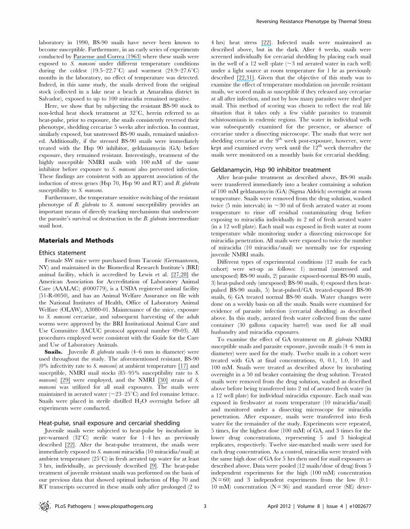
Constitutive expression of Hsp 90 in BS-90 and NMRIsnails is similar
To investigate if the expression of Hsp 90 in the resistant (BS-90)
and susceptible (NMRI) snail would be similar as was shown
previously for the levels of Hsp 70 and the nimbus RT domain,
qualitative RT-PCR was conducted with template cDNA
prepared with and without M-MLV-RT from RNA isolated from
uninfected individual snails. As shown in Figure 1A, qualitative
RT-PCR revealed that basal constitutive expression of Hsp 90 in
both snail stocks was remarkably similar. Thus, the PCR
amplification of cDNA from two individual BS-90 snails (Fig. 1A,
lanes 1 and 2), using the Hsp 90 gene specific primers described in
Materials and Methods, produced a 199 bp expected-size band
that was similar in size and intensity (Fig. 1A, lanes 1 to 4) to the
PCR amplification product produced from cDNA templates from
two individual NMRI susceptible snails (Fig. 1A, lanes 3 and 4).
To rule out any possibility that the amplicons detected in lanes 1 to
4 might have originated from genomic DNA contamination in
RNA preparations utilized for first strand cDNA synthesis, control
reactions performed without reverse transcriptase (2M-MLVRT)
in the first stand reaction were also utilized for PCR amplifications
(Fig. 1A, lanes 5 to 8). Thus, using the Hsp 90 gene-specific
primers for amplification reactions with this control (minus M-
MLV RT) template produced no bands in either the BS-90 (Lanes
Figure 1. Expression of the Hsp 90 transcript in resistant BS-90, and susceptible NMRI juvenile snails, without and with parasiteexposure. (A) Constitutive expression of Hsp 90 and myoglobin in two juvenile BS-90 (lanes 1, 2, 5, 6, 10, 11, 14 and 15) and NMRI (lanes 3, 4, 7, 8, 12,13, 16 and 17) snails by qualitative RT-PCR, using cDNA synthesized with [+M-MLV-RT], or without [2M-MLV-RT] RT in the first strand reaction. Thesame cDNA template was utilized for PCR amplification but with gene specific primers (forward and reverse) corresponding to the specific transcripts.Amplified bands of the expected size, 199 bp for Hsp 90, and 350 bp for myoglobin stained by ethidium bromide after agarose gel electrophoresisare shown. PCR amplification conducted without cDNA template is shown in lanes 9 and 18. The 100 bp ladder run in parallel is shown on the left. (B)Analysis of the fold difference in Hsp 90 expression by real time qPCR between NMRI and BS-90 snails either without (0) S. mansoni miracidiaexposure or after different time points (15 to 120 mins) PE. The histograms in 1B represent pooled data from 5 independent experiments, eachperformed with RNA (assayed in triplicate) isolated from 10 individual size-matched juvenile snails. The asterisk * indicates statistical significant P-values of ,0.05 by using ANOVA.doi:10.1371/journal.ppat.1002677.g001
Reversing Resistance Phenotype by Thermal Stress
PLoS Pathogens | www.plospathogens.org 5 April 2012 | Volume 8 | Issue 4 | e1002677
5 and 6) or NMRI (Lanes 6 and 7) samples, indicating that there
was no genomic DNA contamination in the RNA preparations
utilized for this assay. In parallel RT-PCR was performed with
primers corresponding to the housekeeping myoglobin gene using
equal amounts of the cDNA templates utilized in lanes 1 to 4. In
this case, (also in lanes 10 to 13) an expected 350 bp size PCR
product was obtained, indicating similar constitutive expression of
myoglobin in both these two snail stocks. As another negative
control, PCR was also conducted without cDNA template. The
absence of a product (lanes 9 and 18) showed there was no
contamination in the reagents utilized for the assay.
Early induction of the Hsp 90 transcript occurs insusceptible but not in resistant juvenile snails exposed toS. mansoni
Since previous studies showed dramatic differences in the
temporal modulation and degree of induction of the Hsp 70 and
RT transcripts following S. mansoni infection between resistant and
susceptible snails, in the present study we chose to examine
whether parasite exposure mediates the differential expression of
another major cellular stress gene, Hsp 90. The induction of the
Hsp 90 transcript in susceptible NMRI and resistant BS-90
juvenile snails exposed for different time points (0, 15, 30, 45, 60
and 120 min) to miracidia is shown in Figure 1B. RNA isolated
from the snails was analyzed by real time qPCR as described in
Materials and Methods using the uniform constitutive expression
of the myoglobin transcript in both snail stocks as internal
standard. From 5 independent (biological replicates) assays done
by using 10 individual snails per time point, with each RNA
sample run in triplicate, results showed that as early as 15 min
post- exposure almost 10-fold induction of the Hsp 90 transcript
was obtained in the susceptible NMRI snail. Furthermore, the
transcript remained upregulated in the susceptible snail through-
out the 120 min PE time period examined. In contrast, a smaller
difference in the upregulation of this transcript (1.54 fold change)
occurred in the resistant snail at the early 15 min time point after
infection. Although variations in the induction level (of Hsp 90
transcript) were observed in the infected susceptible snail between
the early 15–120 min PE time period, none of the levels we
detected in the infected resistant snail (1.45 to 1.54 fold change)
exceeded those detected in the infected susceptible snail.
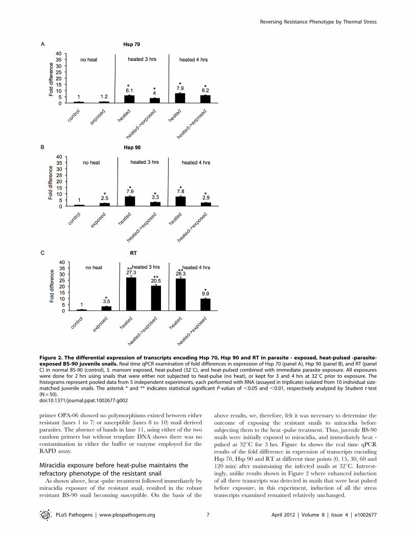
Heat-pulse followed by S. mansoni exposure, enhancesinduction of stress response transcripts in resistant BS-90snails
Previously we ascertained, as mentioned above, that induction
of stress genes occurs in the resistant BS-90 snail after a long 2–
4 hrs time period either in response to parasite exposure, or heat
shock. Accordingly, here we kept the BS-90 snails at 32uC for 3–
4 hrs before exposing them immediately to miracidia as described
in Materials and Methods. Using real time qPCR, modulations in
expression of the three cellular stress genes (Hsp 70, Hsp 90 and
RT) were assessed. The fold changes in expression of transcripts
corresponding to Hsp 70 (Fig. 2a), Hsp 90 (Fig. 2b) and RT
(Fig. 2c) in resistant BS-90 juvenile snails either following heat-
pulse treatment, or subsequently subjecting the heat- pulsed snails
to S. mansoni exposure for 2 hrs were determined (Fig. 2). As shown
in Figure 2a, in comparison to snails exposed without prior heat-
pulse treatment, minor induction (1.2 fold) of the Hsp 70 transcript
was detected in these resistant snails after parasite exposure. In
contrast, however, the heat-pulse for either 3 or 4 hrs promoted a
more significant induction (6.1 and 7.9 fold, respectively) of this
transcript. Interestingly, exposing the snails to miracidia immedi-
ately after 3 and 4 hrs heat-pulse kept the induced Hsp 70
transcript at a level that was higher (4 and 6.2 fold induction,
respectively) than the 1.2 fold induction we observed in snails that
were exposed without being heat pulsed.
In Figure 2b, while a 2.5 fold induction of the Hsp 90 transcript
was observed in the unstressed 2 hrs parasite-exposed BS-90 snail,
we detected a 7.9 and 7.8 fold increase in this transcript in snails
subjected to 3 and 4 hrs of heat-pulse, respectively. The fold
change in the Hsp-90 transcript remained relatively higher in
snails that were heat-pulsed and immediately exposed to miracidia
(3.3 and 2.9 fold increase) compared to the 2.5 fold increase
observed in the normal (minus heat-pulse) parasite-exposed snail.
In Figure 2c, in unstressed BS-90 snails exposed to miracidia, a
3.5 fold increase in the RT transcript was observed compared to a
27.3 and 26.3 fold induction of this transcript when snails were
kept for 3 and 4 hrs at 32uC prior to infection. Thus, in this case,
the dramatic increase in the RT transcript remained elevated in
snails that were heat -pulsed for 3 to 4 hrs and then immediately
exposed to miracidia. Thus, a strong 20.5 and almost 10 fold
increase was detected in these snails (heat-pulse plus exposure)
compared to snails that were exposed without heat-pulse treatment
where only a 3.3 fold increase in the RT transcript was observed.
The above results showed that compared to snails that were
only exposed to miracidia, the induction of all the stress transcripts
examined (Hsp70, Hsp90 and RT) was more elevated in BS-90
snails that were responding to parasite exposure after heat - pulse
treatment.
Heat-pulse immediately preceding parasite exposure ofresistant snails makes them susceptible
Results in Figure 3a shows the effect of combining heat -pulse
treatment with immediate exposure of the stressed resistant snails
to miracidia. Parasite-exposed unstressed snails were monitored as
control. As shown in Figure 3a, as expected, juvenile resistant
snails that were exposed to miracidia without heat -pulse treatment
released no cercariae for the entire 7 weeks duration of the
experiment. In contrast, a significant number, more than half
(60.7% and 53.9%), of resistant snails that were heat -pulsed either
for 3 or 4 hrs prior to exposure were found to shed cercariae
starting at 5 weeks PE. Interestingly, by 7 weeks PE, all such prior
heat -pulsed, and parasite -exposed resistant snails were shedding
cercariae.
To determine if cercariae released from these positive resistant
snails were biologically viable i.e. capable of infecting the
experimental mouse host and developing successfully into adult
worms in this host, cercariae released from ‘resistant’ snails were
utilized for mouse infections as described in Materials and
Methods. Since we were able to perfuse adult worms from the
mice infected with ‘resistant’-snail derived parasites, we can
conclude that larval parasites released from these ‘resistant’ snails
were indeed infectious, developing normally within the expected 6
weeks time-frame into adult worms in the infected mouse. To
determine whether these adult worms were in fact genotypically
identical to parasites harvested from mice exposed to cercariae
released from our representative susceptible snails (NMRI stock),
we analyzed genomic DNA from worms harvested from mice that
were infected by using either resistant, or susceptible snail derived
parasites, with the multi-locus RAPD-PCR genotyping tool. As
shown in Figure 3b, the DNA profile, using random primer OPR-
14, from 7 individual worms (lanes 1 to 7) was comparable to that
of an adult worm recovered from a mouse (by perfusion) that was
exposed to cercariae shed from the representative susceptible snail
(lanes 8 to 10). Likewise, DNA profiling of the same samples as
utilized in lanes 1 and 7 but amplified, this time, with random
Reversing Resistance Phenotype by Thermal Stress
PLoS Pathogens | www.plospathogens.org 6 April 2012 | Volume 8 | Issue 4 | e1002677
primer OPA-06 showed no polymorphisms existed between either
resistant (lanes 1 to 7) or susceptible (lanes 8 to 10) snail derived
parasites. The absence of bands in lane 11, using either of the two
random primers but without template DNA shows there was no
contamination in either the buffer or enzyme employed for the
RAPD assay.
Miracidia exposure before heat-pulse maintains therefractory phenotype of the resistant snail
As shown above, heat -pulse treatment followed immediately by
miracidia exposure of the resistant snail, resulted in the robust
resistant BS-90 snail becoming susceptible. On the basis of the
above results, we, therefore, felt it was necessary to determine the
outcome of exposing the resistant snails to miracidia before
subjecting them to the heat -pulse treatment. Thus, juvenile BS-90
snails were initially exposed to miracidia, and immediately heat -
pulsed at 32uC for 3 hrs. Figure 4a shows the real time qPCR
results of the fold difference in expression of transcripts encoding
Hsp 70, Hsp 90 and RT at different time points (0, 15, 30, 60 and
120 min) after maintaining the infected snails at 32uC. Interest-
ingly, unlike results shown in Figure 2 where enhanced induction
of all three transcripts was detected in snails that were heat pulsed
before exposure, in this experiment, induction of all the stress
transcripts examined remained relatively unchanged.
Figure 2. The differential expression of transcripts encoding Hsp 70, Hsp 90 and RT in parasite - exposed, heat-pulsed -parasite-exposed BS-90 juvenile snails. Real time qPCR examination of fold differences in expression of Hsp 70 (panel A), Hsp 90 (panel B), and RT (panelC) in normal BS-90 (control), S. mansoni exposed, heat-pulsed (32uC), and heat-pulsed combined with immediate parasite exposure. All exposureswere done for 2 hrs using snails that were either not subjected to heat-pulse (no heat), or kept for 3 and 4 hrs at 32uC prior to exposure. Thehistograms represent pooled data from 5 independent experiments, each performed with RNA (assayed in triplicate) isolated from 10 individual size-matched juvenile snails. The asterisk * and ** indicates statistical significant P-values of ,0.05 and ,0.01, respectively analyzed by Student t-test(N = 50).doi:10.1371/journal.ppat.1002677.g002
Reversing Resistance Phenotype by Thermal Stress
PLoS Pathogens | www.plospathogens.org 7 April 2012 | Volume 8 | Issue 4 | e1002677
Accordingly, we monitored these snails (parasite- exposed then
heat -pulsed) for cercarial shedding as described above. Results
(Fig. 4b) showed that all (100%) the resistant snails that were
exposed to the parasite before being subjected to heat -pulse
treatment remained negative. From these results, it is clear that
exposing these resistant snails to the parasite before they are
stressed, maintains the refractory status of these snails, further
demonstrating the temperature sensitivity of BS-90 resistant
phenotype to S. mansoni infection.
Treatment of heat -pulsed and S. mansoni exposed BS-90snails with the Hsp 90 inhibitor, geldanamycin (GA)blocks susceptibility to infection
To determine, more precisely, if significant upregulation of
stress -related transcripts is indeed a factor in rendering the heat -
pulsed resistant snails susceptible to infection, we investigated the
effect of blocking the action of one of the stress proteins, Hsp 90,
by using a specific inhibitor drug geldanamycin (GA) that prevents
this protein from performing its role as an essential chaperone in
the cell [38–40]. In Figure 5, resistant snails that were heat -pulsed
for 3 hrs were exposed either immediately to miracidia as
described above, or were treated with GA (100 mM) before being
exposed to miracidia. Results showed that without inhibitor
treatment of stressed snails, the majority (70%) of heat -pulsed and
exposed snails were found to shed cercariae 9 weeks after
exposure. In contrast, snails that were heat –pulsed, and
immediately treated with GA before exposure failed to shed
cercariae, remaining negative for the entire 9 weeks duration of
the experiment. These data clearly show that blocking the action
of Hsp 90 by GA treatment in the stressed resistant snails before
exposure maintained their refractory phenotype, thereby indicat-
ing that a sustained significant stress induction of Hsp 90 was
involved in the mechanism(s) of B. glabrata susceptibility to S.
mansoni.
Treatment of susceptible NMRI snails with GA beforeparasite infection renders them non-susceptible to S.mansoni
To further demonstrate the link between stress induction in
juvenile B. glabrata snails and their susceptibility to S. mansoni, here
the effect of pre -treating the susceptible NMRI snail with the
aforementioned Hsp 90 inhibitor (GA) on the outcome of infection
was examined. Figure 6a shows the survival of snails after either
infection alone (minus GA inhibitor) or after treatment with
Figure 3. Genotyping of adult S. mansoni parasites generated from cercariae released from resistant BS-90 snails following thecombination of heat-pulse and parasite infection. (A) Percentage of BS-90 resistant snails shedding cercariae after 7 weeks parasite exposure.Note that snails that were exposed to S. mansoni without being subjected to the heat-pulse treatment, as expected, remained negative (0) whilstthose kept for either 3 hrs or 4 hrs prior to exposure began shedding cercariae at 5 weeks PE. (B) RAPD-PCR genotyping of the genomic DNA fromindividual (lanes 1–7) male worms harvested from mice infected with cercariae released from BS-90 ‘resistant’ snails. Note that the DNA profiles ofthese parasites, generated with two different random primers, OPR-14 and OPA-06, were invariant when compared to profiles generated with thesame primers but with DNA from individual (lanes 8–10) worms harvested from mice that were infected with cercariae shed from susceptible (NMRI)snails. The negative control, i.e. amplification without DNA template is shown in lane 11 and the lane labeled M shows position of 100 bp ladder, runin parallel as standard.doi:10.1371/journal.ppat.1002677.g003
Reversing Resistance Phenotype by Thermal Stress
PLoS Pathogens | www.plospathogens.org 8 April 2012 | Volume 8 | Issue 4 | e1002677
100 mM of GA. Results (Fig. 6a) showed that snails tolerated the
drug at this dose and survived at the same rate (100%) as those
that were exposed without drug treatment. Since all the snails
tolerated this relatively high dose of the inhibitor, we proceeded to
examine whether pre -treating the susceptible snail with various
doses of GA would affect their susceptibility phenotype.
As shown in Figure 6b, susceptible snails that were infected
without prior drug treatment, as expected, were found to be
shedding cercariae at 5 weeks PE (18.3%), with all untreated-
exposed susceptible snails shedding cercariae at 9 weeks after
infection. In contrast, susceptible snails that were treated with
100 mM GA prior to exposure failed to shed cercariae at the same
Figure 4. Expression of stress –related transcripts in parasite exposed juvenile resistant BS-90 snails followed by heat-pulsetreatment for various time points (0–120 min). (A) Real time qPCR analysis of the fold difference in expression of transcripts corresponding toHsp 70, Hsp 90, and RT in either normal BS-90 resistant snails, exposed snails, or those that were first exposed to S. mansoni before being subjected toheat-pulse at 32uC for various time points (15–120 mins). These data were pooled from 10 biological replicates of 5 independent experiments. Notethe lack of up-regulation of all transcripts in snails that were exposed and then subsequently kept at 32uC. The asterisk* denotes significantupregulation of one of the transcripts, RT, by Student t-test with P-value,0.05 (N = 50). (B) Results showing 100% shedding of cercariae fromsusceptible NMRI snails after 7 weeks PE to S. mansoni miracidia compared to similarly exposed normal BS-90 snails, or those that were exposedbefore being subjected to heat pulse treatment at 32uC. It is clear that no cercariae were shed from BS-90 snails in either of these two groups. Thesedata were from 5 biological replicates.doi:10.1371/journal.ppat.1002677.g004
Figure 5. Percentage cercarial shedding from heat-pulsed resistant BS-90 snails that were either treated (+GA), or untreated (2GA)prior to S. mansoni exposure. Note that the snails that were not treated with GA but heat- pulsed and exposed began to shed cercariae at 4 weeksPE (with 70% shedding parasites at 9 weeks PE). In contrast, note that the heat -pulsed- GA-treated - infected snails remained negative for the entire 9weeks duration of the experiment.doi:10.1371/journal.ppat.1002677.g005
Reversing Resistance Phenotype by Thermal Stress
PLoS Pathogens | www.plospathogens.org 9 April 2012 | Volume 8 | Issue 4 | e1002677
9 weeks PE time period while only a small percentage (3.7 to 4.8%)
of snails pre-treated with lower doses of GA (0.1 to 10 mM) were
found to be shedding cercariae. The longest surviving miracidial
exposed, drug treated- (100 mM) snails remained negative (no
cercarial shedding) for up to 9 months after infection. All snails not
shedding cercariae by the 9th week after exposure remained
negative at week 12 and continued to be negative for at least 9
months PE.
The reduction of shedding (in percentages) from infected snails
treated with the higher dose of GA compared to the lower doses
was significant as determined by one-way ANOVA (P-value,0.05)
[N = 60 and N = 36].
GA treated miracidia remain infectious and developsuccessfully into the cercarial larval stage
To rule out the possibility that results presented above might
simply reflect the effect of the drug inhibiting the parasite’s Hsp 90
homolog, thereby impairing the ability of miracidia to either
penetrate the snail, or transform successfully into sporocysts, we
treated miracidia directly with the highest dose of GA (100 mM)
that was utilized in this study. These drug -treated miracidia were
then utilized for snail exposures in comparison to exposures done
with untreated miracidia, as control. In these experiments, more
than 75% of miracidiae (with or without GA treatment) penetrated
the snails within 5 min, and all (100%) successfully penetrated the
snail within 1 hr (data not shown). These data were similar to
penetration behavior we previously observed where we used either
normal or irradiated miracidiae for snail exposures [22]. As shown
in Figure 7, 41.7% and 87.5% of NMRI susceptible snails exposed
to GA treated miracidia shed cercariae at 4–5 weeks. At 4 to 5
weeks PE, results analyzed by student’s t-test showed that the
percentage of cercarial shedding between NMRI snails exposed to
either GA treated or normal miracidia was statistically significant
(P –value,0.05). However, after week 5 PE, results showed that
there was no statistically significant difference between the
percentage of cercarial shedding between NMRI exposed to
either GA-treated or normal miracidia. NMRI snails that were
exposed to the untreated miracidia, likewise released cercariae
after week 4 PE as we have routinely come to expect for snail
infections performed by using this parasite strain and snail-host
combination.
Discussion
To date, very little information exists on molecular mechanisms
that determine the outcome of the snail/schistosome interaction.
Here, we have shown that upregulation of stress-related
transcripts, such as those examined in this study, Hsp 70, Hsp
90 and RT in the B. glabrata snail host, soon after infection, plays
an important role in their susceptibility to S. mansoni. These results
are consistent with our previous data that showed a differential
induction of stress genes occurs between juvenile susceptible and
resistant B. glabrata snails after exposure to S. mansoni miracidia.
Figure 6. Survival, and cercarial shedding from juvenile susceptible NMRI snails after GA treatment. A) The percentage of susceptibleNMRI snails surviving after either 9 weeks PE to S. mansoni or after treatment with 100 mM GA as described in Materials and Methods. Note that theinhibitor had no toxic effect on the snails. (B) Percentage cercarial shedding from GA treated and untreated susceptible (NMRI) snails weeks afterinfection to S. mansoni. Note that at 5 weeks PE, more than 50% of untreated (0) snails were shedding cercariae while all GA treated snails, even thosetreated with low doses (0.1–10 mM), remained negative for cercarial release. Data were pooled (12 snails/dose of drug) from 5 independentexperiments for the high (100 mM) concentration (N = 60) and 3 independent experiments from the low (0.1–10 mM) concentration (N = 36) forstandard error (SE) determination. At 9 weeks PE all snails treated prior to exposure with the higher dose of inhibitor remained negative for infection.doi:10.1371/journal.ppat.1002677.g006
Reversing Resistance Phenotype by Thermal Stress
PLoS Pathogens | www.plospathogens.org 10 April 2012 | Volume 8 | Issue 4 | e1002677
Thus, upregulation of transcripts corresponding to Hsp 70 and RT
was detected sooner, and more dramatically in susceptible
compared to resistant snails following either heat shock or parasite
infection [22]. Also, in this previous study, we showed that the
stimulus/stimuli for this stress induction may be present in normal
but not in irradiated attenuated miracidia.
Although we are yet to discover the nature of the parasite stress
elicitor(s), it is most likely released from the incoming parasite as
excretory secretory products (ESP). Interestingly, several studies
have described using miracidial ESP to induce changes in either
snail hemocytes, or the B. glabrata embryonic cell line, Bge [41–43].
Despite our limitation of not knowing what triggers the induction
of stress in the snail (soon after exposure to miracidia) it is clear
from this study that by using the heat -pulse regimen described to
enhance the induction of stress to levels not typically seen under
normal circumstances in resistant snails after exposure, it is
possible to successfully reverse the resistant phenotype of juvenile
resistant BS-90 snails i.e. render them susceptible to S. mansoni.
Therefore, even though our results showed that parasite infection
of the heat –pulsed snails caused a reduction in the induction of
the stress transcripts, a more enhanced induction of all the three
transcripts was still observed in heat-pulsed- infected snails than in
snails that were exposed to miracidia alone without heat -pulse,
helping to switch the phenotype of juvenile BS-90 snails from
being resistant to susceptible. Thus, from these results, it is clear
that resistance to S. mansoni in the juvenile BS-90 snail is a
temperature-sensitive (ts) phenotype. Furthermore, cercariae
released from these ts snails were infectious, developing fully into
adult worms in the infected mouse, and with no change detected in
DNA profiles of adult worms harvested from either these ‘resistant’
snails or our representative susceptible NMRI snail stock.
Historically, studies regarding B. glabrata susceptibility to S.
mansoni have emphasized either the role of genetics, or innate
defense in the snail/parasite association. While these areas of study
have been pivotal in explaining some of the complex dynamics
behind why schistosomes are either destroyed or survive in the
snail host, it is clear from our results that temperature sensitivity of
the stress gene loci reported is a contributing factor in the outcome
of this host/pathogen interaction. Accordingly, we can only
speculate that the ts resistant BS-90 snail’s phenotype must involve
changes in the activation of the three stress response genes to
account for their altered kinetics. In a recent study, we showed that
an unknown external stimulus/stimuli from the parasite was
indeed able to mediate the non-random repositioning of gene loci
of interphase chromosomes in the snail embryonic cell line, Bge
[44]. These gene loci repositioning studies have since been
reproduced in intact snails responding to S. mansoni (Arican,
unpublished), and we are currently investigating if the stress genes
are repositioned upon induction.
Previous studies have shown that the induction of stress is
important for successful outcomes of other host-pathogen
relationships as well. For example, in baculovirus infected Sf-9
cells, an increase in the expression of Hsp 70 was found to
correlate with active virus replication. Thus, in this study, Lyupina
et al. showed that inhibiting Hsp70 expression by the drug
KNK437 suppressed virus replication [45]. Additionally, Hsp 90
has been shown to be essential for the growth of the malaria-
causing agent, P. falciparum, in human erythrocytes [46].
Consequently, derivatives of GA, the Hsp 90 inhibitor used in
the present study, has also been used to inhibit the growth of P.
falciparum and another protozoan, Trypanosoma evansi [46,47]. GA is
a benzoquinone ansamycin antibiotic that binds to the N-terminal
ATPase site of Hsp 90 to inhibit its chaperone activity. Although
the inhibitor and its derivatives have previously been used to
inhibit cell proliferation in cancer [48] and the growth of other
parasites, as mentioned above it has never been shown, until now
to treat any mollusk in the context of examining host-pathogen
interactions. Interestingly, our results showed that GA treatment
neither impaired the penetration behavior of the miracidia nor
their ability to remain infectious. More studies, especially
Figure 7. Cercarial shedding between normal, and GA-treated susceptible juvenile NMRI snails. Percentage cercarial shedding in NMRIsnails infected with GA - treated miracidia (blank box) and normal miracidia (grey box). The snails were releasing cercariae after 4 weeks PE andcontinued to shed cercariae for the 9 weeks duration of the study. The asterisk* denotes a significant difference between GA-treated- and normalmiracidia infection at weeks 4 and 5 PE. However note that after 6 weeks PE there is no significant difference between the two groups as determinedby Student t-test with P-value,0.05 (error bar = SE; N = 60 in each group).doi:10.1371/journal.ppat.1002677.g007
Reversing Resistance Phenotype by Thermal Stress
PLoS Pathogens | www.plospathogens.org 11 April 2012 | Volume 8 | Issue 4 | e1002677
performed in vivo for longer time points will be needed to further
examine this apparent lack of GA toxicity on the larval parasites. A
similar lack of GA toxicity, which might be due to non-binding of
GA to Hsp 90 homologues of free-living nematode larval stages,
has previously been reported [49].
Heat shock proteins are highly conserved proteins that have
been shown to play a critical role in maintaining protein integrity,
preventing the aggregation of misfolded proteins in the cell,
thereby maintaining normal cell function in the face of cellular
injury from physical or physiological stress [50]. Very few studies
have examined stress induction in relation to mollusk/pathogen
interactions. Our results showing the very early (within 15 min)
induction of Hsp 90 in susceptible snails (but not resistant snails)
after infection was surprising and underscores the need for more
studies on this stress protein, especially in relation to snail-
schistosome interactions. Hsp 90 is expressed abundantly even in
the absence of stress and constitutes a large portion of
constitutively expressed protein in cells. The protein is regarded
as being essential to cell viability because of its central role in
forming complexes with a wide variety of co-chaperones and client
proteins that are involved in major cellular pathways, such as
signal transduction and cell-cycle control [51].
How Hsp 90 interacts directly, or indirectly with either the B.
glabrata Hsp 70 or nimbus RT has yet to be investigated.
Particularly, whether (or not) key molecules of the snail’s innate
defense system, such as FREPs are client proteins of Hsp 90,
remain to be investigated. Previously, we showed that co-induction
of Hsp 70 and nimbus RT transcripts occur soon after S. mansoni
infection of juvenile susceptible snails [22]. Mobile Genetic
Elements (MGEs), such as nimbus are responsive to cellular stress
[52,53]. However, the role of the nimbus non-LTR retrotransposon
in the stress pathway of B. glabrata remains unknown. Further
studies are, therefore, required to elucidate the relationship
between all these stress genes (Hsp 90, Hsp 70 and RT) in the
snail’s behavior towards S. mansoni.
In other mollusks, such as the clam, Mercenaria mercenaria, the
upregulation of Hsp 70 was observed in this mollusk in response to
the opportunistic parasite, known as Quahog Parasite Unknown,
QPX [54]. Also, in another clam, Meretrix meretrix, it has recently
been shown that expression of Hsp 70 was upregulated soon after
Vibrio parahaemolyticus infection [55]. Additionally, in the disk
abalone, Haliotis discus, a recent molecular characterization showed
that Hsp 90 is induced within 4 hrs after treatment with
lipopolysaccharide, LPS [56]. In another B. glabrata susceptible
snail, the M-line stock, Hanington et al. [26] showed upregulation
of stress related transcripts following infection of these snails with
trematodes, either S. mansoni or Echinostoma paraensei. The
modulation (down regulation) of Hsp 70 in hemocytes isolated
from exposed B. glabrata resistant and susceptible snails (the cells
most intimately associated with the active destruction of
schistosomes in the snail host) has also been reported, suggesting
an involvement of this stress protein in the snail host’s defense
system [7,23].
Indeed, an immunological role for stress proteins has been
widely documented [57]. Thus, it might be reasonable to assume
that in the snail/schistosome system, cellular stress triggered
against parasite proteins that are recognized in the snail as non-
self, by maintaining the homeostasis of the host, paradoxically
protects the parasite as well. Larval schistosomes have been shown
to express RNA transcripts for heat shock proteins. Presumably,
such heat shock proteins (released from the parasite) might induce
stress genes in the snail host, providing the cytoprotection that the
parasite needs for its own successful invasion. While a strong anti-
schistosome Hsp 70 humoral response has been reported in several
infected (S. mansoni and S. hematobium) mammalian (murine, human
and baboon) hosts [58,59] nothing is known about the role of
schistosome heat shock proteins and the snail’s innate defense
system. Thus far, we have evidence showing that an active defense
system plays an important role in the BS-90 resistant snail’s ability
to ward off the parasite infection. By showing in this study that the
deliberate use of stress in the form of non-lethal heat-pulse
(boosting the level of inducible stress in the resistant BS-90 snail
before infection) was a necessary step in rendering these normally
robust resistant snails susceptible, we can suggest that the stress
induced dampened the anti-schistosome response that is typically
seen in these snails. Previously, it was shown that the resistance
phenotype can be interfered with in resistant snails (10-R2 and 13–
16-R1 stocks) if snails were first infected with other trematodes,
such as E. paraensei and E. lindoense before being exposed to S.
mansoni [60]. While no molecular explanation was given for this
apparent suppression of the defense system by the dual infection
protocol first described by Lie and Heyneman et al. [61], these
early results showed that susceptibility to S. mansoni in these
resistant stocks developed shortly (within 1 hr) after they had been
exposed to E. paraensei. In light of our current results, we can
assume that in this previous study, the primary echinostome
infection, by triggering a stress response, dampening the innate
defense system allowed the secondary S. mansoni infection to
survive and develop. In other host-pathogen systems, there is clear
evidence that expression of stress proteins, in particular Hsp 70 is
an important feature in modulating the host innate immune
response [62].
Another plausible explanation for the results presented here
might be that the initial heat -pulse could have destroyed the
resistant snail’s hemocytes, thereby rendering these cells incapable
of killing the incoming miracidia. It is also possible that the
induction of stress might be a reflection of the successful
establishment of the parasite in the snail. As mentioned above
several factors, and not hemocytes alone govern compatibility/
incompatibility issues between B. glabrata and S. mansoni. The heat
pulse regimen utilized to enhance the induction of stress genes in
the resistant snails was not lethal. All the snails survived at this
elevated temperature, and a colony of BS-90 snails that we
maintain (now in their eighth month) at 32uC continue to thrive.
Interestingly, all (100%) of progeny snails (F1, exposed to
miracidia and kept at 32uC after exposure) bred from BS-90
snails maintained at 32uC were found to shed cercariae at 3 to 4
weeks PE (Ittiprasert, Miller and Knight unpublished).
While we have no data supporting the notion that higher
prevailing environmental temperatures might facilitate snail
susceptibility, our results show that it is possible that climate
change might impact resistant snail susceptibility to schistosomes.
Indeed, a recent study showed that global warming might result in
an increase in cercarial output of infected snails [63].
By using a recently developed gene silencing method based on
soaking snails in siRNA coupled to the inert cationic carrier,
polyethylene imines (PEI) [44], we are currently working to
systematically knock-down transcription of Hsp 70, Hsp 90 and
nimbus RT in the snail by the PEI delivery tool. Thus far,
preliminary results indicate that knocking down these transcripts
to levels comparable to those we routinely obtain for suppressing
the expression of low copy RNA transcripts will be more
challenging. Despite these initial challenges, we hope to elucidate
the role of these stress proteins in the snail host schistosome
relationship by knocking down their corresponding transcripts.
In addition, we have used the Hsp 90 inhibitor drug used in this
study, GA, for treating pre-patent (2 week exposed snails) and
results show consistently that once established, the drug has no
Reversing Resistance Phenotype by Thermal Stress
PLoS Pathogens | www.plospathogens.org 12 April 2012 | Volume 8 | Issue 4 | e1002677
effect on the infection and all treated pre-patent snails go on to
shed cercariae.
In conclusion, we have shown in this study that by applying
stress in the form of mild heat pulse to resistant BS-90 snails before
they are exposed to S. mansoni, renders these snails susceptible. In
contrast, infecting these snails before stressing them does not
reverse their resistance phenotype, suggesting that the stress
induction is an early necessary step in the sequence of molecular
events that contribute towards making a snail susceptible. In
addition, use of the stress inhibitor to treat susceptible snails before
exposure was able to prevent them from shedding cercariae, again
confirming that the stress pathway is indeed required for snail’s to
succumb to the parasite infection. These data open up a new
opportunity to delve into unraveling the mechanism(s) that helps
snails to either overcome or sustain the S. mansoni parasite
infection, investigations that should lead to developing novel tools
to interfere with schistosome-snail infections and thus reduce
transmission of schistosomiasis.
Acknowledgments
We thank Andre Miller and Frances Barnes for their technical assistance.
We also thank Drs Allen Cheever, Fred Lewis, and Jacob Hodgson for
their comments and helpful critique of the manuscript.
Author Contributions
Conceived and designed the experiments: MK WI. Performed the
experiments: MK WI. Analyzed the data: MK WI. Contributed
reagents/materials/analysis tools: MK WI. Wrote the paper: MK WI.
Designed experiment, experiments, manuscript preparation: MK. Exper-
iments, experiment analysis, manuscript preparation WI.
References
1. King CH, Sturrock RF, Kariuki HC, Hamburger J (2006) Transmission control
for schistosomiasis-why it matters now. Trends Parasitol 22: 575–582.
2. Kjetland EF, Ndhlovu PD, Gomo E, Mduluza T, Midzi N, et al. (2006)
Association between genital schistosomiasis and HIV in rural Zimbabwean
women. AIDS 20: 593–600.
3. Rollinson D, Webster JP, Webster B, Nyakaana S, Jorgensen A, et al. (2009)
Genetic diversity of schistosomes and snails: implications for control.
Parasitology. pp 1–11.
4. Lewis FA, Patterson CN, Knight M, Richards CS (2001) The relationship
between Schistosoma mansoni and Biomphalaria glabrata: genetic and molecular
approaches. Parasitology 123 Suppl: S169–179.
5. Richards CS, Knight M, Lewis FA (1992) Genetics of Biomphalaria glabrata and its
effect on the outcome of Schistosoma mansoni infection. Parasitol Today 8:
171–174.
6. Knight M, Miller AN, Patterson CN, Rowe CG, Michaels G, et al. (1999) The
identification of markers segregating with resistance to Schistosoma mansoni
infection in the snail Biomphalaria glabrata. Proc Natl Acad Sci U S A 96:
1510–1515.
7. Lockyer AE, Spinks J, Kane RA, Hoffmann KF, Fitzpatrick JM, et al. (2008)
Biomphalaria glabrata transcriptome: cDNA microarray profiling identifies
resistant- and susceptible-specific gene expression in haemocytes from snail
strains exposed to Schistosoma mansoni. BMC Genomics 9: 634.
8. Adema CM, Hanington PC, Lun CM, Rosenberg GH, Aragon AD, et al. (2010)
Differential transcriptomic responses of Biomphalaria glabrata (Gastropoda,
Mollusca) to bacteria and metazoan parasites, Schistosoma mansoni and Echinostoma
paraensei (Digenea, Platyhelminthes). Mol Immunol 47: 849–860.
9. Ittiprasert W, Miller A, Myers J, Nene V, El-Sayed NM, et al. (2010)
Identification of immediate response genes dominantly expressed in juvenile
resistant and susceptible Biomphalaria glabrata snails upon exposure to Schistosoma
mansoni. Mol Biochem Parasitol 169: 27–39.
10. Rollinson D, Stothard JR, Jones CS, Lockyer AE, de Souza CP, et al. (1998)
Molecular characterisation of intermediate snail hosts and the search for
resistance genes. Mem Inst Oswaldo Cruz 93 Suppl 1: 111–116.
11. Baeza Garcia A, Pierce RJ, Gourbal B, Werkmeister E, Colinet D, et al. (2010)
Involvement of the cytokine MIF in the snail host immune response to the
parasite Schistosoma mansoni. PLoS Pathog 6: e1001115.
12. Hanington PC, Zhang SM (2011) The primary role of fibrinogen-related
proteins in invertebrates is defense, not coagulation. J Innate Immun 3: 17–
27.
13. Bouchut A, Roger E, Gourbal B, Grunau C, Coustau C, et al. (2008) The
compatibiuty polymorphism in invertebrate host/trematodes interactions:
research of molecular determinants. Parasite 15: 304–309.
14. Mitta G, Adema CM, Gourbal B, Loker ES, Theron A (2011) Compatibility
polymorphism in snail/schistosome interactions: From field to theory to
molecular mechanisms. Dev Comp Immunol E-pub ahead of print.
15. Mone Y, Gourbal B, Duval D, Du Pasquier L, Kieffer-Jaquinod S, et al. (2010)
A large repertoire of parasite epitopes matched by a large repertoire of host
immune receptors in an invertebrate host/parasite model. PLoS Negl Trop Dis
4: pii e813.
16. Theron A, Coustau C (2005) Are Biomphalaria snails resistant to Schistosoma
mansoni? J Helminthol 79: 187–191.
17. Paraense W, Correa L (1963) Variations in susceptibility of populations of
Australorbis blabratus to a strain of Schistosoma mansoni. Revista do Instituto de
Medicina Tropical de Sao Paulo 5: 15–22.
18. Richards CS, Shade PC (1987) The genetic variation of compatibility in
Biomphalaria glabrata and Schistosoma mansoni. J Parasitol 73: 1146–1151.
19. Richards CS, Minchella DJ (1987) Transient non-susceptibility to Schistosoma
mansoni associated with atrial amoebocytic accumulations in the snail host
Biomphalaria glabrata. Parasitology 95(Pt 3): 499–505.
20. Niemann GM, Lewis FA (1990) Schistosoma mansoni: influence of Biomphalaria
glabrata size on susceptibility to infection and resultant cercarial production. Exp
Parasitol 70: 286–292.
21. Lockyer AE, Noble LR, Rollinson D, Jones CS (2004) Schistosoma mansoni:
resistant specific infection-induced gene expression in Biomphalaria glabrata
identified by fluorescent-based differential display. Exp Parasitol 107: 97–104.
22. Ittiprasert W, Nene R, Miller A, Raghavan N, Lewis F, et al. (2009) Schistosoma
mansoni infection of juvenile Biomphalaria glabrata induces a differential stress
response between resistant and susceptible snails. Exp Parasitol 123: 203–211.
23. Zahoor Z, Davies AJ, Kirk RS, Rollinson D, Walker AJ (2010) Larval excretory-
secretory products from the parasite Schistosoma mansoni modulate HSP 70
protein expression in defence cells of its snail host, Biomphalaria glabrata. Cell
Stress Chaperones 15: 639–650.
24. Stirewalt MA (1954) Effect of snail maintenance temperatures on development
of Schistosoma mansoni. Exp Parasitol 3: 504–516.
25. Lefcort H, Bayne CJ (1991) Thermal preferences of resistant and susceptible
strains of Biomphalaria glabrata (Gastropoda) exposed to Schistosoma mansoni
(Trematoda). Parasitology 103 Pt 3: 357–362.
26. Hanington PC, Lun CM, Adema CM, Loker ES (2010) Time series analysis of
the transcriptional responses of Biomphalaria glabrata throughout the course of
intramolluscan development of Schistosoma mansoni and Echinostoma paraensei.
Int J Parasitol 40: 819–831.
27. Lewis FA, Stirewalt MA, Souza CP, Gazzinelli G (1986) Large-scale laboratory
maintenance of Schistosoma mansoni, with observations on three schistosome/snail
host combinations. J Parasitol 72: 813–829.
28. Lewis FA, Knight M, Richards CS (1997) A laboratory-based approach to
biological control of snails. Mem Inst Oswaldo Cruz 92: 661–662.
29. Cooper LA, Richards CS, Lewis FA, Minchella DJ (1994) Schistosoma mansoni:
relationship between low fecundity and reduced susceptibility to parasite
infection in the snail Biomphalaria glabrata. Exp Parasitol 79: 21–28.
30. Newton WL (1955) The establishment of a strain of Australorbis glabratus which
combines albinism and high susceptibility to infection with Schistosoma mansoni.
J Parasitol 41: 526–528.
31. Lewis FA, Patterson CN, Grzywacz C (2003) Parasite-susceptibility phenotypes
of F1 Biomphalaria glabrata progeny derived from interbreeding Schistosoma mansoni
-resistant and -susceptible snails. Parasitol Res 89: 98–101.
32. Chomczynski P (1993) A reagent for the single-step simultaneous isolation of
RNA, DNA and proteins from cell and tissue samples. Biotechniques 15:
532–534, 536–537.
33. Raghavan N, Miller AN, Gardner M, FitzGerald PC, Kerlavage AR, et al.
(2003) Comparative gene analysis of Biomphalaria glabrata hemocytes pre- and
post-exposure to miracidia of Schistosoma mansoni. Mol Biochem Parasitol 126:
181–191.
34. Myers J, Ittiprasert W, Raghavan N, Miller A, Knight M (2008) Differences in
cysteine protease activity in Schistosoma mansoni-resistant and -susceptible
Biomphalaria glabrata and characterization of the hepatopancreas cathepsin B
Full-length cDNA. J Parasitol 94: 659–668.
35. Knight M, Raghavan N, Goodall C, Cousin C, Ittiprasert W, et al. (2009)
Biomphalaria glabrata peroxiredoxin: effect of Schistosoma mansoni infection on
differential gene regulation. Mol Biochem Parasitol 167: 20–31.
36. Livak KJ, Schmittgen TD (2001) Analysis of relative gene expression data using
real-time quantitative PCR and the 2(-Delta Delta C(T)) Method. Methods 25:
402–408.
37. Simpson AJ, Dias Neto E, Vidigal TH, Pena HB, Carvalho OS, et al. (1995)
DNA polymorphism of schistosomes and their snail hosts. Mem Inst Oswaldo
Cruz 90: 211–213.
38. Chang YS, Lo CW, Sun FC, Chang MD, Lai YK (2006) Differential expression
of Hsp90 isoforms in geldanamycin-treated 9L cells. Biochem Biophys Res
Commun 344: 37–44.
Reversing Resistance Phenotype by Thermal Stress
PLoS Pathogens | www.plospathogens.org 13 April 2012 | Volume 8 | Issue 4 | e1002677
39. Harrison EM, Sharpe E, Bellamy CO, McNally SJ, Devey L, et al. (2008) Heat
shock protein 90-binding agents protect renal cells from oxidative stress andreduce kidney ischemia-reperfusion injury. Am J Physiol Renal Physiol 295:
F397–405.
40. Kumar R, Musiyenko A, Barik S (2003) The heat shock protein 90 of Plasmodium
falciparum and antimalarial activity of its inhibitor, geldanamycin. Malar J 2: 30.
41. Coustau C, Mitta G, Dissous C, Guillou F, Galinier R, et al. (2003) Schistosoma
mansoni and Echinostoma caproni excretory-secretory products differentially affect
gene expression in Biomphalaria glabrata embryonic cells. Parasitology 127:
533–542.42. Humphries JE, Yoshino TP (2006) Schistosoma mansoni excretory-secretory
products stimulate a p38 signalling pathway in Biomphalaria glabrata embryoniccells. Int J Parasitol 36: 37–46.
43. Zahoor Z, Davies AJ, Kirk RS, Rollinson D, Walker AJ (2009) Nitric oxideproduction by Biomphalaria glabrata haemocytes: effects of Schistosoma mansoni ESPs
and regulation through the extracellular signal-regulated kinase pathway. Parasit
Vectors 2: 18.44. Knight M, Miller A, Liu Y, Scaria P, Woodle M, et al. (2011) Polyethyleneimine
(PEI) mediated siRNA gene silencing in the Schistosoma mansoni snail host,Biomphalaria glabrata. PLoS Negl Trop Dis 5: e1212.
45. Lyupina YV, Dmitrieva SB, Timokhova AV, Beljelarskaya SN, Zatsepina OG,
et al. (2010) An important role of the heat shock response in infected cells forreplication of baculoviruses. Virology 406: 336–341.
46. Banumathy G, Singh V, Pavithra SR, Tatu U (2003) Heat shock protein 90function is essential for Plasmodium falciparum growth in human erythrocytes. J Biol
Chem 278: 18336–18345.47. Pallavi R, Roy N, Nageshan RK, Talukdar P, Pavithra SR, et al. (2010) Heat
shock protein 90 as a drug target against protozoan infections: biochemical
characterization of HSP 90 from Plasmodium falciparum and Trypanosoma evansi andevaluation of its inhibitor as a candidate drug. J Biol Chem 285: 37964–37975.
48. Miyata Y (2005) Hsp 90 inhibitor geldanamycin and its derivatives as novelcancer chemotherapeutic agents. Curr Pharm Des 11: 1131–1138.
49. Him NA, Gillan V, Emes RD, Maitland K, Devaney E (2009) Hsp-90 and the
biology of nematodes. BMC Evol Biol 9: 254.50. Lindquist S, Craig EA (1988) The heat shock proteins. Annu Rev Genet 22:
631–677.
51. Picard D (2002) Heat-shock protein 90, a chaperone for folding and regulation.
Cell Mol Life Sci 59: 1640–1648.52. Kimura RH, Choudary PV, Stone KK, Schmid CW (2001) Stress induction of
Bm1 RNA in silkworm larvae: SINEs, an unusual class of stress genes. Cell
Stress Chaperones 6: 263–272.53. Beauregard A, Curcio MJ, Belfort M (2008) The take and give between
retrotransposable elements and their hosts. Annu Rev Genet 42: 587–617.54. Perrigault M, Allam B (2009) Cytotoxicity of quahog parasite unknown (QPX)
toward hard clam (Mercenaria mercenaria) haemocytes and interactions between
different pathogen isolates and host strains. Parasitology 136: 1281–1289.55. Yue X, Liu B, Sun L, Tang B (2011) Cloning and characterization of a hsp70
gene from Asiatic hard clam Meretrix meretrix which is involved in the immuneresponse against bacterial infection. Fish Shellfish Immunol 30: 791–799.
56. Wang N, Whang I, Lee JS, Lee J (2011) Molecular characterization andexpression analysis of a heat shock protein 90 gene from disk abalone (Haliotis
discus). Mol Biol Rep 38: 3055–3060.
57. Plytycz B, Seljelid R (2002) Stress and immunity: minireview. Folia Biol(Krakow) 50: 181–189.
58. Moser D, Doumbo O, Klinkert MQ (1990) The humoral response to heat shockprotein 70 in human and murine Schistosomiasis mansoni. Parasite Immunol 12:
341–352.
59. Kanamura HY, Hancock K, Rodrigues V, Damian RT (2002) Schistosoma
mansoni heat shock protein 70 elicits an early humoral immune response in S.
mansoni infected baboons. Mem Inst Oswaldo Cruz 97: 711–716.60. Lie KJ, Heyneman D (1977) Schistosoma mansoni, Echinostoma lindoense, and
Paryphostomum segregatum: interference by trematode larvae with acquiredresistance in snails, Biomphalaria glabrata. Exp Parasitol 42: 343–347.
61. Lie KJ, Heyneman D, Jeong KH (1976) Studies on resistance in snails. 7.
Evidence of interference with the defense reaction in Biomphalaria glabrata bytrematode larvae. J Parasitol 62: 608–615.
62. Triantafilou M, Sawyer D, Nor A, Vakakis E, Triantafilou K (2008) Cell surfacemolecular chaperones as endogenous modulators of the innate immune
response. Novartis Found Symp 291: 74–79; discussion 79–85, 137–140.
63. Martorelli SR, Poulin R, Mouritsen KN (2006) A new cercaria and metacercariaof Acanthoparyphium (Echinostomatidae) found in an intertidal snail Zeacu-
mantus subcarinatus (Batillaridae) from New Zealand. Parasitol Int 55: 163–167.
Reversing Resistance Phenotype by Thermal Stress
PLoS Pathogens | www.plospathogens.org 14 April 2012 | Volume 8 | Issue 4 | e1002677