Ago2 facilitates Rad51 recruitment and DNA double-strand break repair by homologous recombination
Upload
petermac-researchCategory
view
2download
0
HYPOTHESIS ANDTHEORY ARTICLEpublished: 03 March 2014
doi: 10.3389/fonc.2014.00034
Reversing platinum resistance in high-grade serousovarian carcinoma: targeting BRCA and the homologousrecombination system
W. Ruprecht Wiedemeyer 1,2*†, Jessica A. Beach1,3† and BethY. Karlan1,2
1 Women’s Cancer Program, Samuel Oschin Comprehensive Cancer Institute, Cedars-Sinai Medical Center, Los Angeles, CA, USA2 Department of Obstetrics and Gynecology, David Geffen School of Medicine, University of California Los Angeles, Los Angeles, CA, USA3 Graduate Program in Biomedical Sciences and Translational Medicine, Cedars-Sinai Medical Center, Los Angeles, CA, USA
Edited by:Ben Davidson, Oslo UniversityHospital, Norway
Reviewed by:Blake Gilks, University of BritishColumbia, CanadaAnne Dørum, Oslo UniversityHospital, Norway
*Correspondence:W. Ruprecht Wiedemeyer , Women’sCancer Program, Samuel OschinComprehensive Cancer Institute,Cedars-Sinai Medical Center, 8700Beverly Blvd., Suite 290W, LosAngeles, CA 90048, USAe-mail: [email protected]†W. Ruprecht Wiedemeyer andJessica A. Beach have contributedequally to this work.
Resistance to platinum chemotherapy is one of the main factors driving ovarian cancer mor-tality, and overcoming platinum resistance is considered one of the greatest challengesin ovarian cancer research. Genetic and functional evidence points to the homologousrecombination (HR) DNA repair system, and BRCA1 and BRCA2 in particular, as maindeterminants of response to platinum therapy. BRCA-mutant ovarian cancers are espe-cially sensitive to platinum, associated with better survival, and amenable to poly ADPribose polymerase inhibitor treatment. Here, we discuss a therapeutic concept that seeksto disrupt HR capacity via targeting of BRCA1 and BRCA2 functionality in order to reverseplatinum resistance in BRCA-proficient high-grade serous ovarian cancers (HGSOC). Wereview the molecular signaling pathways that converge on BRCA1 and BRCA2, their activa-tion status in ovarian cancer, and therapeutic options to modulate BRCA function. Severalrecent publications demonstrate efficient chemosensitization of BRCA-proficient cancersby combining targeted therapy with standard platinum-based agents. Due to its inherentgenomic heterogeneity, molecularly defined subgroups of HGSOC may require differentapproaches. We seek to provide an overview of available agents and their potential useto reverse platinum resistance by inhibiting the HR system, either directly or indirectly, bytargeting oncogenic activators of HR.
Keywords: high-grade serous ovarian cancer, platinum resistance, BRCA1, BRCA2, cyclin-dependent kinases, cyclinE1, cyclin-dependent kinase inhibitors, homologous recombination
INTRODUCTIONPlatinum-based chemotherapy agents, such as cisplatin, havebeen used in the treatment of ovarian carcinoma since the late1970s. Cisplatin significantly improved the overall survival (OS)of women with ovarian cancer, leading to its adoption as the back-bone of most chemotherapeutic regimens (1, 2). Carboplatin, acisplatin analog, with an improved toxicity profile and equivalenttherapeutic efficacy has replaced cisplatin as a standard of caresince the mid-1980s (3). The next major advance in chemother-apy for epithelial ovarian cancer occurred with the introduction ofthe mitotic inhibitor paclitaxel, which further improved OS whencombined with platinum (4, 5). Despite these advances, tumorrecurrences still occur in the majority of ovarian cancer patientsand cures remain too infrequent.
Epithelial ovarian cancer is a heterogeneous disease with mul-tiple histological subtypes and multiple subclones even withina given patient’s tumor. High-grade serous ovarian cancer(HGSOC) accounts for the majority of epithelial ovarian cancers(68%). A recent comprehensive analysis by The Cancer GenomeAtlas (TCGA) revealed HGSOC to be highly genomically unsta-ble with TP53 gene mutations in more than 96% of cases, andless frequent mutations in BRCA1 and BRCA2, while mutationfrequencies for all other genes are each <5% (6). Other ovarian
cancer subtypes, such as clear cell and endometrioid ovarian can-cers have fewer TP53 mutations and are not commonly associatedwith BRCA gene mutation (7, 8). In addition, TCGA identified aplethora of recurrent DNA copy number changes affecting knownoncogenes and tumors suppressor genes. The genomic complex-ity of HGSOC may explain previous failures of targeted therapyapproaches in unselected patient populations. HGSOC has a poorprognosis likely due to a combination of factors, including latestage at presentation and the development of chemoresistance (9).Most patients with advanced (stage III and IV) HGSOC undergocytoreductive surgery followed by combination platinum- andtaxane-based chemotherapy (4, 5). While initial response rates arequite high (~80%), the majority of patients ultimately relapse dueto the emergence of chemoresistant disease (10). Once patientsdevelop resistant disease, the options for effective salvage treat-ment are limited. Clinical trials investigating the inclusion ofalternative chemotherapeutic and biologic agents in recurrentplatinum-resistant ovarian cancer have failed to demonstrate sig-nificant improvements in OS (11), and the 5-year survival ratehas remained relatively unchanged at 43% for several decades(12). Thus, there is a critical need to identify and understand themolecular mechanisms and biological pathways that contribute toplatinum resistance in HGSOC.
www.frontiersin.org March 2014 | Volume 4 | Article 34 | 1
Wiedemeyer et al. BRCA and platinum resistance
Upon entering a cell, platinum-based compounds generateinter- and intra-strand DNA adducts that activate the DNA dam-age response (DDR) and subsequently induce DNA repair (13). Inthe absence of a functional DNA repair system, damage accumu-lates and cell death ensues. Here, we discuss a therapeutic conceptthat seeks to reverse platinum resistance in HGSOC via target-ing the DNA homologous recombination (HR) repair pathwayand the BRCA1 and BRCA2 genes in particular. We will reviewthe role of BRCA1 and BRCA2 in determining the platinumresponse of the cell as well as the concept of synthetic lethal-ity that has led the introduction of poly ADP ribose polymerase(PARP) inhibitors for the treatment of BRCA-mutant HGSOC. Wewill then outline pharmacological strategies to mimic “BRCAness”in BRCA-wildtype HGSOC and explore the use of molecularlytargeted agents to exploit this pathway and sensitize the cell toplatinum-induced lethality.
DNA REPAIR PATHWAYS AND PLATINUM RESISTANCEPlatinum resistance is a complex phenotype characterized bydecreased platinum uptake, increased metabolic turnover, inhi-bition of pro-apoptotic signals, and restored DNA repair capacity[reviewed in Ref. (14, 15)]. Due to this complexity, the devel-opment of chemoresistant disease is assumed to be a dynamicprocess involving multiple mechanisms. As DNA alkylating agents,the cytotoxic effects of platinum drugs are largely dependent onthe cell’s ability to detect and repair DNA damage. Several DNArepair pathways exist and have been linked to platinum resis-tance. Nucleotide excision repair (NER) is the primary pathwayused for intrastrand platinum adduct removal and is an impor-tant mediator of responsiveness to platinum-based chemotherapy(16). High NER activity is correlated with platinum resistance(17, 18). The mismatch repair (MMR) system functions to repairsingle-base pair mismatches and erroneous insertions and dele-tions that occur during DNA replication and recombination.Mutation and decreased expression of MMR components, MLH1and MSH2, have been documented in ovarian and other cancers,and correlated with prognostic indicators including chemotherapyresponse (19, 20). In ovarian cancer, deficiencies in MMR and sub-sequent microsatellite instability (MSI) are estimated to accountfor tumor development in <10% of cases (20). However, the roleof MMR inactivation and MSI in platinum response in HGSOCremains controversial, as several studies have reached conflictingconclusions (21, 22).
The most lethal lesions induced by platinum agents are DNAdouble strand breaks (DSBs), which are a result of platinum-induced interstrand crosslinks. These DSBs are particularly toxicas both strands of DNA are affected and there is no intact com-plimentary strand to utilize as a template for repair. DSBs arerepaired by two major pathways within the cell: non-homologousend-joining (NHEJ) and HR. The preferred method of DSB repairis HR, as NHEJ is inherently mutagenic and can result in undesir-able insertions and/or deletions. HR is a highly conserved pathwaythat provides error-free repair of DSBs by using the intact sisterchromatid as a template, which fixes the break while maintainingsequence integrity. Due to this requirement, HR occurs during G2
and S phases of the cell cycle (23). Two of the most well-known HRproteins are BRCA1 and BRCA2. BRCA1 is a multipurpose protein
that participates in DDR activation, cell cycle checkpoint initia-tion, and DSB repair as a component of several supercomplexes(24). In HR, BRCA1 is localized to DSBs through its associationwith the abraxas-RAP80 complex, and promotes 5′-end resectionof the break in cooperation with other proteins (25, 26). BRCA1 isalso required in the later stages of HR, where its interaction withPALB2 and BRCA2 is necessary for the recruitment of RAD51 toDSBs and subsequent strand invasion of the sister chromatid forDNA repair (27, 28). Of note, the only well-documented functionof BRCA2 is its direct binding of RAD51 in HR (29).
THE BRCA PARADOXMutations in BRCA1 or BRCA2 are found in the majority of hered-itary breast and ovarian cancers and greatly increase lifetime riskfor both cancers. Moreover, somatic mutations in at least one ofthe BRCA genes are present in a significant proportion of spo-radic HGSOC, rendering BRCA1 and BRCA2 as two of the mostfrequently mutated tumor suppressor genes that guard against thetransformation of serous epithelium to HGSOC. However, oncean advanced tumor has developed, BRCA-mutant HGSOC areassociated with better survival than wildtype HGSOC. This seem-ing paradox was first described by comparing outcomes of womenwith hereditary epithelial ovarian cancer to those of women withsporadic ovarian cancer. BRCA mutation carriers had significantlyprolonged survival compared to patients with sporadic disease(30, 31). A meta-analysis of 26 studies comparing 1213 cases withgermline BRCA mutations and 2666 non-carriers determined thatthe 5-year survival rate was 36% for non-carriers, 44% for BRCA1mutation carriers, and 52% for BRCA2 mutation carriers (32).Further, the analysis by TCGA of 316 HGSOC confirmed thatBRCA-mutant HGSOC (both hereditary and sporadic) are associ-ated with better survival than BRCA-wildtype HGSOC (6). Collec-tively, these studies suggest that BRCA-deficient HGSOC respondbetter to standard therapies, specifically platinum chemotherapy,compared to BRCA-wildtype cancers, and further, that an intactHR system seems to be crucial for the survival of platinum-treatedovarian cancer cells.
RESTORED BRCA FUNCTION IN PLATINUM-RESISTANT CANCERSAn independent line of evidence supporting intact BRCA andHR function as one of the main determinants of chemosensi-tivity emerged from the analysis of platinum-resistant cells inwhich BRCA function had been restored by secondary mutations.Sakai et al. analyzed cisplatin-resistant subclones of the CAPAN1pancreatic cancer cell line, which carries a 6174delT frame-shiftmutation and lacks wildtype BRCA2 (33). Fifty percent (7/14) ofthe resistant clones had restored expression of BRCA2 by intra-genic deletions, insertions, or deletions/insertions. In all clones,the reading frame had been restored, and a functional protein wasexpressed. Similarly, frame-shift mutations in the BRCA1 gene canbe reversed by secondary mutations in cisplatin-resistant ovariancancers (34, 35). Mechanistically, secondary mutations could bethe result of error-prone DNA repair in cells that lack a functionalHR system. In the presence of cisplatin, cancer cells with restoredHR function are expected to have a strong selection advantage andmay thus become the dominant cell clone in recurrent cancers. Ina mouse model of mammary tumorigenesis induced by combined
Frontiers in Oncology | Women’s Cancer March 2014 | Volume 4 | Article 34 | 2
Wiedemeyer et al. BRCA and platinum resistance
loss of Brca1 and p53 (K14-Cre; Brca1flox/flox; p53flox/flox), Brca1-null tumors do not become cisplatin-resistant over the course ofat least six cycles of cisplatin treatment (36). In contrast to pointmutations or small insertions/deletions found in human cancers,large genetic deletions resulting from Cre-mediated recombina-tion in this mouse model are irreversible. The inability of thesemurine cancer cells to restore functional Brca1 expression mayexplain their sustained platinum sensitivity. Interestingly, plat-inum treatment cannot fully eradicate the breast tumors in thismodel, leaving a small fraction of surviving cells that can repop-ulate the tumor following withdrawal of cisplatin (36). It istempting to speculate that the few surviving clones escape fromplatinum-induced death by employing mechanisms related toreduced proliferation, such as acquisition of cancer stem cell prop-erties, or complete exit from the cell cycle [dormancy, reviewed inRef. (37)].
EXPLOITING LOSS OF BRCA FUNCTION IN A SYNTHETIC LETHALAPPROACH USING PARP INHIBITORSSynthetic lethality is defined as death resulting from concomi-tant mutation of two genes if mutation of either gene aloneis associated with viability but mutation of both is lethal (38).This concept can be expanded to more than two genes and topharmacologically modulated gene activity, e.g., loss-of-functionfollowing pharmacological inhibition of protein that is criticallyrequired in cancer cells. In the context of anticancer therapy, asynthetic lethal approach may take advantage of somatic muta-tions that render the tumor sensitive to specific chemotherapeuticagents but spare normal cells without the mutation. Alterna-tively, tumor-specific dependency on individual genes or signal-ing pathways (“oncogene addiction”) can expose synthetic lethalvulnerabilities.
In ovarian cancer, the most prominent example of syntheticlethality involves PARP inhibition in BRCA-mutant cancers. PARPis a DNA repair enzyme and part of the base excision repair (BER)pathway. The HR defect in BRCA-mutant cancers renders themparticularly sensitive to inhibition of other DNA repair pathwaysthat compensate for loss of HR activity. Concomitant defects inthe HR and BER pathways are synthetic lethal; DNA damage accu-mulates in PARP inhibitor-treated BRCA-mutant cells and may berepaired by error-prone mechanisms, such as NHEJ. As a result,complex chromatid rearrangements ensue that lead to G2/M phasecell cycle arrest and subsequent cell death (39).
Based on the concept of synthetic lethality, several PARPinhibitors (PARPi), such as Olaparib, have entered clinical trialsfor ovarian cancer and other BRCA-associated cancers. Ovar-ian cancer-specific trials in patients with recurrent BRCA-mutantcancers showed high response rates between 30 and 60% for Ola-parib (40, 41) and increased progression-free survival (42). WhileBRCA-mutant cancers are especially sensitive to PARPi, a sig-nificant proportion of BRCA-proficient HGSOC are thought toexhibit a “BRCAness” phenotype, which is caused by HR defectsother than BRCA mutation (43). HGSOC with the BRCAnessphenotype are also predicted to be sensitive to PARPi, andthe identification of these cancers within the pool of BRCA-wildtype HGSOC could increase the proportion of PARPi-eligiblepatients.
TARGETING HR FUNCTION AS A CHEMOSENSITIZATIONSTRATEGYIn addition to identifying cancers with inherent HR defects, theactive modulation of HR capacity in BRCA-proficient cancers viatargeting of BRCA function is an attractive therapeutic concept.Quinn et al. showed that downregulation of BRCA1 by RNAiincreased sensitivity to cisplatin in ovarian cancer cell lines (44),thus providing a rationale for the use of pharmacological agents inorder to inhibit BRCA function. Several recent publications sug-gest that pharmacological targeting of BRCA1 and BRCA2 cansensitize BRCA-wildtype cancers to platinum-based chemother-apy and PARP inhibition. In the following sections, we will outlinepotential therapeutic strategies that target BRCA loss-of-functionas a result of transcriptional downregulation or inhibition of pro-tein activity. We will describe the molecular pathways regulatingBRCA gene expression and their activation in HGSOC, and thetranscription factors that mediate transcriptional activation ofBRCA1 and BRCA2. Finally, we will discuss different classes oftargeted compounds for their potential use as chemosensitizingagents in BRCA-proficient HGSOC. We hypothesize that molec-ular targeting of HR function can reverse platinum resistance inHGSOC.
REGULATION OF BRCA1 AND BRCA2Control of BRCA1 and BRCA2 activity involves transcriptionalregulation and post-translational modifications of the BRCA pro-teins. As part of the DDR, BRCA1 is phosphorylated by CHEK2and ATM in normal cells and cancer cells following irradiationor exposure to alkylating agents [reviewed in Ref. (24)]. WhileDDR inhibitors may be able to sensitize cells to DNA-damagingagents, this article focuses on targeting genes and pathways that areactivated specifically in cancer cells and required for BRCA geneexpression and/or activity. Importantly, many oncogenic driversand their downstream mediators, such as proliferation-associatedtranscription factors, are positive regulators of BRCA1 and BRCA2.Activation of BRCA1 and BRCA2 by oncogenes offers the oppor-tunity to selectively sensitize cancer cells to platinum by targetingdefined genetic alterations that are not present in normal cells.Inhibition of oncogenic drivers may result in downregulation ofBRCA mRNA and/or inactivation of BRCA proteins.
BRCA1 and BRCA2 are regulated in a cell cycle-dependentmanner. In cultured cells, BRCA1 and BRCA2 mRNA expressionis low under conditions of serum starvation, confluency, or otherfactors that induce G0 cell cycle arrest (45, 46). In contrast, rapidlyproliferating cells express high levels of both BRCA1 and BRCA2mRNA. This is in line with the documented function of BRCA1and BRCA2 in HR, which occurs during the S and G2 phases ofthe cell cycle, and ensures DNA replication fidelity.
Transcriptional regulation of BRCA1 and BRCA2 by ETS, MYC, andE2FSeveral classes of transcription factors are involved in cell cycleprogression and have been shown to regulate BRCA1 and BRCA2expression. Initial analysis of the human BRCA1 promoter iden-tified a core promoter region that extends from about 250 bpupstream of the transcription start site (TSS) into the first exon (47,48). This <300 bp region, which was associated with the highest
www.frontiersin.org March 2014 | Volume 4 | Article 34 | 3
Wiedemeyer et al. BRCA and platinum resistance
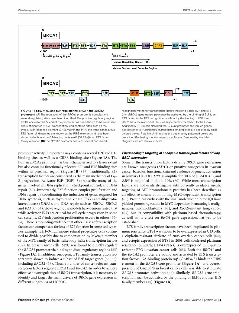
FIGURE 1 | ETS, MYC, and E2F regulate the BRCA1 and BRCA2promoters. (A) The regulation of the BRCA1 promoter is complex andseveral regulatory sites have been identified. The positive regulatory region(PPR) located at the 5′ end of the promoter has been shown to be necessaryand sufficient for BRCA1 transcription, and contains sites such as thecyclic-AMP response element (CRE). Within the PPR, the three consecutiveETS factor binding sites are known as the RIBS element and have beenshown to be bound by GA-binding protein α/β (GABPα/β), an ETS factorfamily member. (B) The BRCA2 promoter contains several conserved
recognition motifs for transcription factors including E-box, E2F, and ETS(49). BRCA2 gene transcription may be activated by the binding of ELF1, anETS factor, to the ETS recognition motifs or by the binding of USF1 andUSF2, basic helix-loop-helix leucine zipper family members, to the E-box.Additionally, NF-κB can also bind the BRCA2 promoter and induce genesexpression (59). Functionally characterized binding sites are depicted by solidcolored boxes. Putative binding sites are depicted by patterned boxes andwere identified using the MatInspector software (Genomatix, Munich).Diagrams are not drawn to scale.
promoter activity in reporter assays, contains several E2F and ETSbinding sites as well as a CREB binding site (Figure 1A). Thehuman BRCA2 promoter has been characterized to a lesser extentbut also contains functionally relevant E2F and ETS binding siteswithin its proximal region (Figure 1B) (49). Traditionally, E2Ftranscription factors are considered as the main mediators of G1–S progression. Activator E2Fs (E2F1–3) transcribe many of thegenes involved in DNA replication, checkpoint control, and DNArepair (50). Importantly, E2F function couples proliferation andDNA repair by coordinating the induction of genes required forDNA synthesis, such as thymidine kinase (TK1) and dihydrofo-latereductase (DHFR), and DNA repair, such as BRCA1, BRCA2,and RAD51 (51). However, mouse models have demonstrated thatwhile activator E2Fs are critical for cell cycle progression in somecell systems, E2F-independent proliferation occurs in others (52–54). There is mounting evidence that other classes of transcriptionfactors can compensate for loss of E2F function in some cell types.For example, E2f1–3-null mouse retinal progenitor cells contin-ued to divide possibly due to compensation by Mycn, a memberof the MYC family of basic helix-loop-helix transcription factors(52). In breast cancer cells, MYC was found to directly regulatethe BRCA1 promoter via binding to distal regulatory regions (55)(Figure 1A). In addition, oncogenic ETS family transcription fac-tors were shown to induce a subset of E2F target genes (56, 57),including BRCA2 (58). Thus, a number of cancer-relevant tran-scription factors regulate BRCA1 and BRCA2. In order to achieveeffective downregulation of BRCA transcription, it is necessary toidentify and target the main drivers of BRCA gene expression indifferent subgroups of HGSOC.
Pharmacologic targeting of oncogenic transcription factors drivingBRCA expressionSome of the transcription factors driving BRCA gene expressionare known oncogenes (MYC) or putative oncogenes in ovariancancer, based on functional data and evidence of genetic activationin primary HGSOC: MYC is amplified in 30% of HGSOC (6), andE2F3 is amplified in about 10% (60). While most transcriptionfactors are not easily druggable with currently available agents,targeting of BET bromodomain proteins has been described asan effective means of inhibiting MYC-dependent transcription(61). Preclinical studies with the small molecule inhibitor JQ1 haveyielded promising results in MYC-dependent hematologic malig-nancies, medulloblastoma (62), and KRAS-mutant lung cancer(63), but its compatibility with platinum-based chemotherapy,as well as its effect on BRCA gene expression, has yet to beestablished.
ETS family transcription factors have been implicated in plat-inum resistance. ETS1 was shown to be overexpressed in C13 cells,a cisplatin-resistant derivate of 2008 ovarian cancer cells (64),and ectopic expression of ETS1 in 2008 cells conferred platinumresistance. Similarly, ETV4 (PEA3) is overexpressed in cisplatin-resistant PEO1 ovarian cancer cells (65). Both the BRCA1 andthe BRCA2 promoter are bound and activated by ETS transcrip-tion factors: GA-binding protein α/β (GABPα/β) binds the RIBSelement in the BRCA1 core promoter (Figure 1A), and overex-pression of GABPα/β in breast cancer cells was able to stimulateBRCA1 promoter activation (66). Similarly, BRCA2 gene tran-scription may be activated by the binding of ELF1, another ETSfamily member (49) (Figure 1B).
Frontiers in Oncology | Women’s Cancer March 2014 | Volume 4 | Article 34 | 4
Wiedemeyer et al. BRCA and platinum resistance
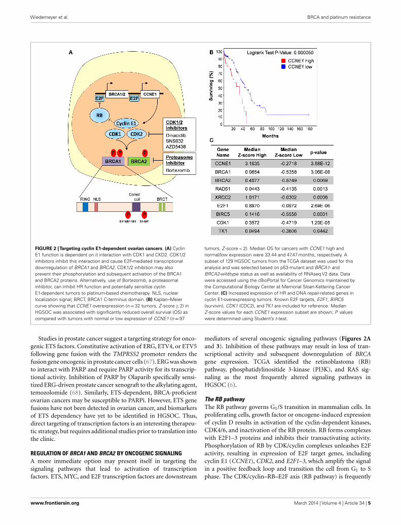
FIGURE 2 |Targeting cyclin E1-dependent ovarian cancers. (A) CyclinE1 function is dependent on it interaction with CDK1 and CKD2. CDK1/2inhibitors inhibit this interaction and cause E2F-mediated transcriptionaldownregulation of BRCA1 and BRCA2. CDK1/2 inhibition may alsoprevent their phosphorylation and subsequent activation of the BRCA1and BRCA2 proteins. Alternatively, use of Bortezomib, a proteasomalinhibitor, can inhibit HR function and potentially sensitize cyclinE1-dependent tumors to platinum-based chemotherapy. NLS, nuclearlocalization signal; BRCT, BRCA1 C-terminus domain. (B) Kaplan–Meiercurve showing that CCNE1 overexpression (n=32 tumors, Z -score≥2) inHGSOC was associated with significantly reduced overall survival (OS) ascompared with tumors with normal or low expression of CCNE1 (n=97
tumors, Z -score < 2). Median OS for cancers with CCNE1 high andnormal/low expression were 33.44 and 47.47 months, respectively. Asubset of 129 HGSOC tumors from the TCGA dataset was used for thisanalysis and was selected based on p53-mutant and BRCA1- andBRCA2-wildtype status as well as availability of RNAseq V2 data. Datawere accessed using the cBioPortal for Cancer Genomics maintained bythe Computational Biology Center at Memorial Sloan-Kettering CancerCenter. (C) Increased expression of HR and DNA repair-related genes incyclin E1-overexpressing tumors. Known E2F targets, E2F1, BIRC5(survivin), CDK1 (CDC2), and TK1 are included for reference. MedianZ -score values for each CCNE1 expression subset are shown; P valueswere determined using Student’s t -test.
Studies in prostate cancer suggest a targeting strategy for onco-genic ETS factors. Constitutive activation of ERG, ETV4, or ETV5following gene fusion with the TMPRSS2 promoter renders thefusion gene oncogenic in prostate cancer cells (67). ERG was shownto interact with PARP and require PARP activity for its transcrip-tional activity. Inhibition of PARP by Olaparib specifically sensi-tized ERG-driven prostate cancer xenograft to the alkylating agent,temozolomide (68). Similarly, ETS-dependent, BRCA-proficientovarian cancers may be susceptible to PARPi. However, ETS genefusions have not been detected in ovarian cancer, and biomarkersof ETS dependency have yet to be identified in HGSOC. Thus,direct targeting of transcription factors is an interesting therapeu-tic strategy, but requires additional studies prior to translation intothe clinic.
REGULATION OF BRCA1 AND BRCA2 BY ONCOGENIC SIGNALINGA more immediate option may present itself in targeting thesignaling pathways that lead to activation of transcriptionfactors. ETS, MYC, and E2F transcription factors are downstream
mediators of several oncogenic signaling pathways (Figures 2Aand 3). Inhibition of these pathways may result in loss of tran-scriptional activity and subsequent downregulation of BRCAgene expression. TCGA identified the retinoblastoma (RB)pathway, phosphatidylinositide 3-kinase (PI3K), and RAS sig-naling as the most frequently altered signaling pathways inHGSOC (6).
The RB pathwayThe RB pathway governs G1/S transition in mammalian cells. Inproliferating cells, growth factor or oncogene-induced expressionof cyclin D results in activation of the cyclin-dependent kinases,CDK4/6, and inactivation of the RB protein. RB forms complexeswith E2F1–3 proteins and inhibits their transactivating activity.Phosphorylation of RB by CDK/cyclin complexes unleashes E2Factivity, resulting in expression of E2F target genes, includingcyclin E1 (CCNE1), CDK2, and E2F1–3, which amplify the signalin a positive feedback loop and transition the cell from G1 to Sphase. The CDK/cyclin–RB–E2F axis (RB pathway) is frequently
www.frontiersin.org March 2014 | Volume 4 | Article 34 | 5
Wiedemeyer et al. BRCA and platinum resistance
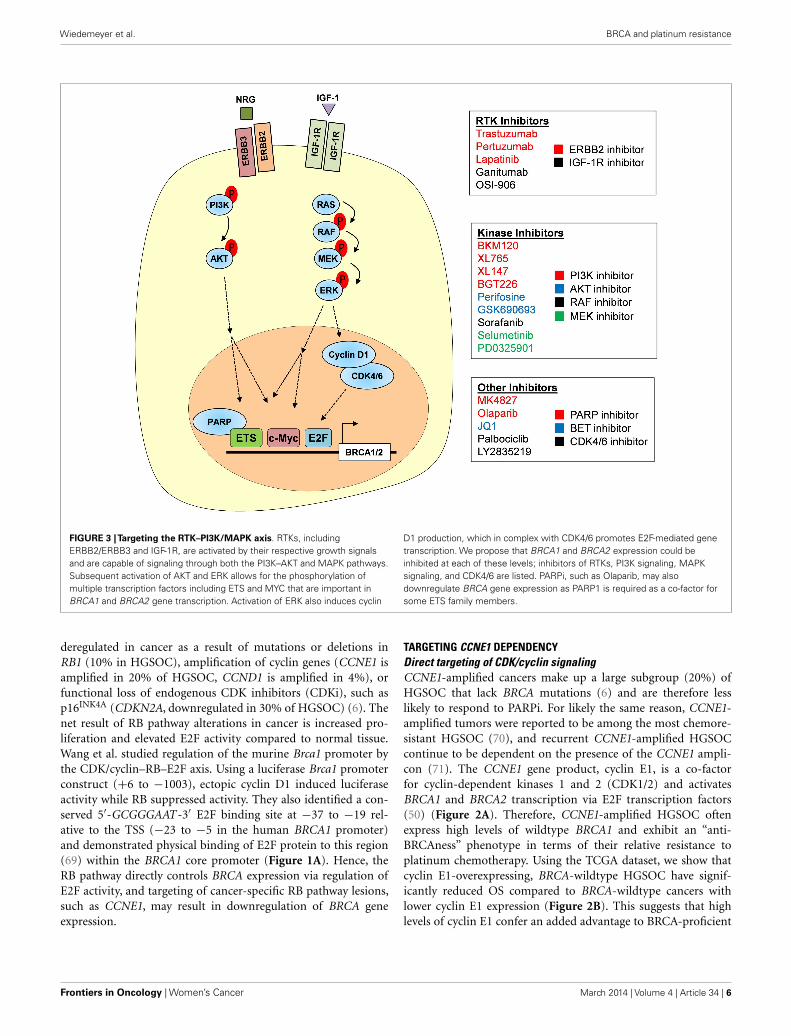
FIGURE 3 |Targeting the RTK–PI3K/MAPK axis. RTKs, includingERBB2/ERBB3 and IGF-1R, are activated by their respective growth signalsand are capable of signaling through both the PI3K–AKT and MAPK pathways.Subsequent activation of AKT and ERK allows for the phosphorylation ofmultiple transcription factors including ETS and MYC that are important inBRCA1 and BRCA2 gene transcription. Activation of ERK also induces cyclin
D1 production, which in complex with CDK4/6 promotes E2F-mediated genetranscription. We propose that BRCA1 and BRCA2 expression could beinhibited at each of these levels; inhibitors of RTKs, PI3K signaling, MAPKsignaling, and CDK4/6 are listed. PARPi, such as Olaparib, may alsodownregulate BRCA gene expression as PARP1 is required as a co-factor forsome ETS family members.
deregulated in cancer as a result of mutations or deletions inRB1 (10% in HGSOC), amplification of cyclin genes (CCNE1 isamplified in 20% of HGSOC, CCND1 is amplified in 4%), orfunctional loss of endogenous CDK inhibitors (CDKi), such asp16INK4A (CDKN2A, downregulated in 30% of HGSOC) (6). Thenet result of RB pathway alterations in cancer is increased pro-liferation and elevated E2F activity compared to normal tissue.Wang et al. studied regulation of the murine Brca1 promoter bythe CDK/cyclin–RB–E2F axis. Using a luciferase Brca1 promoterconstruct (+6 to −1003), ectopic cyclin D1 induced luciferaseactivity while RB suppressed activity. They also identified a con-served 5′-GCGGGAAT -3′ E2F binding site at −37 to −19 rel-ative to the TSS (−23 to −5 in the human BRCA1 promoter)and demonstrated physical binding of E2F protein to this region(69) within the BRCA1 core promoter (Figure 1A). Hence, theRB pathway directly controls BRCA expression via regulation ofE2F activity, and targeting of cancer-specific RB pathway lesions,such as CCNE1, may result in downregulation of BRCA geneexpression.
TARGETING CCNE1 DEPENDENCYDirect targeting of CDK/cyclin signalingCCNE1-amplified cancers make up a large subgroup (20%) ofHGSOC that lack BRCA mutations (6) and are therefore lesslikely to respond to PARPi. For likely the same reason, CCNE1-amplified tumors were reported to be among the most chemore-sistant HGSOC (70), and recurrent CCNE1-amplified HGSOCcontinue to be dependent on the presence of the CCNE1 ampli-con (71). The CCNE1 gene product, cyclin E1, is a co-factorfor cyclin-dependent kinases 1 and 2 (CDK1/2) and activatesBRCA1 and BRCA2 transcription via E2F transcription factors(50) (Figure 2A). Therefore, CCNE1-amplified HGSOC oftenexpress high levels of wildtype BRCA1 and exhibit an “anti-BRCAness” phenotype in terms of their relative resistance toplatinum chemotherapy. Using the TCGA dataset, we show thatcyclin E1-overexpressing, BRCA-wildtype HGSOC have signif-icantly reduced OS compared to BRCA-wildtype cancers withlower cyclin E1 expression (Figure 2B). This suggests that highlevels of cyclin E1 confer an added advantage to BRCA-proficient
Frontiers in Oncology | Women’s Cancer March 2014 | Volume 4 | Article 34 | 6
Wiedemeyer et al. BRCA and platinum resistance
cancers. We attribute this, at least in part, to increased expression ofseveral HR genes, including BRCA1, BRCA2, RAD51, all of whichhave significantly higher RNA levels in cyclin E1-overexpressingcells (Figure 2C).
CDK inhibitors may be a therapeutic option for cyclin E1-dependent HGSOC, including CCNE1-amplified cancers. CyclinE1 has the highest affinity for CDK2, its main binding partner inactively cycling cells (72–74), and CDK1 (CDC2) (75). Both CDK2and CDK1 directly phosphorylate BRCA1 (76, 77) (Figure 2A).Furthermore, a functional link between CDK1 and BRCA1 hasbeen established in lung cancer. Using an inducible shRNA target-ing CDK1 in a human non-small cell lung cancer cell line, Johnsonet al. showed that CDK1 contributes to S phase checkpoint con-trol following DNA damage (77). CDK1 directly phosphorylatedBRCA1 at several serine residues and is required for the forma-tion of BRCA1 foci following DNA damage. Genetic depletion orpharmacological inhibition of CDK1 sensitized BRCA1-proficientcancer cell lines to cisplatin (77) and PARPi (78). Similarly, BRCA2is phosphorylated by both CDK1 and CDK2 (79).
While there are still no selective inhibitors of CDK1 or CDK2,latest generation CDKi have increased specificity and potencycompared to early CDKi, such as flavopiridol, which failed inthe clinic. The compound Dinaciclib inhibits CDK1/2/5/9 atlow nanomolar concentrations (IC50 values: CDK1: 3 nM, CDK2,1 nM, CDK5: 1 nM, CDK9: 4 nM). It induces apoptosis in modelsystems and prevents tumor progression in A2780 ovarian cancerxenografts (80, 81). We hypothesize that of the currently avail-able CDKi, Dinaciclib may have the best therapeutic potential incyclin E1-dependent ovarian cancer. Our unpublished data showthat Dinaciclib exposure results in downregulation of BRCA1 andBRCA2 and sensitized cyclin E1-dependent ovarian cancer cells tocisplatin. Hence, Dinaciclib may have a dual effect on BRCA1 andBRCA2 by causing E2F-mediated transcriptional downregulationand inhibiting CDK1/2-mediated activation of the BRCA proteins(Figure 2A).
Dinaciclib is currently being evaluated in a phase 3 clinical trialfor chronic lymphocytic leukemia (CLL) and may have clinicalpotential in cyclin E1-dependent HGSOC as a chemosensitizingagent. In order to select eligible patients, CCNE1-amplified can-cers can be easily identified by fluorescence in situ hybridization(FISH). Other available CDK2 inhibitors, including Roscovitine,SNS032 (82), and AZD5438 (83), may have similar chemosensi-tizing potential. CDK1, the only mammalian CDK required forproliferation in all cell types (84), is not inhibited by SNS032, ren-dering this compound potentially less potent but also less toxicto normal cells. In principle, other CDKi such as the CDK4/6inhibitor Palbociclib (formerly PD0332991) could be used toreduce BRCA expression in cyclin D-dependent HGSOC. How-ever, Palbociclib was shown to be cytostatic in most systems(85–87) and may thus interfere with the cytotoxic activity ofplatinum agents.
Targeting of HR components induces synthetic lethality inCCNE1-amplified HGSOCAn independent approach to target CCNE1-amplified HGSOCidentified the proteasome inhibitor, Bortezomib (88). In contrastto CDKi, Bortezomib primarily affects the homologous repair
system itself, resulting in synthetic lethality in CCNE1-amplifiedcancer cells: a genome-wide shRNA screen in 102 cancer cell linesrevealed that BRCA1 was specifically required in CCNE1-amplifiedcell lines. Genetic depletion of BRCA1 resulted in significant lossof viability in CCNE1-amplified cells, including the ovarian cancercell line OVCAR3, whereas a lesser effect was observed in CCNE1-wildtype cells, such as the SKOV3 cell line (88). This suggests asynthetic lethal relationship between CCNE1 amplification andloss of BRCA1 function and provides a potential explanation forthe observed mutual exclusivity between CCNE1 amplificationand BRCA mutation (6, 89). In addition to BRCA1, the shRNAscreen identified the DNA repair genes ATR and XRCC2 as specificgenetic hits in CCNE1-amplified cell lines, suggesting that inhibi-tion of the DDR may be a therapeutic strategy in CCNE1-amplifiedovarian cancer. To this end, the authors tested Bortezomib, apotent inhibitor of the HR system, in a panel of ovarian cancercell lines and found that CCNE1-amplified lines were most sensi-tive. While Bortezomib had minimal activity as a single agent inrecurrent ovarian cancer (90) combination with platinum resultedin clinical activity (91). As discussed for CDKi, specific selection ofpatients with CCNE1-amplified cancers for Bortezomib treatmentmay further increase the response rate.
RECEPTOR TYROSINE KINASE, PI3K, AND RAS SIGNALINGIn addition to genetic aberrations within the RB pathway, severaloncogenic signals contribute to cell cycle deregulation and E2Factivity. Oncogenic KRAS (amplified in 11% of HGSOC), MYC,and receptor tyrosine kinases (RTK) all converge on cyclin D andrequire its function for their oncogenic activity (92–94). Moreover,a synthetic lethal relationship was described for KRAS and CDK4,the binding partner of cyclin D (94). KRAS and ERBB2 signalthrough the mitogen-activated kinase (MAPK) signaling pathway,which culminates in activation of ERK (Figure 3). Phosphoryla-tion by ERK activates multiple cellular target proteins, includingETS, CREB, and MYC. The molecular link between oncogenicsignaling pathways and expression of BRCA1 and BRCA2 offersadditional opportunities for therapeutic intervention, includingdirect targeting of ERBB2 and other RTK, targeting of the MAPKpathway by specific MEK or ERK inhibitors, or targeting of thePI3K–AKT axis by specific inhibitors (Figure 3). However, due tothe complex signaling events elicited by RTK and other oncogenicpathways, regulation of BRCA gene expression is only one of manydownstream events, some of which may actually interfere with theintended chemosensitizing effect. Functional studies are neededto determine the compatibility of individual targeted agents withplatinum and PARPi.
Targeting RTK signaling and downstream pathwaysAlthough amplifications or mutations of individual RTK arerare events in primary HGSOC (ERBB2 amplification: 3%,ERBB3 amplification: <5%, IGF1R amplification: <4%, Figure 3),altered ERBB receptor signaling and overexpression of the down-stream RTK mediator ETV4 were found in cisplatin-resistantPEO1 cells (65). RTK can be targeted directly by small mole-cule inhibitors (e.g., Lapatinib for ERBB2) or antagonistic anti-bodies (e.g., Trastuzumab or Pertuzumab for ERBB2). How-ever in unselected patients, the addition of Pertuzumab to
www.frontiersin.org March 2014 | Volume 4 | Article 34 | 7
Wiedemeyer et al. BRCA and platinum resistance
carboplatin-based chemotherapy did not result in prolongedprogression-free survival (95), highlighting the importance ofpatient selection and companion diagnostics to identify likelyresponders to RTK inhibitors. Activation of the MAPK and PI3K–AKT signaling pathways by RTK, or as a result of amplification,offers additional potential therapeutic targets [also reviewed inRef. (96)]. Both PIK3CA, the gene encoding the catalytic subunitof PI3K, and the AKT genes are frequently amplified in HGSOC(PIK3CA: 28%, AKT1: 5%, AKT2: 8%, AKT3: 9%), indicatingtheir oncogenic roles in ovarian cancer. Interestingly, AKT wasshown to directly phosphorylate BRCA1 at serine 694 and pro-mote its protein stability (97). A second AKT phosphorylation siteat threonine 509 was described but its functional implications areunknown (98).
Functionally, a recent study in triple negative breast cancerdemonstrated that PI3K signaling was required for BRCA1 func-tion. Ibrahim et al. showed that inhibition of PI3K phenocopiedloss of BRCA1 and induced synthetic lethality in combinationwith Olaparib (99). Loss of PI3K resulted in reduced BRCA1expression both in cell line models and in patient-derived tumorxenografts. Combined treatment with BKM120, a small moleculePI3K inhibitor, and Olaparib significantly delayed tumor progres-sion in two out of three xenografts whereas single agent treatmenthad little effect on tumor progression. On the molecular level,the MAPK pathway mediated the transcriptional effect on BRCA1via ETS1-dependent downregulation of BRCA1 (99). Interestingly,BKM120 treatment resulted in activation of the MAPK path-way and phosphorylation of ETS1 by ERK, indicating a repressorfunction for ETS1 in this system. This finding highlights the com-plexity of BRCA gene regulation and its dependency on geneticcontext. Due to feedback mechanisms and functional compensa-tion among RTKs, downstream kinases, and transcription factors,specific pharmacological combinations may be limited in theireffectiveness to small genetically defined subsets of HGSOC. More-over, as a result of the genetic complexity and genomic instabilitythat is a hallmark of HGSOC, individual cancers are likely to har-bor or develop resistant cell clones to any given combination. Thus,it will be important to identify multiple agents targeting differentpathways, all of which should be characterized with respect to:(1) their ability to induce BRCA loss-of-function, (2) their com-patibility with platinum and/or PARPi, both in terms of toxicityand mechanism of action, and (3) accompanying biomarkers thatallow for careful patient selection.
TARGETING BRCA PROTEIN STABILITYA different approach to inhibit BRCA1 function involves targetingof the chaperone protein heat shock protein 90 (HSP90), requiredfor BRCA1 protein stability (100). Interestingly, HSP90AB1 is alsoamplified in a small subset of HGSOC (6). Specific inhibition ofHSP90 in BRCA1-proficient breast cancer cells-induced BRCA1ubiquitination and proteasomal degradation ultimately resultingin compromised DSB repair by HR. HSP90 inhibition sensitizedBRCA1-proficient breast and ovarian cancer cells in vitro to car-boplatin treatment at concentrations similar to BRCA1-mutantcells (100). In a subsequent study, treatment with the pan-histonedeacetylase complex (HDAC) inhibitors vorinostat or panobi-nostat induced hyperacetylation of HSP90 gene (HSP90AA1I0),
thereby inhibiting its chaperone function and leading to protea-somal degradation and depletion of BRCA1 (101). Of clinicalsignificance, treatment of human triple negative breast cancer celllines with vorinostat was able to induce BRCA1 degradation anda subsequent BRCAness phenotype, which synergistically inducedcell death in combination with PARPi or cisplatin (102). Basedon these preliminary studies, it is suggested that treatment ofplatinum-resistant HGSOC with HDAC inhibitors may also beable to induce a BRCAness phenotype and resensitize these cellsto platinum or other targeted agents. Trials evaluating vorinos-tat as a single agent in recurrent epithelial ovarian cancer showedit had limited activity (103). Vorinostat may show better ther-apeutic efficacy as a biologic response modifier in combinationwith platinum-based chemotherapy and trials assessing this use inovarian cancer are underway.
CONCLUDING REMARKSIn HGSOC cells, a functional HR system and intact BRCA1 andBRCA2 function are often associated with resistance to platinum-based chemotherapeutic agents and PARPi. A plethora of targetedagents are currently available to modulate BRCA function via tran-scriptional or post-translational intervention. With the advent ofnovel diagnostic tools, such as the use of deep sequencing to repeat-edly profile cancers throughout their evolution, it should becomepossible to predict rational therapeutic combinations that pre-vent or at least delay the onset of platinum resistance and shouldultimately result in improvements in OS.
ACKNOWLEDGMENTSThis work was supported by the Ovarian Cancer Research Fund LizTilberis Scholars Program (W. Ruprecht Wiedemeyer), the PacificOvarian Cancer Research Consortium (W. Ruprecht Wiedemeyerand Beth Y. Karlan, Award Number P50 CA083636 from theNIH/National Cancer Institute), The Borden Family Founda-tion (W. Ruprecht Wiedemeyer), The Garber Foundation (W.Ruprecht Wiedemeyer), an American Cancer Society Early Detec-tion Professorship (Beth Y. Karlan, SIOP-06-258-01-COUN) anda generous donation by Ms. Susie Fragnoli. We thank membersof the Cedars-Sinai Women’s Cancer Program for critical read-ing of the manuscript and apologize to authors whose work wasnot cited.
REFERENCES1. Rossof AH, Talley RW, Stephens R, Thigpen T, Samson MK, Groppe C Jr, et al.
Phase II evaluation of cis-dichlorodiammineplatinum(II) in advanced malig-nancies of the genitourinary and gynecologic organs: a Southwest OncologyGroup Study. Cancer Treat Rep (1979) 63(9–10):1557–64.
2. Thigpen T, Shingleton H, Homesley H, LaGasse L, Blessing J. cis-Dichlorodiammineplatinum(II) in the treatment of gynecologic malignancies:phase II trials by the Gynecologic Oncology Group. Cancer Treat Rep (1979)63(9–10):1549–55.
3. Alberts DS, Green S, Hannigan EV, O’Toole R, Stock-Novack D, Anderson P,et al. Improved therapeutic index of carboplatin plus cyclophosphamide ver-sus cisplatin plus cyclophosphamide: final report by the Southwest OncologyGroup of a phase III randomized trial in stages III and IV ovarian cancer. J ClinOncol (1992) 10(5):706–17.
4. McGuire WP, Hoskins WJ, Brady MF, Kucera PR, Partridge EE, Look KY, et al.Cyclophosphamide and cisplatin compared with paclitaxel and cisplatin inpatients with stage III and stage IV ovarian cancer. N Engl J Med (1996)334(1):1–6. doi:10.1056/NEJM199601043340101
Frontiers in Oncology | Women’s Cancer March 2014 | Volume 4 | Article 34 | 8
Wiedemeyer et al. BRCA and platinum resistance
5. Piccart MJ, Bertelsen K, James K, Cassidy J, Mangioni C, Simonsen E,et al. Randomized intergroup trial of cisplatin-paclitaxel versus cisplatin-cyclophosphamide in women with advanced epithelial ovarian cancer: three-year results. J Natl Cancer Inst (2000) 92(9):699–708. doi:10.1093/jnci/92.17.1446-a
6. Cancer Genome Atlas Research Network. Integrated genomic analyses of ovar-ian carcinoma. Nature (2011) 474(7353):609–15. doi:10.1038/nature10166
7. Jones S, Wang TL, Shih Ie M, Mao TL, Nakayama K, Roden R, et al. Frequentmutations of chromatin remodeling gene ARID1A in ovarian clear cell carci-noma. Science (2010) 330(6001):228–31. doi:10.1126/science.1196333
8. Wiegand KC, Shah SP,Al-Agha OM, Zhao Y, Tse K, Zeng T, et al. ARID1A muta-tions in endometriosis-associated ovarian carcinomas. N Engl J Med (2010)363(16):1532–43. doi:10.1056/NEJMoa1008433
9. Seidman JD, Horkayne-Szakaly I, Haiba M, Boice CR, Kurman RJ, Ron-nett BM. The histologic type and stage distribution of ovarian carcino-mas of surface epithelial origin. Int J Gynecol Pathol (2004) 23(1):41–4.doi:10.1097/01.pgp.0000101080.35393.16
10. Piccart MJ, Lamb H, Vermorken JB. Current and future potential roles ofthe platinum drugs in the treatment of ovarian cancer. Ann Oncol (2001)12(9):1195–203. doi:10.1023/A:1012259625746
11. Matsuo K, Lin YG, Roman LD, Sood AK. Overcoming platinum resistancein ovarian carcinoma. Expert Opin Investig Drugs (2010) 19(11):1339–54.doi:10.1517/13543784.2010.515585
12. Siegel R, Naishadham D, Jemal A. Cancer statistics, 2013. CA Cancer J Clin(2013) 63(1):11–30. doi:10.3322/caac.21166
13. Siddik ZH. Cisplatin: mode of cytotoxic action and molecular basis of resis-tance. Oncogene (2003) 22(47):7265–79. doi:10.1038/sj.onc.1206933
14. Galluzzi L, Senovilla L, Vitale I, Michels J, Martins I, Kepp O, et al. Mole-cular mechanisms of cisplatin resistance. Oncogene (2012) 31(15):1869–83.doi:10.1038/onc.2011.384
15. Stewart DJ. Mechanisms of resistance to cisplatin and carboplatin. Crit RevOncol Hematol (2007) 63(1):12–31. doi:10.1016/j.critrevonc.2007.02.001
16. Martin LP, Hamilton TC, Schilder RJ. Platinum resistance: the role of DNArepair pathways. Clin Cancer Res (2008) 14(5):1291–5. doi:10.1158/1078-0432.CCR-07-2238
17. Ferry KV, Hamilton TC, Johnson SW. Increased nucleotide excision repair incisplatin-resistant ovarian cancer cells: role of ERCC1-XPF. Biochem Pharmacol(2000) 60(9):1305–13. doi:10.1016/S0006-2952(00)00441-X
18. Dabholkar M, Bostick-Bruton F,Weber C, BohrVA, Egwuagu C, Reed E. ERCC1and ERCC2 expression in malignant tissues from ovarian cancer patients. J NatlCancer Inst (1992) 84(19):1512–7. doi:10.1093/jnci/84.19.1512
19. Samimi G, Fink D, Varki NM, Husain A, Hoskins WJ, Alberts DS, et al. Analysisof MLH1 and MSH2 expression in ovarian cancer before and after platinumdrug-based chemotherapy. Clin Cancer Res (2000) 6(4):1415–21.
20. Murphy MA, Wentzensen N. Frequency of mismatch repair deficiency in ovar-ian cancer: a systematic review this article is a US Government work and, assuch, is in the public domain of the United States of America. Int J Cancer(2011) 129(8):1914–22. doi:10.1002/ijc.25835
21. Helleman J, van Staveren IL, Dinjens WN, van Kuijk PF, Ritstier K, Ewing PC,et al. Mismatch repair and treatment resistance in ovarian cancer. BMC Cancer(2006) 6:201. doi:10.1186/1471-2407-6-201
22. Watanabe Y, Koi M, Hemmi H, Hoshai H, Noda K. A change in microsatelliteinstability caused by cisplatin-based chemotherapy of ovarian cancer. Br J Can-cer (2001) 85(7):1064–9. doi:10.1054/bjoc.2001.2037
23. Mao Z, Bozzella M, Seluanov A, Gorbunova V. DNA repair by nonhomologousend joining and homologous recombination during cell cycle in human cells.Cell Cycle (2008) 7(18):2902–6. doi:10.4161/cc.7.18.6679
24. Roy R, Chun J, Powell SN. BRCA1 and BRCA2: different roles in a com-mon pathway of genome protection. Nat Rev Cancer (2012) 12(1):68–78.doi:10.1038/nrc3181
25. Yun MH, Hiom K. CtIP-BRCA1 modulates the choice of DNA double-strand-break repair pathway throughout the cell cycle. Nature (2009)459(7245):460–3. doi:10.1038/nature07955
26. Wang B, Matsuoka S, Ballif BA, Zhang D, Smogorzewska A, Gygi SP, et al.Abraxas and RAP80 form a BRCA1 protein complex required for the DNA dam-age response. Science (2007) 316(5828):1194–8. doi:10.1126/science.1139476
27. Chen J, Silver DP, Walpita D, Cantor SB, Gazdar AF, Tomlinson G, et al.Stable interaction between the products of the BRCA1 and BRCA2 tumor
suppressor genes in mitotic and meiotic cells. Mol Cell (1998) 2(3):317–28.doi:10.1016/S1097-2765(00)80276-2
28. Scully R, Chen J, Ochs RL, Keegan K, Hoekstra M, Feunteun J, et al. Dynamicchanges of BRCA1 subnuclear location and phosphorylation state are initi-ated by DNA damage. Cell (1997) 90(3):425–35. doi:10.1016/S0092-8674(00)80503-6
29. Marmorstein LY, Ouchi T, Aaronson SA. The BRCA2 gene product func-tionally interacts with p53 and RAD51. Proc Natl Acad Sci U S A (1998)95(23):13869–74. doi:10.1073/pnas.95.23.13869
30. Cass I, Baldwin RL, Varkey T, Moslehi R, Narod SA, Karlan BY. Improvedsurvival in women with BRCA-associated ovarian carcinoma. Cancer (2003)97(9):2187–95. doi:10.1002/cncr.11310
31. Chetrit A, Hirsh-Yechezkel G, Ben-David Y, Lubin F, Friedman E, Sadetzki S.Effect of BRCA1/2 mutations on long-term survival of patients with invasiveovarian cancer: the national Israeli study of ovarian cancer. J Clin Oncol (2008)26(1):20–5. doi:10.1200/JCO.2007.11.6905
32. Bolton KL, Chenevix-Trench G, Goh C, Sadetzki S, Ramus SJ, Karlan BY,et al. Association between BRCA1 and BRCA2 mutations and survival inwomen with invasive epithelial ovarian cancer. JAMA (2012) 307(4):382–90.doi:10.1001/jama.2012.20
33. Sakai W, Swisher EM, Karlan BY, Agarwal MK, Higgins J, Friedman C, et al. Sec-ondary mutations as a mechanism of cisplatin resistance in BRCA2-mutatedcancers. Nature (2008) 451(7182):1116–20. doi:10.1038/nature06633
34. Swisher EM, Sakai W, Karlan BY, Wurz K, Urban N, Taniguchi T. SecondaryBRCA1 mutations in BRCA1-mutated ovarian carcinomas with platinum resis-tance. Cancer Res (2008) 68(8):2581–6. doi:10.1158/0008-5472.CAN-08-0088
35. Norquist B, Wurz KA, Pennil CC, Garcia R, Gross J, Sakai W, et al. Sec-ondary somatic mutations restoring BRCA1/2 predict chemotherapy resis-tance in hereditary ovarian carcinomas. J Clin Oncol (2011) 29(22):3008–15.doi:10.1200/JCO.2010.34.2980
36. Borst P, Rottenberg S, Jonkers J. How do real tumors become resistant to cis-platin? Cell Cycle (2008) 7(10):1353–9. doi:10.4161/cc.7.10.5930
37. Aguirre-Ghiso JA. Models, mechanisms and clinical evidence for cancer dor-mancy. Nat Rev Cancer (2007) 7(11):834–46. doi:10.1038/nrc2256
38. Kaelin WG Jr. The concept of synthetic lethality in the context of anticancertherapy. Nat Rev Cancer (2005) 5(9):689–98. doi:10.1038/nrc1691
39. Farmer H, McCabe N, Lord CJ, Tutt AN, Johnson DA, Richardson TB, et al.Targeting the DNA repair defect in BRCA mutant cells as a therapeutic strategy.Nature (2005) 434(7035):917–21. doi:10.1038/nature03445
40. Audeh MW, Carmichael J, Penson RT, Friedlander M, Powell B, Bell-McGuinnKM, et al. Oral poly(ADP-ribose) polymerase inhibitor olaparib in patientswith BRCA1 or BRCA2 mutations and recurrent ovarian cancer: a proof-of-concept trial. Lancet (2010) 376(9737):245–51. doi:10.1016/S0140-6736(10)60893-8
41. Fong PC, Yap TA, Boss DS, Carden CP, Mergui-Roelvink M, Gourley C, et al.Poly(ADP)-ribose polymerase inhibition: frequent durable responses in BRCAcarrier ovarian cancer correlating with platinum-free interval. J Clin Oncol(2010) 28(15):2512–9. doi:10.1200/JCO.2009.26.9589
42. Ledermann J, Harter P, Gourley C, Friedlander M, Vergote I, Rustin G, et al.Olaparib maintenance therapy in platinum-sensitive relapsed ovarian cancer.N Engl J Med (2012) 366(15):1382–92. doi:10.1056/NEJMoa1105535
43. Turner N, Tutt A, Ashworth A. Hallmarks of ‘BRCAness’ in sporadic cancers.Nat Rev Cancer (2004) 4(10):814–9. doi:10.1038/nrc1457
44. Quinn JE, James CR, Stewart GE, Mulligan JM, White P, Chang GK, et al.BRCA1 mRNA expression levels predict for overall survival in ovarian can-cer after chemotherapy. Clin Cancer Res (2007) 13(24):7413–20. doi:10.1158/1078-0432.CCR-07-1083
45. Rajan JV, Wang M, Marquis ST, Chodosh LA. Brca2 is coordinately regulatedwith Brca1 during proliferation and differentiation in mammary epithelialcells. Proc Natl Acad Sci U S A (1996) 93(23):13078–83. doi:10.1073/pnas.93.23.13078
46. Marks JR, Huper G, Vaughn JP, Davis PL, Norris J, McDonnell DP, et al.BRCA1 expression is not directly responsive to estrogen. Oncogene (1997)14(1):115–21. doi:10.1038/sj.onc.1200808
47. Xu CF, Chambers JA, Solomon E. Complex regulation of the BRCA1 gene.J Biol Chem (1997) 272(34):20994–7. doi:10.1074/jbc.272.34.20994
48. Thakur S, Croce CM. Positive regulation of the BRCA1 promoter. J Biol Chem(1999) 274(13):8837–43. doi:10.1074/jbc.274.13.8837
www.frontiersin.org March 2014 | Volume 4 | Article 34 | 9
Wiedemeyer et al. BRCA and platinum resistance
49. Davis PL, Miron A, Andersen LM, Iglehart JD, Marks JR. Isolation and initialcharacterization of the BRCA2 promoter. Oncogene (1999) 18(44):6000–12.doi:10.1038/sj.onc.1202990
50. Bracken AP, Ciro M, Cocito A, Helin K. E2F target genes: unraveling the biology.Trends Biochem Sci (2004) 29(8):409–17. doi:10.1016/j.tibs.2004.06.006
51. Ren B, Cam H, Takahashi Y,Volkert T, Terragni J,Young RA, et al. E2F integratescell cycle progression with DNA repair, replication, and G(2)/M checkpoints.Genes Dev (2002) 16(2):245–56. doi:10.1101/gad.949802
52. Chen D, Pacal M, Wenzel P, Knoepfler PS, Leone G, Bremner R. Division andapoptosis of E2f-deficient retinal progenitors. Nature (2009) 462(7275):925–9.doi:10.1038/nature08544
53. Wenzel PL, Chong JL, Saenz-Robles MT, Ferrey A, Hagan JP, Gomez YM, et al.Cell proliferation in the absence of E2F1-3. Dev Biol (2011) 351(1):35–45.doi:10.1016/j.ydbio.2010.12.025
54. Wu L, Timmers C, Maiti B, Saavedra HI, Sang L, Chong GT, et al. The E2F1-3 transcription factors are essential for cellular proliferation. Nature (2001)414(6862):457–62. doi:10.1038/35106593
55. ChenY, Xu J, Borowicz S, Collins C, Huo D, Olopade OI. c-Myc activates BRCA1gene expression through distal promoter elements in breast cancer cells. BMCCancer (2011) 11:246. doi:10.1186/1471-2407-11-246
56. Bilke S, Schwentner R, Yang F, Kauer M, Jug G, Walker RL, et al. Oncogenic ETSfusions deregulate E2F3 target genes in Ewing sarcoma and prostate cancer.Genome Res (2013) 23(11):1797–809. doi:10.1101/gr.151340.112
57. Roussel MF, Davis JN, Cleveland JL, Ghysdael J, Hiebert SW. Dual controlof myc expression through a single DNA binding site targeted by ETS familyproteins and E2F-1. Oncogene (1994) 9(2):405–15.
58. Llaurado M, Majem B, Castellvi J, Cabrera S, Gil-Moreno A, Reventos J, et al.Analysis of gene expression regulated by the ETV5 transcription factor in OV90ovarian cancer cells identifies FOXM1 overexpression in ovarian cancer. MolCancer Res (2012) 10(7):914–24. doi:10.1158/1541-7786.MCR-11-0449
59. Wu K, Jiang SW, Thangaraju M, Wu G, Couch FJ. Induction of the BRCA2promoter by nuclear factor-kappa B. J Biol Chem (2000) 275(45):35548–56.doi:10.1074/jbc.M004390200
60. Ciriello G, Miller ML, Aksoy BA, Senbabaoglu Y, Schultz N, Sander C. Emerg-ing landscape of oncogenic signatures across human cancers. Nat Genet (2013)45(10):1127–33. doi:10.1038/ng.2762
61. Delmore JE, Issa GC, Lemieux ME, Rahl PB, Shi J, Jacobs HM, et al. BET bro-modomain inhibition as a therapeutic strategy to target c-Myc. Cell (2011)146(6):904–17. doi:10.1016/j.cell.2011.08.017
62. Bandopadhayay P, Bergthold G, Nguyen B, Schubert S, Gholamin S, Tang Y,et al. BET bromodomain inhibition of MYC-amplified medulloblastoma. ClinCancer Res (2014) 20(4):912–25. doi:10.1158/1078-0432.CCR-13-2281
63. Shimamura T, Chen Z, Soucheray M, Carretero J, Kikuchi E, Tchaicha JH, et al.Efficacy of BET bromodomain inhibition in Kras-mutant non-small cell lungcancer. Clin Cancer Res (2013) 19(22):6183–92. doi:10.1158/1078-0432.CCR-12-3904
64. Wilson LA, Yamamoto H, Singh G. Role of the transcription factor Ets-1 incisplatin resistance. Mol Cancer Ther (2004) 3(7):823–32.
65. Macleod K, Mullen P, Sewell J, Rabiasz G, Lawrie S, Miller E, et al. Altered ErbBreceptor signaling and gene expression in cisplatin-resistant ovarian cancer.Cancer Res (2005) 65(15):6789–800. doi:10.1158/0008-5472.CAN-04-2684
66. Atlas E, Stramwasser M, Whiskin K, Mueller CR. GA-binding proteinalpha/beta is a critical regulator of the BRCA1 promoter. Oncogene (2000)19(15):1933–40. doi:10.1038/sj.onc.1203516
67. Han B, Mehra R, Dhanasekaran SM,Yu J, Menon A, Lonigro RJ, et al. A fluores-cence in situ hybridization screen for E26 transformation-specific aberrations:identification of DDX5-ETV4 fusion protein in prostate cancer. Cancer Res(2008) 68(18):7629–37. doi:10.1158/0008-5472.CAN-08-2014
68. Brenner JC, Ateeq B, Li Y, Yocum AK, Cao Q, Asangani IA, et al. Mechanisticrationale for inhibition of poly(ADP-ribose) polymerase in ETS gene fusion-positive prostate cancer. Cancer Cell (2011) 19(5):664–78. doi:10.1016/j.ccr.2011.04.010
69. Wang A, Schneider-Broussard R, Kumar AP, MacLeod MC, Johnson DG. Reg-ulation of BRCA1 expression by the Rb-E2F pathway. J Biol Chem (2000)275(6):4532–6. doi:10.1074/jbc.275.6.4532
70. Nakayama N, Nakayama K, Shamima Y, Ishikawa M, Katagiri A, Iida K, et al.Gene amplification CCNE1 is related to poor survival and potential therapeutictarget in ovarian cancer. Cancer (2010) 116(11):2621–34. doi:10.1002/cncr.24987
71. Etemadmoghadam D, George J, Cowin PA, Cullinane C, Kansara M, GorringeKL, et al. Amplicon-dependent CCNE1 expression is critical for clonogenicsurvival after cisplatin treatment and is correlated with 20q11 gain in ovariancancer. PLoS One (2010) 5(11):e15498. doi:10.1371/journal.pone.0015498
72. Koff A, Giordano A, Desai D, Yamashita K, Harper JW, Elledge S, et al. For-mation and activation of a cyclin E-cdk2 complex during the G1 phase ofthe human cell cycle. Science (1992) 257(5077):1689–94. doi:10.1126/science.1388288
73. Lees E, Faha B, Dulic V, Reed SI, Harlow E. Cyclin E/cdk2 and cyclin A/cdk2kinases associate with p107 and E2F in a temporally distinct manner. GenesDev (1992) 6(10):1874–85. doi:10.1101/gad.6.10.1874
74. Dulic V, Lees E, Reed SI. Association of human cyclin E with a periodic G1-Sphase protein kinase. Science (1992) 257(5078):1958–61. doi:10.1126/science.1329201
75. Koff A, Cross F, Fisher A, Schumacher J, Leguellec K, Philippe M, et al. Humancyclin E, a new cyclin that interacts with two members of the CDC2 gene family.Cell (1991) 66(6):1217–28. doi:10.1016/0092-8674(91)90044-Y
76. Ruffner H, Jiang W, Craig AG, Hunter T, Verma IM. BRCA1 is phosphorylatedat serine 1497 in vivo at a cyclin-dependent kinase 2 phosphorylation site. MolCell Biol (1999) 19(7):4843–54.
77. Johnson N, Cai D, Kennedy RD, Pathania S, Arora M, Li YC, et al. Cdk1 par-ticipates in BRCA1-dependent S phase checkpoint control in response to DNAdamage. Mol Cell (2009) 35(3):327–39. doi:10.1016/j.molcel.2009.06.036
78. Johnson N, Li YC, Walton ZE, Cheng KA, Li D, Rodig SJ, et al. CompromisedCDK1 activity sensitizes BRCA-proficient cancers to PARP inhibition. Nat Med(2011) 17(7):875–82. doi:10.1038/nm.2377
79. Esashi F, Christ N, Gannon J, Liu Y, Hunt T, Jasin M, et al. CDK-dependentphosphorylation of BRCA2 as a regulatory mechanism for recombinationalrepair. Nature (2005) 434(7033):598–604. doi:10.1038/nature03404
80. Feldmann G, Mishra A, Bisht S, Karikari C, Garrido-Laguna I, Rasheed Z, et al.Cyclin-dependent kinase inhibitor Dinaciclib (SCH727965) inhibits pancre-atic cancer growth and progression in murine xenograft models. Cancer BiolTher (2011) 12(7):598–609. doi:10.4161/cbt.12.7.16475
81. Parry D, Guzi T, Shanahan F, Davis N, Prabhavalkar D, Wiswell D, et al. Dinaci-clib (SCH 727965), a novel and potent cyclin-dependent kinase inhibitor. MolCancer Ther (2010) 9(8):2344–53. doi:10.1158/1535-7163.MCT-10-0324
82. Misra RN, Xiao HY, Kim KS, Lu S, Han WC, Barbosa SA, et al.N-(cycloalkylamino)acyl-2-aminothiazole inhibitors of cyclin-dependentkinase 2. N-[5-[[[5-(1,1-dimethylethyl)-2-oxazolyl]methyl]thio]-2-thiazolyl]-4- piperidinecarboxamide (BMS-387032), a highly efficacious and selectiveantitumor agent. J Med Chem (2004) 47(7):1719–28. doi:10.1021/jm0305568
83. Byth KF, Thomas A, Hughes G, Forder C, McGregor A, Geh C, et al.AZD5438, a potent oral inhibitor of cyclin-dependent kinases 1, 2, and 9,leads to pharmacodynamic changes and potent antitumor effects in humantumor xenografts. Mol Cancer Ther (2009) 8(7):1856–66. doi:10.1158/1535-7163.MCT-08-0836
84. Santamaria D, Barriere C, Cerqueira A, Hunt S, Tardy C, Newton K, et al. Cdk1is sufficient to drive the mammalian cell cycle. Nature (2007) 448(7155):811–5.doi:10.1038/nature06046
85. Konecny GE, Winterhoff B, Kolarova T, Qi J, Manivong K, Dering J, et al.Expression of p16 and retinoblastoma determines response to CDK 4/6 inhi-bition in ovarian cancer. Clin Cancer Res (2011) 17(6):1591–602. doi:10.1158/1078-0432.CCR-10-2307
86. Finn RS, Dering J, Conklin D, Kalous O, Cohen DJ, Desai AJ, et al. PD 0332991,a selective cyclin D kinase 4/6 inhibitor, preferentially inhibits proliferationof luminal estrogen receptor-positive human breast cancer cell lines in vitro.Breast Cancer Res (2009) 11(5):R77. doi:10.1186/bcr2419
87. Wiedemeyer WR, Dunn IF, Quayle SN, Zhang J, Chheda MG, Dunn GP,et al. Pattern of retinoblastoma pathway inactivation dictates response toCDK4/6 inhibition in GBM. Proc Natl Acad Sci U S A (2010) 107(25):11501–6.doi:10.1073/pnas.1001613107
88. Etemadmoghadam D, Weir BA, Au-Yeung G, Alsop K, Mitchell G, George J,et al. Synthetic lethality between CCNE1 amplification and loss of BRCA1. ProcNatl Acad Sci U S A (2013) 110(48):19489–94. doi:10.1073/pnas.1314302110
89. Ciriello G, Cerami E, Sander C, Schultz N. Mutual exclusivity analysisidentifies oncogenic network modules. Genome Res (2012) 22(2):398–406.doi:10.1101/gr.125567.111
90. Aghajanian C, Blessing JA, Darcy KM, Reid G, DeGeest K, Rubin SC,et al. A phase II evaluation of bortezomib in the treatment of recurrent
Frontiers in Oncology | Women’s Cancer March 2014 | Volume 4 | Article 34 | 10
Wiedemeyer et al. BRCA and platinum resistance
platinum-sensitive ovarian or primary peritoneal cancer: a Gynecologic Oncol-ogy Group Study. Gynecol Oncol (2009) 115(2):215–20. doi:10.1016/j.ygyno.2009.07.023
91. Kobrinsky B, Joseph SO, Muggia F, Liebes L, Beric A, Malankar A, et al. A phaseI and pharmacokinetic study of oxaliplatin and bortezomib: activity, but dose-limiting neurotoxicity. Cancer Chemother Pharmacol (2013) 72(5):1073–8.doi:10.1007/s00280-013-2295-6
92. Miliani de Marval PL, Macias E, Rounbehler R, Sicinski P, Kiyokawa H, John-son DG, et al. Lack of cyclin-dependent kinase 4 inhibits c-myc tumori-genic activities in epithelial tissues. Mol Cell Biol (2004) 24(17):7538–47.doi:10.1128/MCB.24.17.7538-7547.2004
93. Yu Q, Geng Y, Sicinski P. Specific protection against breast cancers by cyclin D1ablation. Nature (2001) 411(6841):1017–21. doi:10.1038/35082500
94. Puyol M, Martin A, Dubus P, Mulero F, Pizcueta P, Khan G, et al. A syntheticlethal interaction between K-Ras oncogenes and Cdk4 unveils a therapeuticstrategy for non-small cell lung carcinoma. Cancer Cell (2010) 18(1):63–73.doi:10.1016/j.ccr.2010.05.025
95. Kaye SB, Poole CJ, Danska-Bidzinska A, Gianni L, Del Conte G, Gorbunova V,et al. A randomized phase II study evaluating the combination of carboplatin-based chemotherapy with pertuzumab versus carboplatin-based therapy alonein patients with relapsed, platinum-sensitive ovarian cancer. Ann Oncol (2013)24(1):145–52. doi:10.1093/annonc/mds282
96. Yap TA, Carden CP, Kaye SB. Beyond chemotherapy: targeted therapies in ovar-ian cancer. Nat Rev Cancer (2009) 9(3):167–81. doi:10.1038/nrc2583
97. Nelson AC, Lyons TR, Young CD, Hansen KC, Anderson SM, Holt JT. AKT reg-ulates BRCA1 stability in response to hormone signaling. Mol Cell Endocrinol(2010) 319(1–2):129–42. doi:10.1016/j.mce.2010.01.019
98. Altiok S, Batt D, Altiok N, Papautsky A, Downward J, Roberts TM, et al.Heregulin induces phosphorylation of BRCA1 through phosphatidylinositol3-Kinase/AKT in breast cancer cells. J Biol Chem (1999) 274(45):32274–8.doi:10.1074/jbc.274.45.32274
99. Ibrahim YH, Garcia-Garcia C, Serra V, He L, Torres-Lockhart K, Prat A,et al. PI3K inhibition impairs BRCA1/2 expression and sensitizes BRCA-proficient triple-negative breast cancer to PARP inhibition. Cancer Discov(2012) 2(11):1036–47. doi:10.1158/2159-8290.CD-11-0348
100. Stecklein SR, Kumaraswamy E, Behbod F, Wang W, Chaguturu V, Harlan-Williams LM, et al. BRCA1 and HSP90 cooperate in homologous and non-homologous DNA double-strand-break repair and G2/M checkpoint activa-tion. Proc Natl Acad Sci U S A (2012) 109(34):13650–5. doi:10.1073/pnas.1203326109
101. Rao R, Balusu R, Fiskus W, Mudunuru U, Venkannagari S, Chauhan L, et al.Combination of pan-histone deacetylase inhibitor and autophagy inhibitorexerts superior efficacy against triple-negative human breast cancer cells. MolCancer Ther (2012) 11(4):973–83. doi:10.1158/1535-7163.MCT-11-0979
102. Bhalla KN, Rao R, Sharma P, Das Gupta S, Chauhan L, Stecklein S, et al.Treatment with histone deacetylase inhibitors creates ‘BRCAness’ and sensi-tizes human triple negative breast cancer cells to PARP inhibitors and cisplatin.Cancer Res (2012) 72(24 Suppl):S3–7. doi:10.1158/0008-5472.SABCS12-S3-7
103. Modesitt SC, Sill M, Hoffman JS, Bender DP. A phase II study of vorinostatin the treatment of persistent or recurrent epithelial ovarian or primary peri-toneal carcinoma: a Gynecologic Oncology Group Study. Gynecol Oncol (2008)109(2):182–6. doi:10.1016/j.ygyno.2008.01.009
Conflict of Interest Statement: The authors declare that the research was conductedin the absence of any commercial or financial relationships that could be construedas a potential conflict of interest.
Received: 07 January 2014; paper pending published: 28 January 2014; accepted: 11February 2014; published online: 03 March 2014.Citation: Wiedemeyer WR, Beach JA and Karlan BY (2014) Reversing platinum resis-tance in high-grade serous ovarian carcinoma: targeting BRCA and the homologousrecombination system. Front. Oncol. 4:34. doi: 10.3389/fonc.2014.00034This article was submitted to Women’s Cancer, a section of the journal Frontiers inOncology.Copyright © 2014 Wiedemeyer, Beach and Karlan. This is an open-access article dis-tributed under the terms of the Creative Commons Attribution License (CC BY). Theuse, distribution or reproduction in other forums is permitted, provided the originalauthor(s) or licensor are credited and that the original publication in this journal is cited,in accordance with accepted academic practice. No use, distribution or reproduction ispermitted which does not comply with these terms.
www.frontiersin.org March 2014 | Volume 4 | Article 34 | 11
Copyright © 2022 FDOKUMEN