Characterization of three new serous epithelial ovarian cancer cell lines
18
BioMed Central Page 1 of 18 (page number not for citation purposes) BMC Cancer Open Access Research article Characterization of three new serous epithelial ovarian cancer cell lines Véronique Ouellet †1 , Magdalena Zietarska †1 , Lise Portelance 1 , Julie Lafontaine 1 , Jason Madore 1 , Marie-Line Puiffe 1 , Suzanna L Arcand 2 , Zhen Shen 2 , Josée Hébert 3,4 , Patricia N Tonin 2,5,6 , Diane M Provencher 1,4,7 and Anne-Marie Mes-Masson* 1,4 Address: 1 Centre de recherche du Centre hospitalier de l'Université de Montréal (CHUM)/Institut du cancer de Montréal, Montreal, Canada, 2 The Research Institute of McGill University Health Centre, Montreal, Canada, 3 Leukemia Cell Bank of Quebec and Division of Hematology, Maisonneuve-Rosemont Hospital, Montreal, Quebec, Canada, 4 Department of Medicine, Université de Montréal, Montreal, Quebec, Canada, 5 Department of Human Genetics, McGill University, Montreal, Canada, 6 Department of Medicine, McGill University, Montreal, Canada and 7 Department of Obstetrics and Gynecology, Division of Gynecologic Oncology, Université de Montréal, Montreal, Canada Email: Véronique Ouellet - [email protected]; Magdalena Zietarska - [email protected]; Lise Portelance - [email protected]; Julie Lafontaine - [email protected]; Jason Madore - [email protected]; Marie-Line Puiffe - [email protected]; Suzanna L Arcand - [email protected]; Zhen Shen - [email protected]; Josée Hébert - [email protected]; Patricia N Tonin - [email protected]; Diane M Provencher - [email protected]; Anne-Marie Mes-Masson* - anne- [email protected] * Corresponding author †Equal contributors Abstract Background: Cell lines constitute a powerful model to study cancer, and here we describe three new epithelial ovarian cancer (EOC) cell lines derived from poorly differentiated serous solid tumors (TOV-1946, and TOV-2223G), as well as the matched ascites for one case (OV-1946). Methods: In addition to growth parameters, the cell lines were characterized for anchorage independent growth, migration and invasion potential, ability to form spheroids and xenografts in SCID mice. Results: While all cell lines were capable of anchorage independent growth, only the TOV-1946 and OV-1946 cell lines were able to form spheroid and produce tumors. Profiling of keratins, p53 and Her2 protein expression was assessed by immunohistochemistry and western blot analyses. Somatic TP53 mutations were found in all cell lines, with TOV-1946 and OV-1946 harboring the same mutation, and none harbored the commonly observed somatic mutations in BRAF, KRAS or germline BRCA1/2 mutations found to recur in the French Canadian population. Conventional cytogenetics and spectral karyotype (SKY) analyses revealed complex karyotypes often observed in ovarian disease. Conclusion: This is the first report of the establishment of matched EOC cell lines derived from both solid tumor and ascites of the same patient. Published: 28 May 2008 BMC Cancer 2008, 8:152 doi:10.1186/1471-2407-8-152 Received: 31 January 2008 Accepted: 28 May 2008 This article is available from: http://www.biomedcentral.com/1471-2407/8/152 © 2008 Ouellet et al; licensee BioMed Central Ltd. This is an Open Access article distributed under the terms of the Creative Commons Attribution License (http://creativecommons.org/licenses/by/2.0 ), which permits unrestricted use, distribution, and reproduction in any medium, provided the original work is properly cited.
-
Upload
independent -
Category
Documents
-
view
0 -
download
0
Transcript of Characterization of three new serous epithelial ovarian cancer cell lines

BioMed CentralBMC Cancer
ss
Open AcceResearch articleCharacterization of three new serous epithelial ovarian cancer cell linesVéronique Ouellet†1, Magdalena Zietarska†1, Lise Portelance1, Julie Lafontaine1, Jason Madore1, Marie-Line Puiffe1, Suzanna L Arcand2, Zhen Shen2, Josée Hébert3,4, Patricia N Tonin2,5,6, Diane M Provencher1,4,7 and Anne-Marie Mes-Masson*1,4Address: 1Centre de recherche du Centre hospitalier de l'Université de Montréal (CHUM)/Institut du cancer de Montréal, Montreal, Canada, 2The Research Institute of McGill University Health Centre, Montreal, Canada, 3Leukemia Cell Bank of Quebec and Division of Hematology, Maisonneuve-Rosemont Hospital, Montreal, Quebec, Canada, 4Department of Medicine, Université de Montréal, Montreal, Quebec, Canada, 5Department of Human Genetics, McGill University, Montreal, Canada, 6Department of Medicine, McGill University, Montreal, Canada and 7Department of Obstetrics and Gynecology, Division of Gynecologic Oncology, Université de Montréal, Montreal, Canada
Email: Véronique Ouellet - [email protected]; Magdalena Zietarska - [email protected]; Lise Portelance - [email protected]; Julie Lafontaine - [email protected]; Jason Madore - [email protected]; Marie-Line Puiffe - [email protected]; Suzanna L Arcand - [email protected]; Zhen Shen - [email protected]; Josée Hébert - [email protected]; Patricia N Tonin - [email protected]; Diane M Provencher - [email protected]; Anne-Marie Mes-Masson* - [email protected]
* Corresponding author †Equal contributors
AbstractBackground: Cell lines constitute a powerful model to study cancer, and here we describe threenew epithelial ovarian cancer (EOC) cell lines derived from poorly differentiated serous solidtumors (TOV-1946, and TOV-2223G), as well as the matched ascites for one case (OV-1946).
Methods: In addition to growth parameters, the cell lines were characterized for anchorageindependent growth, migration and invasion potential, ability to form spheroids and xenografts inSCID mice.
Results: While all cell lines were capable of anchorage independent growth, only the TOV-1946and OV-1946 cell lines were able to form spheroid and produce tumors. Profiling of keratins, p53and Her2 protein expression was assessed by immunohistochemistry and western blot analyses.Somatic TP53 mutations were found in all cell lines, with TOV-1946 and OV-1946 harboring thesame mutation, and none harbored the commonly observed somatic mutations in BRAF, KRAS orgermline BRCA1/2 mutations found to recur in the French Canadian population. Conventionalcytogenetics and spectral karyotype (SKY) analyses revealed complex karyotypes often observedin ovarian disease.
Conclusion: This is the first report of the establishment of matched EOC cell lines derived fromboth solid tumor and ascites of the same patient.
Published: 28 May 2008
BMC Cancer 2008, 8:152 doi:10.1186/1471-2407-8-152
Received: 31 January 2008Accepted: 28 May 2008
This article is available from: http://www.biomedcentral.com/1471-2407/8/152
© 2008 Ouellet et al; licensee BioMed Central Ltd. This is an Open Access article distributed under the terms of the Creative Commons Attribution License (http://creativecommons.org/licenses/by/2.0), which permits unrestricted use, distribution, and reproduction in any medium, provided the original work is properly cited.
Page 1 of 18(page number not for citation purposes)

BMC Cancer 2008, 8:152 http://www.biomedcentral.com/1471-2407/8/152
BackgroundEpithelial ovarian cancer (EOC) is often described as thesilent killer or the disease that whispers mainly due toabsence of symptoms. This combined with the lack of spe-cific/sensitive markers and/or techniques of screeningleads to the diagnosis at late stages of the disease in morethan 70% of patients. Unfortunately, the five year survivalrate at this point of the disease is less than 30% [1].Although EOC is not the most prevalent of cancers, itaccounts for the highest number of deaths from a gyneco-logic malignancy.
EOC is a complex disease stratified according to his-topathological and morphological criteria. The majorityof EOCs are thought to arise from the ovarian surface epi-thelium (OSE) that is derived from the coelomic epithe-lium. OSE is composed of multipotent cells that candifferentiate and give rise to tumors of different histopa-thology types [1,2]. The latter are defined by the Interna-tional Federation of Gynecology and Obstetrics (FIGO)[3] and represent serous, endometrioid, mucinous, clearcell, de Brenner, mixed and undifferentiated subtypes.Serous type tumors are the most common subtype of EOCidentified in more than 50% of cases. EOC tumors aregraded according to the degree of differentiation of tumorcells which can vary from well (grade 1), moderately(grade 2) or poorly (grade 3) differentiated cells. Finally,EOC tumors are also classified according to the spread ofthe disease varying from stage I when tumors are confinedto the ovaries to stage IV when distant metastases areobserved.
Over the past years several laboratories, including ours[4], have established and characterized cell lines derivedfrom EOC tumors. However, the majority of these EOCcell lines were established from patients ascites [4-29] andonly few were derived from solid tumors [4,12,30-37].Moreover, EOC cell lines have rarely been derived fromchemotherapy-naive patients while others were estab-lished following viral transformation (SV40 Large T anti-gen) (such as NMSO cell line) [38,39] or xenograftpassage in immunocompromised mice (such as the HEY,HO-8910PM, and AMOC-2 cell lines) [10,40,41]. In addi-tion, few cell lines derived from serous EOC tumors areavailable even though this subtype represents the mostfrequently occurring histopathology subtype (such as theTOV-81D, FU-OV-1, and HOC1-7 cell lines)[4,10,11,33,35].
In this study, we describe three new serous EOC cell linesthat were derived in our laboratory from either solidtumors or ascites of two chemotherapy-naïve patients.This is the first report characterizing cell lines derivedfrom both solid tumor and ascites of the same patient.Moreover, the molecular and growth characteristics of the
three cell lines present some unique features thereby pro-viding the research community with new tools in thestudy of different aspects of serous EOC.
MethodsSample and Patient dataTumor samples were collected and banked following sur-geries performed within the Division of GynecologicOncology at the Centre hospitalier de l'Université deMontréal (Hôpital Notre-Dame). The study was approvedby the CHUM institutional ethics committee and writtenconsent was obtained from patients prior to sample col-lection. Stage was determined at the time of surgery. His-topathology and tumor grade were assigned by apathologist according to the International Federation ofGynecology and Obstetrics (FIGO) criteria [3].
Establishment of the cell lines and culture conditionsAll primary cultures and cell lines were cultured in OSEmedium (Wisent, Qc, Canada) supplemented with 10%fetal bovine serum (FBS), 2.5 μg/ml amphotericin B and50 μg/ml gentamicin. Cells were incubated in 5% CO2and 5% O2.
The TOV-1946 cell line was established using the previ-ously described scrape method on tumor tissue frompatient 1946 [42,43]. Briefly, tumor tissue was gentlyscraped into a 100 mm petri dish containing supple-mented OSE medium. TOV-1946 cells were maintained inthe same petri dish for the first 40 days and medium wasreplaced weekly. After 40 days, 80% confluence wasattained and TOV-1946 cells were divided into two petridishes. They were then divided in a proportion of 2:3 oncea week for the first 15 passages and 1:2 twice a week there-after until passage 70. Subsequently, cells were main-tained and divided in a proportion of 1:5 twice a week.
The OV-1946 cell line was established from a mass of cellsfrom the ascites of patient 1946. The mass was macro-dis-sected into small pieces, which were kept in a 100 mmpetri dish for 27 days at which point adherent cellsreached 80% confluence. Pieces of tissue were then dis-carded. Cells were divided in a proportion of 2:3 everyweek for the first 15 passages and then 1:2 twice a weekuntil passage 70. Cells were then maintained and dividedin a proportion of 1:5 twice a week.
The TOV-2223 cell line was established from patient 2223tumor tissue using the collagenase method. Briefly, tumortissue was macro-dissected onto a 100 mm petri dish con-taining serum free OSE medium supplemented with 1000U of collagenase (Sigma-Aldrich, ON, Canada). After 3–4hours at 37°C, cells were resuspended into 8 ml ofmedium and the remaining tumor tissue pieces were dis-carded. The medium containing cells was then divided
Page 2 of 18(page number not for citation purposes)

BMC Cancer 2008, 8:152 http://www.biomedcentral.com/1471-2407/8/152
into four 60 mm petri dishes and 10% FBS was added.Cells were divided 1:2 once a week until passage 19 andthen twice a week until passage 70. Subsequently, cellswere maintained and divided in a proportion of 1:3 twicea week.
AntibodiesFor immunohistochemistry and western blot analyses, thefollowing antibodies were used: beta actin AC-15 (ab6276from Abcam inc. MA, USA), p53 (D0-1) (sc-126 fromSanta Cruz Biotechnology, CA, USA), anti-c-ErbB2/c-Neu(OP15, Calbiochem, ON, Canada), Keratin 19 Ab-1(Ms198-P0, Lab Vision Corp., CA, USA), Keratin 7 Ab-2(MS-1352-P0) and Keratin 8 Ab-4 (MS-997-P0, both fromNeoMarker, Medicorp, Qc, Canada).
ImmunohistochemistryFormalin fixed paraffin embedded tumors were sectionedat 4 μm and the slides were stained using the immunoper-oxidase method. Briefly, tissue sections were heated at60°C for 30 minutes, deparaffinized in toluene and rehy-drated in an ethanol gradient. Slides were submerged inboiling citrate buffer (0.01 M citric acid adjusted to pH6.0) and microwaved for 10 min to unmask antigens. A3% H2O2 treatment was used to eliminate endogenousperoxidase activity. The sections were blocked with a pro-tein blocking serum-free reagent (DakoCytomation Inc.,ON, Canada) and incubated with different antibodies for60 min at room temperature.
The optimal concentration for each primary antibody wasdetermined by serial dilutions. Tissues were incubatedwith either a secondary biotinylated antibody (DakoCyto-mation Inc., ON, Canada) or a rabbit anti-goat biotin-conjugated antibody (1:300) (sc-2774, Santa Cruz Bio-technology, CA, USA) for 20 min followed by incubationwith a streptavidin-peroxidase complex (DakoCytoma-tion Inc., On, Canada) for 20 min at room temperature.Reaction products were developed using diaminobenzi-dine containing 0.3% H2O2 as a peroxidase substrate.Nuclei were counterstained with hematoxylin and all sec-tions were observed by light microscopy at 400× magnifi-cation. Substitution of the primary antibody withphosphate buffered saline served as a negative control.
Growth rateGrowth rates were assessed as previously described [4].On day 0, 1 × 10⁵ cells were seeded onto 60 mm petridishes. On day 1, 3, 5, 7, 9, 11 and 13 the cells weretrypsinized, resuspended in medium and counted using ahemacytometer. Each experiment was performed in tripli-cate for each harvest and repeated once. Saturation den-sity was defined as the mean maximum number of cells atconfluence counted from two independent experimentsperformed with triplicates and the doubling time was cal-
culated according to the slope of the linear portion of thegrowth curve.
Anchorage independent growth in soft agarose and three-dimensional cultureCell lines were assayed for their ability to grow in anchor-age independent conditions by culturing 1 × 10⁴ cells inagarose (0.33 g/100 ml OSE complete medium for theupper layer and 0.66 g/100 ml OSE complete medium forthe base layer) [43]. Cells were cultured in soft agar forthree weeks, colonies were photographed and these wereused for counting. Two independent experiments per-formed in duplicate.
Cell lines were tested for their ability to form three-dimen-sional aggregates or spheroids as previously described[44,45]. Briefly, 4000 cells were suspended in 15 μl ofOSE complete medium. The droplets of medium contain-ing cells are then placed on the cover of non-coated plastictissue culture plate. The cover is placed on a dish contain-ing 10 ml of PBS to prevent dehydration of the droplets.The ability to form spheroids was assessed after four days.
Low serum growthTumor cell growth in low serum conditions was assessedby plating cells in six well plates in OSE medium supple-mented of 1% FBS, 2.5 μg/ml amphotericin B and 50 μg/ml gentamicin and cultured for 21 days. The medium waschanged every seven days. The experiments were per-formed in duplicate.
Wound-healing assayMigration potential was evaluated using the scratch assaymethod as previously described [46-48]. Briefly, cells wereplated onto a 12 well dish and once the cell confluencereached about 90% wounds were created using a 200 μlplastic tip. In order to evaluate cell migration into thewound, cells were methanol fixed and treated withGiemsa Stain (Sigma-Aldrich Inc., MO, USA) at 0, 8, 24and 48 hours after creating the wound. The experimentswere performed twice in triplicate.
In vitro invasion assayCellular invasion was assayed by the ability of cells toinvade a synthetic basement membrane (Matrigel, Bec-ton-Dickenson, NJ, USA) using Boyden chambers. Poly-carbonate membranes (8 μm pore size) of the uppercompartment of transwell culture chambers were coatedwith 0.4 μg/ml Matrigel. Ovarian cancer cells weretrypsinized and resuspended in OSE medium supple-mented with 1% FBS. The cell suspension (20 × 103 cells/well) was placed in the upper compartment of the Boydenchamber, and the lower compartment was filled with OSEmedium with 5% FBS. Cells were incubated at 37°C for24 hours. Following incubation, membranes were metha-
Page 3 of 18(page number not for citation purposes)
BMC Cancer 2008, 8:152 http://www.biomedcentral.com/1471-2407/8/152
nol fixed and stained with Giemsa Stain (Sigma-AldrichInc., MO, USA). Non-invading cells were removed with acotton swab, while invading cells on the underside of themembrane were counted using an inverted microscope.The experiments were performed in duplicate.
In vivo growth in SCID miceThe tumorigenic potential of cell lines was assessed basedon their ability to form tumors in 45 day-old female SCIDmice at subcutaneous (s.c.) left gluteal or intraperitoneal(i.p.) injection sites. Each mouse was injected with 5 × 10⁶
cells suspended in phosphate buffered saline (PBS). Theanimals were housed under sterile conditions in a laminarflow environment with ad-lib access to food and water.Tumor formation was assessed over 180 days. Animalswere sacrificed before neoplastic masses reached limitpoints established by the Institutional Committee on Ani-mal Protection (CIPA) according to the Canadian Councilon Animal Care.
Mutation analysesTP53 mutations were detected by single-strand conforma-tion polymorphism (SSCP) analysis of cell line DNA.Polymerase chain reaction (PCR) was used to amplifyexons 5–9 of TP53 as previously described [49]. Muta-tions were detected as band shift relative to the wild-typepattern, and confirmed by sequence analysis (McGill Uni-versity and Genome Quebec Innovation Centre, Mon-treal, Quebec, Canada). If negative by the SSCP assay,samples were sequenced for exons 2–11 (translatedregion), as previously described [50]. KRAS was investi-gated by sequencing genomic regions corresponding tocodons 12 and 13 as previously described [51]. Microsat-ellite instability (MSI) was established as previouslydescribed [4].
BRAF exons 11 and 15 were analyzed by SSCP analysis.PCR was performed in a 12.5 μl volume containing 200ng of genomic DNA; 1.25 μCi of [35S]dATP (Perkin-Elmer, ON, Canada); 1× PCR buffer (Invitrogen, ON,Canada); 2.5 nmol each dCTP, dGTP and dTTP; 0.3 nmoldATP; 1.5 mM MgCl2; 15 pmol of each primer [52]; and0.5 U of Taq DNA polymerase (Invitrogen, ON, Canada).The PCR conditions were 3 min at 95°C, 35 cycles of94°C for 30 sec, 55°C for 30 sec and 72°C for 30 sec. Thereaction products were diluted 2:3 with stop buffer (90%formamide, 10 mM EDTA, 10% bromophenol blue and10% xylene cyanol) and heated at 95°C for 10 min beforeloading on a 0.5× MDE (Mandel Scientific, ON, Canada)non-denaturing gel. The products were electrophoresed at25 W at 4°C for 6 h. Gels were dried at 80°C and autora-diographed at room temperature for 2–3 days on KodakBiomax MR film (Perkin-Elmer, ON, Canada). Mutationswere detected as band shift relative to the wild-type pat-
tern, and confirmed by sequence analysis (McGill Univer-sity and Genome Quebec Innovation Centre).
The common French Canadian founder mutations4446C>T and 2953delGTAinsC in BRCA1 and8765delAG, 6085G>T and 3398delAAAAG in BRCA2were investigated in DNA from patient matched periph-eral blood lymphocytes as described [53,54].
Conventional cytogenetics and Spectral Karyotyping (SKY) of the cell linesMetaphase preparation and cytogenetic analyses with atrypsin-Giemsa banding technique of the TOV-2223,TOV-1946 and OV-1946 ovarian cancer cell lines wereperformed according to standard cytogenetic procedures.Clonal chromosomal abnormalities and GTG-bandedkaryotypes were described according to the InternationalSystem for Human Cytogenetic Nomenclature [55]. Met-aphase cells from the same culture passage were used forstandard and spectral karyotyping of each ovarian cancercell line. Slide pretreatment, hybridization with the Sky-Paint™ human probes and detection were performed withthe protocol provided by Applied Spectral Imaging (ASI)[56] with minor modifications. Spectral images wereacquired with a SpectraCube® system (ASI) mounted on aZeiss Axioplan II microscope and analyzed using the Sky-View version 1.6.1 software (ASI).
ResultsPrimary culture, cell line and tumor tissue phenotypeCell lines were derived from both solid tumors (TOV-1946 and TOV-2223G) and ascites (OV-1946) of twochemotherapy naive patients. Patients 1946 and 2223from which the cell lines were derived both presentedpoorly differentiated (grade 3) serous papillary cystaden-ocarcinoma at stage IIIC. Based on residual tumor presentfollowing surgery, both patients were considered to besuboptimaly debulked. Patient 2223 received a palliativetreatment only and survived for 18 months while patient1946 died from post-operative complications. Patient1946 did not have any known familial history of cancer.However, two sisters of patient 2223 were diagnosed withovarian cancer (70 and 80 years old) and numerous coloncancers were diagnosed within the family. Clinical datafor both patients are summarized in Table 1.
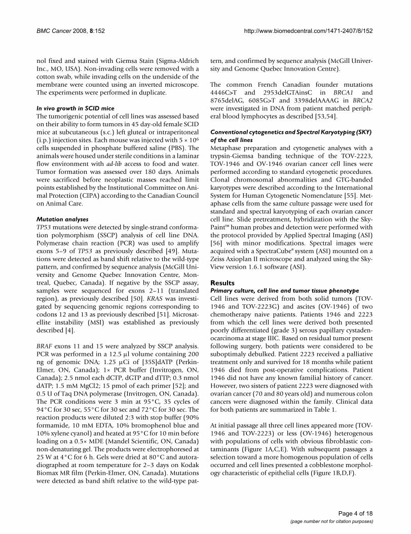
At initial passage all three cell lines appeared more (TOV-1946 and TOV-2223) or less (OV-1946) heterogenouswith populations of cells with obvious fibroblastic con-taminants (Figure 1A,C,E). With subsequent passages aselection toward a more homogenous population of cellsoccurred and cell lines presented a cobblestone morphol-ogy characteristic of epithelial cells (Figure 1B,D,F).
Page 4 of 18(page number not for citation purposes)

BMC Cancer 2008, 8:152 http://www.biomedcentral.com/1471-2407/8/152
Hematoxylin-Eosin stained tumor tissue sections ofpatient 1946 and 2223 (Figure 1G and 1H, respectively)presented typical poorly differentiated tumor masses.While all cell lines exhibited similar cobblestone mor-phologies typical of epithelial cells, the TOV-2223 cellsare larger than the TOV-1946 and OV-1946 cells (Figure1B,D,F). This reflects morphological differences in thecorresponding tumor tissues where cells in the TOV-2223tumors were larger than those observed in the 1946 tumor(Figure 1G,H).
One of the hallmarks of cancer cells is their ability to growin the absence of exogenous growth factors, which can beverified by culturing cancer cells in low serum conditions.All cell lines were able to grow in a medium containingonly 1% of FBS. In general however, growth rates wereslower in low serum conditions than those observed in atypical 10% FBS environments (data not shown).
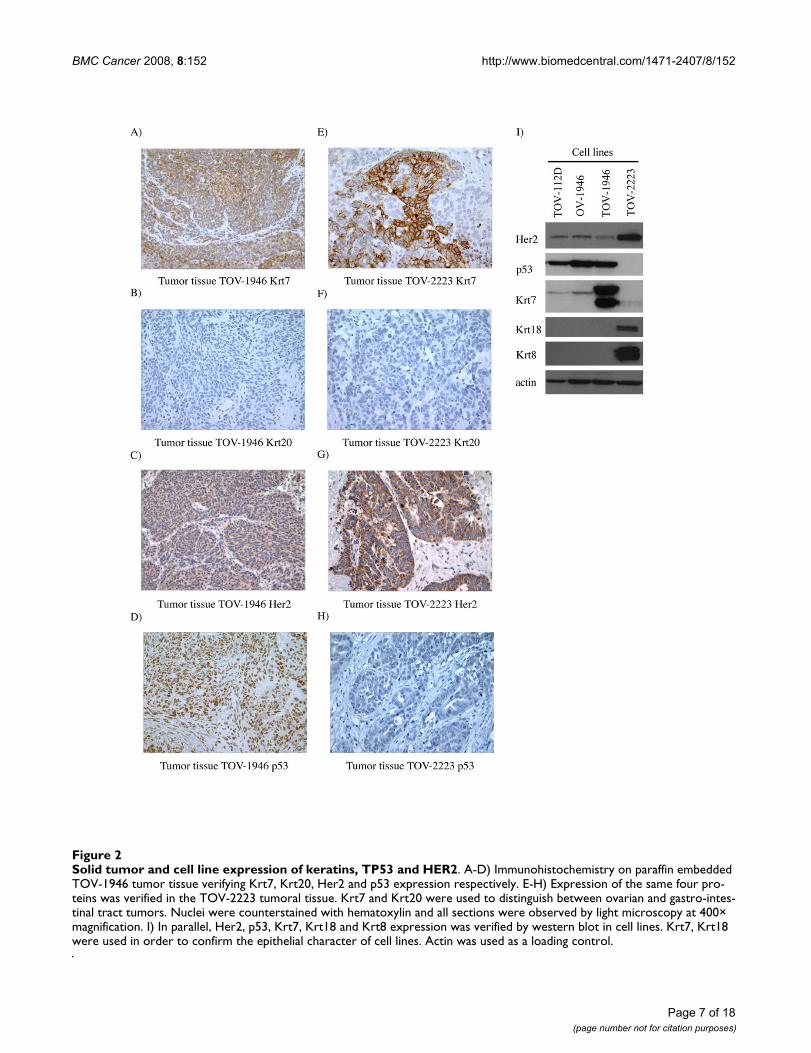
Solid tumor and cell line expression of keratins, TP53 and HER2In order to further establish the epithelial characteristicsof our cell lines keratin expression was assessed. BothTOV-1946 and OV-1946 cell lines expressed Krt7 andhence mirror keratin expression observed in vivo in theoriginal tumor (Figure 2A and 2I). However, the TOV-1946 cell line expressed a much higher level of this keratinwhen compared to its ascites counterpart (Figure 2I). Asreflected by immunohistochemistry and western-blotanalyses, although TOV-2223 tumor tissue expressedKrt7, the corresponding cell line seems to have lost thecapacity to express this particular keratin (Figure 2E and2I). The epithelial characteristics of the cell lines were alsoverified by the expression of Krt18 and Krt8, which arealso markers of epithelial cells. The expression of thesekeratins was observed only in the TOV-2223 cell line (Fig-
ure 2I). The gastro-intestinal tract tumor marker Krt20 wasabsent in both tumor tissues and all cell lines (Figure 2Band 2F).
We assessed the expression of p53 and Her2 in both orig-inal tumor tissues and cell lines. Tumor tissues from bothpatients exhibited Her2 expression (Figure 2C and 2G),however the level of expression was stronger in the TOV-2223 tumor tissue (Figure 2G). This is concordant withHer2 expression in the cell lines by western blot analysis(Figure 2I). We also examined p53 expression by immu-nohistochemistry. Positive staining with the p53 antigenis often indicative of TP53 gene mutation. The tissue frompatient 1946 exhibited positive nuclear staining for p53(Figure 2D) in contrast to that observed with the tumorfrom the patient 2223 (Figure 2H). p53 positive immuno-reactivity was mirrored in the cell lines by western blot(Figure 2I).
Cell growth rate and tumorigenicity assaysThe growth characteristics of the new cell lines were alsoassessed (Table 2 and Figure 3) and compared to TOV-112D, a cell line previously established and characterizedin our laboratory [4]. The new EOC cell lines exhibitedslower growth rates than the very aggressive TOV-112Dreference cell line (Table 2 and Figure 3A). TOV-1946 hasthe shortest doubling time (1.3 days) when compared tothe other two new cell lines (2.5 and 2.6 days for OV-1946and TOV-2223 respectively). All cell lines exhibited simi-lar saturation density although inferior (almost half) tothat of TOV-112D. These results are consistent with theobservation that TOV-112D exhibited small cells with atendency to compact and form foci [4] as opposed toTOV-1946 and TOV-2223 cell lines which respectivelyshow cells of medium and large size. The capacity of thenew cell lines to form foci at high cell inocula and density
Table 1: Patient clinical data
Patient
Clinical Parameter 1946 2223
Age at diagnosis 75 89Tumor type cystadenocarcinoma cystadenocarcinomaHistopathology sub-type serous papillary serous papillaryTumor grade G3 G3Disease stage IIIC IIICAscitis at surgery Yes yesSurgical debulking Sub-optimal Sub-optimalProgression N/A yesDeath yes* yesCause of death gastric hemorrhage disease progressionFollow up (months) 0.5 18Treatment N/A Megace (palliative care only)
N/A = not applicable*patient died from post-operative complication
Page 5 of 18(page number not for citation purposes)

BMC Cancer 2008, 8:152 http://www.biomedcentral.com/1471-2407/8/152
Page 6 of 18(page number not for citation purposes)
Cellular morphology of serous ovarian epithelium cancer cell lines and their corresponding tumoral tissuesFigure 1Cellular morphology of serous ovarian epithelium cancer cell lines and their corresponding tumoral tissues. A, C, E) Morphological appearance of the TOV-1946, OV-1946 and TOV-2223 cell lines respectively at passage 0. More (TOV-1946 and TOV-2223) or less (OV-1946) heterogenous populations of cells with obvious fibroblastic contaminants are visible for all three cell lines. B,D,F) Appearance of TOV-1946, OV-1946 and TOV-2223 cell lines respectively at passage 70. An evo-lution toward a more homogenous population of cobblestone-like cells typical of an epithelial cell type. G,H) Hematoxylin-Eosin stained tumor tissue sections of patient 1946 and 2223. Poorly differentiated tumor masses can be observed. All primary cultures and cell lines were cultured in OSE medium composed of 50:50 medium 199:105 (Sigma) supplemented with 10% fetal bovine serum (FBS), 2.5 μg/mL amphotericin B and 50 μg/mL gentamicin. All photographs were taken at 400× magnification.
BMC Cancer 2008, 8:152 http://www.biomedcentral.com/1471-2407/8/152
Page 7 of 18(page number not for citation purposes)
Solid tumor and cell line expression of keratins, TP53 and HER2Figure 2Solid tumor and cell line expression of keratins, TP53 and HER2. A-D) Immunohistochemistry on paraffin embedded TOV-1946 tumor tissue verifying Krt7, Krt20, Her2 and p53 expression respectively. E-H) Expression of the same four pro-teins was verified in the TOV-2223 tumoral tissue. Krt7 and Krt20 were used to distinguish between ovarian and gastro-intes-tinal tract tumors. Nuclei were counterstained with hematoxylin and all sections were observed by light microscopy at 400× magnification. I) In parallel, Her2, p53, Krt7, Krt18 and Krt8 expression was verified by western blot in cell lines. Krt7, Krt18 were used in order to confirm the epithelial character of cell lines. Actin was used as a loading control.

BMC Cancer 2008, 8:152 http://www.biomedcentral.com/1471-2407/8/152
was also verified and confirmed. The cell lines describedhere exhibit all the qualities of an established immortal-ized cell lines as they were all kept in culture for more than150 passages.
To further characterize the new cell lines, the migrationpotential was assessed (Table 2 and Figure 3B). TOV-1946cells migrated faster than the other three cell lines, as theywere able to close the wound in 24 hours. The migrationpatterns of TOV-112D and OV-1946 were similar andfilled the wound in 48 hours. However, TOV-2223migrated more slowly and needed more than 48 hours toclose the wound.
Using the modified Boyden chambers, we then measuredthe capability of the cells to invade through matrigelmembranes (Table 2 and Figure 3C). We noted anincrease in invasion potential from TOV-112D, OV-1946,TOV-2223 and TOV-1946, where TOV-112D was the leastinvasive and TOV-1946 the most invasive.
We next monitored the capacity of the cells to form three-dimensional structures in hanging droplets, a methodroutinely used in our laboratory. Only TOV-112D cellswere able to form very compact spheroids, OV-1946
formed less compact spheroids, TOV-1946 cells formedloose aggregates of cells. The TOV-2223 cells were unableto form any aggregates and cells were individually spreadacross the droplet (Table 2 and Figure 3D).
We next measured the capability of the cell lines to growin an anchorage independent environment by culturingthe cells in soft agarose (Table 2). All cell lines were ableto form colonies in soft agarose. The size of the colonieswere similar for TOV-112D, TOV-1946 and OV-1946 butwere larger than TOV-2223. The number of coloniesincreased from TOV-1946, TOV-2223, OV-1946 and TOV-112D with TOV-1946 having the lowest number of colo-nies and TOV112D the highest.
Finally, we monitored the potential of in vivo growth byinjecting tumor cells at intraperitoneal or subcutaneoussites in SCID mice (Table 2). Subcutaneous tumors wereobserved only for the TOV-112D cell line. However, intra-peritoneal (IP) tumors were also observed for theTOV1946 and OV1946 cell lines. The TOV-1946 cell lineformed tumors in only 3 mice and only after an average of125 days. The OV-1946 cell line formed tumors more rap-idly (average 63 days) and a greater number of tumors (5mice). The TOV2223 cell lines did not form tumors in
Table 2: Summary of cell line growth characteristics and tumorigenicity
Cell line growth assays TOV-1946 OV-1946 TOV-2223 TOV-112D
Growth characteristic
Doubling time (days) +/- S.D.a 1.3 +/- 0.4 2.5 +/- 0.9 2.6 +/- 0.7 1.0 +/- 0.2
Saturation density (nb cells +/- S.D.)b
3 053 933 +/- 153 933
3 231 400 +/- 962 723
2 536 867 +/-680 852
6 162133 +/- 515 034
Number of passages to date >210 >210 >150 >200Spheroid Formation aggregate semi-compact no compactMigration time for wound filling (h) 24 48 >48 48Invasion Mean number of cells +/-
S.D.c417 +/- 226 149 +/- 70 257 +/- 79 118 +/- 34
Soft agarose Colony number +/- S.D.d 11 +/- 3 24 +/- 8 16 +/- 4 27 +/- 10Colony size large large small large
Low serum Capacity to grow in low serum (1%) conditions
yes yes yes yes
Subcutaneous injection in SCID
mice
Number of mice with tumors (n = 6)
0 0 0 5
Mean time of tumor appearance (days)
N/A N/A N/A 7
Intraperitoneal injection in SCID
mice
Number of mice with tumors (n = 6)
3 5 0 6
Mean time of tumor appearance (days)
125 63 N/A 18
Number of mice with ascitis 2 1 0 2
N/A not applicableacalculated according to the slope of the linear portion of the growth curve.brepresents the mean of maximum number of cells counted among the two independent experiments performed in triplicatatcnumber of cells that migrate through the matrigel insertdmean of colony numbers per representative area (minimum 5) from two independent experiment performed in duplicate
Page 8 of 18(page number not for citation purposes)

BMC Cancer 2008, 8:152 http://www.biomedcentral.com/1471-2407/8/152
Page 9 of 18(page number not for citation purposes)
Cell growth rate and tumorigenicity assaysFigure 3Cell growth rate and tumorigenicity assays. A) Growth curves of the three new cell lines as well as a previously charac-terized EOC TOV-112D cell line. 100 000 cells were plated onto 60 mm petri dishes. Cells were trypsinised and counted every 48 h for two weeks. Experiments were performed two times in triplicate. B) Wound-healing assay of the same four cell lines. Cells were plated onto a 12 well dish and at near confluence wounds were generated. Cells were methanol fixed and treated with Giemsa Stain at different time points in order to evaluate cell migration (0 h, 8 h, 24 h and 48 h after the scratch was performed). The experiments were performed twice in triplicate. C) Invasion assay using modified Boyden chambers. The capability of the cells to invade through matrigel membranes was verified and the invasion potential increased from TOV-112D, OV-1946, TOV-2223 to TOV-1946. The experiments were performed in duplicate. D) The capacity of the cells to form three-dimensional structures in hanging droplets was monitored. TOV-112D cells were able to form very compact spheroids, OV-1946 formed less compact spheroids, TOV-1946 cells formed loose aggregates of cells and TOV-2223 cells were unable to form any 3-D structure. Spheroid formation capability was visualized after four days.

BMC Cancer 2008, 8:152 http://www.biomedcentral.com/1471-2407/8/152
mice (Table 2) however one mouse showed very smallmasses on liver lobes when all mice were sacrificed atapproximately seven months post-injection (data notshown).
Mutation status of the new EOC cell linesIn order to further characterize the cell lines, we per-formed somatic mutation analysis of TP53, KRAS, andBRAF as well as assayed for evidence of microsatelliteinstability (Table 3). Germline mutation (BRCA1 andBRCA2) analysis was limited to the recurrent mutationsfound in women of French-Canadian descent as cell lineswere derived from patients of this ancestral origin [53].Sequence variations in TP53 were identified in the DNAfrom all three EOC cell lines (Table 3). The same variantin exon 8, which is expected to confer an amino acid sub-stitution of arginine to cysteine at codon 273, was identi-fied in both TOV-1946 and OV-1946. The TP53 sequencevariants are classified as deleterious mutations in the IARCTP53 mutation database [57,58]. The TP53 mutation sta-tus of TOV-1946 and OV-1946 is consistent with IHCresults in these cell lines (Figure 2D and 2I). AlthoughTOV-2223 harbors a TP53 mutation, IHC staining wasnegative (Figure 2). This is not inconsistent with inde-pendent reports that only a smaller fraction of cancers(about 12%) harboring nonsense mutations exhibitedIHC-positivity as compared to IHC-negative nonsensemutations (IARC TP53 mutation database) [57]. As TP53mutation analysis revealed no evidence of heterozygosity,it is likely that no functional p53 is encoded in these celllines. No mutations in the other genes studied were foundin the cell lines.
Cytogenetics and Spectral KaryotypingThe cytogenetic alterations of the three cell lines wereassessed by G-banded karyotyping and by spectral karyo-typing (SKY). Twenty-five metaphases in G-banding andthirty-two metaphases in SKY (TOV-2223), twenty-twometaphases in G-banding and thirty-nine metaphases in
SKY (TOV-1946) and nineteen metaphases in G-bandingand fifteen metaphases in SKY (OV-1946) were analyzed.The G-banded analysis of the three cell lines revealed amodal number of 51 to 71 chromosomes with complexkaryotypes that are described as a composite karyotypecontaining the clonal chromosomal abnormalities (Table4) [55].
For the definition of the chromosomal breakpoints andthe characterization of the marker chromosomes, theinverted-DAPI banding and spectral images were com-pared with the SKY-painted chromosomes of the same celland then studied with the G-banded karyotypes for eachcell line. This method allows a better identification of thenumerous chromosomal rearrangements and severalmarkers in each cell line. It is noteworthy that there werecommon but also unique chromosomal abnormalitieswhen comparing the TOV-1946 and OV-1946 cell lines.Some representative metaphases are shown in Figures 4, 5and 6 (see also Additional Files 1, 2, 3, 4, 5).
DiscussionIn order to establish new cellular models of EOC, all sam-ples of ovarian tissues collected for our tumor banks areprocessed to derive primary cell cultures. Occasionally,some of the primary cultures evolve to become immortalcell lines. So far, we have been successful in establishingseven EOC cell lines [4], three of which are described here.All cell lines were derived from patients who were neverexposed to chemotherapy. This is of importance, since themajority of available cellular models originate from sam-ples obtained following neoadjuvant therapy that couldintroduce genetic events not related to the biology of thedisease.
Early in culture, cell lines presented heterogenous popula-tions of cells that eventually progressed toward a moreuniform population of cells with cobblestone-like mor-phology typical of epithelial cells. All cell lines grew as
Table 3: Mutations status of the new EOC cell lines
Gene tested TOV112D TOV-1946 OV-1946 TOV-2223G
Somatic
TP53 EX5-36 G>A, R175H EX8+35 C>T, R273C EX8+35 C>T, R273C EX4+62 G>A, W53XKRAS - - - -BRAF - - - -Microsatellite Instablility (MSI) - - - -
Germline
BRCA1 - - - -BRCA2 - - - -
- = no mutations were found
Page 10 of 18(page number not for citation purposes)

BMC Cancer 2008, 8:152 http://www.biomedcentral.com/1471-2407/8/152
adherent monolayers without evidence of cellular piling.However, we noted that TOV-2223 cells were more adher-ent to the petri dish when compared to TOV- and OV-1946. Indeed, a longer trypsinization time is required todetach these cells from the petri dish. Also, when fixed inmethanol, a large portion of both 1946 cell lines detachedfrom the solid support while 2223 cells did not (data notshown). The homogeneity of cells observed at passage 0 ofOV-1946 could reflect the fact that these cells came froma mass in the patient's ascites that is more homogenousthen a tumor mass often composed of, among others, epi-thelial tumor cells, stromal cells and endothelial cells.
Cell lines were also evaluated on their capacity to surviveand grow in low serum conditions. It is worth noticingthat although in normal growth conditions both TOV andOV-1946 cell lines present similar growth rates, in lowserum conditions the later is almost two times slower.This could be in line with the fact that the OV-1946 cellline comes from ascites which is rich in growth factors andin culture can replace FBS [59]. This cell line could hencebe more dependent on growth signals provided by theenvironment as opposed to cells originating from solidtumors where cells may be predicted to be more self-suffi-cient.
In epithelial cells, intermediate filaments are composed ofkeratins that vary according to the differentiation of thecells. Both OSE and EOC tumors are characterized by theexpression of different keratins such as KRT7, KRT8 andKRT18. In order to determine if our cell lines presentedthese EOC markers, we monitored protein expression ofKrt7, Krt8 and Krt18 by western blot. When antibodieswere appropriate for immunohistochemistry on paraffinembedded tissues, we also compared keratins expression
in the original tumor tissues. It is also well described thatmonitoring expression of Krt7 and Krt20 could distin-guish between ovarian and gastro-intestinal tract tumors(reviewed in [60]). Both the 1946 and the 2223 patientspresented different epithelium specific Krt expression(Figure 2A and 2E) but did not express Krt20. It is worthnoting that the Krt patterns were not all the same betweendifferent cell lines. Stronger expression of Krt7 is observedin TOV-1946 and TOV-2223G was the only cell line thatexpressed Krt18 and Krt8. This underlines the fact thatthese cell lines are biologically different even though theyoriginate from tissues representing the same type of serousEOC disease. Moreover, Krt7 is also differentiallyexpressed between the TOV-1946 and OV-1946 cell linesthat not only originate from the same type of disease butalso from the same patient and differences are related tosolid tumor versus ascites.
In EOC, 25 to 30% of the tumors present an amplificationof the HER2 gene leading to the overexpression of the pro-tein Her2 (reviewed in [2,61-63]). The overexpression ofthis growth factor receptor alone was shown to be suffi-cient to induce malignant transformation and is impli-cated in ovarian cancer as well as many other types ofcancer [64-67]. In EOCs of advanced stage, over 50% ofthe tumors were shown to be mutated in the TP53 gene(reviewed in [2,61-63]). All three cell lines showed Her2protein expression and contained TP53 gene mutations.
Differences in growth rates, migration, invasion and sphe-roid formation between the TOV-1946 and TOV-2223underline the diversity of phenotypes that can beobserved within the same type of serous EOC disease. Theresults from the comparison of the TOV-1946 and OV-1946 growth characteristics, demonstrate that these cell
Table 4: G-banding composite karyotypes of the three ovarian cancer cell lines
Cell line G-banding composite karyotypes
TOV-2223 53~71, X, der(X)t(X;2)(q13;q2?3), +der(1)t(1;17)(p3?4;q21), +add(1)(p?21), +add(1)(p12),?i(2)(q10), der(2)t(2;5)(q31;q31), +der(3)t(3;22)(q2?2;q11.2), del(3)(q23), +4, +4, -5, +5, der(6;12)(q10;q10), +add(6)(p11.2), +7, del(8)(p11.2)x2, +del(8)(p21), +9, der(10)t(5;10)(q?31;q26), +11, +add(12)(q11), +add(12)(p11.2), +add(12)(q24), +der(12;14)(p10;q10), add(13)(p11.2), +i(13)(q10),
add(14)(p11.2), -15, -15, der(15)add(15)(p11.2)add(15)(q26), add(16)(q22), +add(16)(q22), -17, -17, -18, add(18)(p11.2), del(19)(p13), add(19)(q13), -20, add(21)(p11.2), -22, i(22)(q10), +20mar, inc [cp25]
TOV-1946 51~62, X, -X, der(X;12)(q10;q10), del(1)(q41q42), +add(1)(p13), +del(1)(q12), add(2)(q37), t(3;6)(p21;p21), t(3;6)(p21;p21)x2, del(3)(p?14), add(4)(q35), del(5)(p12), +der(5;8)(q10;p10), der(6)t(3;6)(p10;q10)del(6)(q?21), +der(6)add(6)(p25)add(6)(q27),
del(7)(q11.2q21), +add(7)(p11.2), add(8)(p11.2), +der(8)?t(8;12)(p12;q13), +9, +del(9)(p21), +der(9)del(9) (p13)add(9)(q32), +10, add(11)(p15)x2, +del(11)(p11.2), del(12)(p11.2)x2, der(13)?t(11;13)(p11.2;p11.2)x2, add(15)(p11.2)x2, + der(15)?t(8;15)(q21;p11.2), -
16, add(16)(p13), add(16)(q24), -17, add(17)(p11.2), add(17)(p1?3), add(18)(q23), +19, +add(19)(q13.4), +20, +20, -21, -22,+22,+?22,+20mar,inc[cp22]
OV-1946 57~65, X, -X, der(X;12)(q10;q10), del(1)(q41q42), add(1)(p13), +add(1)(p13), +del(1)(q12), -2, der(2)del(2)(p?14)add(2)(q?31), t(3;6)(p21;p21), del(3)(p?14), +5, +del(5)(q11.2), +7, +add(7)(q11.2), +8, +add(8)(p11.2), +der(8)?t(8;12)(p12;q13), del(9)(p21), add(9)(q32), +add(9)(q32), +der(9)del(9)(p13)add(9)(q32), +10, +add(10)(q26), add(11)(p15)x2, +del(11)(p11.2), del(12)(p11.2),
der(13)?t(11;13)(p11.2;p11.2)x2, add(15)(p11.2)x2, +der(15)?t(8;15)(q21;p11.2), add(16)(p13), -17, -17, add(17)(p11.2), add(17)(p1?3), +del(17)(p11.2), -18, +18, +19, +19, +20, +20, -21, +22, +?22, +26mar, inc [cp19]
Page 11 of 18(page number not for citation purposes)

BMC Cancer 2008, 8:152 http://www.biomedcentral.com/1471-2407/8/152
Page 12 of 18(page number not for citation purposes)
A) Representative G-banded metaphases from the TOV-2223 (cell 10) cell lineFigure 4A) Representative G-banded metaphases from the TOV-2223 (cell 10) cell line. Arrows indicate the abnormal chromosomes, mar: marker chromosome. B) Representative metaphases from TOV-2223 (cell 45) analyzed by SKY. The ori-gin of several marker chromosomes (mar) is defined by SKY analysis. Other examples of G-banded metaphases and the com-bined inverted-DAPI and SKY images of different cells are presented (see additional Files 1 and 2).

BMC Cancer 2008, 8:152 http://www.biomedcentral.com/1471-2407/8/152
Page 13 of 18(page number not for citation purposes)
A) Representative G-banded metaphases from the TOV-1946 (cell 34) cell linesFigure 5A) Representative G-banded metaphases from the TOV-1946 (cell 34) cell lines. Arrows indicate the abnormal chromosomes, mar: marker chromosome. B), D), F) Representative metaphases from TOV-1946 (cell 44) cell line analyzed by SKY. The origin of several marker chromosomes (mar) is defined by SKY analysis. Other examples of G-banded metaphases and the combined inverted-DAPI and SKY images of different cells are presented (see additional Files 3 and 4).

BMC Cancer 2008, 8:152 http://www.biomedcentral.com/1471-2407/8/152
Page 14 of 18(page number not for citation purposes)
A) Representative G-banded metaphases from the OV-1946 (cell 1) cell lineFigure 6A) Representative G-banded metaphases from the OV-1946 (cell 1) cell line. Arrows indicate the abnormal chro-mosomes, mar: marker chromosome. B) Representative metaphases from OV-1946 (cell 11) cell line analyzed by SKY. The ori-gin of several marker chromosomes (mar) is defined by SKY analysis. Other examples of G-banded metaphases and the combined inverted-DAPI and SKY images of different cells are presented (see additional Files 5).

BMC Cancer 2008, 8:152 http://www.biomedcentral.com/1471-2407/8/152
lines present with unique phenotypes. Indeed, TOV-1946cells had a better capability to invade and migratealthough their capacity to form spheroid is reduced com-pared to the OV-1946 cells. One might speculate that cellsderived from solid tumor conserve their migration andinvasion property but in ascites, these characteristics areless vital. It has been previously shown that cells presentin ascites form spheres or aggregates that can adhere todifferent extracellular matrices as well as to normalhuman mesothelial cells [68] but are not always invasive.The role of spheroids in ascites of ovarian cancer patientsremains undefined. This is the first report of cell linesderived both from solid tumor and ascites cells of thesame patient and further studies on these cell lines mayprovide useful insights into the biological progression ofEOCs.
As both the TOV-1946 and OV-1946 injected mice devel-oped ascites we hypothesize that the ability for cell linesto induce ascites formation in mice is intrinsic to the givencell line and independent of their origin (solid tumor ver-sus ascites). It has been previously shown that the in vivotumorigenicity can usually be predicted by the ability ofthe cells to grow in soft agar [69]. The OV-1946 cell line,which was the most tumorigenic, also formed the largestand most numerous colonies when seeded in soft agar(Table 2). In previous studies we have also observed thatthe capacity of the cells to form compact spheroids isrelated to their ability to form tumors in vivo [70]. Consist-ent with this notion, here we show that the OV-1946 cellline formed the most compact spheroids among the newcell lines and formed the greatest number of tumors withthe shortest latency in xenograft experiments. However,the TOV-2223 cell line does not even form an aggregate inhanging droplets, only formed small colonies in soft agarand produced no tumors in SCID mice. The tumorigenic-ity results for TOV-2223 are also consistent with the rela-tive indolent disease observed in patient 2223 whosurvived a relatively long period post diagnosis withouttreatment. Indeed, we have previously isolated the TOV-81D cell lines, also from an indolent disease, which failedto form tumors in immuno-compromised mice [4] sug-gesting inherent qualities of the tumor that are reflected inits clinical behavior can affect this biological parameter.
The unique features of the described cell lines providecomplementary models for different aspects of the dis-ease. For example, while TOV-2223 and the TOV-1946cell lines are both derived from solid tumors of the sametype of serous EOC disease, they vary in important aspectsincluding their aggressiveness and karyotype. They mayprove useful for comparative studies to uncover molecularevents that distinguish very aggressive from more indolentserous disease in ovarian cancer. Although the TOV-1946and OV-1946 lines were derived from the same patient,
they were collected from the solid tumor and the corre-sponding ascites respectively. Although common genomemodification are shared between these two cell lines (seeexamples of modifications occurring on chromosome X,1, 2, 7, 8, 9, 11, 12, 13, 15 and 16) (Table 4), others areunique either to the solid tumor or the ascites derived cellline (chromosome 2, 4, 5, 6, 7, 9, 16, 17, 18, 19, 20 and22) (Table 4). These observations suggest that the two celllines acquired common modifications during the earliersteps of tumorigenesis and during cancer progression dif-ferent rearrangements were selected for, depending on themicroenvironment.
The results obtained by SKY and G-banding assays reflectpreviously published karyotype studies on EOC wherehigh genomic instability is observed [71]. Recently, it asbeen shown that recurrent rearrangements resulting in theformation of new fusion genes could be identified usinggenomic and bioinformatic tools [72-75]. So far suchstudies have been difficult to conduct on solid tumors andhence the importance of appropriate cellular models rep-resenting different types of tumors. The future fine cartog-raphy of recurrent lesions in these cell lines may provideinsights into the molecular events that contribute to EOCinitiation and progression.
ConclusionIn conclusion, this paper describes the establishment andcharacterization of three new serous EOC cell linesderived from two chemotherapy naïve patients. These celllines are new and important tools in the study ovariancancer disease, as few cell lines are described to date thatrepresent this frequently diagnosed histopathology typeof EOC. The rich characterization of these cell lines,including epithelial marker expression, growth character-istics, mutation and SKY analysis, provide the foundationfor future experiments using these new models of EOC.
Competing interestsThe authors declare that they have no competing interests.
Authors' contributionsVO and MZ performed the growth assays (growth rate,anchorage independent growth in soft agarose, three-dimensional culture and growth in low serum), thewound-healing assays, mice assays as well as writing andediting of the manuscript according to all authors revi-sions. LP derived and established the new cell lines fromtumor samples and performed the immunohistochemis-try on the primary cultures and cell lines. JM performedthe immunohistochemistry on tumor tissue. JL performedthe Western blot.
M–LP performed the invasion assays. SLA and ZS per-formed mutations analyses. JH analyzed the SKY and G-
Page 15 of 18(page number not for citation purposes)

BMC Cancer 2008, 8:152 http://www.biomedcentral.com/1471-2407/8/152
banding assays. PNT, DMP and A–MM–M contributed tothe conception and design of the study as well as analysisand interpretation of the data. All authors revised themanuscript and gave final verbal approval.
Additional material
AcknowledgementsWe are grateful to Véronique Barrès, Louise Champoux, Manon de Ladurantaye and Marise Roy for technical assistance. We thank Sylvie Lav-
allée and Claude Rondeau (Leukemia Cell Bank of Quebec) for expert tech-nical assistance in cytogenetics and spectral karyotyping. We would like to thank the Gynecologic Oncologist of CHUM for providing specimens. We are grateful to laboratory members for thoughtful discussion. This work was supported by a grant from the Cancer Research Society (CRS) to A.-M.M.-M., P.N.T. and D.M.P. Tumor banking was supported by the Banque de tissus et de données of the Réseau de recherche sur le cancer of the Fonds de la recherche en santé du Québec (FRSQ), affiliated to the Cana-dian Tumor Repository Network (CTRNet). V.O. was supported by stu-dentships from the Canadian Institute of Health Research (CIHR) and the Canderel Fund of the Institut du Cancer de Montréal. M.Z. was supported by studentship from the Marc Bourgie Fund of the Institut du Cancer de Montréal, the Canderel Fund of the Institut du Cancer de Montréal and the Faculté des études supérieures de l'Université de Montréal.
References1. Colombo N, Van Gorp T, Parma G, Amant F, Gatta G, Sessa C, Ver-
gote I: Ovarian cancer. Crit Rev Oncol Hematol 2006, 60:159-179.2. Auersperg N, Wong AS, Choi KC, Kang SK, Leung PC: Ovarian sur-
face epithelium: biology, endocrinology, and pathology.Endocr Rev 2001, 22:255-288.
3. Heintz AP, Odicino F, Maisonneuve P, Beller U, Benedet JL, CreasmanWT, Ngan HY, Pecorelli S: Carcinoma of the ovary. Int J GynaecolObstet 2003, 83 Suppl 1:135-166.
4. Provencher DM, Lounis H, Champoux L, Tetrault M, Manderson EN,Wang JC, Eydoux P, Savoie R, Tonin PN, Mes-Masson AM: Charac-terization of four novel epithelial ovarian cancer cell lines. InVitro Cell Dev Biol Anim 2000, 36:357-361.
5. DiSaia PJ, Morrow M, Kanabus J, Piechal W, Townsend DE: Two newtissue culture lines from ovarian cancer. Gynecol Oncol 1975,3:215-219.
6. Fogh J, Trempe G: New human tumor cell lines. In Human tumorcell lines in vitro Edited by: Fogh J. New York, Plenum Press; 1975.
7. Sinna GA, Beckman G, Lundgren E, Nordenson I, Roos G: Charac-terization of two human ovarian carcinoma cell lines. GynecolOncol 1979, 7:267-280.
8. Bast RC Jr., Feeney M, Lazarus H, Nadler LM, Colvin RB, Knapp RC:Reactivity of a monoclonal antibody with human ovarian car-cinoma. J Clin Invest 1981, 68:1331-1337.
9. Hamilton TC, Young RC, McKoy WM, Grotzinger KR, Green JA, ChuEW, Whang-Peng J, Rogan AM, Green WR, Ozols RF: Characteri-zation of a human ovarian carcinoma cell line (NIH:OVCAR-3) with androgen and estrogen receptors. Cancer Res 1983,43:5379-5389.
10. Buick RN, Pullano R, Trent JM: Comparative properties of fivehuman ovarian adenocarcinoma cell lines. Cancer Res 1985,45:3668-3676.
11. Hill BT, Whelan RD, Gibby EM, Sheer D, Hosking LK, Shellard SA,Rupniak HT: Establishment and characterisation of three newhuman ovarian carcinoma cell lines and initial evaluation oftheir potential in experimental chemotherapy studies. Int JCancer 1987, 39:219-225.
12. Langdon SP, Lawrie SS, Hay FG, Hawkes MM, McDonald A, HaywardIP, Schol DJ, Hilgers J, Leonard RC, Smyth JF: Characterization andproperties of nine human ovarian adenocarcinoma cell lines.Cancer Res 1988, 48:6166-6172.
13. Briers TW, Stroobants P, Vandeputte TM, Nouwen EJ, Conraads MV,Eestermans G, Van Bockstaele D, De Broe ME: Establishment andcharacterization of a human ovarian neoplastic cell line, DO-s. Cancer Res 1989, 49:5153-5161.
14. Golombick T, Dansey R, Bezwoda WR, Rosendorff J: Establishmentand characterization of two new human ovarian cancer celllines UWOV1 and UWOV2 and a subline UWOV2 (Sf)growing in serum-free conditions: growth characteristics,biochemical, and cytogenetic studies. In Vitro Cell Dev Biol 1990,26:447-454.
15. Wong WS, Wong YF, Ng YT, Huang PD, Chew EC, Ho TH, ChangMZ: Establishment and characterization of a new human cellline derived from ovarian clear cell carcinoma. Gynecol Oncol1990, 38:37-45.
16. Golombick T, Bezwoda WR: In vitro maintenance of a new ovar-ian cancer cell line in protein-free media: a potential model
Additional file 1A) G-banded metaphases from the TOV-2223 cell line (cells 15 and 36 respectively). Arrows indicate the abnormal chromosomes, mar: marker chromosome. B) Combined inverted-DAPI and SKY image of cell 45 and 46 respectively from the TOV 2223 cell line with identification of some marker chromosomes.Click here for file[http://www.biomedcentral.com/content/supplementary/1471-2407-8-152-S1.tiff]
Additional file 2A) G-banded metaphases from the TOV-2223 cell line (cells 15 and 36 respectively). Arrows indicate the abnormal chromosomes, mar: marker chromosome. B) Combined inverted-DAPI and SKY image of cell 45 and 46 respectively from the TOV 2223 cell line with identification of some marker chromosomes.Click here for file[http://www.biomedcentral.com/content/supplementary/1471-2407-8-152-S2.tiff]
Additional file 3A) G-banded metaphases from the TOV-1946 cell line (cells 6 and 43 respectively). Arrows indicate the abnormal chromosomes, mar: marker chromosome. B) Combined inverted-DAPI and SKY image of cell 44 and 36 respectively from the TOV-1946 cell line with identification of some marker chromosomes.Click here for file[http://www.biomedcentral.com/content/supplementary/1471-2407-8-152-S3.tiff]
Additional file 4A) G-banded metaphases from the TOV-1946 cell line (cells 6 and 43 respectively). Arrows indicate the abnormal chromosomes, mar: marker chromosome. B) Combined inverted-DAPI and SKY image of cell 44 and 36 respectively from the TOV-1946 cell line with identification of some marker chromosomes.Click here for file[http://www.biomedcentral.com/content/supplementary/1471-2407-8-152-S4.tiff]
Additional file 5A) G-banded metaphase from the OV-1946 cell line (cell 24). Arrows indicate the abnormal chromosomes, mar: marker chromosome. B) Com-bined inverted-DAPI and SKY image of cell 11 from the OV-1946 cell line with identification of some marker chromosomes.Click here for file[http://www.biomedcentral.com/content/supplementary/1471-2407-8-152-S5.tiff]
Page 16 of 18(page number not for citation purposes)
http://www.ncbi.nlm.nih.gov/entrez/query.fcgi?cmd=Retrieve&db=PubMed&dopt=Abstract&list_uids=1193434
http://www.ncbi.nlm.nih.gov/entrez/query.fcgi?cmd=Retrieve&db=PubMed&dopt=Abstract&list_uids=1193434
http://www.ncbi.nlm.nih.gov/entrez/query.fcgi?cmd=Retrieve&db=PubMed&dopt=Abstract&list_uids=7028788
http://www.ncbi.nlm.nih.gov/entrez/query.fcgi?cmd=Retrieve&db=PubMed&dopt=Abstract&list_uids=7028788
http://www.ncbi.nlm.nih.gov/entrez/query.fcgi?cmd=Retrieve&db=PubMed&dopt=Abstract&list_uids=7028788
http://www.ncbi.nlm.nih.gov/entrez/query.fcgi?cmd=Retrieve&db=PubMed&dopt=Abstract&list_uids=6604576
http://www.ncbi.nlm.nih.gov/entrez/query.fcgi?cmd=Retrieve&db=PubMed&dopt=Abstract&list_uids=6604576
http://www.ncbi.nlm.nih.gov/entrez/query.fcgi?cmd=Retrieve&db=PubMed&dopt=Abstract&list_uids=6604576
http://www.ncbi.nlm.nih.gov/entrez/query.fcgi?cmd=Retrieve&db=PubMed&dopt=Abstract&list_uids=4016745
http://www.ncbi.nlm.nih.gov/entrez/query.fcgi?cmd=Retrieve&db=PubMed&dopt=Abstract&list_uids=4016745
http://www.ncbi.nlm.nih.gov/entrez/query.fcgi?cmd=Retrieve&db=PubMed&dopt=Abstract&list_uids=3804493
http://www.ncbi.nlm.nih.gov/entrez/query.fcgi?cmd=Retrieve&db=PubMed&dopt=Abstract&list_uids=3804493
http://www.ncbi.nlm.nih.gov/entrez/query.fcgi?cmd=Retrieve&db=PubMed&dopt=Abstract&list_uids=3804493
http://www.ncbi.nlm.nih.gov/entrez/query.fcgi?cmd=Retrieve&db=PubMed&dopt=Abstract&list_uids=3167863
http://www.ncbi.nlm.nih.gov/entrez/query.fcgi?cmd=Retrieve&db=PubMed&dopt=Abstract&list_uids=3167863
http://www.ncbi.nlm.nih.gov/entrez/query.fcgi?cmd=Retrieve&db=PubMed&dopt=Abstract&list_uids=2548714
http://www.ncbi.nlm.nih.gov/entrez/query.fcgi?cmd=Retrieve&db=PubMed&dopt=Abstract&list_uids=2548714
http://www.ncbi.nlm.nih.gov/entrez/query.fcgi?cmd=Retrieve&db=PubMed&dopt=Abstract&list_uids=2548714
http://www.ncbi.nlm.nih.gov/entrez/query.fcgi?cmd=Retrieve&db=PubMed&dopt=Abstract&list_uids=2351639
http://www.ncbi.nlm.nih.gov/entrez/query.fcgi?cmd=Retrieve&db=PubMed&dopt=Abstract&list_uids=2351639
http://www.ncbi.nlm.nih.gov/entrez/query.fcgi?cmd=Retrieve&db=PubMed&dopt=Abstract&list_uids=2351639
http://www.ncbi.nlm.nih.gov/entrez/query.fcgi?cmd=Retrieve&db=PubMed&dopt=Abstract&list_uids=2354825
http://www.ncbi.nlm.nih.gov/entrez/query.fcgi?cmd=Retrieve&db=PubMed&dopt=Abstract&list_uids=2354825

BMC Cancer 2008, 8:152 http://www.biomedcentral.com/1471-2407/8/152
for autonomous growth and tumor progression. Eur J Cell Biol1991, 56:459-463.
17. Yamada T, Ueda M, Otsuki Y, Ueki M, Sugimoto O: Establishmentand characterization of a cell line (OMC-3) originating froma human mucinous cystadenocarcinoma of the ovary. GynecolOncol 1991, 40:118-128.
18. Mobus V, Gerharz CD, Press U, Moll R, Beck T, Mellin W, Pollow K,Knapstein PG, Kreienberg R: Morphological, immunohisto-chemical and biochemical characterization of 6 newly estab-lished human ovarian carcinoma cell lines. Int J Cancer 1992,52:76-84.
19. Grunt TW, Oeller H, Somay C, Dittrich C: Different propensityfor spontaneous differentiation of cell clones isolated fromthe human ovarian surface epithelial cell line HOC-7. Differ-entiation 1993, 53:45-50.
20. Provencher DM, Finstad CL, Saigo PE, Rubin SC, Hoskins WJ, FedericiMG, Stockert E, Lloyd KO, Lewis JL Jr.: Comparison of antigenexpression on fresh and cultured ascites cells and on solidtumors of patients with epithelial ovarian cancer. GynecolOncol 1993, 50:78-83.
21. Hirte HW, Kaiser JS, Bacchetti S: Establishment and characteri-zation of four human epithelial ovarian carcinoma cell lines.Cancer 1994, 74:900-906.
22. Buller RE, Niemann T, Connor JP, Squatrito RC, Skilling JS, AndersonB: Isolation and preliminary characterization of an ovariancarcinoma cell line from a patient with familial ovarian can-cer. Gynecol Oncol 1995, 56:39-44.
23. Alama A, Barbieri F, Favre A, Cagnoli M, Noviello E, Pedulla F, VialeM, Foglia G, Ragni N: Establishment and characterization ofthree new cell lines derived from the ascites of human ovar-ian carcinomas. Gynecol Oncol 1996, 62:82-88.
24. Kim JW, Lee CG, Lyu MS, Kim HK, Rha JG, Kim DH, Kim SJ, Namk-oong SE: A new cell line from human undifferentiated carci-noma of the ovary: establishment and characterization. JCancer Res Clin Oncol 1997, 123:82-90.
25. Mancini A, Borrelli A, Formisano S, Masucci MT, Maffeo A, Perla G,De Martino L, Bevilacqua N, Botti G, Maggino T: Establishmentand growth regulation of a novel ovarian cancer cell linefrom a poorly-differentiated adenocarcinoma: proposal for anew treatment. Eur J Gynaecol Oncol 1999, 20:45-52.
26. Bar JK, Harlozinska A: Morphological and phenotypic charac-terization of a new established ovarian carcinoma cell line(OvBH-1). Anticancer Res 2000, 20:2975-2980.
27. Sato S, Kobayashi Y, Okuma Y, Kondo H, Kanishi Y, Saito K, KiguchiK: Establishment and characterization of a cell-line origi-nated from human mucinous cystadenocarcinoma of theovary. Hum Cell 2002, 15:171-177.
28. Saga Y, Suzuki M, Machida S, Ohwada M, Sato I: Establishment of anew cell line (TAYA) of clear cell adenocarcinoma of theovary and its radiosensitivity. Oncology 2002, 62:180-184.
29. Nishikawa Y, Yoshida Y, Kawahara K, Kurokawa T, Tajima K, KotsujiF: Establishment of a novel human ovarian cancer cell linewith high anchorage-independent growth ability. Int J Oncol2003, 23:1679-1686.
30. Woods LK, Morgan RT, Quinn LA, Moore GE, Semple TU, StedmanKE: Comparison of four new cell lines from patients with ade-nocarcinoma of the ovary. Cancer Res 1979, 39:4449-4459.
31. Crickard K, Niedbala MJ, Crickard U, Yoonessi M, Sandberg AA,Okuyama K, Bernacki RJ, Satchidanand SK: Characterization ofhuman ovarian and endometrial carcinoma cell lines estab-lished on extracellular matrix. Gynecol Oncol 1989, 32:163-173.
32. Imai S, Kiyozuka Y, Maeda H, Noda T, Hosick HL: Establishmentand characterization of a human ovarian serous cystadeno-carcinoma cell line that produces the tumor markers CA-125 and tissue polypeptide antigen. Oncology 1990, 47:177-184.
33. van den Berg-Bakker CA, Hagemeijer A, Franken-Postma EM, SmitVT, Kuppen PJ, van Ravenswaay Claasen HH, Cornelisse CJ, SchrierPI: Establishment and characterization of 7 ovarian carci-noma cell lines and one granulosa tumor cell line: growthfeatures and cytogenetics. Int J Cancer 1993, 53:613-620.
34. Yonamine K, Hayashi K, Iida T: Establishment and characteriza-tion of human ovarian clear cell adenocarcinoma cell line(SMOV-2), and its cytotoxity by anticancer agents. Hum Cell1999, 12:139-148.
35. Emoto M, Oshima K, Ishiguro M, Iwasaki H, Kawarabayashi T, KikuchiM: Establishment and characterization of a serous papillary
adenocarcinoma cell line of the human ovary in a serum-freeculture. Pathol Res Pract 1999, 195:237-242.
36. Aoki D, Suzuki N, Susumu N, Noda T, Suzuki A, Tamada Y,Higashiguchi A, Oie S, Nozawa S: Establishment and characteri-zation of the RMG-V cell line from human ovarian clear celladenocarcinoma. Hum Cell 2005, 18:143-146.
37. Scoles DR, Pavelka J, Cass I, Tran H, Baldwin RL, Armstrong K, KarlanBY: Characterization of CSOC 882, a novel immortalizedovarian cancer cell line expressing EGFR, HER2, and acti-vated AKT. Gynecol Oncol 2007, 104:120-128.
38. Auersperg N, Maines-Bandiera SL, Dyck HG, Kruk PA: Characteri-zation of cultured human ovarian surface epithelial cells:phenotypic plasticity and premalignant changes. Lab Invest1994, 71:510-518.
39. Nitta M, Katabuchi H, Ohtake H, Tashiro H, Yamaizumi M, OkamuraH: Characterization and tumorigenicity of human ovariansurface epithelial cells immortalized by SV40 large T anti-gen. Gynecol Oncol 2001, 81:10-17.
40. Shenhua X, Lijuan Q, Hanzhou N, Xinghao N, Chihong Z, Gu Z, Wei-fang D, Yongliang G: Establishment of a highly metastatichuman ovarian cancer cell line (HO-8910PM) and its charac-terization. J Exp Clin Cancer Res 1999, 18:233-239.
41. Yabushita H, Ueno N, Sawaguchi K, Higuchi K, Noguchi M, IshiharaM: Establishment and characterization of a new human cell-line (AMOC-2) derived from a serous adenocarcinoma ofovary. Nippon Sanka Fujinka Gakkai Zasshi 1989, 41:888-894.
42. Kruk PA, Maines-Bandiera SL, Auersperg N: A simplified methodto culture human ovarian surface epithelium. Lab Invest 1990,63:132-136.
43. Lounis H, Provencher D, Godbout C, Fink D, Milot MJ, Mes-MassonAM: Primary cultures of normal and tumoral human ovarianepithelium: a powerful tool for basic molecular studies. ExpCell Res 1994, 215:303-309.
44. Keller GM: In vitro differentiation of embryonic stem cells.Curr Opin Cell Biol 1995, 7:862-869.
45. Zietarska M, Maugard CM, Filali-Mouhim A, Alam-Fahmy M, ToninPN, Provencher D, Mes-Masson AM: Molecular description of a3-D in vitro model for the study of epithelial ovarian cancer.Mol Carcinog 2007, In Press:.
46. Bossolasco M, Veillette F, Bertrand R, Mes-Masson AM: HumanTDE1, a TDE1/TMS family member, inhibits apoptosis invitro and stimulates in vivo tumorigenesis. Oncogene 2006,25:4549-4558.
47. Shigeta M, Sanzen N, Ozawa M, Gu J, Hasegawa H, Sekiguchi K:CD151 regulates epithelial cell-cell adhesion through PKC-and Cdc42-dependent actin cytoskeletal reorganization. JCell Biol 2003, 163:165-176.
48. Ronot X, Doisy A, Tracqui P: Quantitative study of dynamicbehavior of cell monolayers during in vitro wound healing byoptical flow analysis. Cytometry 2000, 41:19-30.
49. Arcand SL, Provencher D, Mes-Masson AM, Tonin PN: OGG1Cys326 variant, allelic imbalance of chromosome band3p25.3 and TP53 mutations in ovarian cancer. Int J Oncol 2005,27:1315-1320.
50. Arcand SL, Maugard CM, Ghadirian P, Robidoux A, Perret C, ZhangP, Fafard E, Mes-Masson AM, Foulkes WD, Provencher D, Narod SA,Tonin PN: Germline TP53 mutations in BRCA1 and BRCA2mutation-negative French Canadian breast cancer families.Breast Cancer Res Treat 2007.
51. Samouelian V, Maugard CM, Jolicoeur M, Bertrand R, Arcand SL,Tonin PN, Provencher DM, Mes-Masson AM: Chemosensitivityand radiosensitivity profiles of four new human epithelialovarian cancer cell lines exhibiting genetic alterations inBRCA2, TGFbeta-RII, KRAS2, TP53 and/or CDNK2A. CancerChemother Pharmacol 2004, 54:497-504.
52. Davies H, Bignell GR, Cox C, Stephens P, Edkins S, Clegg S, Teague J,Woffendin H, Garnett MJ, Bottomley W, Davis N, Dicks E, Ewing R,Floyd Y, Gray K, Hall S, Hawes R, Hughes J, Kosmidou V, Menzies A,Mould C, Parker A, Stevens C, Watt S, Hooper S, Wilson R, JayatilakeH, Gusterson BA, Cooper C, Shipley J, Hargrave D, Pritchard-JonesK, Maitland N, Chenevix-Trench G, Riggins GJ, Bigner DD, PalmieriG, Cossu A, Flanagan A, Nicholson A, Ho JW, Leung SY, Yuen ST,Weber BL, Seigler HF, Darrow TL, Paterson H, Marais R, Marshall CJ,Wooster R, Stratton MR, Futreal PA: Mutations of the BRAFgene in human cancer. Nature 2002, 417:949-954.
Page 17 of 18(page number not for citation purposes)
http://www.ncbi.nlm.nih.gov/entrez/query.fcgi?cmd=Retrieve&db=PubMed&dopt=Abstract&list_uids=1802727
http://www.ncbi.nlm.nih.gov/entrez/query.fcgi?cmd=Retrieve&db=PubMed&dopt=Abstract&list_uids=2010102
http://www.ncbi.nlm.nih.gov/entrez/query.fcgi?cmd=Retrieve&db=PubMed&dopt=Abstract&list_uids=2010102
http://www.ncbi.nlm.nih.gov/entrez/query.fcgi?cmd=Retrieve&db=PubMed&dopt=Abstract&list_uids=2010102
http://www.ncbi.nlm.nih.gov/entrez/query.fcgi?cmd=Retrieve&db=PubMed&dopt=Abstract&list_uids=1500230
http://www.ncbi.nlm.nih.gov/entrez/query.fcgi?cmd=Retrieve&db=PubMed&dopt=Abstract&list_uids=1500230
http://www.ncbi.nlm.nih.gov/entrez/query.fcgi?cmd=Retrieve&db=PubMed&dopt=Abstract&list_uids=1500230
http://www.ncbi.nlm.nih.gov/entrez/query.fcgi?cmd=Retrieve&db=PubMed&dopt=Abstract&list_uids=8508947
http://www.ncbi.nlm.nih.gov/entrez/query.fcgi?cmd=Retrieve&db=PubMed&dopt=Abstract&list_uids=8508947
http://www.ncbi.nlm.nih.gov/entrez/query.fcgi?cmd=Retrieve&db=PubMed&dopt=Abstract&list_uids=8508947
http://www.ncbi.nlm.nih.gov/entrez/query.fcgi?cmd=Retrieve&db=PubMed&dopt=Abstract&list_uids=8349167
http://www.ncbi.nlm.nih.gov/entrez/query.fcgi?cmd=Retrieve&db=PubMed&dopt=Abstract&list_uids=8349167
http://www.ncbi.nlm.nih.gov/entrez/query.fcgi?cmd=Retrieve&db=PubMed&dopt=Abstract&list_uids=8349167
http://www.ncbi.nlm.nih.gov/entrez/query.fcgi?cmd=Retrieve&db=PubMed&dopt=Abstract&list_uids=8039117
http://www.ncbi.nlm.nih.gov/entrez/query.fcgi?cmd=Retrieve&db=PubMed&dopt=Abstract&list_uids=8039117
http://www.ncbi.nlm.nih.gov/entrez/query.fcgi?cmd=Retrieve&db=PubMed&dopt=Abstract&list_uids=7821846
http://www.ncbi.nlm.nih.gov/entrez/query.fcgi?cmd=Retrieve&db=PubMed&dopt=Abstract&list_uids=7821846
http://www.ncbi.nlm.nih.gov/entrez/query.fcgi?cmd=Retrieve&db=PubMed&dopt=Abstract&list_uids=7821846
http://www.ncbi.nlm.nih.gov/entrez/query.fcgi?cmd=Retrieve&db=PubMed&dopt=Abstract&list_uids=8690298
http://www.ncbi.nlm.nih.gov/entrez/query.fcgi?cmd=Retrieve&db=PubMed&dopt=Abstract&list_uids=8690298
http://www.ncbi.nlm.nih.gov/entrez/query.fcgi?cmd=Retrieve&db=PubMed&dopt=Abstract&list_uids=8690298
http://www.ncbi.nlm.nih.gov/entrez/query.fcgi?cmd=Retrieve&db=PubMed&dopt=Abstract&list_uids=9030246
http://www.ncbi.nlm.nih.gov/entrez/query.fcgi?cmd=Retrieve&db=PubMed&dopt=Abstract&list_uids=9030246
http://www.ncbi.nlm.nih.gov/entrez/query.fcgi?cmd=Retrieve&db=PubMed&dopt=Abstract&list_uids=2910777
http://www.ncbi.nlm.nih.gov/entrez/query.fcgi?cmd=Retrieve&db=PubMed&dopt=Abstract&list_uids=2910777
http://www.ncbi.nlm.nih.gov/entrez/query.fcgi?cmd=Retrieve&db=PubMed&dopt=Abstract&list_uids=2910777
http://www.ncbi.nlm.nih.gov/entrez/query.fcgi?cmd=Retrieve&db=PubMed&dopt=Abstract&list_uids=2314830
http://www.ncbi.nlm.nih.gov/entrez/query.fcgi?cmd=Retrieve&db=PubMed&dopt=Abstract&list_uids=2314830
http://www.ncbi.nlm.nih.gov/entrez/query.fcgi?cmd=Retrieve&db=PubMed&dopt=Abstract&list_uids=2314830
http://www.ncbi.nlm.nih.gov/entrez/query.fcgi?cmd=Retrieve&db=PubMed&dopt=Abstract&list_uids=8436435
http://www.ncbi.nlm.nih.gov/entrez/query.fcgi?cmd=Retrieve&db=PubMed&dopt=Abstract&list_uids=8436435
http://www.ncbi.nlm.nih.gov/entrez/query.fcgi?cmd=Retrieve&db=PubMed&dopt=Abstract&list_uids=8436435
http://www.ncbi.nlm.nih.gov/entrez/query.fcgi?cmd=Retrieve&db=PubMed&dopt=Abstract&list_uids=7967506
http://www.ncbi.nlm.nih.gov/entrez/query.fcgi?cmd=Retrieve&db=PubMed&dopt=Abstract&list_uids=7967506
http://www.ncbi.nlm.nih.gov/entrez/query.fcgi?cmd=Retrieve&db=PubMed&dopt=Abstract&list_uids=7967506
http://www.ncbi.nlm.nih.gov/entrez/query.fcgi?cmd=Retrieve&db=PubMed&dopt=Abstract&list_uids=2794621
http://www.ncbi.nlm.nih.gov/entrez/query.fcgi?cmd=Retrieve&db=PubMed&dopt=Abstract&list_uids=2794621
http://www.ncbi.nlm.nih.gov/entrez/query.fcgi?cmd=Retrieve&db=PubMed&dopt=Abstract&list_uids=2794621
http://www.ncbi.nlm.nih.gov/entrez/query.fcgi?cmd=Retrieve&db=PubMed&dopt=Abstract&list_uids=2374399
http://www.ncbi.nlm.nih.gov/entrez/query.fcgi?cmd=Retrieve&db=PubMed&dopt=Abstract&list_uids=2374399
http://www.ncbi.nlm.nih.gov/entrez/query.fcgi?cmd=Retrieve&db=PubMed&dopt=Abstract&list_uids=7526992
http://www.ncbi.nlm.nih.gov/entrez/query.fcgi?cmd=Retrieve&db=PubMed&dopt=Abstract&list_uids=7526992

BMC Cancer 2008, 8:152 http://www.biomedcentral.com/1471-2407/8/152
Publish with BioMed Central and every scientist can read your work free of charge
"BioMed Central will be the most significant development for disseminating the results of biomedical research in our lifetime."
Sir Paul Nurse, Cancer Research UK
Your research papers will be:
available free of charge to the entire biomedical community
peer reviewed and published immediately upon acceptance
cited in PubMed and archived on PubMed Central
yours — you keep the copyright
Submit your manuscript here:http://www.biomedcentral.com/info/publishing_adv.asp
BioMedcentral
53. Oros KK, Ghadirian P, Greenwood CM, Perret C, Shen Z, Paredes Y,Arcand SL, Mes-Masson AM, Narod SA, Foulkes WD, Provencher D,Tonin PN: Significant proportion of breast and/or ovarian can-cer families of French Canadian descent harbor 1 of 5BRCA1 and BRCA2 mutations. Int J Cancer 2004, 112:411-419.
54. Tonin PN, Mes-Masson AM, Futreal PA, Morgan K, Mahon M, FoulkesWD, Cole DE, Provencher D, Ghadirian P, Narod SA: FounderBRCA1 and BRCA2 mutations in French Canadian breastand ovarian cancer families. Am J Hum Genet 1998,63:1341-1351.
55. Shaffer LG, Tommerup N: ISCN, An International System forHuman Cytogenetic Nomenclature. Basel, S. Karger AG; 2005.
56. Applied Spectral Imaging (ASI) [http://www.spectral-imaging.com/]
57. Petitjean A, Mathe E, Kato S, Ishioka C, Tavtigian SV, Hainaut P,Olivier M: Impact of mutant p53 functional properties onTP53 mutation patterns and tumor phenotype: lessons fromrecent developments in the IARC TP53 database. Hum Mutat2007, 28:622-629.
58. IARC TP53 mutation database [http://www-p53.iarc.fr/]59. Puiffe ML, Le Page C, Filali-Mouhim A, Zietarska M, Ouellet V, Tonin
PN, Chevrette M, Provencher DM, Mes-Masson AM: Characteriza-tion of ovarian cancer ascites on cell invasion, proliferation,spheroid formation, and gene expression in an in vitro modelof epithelial ovarian cancer. Neoplasia 2007, 9:820-829.
60. Prat J: Ovarian carcinomas, including secondary tumors: diag-nostically challenging areas. Mod Pathol 2005, 18 Suppl2:S99-111.
61. Boente MP, Hurteau J, Rodriguez GC, Bast RC Jr., Berchuck A: Thebiology of ovarian cancer. Curr Opin Oncol 1993, 5:900-907.
62. Berchuck A, Elbendary A, Havrilesky L, Rodriguez GC, Bast RC Jr.:Pathogenesis of ovarian cancers. J Soc Gynecol Investig 1994,1:181-190.
63. Aunoble B, Sanches R, Didier E, Bignon YJ: Major oncogenes andtumor suppressor genes involved in epithelial ovarian cancer(review). Int J Oncol 2000, 16:567-576.
64. Meden H, Kuhn W: Overexpression of the oncogene c-erbB-2(HER2/neu) in ovarian cancer: a new prognostic factor.[Review] [36 refs]. Eur J Obstet Gynecol Rep Biol 1997, 71:173-179.
65. Meden H, Marx D, Schauer A, Wuttke W, Kuhn W: Prognostic sig-nificance of p105 (c-erbB-2 HER2/neu) serum levels inpatients with ovarian cancer. Anticancer Res 1997, 17:757-760.
66. Slamon DJ, Clark GM, Wong SG, Levin WJ, Ullrich A, McGuire WL:Human breast cancer: correlation of relapse and survivalwith amplification of the HER-2/neu oncogene. Science 1987,235:177-182.
67. Calvo BF, Levine AM, Marcos M, Collins QF, Iacocca MV, Caskey LS,Gregory CW, Lin Y, Whang YE, Earp HS, Mohler JL: Human epider-mal receptor-2 expression in prostate cancer. Clin Cancer Res2003, 9:1087-1097.
68. Burleson KM, Boente MP, Pambuccian SE, Skubitz AP: Disaggrega-tion and invasion of ovarian carcinoma ascites spheroids. JTransl Med 2006, 4:6.
69. Freedman VH, Shin SI: Cellular tumorigenicity in nude mice:correlation with cell growth in semi-solid medium. Cell 1974,3:355-359.
70. Cody NA, Ouellet V, Manderson EN, Quinn MC, Filali-Mouhim A,Tellis P, Zietarska M, Provencher DM, Mes-Masson AM, Chevrette M,Tonin PN: Transfer of chromosome 3 fragments suppressestumorigenicity of an ovarian cancer cell line monoallelic forchromosome 3p. Oncogene 2007, 26:618-632.
71. Taetle R, Aickin M, Yang JM, Panda L, Emerson J, Roe D, Adair L,Thompson F, Liu Y, Wisner L, Davis JR, Trent J, Alberts DS: Chro-mosome abnormalities in ovarian adenocarcinoma: I. Non-random chromosome abnormalities from 244 cases. GenesChromosomes Cancer 1999, 25:290-300.
72. Tomlins SA, Laxman B, Dhanasekaran SM, Helgeson BE, Cao X, Mor-ris DS, Menon A, Jing X, Cao Q, Han B, Yu J, Wang L, Montie JE, RubinMA, Pienta KJ, Roulston D, Shah RB, Varambally S, Mehra R, Chinnai-yan AM: Distinct classes of chromosomal rearrangementscreate oncogenic ETS gene fusions in prostate cancer. Nature2007, 448:595-599.
73. Tomlins SA, Rhodes DR, Perner S, Dhanasekaran SM, Mehra R, SunXW, Varambally S, Cao X, Tchinda J, Kuefer R, Lee C, Montie JE, ShahRB, Pienta KJ, Rubin MA, Chinnaiyan AM: Recurrent fusion of
TMPRSS2 and ETS transcription factor genes in prostatecancer. Science 2005, 310:644-648.
74. Martin ES, Tonon G, Sinha R, Xiao Y, Feng B, Kimmelman AC, Proto-popov A, Ivanova E, Brennan C, Montgomery K, Kucherlapati R, Bai-ley G, Redston M, Chin L, DePinho RA: Common and distinctgenomic events in sporadic colorectal cancer and diversecancer types. Cancer Res 2007, 67:10736-10743.
75. Weir BA, Woo MS, Getz G, Perner S, Ding L, Beroukhim R, Lin WM,Province MA, Kraja A, Johnson LA, Shah K, Sato M, Thomas RK, Bar-letta JA, Borecki IB, Broderick S, Chang AC, Chiang DY, Chirieac LR,Cho J, Fujii Y, Gazdar AF, Giordano T, Greulich H, Hanna M, JohnsonBE, Kris MG, Lash A, Lin L, Lindeman N, Mardis ER, McPherson JD,Minna JD, Morgan MB, Nadel M, Orringer MB, Osborne JR, Ozen-berger B, Ramos AH, Robinson J, Roth JA, Rusch V, Sasaki H, Shep-herd F, Sougnez C, Spitz MR, Tsao MS, Twomey D, Verhaak RG,Weinstock GM, Wheeler DA, Winckler W, Yoshizawa A, Yu S,Zakowski MF, Zhang Q, Beer DG, Wistuba, Watson MA, GarrawayLA, Ladanyi M, Travis WD, Pao W, Rubin MA, Gabriel SB, Gibbs RA,Varmus HE, Wilson RK, Lander ES, Meyerson M: Characterizingthe cancer genome in lung adenocarcinoma. Nature 2007.
Pre-publication historyThe pre-publication history for this paper can be accessedhere:
http://www.biomedcentral.com/1471-2407/8/152/prepub
Page 18 of 18(page number not for citation purposes)
http://www.ncbi.nlm.nih.gov/entrez/query.fcgi?cmd=Retrieve&db=PubMed&dopt=Abstract&list_uids=9792861
http://www.ncbi.nlm.nih.gov/entrez/query.fcgi?cmd=Retrieve&db=PubMed&dopt=Abstract&list_uids=9792861
http://www.ncbi.nlm.nih.gov/entrez/query.fcgi?cmd=Retrieve&db=PubMed&dopt=Abstract&list_uids=9792861
http://www.ncbi.nlm.nih.gov/entrez/query.fcgi?cmd=Retrieve&db=PubMed&dopt=Abstract&list_uids=8218503
http://www.ncbi.nlm.nih.gov/entrez/query.fcgi?cmd=Retrieve&db=PubMed&dopt=Abstract&list_uids=8218503
http://www.ncbi.nlm.nih.gov/entrez/query.fcgi?cmd=Retrieve&db=PubMed&dopt=Abstract&list_uids=9419769
http://www.ncbi.nlm.nih.gov/entrez/query.fcgi?cmd=Retrieve&db=PubMed&dopt=Abstract&list_uids=9419769
http://www.ncbi.nlm.nih.gov/entrez/query.fcgi?cmd=Retrieve&db=PubMed&dopt=Abstract&list_uids=9066616
http://www.ncbi.nlm.nih.gov/entrez/query.fcgi?cmd=Retrieve&db=PubMed&dopt=Abstract&list_uids=9066616
http://www.ncbi.nlm.nih.gov/entrez/query.fcgi?cmd=Retrieve&db=PubMed&dopt=Abstract&list_uids=9066616
http://www.ncbi.nlm.nih.gov/entrez/query.fcgi?cmd=Retrieve&db=PubMed&dopt=Abstract&list_uids=3798106
http://www.ncbi.nlm.nih.gov/entrez/query.fcgi?cmd=Retrieve&db=PubMed&dopt=Abstract&list_uids=3798106
http://www.ncbi.nlm.nih.gov/entrez/query.fcgi?cmd=Retrieve&db=PubMed&dopt=Abstract&list_uids=3798106
http://www.ncbi.nlm.nih.gov/entrez/query.fcgi?cmd=Retrieve&db=PubMed&dopt=Abstract&list_uids=4442124