Prediagnosis Food Patterns Are Associated with Length of Survival from Epithelial Ovarian Cancer
Upload
khangminh22Category
view
1download
0
CA125 Targeted Molecular Imaging of Epithelial Ovarian Cancer
by
Sai Kiran Sharma
A thesis submitted in partial fulfillment of the requirements for the degree of
Doctor of Philosophy
in
Pharmaceutical Sciences
Faculty of Pharmacy and Pharmaceutical Sciences
University of Alberta
© Sai Kiran Sharma, 2014

ii
ABSTRACT
Rationale: Ovarian cancer is the most lethal malignancy of the female
reproductive system. ~80% of ovarian cancers are epithelial in origin and
commonly classified as Epithelial Ovarian Cancer (EOC). This malignancy is
characterized by an overexpression of Cancer Antigen-125 (CA125) – a cell
surface mucinous glycoprotein that serves as a USFDA approved ovarian tumor
associated antigen. However, the early detection of EOC is plagued by its
asymptomatic nature of progression and the limitations of currently used front-
line diagnostic tools such as immunoassays that are capable of detecting CA125
as a shed antigen in the serum of patients presenting in the clinic with pelvic
masses suspected for ovarian cancer. At present, there is no technique available
for the in vivo evaluation of CA125 expression in malignant tissues, which has
been shown to be an early event in the recurrence of epithelial ovarian cancer.
Hypothesis: CA125 is a suitable target for the molecular imaging of epithelial
ovarian cancer.
Methods: An immuno-PET strategy was devised to employ CA125-targeted
monoclonal antibody (MAb B43.13) and its derivative single chain Fragment
variable (scFv) as molecular probes for imaging in vivo expression of CA125 via
positron emission tomography. Anti-CA125 MAb and scFv were prepared and
functionally characterized for their targeting capabilities prior to and post
radiolabeling them for use in immuno-PET imaging. In separate applications of
this strategy, we employed three different PET-radionuclides – 18
F, 64
Cu and 89
Zr

iii
with suitable versions of the antibody molecule as targeting vectors to carry out
same-day, next day or later time point in vivo imaging of EOC in subcutaneously
xenografted mice models.
Results: Biochemical methods of analysis including immunofluorescence, flow
cytometry and immunoblotting revealed highly specific binding of the antibody
vectors to CA125-positive NIH:OVCAR-3 cells with no binding to CA125-
negative SKOV3 cells. Radiolabeled versions of the anti-CA125 MAb and scFv
were obtained with high specific activity and both radioimmunoconjugate vectors
demonstrated highly selective binding to NIH:OVCAR-3 cells and virtually no
binding to SKOV3 cells. In vivo radiopharmacological evaluation of the anti-
CA125 radioimmunoconjugate vectors in xenograft mice models provided
consistently high absolute uptake values in NIH:OVCAR-3 tumors and minimal
uptake in SKOV3 tumors. Results from small animal PET imaging were
confirmed by ex vivo digital autoradiography, immunofluorescence and
immunohistochemistry.
Conclusions: CA125 is a suitable target for non-invasive imaging of epithelial
ovarian cancer. Radiolabeling of anti-CA125 MAb and scFv with positron
emitting radionuclides did not compromise their immunoreactivity to the target
antigen. Both the antibody-based radioimmunoconjugates presented targeted
tumor accumulation and an expected in vivo biological clearance profile. This
renders them as potential immuno-PET probes for targeted in vivo molecular
imaging of CA125 in Epithelial Ovarioan Cancer.

iv
PREFACE
This thesis is an original work done by Sai Kiran Sharma. The thesis is a part of a
research project that received research ethics approval from the Animal Care
Committee of the Cross Cancer Institute, Edmonton – In vivo chemistry for
pretargeted molecular imaging and therapy of cancer, No. AC11191 approved on
December 7, 2011.
The introduction in chapter 1 is my original compilation of existing literature on
the subject. However, the highly inter-disciplinary nature of work for this thesis
project necessitated chapters 2, 3, 4 and 6 to be executed in liaison with members
from the Wuest lab and central facilities at the University of Alberta. I
synthesized, engineered, functionally characterized and radiolabeled the antibody-
based vectors used for targeting ovarian cancer cells and tumors in all projects
presented in this thesis. Small animal imaging was performed and analyzed by Dr.
Melinda Wuest. I have authored all the chapters of this thesis under the
supervision of Dr. Frank Wuest.
Chapter 5 of this thesis is the result of collaboration between Dr. Frank Wuest at
the University of Alberta, Canada and Dr. Jason Lewis at the Memorial Sloan-
Kettering Cancer Center, (MSKCC) USA. I engineered and radiolabeled the
antibody vector used for this project, while the animal studies were carried out
with Dr. Brian Zeglis, Kuntal Kumar Sevak and Sean Carlin in the Lewis lab at
MSKCC, who also contributed to the manuscript for this chapter.

v
Chapter 2 of this thesis has been published as Sharma SK, Suresh MR, Wuest FR,
Improved soluble expression of a single-chain antibody fragment in E.coli for
targeting CA125 in epithelial ovarian cancer. Protein Expression and
Purification, 2014 July, Issue 102: 27-37.
Chapters 3 and 4 of this thesis have been submitted to the European Journal of
Nuclear Medicine and Molecular Imaging Research; and are presently under peer
review. Chapter 5 will be submitted for publication to the Journal of Nuclear
Medicine. Chapter 6 is in the works and will be submitted to Molecular Imaging
and Biology.

vi
DEDICATION
I dedicate this thesis and my doctorate to the faith and inspiration of
my loving wife NEHA SHARMA,
to the memory of my beloved mother NEELAM SHARMA
&
to the strength and sacrifice of my dear father
AMAR DUTT SHARMA

vii
ACKNOWLEDGEMENTS
I wish to express sincere gratitude to my supervisor, Dr. Frank Wuest, for his
tremendous support that reflected in his enthusiasm and accessibility for
discussions, collaborative intent, scientific flexibility and motivation for
excellence that meant a lot to me throughout my Ph.D program. I thank Dr.
Mavanur Suresh for inducting me into the University of Alberta as his graduate
student. I wish him well through his retirement. Special thanks are extended to Dr.
Lars-Oliver Klotz for agreeing to be my co-supervisor and supporting me through
the rest of the program in absence of Dr. Suresh. Dr. Hoon Sunwoo is equally
thanked for providing me with access to the facilities and resources in his lab to
accomplish my research.
I sincerely thank the members of my graduate supervisory and candidacy
committee – Dr. Michael Doschak, Dr. John Lewis and Dr. Yangxin Fu for their
excellent insights and support through the program.
I acknowledge every member of the Wuest, Suresh and Klotz labs for their
support in more ways than one. Some of them are central to the work presented in
this thesis and I would like to extend a very special thanks to each of them – Dr.
Melinda Wuest and Monica Wang for excellent work and terrific support with the
animal studies; Dr. Vincent Bouvet and Jenilee Way for training me on several
aspects related to radiochemistry; Dr. Batchu, Dr. Raghuwanshi, and Dr. Ganguly
for helping me set myself up in Edmonton and the Suresh lab through the initial
days of the program – I am deeply grateful to you guys.

viii
There is a reason to be even more grateful when people outside of the
defined research group help a fellow researcher tackle scientific problems. I am
deeply grateful to the following people for helping me by going out of their way
on many occasions and teaching me so much – Bonnie Andrais, Geraldine
Barron, Jingzhou Huang, Darryl Glubrecht, Dr. Razmik Mirzayans, Dr. Jody
Groenendyk, Biwen Xu, Dr. Jack Moore, Dr. Jitendra Kumar, Cheryl Nargang,
Dr. Atul Bhardwaj, Dr. Matthew Hildebrandt and Dr. Hilmar Strickfaden. I am
also grateful to Dr. Jason Lewis and Dr. Brian Zeglis for providing me an
opportunity to work in collaboration and gain hands-on experience with 89
Zr-
labeling in their laboratories at the Memorial Sloan-Kettering Cancer Center, NY,
USA. Dr. John Wilson, Blake Lazurko and David Clendening are also thanked for
their support with 18
F synthesis in the cyclotron facilities at the Cross Cancer
Institute, Edmonton.
I wish to thank the Faculty of Graduate Studies, Faculty of Medicine and
Dentistry and the Faculty of Pharmacy and Pharmaceutical Sciences at the
University of Alberta for funding my graduate studies. Special thanks are
extended to Natural Sciences Engineering and Research Council of Canada
(NSERC) – CREATE program for funding my training as a radiochemist to
develop molecular probes.
Finally, I am deeply grateful to my family and friends for all their love and
support through this journey.

ix
TABLE OF CONTENTS
1. Introduction 1
1.1 Human Ovaries: Structure and Function 2
1.2 Cancer of the Ovary 4
1.2.1 Disease Classification 4
1.3 Pathogenesis of Epithelial Ovarian Cancer 9
1.3.1 Incessant Ovulation Theory 9
1.3.2 Gonadotropin Theory 10
1.4 A New Paradigm in Ovarian Cancer 11
1.5 Tumor Biomarkers 16
1.5.1 A Focus on CA125 18
1.5.2 Structure of CA125 20
1.5.3 Function of CA125 23
1.5.4 Clinical Utility of CA125 25
1.6 Diagnosis of Ovarian Cancer 27
1.6.1 Contemporary Diagnostics for Ovarian Cancer 29
1.7 Molecular Imaging 32
1.7.1 Positron Emission Tomography 35
1.7.2 Targeted Positron Emission Tomography 36
1.7.3 Immuno-Positron Emission Tomography 37
1.8 Thesis Overview 40
1.8.1 Rationale 40
1.8.2 Hypothesis 42
x
1.8.3 Aim 42
1.8.4 Objectives 42
1.9 References 43
2. Improved soluble expression of a single-chain antibody fragment in
E.coli for targeting CA125 in Epithelial Ovarian Cancer 60
2.1 Introduction 61
2.2 Materials 64
2.3 Methods 65
2.3.1 Cloning of anti-CA125 scFv variants 65
2.3.2 Small scale expression analysis 67
2.3.3 Small scale batch binding 68
2.3.4 Medium scale expression and purification of anti-CA125
scFv variants 69
2.3.5 Molecular and functional characterization of anti-CA125
scFv constructs 70
2.4 Results 75
2.4.1 scFv cloning and codon optimization 75
2.4.2 scFv expression analysis and purification yields 78
2.4.3 Characterization of scFv constructs 82
2.5 Discussion 86
2.6 Conclusion 91
2.7 References 92

xi
3. 18
F-labeled single-chain antibody as a targeted radiotracer for same-
day imaging of Epithelial Ovarian Cancer 97
3.1 Introduction 97
3.2 Materials and Methods 102
3.2.1 Production of CA125 targeting vectors 102
3.2.2 Cell lines and culture conditions 103
3.2.3 Functional characterization of anti-CA125 scFv 104
3.2.4 Synthesis of [18
F]SFB 105
3.2.5 18
F-labeling of anti-CA125 scFv 108
3.2.6 In vitro binding analysis 109
3.2.7 In vivo experiments 110
3.3 Results 113
3.3.1 Anti-CA125 scFv production and characterization 113
3.3.2 [18
F]SFB synthesis, radiolabeling of anti-CA125 scFv 116
3.3.3 In vitro functional characterization of anti-CA125
[18
F]FBz-scFv 120
3.3.4 Small animal imaging 122
3.4 Discussion 126
3.5 Conclusions 131
3.6 References 132

xii
4. Immuno-PET of Epithelial Ovarian Cancer: Harnessing the potential
of CA125 for non-invasive imaging 136
4.1 Introduction 137
4.2 Materials and Methods 140
4.2.1 Expression and purification of MAb-B43.13 and its
derivative scFv 140
4.2.2 Cell lines and culture conditions 140
4.2.3 Characterization of CA125 targeting vectors 140
4.2.4 NOTA Functionalization 143
4.2.5 Determination of Bi-functional Chelates
per MAb/scFv 144
4.2.6 Preparation of CA125 targeting
radioimmunoconjugates 145
4.2.7 64
Cu labeling of NOTA-functionalized MAb/scFv 146
4.2.8 Functional characterization of CA125 targeting
radioimmunoconjugates 149
4.2.9 Animal Imaging 150
4.2.10 Ex vivo analyses 151
4.2.11 CA125 serum levels 153
4.2.12 Statistical Analysis 153
4.3 Results 154
4.3.1 Isolation, characterization and modification of CA125
targeting vectors 154

xiii
4.3.2 Development and functional assessment of CA125 targeting
radioimmunoconjugates 159
4.3.3 In vivo experiments and radiopharmacological
evaluation 171
4.3.4 Ex vivo analysis 178
4.4 Discussion 181
4.5 Conclusions 185
4.6 References 186
5. 89
Zr-immuno-PET of Epithelial Ovarian Cancer 190
5.1 Introduction 191
5.2 Materials and Methods 194
5.2.1 Preparation of anti-CA125 immunoconjugate 194
5.2.2 Determination of bi-functional chelates
per MAb-B43.13 195
5.2.3 Functional characterization of MAb –B43.13
immunoconjugates 195
5.2.4 Preparation of radioimmunoconjugates 197
5.2.5 Cell lines and Culture 198
5.2.6 Immunoreactivity measurements 199
5.2.7 Radioimmunoconjugate stability 200
5.2.8 Animal Xenografts 201
5.2.9 Acute Biodistribution 202
5.2.10 PET Imaging 203

xiv
5.2.11 Ex vivo analyses 204
5.3 Results 205
5.3.1 Preparation and functional characterization of
anti-CA125 immunoconjugate 205
5.3.2 Preparation and functional characterization of
89Zr-DFO-MAb-B43.13 208
5.3.3 Acute Biodistribution 210
5.3.4 Small animal imaging 214
5.3.5 Lymph node involvement 225
5.3.6 Ex vivo analysis 230
5.3.7 Histopathological Findings 234
5.4 Discussion 237
5.5 Conclusions 247
5.6 References 248
6. Development of a CA125 targeting Diabody for application in same-day
imaging of Epithelial Ovarian Cancer 253
6.1 Introduction 254
6.2 Materials and Methods 259
6.2.1 Production of anti-CA125 Diabody 259
6.2.2 Functional characterization of anti-CA125 Diabody 261
6.2.3 Radiolabeling of anti-CA125 Cys-Db 264
6.2.4 In vitro immunoreactivity test 266
6.2.5 In vivo studies 266

xv
6.3 Results 268
6.3.1 Anti-CA125 Diabody production and characterization 268
6.3.2 Production and in vitro characterization of
64Cu-labeled anti-CA125 Cys – diabody 276
6.3.3 Small animal imaging 283
6.4 Discussion 285
6.5 Conclusions 291
6.6 References 292
7. General Discussion and Conclusion 298
7.1 General Discussion and Conclusion 299
7.1.1 Summary Discussion 299
7.2 Conclusions 312
7.3 Limitations 313
7.4 Future Work 314
7.5 References 315
Bibliography 321

xvi
LIST OF TABLES
Table 1.1: Representation of different antibody formats employed for immuno-
PET 37
Table 1.2: List of PET-radionuclides and their salient properties suitable for
immuno-PET 38
Table 2.1: Representation of the various anti-CA125 scFv constructs and their
recombinant soluble expression 81
Table 5.1: Ex vivo Biodistribution data for 89
Zr-DFO-MAb-B43.13 versus time in
mice bearing subcutaneous NIH:OVCAR-3 xenografts 213
Table 5.2: Tumor-to-tissue activity ratios for 89
Zr-DFO-MAb-B43.13 versus time
in mice 214
Table 5.3: In vivo biodistribution profile of 89
Zr-DFO-MAb-B43.13 in various
organs as derived from immuno-PET images recorded between 24 – 120 h post-
injection. All values are represented in % ID/g 221
Table 5.4: In vivo biodistribution profile of 89
Zr-DFO-MAb-B43.13 in various
organs as derived from immuno-PET images 222
Table 5.5: Ex vivo Biodistribution data for 89
Zr-DFO-antiCA125 and 89
Zr-DFO-
IgG at 120 h post-injection in mice bearing subcutaneous NIH:OVCAR-3
xenografts (n=4) 228

xvii
LIST OF FIGURES
Figure 1.1: Major structures and hormones of the human female reproductive system 2
Figure 1.2: Ovarian cancer subtypes under the dual classification system 12
Figure 1.3: Stepwise development of High Grade Serous Carcinomas 13
Figure 1.4: Representation of the 3 antibody binding domains 19
Figure 1.5: Proposed molecular structure of MUC16 20
Figure 2.1: Schematic representation of the anti-CA125 MAb 66
Figure 2.2: Sequence Information Codon-optimized nucleotide sequences for amino
acids in the variable light and heavy chains 76
Figure 2.3: Anti-CA125 scFv expression analysis A) Coomassie-stained SDS-PAGE of
total cell protein samples from the six anti-CA125 scFv 78
Figure 2.4: Small-scale expression anlaysis A) Coomassie-stained SDS-PAGE of
samples containing the insoluble and 79
Figure 2.5: Batch Binding Studies A) Representative Coomassie-stained SDS-PAGE of
batch-binding experiments 80
Figure 2.6: Anti-CA125 scFv expression and purification analysis A) Coomassie-stained
SDS-PAGE of the IMAC purified scFv 82
Figure 2.7: Functional characterization of purified anti-CA125 scFv constructs A)
Immunoblots of SKOV3 and NIH:OVCAR-3 cell lysates probed for the presence of
CA125 antigen 84

xviii
Figure 2.8: Immunostaining for biochemical binding activity of anti-CA125 scFv.
Representative confocal images from immunofluorescence 85
Figure 3.1: Scheme of the automated synthesis unit for the synthesis of N-succinimidyl-
4-[18
F]fluorobenzoate 107
Figure 3.2: Purification of anti-CA125 scFv: Immobilized metal affinity chromatography
(IMAC) purification profile of the anti – CA125 scFv 113
Figure 3.3: Functional characterization of anti-CA125 scFv. A three dimensional
representation of the histograms from flow cytometry 114
Figure 3.4: Representative confocal images from immunofluorescence of CA125
expressed on NIH:OVCAR-3 cells 115
Figure 3.5: [18
F]SFB synthesis and purification. A) Chemical reaction scheme for the
synthesis of N-succinimidyl-4-[18
F]fluorobenzoate 116
Figure 3.6: Schematic representation for the 18
F-labeling of anti-CA125 117
Figure 3.7: Quality control for purification of [18
F]FBz-anti-CA125 scFv. A)
Phosphorimage of the size exclusion chromatography (SEC) 118
Figure 3.8: Quality control Radio -TLCs: A) HPLC – purified [18
F]SFB; B) 18
F- labeling
reaction mixture at end of radiotracer synthesis 119
Figure 3.9: In vitro analysis of anti-CA125 scFv binding: A) 18
F – labelled MAb; B) 18
F
– labelled scFv 121
Figure 3.10: In vivo analysis of radiotracer: Comparative micro-PET images of
NIH:OVCAR-3 and SKOV3 xenograft mice 123

xix
Figure 3.11: In vivo analysis of radiotracer: Analyzed radioactive time activity curves
obtained from dynamic micro – PET scans 124
Figure 4.1: Purification and biochemical characterization of anti-CA125 MAb and scFv.
A) Representative images of purified anti-CA125 MAb 155
Figure 4.2: Functional characterization of anti-CA125 vectors. Confocal microscopy
images of immunofluorescence studies; a-b) NIH:OVCAR-3 cells 156
Figure 4.3: Functional characterization of anti-CA125 MAb and scFv. Flow cytometry
analysis of A) anti-CA125 MAb; B) anti-CA125 scFv 157
Figure 4.4: Antibody internalization assay: Confocal microscopy images of
NIH:OVCAR-3 cells stained with FITC- MAb-B43.13 158
Figure 4.5: Determination of number of bi-functional chelators per anti-CA125 vector.
Chromatograms from MALDI-ToF analysis 160
Figure 4.6: Determination of the number of bi-functional chelators (BFCs) per A) MAb-
B43.13 and B) scFv-B43.13 161
Figure 4.7: Diagrammatic representation for 64
Cu-labeling of NOTA-MAb-B43.13 and
NOTA-scFv-B43.13 162
Figure 4.8: Quality control for purification of 64
Cu-radioimmunoconjugates. A) Size
exclusion purification histograms of 64
Cu-NOTA-MAb-B43.13 163
Figure 4.9: Quality control Radio-TLCs of A) crude reaction mixture from 64
Cu-labeling
of NOTA-MAb-B43.13; B) Fraction 8 from SEC-purified
164
Figure 4.10: Quality control and stability study of purified 64
Cu-NOTA- MAb B43.13
radioimmunoconjugate. 165
Figure 4.11: In vitro analysis of purified 64
Cu-radioimmunoconjugates. Representative
graphs for cell uptake of A) 64
Cu-NOTA- MAb B43.13 166

xx
Figure 4.12: Immunoreactivity Test. A) Representative double inverse plots from
Lindmo assays performed with 64
Cu-NOTA-MAb-B43.13 167
Figure 4.13: Biochemical characterization of CA125 binding sites and the binding
affinity of anti-CA125 MAb. Graph of non-linear regression fit analysis 168
Figure 4.14: Quality control and trans-chelation analysis of 64
Cu-NOTA-MAb-B43.13.
Analytical Radio and UV-traces from size exclusion HPLC 169
Figure 4.15: In vivo small animal PET analysis of radioimmunoconjugates 172
Figure 4.16: In vivo analysis for uptake of radioimmunoconjugates. Bar diagram
representation of A) SUV of 64
Cu-NOTA-MAb-B43.13 174
Figure 4.17: Uptake of radioimmunoconjugates in organs of clearance. Bar diagram
representation of A) 64
Cu-NOTA-MAb-B43.13 associated radioactivity 175
Figure 4.18: In vivo tumor-to-background ratios for 64
Cu-labeled anti-CA125
radioimmunoconjugates. 177
Figure 4.19: Ex vivo analysis. A) Autoradiography image from a section of
NIH:OVCAR-3 tumor indicating hot spots of in vivo targeting 179
Fig 4.20: CA125 ELISA 180
Figure 5.1: Diagrammatic representation for bioconjugation of DFO-NCS
(Desferrioxamine-isothiocyanate) to anti-CA125 MAb-B43.13 205
Figure 5.2: Characterization of number of bi-functional chelator (DFO) conjugated per
MAb-B43.13. 206
Figure 5.3: Functional characterization of DFO-MAb-B431.3. A) Immunofluorescence
images of NIH:OVCAR-3 cells 207

xxi
Figure 5.4: Diagrammatic representation for 89
Zr radiolabeling of DFO-MAb 208
Fig 5.5: 89
Zr-DFO-MAb-B43.13 quality control and immunoreactivity test. A) Radio-
Instant Thin Layer Chromatogram for the crude reaction mix 209
Figure 5.6: Biodistribution analysis. Bar diagram representation for ex vivo acute
biodistribution data of the OVCAR3 tumors and various organs 212
Figure 5.7: In vivo small animal PET imaging. Representative transverse and coronal
PET images of 89
Zr-DFO-MAb-B43.13 215
Figure 5.8: In vivo small animal PET imaging. Transverse and coronal PET images of
89Zr-DFO-MAb-B43.13 (10.2 – 12.0 MBq 216
Figure 5.9: In vivo small animal PET imaging. Transverse and coronal PET images of
89Zr-DFO-MAb-B43.13 (10.2 – 12.0 MBq 217
Figure 5.10: In vivo small animal PET imaging. Transverse and coronal PET images of
89Zr-DFO-MAb-B43.13 (10.2 – 12.0 MBq 218
Figure 5.11: In vivo small animal PET imaging. Transverse and coronal PET images of
89Zr-DFO-MAb-B43.13 (10.2 – 12.0 MBq 219
Figure 5.12: In vivo small animal PET imaging analysis. Time activity curves generated
from immuno-PET images of athymic nude mice (n = 4) 221
Figure 5.13: In vivo small animal PET imaging analysis. Time activity curves generated
from immuno-PET images of athymic nude mice (n = 4) 222
Figure 5.14: In vivo small animal PET imaging analysis. Coronal PET images of 89
Zr-
DFO-MAb-B43.13 (10.2 – 12.0 MBq 223

xxii
Figure 5.15: In vivo small animal PET imaging analysis. Coronal PET images of 89
Zr-
DFO-MAb-B43.13 (10.2 – 12.0 MBq 224
Figure 5.16: In vivo small animal PET imaging analysis. A – C) Coronal PET images of
89Zr-DFO-MAb-B43.13 (7 – 9 MBq) 226
Fig 5.17: In vivo small animal PET imaging analysis. D – F) Coronal PET images of
89Zr-DFO-IgG (7 – 9 MBq) 227
Figure 5.18: Ex vivo analysis. Autoradiograph of harvested tissues from OVCAR3
bearing xenograft mouse injected with 89
Zr-DFO-MAb-B43.13 229
Figure 5.19: Ex vivo analysis of anti-CA125 MAb-targeted NIH:OVCAR-3 tumor using
immunofluorescence (left panels), digital autoradiography (central panels) 231
Figure 5.20: Ex vivo analysis of the tumor proximal ipsilateral brachial lymph node from
an anti-CA125 MAb-targeted NIH:OVCAR-3 tumor xenograft 232
Figure 5.21: Ex vivo analysis of the non-tumor contralateral chain brachial lymph node
from an anti-CA125 MAb-targeted NIH:OVCAR-3 tumor xenograft mouse 233
Figure 5.22: In vivo small animal PET imaging 312 h post-injection Transverse and
coronal PET images of 89
Zr-DFO-MAb-B43.13 (10.2 – 12.0 MBq) 236
Figure 6.1: Diagrammatic representation of DNA constructs for the production of anti-
CA125 diabodies 257
Figure 6.2: Representative segments of the sequence verified anti-CA125 VL-(G4S)1-VH
and anti-CA125 Cys-Db DNA constructs 269
Figure 6.3: Analysis of recombinant anti-CA125 Cys-Db expression and purification. A)
Representative immunoblot for IMAC-purified fractions 271

xxiii
Figure 6.4: Gel analysis of anti-CA125 Cys-Db A) Coomassie – stained 12% non-
reducing SDS-PAGE of final preparations 272
Figure 6.5: Functional characterization of anti-CA125 Cys-Db binding to CA125 A)
Immuoblot of electrophoresed NIH:OVCAR-3 and SKOV3 cell lysates 274
Figure 6.6: Functional characterization of anti-CA125 Cys-Db binding to CA125:
Indirect immunostaining of NIH:OVCAR-3 cells 275
Figure 6.7: Diagrammatic representation of 64
Cu - radiolabeling scheme for anti-CA125
Cys-Db 276
Figure 6.8: Quality control for SEC-purified anti-CA125 Cys-Db. A) Phosphorimage of
radio-TLCs of size exclusion chromatography (SEC) - purified fractions 277-278
Figure 6.9: Quality control of 64
Cu – anti-CA125 Cys-Db. A) Radio-TLC of crude
reaction mixture at end of 64
Cu-labeling for anti-CA125 Cys-Db 279-280
Figure 6.10: Immunoreactivity test. Representative double inverse plot from a Lindmo
assay performed with 64
Cu-labeled anti-CA125 Cys-Db in NIH:OVCAR-3 281
Figure 6.11: Quality Control for serum vs. buffer stability of the diabody. A – F) Radio-
TLCs for in vitro stability of 64
Cu – anti-CA125 Cys-Db 282
Figure 6.12: In vivo small animal PET imaging analysis. A – C) Small animal-PET
images of NIH:OVCAR-3 xenograft mice 284

xxiv
LIST OF ABBREVIATIONS AND SYMBOLS USED
aa amino acid
A488 Alexa fluor-488
µm micrometer
µL microliter
µCi microcurie
µg micrograms
% ID/gm percentage injected dose per gram
CA125 Cancer Antigen 125
CO2 Carbon di-oxide
C-terminus Carboxyl terminus of a protein
64Cu Copper-64
C degree Celsius
DNA Deoxyribose Nucleic Acid
DAPI 4’, 6-Diamidino-2-phenylindole
DTT Dithiothrietol
ELISA Enzyme Linked Immunosorbent assay
EOC Epithelial Ovarian Cancer
E.coli Escherichia coli
EPR Enhanced Permeability and Retention
EDTA Ethylene Diamine Tetraacetic Acid

xxv
HPLC High Performance Liquid Chromatography
[18
F]FDG 18
F-fluorodeoxyglucose
18F Fluorine-18
FITC Fluorescein Isothiocyanate
γ gamma
IPTG Isopropyl β – D -1 thiogalactopyranoside
IgG Immunoglobulin G
IMAC Immobilized Metal Affinity Chromatography
immuno-PET immune Positron Emission Tomography
Kb Kilobases
KBq KiloBequerels
KD Dissociation Constant
KDa Kilo Daltons
L Liter
M Molar
Mol moles
MAb Monoclonal Antibody
MW Molecular Weight
MALDI-ToF Matrix Assisted Laser Desorption/Ionization – Time of Flight
mg/mL milligrams per milliliter
MeV Mega Electron Volts

xxvi
MBq MegaBequerels
Min minutes
mCi millicurie
OD Optical Density
PAGE Polyacrylamide Gel Electrophoresis
PCR Polymerase Chain Reaction
p.i post injection
rpm revolutions per minute
ROI Region of Interest
RCY Radiochemical Yield
scFv single chain Fragment variable
SDS Sodium Dodecyl Sulphate
SEC Size Exclusion Chromatography
SOE-PCR Splice Overlap Extension PCR
SUV Standardized Uptake Value
t ½ half-life
VL Variable Light Chain
VL Variable Heavy Chain
89Zr Zirconium-89

1
INTRODUCTION

2
1.1 HUMAN OVARIES: Structure and Function
The ovaries are a pair of primary female reproductive organs suspended by a
mesentery on either side of the uterus and located in shallow depressions called
the ovarian fossae within the lateral walls of the pelvic cavity. Described as ovoid
shaped structures akin to the size of an almond measuring 3.5 cm in length, 2 cm
wide and ~ 1 cm thick, with slight variations in these dimensions occurring as a
function of age and physiological activity, each ovary is attached to the fimbriae
of the fallopian tube that arches over its medial surface.1
Figure 1.1: Major structures and hormones of the human female reproductive system.
Image retrieved July 6, 2014 from Encyclopaedia Britannica
http://www.britannica.com/EBchecked/media/19648/Major-structures-and-hormones-
involved-in-the-initiation-of-pregnancy

3
The ovaries perform two major functions2:
a) Generation of a fertilizable oocyte with full potential for development
b) Secretion of female steroidal hormones required in preparation of the
reproductive tract for fertilization and subsequent establishment of
pregnancy
Anatomically, such a bifunctional unit necessitates a degree of
compartmentalization in its structural organization. This is reflected by the fact
that the ovary is derived from multiple embryonic structures including the
coelomic epithelium, sub-coelomic mesoderm and primordial germ cells from the
yolk sac endoderm. Consequently, it is comprised of several cell types to serve
specified structural, reproductive and hormonal functions.3 On their exterior, the
ovaries are covered with a cuboidal single cell mesothelial layer of cells
commonly referred to as the “Ovarian Surface Epithelium” (OSE). This layer is
also called as the visceral peritoneum and is continuous with the serosa of the
fallopian tube, peritoneal cavity and uterus. Deeper into their anatomy, the ovaries
are divided into a dense granular outer cortex comprising of several ovarian
follicles containing oocytes in different stages of development and an inner
medulla comprised of blood vessels, lymphatics and nerve fibers.1
The functional activity of oogenesis in the ovaries begins early during fetal
development with the formation of primary oocytes that remain suspended in
prophase of meiosis I. On reaching puberty and under the influence of follicle
stimulating hormone, the oocyte and associated follicle mature and get ready for
ovulation that is further triggered by the luteinizing hormone. Each ovulatory

4
cycle releases secondary oocytes into the peritoneum that ultimately reach into the
fallopian tube for potential fertilization.1
1.2 Cancer of the Ovary: One Name, Many Diseases?
In light of the aforementioned embryonic origins of the ovary that lead to its
multi-compartmental organization as a functional apparatus, each cell type in its
composition is thence capable of forming a different neoplasm in itself.3
Therefore, emerging knowledge suggests that cancer of the ovary is a rather
heterogeneous and complex group of diseases than just one entity.4
1.2.1 Disease Classification:
This section briefly illustrates the classification of ovarian cancer as per an
evolving understanding of this complex disease.
Classification based on cell of origin:
Ovarian cancer has been noted to arise from 3 different cell types5, 6
:
1) Malignant Epithelium – > 95 % of all ovarian cancers
2) Germ Cells – 2 - 3 % of all ovarian cancers
3) Gonadal or Sex Cord Stromal – 1 - 2 % of all ovarian cancers
5
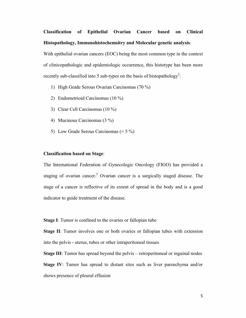
Classification of Epithelial Ovarian Cancer based on Clinical
Histopathology, Immunohistochemsitry and Molecular genetic analysis:
With epithelial ovarian cancers (EOC) being the most common type in the context
of clinicopathologic and epidemiologic occurrence, this histotype has been more
recently sub-classified into 5 sub-types on the basis of histopathology5:
1) High Grade Serous Ovarian Carcinomas (70 %)
2) Endometrioid Carcinomas (10 %)
3) Clear Cell Carcinomas (10 %)
4) Mucinous Carcinomas (3 %)
5) Low Grade Serous Carcinomas (< 5 %)
Classification based on Stage:
The International Federation of Gynecologic Oncology (FIGO) has provided a
staging of ovarian cancer.5 Ovarian cancer is a surgically staged disease. The
stage of a cancer is reflective of its extent of spread in the body and is a good
indicator to guide treatment of the disease.
Stage I: Tumor is confined to the ovaries or fallopian tube
Stage II: Tumor involves one or both ovaries or fallopian tubes with extension
into the pelvis - uterus, tubes or other intraperitoneal tissues
Stage III: Tumor has spread beyond the pelvis – retroperitoneal or inguinal nodes
Stage IV: Tumor has spread to distant sites such as liver parenchyma and/or
shows presence of pleural effusion

6
Independent origin for each histotype?
Given a common embryonic origin of the ovarian surface epithelium and the
lining of the peritoneal cavity from the coelomic epithelium, there tends to be a
high degree of resemblance in the morphological appearance of neoplastic cells
with their non-neoplastic counterparts in many parts of the female genital tract.7
In more recent studies, this feature has linked specific subtypes of neoplastic cells
to their more benign epithelial counterparts in these tissues as being potential sites
of origin unlike previous assumptions whereby all subtypes of ovarian carcinomas
were considered to be primarily ovarian in origin. For example: a benign structure
such as endosalpingiosis in the fallopian tube bears resemblance with serous
carcinomas, benign endometriosis bears resemblance to endometrioid and clear
cell carcinomas, whereas endocervicosis has resemblance with mucinous
carcinomas.8 Despite such a revelation, it is ironic that current clinical treatment
of epithelial ovarian cancer does not take into consideration these differences in
histotypes to treat each of them as separate diseases.7
Classification based on underlying molecular fingerprint:
Furthermore, the objective to identify a common molecular fingerprint for ovarian
cancer has inspired an investigation into the genetic and epigenetic landscape of
neoplastic cells from different histotypes.9 Although no common genetic
alteration specific to ovarian cancer was identified by this approach, molecular
and genetic fingerprints specific for certain ovarian histotypes were identified. In
some cases, these were specific enough to enable distinguishing high grade from

7
low-grade carcinomas – particularly in the case of low grade versus high-grade
carcinomas of the serous and endometrioid histotypes. These findings led to a
proposal for reclassification of ovarian cancers in to two major categories:7, 9
Type I: Indolent tumors that are mostly restricted to the ovaries and constitute
25% of all ovarian carcinomas known to result in 10% of ovarian cancer related
mortality.10
These tumors are generally found to have mutations in KRAS (v-Ki-
ras2 Kirsten rat sarcoma viral oncogene homology), BRAF (v-raf murine sarcoma
viral oncogene homolog B1), PTEN (phosphatase and tensin homolog), PIK3CA
(phoshatidylinositol 3-kinase catalytic subunit), CTNNB1 (gene encoding β-
catenin) and HER2 (human epidermal growth factor receptor 2) and to develop in
a stepwise fashion from well-recognized precursor lesions which are mostly
borderline tumors. Overall, these tumors are genetically stable and thus resistant
to platinum-based chemotherapy. The following histotypes and their
corresponding mutated genes fall under this category:
A) Low Grade Serous Carcinoma (LGSC) - KRAS, BRAF
B) Low Grade Endometrioid Carcinoma (LGEC) – CTNNB1, PTEN, PIK3CA
C) Clear Cell Carcinoma – PIK3CA, PTEN
D) Mucinous Carcinoma – KRAS
Type II: Highly aggressive tumors that are usually metastasized by the time of
diagnosis. This category comprises the most common form of serous ovarian

8
cancer that accounts for 75% of all epithelial ovarian cancers and causes 90%
ovarian cancer related mortality.10
These tumors are rarely diagnosed at early
stages since their transition from occult lesions to malignant disease is rapid
owing to their anatomical location on the surface of ovaries conducive for
sloughing off and spreading of the malignant cells into the peritoneum. These
tumors generally have mutations in the TP53 gene and may also carry
chromosomal gains/ amplifications of CCNE1 (Cyclin E1), which contributes to
genetic instability in the presence of mutated p53. In fact, serous epithelial
ovarian carcinomas are known to have the highest frequency of p53 mutations of
any solid tumors.11
These tumors are thus genetically unstable and highly
susceptible to platinum-based chemotherapy. Nevertheless, they have a high rate
of recurrence. The following histotypes fall under this category:
E) High Grade Serous Carcinoma (HGSC): 70% of epithelial ovarian
cancers.
F) High Grade Endometrioid Carcinoma
G) Undifferentiated Carcinoma
Such a dual system of classification has led to the stratification of this disease
inclusive of its epidemiology, molecular events, patterns of spread, premalignant
origins, response to therapy and prognosis.

9
1.3 Pathogenesis of Epithelial Ovarian Cancer:
One of the most critical aspects to treat a malignancy is to know its cellular
origins. While most cases of epithelial ovarian cancer are sporadic, 5 – 10%
women may be genetically predisposed to the disease by virtue of carrying
mutations in the BRCA gene.
12 Despite the aforementioned identification of the
various subtypes of ovarian cancers and the elaborate classification based on
histopathology and genetic alterations that lead to a malignant phenotype, there
continues to be an incomplete understanding of the pathogenesis of epithelial
ovarian cancers – especially that of high-grade serous carcinoma, which forms the
epidemiologically most aggressive malignant subtype.
1.3.1 Incessant Ovulation Theory:
In 1971, Fathalla proposed the popular “Incessant Ovulation Theory” behind the
origin of EOC, based on observations for high incidence of metastatic ovarian
adenocarcinoma in hens that were forced to lay eggs through uninterrupted
ovulation and consequently had extensive damage to their ovarian surface
epithelium in this process.13
The theory suggested an internalization of the
damaged OSE leading to the formation of inclusion cysts that underwent
metaplasia to differentiate into Mullerian-like epithelium. Such inclusion cysts
were proposed to eventually become dysplastic and manifest as ovarian
carcinoma. Furthermore, this theory resonated with epidemiologic evidence in
human populations wherein risk of ovarian cancer was shown to increase as a
function of the frequency of ovulation. This was evidenced by data that showed

10
women who had breaks in their ovulatory cycles as a consequence of pregnancy
and/or intake of oral contraceptive pills (OCP) were at a reduced risk whereas
nulliparous women were at an increased risk for ovarian cancer. However, this
theory was in disagreement with other epidemiologic data such as evidence from
women with polycystic ovarian syndrome (PCOS) who were at increased risk for
EOC despite naturally infrequent ovulation. Furthermore, it was also shown that
most OCPs functioned similar to progesterone – only formulations that do not
preclude ovulation.3
1.3.2 Gonadotropin Hypothesis:
Alternatively, the gonadotropin hypothesis premised on the end result of an
overstimulated OSE being at risk for malignant transformation under the effect of
Follicle Stimulating Hormone (FSH) and luteinizing hormone (LH) was proposed
to explain the insufficiencies of the incessant ovulation hypothesis. This theory
accounted for the nulliparous women and those with PCOS as having high levels
of gonadotropins and being at greater risk for ovarian cancer when compared to
pregnant women and those taking OCPs, who had low levels of gonadotropins.
Furthermore, it was able to account for perimenopausal women developing
ovarian carcinomas due to an increased production of gonadotropins. However,
there is no conclusive evidence to demonstrate malignant transformation of
inclusion cysts or OSE as a result of gonadotropin exposure, even though few
animal studies have shown that gonadotropin exposure promotes tumor growth.
11
In summary, both the aforementioned theories are based on the premise that
ovulation by itself is an inflammatory process14
involving repetitive disruption of
the ovarian surface and its subsequent exposure to estrogen rich follicular fluid,
release of cytokines and an influx of inflammatory cells in to the ovarian stroma.
This is accompanied by increased oxidative stress via production of reactive
oxygen (ROS) and reactive nitrogen species (RNS), cell damage, elevation of
cytokines, proteases, prostaglandins and an onset of repair mechanisms with
potential epithelial to mesencymal transition (EMT).15
Taken together, all of these
factors increase the susceptibility for occurrence of mutations augmented by the
repetitive nature of this stress on a monthly basis throughout the reproductive age
of women to potentiate oncogenesis.16
1.4 A New Paradigm in Ovarian Cancer: Role of the Fallopian Tube
Even though the anatomical and physiological features of the fallopian tube have
been well documented in literature, it has only recently come to light in a manner
that is challenging all previous understanding of epithelial ovarian cancer
pathogenesis and etiology.17
As mentioned previously, the ovaries are derived from multiple embryonic
structures and are covered by a cuboidal mesothelial layer called as the OSE.
Therefore, the ovaries by themselves do not have a well-differentiated epithelium.
All other organs of the female reproductive system – fallopian tubes, cervix,
uterus and vagina are derivatives of the Mullerian duct. This distinction in
developmental origins is further evidenced in patients with Mullerian agenesis

12
who have fully functional ovaries. Furthermore, the OSE is not reported to
express CA125, which happens to be a marker of well-differentiated epithelium;
rather it is known to express mesenchymal markers – vimentin and N-cadherin.
This has strongly questioned the “ovarian origins” of EOCs in the absence of a
real epithelial cell in the OSE. Instead, further clinicopathologic and genetic
assessment of samples from epithelial ovarian cancers have revealed that most of
these cells resemble the Mullerian – derived epithelium of the female genital tract.
Further evidence was reported by Piek et al18
in 2001, upon finding hyperplastic
or dysplastic lesions with intraepithelial components located at the fimbriated
ends of the fallopian tubal segments from women undergoing risk reduction
bilateral salpingo-oophorectomies (BSO) either on account of harbouring a BRCA
mutation or having breast cancer and/or a strong family history of ovarian cancer.
Figure 1.2: Ovarian cancer subtypes under the dual classification system. Image
reproduced with permission from Jones PM and Drapkin R, Front.Oncol. 3:217 8

13
Such intraepithelial components came to be called as “Tubal Intraepithelial
Carcinomas” (TIC) and have been characterized by strong cytoplasmic
accumulation of mutated TP53, which has been previously mentioned as a
hallmark of almost 100% Type II high-grade serous ovarian carcinomas.
Furthermore, benign regions of the distal tubal epithelium that were found to
express TP53 have been termed as “p53 signatures” and are proposed to appear as
precursor lesions earlier than TICs in the transformative process leading to high-
grade serous carcinomas.19
These observations held strong validity for the
presence of TICs even with immunostaining and analysis of thin sections of distal
tubal fimbriae from women outside the BRCA mutation cohorts.20
This study also
successfully established the clonality of metastatic ovarian cancer through
identification of the same TP53 mutation analyzed in the metastasized cells and
the TIC.21
Furthermore, TICs are a characteristic feature of HGSCs and are not
found in endometrioid or mucinous subtypes of EOC.22
Figure 1.3: Stepwise development of High Grade Serous Carcinomas from the fallopian
tube. Image reproduced with permission from Karst AM, Drapkin R, F1000 Med Rep
2011;3: 22.24

14
In addition, a comparative molecular analysis of p53 mutations from the distal
fimbriae of fallopian tubes and ovarian inclusion cysts in BRCA+
women
undergoing risk reduction surgeries, revealed an absence of p53 mutations in the
inclusion cysts but presence in the fallopian tubes of 38% of women in this
study.23
Finally, this theory proposes that even though TICs may not be present in all
HGSCs, most of them arise in the distal fallopian tubal epithelium as areas of
dysplasia, which further transform into malignant cells. A convenient anatomical
location of these malignant cells located at fimbriated ends, allows them to slough
off and spread into the peritoneum or rub against the OSE of the neighbouring
ovaries. However, in the absence of any ovarian involvement these metastasized
cells may also form peritoneal carcinomas or tubal carcinomas.3 Thus such a
Tubal Intraepithelial Carcinoma theory based on the role of the fallopian tube
explains an organized and logical manner in which HGSCs could potentially
transition from benign to dysplastic lesions to form intraepithelial components
(TICs) that ultimately transform into serous tubal intraepithelial carcinomas
(STIC) prior to spreading peritoneally as disseminated serous carcinomas.24
More recently, the research group of Drapkin et al have developed an ex vivo
model to recapitulate the human fallopian tube epithelium.25
This has been used to
demonstrate the transformation of secretory epithelial cells and their response to
cellular stress such as genotoxic insult etc. They have also reported the

15
development of (hTERT) immortalized secretory fallopian epithelial cells and (c-
myc) / (oncogenic Ras) transformed versions of these cells to recapitulate high-
grade pelvic serous carcinomas (HGPSC) for in vitro and in vivo use with
immunocompromised xenograft mice.26
The TIC theory and the aforementioned recent developments are having major
implications to facilitate the search for new biomarkers, targeted therapies and
imaging techniques directed in the right anatomical, molecular and physiological
contexts respectively. Since most pre-malignant lesions including TICs for HGSC
in women with BRCA+ mutations have been found to exist in the distal tubal
fimbriae, risk reduction surgery restricted to a salpingectomy would get rid of the
fallopian tubes while preserving the ovaries. This bears great significance from an
endocrine point of view as well as to avoid side effects such as cardiovascular
risk, osteoporosis and cognitive impairment as a consequence of a complete
hysterectomy or bilateral salpingo-oophorectomy.3, 12
In summary, it is now apparent that the disease previously considered “Ovarian
Cancer” was grossly a misnomer. This is becoming clearer with an emerging
understanding of the molecular basis for ovarian cancer as a very heterogeneous
and complex set of independent diseases. Furthermore, provocative evidence to
propose a tubal origin to HGSCs that were previously thought to arise from the
OSE, is now challenging the field to delve deeper into the molecular pathogenesis
of this disease. This inspires a new hope in the search for better diagnostic and

16
therapeutic approaches that may enable an early detection of the elusive
malignancy!
1.5 TUMOR BIOMARKERS:
Simply put, tumor biomarkers are molecular signatures of neoplastic cells that
help identify them within a complex cellular milieu. These inlcude genetic
signatures characterized by DNA mutations, chromosomal alterations – gains or
losses in segments, epigenetic changes, overexpressed and/or secreted proteins
that are found in higher than normal amounts in the blood or other body fluids of
individuals having a tumor. Tumor markers are not always limited to those that
are produced by the neoplasms themselves, but could also be produced by the
body in response to a neoplastic entity.
Furthermore, tumor biomarkers have been classified into two broad categories:27
A) Tumor Specific Antigens – molecules that contribute primarily to
oncogenesis and are not expressed in normal cells of the body. For
example: mutated p53 or Ras oncogenes, carcinoembryonic antigen
(CEA) and alfafetoprotein (AFP).
B) Tumor Associated Antigens – molecules that are expressed differently
between normal and neoplastic cells and are of utility in identifying
cancerous cells from their healthy counterparts. For example: CA19-9,
CA125
Some of the desirable features in a clinical biomarker are that it should be
specific, reliable, measurable and predictive in nature. Given the relatively low

17
incidence for occurrence of ovarian cancer in the general population (12.3 per
100,000 in the United States between 2007 – 2011 [SEER, NCI, USA]),
developing a tumor biomarker with relevance for screening faces a special
challenge in that such a test needs to have a very high degree of specificity.
Statistically, this demands a sensitivity of 75% and a specificity of 99.6% in order
to achieve a positive predictive value of 10%.28
CA125 – Cancer Antigen 125, as
such is elevated in 80% of women with EOC and is reported to have a sensitivity
of 50 – 62% for patients with early stage disease and 90% for patients with
advanced stage disease.29
Furthermore, with specificity > 95% for all advanced
stage cases, CA125 is more suited as a biomarker for recurrence rather than
general screening, wherein it tends to have a lower specificity. In recent years,
several molecules such as HE4 (human epididymis 4), mesothelin, M-CSF
(macrophage colony stimulating factor), Osteopontin, KLK6 (Human Kallikrein-
6), CA72-4, CA19.9, CA15.3, MUC1, AFP and others have been assessed alone
or in combination with CA125 in order to increase the sensitivity of a screening
test for primary ovarian cancer.30
Among these, the use of HE4 and CA125
together has demonstrated highest accuracy for the detection of malignancy. As of
now, there is no single biomarker that outperforms CA125 to indicate an ovarian
malignancy.

18
1.5.1 A Focus on CA125: Cancer Antigen 125
With a history of over three decades since its discovery by Bast and colleagues in
1981,31
and despite its limitations for sensitivity and specificity, CA125 has long
served as the most reliable tumor biomarker that has come to be a ‘gold standard’
of reference in the clinical assessment of epithelial ovarian cancer. CA125 is a
mucinous glycoprotein expressed by fetal amniotic and coelomic epithelium.32
Owing to its mucinous nature and a much later discovery of its being encoded by
the MUC16 gene,33
it is common to find literature interchangeably referring to
this tumor biomarker as CA125 or MUC16. Under normal physiologic conditions,
CA125 is expressed in adult tissues such as mesothelial cells of the pericardium,
pleura and peritoneum that are embryonically derived from the coelomic epithelia
and also in derivatives of the Mullerian epithelia such as fallopian tubes,
endometrium and endocervix that constitute organs of the female genital tract.
Other anatomical regions positive for the expression of CA125 include the ocular
epithelium and the upper respiratory tract. CA125 can also be isolated from the
peritoneal ascites of ovarian cancer patients.34
Subsequent to its molecular identification using a murine monocolonal antibody –
OC125, several antibodies were developed for binding to CA125.35-40
This led to
their classification on the basis of three distinct epitopic regions / domains found
in the CA125 antigen:38
Region A: binds OC125 like antibodies
Region B: binds M11 like antibodies
Region C: binds Ov197 like antibodies

19
Figure 1.4: Representation of the 3 antibody binding domains (A-C) on the CA125
epitope. Image reproduced with permission from Nustad K et al, Tumour Biol
1996;17(6): 325-31.37
Biochemically, CA125 has been described as a repeating peptide epitope on a
mucinous glycoprotein – MUC16, which is proposed to be 22,152 amino acids
(aa) long with a molecular weight of 2.5 MDa (Mega Dalton), which potentially
doubles in size as a result of glycosylation to produce a 3 -5 MDa glycoprotein.41,
42 A conformation dependent peptidic nature of the CA125 epitope was revealed
through the seminal works of Davis et al who conclusively demonstrated the
sensitivity of CA125 to high temperature, low pH and treatment with proteases.
This work also showed that there was a minimal effect from periodate treatment
of the glycoprotein, thereby refuting previous claims of CA125 being a
carbohydrate antigen.43
20
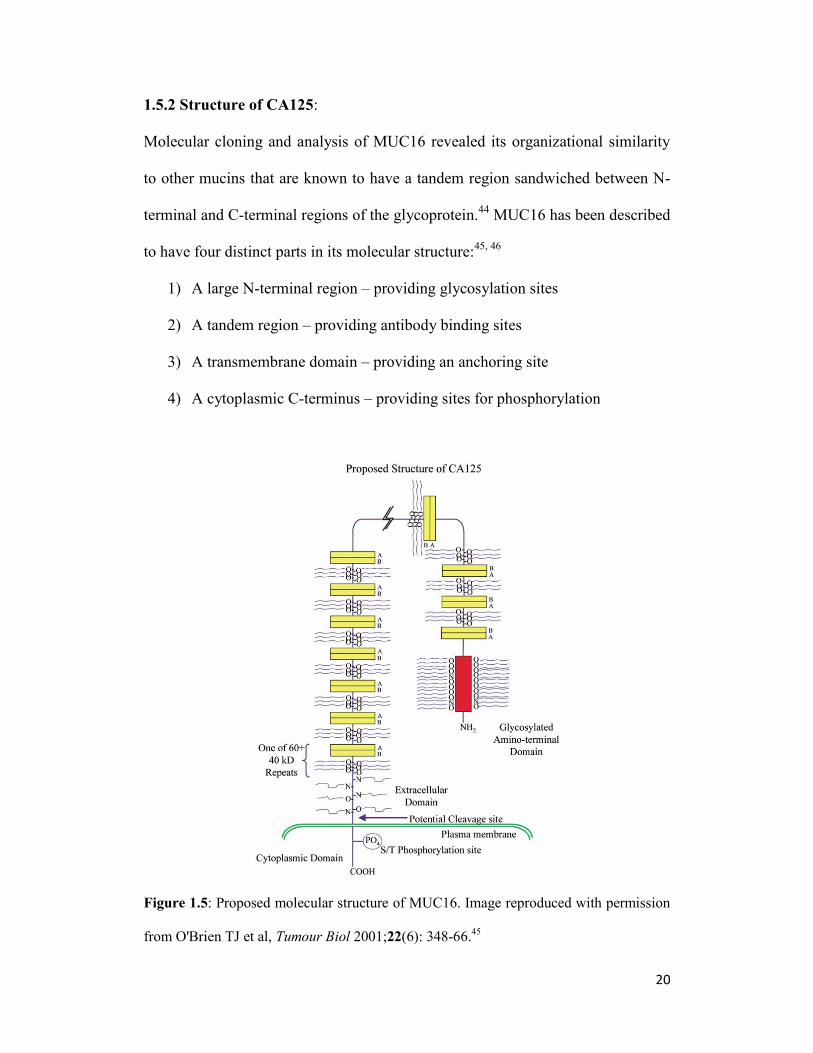
1.5.2 Structure of CA125:
Molecular cloning and analysis of MUC16 revealed its organizational similarity
to other mucins that are known to have a tandem region sandwiched between N-
terminal and C-terminal regions of the glycoprotein.44
MUC16 has been described
to have four distinct parts in its molecular structure:45, 46
1) A large N-terminal region – providing glycosylation sites
2) A tandem region – providing antibody binding sites
3) A transmembrane domain – providing an anchoring site
4) A cytoplasmic C-terminus – providing sites for phosphorylation
Figure 1.5: Proposed molecular structure of MUC16. Image reproduced with permission
from O'Brien TJ et al, Tumour Biol 2001;22(6): 348-66.45

21
N-terminal region:
The extracellular N-terminus is reported to have 12,068 amino acids rich in serine
and threonine residues acting as suitable sites for O-linked glycolsylation.
Tandem Repeat Region:
The tandem region has been described to have up to 60 repeats with each repeat
being composed of 156 amino acids which are not identical but largely
homologous per repeat. Two interesting aspects of the tandem regions are:
A) Presence of a highly conserved pair of cysteines at positions 59 and 79
that are potentially involved in the formation of intramolecular disulfide
bonds that create loop-like structures within the MUC16 molecule.
Alternatively, intermolecular disulfide bonds between the conserved
cysteine residues have been proposed to promote the formation of an
extracellular matrix.
The 21-mer-loop structure between Cys 59 and Cys 79 had previously been
described as potential epitopic site on the CA125 peptide – called as the
‘cysteine loop’. Some of the key characters of the cysteine loop as ideal sites
for antibody binding included: a) an ability of the disulfide bond to push
amino acids comprising the loop to move away from the protein core and thus
become more available for antibody binding; b) the presence of hydrophilic
amino acids at the center of the loop; c) the relative absence of nearby
glycosylation.

22
Nevertheless, unlike previous experiments wherein protease digestion of this
loop abrogated antibody binding with CA125, recent independent reports of
Brennan et al using deletion constructs of the 156 aa repeat region and
Berman et al.47
using synthetic 21-mer peptide have challenged the cysteine
loop theory to suggest that the cysteine loop by itself is insufficient as the
target epitopic site. Taken together, their results questioned the accuracy of
the cysteine-loop model to define a region on the CA125 epitope for antibody
binding. Furthermore, studies with deletion constructs of the 156 aa tandem
repeat regions have shown that deletion of the amino acids between position
129 through 156 did not impact antibody binding, but subsequent deletion of
the first 30 amino acids at the N-terminus of such a construct abrogated
binding to anti-CA125 antibodies from all three categories – OC125, M11 and
Ov197. This has also led to a proposition for CA125 to be a discontinuous
epitope within the 1 – 129 aa region of each tandem repeat.
B) Positively charged SEA (Sea Urchin Sperm Protein, Enterokinase and
Agrin) domains in this region present a sandwich three-dimensional
structure formed by two alpha helices, four antiparallel beta strands and a
hydrophobic core. The presence of multiple SEA domains is a unique
feature of MUC16 although their biological role has not yet been
determined. SEA domains tend to have an autoproteolytic role, but it is
uncertain if this directly contributes to the shedding of CA125 in the
serum.

23
C-Terminal Region:
CA125 is tethered to the surface of neoplastic cells through the C-terminal
domain of MUC16 comprised of 284 amino acids and has a potential site for
phosphorylation that is proposed to play a role in cleavage of the mucin and
release of the MUC16 ectodomain into the serum.
However, despite the recent progress made in deciphering the molecular structure
and organization of the aforementioned regions of CA125 on this complex mucin,
there is limited knowledge about the precise epitope sites for antibody
binding/targeting, the role of multiple SEA domains, the effects of post-
translational modifications and the factors that control the in vivo shedding of
CA125.
1.5.3 Function of CA125:
Despite expression in the epithelia of normal tissues such as the pericardium,
endometrium and cornea, a specific biological role for CA125 in health has not
yet been understood.44
Reports in the literature suggest an immunosuppressive
role for this glycoprotein; particularly owing to its interaction with Natural Killer
(NK) cells to reduce the expression of CD16 expression on their surface.48
Such
an NK-suppressive effect combined with its increased expression in pregnant
women during the first trimester has also been suggestive of a role in preventing
immunological rejection of the fetus.49-52
Further, an interaction between MUC16
and galectin in the corneal epithelium has been proposed to provide a barrier to
24
bacterial and viral infections in the ocular epithelia.53, 54
Additionally, owing to its
expression in the pericardium, a rise in physiol.ogical CA125 levels has been
implicated in congestive heart failure.55, 56
However, MUC16 knock out mice
have not shown any major physiological defects to warrant the mucin’s
indispensability in the health of these animals.57
In the context of ovarian cancer, MUC16 is reported to exercise an
immunoprotective role to shield ovarian cancer cells from NK cells. This renders
the tumor evasive to the immune system. Some of the mechanisms postulated to
achieve this effect include steric hinderance of NK and ovarian cancer cell
synapse formation58
due to the macromolecular size of MUC16 and/or a
consequence of binding between negatively charged terminal sialic acid residues
of MUC16 with NK cell inhibitory receptors such as Siglec-9 that ultimately lead
to an attenuation of natural killer cell activity against ovarian cancer cells.59
This
phenomenon has also been observed in MUC16 binding to monocytes, which are
attracted to ovarian cancer cells and ultimately get attenuated in their downstream
activity.60
Additionally, NK cells within the ovarian tumor microenvironment
have also been indicated in immune editing by virtue of attacking tumor cells with
low levels of CA125 and thus indirectly enriching the tumor for cells with high
levels of CA125 expression.61
Further, CA125 is also known to interact strongly with mesothelin expressed
along the lining of the peritoneum.62
This interaction is proposed to occur with a
Kd of 5 nM between the N-glycans of MUC16 and super-helical ARM-type

25
repeats on mesothelin, that ultimately allows binding of ovarian tumor cells to the
walls of the peritoneum to promote metastasis beyond the ovaries.63, 64
While knockdown of MUC16 in ovarian cancer cells has an anti-proliferative
effect and negative impact on their metastatic potential, expression of its C-
terminal domain in SKOV3 cells has been reported to increase their proliferation
and in vivo tumor burden in xenograft mice.65
These observations have been
linked to downstream cell signaling interactions of MUC16 with components of
the JAK-STAT pathway and/or Src-family kinases that induce E-cadherin
mediated metastasis.65
1.5.4 CLINICAL UTILITY OF CA125:
As a consequence of neoplastic transformation in epithelial ovarian cells, CA125
is overexpressed and eventually shed into the blood pool of subjects. At present,
the clinical usefulness of CA125 comes from its estimation in serum samples of
individuals presenting in the clinic with pelvic masses suspected for ovarian
cancer and in the follow-up of patients who have undergone cytoreductive surgery
and/or chemotherapy. Typically, an immunoassay quantified serum CA125 level
of ≤ 35 U/mL is considered normal while patients with advanced stages of the
disease are known to have relatively higher serum CA125 levels.66, 67
Nonetheless, CA125 levels can be elevated in premenopausal women during
ovulatory cycles and also in the absence of neoplasia.68
Furthermore, CA125 has been employed as a screening biomarker in clinical
trials. The most recent one is the United Kingdom Collaborative Trial of Ovarian

26
Cancer Screening (UKCTOCS), wherein > 200,000 post-menopausal women
have been screened for ovarian cancer.69
The interim results of this trial are
promising and suggestive of > 47% women who tested positive in a combined
assessment of CA125 levels with transvaginal ultrasound had stage I or stage II
disease.70
CA125 has also been used as a diagnostic marker for Epithelial Ovarian
Cancer owing to the fact that elevation of CA125 levels have known to precede
clinical detection of the disease at least by 3 months.32, 71, 72
Also, the prognostic
utility of CA125 has been highlighted by its ability to effect management of the
disease through longitudinal monitoring of its levels in patients pre- and post-
chemotherapy/surgery. This has been demonstrated by the fact that patients with
elevated levels of CA125 post-treatment have a worse prognosis than those whose
levels have normalized.32, 67
In addition to its diagnostic and prognostic utility, CA125 has also been a target
for therapeutic approaches designed and attempted against EOC. Oregovomab (a
MAb-B43.13 formulation), initially used as a radioimmunoscintigraphy agent73
was later transformed into an immunotherapeutic74
due to its ability for eliciting
anti-idiotypic responses and T-cell stimulation against multiple epitopes on
CA125 that resulted in an extended survival of patients treated with this agent.75
After showing no significant effect as a monoimmunotherapy, Oregovomab
(OvaRex-MAb-B43.13) is presently undergoing clinical trials in combination
with routine adjuvant chemotherapeutic agents to treat ovarian cancer.76
Abagovomab is an antibody that was used to generate specific anti-idiotypic

27
response to CA125.77-80
Similarly, anti-mesothelin antibodies81
and HN125 – an
engineered version of the MUC16-binding epitope of mesothelin grafted onto the
Fc portion of the human IgG1 antibody has been developed.82
There have also
been recent reports of antibody-drug conjugates (ADCs) using CA125-targeting
antibodies – 3A5 and 11D10 conjugated with cytotoxic drug – monomethyl
auristatin E (MMAE). These agents yielded promising results for high efficacy
and minimal toxicity in Phase I clinical trials.83, 84
In summary, CA125 (MUC16) plays a vital role in the proliferation of EOC and
its metastatis through an orchestration of intra- and inter-cellular interactions as
described above. Simulatenously, the ability to clinically measure CA125 levels
from the serum of EOC patients helps to monitor disease progression and to
evaluate the response to therapy.
1.6 DIAGNOSIS OF OVARIAN CANCER:
This malady has metaphorically come to be known as the “whispering disease”
and a “silent killer” owing to its synonymous description for asymptomatic
progression. It is not only the fifth leading cause of cancer-related deaths in
women, but also the most lethal gynecologic malignancy. This is exemplified by
the National Cancer Institute’s Surveillance Epidemiology and End Results
(SEER) report for 2014, wherein 21,980 cases of ovarian cancer will be diagnosed
in the United States alone, of which > 14,000 deaths are predicted as a result of
this disease. Cancer Research UK has ranked ovarian cancer as eighteenth in

28
terms of worldwide incidences of cancer by virtue of its annual contribution to
1.7% of all cancers. The National Cancer Institute of Canada has predicted 2700
new cases of ovarian cancers that will be diagnosed in 2014, of which there will
be an estimated 1750 deaths. Clearly, the disease has a high mortality rate that
hasn’t changed much over the last few decades.
As with any diseased condition, diagnosis plays a vital role in the treatment of
ovarian cancer. Although recent knowledge suggests that the disease progresses
with 23 known indications, most of these are abdominal or gastrointestinal in
nature and rarely link directly to a gynecologic aspect. Most of these are mild
indications such as bloating of the abdomen, constipation, early satiety, increased
thirst and frequent urination that either get overlooked as non-specific signals or
potentially mistaken for common disorders such as gastritis, irritable bowel
syndrome, urinary tract infection etc. Consequently, most patients are diagnosed
with the disease at late stages (III or IV). Less than 25% cases are detected at
stage I. This is further hampered by the fact that CA125 is reported to be
expressed in only 50% of stage I epithelial ovarian cancer. Nonetheless, there is a
90% cure rate for epithelial ovarian cancers diagnosed at stage I, whereas the cure
rate plummets to 20 – 25% in patients diagnosed with late stage disease.32
This
raises a critical need for better diagnosis of epithelial ovarian cancer.
More recently, Goff et al introduced a symptom index (SI) for ovarian cancer with
the intent to increase the possibilities for early detection.85
The SI was considered
positive if one or more of the symptoms were present over a period of 12 months
and occurred more than twelve days a month. Independent investigations thus far

29
have clearly demonstrated that the severity, duration and frequency of the
gastrointestinal and/or abdominal symptoms were significantly higher in women
having ovarian cancer at all stages. The SI reported ovarian cancer in women with
a sensitivity of 57% for early stage disease, 80% for advanced stage disease and a
specificity of 90%. This was similar to CA125 serum tests, which are reported to
have a sensitivity of 50 – 79% and specificity between 96 – 99% for advanced
stage ovarian carcinomas. Furthermore, supplementing CA125 and HE4 values to
the SI is reported to improve the detection of early stage EOC to raise the
sensitivity to 84% with a specificity of 98.5%, if any two of the variables were
positive.86, 87
A conscious implementation of such a tool may be instrumental in
triaging patients in a primary care setting.
1.6.1 Contemporary Diagnostics for Ovarian Cancer:
Nevertheless, patients presenting in the clinic with pelvic masses are routinely
diagnosed for ovarian cancer via physical examination, careful analysis of the
medical history, assessment of serum CA125 levels and adnexal imaging via
trans-vaginal ultrasound (TVUS).
1.6.1.1 CA125 ELISA: (Enzyme Linked Immunosorbent Assay)
A two-site binding immunoassay such as CA125 II is currently used in the clinic
to evaluate serum CA125 levels through a capture of the tumor associated antigen
by an M11 antibody coated onto microtiter wells while an OC125 antibody is
employed as a tracer.65
Patients with elevated levels of CA125 at the time of
presentation and post-treatment are known to have a worse prognosis than those

30
with low or normalized levels respectively at these stages. However, tumor
markers such as CA125 have a major limitation in that their expression levels are
often found to be elevated in many benign conditions. Furthermore, even if this
information is specific, it does not indicate the precise location of the tumor in the
body. This calls for an imaging technique that can provide a visual assessment to
precisely assert the diagnosis of ovarian malignancies.
1.6.1.2 Ultrasound: (US)
Given the low cost of performing as well as undertaking an ultrasound
examination combined with the convenience of bedside operation and widespread
availability in almost every gynecologic clinic and the absence of ionizing
radiations, have led to transvaginal ultrasound (TVUS) becoming the first-line
pre-treatment triage imaging modality used in the clinical diagnosis of ovarian
cancer.88
This technique provides the capability to view adnexal masses and also
differentiate them from unilocular ovarian cysts that are benign.91
Though the
addition of morphologic scoring systems and color Doppler have been introduced
to enhance the clinical performance of US, the technique is mostly operator
dependent unlike other tomographic methods and thus has not proven adequate
diagnostic performance.88
Even though an introduction of targeted microbubbles
functionalized with antigen-binding ligands has been proposed to improve the
specificity of US, the size of these agents practically limits them to eventually
binding with epitopes expressed on the surface of endothelial cells in the tumor
vasculature. In other words, such microbubbles are too large to extravasate
through endothelial cells of blood vessels.

31
1.6.1.3 Computed Tomography: (CT)
This technique has been the most widely used imaging method in the surveillance
of women who have already undergone primary treatment of ovarian cancer.90, 91
While CT is capable of successfully identifying a hypodense region of pleural
effusion in the thorax as an indicator of relapse, or pulmonary parenchyma as
round solid nodules and calcifications in serous carcinoma, this technique is
significantly limited in its ability to detect small recurrences due to low tissue
contrast resolution. This is especially true for regions in the small bowel and
mesentery. The other disadvantages of CT include exposure to ionizing radiation,
and its inability to distinguish between recurrence, and post-operative or post-
radiotherapy fibrosis.90
1.6.1.4 Magnetic Resonance Imaging: (MRI)
The combination of high-field magnets, phase arrayed body coils and fast impulse
sequences has enabled magnetic resonance imaging to deliver high quality images
with high tissue contrast and high resolution within a short scanning time. The
introduction of diffusion weighted imaging (DWI) and perfusion imaging have
enhanced the clinical utility of this modality by allowing visualization of
macroscopic disease as well as providing information on the cellularity and
vascularity of the tissue. Most importantly, MRI provides excellent soft-tissue
contrast to reveal anatomical detail without the use of ionizing radiations and is
fairly operator-independent. Nevertheless, some of its limitations include the costs
associated with an MRI scan, a need for patient cooperation and the limited

32
window for regional scanning, unlike whole body scans that can be performed via
CT. Furthermore, from an oncologic viewpoint, MRI is also limited in its
capabilities for detecting micrometastasis in lymph nodes.90
1.7 MOLECULAR IMAGING:
An ability to image real time physiological events at the cellular and molecular
level in disease and health has developed into a new field of medical imaging
expertise commonly referred to as molecular imaging. Molecular imaging now
plays an increasingly important role to equip physicians and oncologists with the
extra pieces of information including the extent of spread of a disease, unique
tumor-specific expression profiles of receptors and/or biomarkers for designing a
targeted therapy, to evaluate response to therapy and monitor disease progression
or recurrence – all of which can positively impact decision making to effect
disease management in the larger scheme of patient care. Furthermore, this
information may not be captured by conventional imaging techniques that deliver
excellent anatomical details as opposed to biochemical information at a cellular
level that can sometimes be obtained even prior to the development of symptoms.
1.7.1 Positron Emission Tomography: (PET)
Unlike the aforementioned conventional imaging techniques which are not
originally designed to perform diagnosis at a molecular level, PET is a natively
functional molecular imaging modality by virtue of its ability to generate real time
information based on the differential in vivo physiological uptake or biochemical
targeting and metabolic processing of a radiotracer/molecular probe between

33
neoplastic versus normal cells. Furthermore, PET is unique in its ability to render
quantitative information in addition to functional imaging. PET relies on the use
of ‘tracers’ formulated with radioisotopes that have an unstable nucleus and decay
via emission of positrons, which travel a short distance in tissues prior to colliding
with electrons to annihilate and produce two co-incident back-to-back 511 KeV
gamma photons detectable by a PET scanner. A reconstruction of the lines of
responses from a PET scan helps map the three-dimensional distribution of the
radionuclide in the body.
The simplest and most widely used radiotracer in oncologic PET imaging is an
analog of glucose – [18
F]FDG (18
F-fluoro-deoxyglucose) which functions as a
radiotracer by virtue of its being taken up more avidly by neoplastic cells that
inherently have a higher degree of glycolytic activity vis-à-vis normal healthy
cells. The radiotracer gets phosphorylated by hexokinase upon entry into the
neoplastic cells but, the lack of a 2’-hydroxyl group in this molecule precludes it
from being further metabolized by such cells. Thus, the radiotracer gets trapped
and continues to decay within the tumor cells that can be detected in a PET scan.
In the context of ovarian cancer, [18
F]FDG-PET is reported to have a low
sensitivity (58 – 78%) and low specificity (76 – 87%) for the diagnosis of primary
ovarian cancer, which may partly be attributed to the cystic nature of ovarian
tumors.88
However, a combination of [18
F]FDG-PET and CA125 evaluation
yielded a sensitivity of 94 – 98%.92
Furthermore, the evidence of recurrence on
PET is reported to have preceded CT findings by 6 months.93

34
With surgery being the primary mode of treatment for epithelial ovarian cancers
and an extremely high probability of recurrence of the disease combined with
recent advances in the development of hybrid imaging technologies, there are
several reports for the utility of PET/CT as a modality that delivers a high degree
of accuracy for the detection of recurrent disease in the post-treatment
surveillance of patients.94-98
Such a combination has improved the sensitivity,
specificity and accuracy for the diagnosis of malignant ovarian lesions.97
Most
importantly, PET/CT was reported to have found positive lesions in many
instances where CT by itself was negative – especially metastatic lesions in the
bowel wall whose detection was difficult with MRI or CT.100-103
Furthermore, in
one study, the addition of PET/CT information to conventional diagnosis was able
to effect a change to the clinical management of the disease in up to 60% of the
cases.104, 105
Taken together, such reports are suggestive of PET/CT being most
useful for the diagnosis of recurrent ovarian lesions in symptomatic patients that
have a rising CA125 levels but negative or equivocal results from primary
diagnosis using conventional imaging techniques. A meta-analysis by Gu et al
illustrated the area under the receiver operator curve to be 0.92 for CA125, 0.93
for PET alone, 0.96 for PET/CT, 0.88 for CT and 0.80 for MRI for the detection
of ovarian cancer.106
It is now apparent that active disease can be diagnosed via PET/CT despite low
levels of CA125, and may have a potential for translation into an early diagnosis
of recurrent ovarian cancer. This may help stratify patients suitable for site-
specific treatment, radiation therapy and to choose optimal candidates for surgery.

35
107, 108 Thus, [
18F]FDG-PET/CT can deliver increased lesion contrast along with
delivering information on the anatomical localization and differentiation of
neoplastic disease from post-treatment fibrosis.109
However, some drawbacks for
diagnosis with [18
F]FDG-PET/CT include a radiation burden to subjects and its
physiological uptake in the bowel, kidneys and bladder. Furthermore, it is limited
in its capability to diagnose small volume disease (5–7 mm) and lymphatic spread
– especially into nodes surrounding the urinary bladder.
1.7.2 Targeted Positron Emission Tomography
In light of the aforementioned limitations and possibilities for non-specific uptake
of generic radiotracers such as [18
F]FDG in cells under the effect of inflammation
and other hypermetabolic benign or physiological conditions, it may be useful to
adopt a more targeted approach to facilitate the in vivo imaging of ovarian
carcinomas. This has been demonstrated through the synthesis and use of specific
radiolabeled ligands as tracers for receptor molecules expressed in ovarian cancer.
Overexpression of folate receptors (FR) on ovarian neoplasms forms an attractive
target for tumor imaging and therapy.110
Some recent efforts to carry out a folate
receptor targeted molecular imaging in ovarian cancer include but are not limited
to – the synthesis of several small molecule based 99m
Tc-labeled folate conjugates
for SPECT imaging,111-114
99m
Tc-etarfolatide115
and 111
In-DTPA folate;116
67Ga/
68Ga-,
18F- and
124I-labeled folate conjugates for PET imaging;
117, 118
18F-
labeled PET radiotracer targeting COX-1 in ovarian cancer;119
fluorescein
conjugated folate for intra-operative procedures;120
a dual imaging affibody
bioconjugate with capabilities for near infrared optical imaging and PET using

36
64Cu;
121 development of a porphyrin-based multimodal PET-Optical probe for
targeting folate receptors in ovarian cancer122
and a smart probe developed from
folate conjugated to a near infrared fluorescent dye by a linker that specifically
gets cleaved by tumor-associated lysosomal cathepsin B enzyme upon FR
mediated uptake into neoplastic ovarian cancer cells.123
Alternatively, radiolabeled antibodies against specific tumor associated antigens
overexpressed by ovarian cancer cells – MoV 18 against Folate Receptor α,124
OC125,125, 126
MAb-B43.13,73
MAb 3A584
against CA125 antigen, MAb MX35
against an unspecified ovarian cancer antigen and MAb MH99 against 17.1A
antigen127
have been described for use in in vivo applications.
1.7.2.1 Immuno-Positron Emission Tomography: (immuno-PET)
In recent years, the latter approach has sprung into a new branch of molecular
imaging commonly referred to as immuno-PET.128
This technology brings
together the high specificity and affinity of antibody targeting vectors with the
great sensitivity of detection rendered by positron emitting radionuclides.129, 130
In
vivo application of such a composite radiotracer is ultimately delivers real time
information on its biochemical targeting capabilities and pharmacokinetics as
analyzed via positron emission tomography. The use of this technology not only
allows the development of a molecular probe that is highly specific for tumor
targeting, but can also provide vital information about off-target effects and
potential in vivo pharmacological profiles and fate of the targeting vector for
therapeutic applications that may be intended in the future.

37
Within a short span of time, preclinical immuno-PET strategies have been
developed against several oncologic targets ranging from applications suited for
same to next–day imaging131-143
and beyond144-151
to image several solid and
hematologic malignancies. This has mainly been facilitated by two factors:
a) Advances in recombinant technology that contributed to the synthesis of
engineered antibodies, antibody fragments and nanobodies; 152-155
Antibody
Format
Complete
Antibody
F(ab’)2 Minibody Diabody Fab scFv Nanobody
MW (KDa) 150 120 80 55 55 28 15
Source
Hybridoma
or
Engineered
Enzymatic Engineered Engineered Enzymatic Engineered Camelids
Valency Bivalent Bivalent Bivalent Bivalent Monovalent Monovalent Monovalent
Clearance
Route
Hepatic
Hepatic /
Renal
Hepatic Renal Renal Renal Renal
Table 1.1: Representation of different antibody formats employed for immuno-
PET
b) An increasing awareness and availability of several positron emitting
radionuclides that have provided a variety and choice to tailor molecular
probes for use in immuno-PET strategies.

38
Radionuclide
T ½
(h)
Positron Yield
(%)
β+
max
(MeV)
68Ga 1.1 89 1.89
18F 1.8 97 0.63
64Cu 12.7 19 0.66
86Y 14.7 33 3.15
76Br 16.2 23 3.98
89Zr 78.4 23 0.90
124I 100.3 23 2.14
Table 1.2: List of PET-radionuclides and their salient properties suitable for immuno-
PET
The aforementioned antibody constructs and radioisotopes thus form integral
components in the armamentarium of a radiochemist to synthesize radiotracers for
immuno-PET applications suited to the nature of the biological target and the in
vivo pharmacokinetics of the targeting vector. This aspect is primarily governed
by a cardinal principle of molecular imaging via PET that recommends a match
between the biological (serum) half-life of the targeting vector with the physical
(decay) half-life of the radioisotope used in conjunction with the biomolecule.152

39
Not to be missed, simultaneous advancements in the manufacture of PET scanners
have been a great technological advance to support the progress in this field.
In summary, although serum CA125 levels serve as a diagnostic tool and
prognostic indicator of disease burden resulting from ovarian carcinoma, it is
considered clinically inadequate as an isolated value to decide further course of
treatment in patients due to its limited specificity as a biomarker in the diagnosis
of primary ovarian cancer. Trans-vaginal and/or trans-abdominal ultrasound is the
current mainstay among imaging modalities used in the clinical diagnosis of
primary ovarian cancer. In other words, serum CA125 and ultrasound form the
major tests currently use to triage patients for further treatment. However, given
the reliability of a rising CA125 level in high-risk group patients and those that
have undergone primary cytoreductive surgery,72
a combined application of
[18
F]FDG-PET/CT has recently enabled relatively superior detection of recurrent
disease even if these individuals were asymptomatic and had negative results
from other conventional imaging modalities including CT and MRI. This may
have potential implications for the preferential use of PET/CT towards an early
detection of ovarian cancer in a recurrent setting, in order to positively impact
disease management for the future.

40
1.8 THESIS OVERVIEW:
1.8.1 Rationale:
Despite the fact that 75% of women diagnosed with advanced stage ovarian
cancer have a favourable clinical response as a result of optimal cytoreductive
surgery and first-line platinum therapy, the mortality rate resulting from this
cancer remains high with a 5 year survival rate of only 44.6%.156
This illustrates
the powerful impact of recurrence from this malignancy and the present-day
limitations to effectively detect and treat the disease in this phase.
Recognizing the ongoing efforts to provide reliable and early detection of primary
EOC through screening with novel biomarkers in combinatorial panels that
incorporate risk stratification algorithms; the work presented in this thesis aims to
develop a targeted non-invasive imaging strategy for the detection of epithelial
ovarian cancer. The central idea is to overcome some limitations of present day
methods used for ovarian cancer diagnosis and to prospectively result in an early
detection of its recurrence to positively impact disease management.
The work in this thesis is premised on two facts:
a) CA125 is a reliable biomarker for recurrent epithelial ovarian cancer and
bears a high diagnostic and prognostic value in this setting.32, 71, 156, 157
b) [18
F]FDG-PET has a high sensitivity and diagnostic accuracy in
identifying recurrent epithelial ovarian cancer in asymptomatic patients
having a rising CA125 level and negative results from conventional
imaging modalities including CT and MRI.90, 156, 159

41
However, both these aspects have the following limitations:
a) Despite having a high prognostic and diagnostic significance in the
recurrent setting, a rising CA125 level does not indicate the in vivo
location of the malignancy.
b) Although CA125 ELISAs perform excellently well with a detection limit
of 1.5 U/mL, these assays rely on the availability of CA125 antigen in
circulation. There is a possibility that small volume recurrent disease may
not shed as much antigen into the blood that can be detected by such
ELISAs. Thus, patients may appear to have normal CA125 levels. This is
further exemplified by a mathematical model proposed by Hori and
Gambhir157
wherein an ovarian tumor may take 10.6 years since its first
neoplastic cell to grow up to a size of 10.52 mm3, after which it sheds just
enough CA125 antigen (1.5 U/mL) that is detectable by the existing high
performance clinical ELISA kits.
c) [18
F]FDG-PET of ovarian cancer is sensitive yet suffers from relatively
low [18
F]FDG uptake in these tumors partly due to their cystic nature.88
This is compounded further by a limited ability to detect small volume
residual disease resulting from very low differential glucose uptake in
small lesions combined with the limitations of PET for spatial resolution.90
Considering these factors, we developed CA125-targeted vectors for non-invasive
imaging of EOC with an aim to expand the diagnostic utility of CA125 and
simultaneously overcome some limitations of present day ELISAs and [18
F]FDG-
PET as diagnostic methods used in the context of ovarian cancer.

42
1.8.2 Hypothesis
Based on the aforementioned rationale, we hypothesized that CA125 is a suitable
target for the molecular imaging of Epithelial Ovarian Cancer (EOC) to reflect in
vivo tumor burden in subjects.
1.8.3 Aim:
To develop a CA125-targeted immuno-PET strategy for the molecular imaging of
epithelial ovarian cancer.
1.8.4 Objectives:
To develop a single-chain antibody fragment for targeting CA125 in
epithelial ovarian cancer
To synthesize an 18
F-labeled single-chain antibody fragment as a CA125-
targeted radiotracer for same-day imaging of epithelial ovarian cancer
To develop 64
Cu-labeled anti-CA125 antibody and single-chain antibody
fragment for immuno-PET of epithelial ovarian cancer
To maximize the potential of full-length antibody imaging through 89
Zr-
immuno-PET of epithelial ovarian cancer
To develop a CA125 targeted diabody for optimal antibody fragment-
based same-day imaging of epithelial ovarian cancer
The forthcoming chapters 2-6 of this thesis illustrate how the aforementioned
objectives were achieved and the findings thereof.

43
1.9 References:
1. National Cancer Institute, SEER Training Modules
http://training.seer.cancer.gov/anatomy/reproductive/female/ovaries.html
2. Oktem O, Oktay K. The ovary: anatomy and function throughout human life. Ann
N Y Acad Sci 2008;1127: 1-9.
3. Erickson BK, Conner MG, Landen CN, Jr. The role of the fallopian tube in the
origin of ovarian cancer. Am J Obstet Gynecol 2013;209(5): 409-14.
4. Prat J. Ovarian carcinomas: five distinct diseases with different origins, genetic
alterations, and clinicopathological features. Virchows Arch 2012;460(3): 237-49.
5. Mutch DG, Prat J. 2014 FIGO staging for ovarian, fallopian tube and peritoneal
cancer. Gynecol Oncol 2014;133(3): 401-4.
6. Holschneider CH, Berek JS. Ovarian cancer: epidemiology, biology, and
prognostic factors. Semin Surg Oncol 2000;19(1): 3-10.
7. Cho KR, Shih Ie M. Ovarian cancer. Annu Rev Pathol 2009;4: 287-313.
8. Jones PM, Drapkin R. Modeling High-Grade Serous Carcinoma: How Converging
Insights into Pathogenesis and Genetics are Driving Better Experimental
Platforms. Front Oncol 2013;3: 217.
9. Cho KR. Ovarian cancer update: lessons from morphology, molecules, and mice.
Arch Pathol Lab Med 2009;133(11): 1775-81.
10. Rescigno P, Cerillo I, Ruocco R, Condello C, De Placido S, Pensabene M. New
hypothesis on pathogenesis of ovarian cancer lead to future tailored approaches.
Biomed Res Int 2013;2013: 852839.
11. Ahmed AA, Etemadmoghadam D, Temple J, et al. Driver mutations in TP53 are
ubiquitous in high grade serous carcinoma of the ovary. J Pathol 2010;221(1): 49-
56.

44
12. Gubbels JA, Claussen N, Kapur AK, Connor JP, Patankar MS. The detection,
treatment, and biology of epithelial ovarian cancer. J Ovarian Res 2010;3: 8.
13. Fathalla MF. Incessant ovulation--a factor in ovarian neoplasia? Lancet
1971;2(7716): 163.
14. Espey LL. Ovulation as an inflammatory reaction--a hypothesis. Biol Reprod
1980;22(1): 73-106.
15. Zhu Y, Nilsson M, Sundfeldt K. Phenotypic plasticity of the ovarian surface
epithelium: TGF-beta 1 induction of epithelial to mesenchymal transition (EMT) in
vitro. Endocrinology 2010;151(11): 5497-505.
16. Murdoch WJ, Murphy CJ, Van Kirk EA, Shen Y. Mechanisms and pathobiology
of ovulation. Soc Reprod Fertil Suppl 2010;67: 189-201.
17. Kurman RJ, Shih Ie M. The origin and pathogenesis of epithelial ovarian cancer: a
proposed unifying theory. Am J Surg Pathol 2010;34(3): 433-43.
18. Piek JM, van Diest PJ, Zweemer RP, et al. Dysplastic changes in prophylactically
removed Fallopian tubes of women predisposed to developing ovarian cancer. J
Pathol 2001;195(4): 451-6.
19. Lee Y, Miron A, Drapkin R, et al. A candidate precursor to serous carcinoma that
originates in the distal fallopian tube. J Pathol 2007;211(1): 26-35.
20. Kindelberger DW, Lee Y, Miron A, et al. Intraepithelial carcinoma of the fimbria
and pelvic serous carcinoma: Evidence for a causal relationship. Am J Surg Pathol
2007;31(2): 161-9.
21. Soussi T, Ishioka C, Claustres M, Beroud C. Locus-specific mutation databases:
pitfalls and good practice based on the p53 experience. Nat Rev Cancer 2006;6(1):
83-90.

45
22. Przybycin CG, Kurman RJ, Ronnett BM, Shih Ie M, Vang R. Are all pelvic
(nonuterine) serous carcinomas of tubal origin? Am J Surg Pathol 2010;34(10):
1407-16.
23. Folkins AK, Jarboe EA, Saleemuddin A, et al. A candidate precursor to pelvic
serous cancer (p53 signature) and its prevalence in ovaries and fallopian tubes
from women with BRCA mutations. Gynecol Oncol 2008;109(2): 168-73.
24. Karst AM, Drapkin R. The new face of ovarian cancer modeling: better prospects
for detection and treatment. F1000 Med Rep 2011;3: 22.
25. Levanon K, Ng V, Piao HY, et al. Primary ex vivo cultures of human fallopian
tube epithelium as a model for serous ovarian carcinogenesis. Oncogene
2010;29(8): 1103-13.
26. Karst AM, Levanon K, Drapkin R. Modeling high-grade serous ovarian
carcinogenesis from the fallopian tube. Proc Natl Acad Sci U S A 2011;108(18):
7547-52.
27. Graziano DF, Finn OJ. Tumor antigens and tumor antigen discovery. Cancer Treat
Res 2005;123: 89-111.
28. Bast RC, Jr. Status of tumor markers in ovarian cancer screening. J Clin Oncol
2003;21(10 Suppl): 200s-05s.
29. Husseinzadeh N. Status of tumor markers in epithelial ovarian cancer has there
been any progress? A review. Gynecol Oncol 2011;120(1): 152-7.
30. Su Z, Graybill WS, Zhu Y. Detection and monitoring of ovarian cancer. Clin Chim
Acta 2013;415: 341-5.
31. Bast RC, Jr., Feeney M, Lazarus H, Nadler LM, Colvin RB, Knapp RC. Reactivity
of a monoclonal antibody with human ovarian carcinoma. J Clin Invest
1981;68(5): 1331-7.

46
32. Gupta D, Lis CG. Role of CA125 in predicting ovarian cancer survival - a review
of the epidemiological literature. J Ovarian Res 2009;2: 13.
33. Yin BW, Dnistrian A, Lloyd KO. Ovarian cancer antigen CA125 is encoded by the
MUC16 mucin gene. Int J Cancer 2002;98(5): 737-40.
34. Madiyalakan R, Kuzma M, Noujaim AA, Suresh MR. An antibody-lectin
sandwich assay for the determination of CA125 antigen in ovarian cancer patients.
Glycoconj J 1996;13(4): 513-7.
35. O'Brien TJ, Raymond LM, Bannon GA, et al. New monoclonal antibodies identify
the glycoprotein carrying the CA 125 epitope. Am J Obstet Gynecol 1991;165(6 Pt
1): 1857-64.
36. Lloyd KO, Yin BW, Kudryashov V. Isolation and characterization of ovarian
cancer antigen CA 125 using a new monoclonal antibody (VK-8): identification as
a mucin-type molecule. Int J Cancer 1997;71(5): 842-50.
37. Nap M, Vitali A, Nustad K, et al. Immunohistochemical characterization of 22
monoclonal antibodies against the CA125 antigen: 2nd report from the ISOBM
TD-1 Workshop. Tumour Biol 1996;17(6): 325-31.
38. Nustad K, Bast RC, Jr., Brien TJ, et al. Specificity and affinity of 26 monoclonal
antibodies against the CA 125 antigen: first report from the ISOBM TD-1
workshop. International Society for Oncodevelopmental Biology and Medicine.
Tumour Biol 1996;17(4): 196-219.
39. Nustad K, Lebedin Y, Lloyd KO, et al. Epitopes on CA 125 from cervical mucus
and ascites fluid and characterization of six new antibodies. Third report from the
ISOBM TD-1 workshop. Tumour Biol 2002;23(5): 303-14.
40. Warren DJ, Nustad K, Beard JB, O'Brien TJ. Expression and epitope
characterization of a recombinant CA 125 repeat: fourth report from the ISOBM
TD-1 workshop. Tumour Biol 2009;30(2): 51-60.

47
41. Perez BH, Gipson IK. Focus on Molecules: human mucin MUC16. Exp Eye Res
2008;87(5): 400-1.
42. Bressan A, Bozzo F, Maggi CA, Binaschi M. OC125, M11 and OV197 epitopes
are not uniformly distributed in the tandem-repeat region of CA125 and require the
entire SEA domain. Dis Markers 2013;34(4): 257-67.
43. Davis HM, Zurawski VR, Jr., Bast RC, Jr., Klug TL. Characterization of the CA
125 antigen associated with human epithelial ovarian carcinomas. Cancer Res
1986;46(12 Pt 1): 6143-8.
44. Hattrup CL, Gendler SJ. Structure and function of the cell surface (tethered)
mucins. Annu Rev Physiol 2008;70: 431-57.
45. O'Brien TJ, Beard JB, Underwood LJ, Dennis RA, Santin AD, York L. The CA
125 gene: an extracellular superstructure dominated by repeat sequences. Tumour
Biol 2001;22(6): 348-66.
46. Weiland F, Martin K, Oehler MK, Hoffmann P. Deciphering the Molecular Nature
of Ovarian Cancer Biomarker CA125. Int J Mol Sci 2012;13(8): 10568-82.
47. Berman ZT, Moore LJ, Knudson KE, Whelan RJ. Synthesis and structural
characterization of the peptide epitope of the ovarian cancer biomarker CA125
(MUC16). Tumour Biol 2010;31(5): 495-502.
48. Patankar MS, Jing Y, Morrison JC, et al. Potent suppression of natural killer cell
response mediated by the ovarian tumor marker CA125. Gynecol Oncol
2005;99(3): 704-13.
49. Baalbergen A, Janssen JW, van der Weiden RM. CA-125 levels are related to the
likelihood of pregnancy after in vitro fertilization and embryo transfer. Am J
Reprod Immunol 2000;43(1): 21-4.

48
50. Miller KA, Deaton JL, Pittaway DE. Evaluation of serum CA 125 concentrations
as predictors of pregnancy with human in vitro fertilization. Fertil Steril
1996;65(6): 1184-9.
51. Mazor M, Bashiri A, Ghezzi F, et al. Maternal serum CA 125 is of prognostic
value in patients with uterine bleeding in the detection of small-for-gestational-age
neonates. Eur J Obstet Gynecol Reprod Biol 1996;67(2): 143-7.
52. Spitzer M, Kaushal N, Benjamin F. Maternal CA-125 levels in pregnancy and the
puerperium. J Reprod Med 1998;43(4): 387-92.
53. Argueso P, Tisdale A, Spurr-Michaud S, Sumiyoshi M, Gipson IK. Mucin
characteristics of human corneal-limbal epithelial cells that exclude the rose bengal
anionic dye. Invest Ophthalmol Vis Sci 2006;47(1): 113-9.
54. Argueso P, Guzman-Aranguez A, Mantelli F, Cao Z, Ricciuto J, Panjwani N.
Association of cell surface mucins with galectin-3 contributes to the ocular surface
epithelial barrier. J Biol Chem 2009;284(34): 23037-45.
55. Durak-Nalbantic A, Resic N, Kulic M, et al. Serum level of tumor marker
carbohydrate antigen-CA125 in heart failure. Med Arh 2013;67(4): 241-4.
56. Yilmaz MB, Nikolaou M, Cohen Solal A. Tumour biomarkers in heart failure: is
there a role for CA-125? Eur J Heart Fail 2011;13(6): 579-83.
57. Cheon DJ, Wang Y, Deng JM, et al. CA125/MUC16 is dispensable for mouse
development and reproduction. PLoS One 2009;4(3): e4675.
58. Gubbels JA, Felder M, Horibata S, et al. MUC16 provides immune protection by
inhibiting synapse formation between NK and ovarian tumor cells. Mol Cancer
2010;9: 11.
59. Belisle JA, Horibata S, Jennifer GA, et al. Identification of Siglec-9 as the receptor
for MUC16 on human NK cells, B cells, and monocytes. Mol Cancer 2010;9: 118.

49
60. Avril T, Floyd H, Lopez F, Vivier E, Crocker PR. The membrane-proximal
immunoreceptor tyrosine-based inhibitory motif is critical for the inhibitory
signaling mediated by Siglecs-7 and -9, CD33-related Siglecs expressed on human
monocytes and NK cells. J Immunol 2004;173(11): 6841-9.
61. Dunn GP, Bruce AT, Ikeda H, Old LJ, Schreiber RD. Cancer immunoediting: from
immunosurveillance to tumor escape. Nat Immunol 2002;3(11): 991-8.
62. Rump A, Morikawa Y, Tanaka M, et al. Binding of ovarian cancer antigen
CA125/MUC16 to mesothelin mediates cell adhesion. J Biol Chem 2004;279(10):
9190-8.
63. Gubbels JA, Belisle J, Onda M, et al. Mesothelin-MUC16 binding is a high
affinity, N-glycan dependent interaction that facilitates peritoneal metastasis of
ovarian tumors. Mol Cancer 2006;5(1): 50.
64. Sathyanarayana BK, Hahn Y, Patankar MS, Pastan I, Lee B. Mesothelin,
Stereocilin, and Otoancorin are predicted to have superhelical structures with
ARM-type repeats. BMC Struct Biol 2009;9: 1.
65. Felder M, Kapur A, Gonzalez-Bosquet J, et al. MUC16 (CA125): tumor biomarker
to cancer therapy, a work in progress. Mol Cancer 2014;13(1): 129.
66. van der Burg ME, Lammes FB, Verweij J. The role of CA 125 in the early
diagnosis of progressive disease in ovarian cancer. Ann Oncol 1990;1(4): 301-2.
67. Niloff JM, Bast RC, Jr., Schaetzl EM, Knapp RC. Predictive value of CA 125
antigen levels in second-look procedures for ovarian cancer. Am J Obstet Gynecol
1985;151(7): 981-6.
68. Trape J, Filella X, Alsina-Donadeu M, Juan-Pereira L, Bosch-Ferrer A, Rigo-
Bonnin R. Increased plasma concentrations of tumour markers in the absence of
neoplasia. Clin Chem Lab Med 2011;49(10): 1605-20.

50
69. Menon U, Gentry-Maharaj A, Ryan A, et al. Recruitment to multicentre trials--
lessons from UKCTOCS: descriptive study. BMJ 2008;337: a2079.
70. Menon U, Gentry-Maharaj A, Hallett R, et al. Sensitivity and specificity of
multimodal and ultrasound screening for ovarian cancer, and stage distribution of
detected cancers: results of the prevalence screen of the UK Collaborative Trial of
Ovarian Cancer Screening (UKCTOCS). Lancet Oncol 2009;10(4): 327-40.
71. Gadducci A, Cosio S, Zola P, Landoni F, Maggino T, Sartori E. Surveillance
procedures for patients treated for epithelial ovarian cancer: a review of the
literature. Int J Gynecol Cancer 2007;17(1): 21-31.
72. Niloff JM, Knapp RC, Lavin PT, et al. The CA 125 assay as a predictor of clinical
recurrence in epithelial ovarian cancer. Am J Obstet Gynecol 1986;155(1): 56-60.
73. McQuarrie SA, Baum RP, Niesen A, et al. Pharmacokinetics and radiation
dosimetry of 99Tcm-labelled monoclonal antibody B43.13 in ovarian cancer
patients. Nucl Med Commun 1997;18(9): 878-86.
74. Berek JS. Immunotherapy of ovarian cancer with antibodies: a focus on
oregovomab. Expert Opin Biol Ther 2004;4(7): 1159-65.
75. Schultes BC, Baum RP, Niesen A, Noujaim AA, Madiyalakan R. Anti-idiotype
induction therapy: anti-CA125 antibodies (Ab3) mediated tumor killing in patients
treated with Ovarex mAb B43.13 (Ab1). Cancer Immunol Immunother 1998;46(4):
201-12.
76. Berek J, Taylor P, McGuire W, Smith LM, Schultes B, Nicodemus CF.
Oregovomab maintenance monoimmunotherapy does not improve outcomes in
advanced ovarian cancer. J Clin Oncol 2009;27(3): 418-25.
77. Reinartz S, Kohler S, Schlebusch H, et al. Vaccination of patients with advanced
ovarian carcinoma with the anti-idiotype ACA125: immunological response and
survival (phase Ib/II). Clin Cancer Res 2004;10(5): 1580-7.

51
78. Reinartz S, Hombach A, Kohler S, et al. Interleukin-6 fused to an anti-idiotype
antibody in a vaccine increases the specific humoral immune response against
CA125+ (MUC-16) ovarian cancer. Cancer Res 2003;63(12): 3234-40.
79. Sabbatini P, Dupont J, Aghajanian C, et al. Phase I study of abagovomab in
patients with epithelial ovarian, fallopian tube, or primary peritoneal cancer. Clin
Cancer Res 2006;12(18): 5503-10.
80. Pfisterer J, Harter P, Simonelli C, et al. Abagovomab for ovarian cancer. Expert
Opin Biol Ther 2011;11(3): 395-403.
81. Hassan R, Ho M. Mesothelin targeted cancer immunotherapy. Eur J Cancer
2008;44(1): 46-53.
82. Xiang X, Feng M, Felder M, et al. HN125: A Novel Immunoadhesin Targeting
MUC16 with Potential for Cancer Therapy. J Cancer 2011;2: 280-91.
83. Chen Y, Clark S, Wong T, et al. Armed antibodies targeting the mucin repeats of
the ovarian cancer antigen, MUC16, are highly efficacious in animal tumor
models. Cancer Res 2007;67(10): 4924-32.
84. Pastuskovas CV, Mallet W, Clark S, et al. Effect of immune complex formation on
the distribution of a novel antibody to the ovarian tumor antigen CA125. Drug
Metab Dispos 2010;38(12): 2309-19.
85. Goff BA, Mandel LS, Drescher CW, et al. Development of an ovarian cancer
symptom index: possibilities for earlier detection. Cancer 2007;109(2): 221-7.
86. Andersen MR, Goff BA, Lowe KA, et al. Combining a symptoms index with CA
125 to improve detection of ovarian cancer. Cancer 2008;113(3): 484-9.
87. Andersen MR, Goff BA, Lowe KA, et al. Use of a Symptom Index, CA125, and
HE4 to predict ovarian cancer. Gynecol Oncol 2010;116(3): 378-83.
88. Lutz AM, Willmann JK, Drescher CW, et al. Early diagnosis of ovarian
carcinoma: is a solution in sight? Radiology 2011;259(2): 329-45.

52
89. McDonald JM, Doran S, DeSimone CP, et al. Predicting risk of malignancy in
adnexal masses. Obstet Gynecol 2010;115(4): 687-94
90. Testa AC, Di Legge A, Virgilio B, et al. Which imaging technique should we use
in the follow up of gynaecological cancer? Best Pract Res Clin Obstet Gynaecol
2014;28(5): 769-91.
91. Pannu HK, Bristow RE, Montz FJ, Fishman EK. Multidetector CT of peritoneal
carcinomatosis from ovarian cancer. Radiographics 2003;23(3): 687-701.
92. Murakami M, Miyamoto T, Iida T, et al. Whole-body positron emission
tomography and tumor marker CA125 for detection of recurrence in epithelial
ovarian cancer. Int J Gynecol Cancer 2006;16 Suppl 1: 99-107.
93. Zimny M, Siggelkow W, Schroder W, et al. 2-[Fluorine-18]-fluoro-2-deoxy-d-
glucose positron emission tomography in the diagnosis of recurrent ovarian cancer.
Gynecol Oncol 2001;83(2): 310-5.
94. Muramatsu T, Yamashita E, Takahashi K, et al. Usefulness of combined PET/CT
for patient with epithelial ovarian cancer showing recurrence based on tumor
marker CA125. Tokai J Exp Clin Med 2007;32(1): 23-7.
95. Soussan M, Wartski M, Cherel P, et al. Impact of FDG PET-CT imaging on the
decision making in the biologic suspicion of ovarian carcinoma recurrence.
Gynecol Oncol 2008;108(1): 160-5.
96. Risum S, Hogdall C, Loft A, et al. The diagnostic value of PET/CT for primary
ovarian cancer--a prospective study. Gynecol Oncol 2007;105(1): 145-9.
97. Risum S, Loft A, Engelholm SA, et al. Positron emission tomography/computed
tomography predictors of overall survival in stage IIIC/IV ovarian cancer. Int J
Gynecol Cancer 2012;22(7): 1163-9.

53
98. Fuccio C, Castellucci P, Marzola MC, Al-Nahhas A, Fanti S, Rubello D.
Noninvasive and invasive staging of ovarian cancer: review of the literature. Clin
Nucl Med 2011;36(10): 889-93.
99. Antunovic L, Cimitan M, Borsatti E, et al. Revisiting the clinical value of 18F-
FDG PET/CT in detection of recurrent epithelial ovarian carcinomas: correlation
with histology, serum CA-125 assay, and conventional radiological modalities.
Clin Nucl Med 2012;37(8): e184-8.
100. Bristow RE, del Carmen MG, Pannu HK, et al. Clinically occult recurrent ovarian
cancer: patient selection for secondary cytoreductive surgery using combined
PET/CT. Gynecol Oncol 2003;90(3): 519-28.
101. Makhija S, Howden N, Edwards R, Kelley J, Townsend DW, Meltzer CC. Positron
emission tomography/computed tomography imaging for the detection of recurrent
ovarian and fallopian tube carcinoma: a retrospective review. Gynecol Oncol
2002;85(1): 53-8.
102. Garcia Velloso MJ, Boan Garcia JF, Villar Luque LM, Aramendia Beitia JM,
Lopez Garcia G, Richter Echeverria JA. [F-18-FDG positron emission tomography
in the diagnosis of ovarian recurrence. Comparison with CT scan and CA 125].
Rev Esp Med Nucl 2003;22(4): 217-23.
103. Kurokawa T, Yoshida Y, Kawahara K, et al. Whole-body PET with FDG is useful
for following up an ovarian cancer patient with only rising CA-125 levels within
the normal range. Ann Nucl Med 2002;16(7): 491-3.
104. Simcock B, Neesham D, Quinn M, Drummond E, Milner A, Hicks RJ. The impact
of PET/CT in the management of recurrent ovarian cancer. Gynecol Oncol
2006;103(1): 271-6.

54
105. Ebina Y, Watari H, Kaneuchi M, et al. Impact of FDG PET in optimizing patient
selection for cytoreductive surgery in recurrent ovarian cancer. Eur J Nucl Med
Mol Imaging 2014;41(3): 446-51.
106. Gu P, Pan LL, Wu SQ, Sun L, Huang G. CA 125, PET alone, PET-CT, CT and
MRI in diagnosing recurrent ovarian carcinoma: a systematic review and meta-
analysis. Eur J Radiol 2009;71(1): 164-74.
107. Thrall MM, DeLoia JA, Gallion H, Avril N. Clinical use of combined positron
emission tomography and computed tomography (FDG-PET/CT) in recurrent
ovarian cancer. Gynecol Oncol 2007;105(1): 17-22.
108. Iyer RB, Balachandran A, Devine CE. PET/CT and cross sectional imaging of
gynecologic malignancy. Cancer Imaging 2007;7 Spec No A: S130-8.
109. Lai CH, Yen TC, Chang TC. Positron emission tomography imaging for
gynecologic malignancy. Curr Opin Obstet Gynecol 2007;19(1): 37-41.
110. Kalli KR, Oberg AL, Keeney GL, et al. Folate receptor alpha as a tumor target in
epithelial ovarian cancer. Gynecol Oncol 2008;108(3): 619-26.
111. Guo W, Hinkle GH, Lee RJ. 99mTc-HYNIC-folate: a novel receptor-based
targeted radiopharmaceutical for tumor imaging. J Nucl Med 1999;40(9): 1563-9.
112. Mathias CJ, Hubers D, Low PS, Green MA. Synthesis of [(99m)Tc]DTPA-folate
and its evaluation as a folate-receptor-targeted radiopharmaceutical. Bioconjug
Chem 2000;11(2): 253-7.
113. Leamon CP, Parker MA, Vlahov IR, et al. Synthesis and biological evaluation of
EC20: a new folate-derived, (99m)Tc-based radiopharmaceutical. Bioconjug Chem
2002;13(6): 1200-10.
114. Okarvi SM, Jammaz IA. Preparation and in vitro and in vivo evaluation of
technetium-99m-labeled folate and methotrexate conjugates as tumor imaging
agents. Cancer Biother Radiopharm 2006;21(1): 49-60

55
115. Maurer AH, Elsinga P, Fanti S, Nguyen B, Oyen WJ, Weber WA. Imaging the
folate receptor on cancer cells with 99mTc-etarfolatide: properties, clinical use,
and future potential of folate receptor imaging. J Nucl Med 2014;55(5): 701-4.
116. Siegel BA, Dehdashti F, Mutch DG, et al. Evaluation of 111In-DTPA-folate as a
receptor-targeted diagnostic agent for ovarian cancer: initial clinical results. J Nucl
Med 2003;44(5): 700-7.
117. Muller C. Folate-based radiotracers for PET imaging--update and perspectives.
Molecules 2013;18(5): 5005-31.
118. AlJammaz I, Al-Otaibi B, Al-Rumayan F, Al-Yanbawi S, Amer S, Okarvi SM.
Development and preclinical evaluation of new (124)I-folate conjugates for PET
imaging of folate receptor-positive tumors. Nucl Med Biol 2014;41(6): 457-63.
119. Perrone MG, Malerba P, Uddin MJ, et al. PET radiotracer [(18)F]-P6 selectively
targeting COX-1 as a novel biomarker in ovarian cancer: Preliminary investigation.
Eur J Med Chem 2014;80: 562-8.
120. van Dam GM, Themelis G, Crane LM, et al. Intraoperative tumor-specific
fluorescence imaging in ovarian cancer by folate receptor-alpha targeting: first in-
human results. Nat Med 2011;17(10): 1315-9.
121. Wang Y, Miao Z, Ren G, Xu Y, Cheng Z. A novel Affibody bioconjugate for dual-
modality imaging of ovarian cancer. Chem Commun (Camb) 2014.
122. Liu TW, Stewart JM, Macdonald TD, et al. Biologically-targeted detection of
primary and micro-metastatic ovarian cancer. Theranostics 2013;3(6): 420-7.
123. Lee H, Kim J, Kim H, Kim Y, Choi Y. A folate receptor-specific activatable probe
for near-infrared fluorescence imaging of ovarian cancer. Chem Commun (Camb)
2014;50(56): 7507-10.

56
124. Buist MR, Kenemans P, den Hollander W, et al. Kinetics and tissue distribution of
the radiolabeled chimeric monoclonal antibody MOv18 IgG and F(ab')2 fragments
in ovarian carcinoma patients. Cancer Res 1993;53(22): 5413-8.
125. Zalutsky MR, Knapp RC, Bast RC, Jr. Influence of circulating antigen on blood
pool activity of a radioiodinated monoclonal antibody. Int J Rad Appl Instrum B
1988;15(4): 431-7.
126. Chatal JF, Saccavini JC, Gestin JF, et al. Biodistribution of indium-111-labeled OC
125 monoclonal antibody intraperitoneally injected into patients operated on for
ovarian carcinomas. Cancer Res 1989;49(11): 3087-94.
127. Rubin SC, Kairemo KJ, Brownell AL, et al. High-resolution positron emission
tomography of human ovarian cancer in nude rats using 124I-labeled monoclonal
antibodies. Gynecol Oncol 1993;48(1): 61-7.
128. van Dongen GA, Visser GW, Lub-de Hooge MN, de Vries EG, Perk LR. Immuno-
PET: a navigator in monoclonal antibody development and applications.
Oncologist 2007;12(12): 1379-89.
129. Wu AM. Antibodies and antimatter: the resurgence of immuno-PET. J Nucl Med
2009;50(1): 2-5.
130. McCabe KE, Wu AM. Positive progress in immunoPET--not just a coincidence.
Cancer Biother Radiopharm 2010;25(3): 253-61.
131. Cai W, Olafsen T, Zhang X, et al. PET imaging of colorectal cancer in xenograft-
bearing mice by use of an 18F-labeled T84.66 anti-carcinoembryonic antigen
diabody. J Nucl Med 2007;48(2): 304-10.
132. Olafsen T, Sirk SJ, Olma S, Shen CK, Wu AM. ImmunoPET using engineered
antibody fragments: fluorine-18 labeled diabodies for same-day imaging. Tumour
Biol 2012;33(3): 669-77.
57
133. Hong H, Zhang Y, Orbay H, et al. Positron emission tomography imaging of tumor
angiogenesis with a (61/64)Cu-labeled F(ab')(2) antibody fragment. Mol Pharm
2013;10(2): 709-16.
134. Zhang Y, Hong H, Orbay H, et al. PET imaging of CD105/endoglin expression
with a (6)(1)/(6)(4)Cu-labeled Fab antibody fragment. Eur J Nucl Med Mol
Imaging 2013;40(5): 759-67.
135. Lepin EJ, Leyton JV, Zhou Y, et al. An affinity matured minibody for PET
imaging of prostate stem cell antigen (PSCA)-expressing tumors. Eur J Nucl Med
Mol Imaging 2010;37(8): 1529-38.
136. Olafsen T, Sirk SJ, Betting DJ, et al. ImmunoPET imaging of B-cell lymphoma
using 124I-anti-CD20 scFv dimers (diabodies). Protein Eng Des Sel 2010;23(4):
243-9.
137. Olafsen T, Betting D, Kenanova VE, et al. Recombinant anti-CD20 antibody
fragments for small-animal PET imaging of B-cell lymphomas. J Nucl Med
2009;50(9): 1500-8.
138. Fu M, Brewer S, Olafsen T, et al. Positron emission tomography imaging of
endometrial cancer using engineered anti-EMP2 antibody fragments. Mol Imaging
Biol 2013;15(1): 68-78.
139. Lutje S, Franssen GM, Sharkey RM, et al. Anti-CEA antibody fragments labeled
with [(18)F]AlF for PET imaging of CEA-expressing tumors. Bioconjug Chem
2014;25(2): 335-41.
140. Fonge H, Leyton JV. Positron emission tomographic imaging of iodine 124 anti-
prostate stem cell antigen-engineered antibody fragments in LAPC-9 tumor-
bearing severe combined immunodeficiency mice. Mol Imaging 2013;12(3): 191-
202.

58
141. McCabe KE, Liu B, Marks JD, Tomlinson JS, Wu H, Wu AM. An engineered
cysteine-modified diabody for imaging activated leukocyte cell adhesion molecule
(ALCAM)-positive tumors. Mol Imaging Biol 2012;14(3): 336-47
142. Girgis MD, Kenanova V, Olafsen T, McCabe KE, Wu AM, Tomlinson JS. Anti-
CA19-9 diabody as a PET imaging probe for pancreas cancer. J Surg Res
2011;170(2): 169-78.
143. Li L, Crow D, Turatti F, et al. Site-specific conjugation of monodispersed DOTA-
PEGn to a thiolated diabody reveals the effect of increasing peg size on kidney
clearance and tumor uptake with improved 64-copper PET imaging. Bioconjug
Chem 2011;22(4): 709-16.
144. Natarajan A, Habte F, Gambhir SS. Development of a novel long-lived
immunoPET tracer for monitoring lymphoma therapy in a humanized transgenic
mouse model. Bioconjug Chem 2012;23(6): 1221-9.
145. Zhang Y, Hong H, Severin GW, et al. ImmunoPET and near-infrared fluorescence
imaging of CD105 expression using a monoclonal antibody dual-labeled with
(89)Zr and IRDye 800CW. Am J Transl Res 2012;4(3): 333-46.
146. Ogasawara A, Tinianow JN, Vanderbilt AN, et al. ImmunoPET imaging of
phosphatidylserine in pro-apoptotic therapy treated tumor models. Nucl Med Biol
2013;40(1): 15-22
147. Viola-Villegas NT, Rice SL, Carlin S, et al. Applying PET to broaden the
diagnostic utility of the clinically validated CA19.9 serum biomarker for oncology.
J Nucl Med 2013;54(11): 1876-82.
148. Stillebroer AB, Franssen GM, Mulders PF, et al. ImmunoPET imaging of renal cell
carcinoma with (124)I- and (89)Zr-labeled anti-CAIX monoclonal antibody cG250
in mice. Cancer Biother Radiopharm 2013;28(7): 510-5.

59
149. Ikotun OF, Marquez BV, Huang C, et al. Imaging the L-type amino acid
transporter-1 (LAT1) with Zr-89 immunoPET. PLoS One 2013;8(10): e77476.
150. Holland JP, Divilov V, Bander NH, Smith-Jones PM, Larson SM, Lewis JS. 89Zr-
DFO-J591 for immunoPET of prostate-specific membrane antigen expression in
vivo. J Nucl Med 2010;51(8): 1293-300.
151. Sham JG, Kievit FM, Grierson JR, et al. Glypican-3-targeted 89Zr PET imaging of
hepatocellular carcinoma. J Nucl Med 2014;55(5): 799-804.
152. Wu AM. Engineered antibodies for molecular imaging of cancer. Methods
2014;65(1): 139-47.
153. Knowles SM, Wu AM. Advances in immuno-positron emission tomography:
antibodies for molecular imaging in oncology. J Clin Oncol 2012;30(31): 3884-92.
154. Wu AM, Olafsen T. Antibodies for molecular imaging of cancer. Cancer J
2008;14(3): 191-7.
155. Chakravarty R, Goel S, Cai W. Nanobody: the "magic bullet" for molecular
imaging? Theranostics 2014;4(4): 386-98.
156. Marcus CS, Maxwell GL, Darcy KM, Hamilton CA, McGuire WP. Current
approaches and challenges in managing and monitoring treatment response in
ovarian cancer. J Cancer 2014;5(1): 25-30.
157. Gadducci A, Sartori E, Zola P, et al. Serum CA125 assay in the early diagnosis of
recurrent epithelial ovarian cancer. Oncol Rep 1996;3(2): 301-3.
158. Hori SS, Gambhir SS. Mathematical model identifies blood biomarker-based early
cancer detection strategies and limitations. Sci Transl Med 2011;3(109): 109ra16.
159. Torizuka T, Nobezawa S, Kanno T, et al. Ovarian cancer recurrence: role of
whole-body positron emission tomography using 2-[fluorine-18]-fluoro-2-deoxy-
D-glucose. Eur J Nucl Med Mol Imaging 2002;29(6): 797-803.

60
2
Improved soluble expression of a
single-chain antibody fragment in E.coli
for targeting CA125 in
Epithelial Ovarian Cancer1
1A version of this chapter has been published: Sharma SK, Suresh
MR, Wuest FR.
Improved soluble expression of a single-chain antibody fragment in E.coli for
targeting CA125 in epithelial ovarian cancer.
Protein Expression and Purification. 2014 July;102: 27-37

61
2.1 Introduction
Recombinant technology has expanded the scope for engineering antibodies
which have now evolved from being sentinels of the immune system to becoming
key components in targeted diagnostic and therapeutic applications. One of the
most popular formats of an engineered antibody is the single-chain fragment
variable (scFv). Comprised of the heavy (VH) and light (VL) chain of an
immunoglobulin’s variable domains connected by a flexible peptide linker, this
class of recombinant molecules are often referred to as the smallest yet complete
active component of the immunoglobulin capable of binding to a target antigen.1, 2
Some advantages of the scFv format over full-length antibodies are a) minimal
immunogenicity due to the lack of Fc regions; b) faster in vivo clearance and
better tissue penetration owing to a relatively smaller molecular size; c) cost
effectiveness due to ease of production in simple host systems such as E.coli and
d) their amenability to recombinant engineering.3 Furthermore, the possibilities to
screen immune libraries for isolation of high affinity scFv binders to practically
any antigen by phage display,4 ribosome display
5 and yeast display
6 technologies
have expanded scientific capabilities and ease of discovery, synthesis and
production of engineered antibodies. Nevertheless, having access to the parent
hybridoma cell for a monoclonal antibody (MAb) can facilitate the isolation of its
derivative scFv with retained immunoreactivity and specificity.7, 8
This gains
translational relevance when the scFv is sought from a clinically approved MAb.9
However, there are challenges with the expression of murine antibody sequences

62
from a heterologous prokaryotic host such as E.coli in order to yield soluble and
functional scFv.
Cancer Antigen 125 (CA125) is a mucinous glycoprotein that serves as a USFDA
approved tumor biomarker for the diagnosis of epithelial ovarian cancer (EOC).10,
11 CA125 can be detected in immunoassay techniques as an antigen shed in the
serum of patients presenting in the clinic with pelvic masses.12
The immunoassay
formats and targeting applications have been greatly facilitated by the production
of several antibodies against CA125.13-16
Among those reported, MAb B43.13 has
shown great translational benefit in the immunotherapy of EOC.17
Interestingly,
there are not as many scFv molecules developed against this tumor-associated
antigen.18-22
Prior literature indicates that the anti-CA125 B43.13 scFv was
purified as a secreted protein from yeast,18
as a secreted bi-specific scFv from
murine myeloma NS0 cells,20
and engineered to be produced as a bi-functional
fusion construct in E.coli, albeit in low yields as a result of soluble expression.21
Considering its previously demonstrated potential for biopharmaceutical
application to target CA125,20, 21, 23
the production of this scFv in multi-milligram
amounts could be a starting point for further engineering and expansion of its
utility as an EOC targeting vector for immunodiagnostics, drug delivery and
potential immunotherapy.24
In the present work, we investigated the effects of variable domain orientation,
different inter-domain linkers and codon optimization to increase the yields of
soluble protein from heterologous expression of anti-CA125 B43.13 scFv in
E.coli. The scFv was isolated from soluble fractions of recombinant cell lysates

63
with high purity using a single step purification via immobilized metal affinity
chromatography and was found to be biochemically active for binding to target
antigen CA125. Codon optimization was found to be the most important factor to
positively impact soluble expression of anti-CA125 scFv resulting in up to 14-
fold higher yields than all the other murine scFv variants examined in this study.

64
2.2 Materials
All chemicals including antibiotics used were purchased from Sigma Aldrich
unless otherwise specified. Phusion high-fidelity DNA polymerase (Finnzymes,
F-530S), and oligonucleotide primers (Integrated DNA Technologies) were used
for PCR amplification of the scFv domains. Nco I (NEB, R0193S), Not I (NEB,
R0189S) and T4 DNA Ligase (NEB, M0202S) were used for directional cloning
of genes encoding the scFv domains. pET-22b(+) vector (Novagen, 69744) and
E.coli Rosetta™ 2(DE3) (Novagen, 71400) were used for expression of the scFv.
Bacto tryptone (BD, 212750), Yeast extract (BD, 211705) and sodium chloride
(Fisher Scientific, 7647-14-5) were used to prepare the 2x YT medium. Isopropyl-
β-D-thiogalactopyranoside (IPTG) (Fisher Scientific, BP1755-10) was used for
induction of recombinant scFv expression. BugBuster Master Mix (Novagen,
71456) was used for cell lysis of harvested E.coli cultures. TALON® Superflow
resin (Clontech, 635506) was used for immobilized metal affinity
chromatography. Glycine (Bio-Rad, 161-0718) and Coomassie brilliant blue R-
250 (Bio-Rad, 161-0400), Pre-stained SDS-PAGE standards – Low Range (Bio-
Rad, 161-0305) were used for protein analysis by gel electrophoresis. Amicon
Ultra-15, 10K MWCO filters (EMD Millipore, UFC901024) were used to
concentrate the purified proteins. Fetal bovine serum (Life Technologies, 12483-
020), penicillin-streptomycin (10,000 U/ml) (Life Technologies, 15140-122) and
recombinant human insulin (SAFC Biosciences, 91077C) were used for culturing
ovarian cancer cell lines. CelLytic™ M (Sigma, C2978) Trans-Blot nitrocellulose
membrane (Bio-Rad, 162-0115), Amersham Hyperfilm ECL (GE Healthcare,
65
28906839), Amersham ECL Plus Western Blotting Detection Reagents (GE
Healthcare, RPN2132), mouse anti-β actin IgG (Sigma, A1978), goat anti-mouse
HRP conjugate (Sigma, A4416), 6X His MAb-HRP conjugate (Clontech, 631210)
were used for immunoblotting. Alexa-fluor® 488 goat anti-mouse antibody (Life
Technologies, A-11001) and Penta·His Alexa Fluor 488 conjugate (Qiagen,
35310) were used in immunofluoresce and flow cytometry studies. Anti-CA125
MAb was isolated from hybridoma B43.13 and anti-RANK receptor scFv was
generously provided by Dr. Michael Doschak, University of Alberta, Canada.
2.3 Methods
2.3.1 Cloning of anti-CA125 scFv variants:
The genes encoding the variable domains of the anti-CA125 scFv were amplified
by polymerase chain reaction (PCR) using a previously reported plasmid
construct pWET820
as the template. Engineering of inter-domain linkers and
production of different orientations of the single chain domains was performed by
splice-overlap extension PCRs to create the following constructs viz. VH-(G2S)5-
VL; VL-(G2S)5-VH; VH-(G4S)3-VL; VL-(G4S)3-VH; VL-(218)-VH, (Fig 1) with Nco I
and Not I as unique 5’ and 3’ cloning sites respectively for ligation into pET-
22b(+) expression vector.

66
Figure 2.1: Schematic representation of the anti-CA125 MAb and its derivative scFv
cloned into pET-22b(+) vector in different orientations for heterologous expression in
E.coli.

67
Based on demonstrated outcomes for other scFv molecules suggested in prior
reports,25-27
the VL-(218)-VH scFv gene was codon-optimized for expression in
E.coli and synthesized by GeneArt® (Life Technologies). The codon-optimized
scFv gene was sub-cloned into pET-22b(+) vector between Nco I and Not I sites.
This construct is hereafter designated as GA218 in this article, whereas the
murine anti-CA125 scFv coding sequences will be referred to as the wild-type
(WT) sequence. The scFv DNA cloned in each recombinant construct was
verified by Sanger sequencing on a 3730 DNA analyzer (Applied Biosystems,
Life Technologies) prior to transformation in the bacterial expression host.
2.3.2 Small scale expression analysis:
Each recombinant plasmid coding for different scFv constructs was transformed
into chemically competent E.coli Rosetta 2(DE3) cells by heat-shock method and
plated onto 1.5% 2x YT agar plates containing 100 µg/mL ampicillin and 34
µg/ml chloramphenicol. Single transformant colonies were picked and inoculated
into 10 ml 2x YT medium (16 g/L tryptone, 10 g/L yeast extract and 5 g/L sodium
chloride, pH 7.5) in the presence of antibiotics and cultured overnight by
incubation at 37˚C with shaking at 200 rpm. 0.2 ml of the overnight grown culture
was added to 20 ml of fresh 2x YT medium with the aforementioned
concentration of antibiotics in a 125 mL shake flask and allowed to grow at 37˚C
with shaking at 220 rpm until an OD600 of 0.6 – 0.8 was achieved. The shake flask
cultures were briefly placed in ice cold water for 15 minutes prior to induction
with a final concentration of 0.8 mM IPTG. The induced cultures were incubated
68
at 26˚C with shaking at 200 rpm overnight for 16 h. Spectrophotometric
measurement of overnight grown cultures was performed to enable normalized
loading (OD600 = 1) for a comparative evaluation of scFv expression from the
different constructs. Samples representative of total cell protein samples were
prepared by adding Laemmli sample buffer (50 mM Tris-HCl pH 6.8, 2% SDS,
10% glycerol, 5 mM 2-mercaptoethanol, 0.1% bromophenol blue) to the
normalized cell pellets and heating them at 95˚C for 5 minutes prior to analysis on
a 12% SDS-PAGE, followed by staining with Coomassie brilliant blue R-250 and
immunoblotting with 6X His MAb-HRP conjugate.
2.3.3 Small scale batch binding:
To detect soluble scFv expressed and extractable at the small scale, pertinent
volumes of the overnight grown bacterial cultures (volumes worth a total OD600 =
20) were processed further by cell lysis using BugBuster® Master Mix reagent to
perform batch binding analyses with TALON®
superflow metal affinity resin. 10
mM imidazole in 50 mM sodium di-hydrogen phosphate [NaH2PO4.H2O] and 100
mM sodium chloride pH 7.0 was used as wash buffer and 150 mM imidazole in
50 mM sodium di-hydrogen phosphate and 100 mM sodium chloride pH 7.0 was
used as the elution buffer. Supernatants harvested from cell lysates were allowed
to incubate with 100 µL of TALON resin at 4˚C for 2 h. Unbound proteins were
isolated by centrifugation of the mixture of lysate supernatant and resin at 500 g
for 5 minutes. Two rinses were performed with 1 mL of wash buffer by adding it
to the resin and allowing this to mix well for 10 minutes prior to centrifugation at

69
500 g for 5 minutes. The resulting supernatants were collected and designated as
W1 and W2 samples. Finally, 2 x 10 minutes rinses were performed with the
elution buffer prior to collection of eluate fractions upon centrifugation at 500 g
for 5 minutes. The resulting fractions were designated as E1 and E2 samples. All
fractions were analyzed by SDS-PAGE under reducing conditions followed by
Coomasie staining and immunoblotting for hexa-histidine tagged scFv.
2.3.4 Medium scale expression and purification of anti-CA125 scFv variants:
To compare recombinant scFv yields from 1L cultures in shake flasks, single
transformant colonies were picked for each construct and inoculated into 12.5 ml
2x YT medium with 100 µg/mL ampicillin, 34 µg/mL chloramphenicol and
cultured overnight by incubation at 37˚C with shaking at 200 rpm. 5 ml of the
overnight grown cultures was added to 500 ml fresh 2x YT medium in the
presence of antibiotics in 2 liter flasks and incubated at 37˚C with shaking at 220
rpm until an OD600 of 0.6 – 0.8 was achieved. The shake flask cultures were
briefly placed in ice cold water for 15 minutes prior to induction with a final
concentration of 0.8 mM IPTG. The induced cultures were incubated at 26˚C with
shaking at 200 rpm overnight for 16 h. The bacteria were harvested by
centrifugation at 7000 rpm for 30 minutes and the pellet obtained thereof was
frozen by storage at -20˚C. Soluble scFv variants were extracted from frozen cell
pellets by lysis with BugBuster® Master Mix reagent as per the manufacturer’s
instructions. The cell lysate was cleared by centrifugation at 16,000 rpm for 30
minutes and the supernatant was collected for purification of soluble scFv. C-

70
terminal hexa-histidine tagged recombinant scFv was purified via immobilized
metal affinity chromatography (IMAC) by passing the supernatant over TALON®
superflow metal affinity resin packed in a column that was operated under gravity
using a Masterflex HV-07520-00 peristaltic pump (Cole Parmer) to provide a
flow rate of 1 mL/min. Non-specifically bound protein was washed using 15
column volumes of 10 mM imidazole in equilibration buffer (50 mM
NaH2PO4.H2O and 100 mM NaCl pH 7.0). Soluble hexa-histidine tagged scFv
bound to the affinity column was eluted by flowing a linear gradient of 10 mM –
150 mM imidazole in equilibration buffer prepared using a gradient maker GM-
200 (CBS Scientific). 1 mL IMAC eluted fractions were collected and analyzed
by 12% SDS-PAGE under reducing conditions, followed by Coomassie staining
and immunoblotting with 6X His MAb-HRP conjugate. High purity eluate
fractions were pooled together for dialysis into phosphate buffered saline (pH 7.4)
and concentrated to a final volume of 1 mL using Amicon Ultra-15, 10K MWCO
filters. Final concentrations of the purified scFv were quantified using a Pierce™
BCA protein assay kit (Thermo Scientific, 23227) according to the manufacturer's
recommendations. Bovine serum albumin supplied with the kit was used to
prepare a standard curve for protein estimation.
2.3.5 Molecular and functional characterization of anti-CA125 scFv
constructs:
2.3.5.1 Protein ID: An in-gel tryptic digest of the purified scFv samples followed
by LC-MS/MS analyses was performed to verify protein identity at the Institute of

71
Biomolecular Design, University of Alberta, Canada. Briefly, the excised SDS-
PAGE bands were de-stained twice in 100 mM ammonium
bicarbonate/acetonitrile (50:50). The samples were then reduced (10 mM β-ME in
100 mM bicarbonate) and alkylated (55 mM iodoacetamide in 100 mM
bicarbonate). After dehydration enough trypsin (6 ng/µL) was added to just cover
the gel piece and the digestion was allowed to proceed overnight (~16 hrs.) at
room temperature. Tryptic peptides were first extracted from the gel using 97%
water/2% acetonitrile/1% formic acid followed by a second extraction using 50%
of the first extraction buffer and 50% acetonitrile.
Fractions containing tryptic peptides dissolved in aqueous 25% v/v ACN and 1%
v/v formic acid were resolved and ionized by using nanoflow HPLC (Easy-nLC
II, Thermo Scientific) coupled to the LTQ XL-Orbitrap hybrid mass spectrometer
(Thermo Scientific). Nanoflow chromatography and electrospray ionization were
accomplished by using a PicoFrit fused silica capillary column (ProteoPepII, C18)
with 100 μm inner diameter (300 Å, 5 μm, New Objective). Peptide mixtures
were injected onto the column at a flow rate of 3000 nL/min and resolved at 500
nL/min using 90 min linear gradients from 0 to 45% v/v aqueous ACN in 0.2%
v/v formic acid. The mass spectrometer was operated in data-dependent
acquisition mode, recording high-accuracy and high-resolution survey Orbitrap
spectra using external mass calibration, with a resolution of 60 000 and m/z range
of 400–2000. The ten most intense multiply charged ions were sequentially
fragmented by using collision induced dissociation, and spectra of their fragments
were recorded in the linear ion trap; after two fragmentations, all precursors
72
selected for dissociation were dynamically excluded for 60 s. Data was processed
using Mascot (Matrix Science) and the NCBInr database was searched. Search
parameters included a precursor mass tolerance of 0.8 Da and a fragment mass
tolerance of 0.8Da. Peptides were searched with carbamidomethyl cysteine as a
fixed modification and oxidized methionine as variable modification.
2.3.5.2 N-terminal Protein Sequencing: 12 µg each of scFv VL-(G4S)3-VH and
GA218 were electrophoresed on a 4-15% Mini-PROTEAN®
TGX™ precast gel
(Bio-Rad) and electroblotted onto Hybond-P 0.45 µm PVDF membrane (GE
Healthcare). The blot was stained with Coomassie brilliant blue R-250, destained
with methanol and washed thoroughly with milli-Q water prior to excision of the
scFv bands. N-terminal protein sequencing by Edman degradation was performed
at the protein facility of the Iowa State University Office of Biotechnology, USA.
2.3.5.3 Cell lines and culture conditions: Ovarian cancer cells NIH:OVCAR-3
(ATCC® HTB-161™) that overexpress CA125 and SKOV3 (ATCC
® HTB-77™)
that do not express CA125 were used for in vitro functional characterization
studies. Cells were cultured in DMEM-F12 medium supplemented with 10% v/v
fetal bovine serum, 50 IU/mL penicillin, 50 μg/mL streptomycin. NIH:OVCAR-3
cells were additionally supplemented with 7 μg/mL recombinant human insulin.
Cells were cultured using sterile techniques and grown in a 37°C incubator
providing humidified atmosphere of 5% CO2 in air.

73
2.3.5.4 Immunoblotting: 7.5 x 10⁶
NIH:OVCAR-3 and SKOV3 cells were lysed
with CelLytic™ M. Cell lysates were electrophoresed on a 4-15% Mini-
PROTEAN® TGX™ precast gel (Bio-Rad) and transferred to a Trans-Blot
nitrocellulose membrane (Bio-Rad). The membranes were probed separately to
evaluate binding capabilities of the different anti-CA125 scFv constructs with
target antigen in cell lysates. The blots were blocked for 45 min with 5% non-fat
dry milk (Carnation) in PBS having 0.1% Tween-20 (PBST). Anti-CA125 MAb
(3 mg/mL), mouse anti-β actin IgG and anti-CA125 scFv variants (~2 mg/mL)
were used as primary antibodies (1: 5000 each) to probe the blots for 1 h at room
temperature. Goat anti-mouse HRP conjugate (1: 5000) was used as secondary
antibody to probe the blot against anti-CA125 MAb and mouse anti-β actin IgG
for 1 h at room temperature. 6X His MAb-HRP conjugate (1:5000) was used as
secondary antibody to probe against antigen-bound anti-CA125 scFv variants by
incubating for 1 h at room temperature. Anti-CA125 targeting monoclonal
antibody MAb B43.1328
was used as a positive control. A hexa-histidine tagged
anti-RANK receptor binding scFv29
was used as an isotype control.
2.3.5.5 Flow Cytometry: 1.5 x 10⁶ NIH:OVCAR-3 cells were harvested by
trypsinization, rinsed twice with FACS buffer (PBS with 0.5% heat inactivated
FBS, 2mM EDTA ,0.05% sodium azide) and resuspended by gentle tapping in
~100 µL FACS buffer. 10 µg of anti-CA125 MAb or scFv variants were
incubated with the cell suspension for 30 min at room temperature. Cells were
rinsed twice in FACS buffer and incubated for 30 min with 1.6 µg of Alexa-fluor®

74
488 goat anti-mouse antibody for the MAb sample and 2.4 µg of Penta·His Alexa
Fluor 488 conjugate for scFv samples. Cells were rinsed twice with FACS buffer
and analyzed by flow cytometry on a BD FACS Calibur with 10,000 events gated
per sample. A hexa-histidine tagged anti-RANK receptor binding scFv was used
as an isotype control. Negative controls included unstained NIH:OVCAR-3 cells
and cells incubated with Alexa fluor 488 conjugated antibodies alone.
2.3.5.6 Immunofluorescence: NIH:OVCAR-3 cells were plated onto glass
coverslips in 35-mm tissue culture dishes (100,000 cells/2 mL medium/dish) and
incubated at 37C for 48 h. The cells were rinsed with PBS and fixed in methanol
for 30 min at -20C. The fixed cells were incubated in 5% non-fat dry milk
(Carnation) in PBS and immunostained separately for 1 h with anti-CA125 MAb
and the purified scFv constructs. Corresponding Alexa fluor-488 labeled
secondary antibodies were used as in the flow cytometry studies. Anti-CA125
MAb and scFv (2 mg/mL) (1:250) followed by secondary antibodies (2 mg/mL)
(1:500) in PBS containing 5% non-fat dry milk. Appropriate blank and control
samples were included in the experiments. All antibody incubations were
followed by three rinses with PBST for 10 min each. Coverslips were mounted on
microscopy slides (Fisherbrand) using Mowiol® mounting medium (Calbiochem,
475904) supplemented with DAPI (50 µg/ml). Immunofluorescence was observed
through a Zeiss Plan Apochromat 40X/1.3 Oil DIC M27 lens on a confocal laser
scanning microscope (Zeiss LSM 710). The images were analyzed using Zen
2011 software and processed further using Adobe Photoshop CS6.

75
2.4 Results:
2.4.1 scFv cloning and codon optimization:
Sanger sequencing of the recombinant plasmid constructs revealed high fidelity
DNA amplification and successful directional cloning of all the scFv variants
between NcoI and NotI restriction sites in pET22b (+) expression vector. Analysis
of the nucleotide sequence for codons in the WT anti-CA125 scFv indicated that
~ 58.3%: 136 out of 233 codons (excluding the linker sequences) had to be
modified for optimal expression in E.coli. This was further exemplified by more
than 90% (32/35) of the original murine codons for serine (S) - the most abundant
residue (15%) in the anti-CA125 scFv variable domain sequence had to be
optimized for E.coli expression (Fig. 2.2). Similarly, among other abundant
residues, 71.4% (15/21) codons for glycine (G), 73% (11/15) codons for leucine
(L), 66.6% (10/15) codons for lysine (K) and 100% (6/6) codons for arginine (R)
had to be modified (Fig. 2.2) to this end. The Codon Adaptation Index (CAI) for
heterologous expression of WT anti-CA125 scFv nucleotide sequence in E.coli as
calculated using an online rare codon analysis tool
(http://www.genscript.com/cgi-bin/tools/rare_codon_analysis) improved from
0.64 to 0.93 after codon optimization. A CAI value between 0.8 -1.0 is considered
ideal for recombinant expression in E.coli.

76

77
Figure 2.2: Sequence Information: Codon-optimized nucleotide sequences for amino
acids in the variable light and heavy chains of anti-CA125 scFv. Single lettered amino
acid residues are depicted sandwiched between the wild-type (WT) murine codons on top
(typed in black) and the E.coli-optimized codons (Opt) for the same residue at the bottom
(typed in color). All codons optimized for the following abundant amino acid residues in
the scFv are coded in the following colors: serine (S) - red, glycine (G) – green, leucine
(L) – orange, lysine (K) – purple, arginine (R) – light blue. Codons for these abundant
residues that did not need optimization for E.coli expression are typed in black and the
corresponding amino acid residue is underlined. All other optimized codons are typed in
dark blue.

78
2.4.2 scFv expression analysis and purification yields:
Small scale analysis of total cell proteins from harvested cultures revealed
successful recombinant expression of all scFv variants constructed in this study
(Figs. 2.3A and 2.3B).
Figure 2.3: Anti-CA125 scFv expression analysis A) Coomassie-stained SDS-PAGE of
total cell protein samples from the six anti-CA125 scFv constructs expressed in E.coli; B)
A replica gel of the total cell protein samples immunoblotted for hexa-histidine tagged
anti-CA125 scFv. Arrows indicate the position of migration corresponding to the
molecular weight of the scFv ~28 KDa and is indicative of its presence in all samples.
Lane M indicates the pre-stained SDS-PAGE low range molecular weight standard. All
MWs are represented in KiloDaltons (KDa).
Most of the heterologously expressed scFvs were found in cell pellets as seen by
Coomassie staining and immunoblotting (Figs. 2.4A and 2.4B). However, a
distinct band of soluble scFv was seen in the cell lysate supernatant of the GA218
construct (Fig. 2.4B).

79
Figure 2.4: Small-scale expression anlaysis A) Coomassie-stained SDS-PAGE of
samples containing the insoluble and soluble proteins obtained after processing of
induced recombinant cultures using BugBuster master mix reagent for cell lysis.
Insoluble fractions are labeled with a prefix Pell and corresponding soluble fractions for
each construct are labeled with a prefix Sup. B) Replica gel of the same samples
immunoblotted for the hexa-histidine tagged anti-CA125 scFv. Arrows indicate the
position of migration of the scFv corresponding to ~28 KDa. All MWs are represented in
KiloDaltons (KDa).

80
Batch binding analysis revealed maximum soluble expression and elution of scFv
GA218 variant followed by scFv VL-(G4S)3-VH (Fig 2.5B).
Figure 2.5: Batch Binding studies A) Representative Coomassie-stained SDS-PAGE of
batch-binding experiments performed using lysates from various anti-CA125 scFv
constructs. Each construct has 5 samples that have been analyzed with the following
codes: UB – Unbound proteins, W1 – Wash 1, W2 – Wash 2, E1 – Eluate 1, E2 – Eluate
2; B) Replica gel of the same samples immunoblotted for the hexa-histidine tagged anti-
CA125 scFv. Lane M represents prestained protein molecular weight standard. All MWs
are represented in KiloDaltons (KDa). Arrow indicate the position and presence of the
anti-CA125 scFv in the samples.

81
IMAC purification of 1L cultures for each scFv construct provided the following
yields (averaged from n=5):
Construct Culture
Vol (L)
Wet cell
weight (g)
scFv Yield
(mg)
scFv yield
(per liter)
scFv yield
(per gram)
VH-(G2S)5-VL 1 6.0 0.55 0.55 0.092
VL-(G2S)5-VH 1 5.5 0.35 0.35 0.064
VH-(G4S)3-VL 1 5.6 1.11 1.11 0.198
VL-(G4S)3-VH 1 7.5 1.05 1.05 0.140
VL-(218)-VH 1 7.3 0.4 0.4 0.055
GA218 1 5.2 4.0 4 0.769
Table 2.1: Representation of the various anti-CA125 scFv constructs and their
recombinant soluble expression yields from E.coli Rosetta 2(DE3) strain using pET-
22b(+) to facilitate C-terminal hexa-histidine tagged purification by immobilized metal
affinity chromatography (IMAC).
The GA218 construct yielded ~ 3.8 fold higher amounts (mg/g) of soluble anti-
CA125 scFv when compared to the next highest expressing construct VH-(G4S)3-
VL. In comparison with the least expressing construct VL-(218)-VH, there was ~
14-fold increase in mg/g yields of soluble anti-CA125 scFv. The difference in
purified scFv yields between the listed constructs is represented in Fig 6, wherein
equal volumes of the soluble scFv variants purified from 1L cultures and
concentrated to a final volume of 1 mL were electrophoresed and analyzed by
Coomassie staining and immunoblotting.

82
Figure 2.6: Anti-CA125 scFv expression and purification analysis A) Coomassie-stained
SDS-PAGE of the IMAC purified scFv constructs expressed in E.coli. B) Replica gel of
the same samples from A) immunoblotted for hexa-histidine tagged anti-CA125 scFv.
2.4.3 Characterization of scFv constructs:
LC-MS/MS analysis of peptides from the tryptic digests of purified scFv yielded
high confidence score identities with mouse immunoglobulin variable light and
heavy chain proteins upon performing a BLAST against the NCBInr database.
The following 5 peptides obtained from the tryptic digests and LC-MS/MS
analysis had 100% identities with amino acid sequences in the anti-CA125 scFv
being examined. They also yielded high score identities with mouse
immunoglobulin heavy and light chains from the NCBInr database:
1. K·LLIYWASTR·E
2. K·NYLAWYQQK·P
3. M·SQSPSSLAVSVGEK·V
4. R·FTGSGSGTDFTLTISSVK·A
5. K·NYLAWYQQKPGQSPK·L

83
N-terminal protein sequencing of the purified scFvs revealed absence of the pelB
leader peptide upstream of the N-terminal scFv variable domain, thus indicating
that the recombinant protein was processed in the bacterial periplasm and purified
thereof. Functional characterization of the scFv variants by immunoblotting (Fig.
2.7A) revealed their highly specific binding to lysates of NIH:OVCAR-3 cells
that overexpress CA125 and no binding to SKOV3 cell lysates. This experiment
also demonstrated the migration of CA125 in a 4-15% gradient SDS-PAGE in
good agreement with previously reported characterization studies for this antigen
by Davis et al. 10
Furthermore, the use of MAb-B43.13 as a positive control and
the anti-RANK receptor scFv as a negative control strengthens the demonstration
for specificity of target binding by the various scFv constructs developed in this
study. Results from flow cytometry (Fig. 2.7B) analyses indicated a shift in the
forward scatter of fluorescence from samples of NIH:OVCAR-3 cells indirectly
stained for CA125 using anti-CA125 MAb and scFv variants, while no shift of
fluorescence was observed in the control samples. Immunofluorescence (Fig. 2.8)
revealed membrane-specific binding of all scFv variants to target CA125 antigen
expressed by NIH:OVCAR-3 cells and virtually no binding to CA125-negative
SKOV3 cells. Control samples that included only Alexa fluor 488 conjugated
secondary antibodies showed no membrane bound fluorescence, thus discounting
any non-specific association observed in these cells.

84
Figure 2.7: Functional characterization of purified anti-CA125 scFv constructs A)
Immunoblots of SKOV3 and NIH:OVCAR-3 cell lysates probed for the presence of
CA125 antigen using various anti-CA125 scFv constructs as represented at the bottom of
the image. Arrows indicate the migratory positions and bands of CA125 antigen and β-
actin in the cell lysates upon electrophoresis in a 4-15% gradient SDS-polyacrylamide
gel; B) A 3-dimensional representation of histograms from flow cytometry analysis for
the binding of various anti-CA125 scFv constructs and controls with NIH:OVCAR-3
cells. The X-axis represents forward scatter and Y-axis represents the cell counts.

85
Figure 2.8: Immunostaining for biochemical binding activity of anti-CA125 scFv.
Representative confocal images from immunofluorescence staining of CA125 using the
various anti-CA125 scFv constructs on fixed NIH:OVCAR-3 and SKOV3 cells. GAM-
A488 represents Alexa fluor 488 conjugated goat anti-mouse antibody. V-His A488
represents Alexa fluor conjugated anti-penta Histidine antibody.

86
2.5 Discussion:
Obtaining high yields of a functional recombinant protein is a prerequisite to its
extensive characterization, molecular engineering and more importantly for
eventual translation as a biopharmaceutical entity. Given the clinical potential of
MAb B43.13 for immunotherapy of epithelial ovarian cancer (EOC)30, 31
and the
previously demonstrated in vitro targeting capabilities of its derivative scFv 20, 21,
23, we revisited the anti-CA125 MAb B43.13 scFv in order to extend its scope for
application in molecular imaging of EOC. Previous reports for this scFv by Luo et
al, had not mentioned purified yields of the scFv produced in P. pastoris18
, while
Kriangkum et al, reported yields of ~ 0.2 mg/L for the scFv produced as a bi-
specific scFv using NS0 myeloma cells 20
and Wang et al, reported 0.75 mg/L of
the same scFv as a bi-functional fusion protein produced in E.coli 21
. Since all the
previously reported yields were practically suboptimal for conducting extensive in
vitro and in vivo studies, it was imperative to increase the soluble expression and
yields of the anti-CA125 B43.13 scFv obtained per batch of recombinant E.coli
culture.
Taking cues from the reported outcomes for other scFvs as cited further in this
article, here we investigated the effect of 3 parameters on the yields of soluble
anti-CA125 scFv expression in E.coli: a) variable domain orientation, b) inter-
domain linkers and c) codon optimization. The (G4S)3 linker was included in this
study as the most canonical flexible peptide linker used between scFv domains 32
,
while the (G2S)5 inter-domain linker was retained from the construct previously

87
reported by Wang et al.21
The 218 linker was chosen for its reported ability to
impart proteolytic stability and decreased aggregation of the scFv.33
Despite achieving successful expression of all the scFv constructs examined in
this study, we observed a typically challenging phenomenon with heterologous
protein expression using E.coli, wherein overexpressed recombinant proteins
aggregate to form inclusion bodies.34, 35
Furthermore, this feature is reported to be
more pronounced in the case of antibody fragments such as scFvs, which tend to
expose hydrophobic patches on the surfaces of their in vivo folding intermediates;
that ultimately favors protein aggregation.36, 37
Though it cannot be generalized,
scFv molecules derived from hybridomas are more likely to be victims of this
phenomenon over those selected from phage display libraries, which may have a
higher propensity for soluble expression in E.coli owing to prior enrichment and
selection as fusion proteins expressed from phagemids.
In our efforts to obviate scFv aggregation, other parameters such as lowering
post-induction bacterial growth temperatures between 16˚C to 30˚C, using various
concentrations of IPTG ranging from 0.01 mM to 1.0 mM for induction and the
use of 0.4 M sucrose for protein expression were tested with no significant
improvement in yields of soluble scFv. Purification of the anti-CA125 scFv VL-
(G4S)3-VH from inclusion bodies provided highly pure scFv with yields up to 6
mg/L. Nonetheless, attempts to refold the denatured scFv in the presence of
arginine and prescribed ratios of redox components repeatedly resulted in
precipitation of > 95% of the protein during dialysis in phosphate buffered saline.

88
Consequently, we chose to express and purify the soluble, in vivo folded and
biochemically active scFv instead of isolating it from inclusion bodies under
denaturing conditions and performing in vitro refolding procedures. In the present
work, small scale batch binding studies were instrumental in demonstrating the
purified soluble scFv yields for all constructs. The intensity of chemiluminiscent
signal corresponding to anti-CA125 scFv in the E1 fraction from GA218 was
clearly indicative of higher amounts of soluble scFv produced from this construct
in comparison to the others; especially since equal volumes of all eluates were
used for such an analysis starting from normalized volumes of recombinant cell
lysate supernatants used in the batch binding procedure.
Unlike reports for some scFv molecules 38, 39
, we found very little difference in
the yields for soluble anti-CA125 scFv (WT) expression in E.coli as a result of
domain orientation. Nonetheless, our results indicated that the VH-VL orientation
consistently provided ~ 1.4 fold higher yields (mg per gram wet cell weight) of
the anti-CA125 scFv, for both the inter-domain linkers: (G2S)5 and (G4S)3. We
also observed that the anti-CA125 scFv construct with (G4S)3 linker yielded more
than twice the amounts of soluble WT scFv (mg/L and mg/g) over the (G2S)5
linker constructs in either domain orientation. This may be partially attributed to
an increase of serine residues in the (G2S)5 linker and its corresponding non-
optimized codons in the WT scFv sequence. Surprisingly, we found that the 218
linker-containing anti-CA125 scFv yielded much lower soluble scFv in contrast to
its expected impact for decreased in vivo aggregation.

89
Going forward, we confined the scope of this study to the VL-VH orientation and
found that codon optimization of the anti-CA125 scFv in this format had the
maximum positive impact on its soluble expression in E.coli. This was evidenced
by the poor codon adaptation index of 0.64 for the WT anti-CA125 scFv sequence
and the fact that 58.3% of the WT codons were modified to suit the bias for
heterologous expression in E.coli. Additionally, the challenge to E.coli’s
translational machinery during heterologous overexpression of such proteins is
briefly exemplified by the fact that while a native E.coli protein would comprise
of ~ 5.7% serine residues 40
, the anti-CA125 scFv constructs examined in this
study were comprised of ~ 16% serine residues inclusive of those present in the
inter-domain linkers. Furthermore, ~ 63% of these serine residues were encoded
by rare codons such as TCA, AGT, TCG, TCC (arranged in the increasing order
of their average frequency and abundance to code for serine) in E.coli.40
Using
E.coli Rosetta 2(DE3) as the expression host strain supplemented with tRNAs to
provide the canonical rare codons for arginine (R), isoleucine (I), proline (P) and
leucine (L) may have worked to our advantage by preventing truncated translation
of the scFv during in vivo expression. However, the tendency of GA218 scFv to
aggregate despite codon optimization and expression in a suitable host can be
attributed to the primary protein sequence of the scFv itself and a plausible
exposure of its hydrophobic patches in a folding intermediate that may result in
off-pathway aggregation to form periplasmic inclusion bodies.41
Nevertheless, in
the present study, we were successfully able to demonstrate and achieve highest
production of soluble anti-CA125 B43.13 scFv primarily as a result of codon

90
optimization. Further improvement of the soluble expression and corresponding
purified yields relevant to the biopharmaceutical production of this scFv may be
achieved via engineering of hydrophobic amino acid residues in the framework
regions of this molecule without negatively affecting its immunoreactivity
combined with the use of solubility enhancing tags and fermentation scale
bioreactors.
In our hands, we found consistently higher yields of the scFv in soluble fractions
from recombinant cell lysates upon using BugBuster master mix reagent when
compared to the traditional osmotic shock method. Additionally, using the cobalt
based metal affinity resin TALON® allowed single step purification of the hexa-
histidine tagged scFv in high purity due to reduced non-specific binding of
proteins from the cell lysate supernatant and convenient downstream processing
on account of lower concentrations of imidazole used in elution buffers. N-
terminal protein sequencing performed for two of the purified scFv variants VL-
(G4S)3-VH and GA218 revealed the absence of pelB leader peptide; thus
indicating that the scFv(s) were processed and successfully purified from the
soluble periplasmic fractions. Successful translocation of the scFv into the
oxidized environment of the periplasm increases the possibilities for intradomain
disulfide bond formation, which eventually results in the molecule attaining a
properly folded structure to be a functionally active scFv.
Finally, a set of qualitative assessments to evaluate the biochemical functionality
by virtue of the purified scFv’s antigen binding capability demonstrated that all
constructs examined were able to bind the target antigen with high specificity as

91
observed from immunoblotting and flow cytometry experiments. The pattern of
membrane bound fluorescence of CA125 observed upon immunostaining
NIH:OVCAR-3 cells was in agreement with a previous report 42
, which employed
a FITC-labeled anti-CA125 targeting MAb B27.1. However, we were able to
obtain higher resolution and superior quality images for immunostaining as
compared to previous reports employing anti-CA125 targeting MAb 42
and scFv
21. Furthermore, these studies also prompt the use of NIH:OVCAR-3 and SKOV3
cells to develop tumor xenograft models using mice for in vivo testing of targeting
by anti-CA125 B43.13 scFv.
Conclusion:
In the present work, we improved the soluble expression of anti-CA125 B43.13
scFv in E.coli to obtain significantly higher yields of the epithelial ovarian cancer
targeting vector. We found that codon optimization of the murine anti-CA125
scFv sequence and its expression in a tRNA supplemented host provided up to 14
fold higher yields of soluble anti-CA125 scFv when all other parameters for
expression and purification were kept constant. This is a significant step forward
in obtaining high yields of functionally active anti-CA125 scFv towards a greater
scope for molecular characterization and engineering with extended applications
for biomedical research.

92
2.7 References:
1. Holliger P, Hudson PJ. Engineered antibody fragments and the rise of single
domains. Nat Biotechnol 2005;23(9): 1126-36.
2. Bird RE, Hardman KD, Jacobson JW, et al. Single-chain antigen-binding proteins.
Science 1988;242(4877): 423-6.
3. Kipriyanov SM. High-level periplasmic expression and purification of scFvs.
Methods Mol Biol 2009;562: 205-14.
4. Clackson T, Hoogenboom HR, Griffiths AD, Winter G. Making antibody
fragments using phage display libraries. Nature 1991;352(6336): 624-8.
5. Schaffitzel C, Hanes J, Jermutus L, Pluckthun A. Ribosome display: an in vitro
method for selection and evolution of antibodies from libraries. J Immunol
Methods 1999;231(1-2): 119-35.
6. Feldhaus MJ, Siegel RW. Yeast display of antibody fragments: a discovery and
characterization platform. J Immunol Methods 2004;290(1-2): 69-80.
7. J.V. Schaefer, A. Honneger, A. Pluckthun, Construction of scFv fragments from
hybridoma or spleen cells by PCR assembly, in: R. Kontermann, S. Dubel (Eds.),
Antibody Engineering, Vol 1, Springer-Verlag, Berlin, Heidelberg, 2010, pp. 21-
44.
8. Toleikis L, Frenzel A. Cloning single-chain antibody fragments (ScFv) from
hyrbidoma cells. Methods Mol Biol 2012;907: 59-71.
9. Fields C, O'Connell D, Xiao S, Lee GU, Billiald P, Muzard J. Creation of
recombinant antigen-binding molecules derived from hybridomas secreting
specific antibodies. Nat Protoc 2013;8(6): 1125-48.
10. Davis HM, Zurawski VR, Jr., Bast RC, Jr., Klug TL. Characterization of the CA
125 antigen associated with human epithelial ovarian carcinomas. Cancer Res
1986;46(12 Pt 1): 6143-8.
93
11. Scholler N, Urban N. CA125 in ovarian cancer. Biomark Med 2007;1(4): 513-23.
12. Capstick V, Maclean GD, Suresh MR, et al. Clinical evaluation of a new two-site
assay for CA125 antigen. Int J Biol Markers 1991;6(2): 129-35.
13. Nustad K, Bast RC, Jr., Brien TJ, et al. Specificity and affinity of 26 monoclonal
antibodies against the CA 125 antigen: first report from the ISOBM TD-1
workshop. International Society for Oncodevelopmental Biology and Medicine.
Tumour Biol 1996;17(4): 196-219.
14. Nap M, Vitali A, Nustad K, et al. Immunohistochemical characterization of 22
monoclonal antibodies against the CA125 antigen: 2nd report from the ISOBM
TD-1 Workshop. Tumour Biol 1996;17(6): 325-31.
15. Chen Y, Clark S, Wong T, et al. Armed antibodies targeting the mucin repeats of
the ovarian cancer antigen, MUC16, are highly efficacious in animal tumor
models. Cancer Res 2007;67(10): 4924-32.
16. Singleton J, Guillen DE, Scully MS, et al. Characterization of antibodies to CA
125 that bind preferentially to the cell-associated form of the antigen. Tumour Biol
2006;27(3): 122-32.
17. Berek JS. Immunotherapy of ovarian cancer with antibodies: a focus on
oregovomab. Expert Opin Biol Ther 2004;4(7): 1159-65.
18. Luo D, Geng M, Schultes B, et al. Expression of a fusion protein of scFv-biotin
mimetic peptide for immunoassay. J Biotechnol 1998;65(2-3): 225-8.
19. Hashimoto Y, Tanigawa K, Nakashima M, et al. Construction of the single-chain
Fv from 196-14 antibody toward ovarian cancer-associated antigen CA125. Biol
Pharm Bull 1999;22(10): 1068-72.
20. Kriangkum J, Xu B, Gervais C, et al. Development and characterization of a
bispecific single-chain antibody directed against T cells and ovarian carcinoma.
Hybridoma 2000;19(1): 33-41.

94
21. Wang WW, Das D, McQuarrie SA, Suresh MR. Design of a bifunctional fusion
protein for ovarian cancer drug delivery: single-chain anti-CA125 core-streptavidin
fusion protein. Eur J Pharm Biopharm 2007;65(3): 398-405.
22. Boivin M, Lane D, Piche A, Rancourt C. CA125 (MUC16) tumor antigen
selectively modulates the sensitivity of ovarian cancer cells to genotoxic drug-
induced apoptosis. Gynecol Oncol 2009;115(3): 407-13.
23. Ma J, Luo D, Kwon GS, Samuel J, Noujaim AA, Madiyalakan R. Use of
encapsulated single chain antibodies for induction of anti-idiotypic humoral and
cellular immune responses. J Pharm Sci 1998;87(11): 1375-8.
24. Accardi L, Di Bonito P. Antibodies in single-chain format against tumour-
associated antigens: present and future applications. Curr Med Chem 2010;17(17):
1730-55.
25. Burgess-Brown NA, Sharma S, Sobott F, Loenarz C, Oppermann U, Gileadi O.
Codon optimization can improve expression of human genes in Escherichia coli: A
multi-gene study. Protein Expr Purif 2008;59(1): 94-102.
26. Tiwari A, Sankhyan A, Khanna N, Sinha S. Enhanced periplasmic expression of
high affinity humanized scFv against Hepatitis B surface antigen by codon
optimization. Protein Expr Purif 2010;74(2): 272-9.
27. Gustafsson C, Govindarajan S, Minshull J. Codon bias and heterologous protein
expression. Trends Biotechnol 2004;22(7): 346-53.
28. Noujaim AA, Baum RP, Sykes TR, Sykes CJ, Hertel A, Niesen, A, et al.
Monoclonal antibody B43.13 for immunoscintigraphy and immunotherapy of
ovarian cancer, in: Klapdor, R. (Ed.), Current Tumor Diagnosis: Applications,
Clinical Relevance, Trends. W Zuckschwerdt Verlag, Munich, 1994, pp. 823–829.

95
29. Newa M, Lam M, Bhandari KH, Xu B, Doschak MR. Expression, characterization,
and evaluation of a RANK-binding single chain fraction variable: an osteoclast
targeting drug delivery strategy. Mol Pharm 2014;11(1): 81-9.
30. Mobus VJ, Baum RP, Bolle M, et al. Immune responses to murine monoclonal
antibody-B43.13 correlate with prolonged survival of women with recurrent
ovarian cancer. Am J Obstet Gynecol 2003;189(1): 28-36.
31. Noujaim AA, Schultes BC, Baum RP, Madiyalakan R. Induction of CA125-
specific B and T cell responses in patients injected with MAb-B43.13--evidence
for antibody-mediated antigen-processing and presentation of CA125 in vivo.
Cancer Biother Radiopharm 2001;16(3): 187-203.
32. Kortt AA, Dolezal O, Power BE, Hudson PJ. Dimeric and trimeric antibodies: high
avidity scFvs for cancer targeting. Biomol Eng 2001;18(3): 95-108.
33. Whitlow M, Bell BA, Feng SL, et al. An improved linker for single-chain Fv with
reduced aggregation and enhanced proteolytic stability. Protein Eng 1993;6(8):
989-95.
34. Palomares LA, Estrada-Mondaca S, Ramirez OT. Production of recombinant
proteins: challenges and solutions. Methods Mol Biol 2004;267: 15-52.
35. Carrio MM, Villaverde A. Construction and deconstruction of bacterial inclusion
bodies. J Biotechnol 2002;96(1): 3-12.
36. Nieba L, Honegger A, Krebber C, Pluckthun A. Disrupting the hydrophobic
patches at the antibody variable/constant domain interface: improved in vivo
folding and physical characterization of an engineered scFv fragment. Protein Eng
1997;10(4): 435-44.
37. Chowdhury PS, Vasmatzis G. Engineering scFvs for improved stability. Methods
Mol Biol 2003;207: 237-54.

96
38. Tsumoto K, Nakaoki Y, Ueda Y, et al. Effect of the order of antibody variable
regions on the expression of the single-chain HyHEL10 Fv fragment in E. coli and
the thermodynamic analysis of its antigen-binding properties. Biochem Biophys
Res Commun 1994;201(2): 546-51.
39. Hamilton S, Odili J, Gundogdu O, Wilson GD, Kupsch JM. Improved production
by domain inversion of single-chain Fv antibody fragment against high molecular
weight proteoglycan for the radioimmunotargeting of melanoma. Hybrid
Hybridomics 2001;20(5-6): 351-60.
40. Kane JF. Effects of rare codon clusters on high-level expression of heterologous
proteins in Escherichia coli. Curr Opin Biotechnol 1995;6(5): 494-500.
41. Knappik A, Pluckthun A. Engineered turns of a recombinant antibody improve its
in vivo folding. Protein Eng 1995;8(1): 81-9.
42. Xiao Z, McQuarrie SA, Suresh MR, Mercer JR, Gupta S, Miller GG. A three-step
strategy for targeting drug carriers to human ovarian carcinoma cells in vitro. J
Biotechnol 2002;94(2): 171-84.

97
3
18F-labeled single-chain antibody as a
targeted radiotracer for same day
imaging of Epithelial Ovarian Cancer2
2A version of this chapter will be submitted to the European Journal of Nuclear
Medicine and Molecular Imaging Research: Sharma SK, Wuest M, Way JD,
Bouvet VR, Wang M, Wuest F. 18
F-labeled single-chain antibody as a targeted
radiotracer for same day imaging of Epithelial Ovarian Cancer.

98
3.1 Introduction:
Rapid advances in recombinant technology combined with an expanding scope to
use a variety of radionulcides for positron emission tomography (PET) have
empowered immuno-PET as a versatile tool that employs engineered antibody
formats tailored for applications in the molecular imaging of cancer.1, 2
A single-
chain antibody fragment (scFv), comprised of the paratopic variable heavy and
light chain domains of an immunoglobulin molecule is considered to be the
smallest yet complete active component of the immunoglobulin capable of
binding to a target antigen.3 Therefore, it is not surprising that scFvs act as a good
starting point and happen to be one of the most popular formats for further
engineering of antibodies. In contrast to the molecular weight of ~150 KDa for a
full-length antibody, the single-chain antibody fragments are one-fifth the size
(~28 KDa) and propose to offer the following advantages: a) better tissue
penetration – especially within the tumor’s poorly vascularized environment; b)
rapid systemic clearance – which can result in high tumor-to-background ratios to
yield high contrast images at early time points and c) amenability to genetic
engineering for various applications such as site-specific labeling;
multimerization in order to improve avidity and affinity enhancement via site-
directed mutagenesis etc. Bereft of an immunoglobulin’s Fc portion and its
associated downstream activation of immune modulators, scFv molecules and
their engineered formats can potentially serve as non-immunogenic targeting
vectors in vivo.4 Such inherent properties of engineered antibody fragments can be
particularly advantageous to oncologic imaging.

99
While the selection of an appropriate targeting vector for immuno-PET forms one
side of the coin, the other facet involves the selection of an appropriate
radionuclide for the preparation of a suitable radiotracer. This is largely governed
by a cardinal principle of in vivo molecular imaging whereby maximum contrast
and meaningful information from this application can be obtained as a result of an
ideal match between the biological half-life of the targeting vector and the
physical half-life of the radionuclide it is being labeled with.5 Following this
principle, it is only befitting that recombinant antibody formats such as single-
chain variable fragments (scFv ~28 KDa) and diabodies (Db ~56 KDa), that
possess a molecular weight below the cut-off for renal clearance and have
relatively short biological half-lives (~ 0.5 h - 7 h) should be radiolabeled with
fluorine-18 [18
F] or [68
Ga], having a physical half – life of 1.8 h (109.8 min) and
68 min respectively.6
More recently, the establishment of biomedical cyclotrons at several sites
worldwide has contributed to the easy availability of 18
F and its routine use in
[18
F]FDG scans to evaluate metabolic activity status of tumors and response to
therapy in the clinic. This has concomitantly resulted in most clinical PET
scanners being calibrated for use with this radioisotope. One other enticing feature
to use 18
F is its 97% positron emission yield and lack of interfering radiations that
ultimately provide high resolution images. Thus, there can be great potential for
the clinical translation of a suitable radiotracer labeled with this isotope. These
factors indicate a degree of ease and potential for the clinical translation of
radiotracers labeled with this isotope. The greatest advantage however comes in

100
the form of a possibility to accomplish same-day imaging with a significant
reduction of radioactivity burden to patients. In an immuno-PET setting, these
objectives may be achieved by combining the short biological half-lives of
antibody fragments with short-lived PET-radionuclides to formulate novel
radiotracers. Conversely, full-length antibody radiotracers necessitate the use of
relatively long-lived radioisotopes that continue to generate signals from tumor
accretion while the radiotracer is systemically cleared from circulation over a
period of days.
Epithelial ovarian cancer (EOC) is the most lethal gynecologic malignancy and is
characterized by the overexpression of Cancer Antigen 125 (CA125) – a high
molecular weight mucinous glycoprotein.7
This antigen has also been found to
express in breast cancer, mesothelioma, leiomyoma, leiomyosarcoma of
gastrointestinal origin and some benign conditions such as endometriosis,
ovulatory cycles, congestive heart failure, and liver disease.8 A USFDA approved
tumor biomarker that has served as a prognostic indicator of disease; it is
currently assessed by immunoassays in serum samples of patients presenting in
the clinic with pelvic masses and being diagnosed for ovarian cancers.9 Most
immunoassays have been developed using antibodies reported to bind with
CA125.10, 11
Among these, MAb B43.13 has been used for CA125-targeted
immunotherapy of EOC.12
Furthermore, apart from [18
F]FDG, which is used as a
generic metabolic radiotracer for functional imaging of several cancers including
ovarian tumors,13
there is currently no targeted PET radiotracer available for
same-day non-invasive imaging of EOC.
101
In the present work, we developed a single-chain antibody fragment to target
CA125 and transformed it for use as a molecular imaging probe for same-day
imaging of epithelial ovarian cancer via immuno-PET. To this end, an anti-
CA125 scFv derived from murine monoclonal antibody MAb-B43.13 was
produced and radiolabeled with 18
F (t1/2 109.8 min) using N-succinimidyl-
4[18
F]fluorobenzoate ([18
F]SFB) as a prosthetic group in order to obtain [18
F]FBz-
anti-CA125 scFv. The radiotracer was analyzed in vitro and in vivo using CA125
overexpressing NIH:OVCAR-3 cells and antigen negative SKOV3 cells in
preclinical subcutaneously xenografted EOC mouse models.

102
3.2 Materials and Methods:
3.2.1 Production of CA125 targeting vectors:
The genes encoding for individual variable domains of the anti-CA125 scFv were
amplified by polymerase chain reaction (PCR) from a previously reported plasmid
construct pWET8.14
The inter-domain peptide linker was changed to a canonical
(Gly4Ser)3 linker by splice overlap extension PCR. The anti-CA125 scFv oriented
as VL-(G4S)3-VH with the variable light chain domain at the N-terminus and
variable heaving chain domain at the C-terminus. The construct was sub-cloned
between Nco I and Not I restriction sites of pET-22b(+) vector (Novagen, 69744)
for recombinant expression in E.coli Rosetta 2(DE3) (Novagen, 71400) . Single
transformant colonies were grown in 2 litre bacterial cultures, induced with 0.8
mM Isopropyl-β-D-thiogalactopyranoside (IPTG) (Fisher Scientific, BP1755-10)
and allowed to grow for 16 h at 26ºC prior to harvesting cells by centrifugation at
7000 rpm for 30 minutes. The cell pellet was treated with BugBuster Master Mix
(Novagen, 71456) and soluble protein was extracted as per the manufacturer’s
instructions. The anti-CA125 scFv was purified by immobilized metal affinity
chromatography using a cobalt-based TALON® Superflow resin (Clontech,
635506). The cell lysate supernatant was passed over the resin at a flow rate of 1
mL/min to allow binding of the hexa-histidine tagged scFv to the resin. The
captured scFv was eluted from the column using a linear gradient of 10 mM – 150
mM imidazole as the eluant. 1 mL fractions were collected and analyzed by 12%
SDS-PAGE stained with Coomassie brilliant blue R-250 and immunoblotted for
the C-terminal hexa-histidine tag using 6X His MAb-HRP conjugate (Clontech,

103
631210). Eluted fractions containing high concentration of purified scFv were
pooled together and dialyzed in phosphate buffered saline (pH 7.4) to remove
excess imidazole and concentrated via centrifugation using Amicon Ultra-15, 10K
MWCO filters (EMD Millipore, UFC901024). Anti-CA125 murine monoclonal
antibody (MAb) was purified from MAb B43.13 hybridoma15
cell culture
supernatant by protein G affinity (Sigma, P-7700) on a BioLogic DuoFlow™
chromatography system (Bio-Rad, 760-0135). Final concentrations of the purified
scFv and MAb were quantified using a Pierce™ BCA protein assay kit (Thermo
Scientific, 23227) according to the manufacturer's recommendations.
3.2.2 Cell lines and culture conditions: Ovarian cancer cells NIH:OVCAR-3
(ATCC® HTB-161™) that overexpress CA125 and SKOV3 (ATCC
® HTB-77™)
that do not express CA125 were used for in vitro functional characterization
studies. Cells were cultured in DMEM-F12 medium supplemented with 10% v/v
fetal bovine serum (Life Technologies, 12483-020), 50 IU/mL penicillin, 50
μg/mL streptomycin (Life Technologies, 15140-122). NIH:OVCAR-3 cells were
additionally supplemented with 7 μg/mL recombinant human insulin (SAFC
Biosciences, 91077C). Cells were cultured using sterile techniques and grown in a
37°C incubator providing humidified atmosphere of 5% CO2 in air.

104
3.2.3 Functional characterization of anti-CA125 scFv
Fluorescent labeling of 2 mg anti-CA125 MAb (4 mg/mL) and scFv (2 mg/mL)
was carried out using the Pierce FITC antibody labeling kit (Thermo Scientific,
53027) as per the manufacturer’s instructions.
3.2.3.1 Flow Cytometry: 1.5 x 10⁶ NIH:OVCAR-3 cells were harvested by
trypsinization, rinsed twice with FACS buffer (PBS with 0.5% heat inactivated
FBS, 2mM EDTA, 0.05% sodium azide) and resuspended by gentle tapping in
~100 µL of this buffer. 10 µg of FITC-labeled anti-CA125 MAb or scFv,
unmodified anti-CA125 scFv and a hexa-histidine tagged anti-RANK receptor
binding scFv used as an isotype control in this experimental setup, were incubated
with the NIH:OVCAR-3 cell suspension for 30 min at room temperature. Cells
were rinsed twice in FACS buffer and incubated for another 30 min with 2.4 µg
of Penta·His Alexa Fluor 488 conjugate (Life Technologies, A-11001) for
samples previously incubated with unmodified scFv. Cells were rinsed twice with
FACS buffer and analyzed by flow cytometry on a BD FACS Calibur with 10,000
events gated per sample. Negative controls included unstained NIH:OVCAR-3
cells and cells incubated with Penta·His Alexa fluor 488 – conjugated antibody
alone.
3.2.3.2 Immunofluorescence: NIH:OVCAR-3 and SKOV3 cells were plated
onto glass coverslips in 35-mm tissue culture dishes (100,000 cells/2 mL
medium/dish) and incubated at 37C for 48 h. The cells were rinsed with PBS and
fixed in methanol for 30 min at -20C. The fixed cells were incubated in 5% non-

105
fat dry milk (Carnation) in PBS and immunostained separately for 1 h with FITC-
labeled versions of the anti-CA125 MAb and scFv, unmodified anti-CA125 scFv
and anti-RANK receptor scFv used (isotype control). Penta·His Alexa Fluor 488
conjugate was used as a secondary antibody in the case of unmodified anti-CA125
scFv and anti-RANK receptor scFvs. FITC-labeled anti-CA125 MAb and scFv (1
mg/mL each) were used at 1:250 dilution Un-modified anti-CA125 scFv and anti-
RANK receptor scFv were further incubated with Penta·His Alexa Fluor 488
conjugate (1:500) in PBS containing 5% non-fat dry milk. Appropriate blank and
control samples were included in the experiments. All antibody incubations were
followed by three rinses with PBST for 10 min each. Coverslips were mounted on
microscopy slides (Fisherbrand) using Mowiol® mounting medium (Calbiochem,
475904) supplemented with DAPI (50 µg/mL). Immunofluorescence was
observed through a Zeiss Plan Apochromat 40X/1.3 Oil DIC M27 lens on a
confocal laser scanning microscope (Zeiss LSM 710). The images were analyzed
using Zen 2011 software and processed further using Adobe Photoshop CS6.
3.2.4 Synthesis of [18
F]SFB: [18
F]SFB was synthesized on an automated
synthesis unit as per a previously reported method.16
Non carrier added
[18
F]fluoride was produced via an 18
O(p,n)18
F nuclear reaction from [18
O]H2O
(Rotem Industries Ltd, Hyox oxygen-18 enriched water, min. 98%) on an ACSI
TR19/9 Cyclotron (Advanced Cyclotron Systems Inc., Richmond, Canada).
Radiosynthesis of N-succinimidyl-4-[18
F]fluorobenzoate was performed on a GE
TRACERlab™ FX (General Electric Company, Fairfield, Connecticut, United

106
States.) This ASU was modified for the program and hardware as shown in Fig
3.1. The synthesis was initiated by the elution of resin-bound cyclotron-produced
[18
F]fluoride from a Waters Sep-Pak
light QMA anion exchange column into
Reactor 1 (R1) of the GE TRACERlab™ FX using a solution of 86%
K2.2.2./K2CO3 (1.5 mL). [18
F]Fluoride was dried azeotropically (V2, CH3CN, 1.5
mL) under vacuum and a steady stream of nitrogen at 50°C and 95°C. To dried
[18
F]fluoride, (4-Iodophenyl) diphenylsulfoniumtriflate (V3) in CH3CN (1 mL)
was added and reacted at 80°C for 10 minutes. Once the reaction was completed
and cooled, 1 N HCl (V4,1.0 mL) was added and allowed to react for an
additional 5 minutes at 100°C. After de-protection the mixture was diluted with
water (V5, 12 mL) and passed through a Macherey-Nagel Chromabond® HR-P
M (330 mg, Purification Column 1). 4-[18
F]fluorobenzoic acid ([18
F]FB) was
eluted off in CH3CN (V6, 3mL) into Reactor 2 (R2), which pre-contains 25 wt%
tetramethylammonium hydroxide in methanol (35 µL) in CH3CN (0.5 mL).
[18
F]FB was additionally dried azeotropically (V8, CH3CN, 1.5 mL) under
vacuum and a steady stream of nitrogen at 50°C and 95°C to reduce the volume
from 3 mL to 0.5 mL. To volume reduced [18
F]FB, N,N,N,N-Tetramethyl-O-(N-
succinimidyl) uronium tetrafluoroborate (V9, 40 mg) in CH3CN (1 mL) was
added and reacted for 90°C for 2 minutes. Once completed, the mixture was
diluted with 5 % acetic acid (V10, 12 mL) and passed through a Macherey-Nagel
Chromabond® HR-P M (330 mg, purification column 2). The purification column
2 was additionally washed with water (V11, 10 mL) and the final product was

107
eluted in CH3CN (4 mL) through a Waters SepPak Silica Plus column in to a 10
mL sealed collection vial with a vent needle.
Figure 3.1: Scheme of the automated synthesis unit for the synthesis of N-succinimidyl-
4-[18
F]fluorobenzoate [18
F]SFB

108
3.2.5 18
F labeling of anti-CA125 scFv:
The [18
F]SFB was further purified by HPLC on a semi-preparative Luna C18
column (100 Å, 10 µm, 250 x 10 mm) using a gradient elution of acetonitrile –
water at a flow rate of 3 mL/min with monitored UV detection at 254 nm and a
HERM detection of the radiotrace. 1 GBq of the ASU – synthesized [18
F]SFB was
injected into the semi-preparative HPLC column and the purification was initiated
with an acetonitrile – water elution gradient from 15/85 to 50/50 (v/v) for 12 min,
which was then allowed to stay at 50/50 (v/v) for a minute followed by a 6 min
gradient elution from 50/50 to 70/30 (v/v), continued as a final eluent ratio of
acetonitrile – water for an additional 11 min in a programmed run time of 30 min.
[18
F]SFB corresponding to the area under the curve from the HPLC radiotrace
was collected in a 50 ml pear shaped flask (Ace glass, 9477-27). The HPLC-
purified [18F]SFB was concentrated to dryness on a rotary evaporator set at 35˚C.
100 µg of anti-CA125 scFv (2 mg/mL) in borate buffer pH 8.3 was added to the
dried [18
F]SFB and mixed well to dissolve the two reactants prior to transferring
the reaction mixture into a Protein LoBind 1.5 mL tube (Eppendorf, 13698794).
The reaction was allowed to proceed in a thermal shaker set at 37˚C, 550 rpm for
45 minutes. 18
F-labeled scFv was further purified by passing the reaction mixture
over an Econo-Pac® 10DG desalting column (Bio-Rad, 732-2010) pre-
equilibrated with 0.9% saline (Becton Dickinson, 306572) which was also used as
the eluent for this purification. 350 μL elution fractions were collected from the
desalting column and associated radioactivity was measured on an Atomlab 400
dose calibrator (Biodex). 15 μL of each elution fraction was electrophoresed in a

109
12% SDS-PAGE gel under reducing conditions and evaluated by autoradiography
on a BAS-5000 phoshorimager (Fujifilm). Radiochemical yields and purity were
determined by thin layer chromatography on TLC Silica Gel 60 F254 (Merck
KGaA, Germany) using a 5:1 (v/v) mixture of ethyl acetate – hexane as the
eluent. Fractions containing high specific activity [18
F]FBz-anti-CA125 scFv were
further used for in vitro and in vivo radiopharmacological experiments. The same
procedure was also applied for 18
F-labeling of the anti-CA125 MAb-B43.13,
which was used as a positive control for in vitro binding and cell uptake studies.
3.2.6 In vitro binding analysis:
3.2.6.1 Cell Uptake Studies: NIH:OVCAR-3 and SKOV3 cells were grown to
obtain 250,000 cells per well in 12-well tissue culture plates. Prior to the uptake
experiment, growth media was removed and cells were rinsed twice with PBS and
incubated in Krebs buffer at 37°C for 1 h. In separate experiments, the 18
F-labeled
anti-CA125 MAb and scFv (100 KBq) were added to each well except those
assigned for measuring background activity alone. Cell uptake was terminated at
5, 10, 15, 30, 60, 90, 120 min by adding ice cold Krebs buffer and rinsing the
wells twice to wash away any un-bound [18
F]FBz-MAb/scFv prior to cell lysis
with RIPA buffer. The cell lysates were transferred to scintillation vials and
measured for radioactivity using a γ-counter (Wizard2®
2480 Automatic Gamma
Counter, Perkin-Elmer, Canada). Protein levels were quantified using a Pierce™
BCA protein assay kit according to the manufacturer's recommendations. Cell
uptake levels were normalized to percent of the total amount of radioactivity per
110
milligram of protein (% radioactivity/mg protein) and plotted as a function of
time. All experiments were performed in triplicates.
3.2.6.2 Determination of EC50 values: Using a similar setup as that used for the
cell uptake experiments, blocking studies were performed using NIH:OVCAR-3
cells in two sets of 12-well tissue culture plates containing ~250,000 cells per
well. One set was used to evaluate cell uptake of 18
F-labeled MAb from 5 -120
min as mentioned previously. In the other set, a competition was allowed to occur
over the same periods of time by co-incubation of 1 mg unmodified anti – CA125
MAb B43.13 with 100 KBq of 18
F-labeled MAb per well. The uptake was
terminated by addition of ice-cold Krebs buffer and the cells were further
processed for the measurement of radioactivity and quantitative evaluation of cell
uptake as outlined above. Furthermore, EC50 values for anti-CA125 MAb B43.13
were determined via titration of the cell uptake using 100 KBq 18
F-labeled MAb
or scFv with the co-incubated concentrations of un-labeled MAb B43.13 ranging
from 1 x 10⁻⁵
through 1 x 10-12
over a fixed time period of 90 min at 37˚C.
3.2.7 In vivo experiments:
3.2.7.1 Xenograft models: All experiments were carried out according to
guidelines of the Canadian Council on Animal Care (CCAC) and approved by the
local animal care committee of the Cross Cancer Institute, Edmonton. Six weeks
old BALB/c and NIH-III female mice were obtained from Charles River labs
(Quebec, Canada). The animals were housed in ventilated cages and provided

111
food and water ad libitum. NIH:OVCAR-3 tumors were induced on the left
shoulder by two subsequent subcutaneous injections of 15 x 106
and 10 x 106
cells
in a 300 µL suspension of 1:1 mixture of PBS and matrigel (BD Biosciences).
The second injection of cells was administered at the same site within 7-10 days.
NIH:OVCAR-3 tumors grew for 6-8 weeks before reaching suitable tumor sizes
of 150 – 200 mm3. In separate animals, SKOV3 tumors were induced on the left
shoulder by a single subcutaneous injection of 5 x 10⁶ cells in PBS and were
grown for about 2-3 weeks before achieving similar tumor sizes.
3.2.7.2 Small animal PET imaging:
Positron emission tomography (PET) experiments were performed with SKOV3
or NIH:OVCAR-3 tumor-bearing BALB/c nude or NIH-III mice after injecting
[18
F]FBz-anti-CA125 scFv and analyzing its tumor uptake in vivo. For radiotracer
injection, a catheter was placed into the tail vein of the mouse. Under anesthesia,
mice were placed in prone position into the centre of field of view of the
microPET
R4 or Inveon
PET scanner (Siemens Preclinical Solutions,
Knoxville, TN, USA). A transmission scan for attenuation correction was not
acquired. 2-7 MBq of [18
F]FBz-anti-CA125 scFv or [18
F]FDG in 100-150 μL
saline was injected intravenously through the catheter into the tail vein. PET
acquisition continued for 60 min in 3D list mode. The dynamic list mode data
were sorted into sinograms with 53 time frames (10x2, 8x5, 6x10, 6x20, 8x60,
10x120, 5x300 s). The frames were reconstructed using Ordered Subset
Expectation Maximization (OSEM) or maximum a posteriori (MAP)

112
reconstruction modes. Correction for partial volume effects was not performed.
The image files were further processed using the ROVER v2.0.51 software (ABX
GmbH, Radeberg, Germany). Masks defining 3D regions of interest (ROI) were
set and the ROIs were defined by thresholding. Mean standardized uptake values
[SUVmean = (activity/mL tissue) / (injected activity/body weight), mL/g] were
calculated for each ROI. Time-activity curves (TAC) were generated and the
semi-quantified PET data are presented as means ± SEM.

113
3.3 Results:
3.3.1 Anti-CA125 scFv production and characterization:
Anti-CA125 scFv was purified in yields of 1 milligram per liter of recombinant
bacterial culture using immobilized metal affinity chromatography (Fig 3.2). The
anti-CA125 MAb (used as a positive control for binding to CA125) was purified
by protein G affinity chromatography from the cell culture supernatant of B43.13
hybridoma. We were able to conjugate two moles of FITC per mole of MAb and
scFv.
Figure 3.2: Purification of anti-CA125 scFv. Immobilized metal affinity chromatography
(IMAC) purification profile of the anti-CA125 scFv. A) Coomassie-stained SDS PAGE
gel; B) corresponding immunoblot of a replica gel probed for hexa-histidine tag. UB:
Unbound fraction; P: Insoluble – Pellet fraction; M: Protein Molecular Weight Standard;
lanes numbered 1-44 indicate the IMAC fractions. Molecular weights (represented in
KiloDaltons – Kda) corresponding to the protein standards used are mentioned on the left

114
hand side of the images. Red arrows indicate the migration position of the anti-CA125
scFv (~ 28KDa).
Immunoreactivity of the FITC-labeled vectors for binding to CA125 as assessed
by flow cytometry revealed a shift in forward scatter of fluorescence indicative of
binding to NIH:OVCAR-3 cells only in samples incubated with anti-CA125
targeting vectors whereas the control samples had no shift of forward scatter as
seen in the histogram (Fig 3.3).
Figure 3.3: Functional characterization of anti-CA125 scFv. A three dimensional
histogram from flow cytometry analyses of anti-CA125 scFv and control samples using
NIH:OVCAR-3 cells. X axis represents forward scatter of fluorescence and Y axis
represents the cell counts.
Immunofluorescence revealed membrane bound fluorescence in NIH:OVCAR-3
cells incubated with anti-CA125 MAb and scFv but, no binding to CA125-
negative SKOV3 cells with these targeting vectors as seen in Fig 3.4.

115
Figure 3.4: Representative confocal images from immunofluorescence of CA125
expressed on NIH:OVCAR-3 cells using a) FITC-labeled anti-CA125 MAb B43.13; b)
FITC-labeled anti-CA125 scFv; The following samples were indirectly immunostained
with c) anti-CA125 scFv; d) anti-RANK receptor scFv used as isotype control; e) 18
F-
labeled anti-CA125 scFv from experiment – 1; f) 18
F-labeled anti-CA125 scFv from
experiment – 2; g) 18
F-labeled anti-CA125 scFv from experiment – 3; Negative Control –
h) Alexa fluor 488 conjugated anti-penta histidine (V-His A488) used as a secondary
antibody control. i – l) representative images of CA125 negative SKOV3 cells
immunostained to validate specific binding of anti-CA125 targeting vectors (MAb/scFv)
and Alexa fluor 488 – conjugated secondary antibodies.

116
3.3.2 [18
F]SFB synthesis and radiolabeling of anti-CA125 scFv:
Automated synthesis of N-succinimidyl-4-[18
F]fluorobenzoate [18
F]SFB afforded
67 ± 19% radiochemical yield, (decay corrected) over a total synthesis time of 58
± 4 min with >95 % radiochemical purity (n = 186). The maximum activity
achieved was 9.77 GBq from 17.0 GBq of starting [18
F]Fluoride (Fig. 3.5)
Figure 3.5: [18
F]SFB synthesis and purification. A) Chemical reaction scheme for the
synthesis of N-succinimidyl-4-[18
F]fluorobenzoate; B) Radioactive HPLC trace of
purified [18
F]SFB for radiolabeling the anti – CA125 scFv. The retention time of
[18
F]SFB is indicated at 20.404 min.

117
Figure 3.6: Schematic representation for the 18
F-labeling of anti-CA125 scFv
[18
F]FBz- anti-CA125 scFv was obtained in radiochemical yields of 3.68 ± 1.81%
(n=17) and specific activity of 3.88 ± 0.76 GBq/µmol (n=5). Further purification
of the [18
F]FBz-anti-CA125 scFv from unreacted [18
F]SFB and its degradation
intermediate in borate buffer pH 8.3 by size exclusion chromatography on an
EconoPac® 10DG desalting column, provided
18F-labeled scFv in > 97% purity
as analyzed from phosphorimages of eluted samples electrophoresed in a 12%
SDS-polyacrylamide gel (Fig 3.7) and radio-TLCs (Fig 3.8).

118
Figure 3.7: Quality control for purification of [18
F]FBz-anti-CA125 scFv. A)
Phosphorimage of the size exclusion chromatography (SEC) purified 18
F-labeled anti-
CA125 scFv fractions electrophoresed in a 12% SDS-polyacrylamide gel; B) A
representative plot of the corresponding elution profile as a result of size based exclusion
of radioactive samples from the 18
F-labeling reaction mixture.
B A

119

120
Figure 3.8: Quality control Radio-TLCs: A) HPLC-purified [18
F]SFB; B) 18
F- labeling
reaction mixture at end of radiotracer synthesis [45 min]; C) size exclusion column
(SEC)-purified fraction 7; D) SEC-fraction 9; E) SEC-fraction 10; F) SEC-fraction 11;
G) SEC-fraction 16; H) SEC-fraction 24.
3.3.3 In vitro functional characterization of anti-CA125 [18
F]FBz-scFv:
The purified [18
F]FBz-anti-CA125 scFv showed highly specific binding to
NIH:OVCAR-3 and virtually no binding to SKOV3 cells used for in vitro cell
uptake studies (Fig 3.9B). This was also observed for the 18
F-labeled anti-CA125
MAb (Fig. 3.9A). Inhibition studies carried out using 18
F-labeled anti-CA125
MAb and scFv provided in vitro EC50 values of 48 nM for the [18
F]FBz-anti-
CA125 MAb and 40 nM for the [18
F]FBz-anti-CA125 scFv . These values were
within a comparable range for both the radiotracers co-incubated with different
concentrations of un-labeled anti-CA125 MAb (Fig. 3.9C).
Further, immunofluorescence experiments performed after complete decay of the
purified [18
F]FBz-anti-CA125 scFvc also revealed retention of immunoreactivity
of the labeled antibody fragment (Fig. 3.4 e – g).

121
Figure 3.9: In vitro analysis of anti-CA125 scFv binding: A) 18
F-labelled MAb; B) 18
F-
labeled scFv; C) Non-linear regression analysis of 18
F-labeled MAb versus scFv binding
in competition with various concentrations of un-labeled MAb B43.13 for EC50
calculation.

122
3.3.4 Small animal imaging:
In vivo analysis of [18
F]FBz-anti-CA125 scFv in NIH-III mice bearing
NIH:OVCAR-3 and SKOV3 xenografts as documented in micro-PET scans
revealed most of the radioactivity present as either circulating in the bloodstream
or in the organs of clearance such as kidney and liver (Fig. 3.10C and D). Overall,
within the first hour post-injection of the radiotracer, there was significantly
higher amounts of radioactivity present in the blood pool of NIH:OVCAR-3
xenograft mice, while the tumor itself revealed an SUVmean of 0.5 (n=3). SKOV3
tumor had a SUVmean of 0.3 (n=2). In comparison, a metabolic radiotracer -
[18
F]FDG provided an SUVmean of 0.3 for SKOV3 tumors xenografted in NIH-III
mice and an SUVmean of 0.6 in NIH:OVCAR-3 tumors xenografted in BALB/c
nude mice (Fig. 3.10A and B)

123
Figure 3.10: In vivo analysis of radiotracer: Comparative micro-PET images of
NIH:OVCAR-3 and SKOV3 xenograft mice injected with [18
F]FBz-anti-CA125 scFv and
standard clinical PET radiotracer [18
F]FDG.

124

125
Figure 3.11: In vivo analysis of radiotracer: Analyzed radioactive time activity curves
obtained from dynamic micro-PET scans for [18
F]FBz-anti-CA125 scFv in NIH:OVCAR-
3 versus SKOV3 tumor-bearing NIH-III mice.
An analysis of the radioactivity time activity curves from 1 h dynamic micro –
PET scans revealed a concurrence with the aforementioned observation for
considerably higher amounts of [18
F]FBz-anti-CA125 scFv in the blood pool of
NIH:OVCAR-3 xenograft mice than in SKOV3 mice injected with the same
amount of radiotracer (Fig 3.11A). Although [18
F]FBz-anti-CA125 scFv is
expected to be renally cleared (via kidneys and bladder), the radiotracer seems to
conform with this clearance pathway only in SKOV3 xenograft animals (Fig. 3.11
C and 3.11D), wherein most of it is being processed by the kidneys (Fig. 3.11C)
or is present in the bladder for excretion (Fig. 3.11D). In contrast, there appears to
be a tendency for [18
F]FBz-anti-CA125 scFv to be cleared hepatically in
NIH:OVCAR-3 bearing xenograft mice (Fig. 3.11B) rather than the expected
renal route.
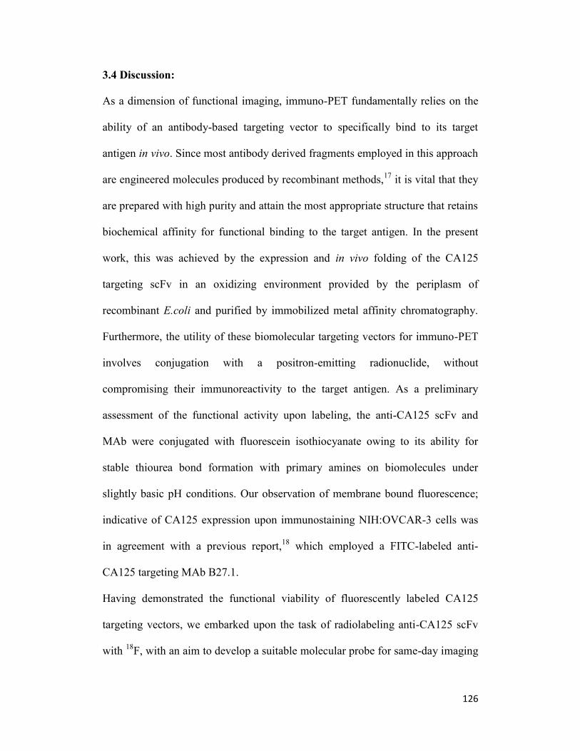
126
3.4 Discussion:
As a dimension of functional imaging, immuno-PET fundamentally relies on the
ability of an antibody-based targeting vector to specifically bind to its target
antigen in vivo. Since most antibody derived fragments employed in this approach
are engineered molecules produced by recombinant methods,17
it is vital that they
are prepared with high purity and attain the most appropriate structure that retains
biochemical affinity for functional binding to the target antigen. In the present
work, this was achieved by the expression and in vivo folding of the CA125
targeting scFv in an oxidizing environment provided by the periplasm of
recombinant E.coli and purified by immobilized metal affinity chromatography.
Furthermore, the utility of these biomolecular targeting vectors for immuno-PET
involves conjugation with a positron-emitting radionuclide, without
compromising their immunoreactivity to the target antigen. As a preliminary
assessment of the functional activity upon labeling, the anti-CA125 scFv and
MAb were conjugated with fluorescein isothiocyanate owing to its ability for
stable thiourea bond formation with primary amines on biomolecules under
slightly basic pH conditions. Our observation of membrane bound fluorescence;
indicative of CA125 expression upon immunostaining NIH:OVCAR-3 cells was
in agreement with a previous report,18
which employed a FITC-labeled anti-
CA125 targeting MAb B27.1.
Having demonstrated the functional viability of fluorescently labeled CA125
targeting vectors, we embarked upon the task of radiolabeling anti-CA125 scFv
with 18
F, with an aim to develop a suitable molecular probe for same-day imaging
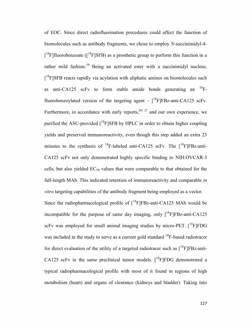
127
of EOC. Since direct radiofluorination procedures could affect the function of
biomolecules such as antibody fragments, we chose to employ N-succinimidyl-4-
[18
F]fluorobenzoate ([18
F]SFB) as a prosthetic group to perform this function in a
rather mild fashion.19
Being an activated ester with a succinimidyl nucleus,
[18
F]SFB reacts rapidly via acylation with aliphatic amines on biomolecules such
as anti-CA125 scFv to form stable amide bonds generating an 18
F-
fluorobenzoylated version of the targeting agent – [18
F]FBz-anti-CA125 scFv.
Furthermore, in accordance with early reports,20, 21
and our own experience, we
purified the ASU-provided [18
F]SFB by HPLC in order to obtain higher coupling
yields and preserved immunoreactivity, even though this step added an extra 23
minutes to the synthesis of 18
F-labeled anti-CA125 scFv. The [18
F]FBz-anti-
CA125 scFv not only demonstrated highly specific binding to NIH:OVCAR-3
cells, but also yielded EC50 values that were comparable to that obtained for the
full-length MAb. This indicated retention of immunoreactivity and comparable in
vitro targeting capabilities of the antibody fragment being employed as a vector.
Since the radiopharmacological profile of [18
F]FBz-anti-CA125 MAb would be
incompatible for the purpose of same day imaging, only [18
F]FBz-anti-CA125
scFv was employed for small animal imaging studies by micro-PET. [18
F]FDG
was included in the study to serve as a current gold standard 18
F-based radiotracer
for direct evaluation of the utility of a targeted radiotracer such as [18
F]FBz-anti-
CA125 scFv in the same preclinical tumor models. [18
F]FDG demonstrated a
typical radiopharmacological profile with most of it found in regions of high
metabolism (heart) and organs of clearance (kidneys and bladder). Taking into

128
consideration the data from micro-PET images and the analyzed time activity
curves, [18
F]FBz-anti-CA125 scFv clearly revealed a higher persistence in the
systemic circulation of both the tumor xenograft models used in this study; with
the effect being more pronounced in NIH:OVCAR-3 tumor-bearing mice.
Furthermore, a major proportion of the [18
F]FBz-anti-CA125 scFv tends to be
cleared hepatically in stark contrast with the expected in vivo
radiopharmacologically preferred renal route of clearance for radiotracers below
MW ~ 60 KDa as conformed to by the same radiotracer in SKOV3 tumor-bearing
animals. This unique observation may be attributed to the potential formation of
immunocomplexes between trace amounts of both [18
F]FBz-anti-CA125 scFv and
CA125 molecules that may be present as shed antigen in the bloodstream of
corresponding NIH:OVCAR-3 tumor-bearing animals. A similar observation of
potential immunocomplex formation with shed antigen within the first 90 minutes
of the radiotracer injection and subsequent clearance via the liver has been
previously reported by McQuarrie et al, dealing with the administration of the
parental anti-CA125 MAb-B43.13 formulated as a radiotracer using 99m
Tc for
radioimmunoscintigraphy in human patients.22
However, in that report an excess
dose of radioimmunoconjugate employed (2 mg of 99
mTc-MAb B43.13 per
patient) ensured tumor bioavailability of the tracer for use in
radioimmunoscintigraphy, despite immunocomplex formation.22, 23
Additionally, despite having good immunoreactivity and promising in vitro
binding capabilities, the scFv format appears to be greatly limited by its molecular
weight / size, which was initially proposed as its prime advantage for enhanced

129
tumor penetration and rapid systemic clearance in order to translate into optimal
tumor-to-background ratios to deliver high contrast images within a shorter period
of time. This has also been demonstrated in previous reports,25, 26 27
wherein the
monomeric scFv format becomes a victim of first pass clearance via the renal
route. An evaluation of the ideal size for tumor targeting agents by Wittrup et al,28
described scFv molecules as residing in the death-valley of their evaluation curve.
This has been attributed to the plausible explanation that scFv molecules are too
small to escape renal clearance and too big for rapid extravasation from blood
vessels carrying them to the tumor. Furthermore, a lack of sufficient avidity also
contributes to a loss of in vivo binding efficacy for the monomeric scFv.
Therefore, diabodies have been proposed and demonstrated to yield relatively
better in vivo tumor uptake of antibody fragments which are below the renal cut-
off for clearance.21, 29, 30
It is also noteworthy that although the scFv seemed to
follow its expected route for pharmacological clearance, the presence of a polar
hexa-histidine tag at its C-terminus and the overall positive charge of the
molecule owing to its amino acid composition may also be factors that contribute
to its re-absorption and retention in the kidneys.30, 31
Pre-dosing the animal
subjects with positively-charged amino acids such as poly-L-lysine (1.2 g/kg) as a
competitive strategy routinely practiced in the clinic for administration of peptide-
receptor radionuclide therapy (PRRT) or co-infusion of succinylated gelatin (150
mg/kg) reported for preclinical immuno-PET applications using nanobodies may
be options to help circumvent excessive renal trapping of the radiotracer and
concomitantly improve its tumor bioavailability.31
Nevertheless, these approaches

130
manifest side effects related to nausea and vomiting. Finally, despite similar
tumor-to-muscle ratios for the radiotracer used in both xenograft bearing mice,
there was a slightly higher and differential absolute uptake value of [18
F]FBz-anti-
CA125 scFv in NIH:OVCAR-3 tumors when compared with SKOV3 tumors.
This feature highlights the tumor targeting specificity of the scFv radiotracer
despite its aforementioned limitations for in vivo functioning.
Further engineering of the anti-CA125 scFv into a diabody is warranted in order
to improve avidity so that better tumor binding and favourable in vivo
radiopharmacological profiles for same-day imaging can be achieved.
Nonetheless, the anti-CA125 scFv can be employed as an analytical reagent for in
vitro and ex vivo analysis of samples being investigated for the presence of
CA125. It may also be applied to develop immunoaffinity resins for the isolation
of CA125 antigen for biomedical research applications.

131
3.5 Conclusions:
An scFv derived from MAb-B43.13 was produced by recombinant methods and
purified as a biochemically active protein capable of binding to target antigen –
CA125. The scFv was radiolabeled using [18
F]SFB as a prosthetic group to yield
[18
F]FBz-anti-CA125 scFv with retained immunoreactivity. In vivo application of
the [18
F]FBz-anti-CA125 scFv as a radiotracer in preclinical ovarian cancer
xenograft mice revealed a modest absolute tumor uptake in NIH:OVCAR-3
xenograft mice. [18
F]FBz-anti-CA125 scFv demonstrated remarkably different in
vivo biodistribution and radiopharmacological profiles between the test and
control tumors and also in comparison with [18
F]FDG. In summary, the present
work demonstrates the development of a CA125-targeted antibody-fragment
based molecular probe for same-day imaging of epithelial ovarian cancer via
immuno-PET. This study demonstrates the feasibility of prosthetic group
mediated radiofluorination of biomolecules such as antibody fragments and its
subsequent use for molecular imaging of EOC. Moreover, the limitations of using
such a vector in a setting that involves circulating shed antigens, which may be
easier targets for antibody fragment binding at early time points in PET imaging,
are exemplified through this work. Clearly, a monomeric scFv is not a suitable
molecule of choice as a radiotracer for oncologic imaging of solid tumors.

132
3.6 References:
1. Knowles SM, Wu AM: Advances in immuno-positron emission tomography:
antibodies for molecular imaging in oncology. J Clinical Oncol 2012, 30:3884-
3892.
2. Wu AM: Engineered antibodies for molecular imaging of cancer. Methods 2014,
65:139-147.
3. Holliger P, Hudson PJ: Engineered antibody fragments and the rise of single
domains. Nat Biotechnol 2005, 23:1126-1136.
4. Wu AM, Yazaki PJ: Designer genes: recombinant antibody fragments for
biological imaging. Q J Nucl Med 2000, 44:268-283.
5. Wu AM: Antibodies and antimatter: the resurgence of immuno-PET. J Nucl Med
2009, 50(1):2-5.
6. Wu AM, Olafsen T: Antibodies for molecular imaging of cancer. Cancer J 2008,
14: 191-197.
7. Davis HM, Zurawski VR, Jr., Bast RC, Jr., Klug TL: Characterization of the CA
125 antigen associated with human epithelial ovarian carcinomas. Cancer Res
1986, 46:6143-6148.
8. Scholler N, Urban N: CA125 in ovarian cancer. Biomark Med 2007, 1:513-523.
9. Capstick V, Maclean GD, Suresh MR, Bodnar D, Lloyd S, Shepert L, Longenecker
BM, Krantz M: Clinical evaluation of a new two-site assay for CA125 antigen. Int
J Biol Markers 1991, 6:129-135.
10. Nustad K, Bast RC, Jr., Brien TJ, Nilsson O, Seguin P, Suresh MR, Saga T,
Nozawa S, Bormer OP, de Bruijn HW et al: Specificity and affinity of 26
monoclonal antibodies against the CA 125 antigen: first report from the ISOBM
TD-1 workshop. International Society for Oncodevelopmental Biology and
Medicine. Tumour Biol 1996, 17:196-219.

133
11. Nap M, Vitali A, Nustad K, Bast RC, Jr., O'Brien TJ, Nilsson O, Seguin P, Suresh
MR, Bormer OP, Saga T et al: Immunohistochemical characterization of 22
monoclonal antibodies against the CA125 antigen: 2nd report from the ISOBM
TD-1 Workshop. Tumour Biol 1996, 17:325-331.
12. Berek JS: Immunotherapy of ovarian cancer with antibodies: a focus on
oregovomab. Expert Opin Biol Ther 2004, 4:1159-1165.
13. Iagaru AH, Mittra ES, McDougall IR, Quon A, Gambhir SS: 18F-FDG PET/CT
evaluation of patients with ovarian carcinoma. Nucl Med Commun 2008, 29:1046-
1051.
14. Wang WW, Das D, McQuarrie SA, Suresh MR: Design of a bifunctional fusion
protein for ovarian cancer drug delivery: single-chain anti-CA125 core-streptavidin
fusion protein. Eur J Pharm Biopharm 2007, 65:398-405.
15. Noujaim AA, Baum RP, Sykes TR, Sykes CJ, Hertel A, Niesen, A, et al.
Monoclonal antibody B43.13 for immunoscintigraphy and immunotherapy of
ovarian cancer. In Current Tumor Diagnosis: Applications, Clinical Relevance,
Trends. Edited by Klapdor, R. Munich: W Zuckschwerdt Verlag; 1994; 823–829.
16. Mading P, Fuchtner F, Wust F: Module-assisted synthesis of the bifunctional
labelling agent N-succinimidyl 4-[(18)F]fluorobenzoate ([(18)F]SFB). Appl Radiat
Isot 2005, 63:329-332.
17. Olafsen T, Wu AM: Antibody vectors for imaging. Seminars in nuclear medicine
2010, 40:167-181.
18. Xiao Z, McQuarrie SA, Suresh MR, Mercer JR, Gupta S, Miller GG: A three-step
strategy for targeting drug carriers to human ovarian carcinoma cells in vitro. J
Biotechnol 2002, 94:171-184.

134
19. Wester HJ, Hamacher K, Stocklin G: A comparative study of N.C.A. fluorine-18
labeling of proteins via acylation and photochemical conjugation. Nucl Med Biol
1996, 23:365-372.
20. Vaidyanathan G, Zalutsky MR: Improved synthesis of N-succinimidyl 4-
[18
F]fluorobenzoate and its application to the labeling of a monoclonal antibody
fragment. Bioconjug Chem 1994, 5:352-356.
21. Garg PK, Garg S, Zalutsky MR: Fluorine-18 labeling of monoclonal antibodies
and fragments with preservation of immunoreactivity. Bioconjug Chem 1991, 2:44-
49.
22. McQuarrie SA, Riauka T, Baum RP, Sykes TR, Noujaim AA, Boniface G,
MacLean GD, McEwan AJ: The effects of circulating antigen on the
pharmacokinetics and radioimmunoscintigraphic properties of 99m
Tc labelled
monoclonal antibodies in cancer patients. J Pharm Pharm Sci 1998, 1:115-125.
23. McQuarrie SA, Baum RP, Niesen A, Madiyalakan R, Korz W, Sykes TR, Sykes
CJ, Hor G, McEwan AJB, Noujaim AA: Pharmacokinetics and radiation dosimtery
of 99
Tcm – labelled monoclonal antibody B43.13 in ovarian cancer patients. Nucl
Med Commun 1997, 18:878-886.
24. Williams LE, Wu AM, Yazaki PJ, et al. Numerical selection of optimal tumor
imaging agents with application to engineered antibodies. Cancer Biother
Radiopharm 2001;16(1): 25-35.
25. Cai W, Olafsen T, Zhang X, Cao Q, Gambhir SS, Williams LE, Wu AM, Chen X:
PET imaging of colorectal cancer in xenograft-bearing mice by use of an 18F-
labeled T84.66 anti-carcinoembryonic antigen diabody. J Nucl Med 2007, 48:304-
310.

135
26. Olafsen T, Sirk SJ, Olma S, Shen CK, Wu AM: ImmunoPET using engineered
antibody fragments: fluorine-18 labeled diabodies for same-day imaging. Tumour
Biol 2012, 33:669-677.
27. Wittrup KD, Thurber GM, Schmidt MM, Rhoden JJ: Practical theoretic guidance
for the design of tumor-targeting agents. Methods enzymol 2012, 503:255-268.
28. Schneider DW, Heitner T, Alicke B, Light DR, McLean K, Satozawa N, Parry G,
Yoo J, Lewis JS, Parry R: In vivo biodistribution, PET imaging, and tumor
accumulation of 86Y- and 111In-antimindin/RG-1, engineered antibody fragments
in LNCaP tumor-bearing nude mice. J Nucl Med 2009, 50:435-443.
29. Eder M, Knackmuss S, Le Gall F, et al. 68Ga-labelled recombinant antibody
variants for immuno-PET imaging of solid tumours. Eur J Nucl Med Mol Imaging
2010;37(7): 1397-407.
30. Vegt E, de Jong M, Wetzels JF, Masereeuw R, Melis M, Oyen WJ, Gotthardt M,
Boerman OC: Renal toxicity of radiolabeled peptides and antibody fragments:
mechanisms, impact on radionuclide therapy, and strategies for prevention. J Nucl
Med 2010, 51:1049-1058.
31. D'Huyvetter M, Vincke C, Xavier C, Aerts A, Impens N, Baatout S, De Raeve H,
Muyldermans S, Caveliers V, Devoogdt N et al: Targeted Radionuclide Therapy
with a 177
Lu-labeled Anti-HER2 Nanobody. Theranostics 2014, 4:708-720.

136
4
Immuno-PET of Epithelial Ovarian Cancer:
Harnessing the potential of CA125 for
non-invasive imaging3
3A version of this chapter will be submitted to the European Journal of Nuclear
Medicine and Molecular Imaging Research: Sharma SK, Wuest M, Wang M,
Glubrecht D, Andrais B, Lapi SE, Suresh MR, Wuest F.
Immuno-PET of Epithelial Ovarian Cancer: Harnessing the potential of CA125
for non-invasive imaging

137
4.1 Introduction
Despite its relative rarity in the general population and the availability of standard
treatment through surgical intervention and platinum chemotherapy, epithelial
ovarian cancer (EOC) continues to be the most insidious and lethal gynecologic
malignancy in the western world. The Surveillance Epidemiology and End
Results (SEER) program estimated 22,240 new cases of ovarian cancer in 2014
alongside 14,270 deaths resulting from this disease in the United States alone.1 A
combination of the asymptomatic nature of this malignancy and high incidence of
recurrence results in a low 5-year survival rate of 44%.1
Cancer Antigen 125 (CA125) is a high molecular weight mucinous glycoprotein
overexpressed on the membrane of EOC cells.2-4
It has long served as a standard
of care serum biomarker for ovarian cancer surveillance although its presence is
also reported in some non-gynecologic malignancies and benign conditions such
as pregnancy, menstruation, endometriosis, liver disease and congestive heart
failure.3, 4
Post-treatment elevation of serum CA125 level in EOC patients serves
as an indicator of progressive disease and finds greatest use in the management of
patients with documented evidence of ovarian cancer.3-5
However, a normal
CA125 serum titer does not always imply an absence of the disease. An estimated
50% of patients with normal levels of CA125 post-chemotherapy have small
volumes of active disease at second-look surgery, and a large portion of this
population succumbs to recurrence from the disease.6 Presently, serial CA125
levels in patients are tested using ELISA-based immunoassays. Despite their
indicative advantage, such assays can be far from delivering a true representation
138
of the in vivo pathophysiological pattern, and may be limited in their capabilities
for early detection of ovarian neoplasms.
From a diagnostic standpoint, it is known that radiological progression precedes
clinical progression by 2-3 months and when combined with CA125, it can be
preceded by 5 months.5 Among contemporary imaging modalities, [
18F]FDG-PET
(18
F-Fluorodeoxyglucose positron emission tomography) has shown highest
sensitivity and accuracy in detecting recurrent ovarian cancer whilst simultaneous
investigations with CT, MRI and ultrasound yielded negative or equivocal
results.7-9
Over the years, several antibodies have been developed to target CA125.10-13
Among those reported, MAb-B43.13 has seen the most progress towards clinical
translation for the immunotherapy of EOC.14-16
More recently, immuno-PET has
emerged as a strategy that unifies the specificity of antibody-based targeting with
the sensitivity for detection by positron emitting radionuclides via PET in order to
render a superlative diagnostic potential.17
We hypothesized that in vivo imaging of CA125 expression while it is present on
the surface of ovarian carcinomas, could lead to a much earlier detection of
recurrent disease.
The goal of this study was to develop a targeted non-invasive method for the in
vivo detection of CA125 present on ovarian carcinomas. This would allow the use
of CA125 as a biomarker for early detection of ovarian cancer by providing a
more accurate in vivo evaluation of tumor load and residual disease for effective
disease management. Previous research with MAb B43.13 and the recent reports

139
of antibody-based radiotracers targeting biomarkers of similar pathophysiological
nature in other cancers,18, 19
prompted us to develop an immuno-PET strategy for
the in vivo imaging of EOC. Towards this goal, anti-CA125 MAb (MAb-B43.13)
and its derivative single chain Fragment variable (scFv-B43.13) were radio-
labeled with positron emitter 64
Cu (t1/2 12.7 h) and analyzed in vitro and in vivo
using CA125 overexpressing NIH:OVCAR-3 cells and antigen negative SKOV3
cells in preclinical EOC xenograft mouse models.

140
4.2 Materials and Methods
4.2.1 Expression and purification of MAb-B43.13 and its derivative scFv:
Anti-CA125 murine monoclonal antibody was purified from B43.13 hybridoma15
by protein G affinity chromatography (Sigma, P-7700). Anti-CA125 scFv was
produced after deletion of the core-streptavidin from previously described fusion
constructs.20
The scFv was expressed as a C-terminal hexa-histidine tagged
protein in E.coli Rosetta 2(DE3) (Novagen, 71400) and purified by immobilized
metal affinity chromatography using a TALON® superflow resin (Clontech,
635506).
4.2.2 Cell lines and culture conditions: Ovarian cancer cell lines NIH:OVCAR-
3 and SKOV3 were obtained from ATCC and used for in vitro and in vivo studies.
Cells were cultured in DMEM-F12 medium supplemented with 10% v/v fetal
bovine serum (Life Technologies, 12483-020), 50 IU/mL penicillin, 50 μg/mL
streptomycin (Life Technologies, 15140-122). NIH:OVCAR-3 cells were
additionally supplemented with 7 μg/mL recombinant human insulin (SAFC
Biosciences, 91077C). Cells were cultured using sterile techniques and grown in a
37°C incubator providing humidified atmosphere of 5% CO2 in air.
4.2.3 Characterization of CA125 targeting vectors:
MAb-B43.13 and scFv-B43.13 were characterized for binding to the target
antigen by immunoblotting, immunofluorescence and flow cytometry. FITC-
labeled MAb-B43.13 was also tested for uptake in live cells over a period of 48 h.

141
4.2.3.1 Western blotting: 7.5 x 10⁶
NIH:OVCAR-3 and SKOV3 cells were lysed
with CelLytic™M (Sigma, C2978). Cell lysates were electrophoresed on a 4-15%
Mini-PROTEAN® TGX™ precast gel (Bio-Rad, #456-1086) and transferred to a
Trans-Blot nitrocellulose membrane (Bio-Rad, 162-0115). The membranes were
probed separately to evaluate MAb versus scFv binding with target antigen –
CA125 in cell lysates. The blots were blocked for 45 min with 5% non-fat dry
milk (Carnation) in PBS having 0.1% Tween-20 (PBST). MAb-B43.13 (3
mg/mL), mouse anti-β actin IgG (Sigma) and scFv-B43.13 (2 mg/mL) were used
as primary antibodies (1: 5000) to probe the blots for 1 h at room temperature.
Goat anti-mouse HRP conjugate (Sigma, A4416) was used as secondary antibody
(1: 5000) to probe the blot against anti-CA125 MAb and mouse anti-β actin IgG
(Sigma, A1978) for 1 h at room temperature. 6X His MAb-HRP conjugate
(Clontech, 631210) was used as secondary antibody (1:5000) to probe against
scFv-B43.13 for 1 h at room temperature. The blots were washed with PBST and
developed on Amersham Hyperfilm ECL (GE Healthcare, 28906839) using
Amersham ECL Plus Western Blotting Detection Reagents (GE Healthcare,
RPN2132).
4.2.3.2 Fluorescent labeling of anti-CA125 MAb/scFv: 1 mg each of MAb-
B43.13 and scFv-B43.13 (2 mg/mL) were fluorescently labeled using the Pierce®
FITC antibody labeling kit (Thermo Scientific, 53027) according to the
manufacturer’s instructions.

142
4.2.3.3 Flow Cytometry: 1.5 x 10⁶ NIH:OVCAR-3 cells were harvested by
trypsinization, rinsed twice with FACS buffer (PBS with 0.5% heat inactivated
FBS, 2mM EDTA, 0.05% sodium azide) and resuspended by gentle tapping in
~100 µL FACS buffer. 10 µg of MAb-B43.13 or scFv-B43.13 was incubated with
the cell suspension for 30 min at room temperature. Cells were rinsed twice in
FACS buffer and incubated for 30 min with 4 µg of Alexa-fluor® 488 goat anti-
mouse antibody (Life Technologies, A-11001) for the MAb samples and 4 µg of
Penta·His Alexa Fluor 488 conjugate (Qiagen, 35310) for scFv samples. Cells
were rinsed twice with FACS buffer and analyzed by flow cytometry on a BD
FACS Calibur with 10,000 events gated per sample. A dengue virus targeting IgG
(12A1) and a hexa-histidine tagged anti-RANK receptor scFv were used as
isotype controls for the experiments. Negative controls included unstained
NIH:OVCAR-3 cells and cells incubated with Alexa fluor 488 conjugated
antibodies alone.
4.2.3.4 Immunofluorescence: NIH:OVCAR-3 and SKOV3 cells were plated
onto glass coverslips in 35-mm tissue culture dishes (100,000 cells/2 mL
medium/dish) and incubated at 37C for 48 h. The cells were rinsed with PBS and
fixed in methanol for 30 min at -20C. The fixed cells were incubated in 5% non-
fat dry milk (Carnation) in PBS for 30 min and immunostained separately for 1 h
with FITC-labeled versus un-labeled MAb-B43.13 and scFv-B43.13. The un-
labeled MAb and scFv samples were indirectly stained with corresponding Alexa
fluor 488 labeled secondary antibodies as used in flow cytometry experiments.

143
MAb-B43.13 and scFv-B43.13 (2 mg/mL) were used at 1:250 dilution followed
by 1:500 dilutions of secondary antibodies (2 mg/mL) in PBS containing 5% non-
fat dry milk. Appropriate blank and control samples were included in the
experiments. Antibody incubations were followed by three rinses with PBST for
10 min each. Coverslips were mounted on microscopy slides (Fisherbrand) using
Mowiol® mounting medium (Calbiochem, 475904) supplemented with DAPI (50
µg/mL). Immunofluorescence was observed through a Zeiss Plan Apochromat
40X/1.3 Oil DIC M27 lens on a confocal laser scanning microscope (Zeiss LSM
710) and images were analyzed using Zen 2011 software. Separately, 30 µg of
FITC-labeled anti-CA125 MAb was added to NIH:OVCAR-3 cells grown on
coverslips and allowed to incubate under standard cell culture conditions over a
period of 48 h to study uptake in live cells. Coverslips were washed and mounted
on glass slides for analysis by confocal microscopy at 0.5, 1, 4, 12, 24 and 48 h
time points.
4.2.4 NOTA Functionalization: p-SCN-Bn-NOTA [S-2-(4-
Isothiocyanatobenzyl)-1,4,7-triazacyclononane-1,4,7-triacetic acid] (Macrocyclics
Inc, B-605) was conjugated to MAb-B43.13 and scFv-B43.13 as a bi-functional
chelator for 64
Cu radiolabeling. Briefly, a 6 molar excess of p-SCN-Bn-NOTA in
DMSO was added to MAb/scFv-B43.13 in 0.1 M sodium bi-carbonate buffer pH
8.5 and allowed to react for 1 h at 37C. NOTA-functionalized MAb/scFv-B43.13
was purified from un-conjugated NOTA while simultaneously achieving buffer
exchange into 0.25 M sodium acetate pH 5.5 by using an Econo-Pac®
10DG

144
desalting column (Bio-Rad, 732-2010). The number of NOTA molecules
conjugated per MAb and scFv was determined by MALDI-ToF MS analysis and
according to the method described by Cooper et al.21
4.2.5 Determination of Bi-functional Chelates per MAb/scFv:
NOTA conjugated anti-CA125 MAb and scFv were analyzed by MALDI-ToF-
MS at the Institute for Biomolecular Design, University of Alberta, to determine
the number of bi-functional chelate molecules per targeting vector. 1 µL of each
sample was mixed with 1 µL of sinapic acid (10 mg/mL in 50% acetonitrile:water
and 0.1% trifluoroacetic acid). 1 µL of the sample/matrix solution was then
spotted onto a stainless steel target plate and allowed to air dry. All mass spectra
were obtained using a Bruker Ultraflex MALDI-ToF/ToF (Bruker Daltonic
GmbH). Ions were analyzed in positive mode and external calibration was
performed by use of a standard protein mixture.
An isotopic dilution assay as per the method of Cooper et al 21
was also performed
to verify data obtained from MALDI-ToF analysis of immunoconjugates.
Percentage radiolabeling efficiency was plotted as a function of different moles of
copper added to a fixed concentration (moles) of MAb/scFv in order to determine
the average number of bi-functional chelate molecules conjugated per targeting
vector.
4.2.6 Preparation of CA125 targeting radioimmunoconjugates:
4.2.6.1 General: All glassware was rinsed with ultra-pure HCl (Fisherbrand,
A508-P500). Trace metals basis ultra-pure chemicals for buffer preparations were

145
purchased from Sigma Aldrich. All buffer solutions were treated with
biotechnology grade Chelex 100 (Bio-Rad, 143-2832).
4.2.6.2 64
Cu labeling of NOTA-functionalized MAb/scFv: 64
Cu was produced
via a 64
Ni(p,n)64
Cu nuclear reaction on a CS-15 biomedical cyclotron at
Washington University, St. Louis, USA as previously described.22
85 MBq of
64Cu-acetate was added to 100 µg NOTA-functionalized MAb/scFv-B43.13 and
allowed to react at 30°C, 550 rpm for 1 h in a thermomixer. 1 mM EDTA was
added to quench the reaction over 10 min. 64
Cu-NOTA-MAb-B43.13 and 64
Cu-
NOTA-scFv-B43.13 radioimmunoconjugates were purified using an Econo-Pac®
10DG desalting column pre-equilibrated with 0.25 M sodium acetate, pH 5.5 used
as the eluent. 350 μL elution fractions were collected from the column and
radioactivity was measured on an Atomlab 400 dose calibrator (Biodex Inc). High
specific activity radioimmunoconjugates were used for in vitro and in vivo
radiopharmacological experiments. 64
Cu-labeled 12A1 MAb was used as an
isotype control for in vivo experiments.
4.2.7 Functional characterization of CA125 targeting
radioimmunoconjugates:
64Cu-labeled anti-CA125 MAb and scFv were evaluated for target vs non-target
cell uptake over a period of 2 h in an in vitro assay. Briefly, NIH:OVCAR-3 and
SKOV3 cells were allowed to incubate for various time intervals with a fixed
amount (100 KBq) of the aforementioned radioimmunoconjugates, then rinsed

146
and counted for cell-bound radioactivity. Immunoreactivity of the
radioimmunoconjugates was assessed as per the method of Lindmo et al.23
Direct
binding studies were performed to determine the dissociation constant (KD) and
maximum number of binding sites (Bmax) on target cells.
4.2.7.1 Quality control of purified radioimmunoconjugates:
64Cu-NOTA-MAb-B43.13 and
64Cu-NOTA-scFv-B43.13 were purified by size
exclusion chromatography using a pre-packed desalting column. The
radioimmunoconjugates eluted out in the earlier fractions owing to their
molecular size compared with the EDTA chelated 64
Cu from the reaction mixture.
1 µL of the quenched radiolabeling reaction mixtures and selected fractions from
size exclusion purification were spotted on ITLC-SG strips, allowed to dry and
separate using 10mM EDTA pH 5.5 as the eluent and analyzed on a radio-TLC
scanner (Bioscan AR-2000). As per previously established results in such
experiments for quality control, 64
Cu-labeled anti-CA125 MAb and scFv
remained at the baseline and any un-conjugated or EDTA-chelated 64
Cu moved
with the solvent front on ITLC strips. Additionally, 15 μL of each elution fraction
was electrophoresed in a 12% SDS-PAGE gel under non-reducing conditions and
evaluated by autoradiography on a BAS-5000 phoshorimager (Fujifilm).
4.2.7.2 Stability of radioimmunoconjugates:
64Cu-labeled MAb and scFv was analyzed by incubating 5 MBq of purified
radioimmunoconjugates in 500 µL human AB type serum at 37°C for 48 h.

147
Samples were analyzed by size exclusion HPLC at 0, 24 and 48 h on a Biosep
SEC-S2000 column (Phenomenex, 00F-2145-B0), using an isocratic mobile phase
of 0.1 M phosphate buffer at a flow rate of 1 mL/min and by ITLC using 10 mM
EDTA pH 5.5 as the eluent or via ITLC.
4.2.7.3 SOD Challenge experiments:
64Cu-labeled MAb (5MBq) was challenged with a 1000 fold excess of Superoxide
dismutase (Sigma, S8160) and allowed to incubate at 37°C over a period of 48 h.
Samples were analyzed for trans-chelation by size exclusion chromatography on a
Shimadzu analytical HPLC using the conditions mentioned above.
4.2.7.4 Cell Uptake Studies: NIH:OVCAR-3 and SKOV3 cells were seeded to
obtain 250,000 cells per well in 12-well tissue culture plates. Prior to the uptake
experiment, growth media was removed and cells were rinsed twice with PBS and
incubated in Krebs buffer at 37°C for 1 h. The radioimmunoconjugate (100 KBq)
was added to each well except those assigned to measure background activity
alone. Cell uptake was terminated at 5, 10, 15, 30, 60, 90, 120 min by adding ice
cold Krebs buffer and rinsing the wells twice to wash away any un-bound
radioimmunoconjugates prior to lysing cells with RIPA buffer. The cell lysates
were transferred to scintillation vials and measured for radioactivity using a γ-
counter (Wizard2®
2480 Automatic Gamma Counter, Perkin-Elmer, Canada).
Protein levels were quantified using a Pierce™ BCA protein assay kit according
to the manufacturer's recommendations. Cell uptake levels were normalized to

148
percent of the total amount of radioactivity per milligram of protein (%
radioactivity / mg protein) and plotted as a function of time. All experiments were
performed in triplicates.
4.2.7.5 Lindmo assay for determination of immunoreactive fraction:
NIH:OVCAR-3 cells were aliquoted into microcentrifuge tubes at concentrations
of 5, 4, 3, 2.5, 2, 1.5 and 0.5 x 10⁶ cells in 500 μL PBS (pH 7.4). A stock solution
of the radioimmunoconjugate was prepared in PBS (pH 7.4) supplemented with
1% BSA such that 50 μL of this solution contained 20,000 cpm which was then
uniformly aliquoted into individual tubes. This setup was allowed to incubate for
1 h at room temperature on a platform mixer. Thereafter, cells were pelleted by
centrifugation (5,000 rpm for 3 mins), rinsed twice with ice cold PBS and the
supernatant was aspirated prior to measuring cell-bound radioactivity on a γ-
counter (Wizard2®
2480 Automatic Gamma Counter, Perkin-Elmer, Canada).
Radioactivity data obtained thereof was background corrected and compared with
counts from the total activity control samples from the experiment. The
immunoreactive fraction was determined by performing a linear regression
analysis on a double-inverse plot of (total/bound) activity versus normalized cell
concentration. All data was obtained in triplicates and no weighting was applied.
4.2.7.6 Direct binding studies were performed to determine the dissociation
constant (KD) and maximum number of binding sites (Bmax) by incubating 0.5 x
106
cells with increasing concentrations (0.2 nM – 19.2 nM) of 64
Cu-labeled anti-

149
CA125 MAb in 250 μl PBS supplemented with 0.2% bovine serum albumin. To
evaluate non-specific binding, a corresponding experimental set was prepared
with 100 nM of un-labeled immunoconjugate added to the reaction in order to
provide this reagent in 5–500 fold molar excess to the radioimmunoconjugate
concentrations used. This setup was allowed to incubate at room temperature for 1
h with gentle shaking. Cells were pelleted by centrifugation at 1200 rpm for 5 min
followed by 2 rounds of rinsing with ice cold PBS. Cell-bound radioactivity was
measured using a γ-counter. Data was plotted using a non-linear regression fit
analysis on GraphPad Prism6.
4.2.8 Animal Imaging: Small animal PET experiments were performed on a
MicroPET® R4 or INVEON PET scanner (Siemens Preclinical Solutions). Mice
were anesthetized by inhalation of isoflurane (Baxter Healthcare) in 40%
oxygen/60% nitrogen, 1 L/min, while maintaining body temperature at 37°C
throughout the experiment. 6–10 MBq of high specific activity
radioimmunoconjugate (~30-40 µg) in 150 – 200 µL of 0.25M sodium acetate
(pH 5.5) was administered intravenously via a tail vein catheter. For blocking
experiments, 1 mg of unmodified anti-CA125 MAb was administered
intraperitoneally 24 h prior to injection of the 64
Cu-labeled
radioimmunoconjugates. In separate animals, 8-10 MBq of 64
Cu-labeled 12A1
IgG was administered via tail injection to evaluate non-specific uptake. Whole
body PET data was acquired by performing static scans for each animal at 24 h p.i
and 48 h p.i. Data acquisition continued for 60 min in 3D list mode. All PET

150
image files were reconstructed using the maximum a posteriori (MAP) algorithm.
The image files were further processed using the ROVER v 2.0.51 software (ABX
GmbH, Radeberg, Germany). Masks for defining 3D regions of interest (ROI)
were set and the ROIs were defined by thresholding. Standardized uptake values
[SUV = (activity/ml tissue) / (injected activity/body weight), mlg-1
] were
calculated for each ROI. Data are expressed as means ± SEM from n investigated
animals.
4.2.9 Animal Imaging: All experiments were carried out according to guidelines
of the Canadian Council on Animal Care (CCAC) and approved by the local
animal care committee of the Cross Cancer Institute, Edmonton. Six weeks old
BALB/c nude female mice were obtained from Charles River labs (Quebec,
Canada). The animals were housed in ventilated cages and provided food and
water ad libitum. NIH:OVCAR-3 tumors were induced on the left shoulder by
two subsequent subcutaneous injections of 15 x 106
and 10 x 106
cells in a 300 µL
suspension of 1:1 mixture of PBS and matrigel (BD Biosciences). The second
injection of cells was administered at the same site within 7-10 days.
NIH:OVCAR-3 tumors grew for 6-8 weeks before reaching suitable tumor sizes
of 150 – 200 mm3. SKOV3 tumors were induced on the left shoulder by a single
subcutaneous injection of 5 x 10⁶ cells in PBS and were grown for about 2-3
weeks before achieving similar tumor sizes.

151
4.2.10 Ex vivo analyses:
NIH:OVCAR-3 tumor-bearing animal was injected with 4.55 MBq of 64
Cu-
labeled anti-CA125 MAb via tail vein. At 24 h p.i the animal was euthanized by
cervical dislocation. The tumor was submerged into O.C.T (Optimum Cutting
Temperature) medium and flash frozen using a bath of dry ice cooled methanol. 7
micron slices were cut on a Leica CM 1850 cryostat. The tissue sections were
further analyzed by autoradiography, immunofluorescence and
immunohistochemistry.
4.2.10.1 Autoradiography: Tissue sections were placed into a BAS-Cassette
(Fujifilm) and exposed to a phosphorimaging plate (Fujifilm) for 15 h at room
temperature. The images were developed on a BAS-5000 reader (Fuji Photo Film)
and analyzed with Adaptive Image Deconvolution Algorithm (AIDA) Image
Analyzer v.450 software.
4.2.10.2 Immunofluorescence: Upon thawing, tissue sections were fixed with
formalin for 30 min. The fixed sections were blocked overnight at 4°C using 0.12
mg/mL unconjugated goat anti-mouse Fab fragment (Jackson Immunoresearch) in
0.5% fish skin gelatin (Sigma) pH 7.4, supplemented with 0.1% triton X-100. The
sections were rinsed 3x with Tris-buffered saline supplemented with 0.5% Tween
20 (TBST) for 5 min each. B43.13 MAb was used as primary antibody (1: 6000)
in Dako antibody diluent and allowed to incubate overnight at 4°C. The sections
were rinsed 3x with TBST and incubated with Alexa-fluor® 488 goat anti-mouse

152
antibody (Life Technologies) used as secondary antibody (1:400) in Dako
antibody diluent for 2 h at room temperature. After three washes with TBST, the
sections were rinsed with water and counterstained using Hoechst H33342 (2
μg/mL) for 5 min. The sections were rinsed and mounted under a coverslip using
FluorSave (Calbiochem). The slides were analyzed with a Zeiss Plan Apochromat
10X/0.45 na lens on a confocal laser scanning microscope (Zeiss LSM 710) using
Z-stack for image acquisition. The images were registered using Zen 2011
software (ZEISS) and further processed with Adobe Photoshop CS6.
4.2.10.3 Immunohistochemistry: To analyze the samples for
immunohistochemistry, the same procedures for treatment, fixation, blocking and
probing of tissue with primary antibody were followed. Dako EnVision™
+ HRP
conjugated anti-mouse antibody was used as a secondary antibody and no
counterstain was employed. The sections were observed with a Zeiss Fluar
2.5X/0.12 na lens on an Axioskop 2 Plus microscope. Images were processed and
analyzed on AxioVision 4.7 (ZEISS) and further processed with Adobe
Photoshop CS6.
4.2.11 CA125 serum levels: A CA125 (Human) ELISA kit (Abnova, KA0205)
was used to analyze CA125 levels in xenograft mouse serum. The kit is based on
the principle of solid-phase enzyme-linked immunosorbent assay. Analysis was
performed according to the manufacturer’s instructions.

153
4.2.12 Statistical Analysis:
All data are expressed as means ± SEM. Graphs were constructed using GraphPad
Prism 4.0 (GraphPad Software). Where applicable, statistical differences were
tested by un-paired Student’s t-test and were considered significant for p < 0.05.

154
4.3 Results
4.3.1 Isolation, characterization and modification of CA125 targeting
vectors:
CA125 targeting vectors were purified in yields of 6 mg/L for MAb-B43.13 and
0.7 mg/L for the scFv (Fig. 4.1A). Analysis by western blot (Fig. 4.1B) and
immunofluorescence staining (Fig. 4.2) ascertained the following: a) expression
of CA125 in NIH:OVCAR-3 cells; b) absence of CA125 expression in SKOV3
cells; c) binding of MAb and scFv to target antigen CA125. Isothiocyanate-based
labeling of anti-CA125 MAb and scFv with fluorescein yielded 2 moles of FITC
per mole of MAb and scFv. Flow cytometry analysis with FITC-labeled MAb-
B43.13 and scFv-B43.13 demonstrated preserved immunoreactivity by binding to
CA125 antigen expressed on the membrane of NIH:OVCAR-3 cells (Fig. 4.3).
FITC-labeled anti-CA125 MAb remained membrane bound over a period of 48 h
with minimal cellular internalization observed upon incubation with
NIH:OVCAR-3 cells maintained in culture over the period of analysis (Fig. 4.4).

155
Figure 4.1: Purification and biochemical characterization of anti-CA125 MAb and scFv.
A) Representative images of purified anti-CA125 MAb (148 KDa) and scFv (28 KDa)
upon SDS-PAGE under non-reducing conditions. B) Western blots of SKOV3 (CA125-
negative) and NIH:OVCAR-3 (CA125-positive) cells with anti-CA125 MAb, and anti-β
actin antibody (left half of blot); anti-CA125 scFv and anti-β actin antibody (right half of
blot).

156
Figure 4.2: Functional characterization of anti-CA125 vectors. Confocal microscopy
images of immunofluorescence studies; a-b) NIH:OVCAR-3 cells stained with unlabeled
versus FITC-labeled anti-CA125 MAb. Goat anti-mouse Alexa fluor 488 IgG (GAM-
A488) was used as secondary antibody; c-d) NIH:OVCAR-3 cells stained with unlabeled
versus FITC-labeled anti-CA125 scFv. Penta-His Alexa fluor 488 antibody was used as
secondary antibody; e) Unstained NIH:OVCAR-3 cells (Blank); f) NIH:OVCAR-3 cells
stained with GAM-A488 antibody (Negative control); g) NIH:OVCAR-3 cells stained
with Penta-His Alexa fluor 488 antibody (Negative control); h) NIH:OVCAR-3 cells
stained with MAb 12A1 and GAM-A488 antibody (Isotype control); i-j) SKOV3 cells
stained with unlabeled versus FITC-labeled anti-CA125 MAb; k-l) SKOV3 cells stained
with unlabeled versus FITC-labeled anti-CA125 scFv. Scale bar represents 10 µm.
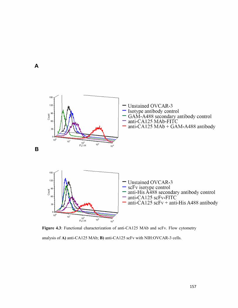
157
Figure 4.3: Functional characterization of anti-CA125 MAb and scFv. Flow cytometry
analysis of A) anti-CA125 MAb; B) anti-CA125 scFv with NIH:OVCAR-3 cells.
A
B

158
Figure 4.4: Antibody internalization assay: Confocal microscopy images of
NIH:OVCAR-3 cells stained with FITC-labeled MAb-B43.13. Minimal internalization of
anti-CA125 antibody was observed over a period of 48 h.

159
4.3.2 Development and functional assessment of CA125 targeting
radioimmunoconjugates:
MALDI-ToF analyses and isotopic dilution assays revealed an average of 2.4
NOTA molecules conjugated per MAb and 1.9 NOTA molecules conjugated per
scFv (Figs. 4.5 and 4.6). 64
Cu-labeling and purification of radioimmunoconjugates
provided isolated radiochemical yields of 65 ± 8 % (n= 14) with specific activity
of 296 ± 37 MBq/mg for 64
Cu-NOTA-MAb-B43.13 and 56 ± 14 % (n= 9) with
specific activity of 122 ± 44 MBq/mg for 64
Cu-NOTA-scFv-B43.13.
Radiochemical purity of the isolated radioimmunoconjugates was > 99% as
analyzed from phosphorimages of electrophoresed fractions (Figs. 4.8) and ITLC
(Fig. 4.9). SEC-purified 64
Cu-NOTA-MAb-B43.13 was > 95% stable in human
AB type serum over 48 h (Fig 4.10) and demonstrated highly specific binding to
CA125 expressed on NIH:OVCAR-3 cells with virtually no binding to SKOV3
cells as evaluated by cell uptake studies (Figs 4.11A and 4.11B).
Immunoreactivity of 64
Cu-NOTA-MAb-B43.13 and 64
Cu-NOTA-scFv-B43.13
preparations was found to be > 90% as obtained from the inverse of the intercept
of plots (Figs. 4.12A and 4.12B respectively). 64
Cu-NOTA-MAb-B43.13
displayed negligible trans-chelation of the radiometal from the
radioimmunoconjugate (Fig. 4.13).

160
Figure 4.5: Determination of number of bi-functional chelators per anti-CA125 vector.
Chromatograms from MALDI-ToF analysis of MAb-B43.13 and scFv-B43.13 (left)
versus their NOTA conjugated counterparts (right). The number of NOTA molecules
conjugated per MAb/scFv was calculated by dividing the difference between the masses
of the NOTA-conjugated version and plain MAb or scFv by the molecular weight of
NOTA (559.9 Da).

161
Figure 4.6: Determination of the number of bi-functional chelators (BFCs) per A) MAb-
B43.13 and B) scFv-B43.13 by radiometric isotopic dilution method. This was
determined by plotting the radiolabeling efficiencies obtained using different
concentrations of 64
Cu and immunoconjugate. The percentage (%) radiolabeling
efficiency was used to indirectly derive the number of moles of 64
Cu chelated to the
immunoconjugate by knowing the number of moles of 64
Cu added initially. The moles of
64Cu chelated are divided by the (fixed) moles of MAb-B43.13 (0.25 nmols) or scFv-
B43.13 (1.285 nmols) added per radiolabeling reaction. Finally, the average ratio across
different concentrations is considered as the number of BFCs conjugated per MAb-
B43.13 or scFv-B43.13 molecule.

162
Figure 4.7: Diagrammatic representation for 64
Cu-labeling of NOTA-MAb-B43.13 and
NOTA-scFv-B43.13

163
Figure 4.8: Quality control for purification of
64Cu-radioimmunoconjugates. A)
Histograms for size exclusion purification of 64
Cu-NOTA-MAb-B43.13; B)
Phosphorimage of SEC-purified 64
Cu-NOTA-MAb-B43.13 fractions upon SDS-PAGE;
C) Histograms for size exclusion purification of 64
Cu-NOTA-scFv-B43.13; D)
Phosphorimage of SEC-purified 64
Cu-NOTA-MAb-B43.13 fractions upon SDS-PAGE.
A B
D C

164
Figure 4.9: Quality control Radio-TLCs of A) crude reaction mixture from 64
Cu-labeling
of NOTA-MAb-B43.13; B) Fraction 8 from SEC-purified 64
Cu-NOTA-MAb-B43.13; C)
Fraction 11 from SEC-purified 64
Cu-NOTA-MAb-B43.13; D) crude reaction mixture
from 64
Cu-labeling of NOTA-scFv-B43.13; E) Fraction 8 from SEC-purified 64
Cu-
NOTA-scFv-B43.13; F) Fraction 24 from SEC-purified 64
Cu-NOTA-scFv-B43.13. The
antibody-based radioimmunoconjugate construct remains at the baseline (extreme left of
the TLC plate), while 64
Cu ions and [64
Cu]-EDTA elute with the solvent front (far right
side of each TLC plate).

165
Figure 4.10: Quality control and stability study of purified 64
Cu-NOTA- MAb B43.13
radioimmunoconjugate. A) Analytical HPLC UV-trace of NOTA-MAb-B43.13; B)
Radio-HPLC trace of 64
Cu-NOTA-MAb-B43.13 at t=0 h; C) Overlay of radio-HPLC
traces of 64
Cu-NOTA-MAb-B43.13 at t=0 h, 24 h and 48 h to analyze any degradation or
loss of 64
Cu from the radioimmunoconjugate upon incubation in serum for the indicated
periods of analysis.

166
Figure 4.11: In vitro analysis of purified 64
Cu-radioimmunoconjugates. Representative
graphs for cell uptake of A) 64
Cu-NOTA- MAb B43.13 and B) 64
Cu-NOTA-scFv-B43.13
in NIH:OVCAR-3 and SKOV3 cells.
A
B

167
Figure 4.12: Immunoreactivity Test. A) Representative double inverse plots from
Lindmo assays performed with 64
Cu-NOTA-MAb-B43.13 and B) 64
Cu-NOTA-scFv-
B43.13 in NIH:OVCAR-3 cells. The immunoreactive fraction is calculated by
multiplying the inverse of the intercept on these plots with 100. (Eg: 64
Cu-NOTA-MAb-
B43.13 IR% = (1/1.0929) x 100 = 91.5%)

168
Figure 4.13: Biochemical characterization of CA125 binding sites and the binding
affinity of anti-CA125 MAb. Graph of non-linear regression fit analysis of data from
saturation binding study to determine affinity constant (KD) of 64
Cu-NOTA-MAb-B43.13
and maximum number of antigen sites (Bmax) on NIH:OVCAR-3 cells. The KD was
calculated to be 1.066 x 10⁻⁹
M and the Bmax was 86,591.

169
Figure 4.14: Quality control and trans-chelation analysis of 64
Cu-NOTA-MAb-B43.13.
Analytical Radio and UV-traces from size exclusion HPLC showing elution profile and
retention times for A) 64
Cu-NOTA-MAb-B43.13; B) UV-traces for Superoxide

170
Dismutase (SOD); C) 64
Cu-NOTA-MAb-B43.13 challenged with excess SOD to evaluate
trans-chelation of 64
Cu after incubation in 0.25M sodium acetate for 24 h; D) 64
Cu-
NOTA-MAb-B43.13 challenged with excess SOD to evaluate trans-chelation of 64
Cu
after incubation in 0.25M sodium acetate for 48 h.

171
4.3.3 In vivo experiments and radiopharmacological evaluation:
PET imaging studies performed using 64
Cu-NOTA-MAb-B43.13 and using 64
Cu-
NOTA-scFv-B43.13 in NIH:OVCAR-3 and SKOV3 tumor-bearing mice as well
as experiments with 64
Cu-labeled isotype IgG and [18
F]FDG are summarized in
Fig. 4.15. In NIH:OVCAR-3 tumors, 64
Cu-NOTA-MAb-B43.13 reached
maximum SUVmean of 5.76 ± 0.85 at 24 h post injection (p.i), which rose to 7.06 ±
0.67 at 48 h p.i (n=4). NIH:OVCAR-3 xenograft mice pre-dosed with unlabeled
MAb revealed a SUVmean of 2.90 ± 0.45 at 24 h p.i (n=4, p<0.05) and 3.96 ± 0.76
at 48 h p.i (n=4, p<0.05). Non-specific tumor uptake using 64
Cu-labeled isotype
IgG was found to have SUVmean of 1.77 ± 0.44, at 24 h p.i (n=3, p<0.05) with no
further change at 48 h p.i. A SUVmean of 1.80 ± 0.69, 24 h p.i (n=5, p<0.01 vs.
uptake in OVCAR3 24 h p.i) was observed in SKOV3 tumors. In contrast, PET
studies using 64
Cu-labeled anti-CA125 scFv in xenograft mice revealed SUVmean
of 0.64 ± 0.04, 24 h p.i (n=3) in NIH:OVCAR-3 tumors and 0.25 ± 0.11 (n=3,
p<0.05) in SKOV3 tumors.

172

173
Figure 4.15: In vivo small animal PET analysis of radioimmunoconjugates. A-E and G-
I) Representative 24 h post injection (p.i) small animal-PET images of xenograft mice
injected with anti-CA125 radioimmunoconjugates. F) 1 h p.i PET image of
NIH:OVCAR-3 xenograft mouse injected with 18
F-FDG. Color intensity scale bars
represent Standardized Uptake Value (SUV) of radiotracer in animals. Standardized
uptake values [SUV = (activity/mL tissue) / (injected activity/body weight), mLg-1
]

174
Figure 4.16: In vivo analysis for uptake of radioimmunoconjugates. Bar diagram
representation of A) SUV of 64
Cu-NOTA-MAb-B43.13 in NIH:OVCAR-3 tumors of
experimental and control animals. B) SUV 64
Cu-NOTA-MAb-B43.13 in NIH:OVCAR-3
tumors after correction for non-specific uptake. C) Comparative tumor SUV of 64
Cu-
NOTA-MAb-B43.13 in NIH:OVCAR-3 versus SKOV3 xenograft mice. D) SUV of 64
Cu-
NOTA-scFv-B43.13 in NIH:OVCAR-3 versus SKOV3 xenograft mice. The number of
animals (n) per analysis are indicated at the bottom of each bar.
A
C
B
D

175
Overall, in vivo clearance of the radioimmunoconjugates was evaluated from the
heart (representing blood pool content), liver and kidneys (Fig 4.17). After 24 h,
SUVmean in the heart and liver of xenograft animals imaged with 64
Cu-labeled
anti-CA125 MAb was found to be 2.40 ± 0.20 and 2.59 ± 0.20 (n=4) respectively.
SUVmean in the kidneys of these animals was 1.64 ± 0.09. In contrast, injection of
64Cu-labeled anti-CA125 scFv led to SUVmean (n=3) of 0.31 ± 0.06 in the heart,
0.98 ± 0.11 in the liver and 13.2 ± 1.2 in the kidneys after 24 h. This information
demonstrated the different clearance profiles between the full-length antibody and
the fragment. Muscle SUVmean at 24 h p.i were comparable between injections of
64Cu-labeled anti-CA125 MAb (0.46 ± 0.07; n=4), in blocked experiments (0.45 ±
0.05; n=4) and non-specific 64
Cu-isotype IgG (0.44 ± 0.04; n=3), respectively.
Figure 4.17: Uptake of radioimmunoconjugates in organs of clearance. Bar diagram
representation of A) 64
Cu-NOTA-MAb-B43.13-associated radioactivity measured in vital
organs – the heart, liver and kidneys at 24 h p.i; B) 64
Cu-NOTA-scFv-B43.13-associated
radioactivity measured in vital organs – the heart, liver and kidneys at 24 h p.i.
A B

176
Tumor-to-muscle ratios calculated at 24 h p.i for 64
Cu-labeled anti-CA125 MAb
was found to be 13.07 ± 1.72 (n=4); 6.85 ± 1.73 (n=4) in animals used for
blocking experiments and 4.12 ± 0.49 (n=3) in animals injected with isotype
radioimmunoconjugate. (Fig. 4.18) Tumor-to-muscle ratios calculated at 24 h p.i
for 64
Cu-labeled anti-CA125 scFv were found to be 5.03 ± 0.77 (n=3) and 4.41
(n=1) in the test and blocking experiments respectively.

177
Figure 4.18: In vivo tumor-to-background ratios for 64
Cu-labeled anti-CA125
radioimmunoconjugates. Bar diagram representation for A) Muscle uptake of
radioactivity in different NIH:OVCAR-3 bearing xenograft animal groups injected with
64Cu-NOTA-MAb-B43.13 or
64Cu-NOTA-isotype IgG; B) Corresponding tumor-to-
muscle ratios obtained in the experimental and control groups shown in A); C) Muscle
uptake of radioactivity in NIH:OVCAR-3 bearing xenograft animals injected with 64
Cu-
NOTA-scFv-B43.13; D) Corresponding tumor-to-muscle ratios obtained in the
experimental and control groups shown in C). The numbers of animals (n) per analysis
are indicated at the bottom of each bar.
A B
C D

178
4.3.4 Ex vivo analysis:
The NIH:OVCAR-3 tumor tested positive for CA125 expression as seen from
immunofluorescence and immunohistochemistry of tissue sections (Figs. 4.19A-
F). Immunohistochemistry revealed necrotic and apoptotic regions spanned by
connective tissue in the tumor’s upper half. Overall, the lower half had more
radioactive hot-spots, stained stronger for CA125 expression and had more
vasculature surrounded by positively staining tumor foci, which collectively
formed pockets of high signals. CA125 staining was observed on the surface of
neoplastic cells in the tumor. Hot-spots representative of in vivo targeting by the
radiotracer as seen in autoradiography of tumor slices correlated well with signals
from the other two techniques. A good correlation of all three types of signals was
observed in regions of tumor foci interspersed with blood vessels and/or
supported by mouse connective tissue.
No CA125 was detected as circulating antigen in the serum of NIH:OVCAR-3
xenograft animals although cell culture supernatants of NIH:OVCAR-3 tested
positive for presence of CA125 (Fig. 4.20).

179
Figure 4.19: Ex vivo analysis. A) Autoradiography image from a section of
NIH:OVCAR-3 tumor indicating hot spots of in vivo targeting by 64
Cu-NOTA-MAb-
B43.13. B) Immunofluorescence image of the same section staining green in regions of
CA125 expression (Alexa fluor 488) and counterstained blue (Hoechst 33342) for nuclei.
C) Overlay of images A and B to establish concurrence between signals from
independent methods of ex vivo analysis to indicate CA125 expression and targeting. D)
Autoradiography image from a separate section of NIH:OVCAR-3 tumor indicating hot
spots of in vivo targeting by 64
Cu-NOTA-MAb-B43.13. E) Immunohistochemistry image
of corresponding tissue section. Intensity of staining is indicative of CA125 expression in
the sample. F) Overlay of images D and E to establish concurrence between signals from
independent methods of ex vivo analysis to indicate CA125 expression and targeting.

180
Fig 4.20: CA125 ELISA
A1, A2: PBS Blank
B1: Kit CA125 Standard 0 U/ml
C1: Kit CA125 Standard 15 U/ml
D1: Kit CA125 Standard 50 U/ml
E1: Kit CA125 Standard 100 U/ml
F1: Kit CA125 Standard 200 U/ml
G1: Kit CA125 Standard 400 U/ml
B2: Mouse serum BALB/c OVCAR3 xenograft-1
C2: Mouse serum BALB/c OVCAR3 xenograft-2
D2, E2: OVCAR3 spent cell culture media (100%)
F2, G2: OVCAR3 unused cell culture media (100%)
H1, H2: OVCAR3 spent cell culture media concentrate (Conc)
A3, A4: OVCAR3 spent cell culture media (1:5 of Conc)
B3, B4: OVCAR3 spent cell culture media (1:10 of Conc)
C3, C4: OVCAR3 spent cell culture media (1:100 of Conc)
D3, D4: OVCAR3 spent cell culture media (1:1000 of Conc)
E3, E4: OVCAR3 unused cell culture media (1:5 of Conc)
F3, F4: OVCAR3 unused cell culture media (1:10 of Conc)
G3, G4: OVCAR3 unused cell culture media (1:100 of Conc)
H3, H4: OVCAR3 unused cell culture media (1:1000 of Conc)
A5, B5: OVCAR3 Cell lysate (1:10)
C5, D5: OVCAR3 Cell lysate (1:100)
E5, F5: OVCAR3 Cell lysate (1:1000)
G5: SKOV3 Cell lysate (1:10)
H5: SKOV3 Cell lysate (1:100)

181
4.4 Discussion
Epithelial ovarian cancer (EOC) accounts for more than 90% of all ovarian
carcinomas presented in the clinic. 70% of these cases are High Grade Serous
Cancers (HGSC) overexpressing CA125.24
This offers a premise to target CA125
for in vivo diagnosis of ovarian neoplasms. For the present study, NIH:OVCAR-3
human adenocarcinoma cells representative of HGSCs and SKOV3 cells
representative of human clear cell ovarian carcinoma that do not express CA125
were utilized in order to develop a robust preclinical testing platform to validate
CA125 antigen expression and targeting. Both cell lines displayed consistent
biochemical characteristics across in vitro, in vivo and ex vivo analyses.
Compared with previous reports,25
MAb-B43.13 was engineered using a universal
bioconjugation strategy to enable radiolabeling using 64
Cu to exploit its specific
targeting capabilities as a radiotracer for immuno-PET.
In contrast with NIH:OVCAR-3 cells in culture,26
in vivo uptake of [18
F]FDG in
OVCAR3 tumors was found to be relatively low (Fig. 4.15F). Utility of a targeted
radiotracer may thus be more sensitive and efficient in delineating EOC tumors
over a metabolic radiotracer. Isothiocyanate based bioconjugation of a fluorescent
tag or macrocyclic chelator within prescribed molar ratios to the MAb/scFv and
subsequent radiolabeling yielded consistent results without compromising the
immunoreactivity of CA125 targeting vectors. This widens the scope for labeling
with different radionuclides, fluorophores, and functional groups for use in
diverse imaging and therapeutic applications. Based on indications from a
previous report27
and our observations for non-internalization of surface antigen-

182
bound MAb-B43.13 over 48 h; pre-targeting applications via conjugating click
components to MAb-B43.13 are presently being explored.
Our studies with MAb-B43.13 and scFv-B43.13 in preclinical ovarian cancer
models found higher targeted tumor uptake of 64
Cu-NOTA-MAb-B43.13.
Specificity of the observed in vivo tumor uptake was validated by pre-dosing
OVCAR3 xenograft animals with 1 mg of un-modified MAb-B43.13 24 h prior to
injection of 64
Cu-labeled anti-CA125 MAb/scFv. Non-specific tumor uptake was
determined to be arising from enhanced permeability and retention effect,28
whose
numerical value fell in range with 64
Cu-MAb B43.13 uptake observed in antigen
negative SKOV3 tumors. Thus, a correction factor was used to evaluate all final
tumor uptake values obtained with this radiotracer.
Background radiation in all PET imaging studies using 64
Cu-NOTA-MAb B43.13
could be accounted by: a) its longer residency time in systemic circulation; b)
consistent non-specific muscle uptake of radiotracer in all experimental animals;
c) hepatobiliary clearance of radiotracer and/or immunocomplexes; d) potential
trans-chelation of 64
Cu from NOTA by hepatic enzymes such as Superoxide
dismutase (SOD). However, NOTA has demonstrated high in vivo kinetic stability
as a chelator for 64
Cu2+
ions29
and in vitro challenge experiments of 64
Cu-MAb
B43.13 with an excess of purified SOD showed negligible trans-chelation (Fig.
4.14).
In contrast with its favourable in vitro targeting properties and proposed
advantages for better tumor penetration and pharmacokinetics attributed to
molecular size, the 64
Cu-labeled antibody fragment was less efficient in its role as

183
a radiotracer for in vivo targeting of CA125. These observations are supported by
other reports from in vivo use of such monomeric antibody fragments.30, 31
Although the scFv is expected to be renally cleared, we observed maximum
trapping of the radioimmunoconjugate in kidneys as early as 1 h p.i. This
phenomenon precludes the scFv’s bioavailability to the tumor for any substantial
accumulation, thus accounting for the lower tumor SUV observed with this
vector. Further molecular engineering of the scFv into a diabody in order to
provide competitive avidity and better in vivo performance is currently underway.
While autoradiography revealed successful in vivo targeting of the tumor by 64
Cu-
labeled anti-CA125 MAb, immunohistochemistry provided a panoramic view of
CA125 expression in tumor slices to indicate regions of high vascularity and other
constituents of the tumor mass. Lower half of the tumor had more vascular
perfusion, thus forming suitable sites for neoplastic cell growth appearing as
densely stained tumor foci in this area. The vasculature also serves as a route of
entry for targeted radioimmunoconjugates such as antibodies into the tumor. Most
of the correlating signals from results between the three ex vivo techniques seem
to go hand in glove with strong CA125 staining in the neighbourhood of dense
vascular perfusion. This can be explained by a combinatorial effect of the aberrant
tumor vasculature and limited penetration capability of antibody
radioimmunoconjugates for extravasation across blood vessels to further reach the
tumor cells. The shedding properties of CA125 antigen and diffuse lymphatics in
the tumor may create reservoirs of shed antigen in regions of high CA125
expression surrounding tumor vasculature. Thus most of the antibody tracer

184
coming into the tumor would form immunocomplexes (IC) with shed antigen at
such junctions, only to be cleared via lymphatics in due course of time.
Subcutaneous tumor xenograft models were used in this preclinical study to allow
for easy assessment of tumor burden and to evaluate the extent of tumor targeting.
However, this strategy warrants testing in alternative xenograft or transgenic
animal models that more closely represent human epithelial ovarian cancer in its
natural setting of the peritoneum.
The concurrence of in vitro, in vivo and ex vivo data taken from this preclinical
study represents an initial step in fulfillment of the goal to investigate CA125 as a
suitable target for in vivo imaging of epithelial ovarian cancer. Drawing
information from the present findings and prior literature reports,32-35
it can be
speculated that along the course of EOC progression, there may be a
pathophysiological time interval between the overexpression of CA125 antigen on
surface of ovarian neoplasms and its subsequent shedding into the bloodstream of
subjects at high risk for epithelial ovarian cancer and during recurrence. This time
interval could be a gap between active small volume disease brewing to relapse
and the limitations of present day methods for detection of CA125. Molecular
imaging by immuno-PET may possibly bridge this gap to achieve early detection
of EOC.

185
4.5 Conclusion
In the work at hand, we have described the synthesis and preclinical
radiopharmacological evaluation of 64
Cu-labeled MAb-B43.13 and its derivative
scFv for the PET imaging of CA125 in EOC. This approach offers both an
opportunity for early detection of EOC in patients at high risk and a method to
monitor patients for recurrence, by providing a non-invasive in vivo assessment of
CA125 status. A robust model system was developed for preclinical testing, and
antibody-based CA125 targeting radioimmunoconjugates were successfully
produced with excellent immunoreactivity. Overall, the more efficient tumor
targeting capabilities and favourable radiopharmacological profiles of the full-
length antibody make it a suitable radiotracer for non-invasive in vivo evaluation
of CA125 in EOC.

186
4.6 References:
1. Howlader N, Noone AM, Krapcho M, et al. SEER Cancer Statistics Review, 1975-
2010, National Cancer Institute. Bethesda, MD,
http://seer.cancer.gov/csr/1975_2010/, based on November 2012 SEER data
submission, posted to the SEER web site, April 2013.
2. Davis HM, Zurawski VR, Jr., Bast RC, Jr., Klug TL. Characterization of the CA
125 antigen associated with human epithelial ovarian carcinomas. Cancer Res
1986;46(12 Pt 1): 6143-8.
3. Bast RC, Jr., Xu FJ, Yu YH, Barnhill S, Zhang Z, Mills GB. CA 125: the past and
the future. Int J Biol Markers 1998;13(4): 179-87.
4. Scholler N, Urban N. CA125 in ovarian cancer. Biomark Med 2007;1(4): 513-23.
5. Pignata S, Cannella L, Leopardo D, Bruni GS, Facchini G, Pisano C. Follow-up
with CA125 after primary therapy of advanced ovarian cancer: in favor of
continuing to prescribe CA125 during follow-up. Ann Oncol 2011;22 Suppl 8:
viii40-viii44.
6. Son H, Khan SM, Rahaman J, et al. Role of FDG PET/CT in staging of recurrent
ovarian cancer. Radiographics 2011;31(2): 569-83.
7. Murakami M, Miyamoto T, Iida T, et al. Whole-body positron emission
tomography and tumor marker CA125 for detection of recurrence in epithelial
ovarian cancer. Int J Gynecol Cancer 2006;16 Suppl 1: 99-107.
8. Zimny M, Siggelkow W, Schroder W, et al. 2-[Fluorine-18]-fluoro-2-deoxy-d-
glucose positron emission tomography in the diagnosis of recurrent ovarian cancer.
Gynecol Oncol 2001;83(2): 310-5.
9. Gadducci A, Cosio S. Surveillance of patients after initial treatment of ovarian
cancer. Crit Rev Oncol Hematol 2009;71(1): 43-52.

187
10. Capstick V, Maclean GD, Suresh MR, et al. Clinical evaluation of a new two-site
assay for CA125 antigen. Int J Biol Markers 1991;6(2): 129-35.
11. Nustad K, Bast RC, Jr., Brien TJ, et al. Specificity and affinity of 26 monoclonal
antibodies against the CA 125 antigen: first report from the ISOBM TD-1
workshop. International Society for Oncodevelopmental Biology and Medicine.
Tumour Biol 1996;17(4): 196-219.
12. Nap M, Vitali A, Nustad K, et al. Immunohistochemical characterization of 22
monoclonal antibodies against the CA125 antigen: 2nd report from the ISOBM
TD-1 Workshop. Tumour Biol 1996;17(6): 325-31
13. Chen Y, Clark S, Wong T, et al. Armed antibodies targeting the mucin repeats of
the ovarian cancer antigen, MUC16, are highly efficacious in animal tumor
models. Cancer Res 2007;67(10): 4924-32.
14. Baum RP, Noujaim AA, Nanci A, et al. Clinical course of ovarian cancer patients
under repeated stimulation of HAMA using MAb OC125 and B43.13. Hybridoma
1993;12(5): 583-9.
15. Schultes BC, Zhang C, Xue LY, Noujaim AA, Madiyalakan R. Immunotherapy of
human ovarian carcinoma with OvaRex MAb-B43.13 in a human-PBL-SCID/BG
mouse model. Hybridoma 1999;18(1): 47-55.
16. Berek JS. Immunotherapy of ovarian cancer with antibodies: a focus on
oregovomab. Expert Opin Biol Ther 2004;4(7): 1159-65.
17. Wu AM. Antibodies and antimatter: the resurgence of immuno-PET. J Nucl Med
2009;50(1): 2-5.
18. Holland JP, Divilov V, Bander NH, Smith-Jones PM, Larson SM, Lewis JS. 89Zr-
DFO-J591 for immunoPET of prostate-specific membrane antigen expression in
vivo. J Nucl Med 2010;51(8): 1293-300.

188
19. Viola-Villegas NT, Rice SL, Carlin S, et al. Applying PET to broaden the
diagnostic utility of the clinically validated CA19.9 serum biomarker for oncology.
J Nucl Med 2013;54(11): 1876-82.
20. Wang WW, Das D, McQuarrie SA, Suresh MR. Design of a bifunctional fusion
protein for ovarian cancer drug delivery: single-chain anti-CA125 core-streptavidin
fusion protein. Eur J Pharm Biopharm 2007;65(3): 398-405.
21. Cooper MS, Ma MT, Sunassee K, et al. Comparison of (64)Cu-complexing
bifunctional chelators for radioimmunoconjugation: labeling efficiency, specific
activity, and in vitro/in vivo stability. Bioconjug Chem 2012;23(5): 1029-39.
22. Kume M, Carey PC, Gaehle G, et al. A semi-automated system for the routine
production of copper-64. Appl Radiat Isot 2012;70(8): 1803-6.
23. Lindmo T, Boven E, Cuttitta F, Fedorko J, Bunn PA, Jr. Determination of the
immunoreactive fraction of radiolabeled monoclonal antibodies by linear
extrapolation to binding at infinite antigen excess. J Immunol Methods 1984;72(1):
77-89.
24. Prat J. New insights into ovarian cancer pathology. Ann Oncol 2012;23 Suppl 10:
x111-7.
25. McQuarrie SA, Baum RP, Niesen A, et al. Pharmacokinetics and radiation
dosimetry of 99Tcm-labelled monoclonal antibody B43.13 in ovarian cancer
patients. Nucl Med Commun 1997;18(9): 878-86.
26. Lutz AM, Ray P, Willmann JK, Drescher C, Gambhir SS. 2-deoxy-2-[F-18]fluoro-
D-glucose accumulation in ovarian carcinoma cell lines. Mol Imaging Biol
2007;9(5): 260-6.
27. Xiao Z, McQuarrie SA, Suresh MR, Mercer JR, Gupta S, Miller GG. A three-step
strategy for targeting drug carriers to human ovarian carcinoma cells in vitro. J
Biotechnol 2002;94(2): 171-84.

189
28. Fang J, Nakamura H, Maeda H. The EPR effect: Unique features of tumor blood
vessels for drug delivery, factors involved, and limitations and augmentation of the
effect. Adv Drug Deliv Rev 2011;63(3): 136-51.
29. Dearling JL, Voss SD, Dunning P, et al. Imaging cancer using PET--the effect of
the bifunctional chelator on the biodistribution of a (64)Cu-labeled antibody. Nucl
Med Biol 2011;38(1): 29-38.
30. Wittrup KD, Thurber GM, Schmidt MM, Rhoden JJ. Practical theoretic guidance
for the design of tumor-targeting agents. Methods Enzymol 2012;503: 255-68.
31. Schneider DW, Heitner T, Alicke B, et al. In vivo biodistribution, PET imaging,
and tumor accumulation of 86Y- and 111In-antimindin/RG-1, engineered antibody
fragments in LNCaP tumor-bearing nude mice. J Nucl Med 2009;50(3): 435-43.
32. Hori SS, Gambhir SS. Mathematical model identifies blood biomarker-based early
cancer detection strategies and limitations. Sci Transl Med 2011;3(109): 109ra16.
33. McQuarrie SA, Riauka T, Baum RP, et al. The effects of circulating antigen on the
pharmacokinetics and radioimmunoscintigraphic properties of 99m Tc labelled
monoclonal antibodies in cancer patients. J Pharm Pharm Sci 1998;1(3): 115-25.
34. Masuho Y, Zalutsky M, Knapp RC, Bast RC, Jr. Interaction of monoclonal
antibodies with cell surface antigens of human ovarian carcinomas. Cancer Res
1984;44(7): 2813-9.
35. Marth C, Zeimet AG, Widschwendter M, Daxenbichler G. Regulation of CA 125
expression in cultured human carcinoma cells. Int J Biol Markers 1998;13(4): 207-
9.

190
5
89Zr immuno-PET
of
Epithelial Ovarian Cancer4
4This work has been carried out in collaboration with
Dr. Jason Lewis at the Memorial Sloan Kettering Cancer Center, New York.
A version of this chapter will be submitted to the Journal of Nuclear Medicine:
Sharma SK, Zeglis BM, Sevak KK, Carlin S, Wuest M, Knight JK, Lewis JS,
Wuest F. 89
Zr immuno-PET of Epithelial Ovarian Cancer

191
5.1 Introduction:
Nuclear medicine has benefitted greatly from the high specificity and affinity of
antibodies used in conjunction with radioisotopes to function as molecular agents
for applications in diagnostic imaging and therapy.1 The maximum potential of
this combination is usually achieved when the radioisotope used in such a
composite targeting entity has a physical half-life comparable to the in vivo
biological/pharmacokinetic half-life of the antibody. This is especially true from
the perspective of molecular imaging strategies such as positron emission
tomography (PET), wherein a radiotracer is developed as a concerted function of
the targeting vector (antibody) and corresponding effector molecule
(radionuclide) brought together into a single unit. In other words, PET-
radionuclides such as Gallium-68 (t1/2 = 1.1 h) and Fluorine-18 (t1/2 = 1.8 h)
would have insufficient physical half-lives to match the pharmacokinetics of full-
length antibodies while Copper-64 (t1/2 = 12.7 h) and Yttrium-86 (t1/2 = 14.7 h)
may be more suitable, yet imperfect to harness the full potential of antibodies as
radiotracers for human use.
On the other hand, Iodine-124 (t1/2 = 100.2 h, 4.18 d) has been a radionuclide of
choice for immuno-PET strategies using full-length antibodies and has shown
success in preclinical and clinical applications.2, 3, 4
However, this isotope suffers
from limitations due to its cost of production, low positron yield (23%) and in
vivo dehalogenation that results in uptake of free radioiodine in the thyroid and
gut at later time points. More importantly, it renders poor resolution due to higher

192
positron energy and long range positrons emitted in coincidence with γ-rays that
potentially contributes to radiation burden in patients.
More recently, there has been a widespread interest and greater clinical
acceptance of Zirconium-89 (89
Zr: t1/2 78.4 h or 3.2 d) for diagnostic imaging via
immuno-PET with full-length antibodies.5, 6, 7
Some attractive features of this
radioisotope include a favourable physical half that is well matched with the
biological half-life of full-length antibodies, its relative ease and cost of
production,8 in vivo stability, low energy positron emissions that contribute to
higher resolution images.9, 10
Furthermore, immuno-PET imaging with 89
Zr-MAbs
can potentially serve as a scout for potential immunotherapy using 177
Lu versions
of the same tumor targeting antibody.11
This aspect renders a translational benefit
to using 89
Zr for preclinical testing of radiotracers. In addition, better in vivo
residualizing properties combined with an ongoing development of ligands12, 13
for
efficient chelation of this radionuclide are proving advantageous for its
application in preclinical and clinical cancer research
In this chapter, we utilized 89
Zr in order to maximize the previously demonstrated
diagnostic potential of anti-CA125 MAb-B43.13 and deliver a translational
benefit to the immuno-PET strategy for in vivo evaluation of CA125 expression in
epithelial ovarian cancer. This was achieved by functionalizing the anti-CA125
MAb-B43.13 via conjugation with desferrioxamine (DFO), used as a ligand for
chelation of 89
Zr+4
. The resultant radioimmunoconjugate – 89
Zr-DFO-MAb-
B43.13 was subsequently tested in a preclinical setting for its in vitro

193
functionality using CA125-positive NIH:OVCAR-3 and CA125-negative SKOV3
cells. Immunocompromised mice were xenografted with these cell lines to
develop corresponding subcutaneous tumor models for evaluation of the in vivo
tumor targeting capabilities and associated pharmacokinetics of the
radioimmunoconjugate as an immuno-PET tracer. Here, we show successful in
vivo tumor targeting properties of 89
Zr-DFO-MAb-B43.13 by virtue of its ability
to dileneate ovarian tumors within 24 h p.i and furthermore provide an
opportunity to track the in vivo radiopharmacological profile of the radiotracer up
to 120 h p.i

194
5.2 Materials and Methods:
5.2.1 Preparation of anti-CA125 immunoconjugate:
Murine monoclonal anti-CA125 antibody was produced from B43.13
hybridoma14
and purified from its cell culture supernatant by protein G affinity
(Sigma, P-7700) on a BioLogic DuoFlow™ chromatography system (Bio-Rad,
760-0135). The concentration of purified MAb-B43.13 was quantified using a
Pierce™ BCA protein assay kit (Thermo Scientific, 23227) according to the
manufacturer's recommendations. IgG from mouse serum (Sigma, I5381) was
used as an isotype control for the experiments involved in this study and also to
generate a standard curve for protein estimation of MAb-B43.13. p-SCN-DFO; p-
isothiocyanatobenzoyl-desferrioxamine (Macrocyclics, B-705) was conjugated to
MAb-B43.13 and mouse IgG (isotype) as a bi-functional chelator for 89
Zr
radiolabeling. Briefly, a 10 molar excess of p-SCN-DFO dissolved in DMSO was
added to MAb-B43.13 in 0.1 M sodium bi-carbonate buffer pH 9.0 and allowed to
react for 1 h at 37C. DFO-functionalized MAb-B43.13 was purified from un-
conjugated DFO whilst simultaneously achieving buffer exchange into phosphate
buffered saline (PBS) pH 7.4 by using an Econo-Pac® 10DG desalting column
(Bio-Rad, 732-2010). The number of DFO molecules conjugated per MAb was
determined by MALDI-ToF MS analysis.

195
5.2.2 Determination of bi-functional chelates per MAb-B43.13:
To determine the average number of bi-functional chelate molecules conjugated
per molecule of MAb-B43.13, MALDI-ToF-MS analysis was performed at the
Institute for Biomolecular Design, University of Alberta. 1 µL of the DFO-MAb-
B43.13 samples was mixed with 1 µL of sinapic acid (10 mg/ml in 50%
acetonitrile:water and 0.1% trifluoroacetic acid). 1 µL of the sample/matrix
solution was then spotted onto a stainless steel target plate and allowed to air dry.
All mass spectra were obtained using a Bruker Ultraflex MALDI-ToF/ToF
(Bruker Daltonic GmbH). Ions were analyzed in positive mode and external
calibration was performed by use of a standard protein mixture. The average
number of DFOs conjugated per MAb-B43.13 molecule was derived by
calculating the difference of masses between the DFO-conjugated versus un-
modified MAb-B43.13 and dividing this difference by the molecular weight of
DFO (752.9 Da).
5.2.3 Functional characterization of MAb –B43.13 immunoconjugates:
5.2.3.1 Cell lines and culture conditions: Ovarian cancer cells NIH:OVCAR-3
(ATCC® HTB-161™) that overexpress CA125 were used for in vitro functional
characterization studies. Cells were cultured in DMEM-F12 medium
supplemented with 10% v/v fetal bovine serum (Life Technologies, 12483-020),
50 IU/mL penicillin, 50 μg/mL streptomycin (Life Technologies, 15140-122) and
were additionally supplemented with 7 μg/mL recombinant human insulin (SAFC

196
Biosciences, 91077C). Cells were cultured using sterile techniques and grown in a
37°C incubator providing humidified atmosphere of 5% CO2 in air.
5.2.3.2 Immunofluorescence: NIH:OVCAR-3 cells were plated onto glass
coverslips in 35-mm tissue culture dishes (100,000 cells/2 mL medium/dish) and
incubated at 37C for 48 h. The cells were rinsed with PBS and fixed in methanol
for 30 min at -20C. The fixed cells were incubated in 5% non-fat dry milk
(Carnation) in PBS and immunostained separately for 1 h with 1:250 dilution of 2
mg/mL anti-CA125 MAb-B43.13, DFO-MAb-B43.13 and anti-dengue MAb12A1
(isotype control). Alexa-fluor® 488 conjugated goat anti-mouse antibody (1
mg/mL) (Life Technologies, A-11001) was used as a secondary antibody (1:500)
in PBS containing 5% non-fat dry milk. Appropriate control samples were
included in the experiments. All antibody incubations were followed by three
rinses with PBST for 10 min each. Coverslips were mounted on microscopy slides
(Fisherbrand) using Mowiol® mounting medium (Calbiochem, 475904)
supplemented with DAPI (50 µg/ml). Immunofluorescence was observed through
a Zeiss Plan Apochromat 40X/1.3 Oil DIC M27 lens on a confocal laser scanning
microscope (Zeiss LSM 710). The images were analyzed using Zen 2011 software
and processed further using Adobe Photoshop CS6.
5.2.3.3 Flow Cytometry: 1.5 x 10⁶ NIH:OVCAR-3 cells were harvested by
trypsinization, rinsed twice with FACS buffer (PBS with 0.5% heat inactivated
FBS, 2mM EDTA, 0.05% sodium azide) and resuspended by gentle tapping in
~100 µL of this buffer. 10 µg of MAb B43.13, FITC-conjugated MAb-B43.13,

197
DFO-conjugated MAb-B43.13 and MAb12A1 (isotype control), were incubated
with the NIH:OVCAR-3 cell suspension for 30 min at room temperature. Cells
were rinsed twice in FACS buffer and incubated for another 30 min with 1.6 µg
of Alexa-fluor® 488 conjugated goat anti-mouse IgG (Life Technologies, A-
11001) used as a secondary antibody. Cells were rinsed twice with FACS buffer
and analyzed by flow cytometry on a BD FACS Calibur. Ten thousand events
were gated. Negative controls included unstained NIH:OVCAR-3 cells and
coverslips of cells incubated separately with MAb-B43.13 only and secondary
antibody alone.
5.2.4 Preparation of radioimmunoconjugates: (Performed at MSKCC, NYC)
To prepare an isotype mouse IgG immunonjugate for 89
Zr-labeling, a 5 molar
excess of p-SCN-DFO was added to mouse IgG in 0.1 M sodium bi-carbonate
buffer pH 9.0 and processed similarly as DFO-MAb-B43.13. To radiolabel the
immunoconjugate constructs, 300-400 g of the DFO-conjugated antibody was
first added to 200 L buffer (PBS, pH 7.4). [89
Zr]Zr-oxalate (2000-2500 Ci, 74 –
95 MBq) in 1.0 M oxalic acid was then adjusted to pH 7.0-7.5 with 1.0 M
Na2CO3. After adjustment of the pH, 89
Zr was added to the antibody solution, and
the resultant mixture was incubated at room temperature for 1 h. The reaction
progress was assayed using radio-TLC with an eluent of 50 mM EDTA, pH 5, and
the reaction was quenched with 50 L of the same EDTA solution. The antibody
construct was purified using size-exclusion chromatography (Sephadex G-25 M,
PD-10 column, GE Healthcare; dead volume = 2.5 mL, eluted with 500 L

198
fractions of PBS, pH 7.4) and concentrated, if necessary, with centrifugal
filtration. The radiochemical purity of the crude and final radioimmunoconjugate
was assayed by radio-TLC. In the ITLC experiments, the antibody construct
remains at the baseline, while 89
Zr4+
ions and [89
Zr]-EDTA elute with the solvent
front.
5.2.5 Cell lines and Culture:
Human ovarian adenocarcinoma NIH:OVCAR-3 and SKOV3 cells were obtained
from American Type Culture Collection (ATCC, Manassas, VA). The cells were
grown as adherent mono layers according to recommendations of ATCC in a
37°C incubator providing humidified atmosphere of 5% CO2 in air. The
NIH:OVCAR-3 cells were cultured using RPMI 1640 medium supplemented with
heat inactivated fetal bovine serum (20% v/v, GIBCO, Life Technologies,
Carlsbad, CA), 2 mM L-glutamine, 10 mM HEPES, 1 mM sodium pyruvate, 4.5
g/L glucose, 1.5 g/L sodium bicarbonate, bovine insulin (0.01 mg/mL, Gemini
Bioproducts), 100 units/mL penicillin and 100 µg/mL streptomycin. The SKOV3
cells were cultured using McCoy’s 5A medium supplemented with heat
inactivated fetal bovine serum (10% v/v, GIBCO, Life Technologies, Carlsbad,
CA), 1.5 mM L-glutamine, 2.2 g/L sodium bicarbonate, 100 units/mL penicillin
and 100 µg/mL streptomycin. Adherent cells were harvested with 0.25% trypsin
and 0.53 mM EDTA in Hank’s Buffered Salt Solution (HBSS) without any traces
of calcium or magnesium. The cells were passaged regularly at 70-80%

199
confluence in a ratio of 1:2 to 1:4 (NIH:OVCAR-3) and 1:3 to 1:6 (SKOV3) for
sub-cultivation.
5.2.6 Immunoreactivity measurements:
5.2.6.1 Lindmo assay15
for determination of immunoreactive fraction:
NIH:OVCAR-3 cells were aliquoted into microcentrifuge tubes at concentrations
of 5, 4, 3, 2.5, 2, 1.5 and 0.5 x 10⁶ cells in 500 μL PBS (pH 7.4). A stock solution
of the 89
Zr-DFO-MAb-B43.13 radioimmunoconjugate was prepared in PBS (pH
7.4) supplemented with 1% BSA such that 50 μL of this solution contained
20,000 cpm, which was then uniformly aliquoted into individual tubes. This setup
was allowed to incubate for 1 hr at room temperature on a platform mixer.
Thereafter, NIH:OVCAR-3 cells were pelleted by centrifugation (5,000 rpm for 3
mins), followed by two rinses with ice cold PBS before aspirating out the
supernatant prior to measuring cell-bound radioactivity on a γ-counter (Wizard2®
2480 Automatic Gamma Counter, Perkin-Elmer, Canada). Radioactivity data
obtained thereof was background corrected and compared with counts from the
total activity control samples in the experiments. A similar set up was organized
to conduct experiments with CA125-negative SKOV3 cells as well. The
immunoreactive fraction was determined by performing a linear regression
analysis on a double-inverse plot of (total/bound) activity versus normalized cell
concentration. This value is obtained from the inverse of the intercept on the plot.
All data was obtained in triplicates and no weighting was applied.

200
5.2.6.2 Antigen Saturation Binding Assay: Additionally, the in vitro
immunoreactivity of 89
Zr-DFO-MAb-B43.13 radioimmunoconjugate was
determined using an antigen saturation binding assay. To this end, both antigen-
expressing NIH:OVCAR-3 and antigen-negative SKOV3 cells were prepared in
culture. For the assay, a 0.4 ng/L solution of 89
Zr-DFO-MAb-B43.13 was
prepared in PBS supplemented with 1% human serum albumin. Subsequently, 20
L of this radioimmunoconjugate solution was added to a microcentrifuge tube
containing 10 x 106
cells in 200 L of culture media. The resulting solution was
then thoroughly mixed using a micropipette and incubated for 1 h on ice. After 1
h, the cells were pelleted via centrifugation (600 g for 5 min), and the
supernatant was removed to another microcentrifuge tube. The cells were
subsequently washed with 1 mL of ice-cold PBS and re-pelleted via
centrifugation (600 g for 5 min), and the supernatant was again removed to
another centrifuge tube. This washing procedure was repeated two more times.
Subsequently, the cell pellet, the media supernatant, and the three wash fractions
were counted for radioactivity on a gamma counter calibrated for 89
Zr. Finally, the
immunoreactive fraction of the radioimmunoconjugate was determined by the
following formula:
Immunoreactive Fraction = [CountsCell Pellet]/[[CountsCell Pellet + CountsMedia Supernatant
+ CountsWash1 + CountsWash2 + CountsWash3]
No weighting was applied to the data, and the data were obtained in triplicate.

201
5.2.7 Radioimmunoconjugate stability: In vitro stability of 89
Zr-DFO-MAb-
B43.13 radioimmunoconjugates with respect to radiochemical purity and loss of
radioactivity from the antibody was investigated by incubation of the antibodies
in human AB type serum for 7 d at 37 °C. The radiochemical purity of the
antibodies was determined via radio-TLC with an eluent of 50 mM EDTA pH 5.0.
All experiments were performed in triplicate.
5.2.8 Animal Xenografts: All animals were treated according to the guidelines
set and approved by the Research Animal Resource Center and Institutional
Animal Care and Use Committee at Memorial Sloan Kettering Cancer Center.
Female athymic nude mice (01B74-Athymic NCr-nu/nu, 20-22 gm, 6-8 wk old)
were obtained from National Cancer Institute and were allowed to acclimatize at
the Memorial Sloan Kettering Cancer Center vivarium for 1 week before tumors
were implanted. Mice were provided with food and water ad libitum. A total of 10
x 106 NIH:OVCAR-3 cells in 150 µL suspension of 1:1 media/Matrigel Basement
Membrane Matrix (BD Biosciences) were injected subcutaneously on the right
shoulder. Due to the slow in vivo tumorigenicity of NIH:OVCAR-3 cells and a
tendency of the tumor to show regression after initial growth, a second xenograft
inoculation of 10 x 106 NIH:OVCAR-3 cells (in 150µL suspension of 1:1 media/
Matrigel Basement Membrane Matrix (BD Biosciences) was made at the same
site 10 days after the first xenograft inoculation. Tumor growth was monitored
weekly. Tumor volume (V) was calculated using Vernier Calipers, where V = π/6
x length x width x height. Mice were used for imaging and acute biodistribution

202
studies after tumor volumes reached 150-300 mm3. Separately, a cohort of mice
were implanted with NIH:OVCAR-3 cells on the left shoulder and SKOV3 cells
on the right in order to develop a bilateral tumor xenograft model for in vivo
evaluation of the radioimmunoconjugates.
5.2.9 Acute Biodistribution: Acute in vivo biodistribution studies were
performed in order to evaluate the uptake of 89
Zr-DFO-MAb-B43.13 and 89
Zr-
DFO-IgG in mice bearing subcutaneous NIH:OVCAR-3 xenografts (right
shoulder, 150-300 mm3, 8-10 weeks post inoculation). Tumor-bearing mice were
randomized before the study and were warmed gently with a heat lamp for 5 min
before administration of the appropriate 89
Zr-DFO-antibody construct (0.55 – 0.75
MBq [15-20 µCi] in 200 µL 0.9% sterile saline, 4-6 µg) via intravenous tail vein
injection (t = 0). Animals (n = 4 per group) were euthanized by CO2(g)
asphyxiation at 24, 48, 72, 96, and 120 h post-injection. To probe for target
antigen saturation, an additional cohort of animals were administered 89
Zr-DFO-
MAb-B43.13 with a dramatically lowered specific activity – achieved by co-
injection of the standard dose of 89
Zr-DFO-MAb-B43.13 mixed with 480 µg of
the cold, unlabeled DFO-MAb-B43.13 – and euthanized at 72 h post-injection.
After asphyxiation, relevant tissues (including tumor) were removed, rinsed in
water, dried in air for 5 min, weighed, and counted in a gamma counter calibrated
for 89
Zr. Counts were converted into activity using a calibration curve generated
from known standards. Count data were background- and decay-corrected to the

203
time of injection, and the %ID/g for each tissue sample was calculated by
normalization to the total activity injected.
5.2.10 PET Imaging. PET imaging experiments were conducted on a microPET
Focus rodent scanner (Concorde Microsystems). Mice bearing subcutaneous
NIH:OVCAR-3 (right shoulder) xenografts (150-200 mm3) were administered
89Zr-DFO-MAb-B43.13 (10.2 – 12.0 MBq [275-325 µCi] in 200 µL 0.9% sterile
saline) via intravenous tail vein injection (t = 0). Approximately 5 minutes prior
to the PET images, mice were anesthetized by inhalation of 2% isoflurane (Baxter
Healthcare, Deerfield, IL)/oxygen gas mixture and placed on the scanner bed;
anesthesia was maintained using 1% isoflurane/gas mixture. PET data for each
mouse were recorded via static scans at time points between 24 and 120 h. A
minimum of 20 million coincident events were recorded for each scan, which
lasted between 10-45 min. An energy window of 350-700 keV and a coincidence
timing window of 6 ns were used. Data were sorted into 2-dimensional
histograms by Fourier re-binning, and transverse images were reconstructed by
filtered back-projection (FBP) into a 128 × 128 × 63 (0.72 × 0.72 × 1.3 mm)
matrix. The image data were normalized to correct for non-uniformity of response
of the PET, dead-time count losses, positron branching ratio, and physical decay
to the time of injection but no attenuation, scatter, or partial-volume averaging
correction was applied. The counting rates in the reconstructed images were
converted to activity concentrations (percentage injected dose [% ID] per gram of
tissue) by use of a system calibration factor derived from the imaging of a mouse-

204
sized water-equivalent phantom containing 89
Zr. Images were analyzed using
ASIPro VMTM software (Concorde Microsystems).
5.2.11 Ex vivo analyses:
Ex vivo autoradiography and IHC
Immediately following sacrifice, tumors were excised and frozen. Cryosectioning
and digital autoradiography was carried out as previously described16
with the
exception of the plate reader in this instance being a GE Typhoon 7000IP, with
maximum pixel resolution of 25 µm.
Immediately following autoradiography exposure, sections were fixed in 10%
buffered formalin, followed by blocking with 0.5% fish skin gelatin and
immunostaining for CA125 using MAb-B43.13 at 1:100 dilution and secondary
detection using goat anti-mouse Fab-Alexa 594 (Jackson Immuno Research) at
0.12 mg/ml. Immunofluorescence imaging of the entire specimen was carried out
as previously described.16
Sequential sections were used for H&E staining and
images were acquired in a similar manner. Images were adjusted for visual quality
and registered using ImageJ software.
Separately, fixed tissue slices of the tumor, ipsilateral and contralateral chain of
brachial and axillary lymph nodes were submitted for analysis by histopathology
to the laboratory for comparative pathology at the Memorial Sloan Kettering
Cancer Center.

205
5.3 Results:
5.3.1 Preparation and functional characterization of anti-CA125
immunoconjugate:
MAb-B43.13 was obtained in high purity with yields of ~ 7 mg/L of hybridoma
cell culture supernatant. Isothiocyanate-based bio-conjugation of desferrioxamine
(Fig. 5.1) yielded ~ 4 molecules of DFO conjugated per molecule of MAb-
B43.13, as determined via MALDI-ToF analysis (Fig. 5. 2).
Figure 5.1: Diagrammatic representation for bioconjugation of DFO-NCS
(Desferrioxamine-isothiocyanate) to anti-CA125 MAb-B43.13

206
Figure 5.2: Characterization of number of bi-functional chelator (DFO) conjugated per
MAb-B43.13. MALDI-ToF analysis of A) MAb-B43.13; B) DFO-MAb-B43.13. The
masses (m/z) are highlighted in blue.
Functional characterization of the DFO-MAb-B43.13 revealed characteristic
surface staining of CA125 as analyzed via immunofluorescence under the
confocal microscope (Fig. 5.3A) and also by flow cytometry analysis of
NIH:OVCAR-3 cells incubated with un-modified versions of this antibody and
appropriate experimental controls (Fig. 5.3B).

207
Figure 5.3: Functional characterization of DFO-MAb-B431.3. A) Immunofluorescence
images of NIH:OVCAR-3 cells indirectly stained with a) MAb-B43.13 (positive control);
c) DFO-MAb-B43.13; d) MAb-12A1 (isotype control); b) GAM-A488 (Goat anti-mouse
Alexa Fluor 488 conjugated antibody) was used as a secondary antibody for
immunostaining in all samples. B) 3D plot of histograms from flow cytometry analyses
of DFO-MAb-B43.13 in comparison with control samples as indicated in the color code.

208
5.3.2 Preparation and functional characterization of 89
Zr-DFO-MAb-B43.13:
The DFO-MAb-B43.13 immunconjugate was radiolabeled with 89
Zr4+
via
incubation of the antibody (300-400 g) with 89
Zr (2.0-2.5 mCi, 74 – 92.5 MBq)
in phosphate buffered saline at pH 7.0 – 7.5 for 1 h at room temperature (Fig.
5.4), followed by purification with size exclusion chromatography.
Figure 5.4: Diagrammatic representation for 89
Zr radiolabeling of DFO-MAb-B43.13
After purification, the final 89
Zr-DFO-MAb-B43.13 radioimmunoconjugate was
isolated in > 99% radiochemical purity (Fig 5.5A, 5.5B) with a specific activity of
5.5 0.8 mCi/mg (203.5 29.6 MBq/mg). In vitro immunoreactivity experiments
using the CA125-expressing NIH:OVCAR-3 cells revealed an average
immunoreactive fraction of 93.4 ± 5.8% as determined by Lindmo assays (Fig
5.5C) and 0.91 0.03 for 89
Zr-DFO-MAb-B43.13 as analyzed from antigen
saturation binding assays. Conversely, in vitro analyses with the SKOV3 ovarian
cancer cell line, which does not express the CA125 antigen, resulted in < 2%
binding of the radioimmunoconjugate to the cells in question. Finally, to assay the
stability of the radioimmunoconjugate to demetallation under biological
conditions, 89
Zr-DFO-MAb-B43.13 was incubated in human AB type serum for 7

209
days at 37ºC. Radio-TLC analysis with an eluent of 50 mM EDTA (pH 5.0)
performed over the course of the experiment demonstrated the
radioimmunoconjugate to be > 96% stable in human serum.
Fig 5.5: 89
Zr-DFO-MAb-B43.13 quality control and immunoreactivity test. A) Radio-
TLC for the crude reaction mix of 89
Zr – labeling of DFO-MAb-B43.13; B) Radio-TLC
of size exclusion PD-10 column purified 89
Zr-DFO-MAb-B43.13; C) Representative non-
linear regression curve on a double inverse plot for measuring immunoreactive fraction of
89Zr-DFO-MAb-B43.13 by Lindmo assay.

210
5.3.3 Acute Biodistribution:
In order to investigate the potential of 89
Zr-DFO-MAb-B43.13 for in vivo
imaging, an acute biodistribution experiment was performed. To this end, mice
bearing subcutaneous NIH:OVCAR-3 ovarian cancer xenografts (n = 4 per group)
were injected via the tail vein with 89
Zr-DFO-MAb-B43.13 [15-20 Ci, (0.55 –
0.74 MBq), 4-6 g] and euthanized at 24, 48, 72, 96, and 120 h post-injection,
followed by the collection and weighing of tissues and an assay of the amount of
89Zr activity in each tissue (Fig. 5.6, Tables 5.1 and 5.2). The amount of
radioactivity in the tumor at 24 h was rather modest (7.2 0.3 % ID/g) but
increased significantly over the course of the experiment, reaching 22.3 6.3 %
ID/g at 72 h post-injection and a maximum of 24.7 7.5 % ID/g at 120 h post-
injection. As is typical for radioimmunoconjugates, a concomitant slow decrease
in the amount of radioactivity in the blood was also observed, from 9.1 0.6 %
ID/g at 24 h to 4.0 3.0 % ID/g at 120 h. The non-target organ with the highest
activity concentration was the liver, with approximately ~15% ID/g at all time
points in the experiment. All other organs including heart, lung, liver, spleen,
stomach, large and small intestine, kidney, uterus/ovaries, muscle, and bone
displayed activity concentrations often well below 5% ID/g. As a control, an
additional cohort of mice (n=4) was co-injected 480 g unlabeled DFO-MAb-
B43.13 in order to saturate the antigen and thus illustrate selective blocking.
Critically, the blocking experiment lowered the uptake of the
radioimmunoconjugate in the tumor from 22.29 6.3 % ID/g to 7.56 3.2 %

211
ID/g at 72 h post-injection, clearly indicating that 89
Zr-DFO-MAb-B43.13
selectively targets the CA125 antigen.

212
Figure 5.6: Biodistribution analysis. Bar diagram representation for ex vivo acute
biodistribution data of the OVCAR3 tumors and various organs from xenograft mice
injected with 89
Zr-DFO-MAb-B43.13 [15-20 Ci, (0.55 – 0.74 MBq), 4-6 g] and
euthanized at 24, 48, 72, 96, and 120 h post-injection. The Y axis represents 89
Zr
radioactivity uptake represented as % injected dose per gram (% ID/g).

213
24 h 48 h 72 h 72 h
block
96 h 120 h
Blood 9.1 ± 0.6 6.2 ± 3.3 3.3 ± 1.6 7.2 ± 2.1 5.2 ± 2.8 4 ± 3
Tumor 7.2 ± 0.3 19.3 ± 4.8 22.3 ± 6.3 7.6 ± 3.2 19.2 ± 3.1 24.7 ± 7.5
Heart 3.2 ± 0.6 2.6 ± 1.1 1.6 ± 0.5 3.2 ± 0.5 2.2 ± 1.1 1.7 ± 1
Lung 2.4 ± 0.6 3.1 ± 2.1 1.4 ± 0.8 3.2 ± 2 2.2 ± 0.5 2 ± 1.9
Liver 14.9 ± 8 13.3 ± 8.8 12.7 ± 6.4 15.4 ± 5.6 14.7 ± 2.1 16.8 ± 5.7
Spleen 2.6 ± 1.6 2.5 ± 1 2.6 ± 1.7 4.2 ± 1.8 2.9 ± 2 4.8 ± 2
Stomach 1.0 ± 0.6 0.2 ± 0.1 0.5 ± 0.4 0.9 ± 0.3 0.5 ± 0.2 0.2 ± 0.1
Large
Intestine
1.1 ± 0.3 0.6 ± 0.2 0.5 ± 0.2 0.8 ± 0.1 0.6 ± 0.2 0.4 ± 0.3
Small
Intestine
0.9 ± 0.3 0.7 ± 0.1 0.5 ± 0.1 1 ± 0.1 0.7 ± 0.2 0.6 ± 0.6
Kidney 4.2 ± 0.7 4 ± 0.8 3.5 ± 0.9 4 ± 1.2 3.9 ± 1.7 3 ± 1.3
Uterus/Ovaries 2.2 ± 2.1 1.0 ± 0.3 1.0 ± 0.3 2.4 ± 1.4 2.9 ± 2.2 2.4 ± 2.3
Muscle 0.4 ± 0.2 0.3 ± 0.1 0.2 ± 0.1 0.2 ± 0.1 0.1 ± 0.1 0.1 ± 0.1
Bone 0.6 ± 0.2 1.1 ± 0.2 0.9 ± 0.4 0.7 ± 0.6 0.6 ± 0.6 0.5 ± 0.4
Table 5.1: Ex vivo biodistribution data for 89
Zr-DFO-MAb-B43.13 versus time in mice
bearing subcutaneous OVCAR3 xenografts (n=4 for each time point). Mice were
administered 89
Zr-DFO-MAb-B43.13 (0.55 – 0.75 MBq [15-20 Ci] in 200 L 0.9%
sterile saline) via tail vein injection (t = 0). For the 72 h time point, an additional cohort
of animals were given 89
Zr-DFO-MAb-B43.13 with dramatically lowered specific
activity (LSA), achieved by the coinjection of the standard dose of the 89
Zr-labeled
construct mixed with 480 µg of cold, unlabeled DFO-MAb-B43.13.

214
24 h 48 h 72 h 72 h block 96 h 120 h
Tumor/Blood 0.8 ± 0.1 3.1 ± 1.8 6.8 ± 3.8 1.0 ± 0.5 3.7 ± 2.1 6.2 ± 5.0
Tumor/Heart 2.2 ± 0.4 7.3 ± 3.6 13.5 ± 5.4 2.4 ± 1.1 8.7 ± 4.6 14.7 ± 10.0
Tumor/Lung 3 ± 0.8 6.3 ± 4.5 15.6 ± 10 2.4 ± 1.8 8.6 ± 2.5 12.2 ± 12.0
Tumor/Liver 0.5 ± 0.3 1.5 ± 1.0 1.8 ± 1.0 0.5 ± 0.3 1.3 ± 0.3 1.5 ± 0.7
Tumor/Spleen 2.8 ± 1.7 7.8 ± 3.7 8.7 ± 6.3 1.8 ± 1.1 6.7 ± 4.8 5.2 ± 2.7
Tumor/Stomach 7.4 ± 4.5 83.9 ± 55.7 41.8 ± 32.3 8.6 ± 4.9 38.5 ± 18.5 106.1 ± 65.4
Tumor/Large Intestine 6.8 ± 2 32.4 ± 11.5 45.9 ± 20.6 9.8 ± 4.3 30.9 ± 10.1 61.4 ± 49.7
Tumor/Small Intestine 7.9 ± 2.6 29.6 ± 9.6 43.4 ± 16.7 7.3 ± 3.3 25.6 ± 7.4 38.9 ± 38.1
Tumor/Kidney 1.7 ± 0.3 4.8 ± 1.6 6.5 ± 2.4 1.9 ± 1.0 4.9 ± 2.3 8.1 ± 4.3
Tumor/Uterus + Ovary 3.2 ± 3.1 19.3 ± 7.0 21.5 ± 8 3.1 ± 2.3 6.6 ± 5.3 10.5 ± 10.8
Tumor/Muscle 18.5 ± 8.5 60.4 ± 23.6 114.4 ± 46.6 47.5 ± 24.7 171.2 ± 144.9 565.4 ± 256.2
Tumor/Bone 12.6 ± 4.6 18.2 ± 6.1 25.7 ± 13.1 10.3 ± 9.3 32 ± 33.8 48.4 ± 44.3
Table 5.2: Tumor-to-tissue activity ratios for 89
Zr-DFO-MAb-B43.13 versus time in mice
bearing subcutaneous NIH:OVCAR-3 xenografts (n = 4 for each time point).
5.3.4 Small animal imaging:
Small animal PET imaging experiments were employed to further investigate the
in vivo behavior of 89
Zr-DFO-MAb-B43.13. Preliminary experiments with
bilateral tumor xenograft mice revealed selective targeting of the 89
Zr-DFO-MAb-
B43.13 in NIH:OVCAR-3 tumors and minimal non-specific uptake in SKOV3
tumors with major accumulation of the radioactivity in the liver as an organ of
metabolism and clearance of antibody-based radioimmunoconjugates (Fig. 5.7).

215
Figure 5.7: In vivo small animal PET imaging. Representative transverse and coronal
PET images of 89
Zr-DFO-MAb-B43.13 (7 – 10 MBq [189-270 µCi] injected via tail vein
in 200 L 0.9 % sterile saline) in athymic nude mice bearing bilateral subcutaneous
CA125-expressing NIH:OVCAR-3 xenografts on the left shoulder (indicated by white
arrows) and CA125-negative SKOV3 on the right shoulder (indicated by orange arrows).
Images were taken between 24 and 120 h post-injection.
In the experiments conducted with mice bearing subcutaneous NIH:OVCAR-3
tumor xenografts alone (n=4), 89
Zr-DFO-MAb-B43.13 (275-325 Ci; 10.2 – 12.0
MBq) was injected via the tail vein and the mice were imaged at 24, 48, 72, 96,
and 120 h post-injection (Figs. 5.8 - 5.11).

216
Figure 5.8: In vivo small animal PET imaging. Transverse and coronal PET images of
89Zr-DFO-MAb-B43.13 (10.2 – 12.0 MBq [275-325 µCi] injected via tail vein in 200 L
0.9 % sterile saline) in athymic nude mouse # 1 bearing subcutaneous CA125-expressing
NIH:OVCAR-3 xenografts on the right shoulder. Images were taken between 24 and 120
h post-injection. Images in the top 2 panels are scaled with signal maxima set at 25 %
ID/g, the middle set of coronal images are scaled with signal maxima set at 50 % ID/g.
The bottom panel represents maximum intensity projection (MIP) images at all
corresponding time points of evaluation. White arrow indicates the tumor, orange arrow
indicates the liver.

217
Figure 5.9: In vivo small animal PET imaging. Transverse and coronal PET images of
89Zr-DFO-MAb-B43.13 (10.2 – 12.0 MBq [275-325 µCi] injected via tail vein in 200 L
0.9 % sterile saline) in athymic nude mouse # 2 bearing subcutaneous CA125-expressing
NIH:OVCAR-3 xenografts on the right shoulder. Images were taken between 24 and 120
h post-injection. Images in the top 2 panels are scaled with signal maxima set at 25 %
ID/g, the middle set of coronal images are scaled with signal maxima set at 50 % ID/g.
The bottom panel represents maximum intensity projection (MIP) images at all
corresponding time points of evaluation.

218
Figure 5.10: In vivo small animal PET imaging. Transverse and coronal PET images of
89Zr-DFO-MAb-B43.13 (10.2 – 12.0 MBq [275-325 µCi] injected via tail vein in 200 L
0.9 % sterile saline) in athymic nude mouse # 3 bearing subcutaneous CA125-expressing
NIH:OVCAR-3 xenografts on the right shoulder. Images were taken between 24 and 120
h post-injection. Images in the top 2 panels are scaled with signal maxima set at 25 %
ID/g, the middle set of coronal images are scaled with signal maxima set at 50 % ID/g.
The bottom panel represents maximum intensity projection (MIP) images at all
corresponding time points of evaluation.

219
Figure 5.11: In vivo small animal PET imaging. Transverse and coronal PET images of
89Zr-DFO-MAb-B43.13 (10.2 – 12.0 MBq [275-325 µCi] injected via tail vein in 200 L
0.9 % sterile saline) in athymic nude mouse # 4 bearing subcutaneous CA125-expressing
NIH:OVCAR-3 xenografts on the right shoulder. Images were taken between 24 and 120
h post-injection. Images in the top 2 panels are scaled with signal maxima set at 25 %
ID/g, the middle set of coronal images are scaled with signal maxima set at 50 % ID/g.
The bottom panel represents maximum intensity projection (MIP) images at all
corresponding time points of evaluation.

220
The PET images from these animals largely confirm the data obtained from the
acute biodistribution study. Even at early time points, the tumor can be clearly
delineated from non-target tissue, and the amount of radiotracer in the tumor
increases to a maximum of ~ 68.27 ± 17.26 %ID/g. Further, the organ with the
highest amount of uptake was the liver, while all other non-target tissues
displayed low levels of uptake. Time activity curves generated from the immuno-
PET images at all the time points showing mean % ID/g and SUVmax from the
tumor, muscle, heart, liver and kidneys of all animals (n=4) are presented in Fig.
5.12, Table 5.3 and Fig. 4.13, Table 5.4 respectively.

221
Figure 5.12: In vivo small animal PET imaging analysis. Time activity curves generated
from immuno-PET images of athymic nude mice (n=4) bearing subcutaneous CA125-
expressing NIH:OVCAR-3 ovarian cancer xenografts, injected with 89
Zr-DFO-MAb-
B43.13 (10.2 – 12.0 MBq [275-325 µCi]). The data points represent uptake of radiotracer
in various organs between 24 – 120 h post-injection. Data on X axis is represented in %
ID/g.
24 h 48 h 72 h 96 h 120 h
Tumor 7.83 ± 1.09 19.61 ± 3.72 31.58 ± 6.14 47.49 ± 10.92 68.27 ± 17.26
Muscle 1 ± 0.06 1.09 ± 0.07 1.12 ± 0.15 1.34 ± 0.11 1.50 ± 0.25
Heart 5.31 ± 0.49 4.98 ± 0.59 4.54 ± 0.76 4.39 ± 0.86 4.17 ± 0.95
Liver 7.84 ± 0.46 9.86 ± 0.67 12.96 ± 1.09 15.40 ± 0.81 20.20 ± 1.86
Kidney 2.23 ± 0.10 2.42 ± 0.23 2.47 ± 0.15 2.75 ± 0.24 3.17 ± 0.33
Table 5.3: In vivo biodistribution profile of 89
Zr-DFO-MAb-B43.13 in various organs as
derived from immuno-PET images recorded between 24 – 120 h post-injection. All
values are represented in % ID/g.

222
Figure 5.13: In vivo small animal PET imaging analysis. Time activity curves generated
from immuno-PET images of athymic nude mice (n=4) bearing subcutaneous CA125-
expressing NIH:OVCAR-3 xenografts, injected with 89
Zr-DFO-MAb-B43.13 (10.2 –
12.0 MBq [275-325 µCi]). The data points represent uptake of radiotracer in various
organs between 24 – 120 h post-injection. Data on X axis is represented in SUVmax:
Standardized Uptake Value.
24 h 48 h 72 h 96 h 120 h
Tumor 3.81 ± 0.59 7.53 ± 1.42 11.72 ± 2.09 17.88 ± 4.05 24.48 ± 5.82
Muscle 0.37 ± 0.03 0.40 ± 0.03 0.41 ± 0.06 0.49 ± 0.04 0.58 ± 0.09
Heart 1.54 ± 0.15 1.40 ± 0.17 1.32 ± 0.24 1.23 ± 0. 25 1.22 ± 0.30
Liver 2.33 ± 0.16 2.87 ± 0.20 3.69 ± 0.27 4.54 ± 0.25 6.15 ± 0.61
Kidney 0.79 ± 0.03 0.81 ± 0.07 0.84 ± 0.05 0.94 ± 0.07 1.11 ± 0.12
Table 5.4: In vivo biodistribution profile of 89
Zr-DFO-MAb-B43.13 in various organs as
derived from immuno-PET images recorded between 24 – 120 h post-injection. All
values are represented in SUVmax.

223
The most intriguing aspect of the PET images, however, was the amount of
uptake of the radiotracer observed in the lymph nodes proximal to the tumor. The
brachial and axillary lymph nodes proximal to the tumor displayed activity
concentrations ~ 50% ID/g, while the analogous distal lymph nodes displayed
little to no uptake of the tracer (Fig. 5.14).
Figure 5.14: In vivo small animal PET imaging analysis. Coronal PET images of 89
Zr-
DFO-MAb-B43.13 (10.2 – 12.0 MBq [275-325 µCi] injected via tail vein in 200 L 0.9
% sterile saline) in athymic nude mice bearing subcutaneous, CA125-expressing
NIH:OVCAR-3 ovarian cancer xenografts between 24 and 120 h post-injection. T =
tumor; LN = lymph nodes; L = liver; ALN= axillary lymph node; BLN = brachial lymph
node.

224
Interestingly, the uptake of the radiotracer in the proximal lymph nodes seemed to
vary from animal to animal. In all cases, high levels of uptake were observed in
the proximal brachial and axillary nodes, but in one case, high levels of uptake of
the radiotracer were observed in the cervical lymph node proximal to the tumor
(Fig. 5.15).
Figure 5.15: In vivo small animal PET imaging analysis. Coronal PET images of 89
Zr-
DFO-MAb-B43.13 (10.2 – 12.0 MBq [275-325 µCi] injected via tail vein in 200 L 0.9
% sterile saline) in three different athymic nude mice bearing subcutaneous CA125-
expressing NIH:OVCAR-3 ovarian cancer xenografts at 120 h post-injection. T = tumor;
LN = lymph nodes; L = liver; ALN= axillary lymph node; BLN = brachial lymph node;
CLN = cervical lymph node.

225
5.3.5 Lymph node involvement:
In order to further investigate this phenomenon, an additional set of small animal
PET imaging and acute biodistribution experiments were performed to compare
the uptake of 89
Zr-DFO-MAb-B43.13 with that of a non-specific
radioimmunoconjugate – 89
Zr-DFO-IgG. The DFO-IgG conjugate was prepared
in a manner analogous to DFO-MAb-B43.13 and was labeled with 89
Zr via
standard protocols to achieve high radiochemical purity (> 99 %) and specific
activity (5.6 mCi/mg). For small animal imaging, 190 – 243 µCi (7 – 9 MBq) of
89Zr-DFO-MAb-B43.13 or
89Zr-DFO-IgG was injected into the tail vein of
NIH:OVCAR-3 xenograft bearing mice (n=3 per group) and images were
obtained at 24, 48, 72, 96, and 120 h post-injection (Figs. 5.16 and 5.17).

226
Figure 5.16: In vivo small animal PET imaging analysis. A – C) Coronal PET images of
89Zr-DFO-MAb-B43.13 (7 – 9 MBq [190-243 µCi] injected via tail vein in 200 L 0.9 %
sterile saline) in athymic nude mice (n = 3) bearing subcutaneous CA125-expressing
NIH:OVCAR-3 xenografts. Images were taken between 24 and 120 h post-injection and
scaled using a signal maxima of 30 % ID/g.

227
Fig 5.17: In vivo small animal PET imaging analysis. D – F) Coronal PET images of
89Zr-DFO-IgG (7 – 9 MBq [190-243 µCi] injected via tail vein in 200 L 0.9 % sterile
saline) in athymic nude mice (n = 3) bearing subcutaneous CA125-expressing
NIH:OVCAR-3 xenografts. Images taken between 24 and 120 h post-injection and scaled
using a signal maxima of 10 % ID/g.

228
For acute biodistribution studies, mice bearing subcutaneous NIH:OVCAR-3
xenografts (n = 4 per group) were injected via the tail vein with either 89
Zr-DFO-
MAb-B43.13 or 89
Zr-DFO-IgG [15-20 Ci, (0.5 – 0.7 MBq) 4 - 6 g] and
euthanized at 72 h post-injection, followed by the collection and weighing of
tissues and an assay of the amount of 89
Zr activity in each tissue (Table 5.5).
89
Zr-DFO-
antiCA125
89Zr-DFO-IgG
Blood 1.3 ± 1 9.9 ± 0.6
Tumor 18 ± 3.4 6.1 ± 0.4
Proximal Brachial
Node
56.6 ± 39.4 6.1 ± 5
Proximal Axillary
Node
104.8 ± 45.1 2.1 ± 0.9
Distal Brachial Node 6.4 ± 3.7 6 ± 2.7
Distal Axillary Node 3.7 ± 3.2 5.6 ± 4.5
Table 5.5: Ex vivo Biodistribution data for 89
Zr-DFO-antiCA125 and 89
Zr-DFO-IgG at
120 h post-injection in mice bearing subcutaneous NIH:OVCAR-3 xenografts (n=4).
Mice were administered 89
Zr-DFO-antiCA125 or 89
Zr-DFO-IgG (0.55 – 0.75 MBq [15-20
Ci] in 200 L 0.9 % sterile saline) via tail vein injection (t = 0).
Not surprisingly, the 89
Zr-DFO-MAb-B43.13 displayed higher levels of uptake in
the tumor (18.1 3.4 % ID/g) compared to 89
Zr-DFO-IgG (6.1 0.4 % ID/g),
while the amount of 89
Zr-DFO-IgG in the blood (9.9 0.6 % ID/g) was far greater
than that of 89
Zr-DFO-MAb-B43.13 (1.3 1.0 % ID/g). This was consistent with
observations from small animal PET images. Furthermore, acute biodistribution
studies revealed that in the case of 89
Zr-DFO-MAb-B43.13, the ipsilateral axillary

229
and brachial nodes proximal to the tumor displayed uptake values of 56.6 39.4
%ID/g and 104.8 45.1 % ID/g, respectively. These uptake values were in stark
contrast with the analogous contralateral nodes distal from the tumor, which had
uptake values of 6.3 3.7 % ID/g (brachial) and 3.7 3.2 % ID/g (axillary). This
is also represented in Fig 5.18.
Figure 5.18: Ex vivo analysis. Digital autoradiograph of harvested tissues from
NIH:OVCAR-3 bearing xenograft mouse injected with 89
Zr-DFO-MAb-B43.13 (7 – 9
MBq [190-243 µCi] injected via tail vein in 200 L 0.9 % sterile saline) and sacrificed at
120 h p.i; tumor (top center), ipsilateral tumor proximal brachial lymph node (top right),
ipsilateral tumor proximal axillary lymph node (bottom right), contralateral tumor distal
brachial lymph node (top left) and contralateral tumor distal axillary lymph node (bottom
left).

230
For the non-specific 89
Zr-DFO-IgG construct, these values were far lower and did
not display significant differences between the proximal and distal lymph nodes.
The proximal brachial and axillary nodes had activity concentrations of 6.1 5.0
% ID/g and 2.1 0.9 % ID/g, respectively, while the distal brachial and axillary
nodes had activity concentrations of 5.9 2.7 % ID/g and 5.6 4.5 % ID/g,
respectively.
5.3.6 Ex vivo analysis:
These observations led us to ask the question if this was a result of potential
metastatic spread of epithelial ovarian cancer to these lymph nodes or a case of
radioactive immunocomplexes of shed antigen draining into the ipsilateral
proximal nodes. To this end, we performed ex vivo analysis after harvesting the
relevant tissues – tumors, proximal and corresponding distal axillary and brachial
lymph nodes from xenograft animals at the end of the experiments (120 h p.i).
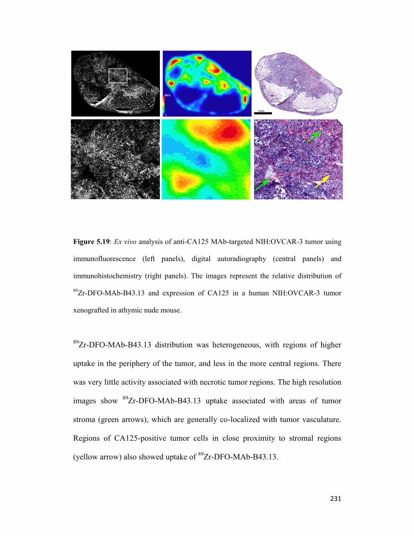
231
Figure 5.19: Ex vivo analysis of anti-CA125 MAb-targeted NIH:OVCAR-3 tumor using
immunofluorescence (left panels), digital autoradiography (central panels) and
immunohistochemistry (right panels). The images represent the relative distribution of
89Zr-DFO-MAb-B43.13 and expression of CA125 in a human NIH:OVCAR-3 tumor
xenografted in athymic nude mouse.
89Zr-DFO-MAb-B43.13 distribution was heterogeneous, with regions of higher
uptake in the periphery of the tumor, and less in the more central regions. There
was very little activity associated with necrotic tumor regions. The high resolution
images show 89
Zr-DFO-MAb-B43.13 uptake associated with areas of tumor
stroma (green arrows), which are generally co-localized with tumor vasculature.
Regions of CA125-positive tumor cells in close proximity to stromal regions
(yellow arrow) also showed uptake of 89
Zr-DFO-MAb-B43.13.

232
Figure 5.20: Ex vivo analysis of the tumor proximal brachial lymph node from an anti-
CA125 MAb-targeted NIH:OVCAR-3 tumor xenograft mouse using
immunofluorescence (left panels), digital autoradiography (central panels) and
immunohistochemistry (right panels). The images represent the relative distribution of
89Zr-DFO-MAb-B43.13 in the brachial lymph node in the ipsilateral chain proximal to
the tumor.
CA125 staining was observed in discrete regions within the lymph node, but no
tumor cells were observed. Uptake of 89
Zr-DFO-MAb-B43.13 was associated
with regions of CA125 positivity (green arrow), but decreased in regions of low
CA125 staining (red arrow).

233
Figure 5.21: Ex vivo analysis of the non-tumor brachial lymph node from an anti-CA125
MAb-targeted NIH:OVCAR-3 tumor xenograft mouse using immunofluorescence(left
panels), digital autoradiography (central panels) and immunohistochemistry (right
panels). Uptake and relative distribution of 89
Zr-DFO-MAb-B43.13 in the brachial lymph
node contralateral to the tumor location.
The appearance of this node was very similar to that of the ipsilateral tumor, with
distinct heterogeneous staining of CA125, but no visible tumor cells. Again,
uptake of 89
Zr-DFO-MAb-B43.13 was associated with regions of CA125
positivity (green arrow), but decreased in regions of low CA125 staining (red

234
arrow). However, it should be noted that the scale bar indicating the relative
radioactive concentration on the autoradiographic images indicate that this lymph
node contains a significantly lower 89
Zr-DFO-MAb-B43.13 concentration per unit
area when compared to the ipsilateral node.
5.3.7 Histopathological Findings:
Histopathologic evaluation of the harvested and fixed tumor by a veterinarian
pathologist at MSKCC, NY, revealed this tissue to be an ovoid neoplasm
surrounded by a small amount of fibrous connective tissue, skeletal muscle,
adipose tissue, and mammary glands. The neoplasm was composed of cells
forming nests and papillary structures with lumens filled with proteinaceous fluid,
and supported by a small to moderate amount of collagenous stroma. Cells were
polygonal, with distinct borders, and a moderate amount of eosinophilic
cytoplasm. The nuclei were oval, with finely stippled chromatin and one or two
large prominent nucleoli. There was four fold anisokaryosis and multinucleated
cells with an average of 55 mitoses per 400x fields. The tumor cells showed
diffuse cytoplasmic staining upon performing immunohistochemical (IHC)
staining for cytokeratin WSS.
Similar examination of the tumor proximal axillary lymph node revealed a
lymphoid depletion of the paracortex, which is a normal finding in this strain.
However, the paracortex had a focal area of coagulative necrosis. This area
contained rare single polygonal cells with large nuclei, which resemble the cells
described above in the tumor. However, upon immunohistochemical staining for

235
cytokeratin WSS, there were no positive cells and the area of necrosis was not
present. On a deeper section stained with Haematoxylin and Eosin (H&E), the
area of necrosis and atypical cells were absent. Histopathology performed on the
the ipsilateral brachial and contralateral axillary and brachial showed no abnormal
observations except for lymphoid depletion of the paracortex and cyst-like ectasia
of some sinuses, which are normal findings in this strain.

236
Figure 5.22: In vivo small animal PET imaging 312 h post-injection. Transverse and
coronal PET images of 89
Zr-DFO-MAb-B43.13 (10.2 – 12.0 MBq [275-325 µCi] injected
via tail vein in 200 L 0.9% sterile saline) in athymic nude mice # 1, 2 and 3 bearing
subcutaneous CA125-expressing OVCAR3 ovarian cancer xenografts at 312 h post-
injection. Images in the top 2 panels are scaled with signal maxima set at 25 % ID/g, the
middle set of coronal images are scaled with signal maxima set at 50 % ID/g. The bottom
panel represents maximum intensity projection (MIP) images at all corresponding time
points of evaluation.

237
5.4 Discussion:
Preparation of stable radioimmunoconjugates:
An impetus to generate novel antibodies and their derivatives for applications in
targeted strategies such as immuno-PET for oncologic imaging has been
accompanied by a need to use suitable radioisotopes that have a physical half-life
compatible with the biological half-life of such antibody vectors. Despite its
presence for decades, 89
Zr has recently gained popularity for its advantageous
features9 suited to immuno-PET strategies for imaging prostate specific
membrane antigen,17
and free prostate specific antigen,18
in prostate cancer,
Her2/neu in breast cancer,19
CD8+ T cells in lymphomas,20
CA19.9 in pancreatic
cancer,21
glypican3 in hepatocellular cancer,22
L-type amino acid receptor in
colorectal cancer23
and many other neoplasms. In this chapter, we extended the
potential for immuno-PET of epithelial ovarian cancer using the CA125 targeting
MAb-B43.13 by radiolabeling it with 89
Zr. This enabled us to track the in vivo
pharmacokinetic biodistribution of the full-length antibody radiotracer while
allowing it to gain maximal tumor accretion up to 120 h post injection (p.i). This
is a significant step forward in terms of the information we obtained from this
study when compared with a 64
Cu-labeled version of the same antibody
(presented in chapter 3 of this thesis), which was somewhat limited in its potential
to deliver information only up to 48 h p.i due primarily to the physical half-life of
the radioisotope.
Similar to the use of a tridentate azamacrocyclic chelator (NOTA) for 64
Cu-
labeling of the MAb-B43.13 antibody, here we used desferrioxamine (DFO) – an

238
acyclic hexadentate siderophore with three hydroxamate groups for chelating
radiometals such as 89
Zr4+
. Owing to its ability for quantitative radiolabeling with
89Zr in 1 hour at ambient room temperature and a demonstrated in vitro and in
vivo stability with respect to demetallation, DFO has come to be the most widely
used chelator for 89
Zr labeling of antibodies in immuno-PET.12, 9, 24
In the present
study, conjugation of an isothiocyanate-based bi-functional version of
desferrioxamine (p-SCN-DFO) to the MAb-B43.13 in 10-fold molar excess
resulted in ~ 4 molecules of DFO per MAb. Moreover, this did not compromise
its immunoreactivity to the target antigen – CA125. This was reflected in the
results from functional characterization of the DFO-MAb-B43.13
immunoconjugate via immunofluorescence and flow cytometry. Compared to the
64Cu-NOTA-MAb-B43.13 study, the ratio of bifunctional chelator – to – MAb
was increased in this study with intent to improve the specific activity of the
resultant radioimmunoconjugate by virtue of having a reasonably higher number
of DFOs conjugated per MAb without compromising its immunoreactivity. This
objective was achieved upon obtaining high isolated radiochemical yields of
purified and high specific activity 89
Zr-DFO-MAb-B43.13, which retained its
immunoreactivity to CA125 expressed on NIH:OVCAR-3 cells as analyzed by
two different methods. Furthermore, the stability of DFO chelated 89
Zr in
biological medium such as serum was demonstrated by a less than 4%
transmetallation from 89
Zr-DFO-MAb-B43.13 over 7 days. Even if this may not
exactly be the case in vivo due to catabolism of the radioimmunoconjugate by

239
active cellular processes, it is a good indicator to suggest the use of such a
chelator as far as resistance to transmetallation is concerned.
In vivo radiopharmacologic profile:
The acute biodistribution experiments provided an ex vivo evaluation
representative of the in vivo radiopharmacologic profile for 89
Zr-DFO-MAb-
B43.13 in xenograft mice models. Except in the cohort used for the 72 h blocking
experiment, 89
Zr-DFO-MAb-B43.13 showed a consistent trend for increasing
tumor accretion over time up to 120 h, with a concomitantly decreasing activity in
the blood pool of all animals. All other non-target organs maintained a baseline
amount uptake of radioactivity which ultimately translates into a high tumor-to-
non-tumor ratio with progression of time. The consistently high amount of
radioactivity in the liver at all the time points evaluated in this study may be
explained by more than one reason: a) the liver is a highly perfused organ and
generally acts as the site of clearance for high molecular weight entities such as
antibodies; b) immunocomplexes formed as a result of rapid sequestration of shed
CA125 antigen by 89
Zr-DFO-MAb-B43.13 in the bloodstream of OVCAR-3
bearing xenograft mice potentially gets routed for clearance through the liver. The
latter phenomenon has been reported to occur for CA125-MAb-B43.13
immunocomplexes formed within 2 h after injecting the antibody in a previous
study by McQuarrie et al, using a 99m
Tc-labeled MAb-B43.13 for in vivo
evaluation of its pharmacokinetics in human patients.25, 26
However, in those
reports, most of the immunocomplexes were cleared via the spleen due to an
immunologic basis to this process. The possibility of an immunologic

240
involvement for clearance of such immunocomplexes in the present study seems
negligible due to the relatively low level uptake of radioactivity found in the
spleen of all animals at all times as compared to the heightened radioactivity
levels in the liver. This observation for hepatic accumulation and clearance of
potential immunocomplexes formed at early time points post-injection of the
radiotracer is also supported by the differential clearance of an 18
F-labeled anti-
CA125 scFv (Chapter 2) 1 h p.i through the hepatic route specifically in
NIH:OVCAR-3 bearing xenograft mice, while owing to its molecular weight
being below the renal threshold for clearance, the scFv by itself tends to be routed
for clearance through the renal pathway. Furthermore, the specificity of CA125
targeting by 89
Zr-DFO-MAb-B43.13 was established by the blocking experiment
which dramatically reduced the absolute tumor uptake by ~ 66% while
demonstrating a concomitant increase of radioactivity uptake in most non-target
organs possibly on account of achieving a high degree of saturation by un-labeled
excess DFO-MAb-B43.13 at target sites in the tumor.
Small animal PET imaging:
The results from acute biodistribution studies corroborated well with our
observations from small animal imaging studies, wherein the CA125 expressing
tumors were delineated as early as 24 h p.i, albeit with modest uptake values that
progressively increased during later time points of evaluation. This observation
was consistent throughout the study and was also observed in the case of bi-lateral
tumors, wherein CA125-positive NIH:OVCAR-3 tumors showed a modest uptake

241
of radiotracer at 24 h p.i but, progressively accumulated the radiotracer by 48 h
and beyond, while the CA125-negative SKOV3 tumors showed a consistent non-
specific tumor uptake by virtue of an enhanced permeability and retention
effect.27
Consistent with results from the acute biodistribution studies, the liver
was the major non-target organ with progressive accumulation of radioactivity,
possibly due to its physiological role as the organ of clearance for
radioimunoconjugates and any associated immunocomplexes. Consequently, high
contrast images were obtained at later time points (72 h and beyond) when
maximum tumor accretion was achieved alongside minimal systemic background
activity as a result of clearance. This aspect can be particularly interesting for the
visualization of residual / recurring disease which tends to be present as small
ovarian carcinomatous nodules within the peritoneum and may be confounded by
background activity in scans performed at earlier time points. Such a non-invasive
follow-up diagnosis via immuno-PET prior to second look surgery in patients
who have already undergone surgical debulking and chemotherapy, could be of
clinical benefit from the perspective of disease management. Nevertheless, a
caveat to this approach is the persisting activity in the liver, which could
potentially hinder the detection of epithelial ovarian tumors in this region of the
peritoneum even at later time points, despite using 89
Zr immuno-PET. Although
the tumor signals for radioactivity were strong until 312 h p.i, the liver continued
to show persistent radioactivity even at this time point (Fig 22), plausibly
attributed to its role as the organ of clearance.

242
Lymph node involvement and Ex vivo analysis:
An involvement of the tumor proximal lymph nodes as tissues with maximum
uptake of the 89
Zr-DFO-MAb-B43.13 as demonstrated by small animal PET
images and further verified through additional acute biodistribution studies
including the use of a 89
Zr-DFO-IgG isotype control was the most stimulating part
of this study. A completely different in vivo biodistribution profile between the
CA125 targeted antibody and the isotype control in OVCAR3 xenograft mice,
furthered by the selectively high uptake of 89
Zr-DFO-MAb-B43.13 in the
harvested OVCAR3 tumor proximal lymph nodes ascertained this phenomenon
beyond a sporadic occurrence of chance.
Ex vivo analyses of the 89
Zr-DFO-MAb-B43.13 distribution in OVCAR xenograft
tumors was compared to both CA125 expression and the underlying tumor
histology at 72 h p.i. The heterogeneous distribution of 89
Zr-DFO-MAb-B43.13
was consistent with our previous observations using 64
Cu-NOTA-MAb-B43.13,
with generally higher uptake in the tumor periphery and reduced uptake in the
central tumor regions (Fig. 5.19). This is not unusual since solid tumors have been
described to have four distinct morphologic regions based on perfusion – an
avascular necrotic region (usually at the center), a semi-necrotic region, a
stabilized microcirculation region (at the periphery) and an advancing front
(region with dense tumor nests surrounding the vasculature).28
At higher
magnification, we observed the uptake to be higher in stroma-dense tumor
regions, suggesting a vascular component to the 89
Zr distribution of the
radiotracer. Possible explanations for such localization include a limited tumor

243
penetration capability of full-length antibodies29, 30, 31, 32
due primarily to their size
that impedes extravasation across endothelial cells. Even after extravasation, the
transport of antibodies within the tumor by diffusion has been noted to be as slow
as 1 h to cover 100 µm.28
This is exacerbated by the interstitial fluid pressure in
solid tumors which prevents efficient transport of antibodies within the tumor.33
Furthermore, it has also been demonstrated that perivascular distribution of
antibodies in the tumor is dose – dependent, such that an injected dose below 50
µg would predominantly be localized to the perivascular space and not be able to
penetrate the tumor as extensively as a 150 µg dose would. A contribution from
the enhanced permeability and retention (EPR) effect34
and / or binding-site
barrier,35
whereby high affinity of the antibody prevents complete antibody
penetration into the tumor may be additional factors playing a role. However,
regions of CA125-positive tumor cells in proximity to regions of stroma did show
significant accumulation of 89
Zr-DFO-MAb-B43.13, implying that the clearance
of unbound antibody from the tumor is not yet complete at this time-point.
On the other hand, the pattern of uptake in the brachial lymph nodes from the
ipsilateral and contralateral chains to the tumor location shown as representative
examples (Fig 5.20 and 5.21), revealed CA125 staining in both nodes, with no
obvious difference in either the localization within the node, or the subjective
intensity of the staining. 89
Zr distribution appeared to correspond to CA125
staining in both cases, but the fixed 25 µm resolution of the digital autoradiogram
was clearly a limitation in this comparison. The use of appropriate photographic
emulsions could potentially be used to further resolve the isotope distribution in

244
these tissues for future studies. Nonetheless, a major difference between the two
representative lymph nodes shown was the relative 89
Zr radioactivity
concentrations observed, with the ipsilateral node measuring over 8-fold higher
average counts per unit area when compared with the contralateral node. This
pattern was repeated in the axillary and inguinal nodes takes from both lymph
node chains.
Such a pattern for uptake of 89
Zr-DFO-MAb-B43.13 in the lymph nodes may be
suggestive of the presence of metastatic tumor deposits in the ipsilateral chain,
absent in the contralateral chain. However, we were unable to detect tumor cells
in any of the lymph nodes examined. The pattern of CA125 staining in the lymph
nodes had the appearance of being cell-associated (Figs 5.20 and 5.21 green
arrows), but this would also be consistent with the non-specific association of
CA125 circulating with tumor infiltrating lymphocytes. The evaluation of the
presence of tumor cells in these lymph nodes was a subjective assessment based
on H&E staining and the possibility exists that small metastatic deposits are
present. However, the CA125 staining and 89
Zr-DFO-MAb-B43.13 data point to
the drainage of shed CA125 immunocomplexes from the tumor as being the most
likely explanation of the lymph node observations presented thus far in this study.
Histopathologic findings confirmed the authenticity of the tumor to be an ovarian
carcinoma and identified all lymph nodes as having some abnormalities that are
expected as part of the nude mouse phenotype (lymphoid depletion of the
paracortex and dilation of sinuses). However, the ipsilateral axillary lymph node
had an area of necrosis, which was not an expected spontaneous finding in a nude
245
mouse lymph node. This area of necrosis contained rare large cells
morphologically similar to the neoplastic cells that composed the tumor.
However, a section from this lymph node stained negative for cytokeratin WSS –
an epithelial marker that was positive on the tumor sample. A lack of further
representation of such an area of necrosis with atypical cells in the ipsilateral
axillary node in spite of cutting deeper into the block to obtain additional sections
for analysis, left us to speculate if the abnormal area observed initially may have
represented metastasis, even if very few cells were present. We were not able to
confirm metastasis on additional sections due to the small size of the lesion while
the three other lymph nodes did not have evidence of metastasis.
Metastasis or circulating immunocomplexes?
Along its course of disease progression, serous epithelial ovarian carcinoma has
been reported as the most metastatic subtype of ovarian cancers which are
generally known to be highly lymphophillic.36
While a transcoelomic route37
would be the most common mechanism for metastatic spread of ovarian cancers
in their natural setting, they are also known to use the para-aortic and pelvic
lymph nodes to metastasize within the body. Although rare, there have been a few
reports of axillary lymph node metastasis of ovarian cancer.38
Furthermore,
increasing evidence for lymphangeogenesis promoted by growth factors (VEGF-
C and D) secreted from solid tumors augment the possibilities for lymphatic
spread of metastatic neoplasms.39, 40
In epithelial ovarian cancers, high levels of
pre-operative CA125 have clinically served as a prognostic indicator for

246
lymphatic metastasis in human patients in order to identify candidates for surgical
lymphadenectomy.41, 42
In the light of these facts and considering contemporary
reports for immuno-PET imaging of tumor-induced lymphangeogenesis and
metastasis,43, 44, 45
alongwith a mathematically modeled proposition46
to suggest
that antibodies may find it easier to target micrometastasis than vascularized
tumors encourages us to think that a case for metastasis should not be completely
ruled out until further investigation.
Nevertheless, the presence of papillary structures filled with proteinaceous fluid
interlacing the tumor nests in this study is consistent with our previous
observations from the 64
Cu-NOTA-MAb-B43.13 study wherein tumor slices
examined revealed dense CA125 staining in similar areas observed with H&E
staining. These channels may be representative of lymphatics in the periphery of
the tumor that potentially contribute to the draining of bound immunocomplexes
or extravasated yet unbound 89
Zr-DFO-MAb-B43.13 that might find it easier to
slip away into the proximal draining lymph nodes in the face of resistance to
penetrate the tumor on account of interstitial fluid pressure and other factors
mentioned previously. Lastly, given the immunotherapeutic aspect of MAb-
B43.13 there could be a potential role for an immunologic basis to this
observation or an interplay between one and more of these speculated factors
leading to lymph node involvement. Further investigation of this phenomenon in a
suitable disease model with inclusion of immunologic aspects is warranted.

247
5.5 Conclusion:
The present chapter is a leap forward in our approach to CA125 targeted immuno-
PET for epithelial ovarian cancer. This study maximizes the potential of using the
CA125 targeting antibody as a radiotracer for pre-clinical and clinical non-
invasive imaging of EOC via immuno-PET. In the present work, we produced a
89Zr-labeled version of MAb-B43.13 and performed preclinical evaluation of this
radiotracer in sub-cutaneous xenograft mouse models for EOC. A wider window
of opportunity was provided to trace the in vivo radiopharmacological fate of an
antibody-based targeting vector to be applied beyond the 48 h window previously
available to us using a 64
Cu-labeled version of the same antibody. Furthermore,
this study imparted a translational relevance by knowing the destination and
potential off-target effects of the radiopharmaceutical prior to designing potential
clinical applications for imaging and therapy in future. In summary, this study
demonstrated an amalgamation of the efficient in vivo tumor targeting capabilities
of MAb-B43.13 and the favourable properties of 89
Zr towards developing a
radiopharmaceutical for immuno-PET of epithelial ovarian tumors. Most
importantly, it extended the scope of this technique and the radiotracer for the
detection of potential metastasis and/or draining lymph nodes, which may be of
clinical significance in the management of this disease.

248
5.6 References:
1. Wu AM, Senter PD. Arming antibodies: prospects and challenges for
immunoconjugates. Nat Biotechnol 2005;23(9): 1137-46.
2. Sundaresan G, Yazaki PJ, Shively JE, et al. 124I-labeled engineered anti-CEA
minibodies and diabodies allow high-contrast, antigen-specific small-animal PET
imaging of xenografts in athymic mice. J Nucl Med 2003;44(12): 1962-9
3. O'Donoghue JA, Smith-Jones PM, Humm JL, et al. 124I-huA33 antibody uptake is
driven by A33 antigen concentration in tissues from colorectal cancer patients
imaged by immuno-PET. J Nucl Med 2011;52(12): 1878-85.
4. Knowles SM, Wu AM. Advances in immuno-positron emission tomography:
antibodies for molecular imaging in oncology. J Clin Oncol 2012;30(31): 3884-92.
5. Holland JP, Williamson MJ, Lewis JS. Unconventional nuclides for
radiopharmaceuticals. Mol Imaging 2010;9(1): 1-20.
6. Wright BD, Lapi SE. Designing the magic bullet? The advancement of immuno-
PET into clinical use. J Nucl Med 2013;54(8): 1171-4.
7. Zhang Y, Hong H, Cai W. PET tracers based on Zirconium-89. Curr Radiopharm
2011;4(2): 131-9.
8. Holland JP, Sheh Y, Lewis JS. Standardized methods for the production of high
specific-activity zirconium-89. Nucl Med Biol 2009;36(7): 729-39.
9. Deri MA, Zeglis BM, Francesconi LC, Lewis JS. PET imaging with (8)(9)Zr: from
radiochemistry to the clinic. Nucl Med Biol 2013;40(1): 3-14.
10. Fischer G, Seibold U, Schirrmacher R, Wangler B, Wangler C. (89)Zr, a
radiometal nuclide with high potential for molecular imaging with PET: chemistry,
applications and remaining challenges. Molecules 2013;18(6): 6469-90.
11. Perk LR, Visser GW, Vosjan MJ, et al. (89)Zr as a PET surrogate radioisotope for
scouting biodistribution of the therapeutic radiometals (90)Y and (177)Lu in

249
tumor-bearing nude mice after coupling to the internalizing antibody cetuximab. J
Nucl Med 2005;46(11): 1898-906.
12. Price EW, Zeglis BM, Lewis JS, Adam MJ, Orvig C. H6phospa-trastuzumab:
bifunctional methylenephosphonate-based chelator with 89Zr, 111In and 177Lu.
Dalton Trans 2014;43(1): 119-31.
13. Deri MA, Ponnala S, Zeglis BM, et al. Alternative Chelator for (89)Zr
Radiopharmaceuticals: Radiolabeling and Evaluation of 3,4,3-(LI-1,2-HOPO). J
Med Chem 2014;57(11): 4849-60.
14. Noujaim AA, Baum RP, Sykes TR, Sykes CJ, Hertel A, Niesen, A, et al.
Monoclonal antibody B43.13 for immunoscintigraphy and immunotherapy of
ovarian cancer. In Current Tumor Diagnosis: Applications, Clinical Relevance,
Trends. Edited by Klapdor, R. Munich: W Zuckschwerdt Verlag; 1994; 823–829.
15. Lindmo T, Boven E, Cuttitta F, Fedorko J, Bunn PA, Jr. Determination of the
immunoreactive fraction of radiolabeled monoclonal antibodies by linear
extrapolation to binding at infinite antigen excess. J Immunol Methods 1984;72(1):
77-89.
16. Carlin S, Khan N, Ku T, Longo VA, Larson SM, Smith-Jones PM. Molecular
targeting of carbonic anhydrase IX in mice with hypoxic HT29 colorectal tumor
xenografts. PLoS One 2010;5(5): e10857
17. Holland JP, Divilov V, Bander NH, Smith-Jones PM, Larson SM, Lewis JS. 89Zr-
DFO-J591 for immunoPET of prostate-specific membrane antigen expression in
vivo. J Nucl Med 2010;51(8): 1293-300.
18. Ulmert D, Evans MJ, Holland JP, et al. Imaging androgen receptor signaling with a
radiotracer targeting free prostate-specific antigen. Cancer Discov 2012;2(4): 320-
7.

250
19. Holland JP, Caldas-Lopes E, Divilov V, et al. Measuring the pharmacodynamic
effects of a novel Hsp90 inhibitor on HER2/neu expression in mice using Zr-DFO-
trastuzumab. PLoS One 2010;5(1): e8859.
20. Tavare R, McCracken MN, Zettlitz KA, et al. Engineered antibody fragments for
immuno-PET imaging of endogenous CD8+ T cells in vivo. Proc Natl Acad Sci U
S A 2014;111(3): 1108-13.
21. Viola-Villegas NT, Rice SL, Carlin S, et al. Applying PET to broaden the
diagnostic utility of the clinically validated CA19.9 serum biomarker for oncology.
J Nucl Med 2013;54(11): 1876-82.
22. Sham JG, Kievit FM, Grierson JR, et al. Glypican-3-targeted 89Zr PET imaging of
hepatocellular carcinoma. J Nucl Med 2014;55(5): 799-804.
23. Ikotun OF, Marquez BV, Huang C, et al. Imaging the L-type amino acid
transporter-1 (LAT1) with Zr-89 immunoPET. PLoS One 2013;8(10): e77476
24. Tinianow JN, Gill HS, Ogasawara A, et al. Site-specifically 89
Zr-labeled
monoclonal antibodies for ImmunoPET. Nucl Med Biol 2010;37(3): 289-97.
25. McQuarrie SA, Baum RP, Niesen A, et al. Pharmacokinetics and radiation
dosimetry of 99
Tcm-labelled monoclonal antibody B43.13 in ovarian cancer
patients. Nucl Med Commun 1997;18(9): 878-86.
26. McQuarrie SA, Riauka T, Baum RP, et al. The effects of circulating antigen on the
pharmacokinetics and radioimmunoscintigraphic properties of 99m
Tc labelled
monoclonal antibodies in cancer patients. J Pharm Pharm Sci 1998;1(3): 115-25.
27. Fang J, Nakamura H, Maeda H. The EPR effect: Unique features of tumor blood
vessels for drug delivery, factors involved, and limitations and augmentation of the
effect. Adv Drug Deliv Rev 2011;63(3): 136-51.
28. Jain RK. Transport of molecules, particles, and cells in solid tumors. Annu Rev
Biomed Eng 1999;1: 241-63.

251
29. Buchegger F, Haskell CM, Schreyer M, et al. Radiolabeled fragments of
monoclonal antibodies against carcinoembryonic antigen for localization of human
colon carcinoma grafted into nude mice. J Exp Med 1983;158(2): 413-27.
30. Thurber GM, Schmidt MM, Wittrup KD. Factors determining antibody distribution
in tumors. Trends Pharmacol Sci 2008;29(2): 57-61.
31. Rhoden JJ, Wittrup KD. Dose dependence of intratumoral perivascular distribution
of monoclonal antibodies. J Pharm Sci 2012;101(2): 860-7.
32. Thurber GM, Schmidt MM, Wittrup KD. Antibody tumor penetration: transport
opposed by systemic and antigen-mediated clearance. Adv Drug Deliv Rev
2008;60(12): 1421-34.
33. Heldin CH, Rubin K, Pietras K, Ostman A. High interstitial fluid pressure - an
obstacle in cancer therapy. Nat Rev Cancer 2004;4(10): 806-13.
34. Heneweer C, Holland JP, Divilov V, Carlin S, Lewis JS. Magnitude of enhanced
permeability and retention effect in tumors with different phenotypes: 89Zr-
albumin as a model system. J Nucl Med 2011;52(4): 625-33.
35. Adams GP, Schier R, McCall AM, et al. High affinity restricts the localization and
tumor penetration of single-chain fv antibody molecules. Cancer Res 2001;61(12):
4750-5.
36. Di Re F, Baiocchi G. Value of lymph node assessment in ovarian cancer: Status of
the art at the end of the second millennium. Int J Gynecol Cancer 2000;10(6): 435-
42.
37. Tan DS, Agarwal R, Kaye SB. Mechanisms of transcoelomic metastasis in ovarian
cancer. Lancet Oncol 2006;7(11): 925-34.
38. Ceccarelli F, Barberi S, Pontesilli A, Zancla S, Ranieri E. Ovarian carcinoma
presenting with axillary lymph node metastasis: a case report. Eur J Gynaecol
Oncol 2011;32(2): 237-9.

252
39. Detmar M, Hirakawa S. The formation of lymphatic vessels and its importance in
the setting of malignancy. J Exp Med 2002;196(6): 713-8.
40. Du LC, Chen XC, Wang D, et al. VEGF-D-induced draining lymphatic
enlargement and tumor lymphangiogenesis promote lymph node metastasis in a
xenograft model of ovarian carcinoma. Reprod Biol Endocrinol 2014;12: 14.
41. Ayhan A, Gultekin M, Dursun P, et al. Metastatic lymph node number in epithelial
ovarian carcinoma: does it have any clinical significance? Gynecol Oncol
2008;108(2): 428-32.
42. Kim HS, Park NH, Chung HH, Kim JW, Song YS, Kang SB. Significance of
preoperative serum CA-125 levels in the prediction of lymph node metastasis in
epithelial ovarian cancer. Acta Obstet Gynecol Scand 2008;87(11): 1136-42.
43. Mumprecht V, Honer M, Vigl B, et al. In vivo imaging of inflammation- and
tumor-induced lymph node lymphangiogenesis by immuno-positron emission
tomography. Cancer Res 2010;70(21): 8842-51.
44. Mumprecht V, Detmar M. In vivo imaging of lymph node lymphangiogenesis by
immuno-positron emission tomography. Methods Mol Biol 2013;961: 129-40.
45. Borjesson PK, Jauw YW, Boellaard R, et al. Performance of immuno-positron
emission tomography with zirconium-89-labeled chimeric monoclonal antibody
U36 in the detection of lymph node metastases in head and neck cancer patients.
Clin Cancer Res 2006;12(7 Pt 1): 2133-40.
46. Thurber GM, Zajic SC, Wittrup KD. Theoretic criteria for antibody penetration
into solid tumors and micrometastases. J Nucl Med 2007;48(6): 995-9.

253
6
Development of a CA125 targeting
Diabody for application in same-day
imaging of Epithelial Ovarian Cancer5
5This is a work in progress. A version of this chapter will be submitted to the
European Journal of Nuclear Medicine and Molecular Imaging Research:
Sharma SK, Wuest M, Lapi SE, Wuest F.
CA125 Targeted Cys-Diabody for same day imaging of epithelial ovarian cancer

254
6.1 Introduction:
The in vivo pharmacokinetics of a molecular probe is central to its success as a
radiotracer for application in PET imaging strategies. With recent advances and
the promise of immuno-PET as a unique functional imaging strategy, a number of
antibodies are being engineered for use in this technique.1, 2
Nonetheless, despite
excellent tumor targeting capabilities and ideal size for in vivo tumor targeting,3
full-length antibodies are not the vectors of choice for translation as radiotracers
for same-day or next-day imaging strategies. Their long residency in systemic
circulation and slow clearance, which contribute to a tremendous therapeutic
potential, paradoxically serve as a major disadvantage to application as
radiotracers for molecular imaging. Consequently, they end up being
incompatible for use with short-lived radioisotopes and when labeled with long-
lived positron emitting radionuclides, they deliver a high systemic background to
result in low tumor-to-normal tissue ratios at early time points. Furthermore, from
a dosimetry perspective, they contribute to radioactivity burden in subjects over
an extended period of time until sufficient tumor accumulation and systemic
clearance of the radiotracer are achieved.
Considering these shortcomings, a pursuit to achieve high image quality as a
consequence of high absolute tumor uptake values combined with high tumor-to-
background ratios while simultaneously reducing the radiation burden in subjects,
has propelled the development of a family of radiotracers based on engineered
versions of the antibody. Recently, many such molecules have been deployed as
vectors for molecular imaging of cancer via immuno-PET.1,4
This paradigm is

255
based on the premise of an inverse correlation between size of the vector
employed as a radiotracer and its in vivo pharmacokinetics.2 Historically, F(ab)
~50 KDa and F(ab’)2 fragments (~ 110 KDa) had been used to achieve this goal.
However, while the former showed low absolute tumor uptake values,5, 6
the latter
yielded high contrast at later time points (12-18 h) and does not qualify for use in
same-day imaging strategies. On the other hand, single chain Fragment variable
(scFv) formats considered as the smallest yet complete active binding unit of the
antibody capable of binding to a target antigen become a victim of their own
molecular weight, which ultimately manifests in anemic tumor uptake. This is
primarily due to a combined effect of first-pass clearance and renal trapping that
precludes tumor bioavailability of these targeting agents.7, 8
Most importantly,
despite retaining the specificity of the parental antibody for the target antigen, the
scFv suffers from a lack of sufficient avidity to result in a short mean residency
time within the tumor. However, the lack of Fc (Fragment crystallizable) region in
this engineered format makes this class of engineered molecules immunologically
inert for in vivo application in a variety of preclinical models and clinical subjects
and contributes greatly towards achieving rapid systemic clearance as a desirable
trait in an immuno-PET radiotracer for same-day imaging.
Collectively, these observations have prompted redesigning strategies to increase
the avidity of such antibody fragments to render a bivalent binding capability
comparable to that of the parent antibody molecule. Some such engineering
approaches include: a) chemical cross-linking of two or more monomeric scFv
units; b) tandem cloning of scFv units and its expression to yield dimers; c)

256
introduction of dimerization domains by recombinant methods; d) non-covalent
dimerization to yield ‘diabodies’ as a result of shortening the length of inter-
domain peptide linker.9, 10, 11
More recently, the introduction of unpaired cysteine
residues at the C-terminus of the scFv has led to the development of disulfide-
linked dimers commonly referred to as Cys-Diabodies.12, 13, 14, 15

257
Figure 6.1: Diagrammatic representation of DNA constructs for the production of anti-
CA125 diabodies: A) Two anti-CA125 scFvs cloned in tandem to form bivalent scFv; B)
Shortening of inter-domain flexible peptide linker from (G4S)3 to (G4S)1 to form non-
covalently-linked dimers (Diabodies); C) Introduction of unpaired cysteine residues at C-
terminus of individual anti-CA125 scFv genes to form disulfide bonds upon periplasmic
expression and in vivo folding in E.coli to form covalently-linked dimers (Cys-
Diabodies). G: Glycine; S: Serine; 6X-His: hexa-histidine tag; S~S: disulfide bond.

258
The motivation for the generation of diabodies stems from the fact that this class
of molecules would possess the best of all worlds in that they have a comparable
functional avidity for binding to antigen as that of a complete antibody, yet they
have a molecular weight (~ 55 KDa), which is intermediate to that of an scFv and
a full-length antibody, and is well below the renal threshold for rapid systemic
clearance. Diabodies targeting several tumor associated antigens have been
proposed and demonstrated as suitable targeted radiotracers for same-day imaging
strategies via immuno-PET.16, 17, 18, 19
In this chapter, we engineered the anti-CA125 scFv into a covalently linked dimer
to develop it as a suitable immuno-PET radiotracer for same-day imaging of
epithelial ovarian cancer. To this end, we introduced an unpaired cysteine at the
C-terminus of the anti-CA125 scFv, such that recombinant expression and in vivo
folding of this molecule within the periplasm of E.coli would result in a disulfide
linked dimeric version of the scFv – the anti-CA125 Cys-Db. The
immunoreactivity of the anti-CA125 Cys-Db was verified by biochemical assays
such as immunoblotting, flow cytometry and immunofluorescence. Subsequently,
the anti-CA125 Cys-Db was radiolabeled with 64
Cu (t1/2 12.7 h) and preliminary
studies were performed using NIH:OVCAR-3 cells for in vitro analysis and
corresponding xenograft bearing mice to evaluate its in vivo radiopharmacologic
profiles and tumor targeting capabilities.

259
6.2 Materials and Methods:
6.2.1 Production of anti-CA125 Diabody:
A) Cloning: The genes for individual variable domains of the anti-CA125 scFv
were amplified by polymerase chain reaction (PCR) from a previously reported
plasmid construct pWET8.20, 21
The plasmid encoding for anti-CA125 VL-(G4S)3-
VH scFv reported in chapter 2 of this thesis was used as a template for splice
overlap extension PCR in order to shorten the inter-domain peptide linker from
the canonical (Gly4Ser)3 to (Gly4Ser)1. The anti-CA125 scFv was oriented as VL-
(G4S)3-VH with the variable light chain domain at the N-terminus and variable
heavy chain domain at the C-terminus. Furthermore, in a separate construct, a
Gly-Gly-Cys tri-peptide was inserted between the last residue (Ser) at the end of
the scFv heavy chain domain and the Not I cloning site by introducing the
corresponding tri-peptide encoding oligonucleotide sequence into the antisense
primer during PCR amplification of the anti-CA125 scFv. This construct is
hereafter referred to as anti-CA125 Cys-Db in this thesis. Ultimately, both
constructs were sub-cloned separately between Nco I and Not I restriction sites of
pET-22b(+) vector (Novagen, 69744) for recombinant expression in E.coli
Rosetta 2(DE3) (Novagen, 71400). The scFv DNA cloned in each recombinant
construct was further verified by Sanger sequencing on a 3730 DNA analyzer
(Applied Biosystems, Life Technologies) prior to transformation in the bacterial
expression host.

260
B) Expression and Purification: Each recombinant plasmid coding for the two
diabody constructs was transformed into chemically competent E.coli Rosetta
2(DE3) cells by heat-shock method and plated onto 1.5% 2x YT agar plates
containing 100 µg/mL ampicillin and 34 µg/mL chloramphenicol. Single
transformant colonies were picked for each construct and inoculated into 12.5 ml
2x YT medium (16 g/L tryptone, 10 g/L yeast extract and 5 g/L sodium chloride,
pH 7.5) with 100 µg/ml ampicillin, 34 µg/mL chloramphenicol and cultured
overnight by incubation at 37˚C with shaking at 200 rpm. 5 ml of the overnight
grown cultures was added to 500 ml fresh 2x YT medium in the presence of
antibiotics in 2 liter flasks and incubated at 37˚C with shaking at 220 rpm until an
OD600 of 0.6 – 0.8 was achieved. The shake flask cultures were briefly placed in
ice cold water for 15 minutes prior to induction with a final concentration of 0.8
mM IPTG. The induced cultures were incubated at 26˚C with shaking at 200 rpm
overnight for 16 h. The bacteria were harvested by centrifugation at 7000 rpm for
30 minutes and the pellet obtained thereof was frozen by storage at -20˚C. Soluble
diabodies were extracted from frozen cell pellets by lysis with BugBuster®
Master
Mix reagent as per the manufacturer’s instructions. The cell lysate was cleared by
centrifugation at 16,000 rpm for 30 minutes and the supernatant was collected for
purification of soluble diabodies. C-terminal hexa-histidine tagged recombinant
diabody was purified via immobilized metal affinity chromatography (IMAC)
using a cobalt-based TALON®
superflow metal affinity resin (Clontech, 635506).
The cell lysate supernatant was passed over the resin at a flow rate of 1 mL/min to
allow binding of the hexa-histidine tagged recombinant protein to the metal

261
affinity resin. Initial attempts to elute the captured protein from the column
employed a step gradient of 10 mM, 20 mM, 40 mM, 80 mM, 120 mM and 150
mM of imidazole in 50 mM sodium di-hydrogen phosphate [NaH2PO4.H2O] and
100 mM sodium chloride pH 7.0 used as the buffer solution. Later trials for
purification were performed using a linear gradient of 10 mM – 150 mM
imidazole as the eluant. 1 mL IMAC-eluted fractions were collected and analyzed
by 12% SDS-PAGE under non-reducing conditions, followed by Coomassie
staining and immunoblotting with 6X His MAb-HRP conjugate (Clontech,
631210). Eluted fractions containing high concentration of purified diabody were
pooled together and dialyzed in phosphate buffered saline (pH 7.4) to remove
excess imidazole and concentrated via centrifugation using Amicon Ultra-15, 10K
MWCO filters (EMD Millipore, UFC901024). Anti-CA125 murine monoclonal
antibody (MAb) was purified from MAb B43.13 hybridoma22
cell culture
supernatant by protein G affinity (Sigma, P-7700) on a BioLogic DuoFlow™
chromatography system (Bio-Rad, 760-0135). Final concentrations of the purified
diabody and MAb were quantified using a Pierce™ BCA protein assay kit
(Thermo Scientific, 23227) according to the manufacturer's recommendations.
6.2.2 Functional characterization of anti-CA125 Diabody:
6.2.2.1 Cell lines and culture conditions: Ovarian cancer cells NIH:OVCAR-3
(ATCC® HTB-161™) that overexpress CA125 were used for in vitro functional
characterization studies. Cells were cultured in DMEM-F12 medium
supplemented with 10% v/v fetal bovine serum (Life Technologies, 12483-020),

262
50 IU/mL penicillin, 50 μg/mL streptomycin (Life Technologies, 15140-122).
NIH:OVCAR-3 cells were additionally supplemented with 7 μg/mL recombinant
human insulin (SAFC Biosciences, 91077C). Cells were cultured using sterile
techniques and grown in a 37°C incubator providing humidified atmosphere of
5% CO2 in air.
6.2.2.2 Immunoblotting: 7.5 x 10⁶
NIH:OVCAR-3 and SKOV3 cells were lysed
with CelLytic™ M. Cell lysates were electrophoresed on a 4-15% Mini-
PROTEAN® TGX™ precast gel (Bio-Rad) and transferred to a Trans-Blot
nitrocellulose membrane (Bio-Rad). The membranes were probed separately to
evaluate binding capabilities of the recombinant anti-CA125 targeting antibody-
based constructs. The blots were blocked for 45 min with 5% non-fat dry milk
(Carnation) in PBS having 0.1% Tween-20 (PBST). Anti-CA125 MAb (3
mg/mL), mouse anti-β actin IgG (Sigma, A1978), anti-CA125 scFv (~2 mg/mL)
and anti-CA125 Cys-Db (~1 mg/mL) were used as primary antibodies (1: 5000
each) to probe the blots for 1 h at room temperature. Goat anti-mouse HRP
conjugate (1: 5000) (Sigma, A4416), was used as secondary antibody to probe the
blot against anti-CA125 MAb and mouse anti-β actin IgG for 1 h at room
temperature. 6X His MAb-HRP conjugate (1:5000) (Clontech, 631210) was used
as secondary antibody to probe against antigen-bound anti-CA125 scFv variants
by incubating for 1 h at room temperature. Anti-CA125 targeting monoclonal
antibody MAb B43.13 was used as a positive control. A hexa-histidine tagged
anti-RANK receptor binding scFv23
was used as an isotype control.

263
6.2.2.3 Flow Cytometry: 1.5 x 10⁶ NIH:OVCAR-3 cells were harvested by
trypsinization, rinsed twice with FACS buffer (PBS with 0.5% heat inactivated
FBS, 2mM EDTA, 0.05% sodium azide) and resuspended by gentle tapping in
~100 µL of this buffer. 10 µg of anti-CA125 MAb B43.13, anti-CA125 scFv and
anti-CA125 Cys-Db and a hexa-histidine tagged anti-RANK receptor binding
scFv (isotype control), were incubated with the NIH:OVCAR-3 cell suspension
for 30 min at room temperature. Cells were rinsed twice in FACS buffer and
incubated for another 30 min with 2.4 µg of Penta·His Alexa Fluor 488 conjugate
(Life Technologies, A-11001) for samples previously incubated with scFv/Db
constructs. Cells were rinsed twice with FACS buffer and analyzed by flow
cytometry on a BD FACS Calibur. Negative controls included unstained
NIH:OVCAR-3 cells and cells incubated with Penta·His Alexa fluor 488 –
conjugated antibody alone.
6.2.2.4 Immunofluorescence: NIH:OVCAR-3 cells were plated onto glass
coverslips in 35-mm tissue culture dishes (100,000 cells/2 mL medium/dish) and
incubated at 37C for 48 h. The cells were rinsed with PBS and fixed in methanol
for 30 min at -20C. The fixed cells were incubated in 5% non-fat dry milk
(Carnation) in PBS and immunostained separately for 1 h with anti-CA125 MAb-
B43.13, anti-CA125 scFv, anti-CA125 Cys-Db (1:250). Alexa-fluor® 488
conjugated goat anti-mouse antibody and anti-Penta·His Alexa Fluor-488
conjugate were used as a secondary antibodies (1:500) in PBS containing 5% non-
fat dry milk for samples previously incubated with MAb-B43.13 and anti-CA125

264
scFv/Cys-Db respectively. Appropriate control samples were included in the
experiments. All antibody incubations were followed by three rinses with PBST
for 10 min each. Coverslips were mounted on microscopy slides (Fisherbrand)
using Mowiol® mounting medium (Calbiochem, 475904) supplemented with
DAPI (50 µg/mL). Immunofluorescence was observed through a Zeiss Plan
Apochromat 40X/1.3 Oil DIC M27 lens on a confocal laser scanning microscope
(Zeiss LSM 710). The images were analyzed using Zen 2011 software and
processed further using Adobe Photoshop CS6.
6.2.3 Radiolabeling of anti-CA125 Cys-Db:
6.2.3.1 General: All glassware was rinsed with ultra-pure HCl (Fisherbrand).
Trace metals basis ultra-pure chemicals for buffer preparations were purchased
from Sigma Aldrich. All buffer solutions were treated with biotechnology grade
Chelex 100 (Bio-Rad, 143-2832).
6.2.3.2 NOTA Functionalization: p-SCN-Bn-NOTA [S-2-(4-
Isothiocyanatobenzyl)-1,4,7-triazacyclononane-1,4,7-triacetic acid] (Macrocyclics
Inc, B-605) was conjugated to anti-CA125 Cys-Db as a bi-functional chelator for
64Cu radiolabeling. Briefly, a 6 molar excess of p-SCN-Bn-NOTA in DMSO was
added to anti-CA125 Cys-Db in 0.1 M sodium bi-carbonate buffer pH 8.5 and
allowed to react for 1 h at 37C. NOTA-functionalized anti-CA125 Cys-Db was
purified from un-conjugated NOTA while simultaneously achieving buffer

265
exchange into 0.25 M sodium acetate pH 5.5 by using an Econo-Pac®
10DG
desalting column (Bio-Rad, 732-2010). Estimation of protein concentration of
column eluted fractions was performed via bicinchoninic assay using a Pierce
BCA Protein Assay Kit (Thermo, 23227) and A280 measurements on a nanodrop.
Fractions with highest protein concentrations were identified and employed for
further use in radiolabeling experiments.
6.2.3.3 64
Cu labeling of NOTA-functionalized MAb/scFv: 64
Cu was produced
via a 64
Ni(p,n)64
Cu nuclear reaction on a CS-15 biomedical cyclotron at
Washington University, St. Louis, USA as described by Kume et al.24
85 MBq of
64Cu-acetate was added to 100 µg NOTA-functionalized anti-CA125 Cys-Db and
allowed to react at 30°C, 550 rpm for 1 h in a thermomixer. 1 mM EDTA was
added to quench the reaction over 10 min. 64
Cu-labeled anti-CA125 Cys-Db
radioimmunoconjugates were purified using an Econo-Pac®
10DG desalting
column pre-equilibrated with 0.25 M sodium acetate, pH 5.5 used as the eluant.
350 μL elution fractions were collected from the column and radioactivity was
measured on an Atomlab 400 dose calibrator (Biodex Inc). 15 μL of each elution
fraction was electrophoresed on a 12% SDS-PAGE gel under non-reducing
conditions and evaluated by autoradiography on a BAS-5000 phoshorimager
(Fujifilm). Radiochemical yields and purity were determined by instant thin layer
chromatography on ITLC-SG (Varian) using 10 mM EDTA pH 5.5 as the eluant.
Fractions containing high specific activity radioimmunoconjugates were used for
in vitro and in vivo radiopharmacological experiments.

266
6.2.4 In vitro immunoreactivity test:
The immunoreactive fraction of the prepared radioimmunoconjugate was
determined as per the method of Lindmo et al.25
Briefly, NIH:OVCAR-3 cells
were aliquoted into microcentrifuge tubes at concentrations of 5, 4, 3, 2.5, 2, 1.5
and 0.5 x 10⁶ cells in 500 μL PBS (pH 7.4). A stock solution of the
radioimmunoconjugate (NOTA functinalized anti-CA125 Cys-Db) was prepared
in PBS (pH 7.4) supplemented with 1% BSA such that 50 μL of this solution
contained 20,000 cpm which was then uniformly aliquoted into individual tubes.
This setup was allowed to incubate for 1 h at room temperature on a platform
mixer. Thereafter, cells were pelleted by centrifugation (5,000 rpm for 3 mins),
rinsed twice with ice cold PBS and the supernatant was aspirated prior to
measuring cell-bound radioactivity on a γ-counter (Wizard2®
2480 Automatic
Gamma Counter, Perkin-Elmer, Canada). Radioactivity data obtained thereof was
background corrected and compared with counts from the total activity added
control samples in the experiments. The immunoreactive fraction was determined
by performing a linear regression analysis on a double-inverse plot of
(total/bound) activity versus normalized cell concentration. All data was obtained
in triplicates and no weighting was applied.
6.2.5 In vivo studies:
6.2.5.1 Xenograft models: All experiments were carried out according to
guidelines of the Canadian Council on Animal Care (CCAC) and approved by the

267
local animal care committee of the Cross Cancer Institute, Edmonton. Six weeks
old BALB/c nude female mice were obtained from Charles River labs (Quebec,
Canada). The animals were housed in ventilated cages and provided food and
water ad libitum. NIH:OVCAR-3 tumors were induced on the left shoulder by
two subsequent subcutaneous injections of 15 x 106
and 10 x 106
cells in a 300 µL
suspension of 1:1 mixture of PBS and matrigel (BD Biosciences). The second
injection of cells was administered at the same site within 7-10 days.
NIH:OVCAR-3 tumors grew for 6-8 weeks before reaching suitable tumor sizes
of 150 – 200 mm3.
6.2.5.2 Animal Imaging: Small animal PET experiments were performed on a
MicroPET® R4 or INVEON PET scanner (Siemens Preclinical Solutions,
Knoxville, TN, USA). Mice were anesthetized by inhalation of isoflurane in 40%
oxygen/60% nitrogen, 1 L/min, while maintaining body temperature at 37°C
throughout the experiment. 5 – 8 MBq of the radioimmunoconjugate (~30-40 µg)
in 150 – 200 µL of 0.25M sodium acetate (pH 5.5) was administered
intravenously via a tail vein catheter. Whole body PET data was acquired by
performing static scans for each animal at 1 h p.i and 4 h p.i and 24 h p.i. Data
acquisition continued for 60 min in 3D list mode. Standardized uptake values
[SUV = (activity/ml tissue) / (injected activity/body weight), mlg-1
] were
calculated for each ROI. Data are expressed as means ± SEM from n investigated
animals. All PET image files were reconstructed using the maximum a posteriori
(MAP) algorithm. The image files were further processed using the ROVER v

268
2.0.51 software (ABX GmbH, Radeberg, Germany). Masks for defining 3D
regions of interest (ROI) were set and the ROIs were defined by thresholding.
6.3 Results:
6.3.1 Anti-CA125 Diabody production and characterization:
Two plasmid constructs, i) CA125 VL-(G4S)1-VH (with shortened inter-domain
peptide linker) and ii) VL-(G4S)1-VH-GGC-6XHis (with C-terminal Cysteine) for
the production of anti-CA125 diabodies were produced by recombinant methods
and verified by Sanger’s di-deoxy DNA sequencing (Fig. 6.2).

269
Figure 6.2: Representative segments of the sequence verified anti-CA125 VL-(G4S)1-VH
and anti-CA125 Cys-Db DNA constructs: A) Highlighted inter-domain (G4S)1 linker of
the anti-CA125 VL-(G4S)1-VH construct; B) C-terminus of the anti-CA125 Cys-Db
construct highlighting the Gly-Gly-Cys tripeptide and hexa-histidine tag.
270
The anti-CA125 VL-(G4S)1-VH construct only yielded scFv monomers and did not
show presence of diabodies (Fig. 3A). Anti-CA125 Cys-Db was purified in yields
of 0.35 milligram per liter of recombinant bacterial culture using immobilized
metal affinity chromatography (Fig. 3B and 3C). In a step gradient purification of
the expressed protein using different concentrations of imidazole in the elution
buffers, a mixture of monomeric anti-CA125 scFv and dimeric scFv (Cys-Db)
were found. While lower imidazole concentrations up to 40 mM predominantly
eluted monomeric scFv, later elution fractions containing higher concentrations of
imidazole predominantly yielded the anti-CA125 Cys-Db (Fig. 6.3D).

271
Figure 6.3: Analysis of recombinant anti-CA125 Cys-Db expression and purification. A)
Representative immunoblot for IMAC-purified fractions of anti-CA125 VL-(G4S)1-VH
diabody construct (fractions numbered in black) showing only scFv monomers under

272
non-reducing conditions; B) Coomassie-stained 12% non-reducing SDS-PAGE of anti-
CA125 Cys-Db fractions purified by IMAC using a step – gradient of imidazole for
elution of protein; C) Coomassie-stained 12% non-reducing SDS-PAGE of anti-CA125
Cys-Db fractions purified by IMAC using a linear gradient of imidazole for elution of
protein; Migratory bands of pre-stained molecular weight markers are labeled in blue.
MW is represented in KiloDaltons (KDa). D) Corresponding immunoblot of SDS-PAGE
from B showing the presence of scFv monomers and anti-CA125 Cys-diabody.
The final preparation of a single-step IMAC-purified anti-CA125 Cys-Db in
direct comparison with the monomeric anti-CA125 scFv is shown in Figs. 6.4A
and 6.4B.
Figure 6.4: Gel analysis of anti-CA125 Cys-Db. A) Coomassie-stained 12% non-
reducing SDS-PAGE of final preparations of purified anti-CA125 Cys-Db; B)
Comparative immunoblot of purified anti-CA125 scFv and Cys-Db.
273
Immunoblotting to assess the biochemical activity of anti-CA125 Cys-Db as a
specific targeting vector revealed its binding to high molecular weight CA125
antigen in NIH:OVCAR-3 cell lysate and absence of the corresponding band in
SKOV3 cell lysates (Fig. 6.5A). Robust in vitro immunoreactivity was further
demonstrated by flow cytometry experiments wherein a shift of forward scatter of
fluorescence indicative of binding to CA125 on NIH:OVCAR-3 cells was
observed in samples containing cells incubated with anti-CA125 scFv and Cys-Db
whereas control samples revealed absence of a shift as seen in the histogram (Fig.
6.5B).

274
Figure 6.5: Functional characterization of anti-CA125 Cys-Db binding to CA125: A)
Immuoblot of electrophoresed NIH:OVCAR-3 and SKOV3 cell lysates to verify
biochemical binding activity of purified anti-CA125 Cys-Db; B) Flow cytometry analysis
of NIH:OVCAR-3 cells indirectly stained with anti-CA125 scFv and anti-CA125 Cys-
Db.

275
Furthermore, immunofluorescence experiments performed with anti-CA125 Cys-
Db revealed membrane bound fluorescence in NIH:OVCAR-3 cells similar to the
pattern demonstrated by anti-CA125 MAb-B43.13 and anti-CA125 scFv which
were used as positive control samples as seen in Fig. 6.6.
Figure 6.6: Functional characterization of anti-CA125 Cys-Db binding to CA125:
Indirect immunostaining of NIH:OVCAR-3 cells with a) anti-CA125 MAb-B43.13
(positive control); b) anti-CA125 scFv; c) Goat Anti Mouse Alexa fluor-488 conjugated
antibody (negative control); d – e) anti-CA125 Cys-Db; f) Anti – Penta His Alexa fluor-
488 conjugated antibody (negative control). Scale bar represents 10 µm.

276
6.3.2 Production and in vitro characterization of 64
Cu-labeled anti-CA125
Cys-diabody:
64Cu-labeling and purification of anti-CA125 Cys-Db provided the
radioimmunoconjugate in isolated radiochemical yields of 35 ± 4 % (n=4) with
specific activity of 54 MBq/mg.
Figure 6.7: Diagrammatic representation of 64
Cu - radiolabeling scheme for anti-CA125
Cys-Db.
Assessment of the radiochemical purity of isolated radioimmunoconjugate
fractions is shown as analyzed by ITLC in Figs. 6.8A and 6.9A-H and
phosphorimages of electrophoresed fractions Figs. 6.8B and 6.8C.

277

278
Figure 6.8: Quality control for SEC-purified anti-CA125 Cys-Db. A) Phosphorimage of
radio-TLCs of size exclusion chromatography (SEC)-purified fractions of 64
Cu-labeled
anti-CA125 Cys-Db; B) Phosphorimage of corresponding non-reducing SDS-PAGE of
the same SEC-purified fractions of 64
Cu-labeled anti-CA125 Cys-Db; C) Corresponding
phosphorimage and Coomassie-stained SDS-PAGE of 64
Cu-labeled anti-CA125 Cys-Db
SEC-purified fractions. Blue arrows indicate 64
Cu-NOTA-anti-CA125 Cys-Db. Red
arrows indicate un-conjugated 64
Cu chelated by EDTA.

279
Figure 6.9: Quality control of 64
Cu – anti-CA125 Cys-Db. A) Radio-TLC of crude
reaction mixture at end of 64
Cu-labeling for anti-CA125 Cys-Db; B – H) Radio-TLCs of
various SEC – purified fractions for 64
Cu – anti-CA125 Cys-Db.

280

281
The immunoreactivity of 64
Cu-labeled anti-CA125 Cys-Db preparations was
found to be > 92% as obtained from the inverse of the intercept of plot seen in
Fig. 6.10.
Figure 6.10: Immunoreactivity test: Representative double inverse plot from a Lindmo
assay performed with 64
Cu-labeled anti-CA125 Cys-Db in NIH:OVCAR-3 cells. The
intercept of the plot represents the immunoreactive fraction.
Purified radioimmunoconjugates were > 90% stable in human AB type serum
over 48 h (Fig. 6.10A - F).

282
Figure 6.11: Quality Control for serum vs. buffer stability of the diabody. A – F) Radio-
TLCs for in vitro stability of 64
Cu – anti-CA125 Cys-Db.

283
6.3.3 Small animal imaging:
In vivo analysis of 64
Cu-labeled anti-CA125 Cys-Db in BALB/c nude mice
bearing NIH:OVCAR-3 xenografts as documented by micro-PET scans (Fig. 12)
at three different time points post injection revealed the following:
A) at 1 h p.i, most of the radioactivity was seen circulating in the bloodstream,
heart, liver, kidneys and bladder. At this time, the OVCAR3 tumor showed an
accretion of the radiotracer equivalent to a SUVmean of 0.41 ± 0.05 (n=4).
B) At 4 h p.i, there was a considerable reduction of circulating radiotracer and
most of it was restricted to the kidneys with some of it present in the liver. At this
time, the tumor showed an increase in accretion of 64
Cu-labeled anti-CA125 Cys-
Db indicated by a SUVmean of 0.60 (n=2).
C) At 24 h p.i, most of the radiotracer was restricted to the organs of clearance:
kidneys, liver and bladder, while there was a relative decrease in tumor related
activity to SUVmean of 0.41 ± 0.07 (n=4).

284
Figure 6.12: In vivo small animal PET imaging analysis. A – C) Small animal-PET
images of NIH:OVCAR-3 xenograft mice injected with 64
Cu-labeled anti-CA125 Cys-
Db. Color intensity scale bars represent Standardized Uptake Value (SUV) of radiotracer
in animals; D) Bar diagram representation of radiotracer SUV at 1 h p.i and 24 h p.i; E)
Comparative Time Activity Curve for kidney uptake of 18
F-labeled anti-CA125 scFv and
64Cu -anti-CA125 Cys-Db.

285
6.4 Discussion:
Given the practical limitations for in vivo tumor targeting by the anti-CA125 scFv
format, here we produced an anti-CA125 diabody. The aim was to fulfill the
unmet needs for avidity and increased molecular weight to synthesize a suitable
molecular probe that can be used in an immuno-PET strategy for same-day
imaging of epithelial ovarian cancer. Among the possibilities for recombinant
engineering of the scFv into a diabody (Fig. 1), the option for tandem cloning of
scFv subunits has been routinely used to produce bi-specific antibody fragments
based on the scFv format. However, we refrained from using this strategy due to
pre-existing low yields (~1 mg/L from shake flask cultures) of the monomeric
anti-CA125 scFv, owing to its un-optimized codon sequence and generally
hydrophobic nature of antibody fragments and their inherent tendency for
aggregation as inclusion bodies in recombinant E.coli.26, 27
Furthermore, given the
limited translational capability of prokaryotic systems for recombinant synthesis
of un-optimized eukaryotic genes28
, we considered that doubling the length and
degree of hydrophobicity of such a protein encoded in a single plasmid construct
could potentially be an inefficient strategy to obtain high enough yields of full-
length protein.
Instead, we chose to adopt the simplest option to shorten the length of the inter-
domain peptide linker. Ideally, this strategy is proposed to cause the non-covalent
dimerization of two scFv molecules on account of limited steric flexibility due to
a shortened linker that prevents interaction of variable domains within the
monomeric scFv. This ultimately promotes hydrophobic interactions between the

286
variable light chain domain of one scFv molecule with the complementary heavy
chain domain of the neighbouring scFv. However, in the case of anti-CA125
scFv, shortening of the inter-domain peptide linker from the canonical (G4S)3 to
(G4S)1 did not result in dimerization. This may be explained by the fact that such
a multimerization strategy that capitalizes on the length of the linker is also
dependent on the variable domain orientation and amino acid composition of the
scFv.29
Ultimately, we resorted to a more recently reported strategy for the production of
diabodies that are covalently linked by intermolecular disulfide bonds formed
between unpaired cysteine residues introduced at the C-terminus of monomeric
scFv constructs. This strategy offers the benefits of producing in vivo folded
diabodies with a potential for site specific radiolabeling for same-day imaging via
immuno-PET. Nevertheless, we found a significant decrease in the overall yield
of recombinant protein produced per litre and poor biochemical purity in the
preparation of anti-CA125 Cys-Db produced from shake flask cultures. This
could be an effect of the extra cysteine residue introduced into the scFv unit and
has been previously reported to result in low yields, low purity and compromised
antigen binding abilities upon periplasmic expression of recombinant scFv
molecules similarly engineered to have unpaired cysteine residues.30
Taking this
into consideration, even though the cost of production may increase slightly, the
gain in overall yields can be higher with alternative expression systems such as
yeast, baculovirus or mammalian cells, engineered to preferentially secrete the
recombinant protein into culture media, thus obviating the chances for in vivo
287
aggregation and non-specific interaction with other proteins as in the periplasmic
space of E.coli.
Interestingly though, we found a mixed population of monomeric and dimeric
scFv in our purification of the recombinant anti-CA125 Cys-Db. This may be
explained by the dynamic equilibrium that tends to exist between these two
protein forms of non-covalently associated scFv units in aqueous buffered
environments. However, the selective elution of anti-CA125 Cys-Db under high
imidazole concentrations in elution buffers used for step-gradient purification
may plausibly be a result of stronger binding of this format with the metal affinity
resin due to the doubled hexa-histidine tag per unit of covalently-linked
recombinant protein (Cys-Db). Despite low yields, the purified anti-CA125 Cys-
Db demonstrated CA125 targeting capabilities upon biochemical analysis via
immunoblotting, flow cytometry and immunofluorescence. In particular, the
immunofluorescence studies demonstrated characteristic membrane-bound signal
for CA125 overexpressed by NIH:OVCAR-3 cells in comparison with anti-
CA125 MAb-B43.13 used as a positive control and the monomeric anti-CA125
scFv molecule. This pattern of CA125 expression on NIH:OVCAR-3 cells has
been observed in our own studies and demonstrated previously using an
alternative FITC-labeled anti-CA125 antibody MAb B27.1.31
Furthermore, although the anti-CA125 Cys-Db was modified via conjugation to a
bi-functional macrocyclic chelator and successfully radiolabeled with 64
Cu, we
obtained low radiochemical yields and specific activity vis-à-vis previously
reported (Chapter 3) values for the monomeric scFv radiolabeled in an identical

288
manner. This may have been due to the low overall recombinant yields obtained
for this vector and subsequently low concentration of the diabody preparations,
which may not have been ideal to allow conjugation of sufficient molecules of
NOTA per diabody. Conversely, the yields for the the monomeric scFv were ~ 3
folds higher and yielded suitable concentration of this antibody fragment for
downstream modifications such as NOTA conjugation and 64
Cu-labeling.
Nevertheless, the purified radioimmunoconjugate demonstrated a high in vitro
immunoreactive fraction when assayed with NIH:OVCAR-3 cells.
In vivo evaluation of the anti-CA125 Cys-Db demonstrated an expected
radiopharmacologic profile which was suggestive of renal clearance for this
molecule possessing a molecular weight of ~ 55 KDa. Furthermore, similar to the
observations made in the case of 18
F-labeled anti-CA125 scFv (Chapter 3), the
presence of a significant amount of radiotracer in the bloodstream 1 h p.i may be
representative of circulating immunocomplexes (IC) between the 64
Cu-labeled
anti-CA125 Cys-Db and CA125 antigen shed in the blood pool of the xenograft
animals. Such high molecular weight ICs may eventually be cleared via the
hepatic/splenic route and could plausibly account for the involvement of high
liver signal until 4 h p.i. Although the increased avidity of diabodies was expected
to yield a significantly improved absolute tumor accumulation, we found that the
anti-CA125 Cys-Db did not perform any better than the monomeric scFv in the
preliminary in vivo evaluations presented here.
In fact, a comparative analysis of the 1 h p.i time activity curves from dynamic
micro-PET scans of 18
F-labeled anti-CA125 scFv and the 64
Cu-labeled anti-

289
CA125 Cys-Db revealed a significantly higher accumulation of Cys-Db
radiotracer in the kidneys than with the monomeric scFv radiotracer (Fig. 12E).
This may be attributed to the greater tendency of radio-metal chelated antibody
fragments to be reabsorbed in the kidneys and a high retention of radiometabolites
resulting from endocytosis in lysosomal compartments of renal cells.32, 33, 34
However, a higher renal uptake of the diabody over the monomeric scFv has also
been observed by Schneider et al 8 although they rationalized this to be a result of
most of the scFv radiotracer being flushed out of the system within 1 h p.i.
Nonetheless, existing evidence and results from various studies with other
scFvs35, 36
and nanobodies37, 38
using hexa-histidine tagged recombinant molecules
and comparing the effect of this polar group on the in vivo biodistribution of such
molecules prompts us to speculate that besides being a radiometal
immunoconjugate, the doubled hexa-histidine tags in the anti-CA125 Cys-Db
molecule may be contributing greatly to an increased renal trapping of this
radiotracer as observed in the kidneys of xenograft mice examined in this study.
This aspect has been previously explained37, 38
to occur as a result of the pH in the
kidney cortex being lower than the pKa of imidazole rings in histidine residues of
reabsorbed proteins/peptides in the renal tubules. Furthermore, a strong
interaction between the protonated imidazole units of histidine residues and the
negatively charged lumen of renal tubules rich in megalin and cubulin endocytic
receptors32
may preclude the anti-CA125 Cys-Db from attaining a desirable in
vivo tumor uptake and realizing its proposed tumor targeting potential. Taken
together, an evaluation of the in vivo radiopharmacologic profile and tumor

290
targeting capabilities of the anti-CA125 Cys-Db without the hexa-histidine tag
and in the form of a radiofluorinated version is warranted to fully investigate the
utility of this molecule for same-day imaging of epithelial ovarian cancer.
However, it is noteworthy that owing to their molecular weight and associated
theoretical advantages thereof, diabodies would continue to be renally cleared
even in the absence of the polar hexa-histidine tag. Nonetheless, substantial renal
toxicity from the in vivo application of such radiotracers can be reduced while
simultaneously achieving suitable tumor uptake values as demonstrated by further
engineering the overall charge of the molecule by introducing negatively charged
amino acids into the variable light chain domains of scFv units,39
introducing
cleavable linkers between radiometal chelate and antibody fragment to release the
radiometabolites from lysosomal compartments of renal cells,40
or site-specific
PEGylation,33
without significantly compromising the immunoreactivity of the
targeting vector. Alternatively, administering a relatively excess dose of
positively charged amino acids or succinylated gelatin to saturate the endocytic
receptors on renal tubules32, 38, 41
are options routinely exercised in the clinic for
peptide receptor radionuclide therapy (PRRT).

291
6.5 Conclusion:
In the present chapter, we have demonstrated the development of a covalent dimer
of an anti-CA125 scFv by engineering an additional unpaired cysteine at the C-
terminus of the scFv construct. The recombinant expression and in vivo folding of
such scFv molecules within the oxidizing periplasmic environment of
recombinant E.coli promoted the production of anti-CA125 Cys-Db. Although
recombinant yields of the anti-CA125 Cys-Db obtained in this study were
relatively low, the construct demonstrated robust in vitro binding to target antigen
CA125 overexpressed on NIH:OVCAR-3 cells. In vivo application of the anti-
CA125 Cys-Db as a radiometal immunoconjugate in OVCAR3 xenograft mice
was able to delineate the tumor as early as 1 h p.i albeit with low absolute tumor
uptake values at this time point but a slight increase in tumor accretion up to 4 h
p.i. The 64
Cu-labeled anti-CA125 Cys-Db continued to suffer from first pass
clearance and high renal uptake attributed to its size, charge, presence of a
doubled hexa-histidine tag and by virtue of being a radiometal immunoconjugate.
Since these are preliminary analysis from a work in progress, further engineering
and evaluation of the in vivo radiopharmacological profiles of this construct are
warranted to make it a suitable radio-tracer for same-day imaging of epithelial
ovarian cancer.

292
6.6 References:
1. Wu AM: Engineered antibodies for molecular imaging of cancer. Methods 2014,
65(1):139-147.
2. Olafsen T, Wu AM: Antibody vectors for imaging. Semin Nucl Med 2010,
40(3):167-181.
3. Wittrup KD, Thurber GM, Schmidt MM, Rhoden JJ: Practical theoretic guidance
for the design of tumor-targeting agents. Methods Enzymol 2012, 503:255-268.
4. Knowles SM, Wu AM: Advances in immuno-positron emission tomography:
antibodies for molecular imaging in oncology. J Clin Oncol 2012, 30(31):3884-
3892.
5. Vogel CA, Bischof-Delaloye A, Mach JP, Pelegrin A, Hardman N, Delaloye B,
Buchegger F: Direct comparison of a radioiodinated intact chimeric anti-CEA
MAb with its F(ab')2 fragment in nude mice bearing different human colon cancer
xenografts. Br J Cancer 1993, 68(4):684-690.
6. Milenic DE, Yokota T, Filpula DR, Finkelman MA, Dodd SW, Wood JF, Whitlow
M, Snoy P, Schlom J: Construction, binding properties, metabolism, and tumor
targeting of a single-chain Fv derived from the pancarcinoma monoclonal antibody
CC49. Cancer Res 1991, 51(23 Pt 1):6363-6371.
7. Shively JE: 18
F labeling for immuno-PET: where speed and contrast meet. J Nucl
Med 2007, 48(2):170-172.
8. Schneider DW, Heitner T, Alicke B, Light DR, McLean K, Satozawa N, Parry G,
Yoo J, Lewis JS, Parry R: In vivo biodistribution, PET imaging, and tumor
accumulation of 86
Y- and 111
In-antimindin/RG-1, engineered antibody fragments in
LNCaP tumor-bearing nude mice. J Nucl Med 2009, 50(3):435-443.
9. Pluckthun A, Pack P: New protein engineering approaches to multivalent and
bispecific antibody fragments. Immunotechnology 1997, 3(2):83-105.

293
10. Kortt AA, Dolezal O, Power BE, Hudson PJ: Dimeric and trimeric antibodies: high
avidity scFvs for cancer targeting. Biomol Eng 2001, 18(3):95-108.
11. Le Gall F, Kipriyanov SM, Moldenhauer G, Little M: Di-, tri- and tetrameric single
chain Fv antibody fragments against human CD19: effect of valency on cell
binding. FEBS Lett 1999, 453(1-2):164-168.
12. Olafsen T, Sirk SJ, Betting DJ, Kenanova VE, Bauer KB, Ladno W, Raubitschek
AA, Timmerman JM, Wu AM: ImmunoPET imaging of B-cell lymphoma using
124I-anti-CD20 scFv dimers (diabodies). Protein Eng Des Sel 2010, 23(4):243-
249.
13. McCabe KE, Liu B, Marks JD, Tomlinson JS, Wu H, Wu AM: An engineered
cysteine-modified diabody for imaging activated leukocyte cell adhesion molecule
(ALCAM)-positive tumors. Mol Imaging Biol 2012, 14(3):336-347.
14. Girgis MD, Federman N, Rochefort MM, McCabe KE, Wu AM, Nagy JO, Denny
C, Tomlinson JS: An engineered anti-CA19-9 cys-diabody for positron emission
tomography imaging of pancreatic cancer and targeting of polymerized liposomal
nanoparticles. J Surg Res 2013, 185(1):45-55.
15. Viola-Villegas NT, Sevak KK, Carlin SD, Doran MG, Evans HW, Bartlett DW,
Wu AM, Lewis JS: Noninvasive Imaging of PSMA in Prostate Tumors with Zr-
Labeled huJ591 Engineered Antibody Fragments: The Faster Alternatives. Mol
Pharm 2014.
16. Williams LE, Wu AM, Yazaki PJ, Liu A, Raubitschek AA, Shively JE, Wong JY:
Numerical selection of optimal tumor imaging agents with application to
engineered antibodies. Cancer Biother Radiophar 2001, 16(1):25-35.
17. Cai W, Olafsen T, Zhang X, Cao Q, Gambhir SS, Williams LE, Wu AM, Chen X:
PET imaging of colorectal cancer in xenograft-bearing mice by use of an 18F-

294
labeled T84.66 anti-carcinoembryonic antigen diabody. J Nucl Med 2007,
48(2):304-310.
18. Olafsen T, Sirk SJ, Olma S, Shen CK, Wu AM: ImmunoPET using engineered
antibody fragments: fluorine-18 labeled diabodies for same-day imaging. Tumour
Biol 2012, 33(3):669-677.
19. Eder M, Knackmuss S, Le Gall F, Reusch U, Rybin V, Little M, Haberkorn U,
Mier W, Eisenhut M: 68Ga-labelled recombinant antibody variants for immuno-
PET imaging of solid tumours. Eur J Nucl Med Mol Imaging 2010, 37(7):1397-
1407.
20. Wang WW, Das D, McQuarrie SA, Suresh MR: Design of a bifunctional fusion
protein for ovarian cancer drug delivery: single-chain anti-CA125 core-streptavidin
fusion protein. EurJ Pharm Biopharm 2007, 65(3):398-405.
21. Kriangkum J, Xu B, Gervais C, Paquette D, Jacobs FA, Martin L, Suresh MR:
Development and characterization of a bispecific single-chain antibody directed
against T cells and ovarian carcinoma. Hybridoma 2000, 19(1):33-41.
22. Noujaim AA, Baum RP, Sykes TR, Sykes CJ, Hertel A, Niesen, A, et al.
Monoclonal antibody B43.13 for immunoscintigraphy and immunotherapy of
ovarian cancer. In Current Tumor Diagnosis: Applications, Clinical Relevance,
Trends. Edited by Klapdor, R. Munich: W Zuckschwerdt Verlag; 1994; 823–829.
23. Newa M, Lam M, Bhandari KH, Xu B, Doschak MR: Expression, characterization,
and evaluation of a RANK-binding single chain fraction variable: an osteoclast
targeting drug delivery strategy. Mol Pharm 2014, 11(1):81-89.
24. Kume M, Carey PC, Gaehle G, Madrid E, Voller T, Margenau W, Welch MJ, Lapi
SE: A semi-automated system for the routine production of copper-64. Appl Radiat
Isot 2012, 70(8):1803-1806.

295
25. Lindmo T, Boven E, Cuttitta F, Fedorko J, Bunn PA, Jr.: Determination of the
immunoreactive fraction of radiolabeled monoclonal antibodies by linear
extrapolation to binding at infinite antigen excess. J Immunol Methods 1984,
72(1):77-89.
26. Palomares LA, Estrada-Mondaca S, Ramirez OT: Production of recombinant
proteins: challenges and solutions. Methods Mol Biol 2004, 267:15-52.
27. Chowdhury PS, Vasmatzis G: Engineering scFvs for improved stability. Methods
Mol Biol 2003, 207:237-254.
28. Gustafsson C, Govindarajan S, Minshull J: Codon bias and heterologous protein
expression. Trends in biotechnology 2004, 22(7):346-353.
29. Lawrence LJ, Kortt AA, Iliades P, Tulloch PA, Hudson PJ: Orientation of antigen
binding sites in dimeric and trimeric single chain Fv antibody fragments. FEBS
Lett 1998, 425(3):479-484.
30. Schmiedl A, Breitling F, Winter CH, Queitsch I, Dubel S: Effects of unpaired
cysteines on yield, solubility and activity of different recombinant antibody
constructs expressed in E. coli. J Immunol Methods 2000, 242(1-2):101-114.
31. Xiao Z, McQuarrie SA, Suresh MR, Mercer JR, Gupta S, Miller GG: A three-step
strategy for targeting drug carriers to human ovarian carcinoma cells in vitro. J
Biotechnol 2002, 94(2):171-184.
32. Vegt E, de Jong M, Wetzels JF, Masereeuw R, Melis M, Oyen WJ, Gotthardt M,
Boerman OC: Renal toxicity of radiolabeled peptides and antibody fragments:
mechanisms, impact on radionuclide therapy, and strategies for prevention. J Nucl
Med 2010, 51(7):1049-1058.
33. Li L, Crow D, Turatti F, Bading JR, Anderson AL, Poku E, Yazaki PJ, Carmichael
J, Leong D, Wheatcroft D et al: Site-specific conjugation of monodispersed
DOTA-PEGn to a thiolated diabody reveals the effect of increasing peg size on

296
kidney clearance and tumor uptake with improved 64-copper PET imaging.
Bioconjug Chem 2011, 22(4):709-716.
34. Yazaki PJ, Wu AM, Tsai SW, Williams LE, Ikler DN, Wong JY, Shively JE,
Raubitschek AA: Tumor targeting of radiometal labeled anti-CEA recombinant
T84.66 diabody and t84.66 minibody: comparison to radioiodinated fragments.
Bioconjug Chem 2001, 12(2):220-228.
35. Waibel R, Alberto R, Willuda J, Finnern R, Schibli R, Stichelberger A, Egli A,
Abram U, Mach JP, Pluckthun A et al: Stable one-step technetium-99m labeling of
His-tagged recombinant proteins with a novel Tc(I)-carbonyl complex. Nat
Biotechnol 1999, 17(9):897-901.
36. Kogelberg H, Miranda E, Burnet J, Ellison D, Tolner B, Foster J, Picon C, Thomas
GJ, Meyer T, Marshall JF et al: Generation and characterization of a diabody
targeting the alphavbeta6 integrin. PloS One 2013, 8(9):e73260.
37. Xavier C, Vaneycken I, D'Huyvetter M, Heemskerk J, Keyaerts M, Vincke C,
Devoogdt N, Muyldermans S, Lahoutte T, Caveliers V: Synthesis, preclinical
validation, dosimetry, and toxicity of 68Ga-NOTA-anti-HER2 Nanobodies for
iPET imaging of HER2 receptor expression in cancer. J Nucl Med 2013,
54(5):776-784.
38. D'Huyvetter M, Vincke C, Xavier C, Aerts A, Impens N, Baatout S, De Raeve H,
Muyldermans S, Caveliers V, Devoogdt N et al: Targeted Radionuclide Therapy
with A (177)Lu-labeled Anti-HER2 Nanobody. Theranostics 2014, 4(7):708-720.
39. Colcher D, Pavlinkova G, Beresford G, Booth BJ, Choudhury A, Batra SK:
Pharmacokinetics and biodistribution of genetically-engineered antibodies. Q J
Nucl Med 1998, 42(4):225-241.
40. Arano Y: Strategies to reduce renal radioactivity levels of antibody fragments. Q J
Nucl Med 1998, 42(4):262-270.

297
41. Behr TM, Goldenberg DM, Becker W: Reducing the renal uptake of radiolabeled
antibody fragments and peptides for diagnosis and therapy: present status, future
prospects and limitations. Eur J Nucl Med 1998, 25(2):201-212.

298
7
General Discussion and Conclusions

299
7.1 General Discussion and Conclusions:
7.1.1 Summary Discussion:
The work presented in this thesis focuses on the development of an
immuno-PET strategy for the molecular imaging of epithelial ovarian cancer
(EOC). The motivation for this work were the existing challenges for early
detection of recurrent EOC and the glaring fact that elevation of CA125 levels
occurs at least 3 months prior to recurrence and there is a median predicted lag
time of 2 months between its detection by radiologic methods.1, 2
This could be a
good indication of recurrent disease brewing from post-treatment residual
neoplasms in the peritoneum and conveniently escaping diagnostic surveillance
from conventional radiologic imaging such as ultrasound, CT and MRI that
deliver anatomical / morphologic information. Presently, PET is not used as a
front-line imaging modality in the diagnosis of recurrent ovarian disease unless
patients with rising CA125 levels show negative results with the aforementioned
radiologic methods.3 Although [
18F]FDG-PET has demonstrated higher sensitivity
and accuracy for the detection of recurrent disease in such a scenario, it has its
limitations with respect to uptake in the cystic ovarian tumors4 and the small size
of recurrent lesions, which may not have adequate [18
F]FDG uptake to be detected
or resolved by clinical PET scanners.3 With PET being the most functional
imaging modality at hand, we devised a strategy to directly target CA125 and
carry out non-invasive imaging of its expression in epithelial ovarian tumors,
which were subcutaneously xenografted into immunocompromised mice. In doing

300
so, we enabled ourselves to test the hypothesis for in vivo targeting of CA125 and
its utility for the imaging of EOC tumor burden in subjects.
The prospects of using a magic bullet:
Antibodies are naturally synonymous with the term ‘targeting’ owing to
their primary affiliation with the immune system and their analogy as ‘magic
bullets’.5 With several preclinical and clinical success stories
6, 7 for antibody-
based tumor targeting, such a molecule was considered an ideal candidate as a
targeting vector to be employed in our strategy.
Being a part of Dr. Suresh’s research group provided a unique opportunity
for access to prior research done by the groups of Noujaim, Schultes, Baum,
Sykes, McQuarrie and Suresh et al.8-10
They had developed and tested murine
antibodies targeting various oncologic and infectious disease targets including
CA125 in epithelial ovarian cancer.11, 12
One of these antibodies – MAb-B43.13
had demonstrated immunotherapeutic potential early in the 1990s13-17
and has
only recently been through Phase III clinical trials with no significant effect as a
maintenance monoimmunotherapeutic agent.18
Interestingly though, it was
initially intended for use as a 99m
Tc-labeled radioimmunoscintigraphic agent in
SPECT imaging of human ovarian cancer patients.19
More recent work from Dr.
Suresh’s group had demonstrated successful in vitro application of a bi-functional
fusion protein20
and a bi-specific single-chain antibody21
targeting CA125 in EOC
cells. The active CA125-targeting component in both these constructs was a
single-chain Fragment variable (scFv) derivative of MAb-B43.13.

301
The recent surge of immuno-PET22, 23
as a functional molecular imaging
strategy has opened the doors to employ engineered antibodies24
in conjunction
with positron-emitting radionulcides for the synthesis and preclinical assessment
of several molecular probes with potential clinical applications in oncologic
imaging. Our access to a full-length antibody and its derivative fragment for
CA125 targeting combined with a unique opportunity to perform radiochemistry
and small animal PET imaging in the group of Dr. Wuest, set our goals towards
the development of an immuno-PET strategy for imaging CA125 in EOC.
The promise, challenges and perils of using a fragment of the entire bullet:
Where did the bullet’s “magic” go?
Given the attractive propositions for retained immunoreactivity but better
tumor penetration and faster in vivo clearance of single-chain Fragment variables
(scFv) as compared with full-length antibodies; transforming an anti-CA125 scFv
into a targeted molecular imaging probe by virtue of its radiolabeling with a
relatively short-lived positron emitting radioisotope such as 18
F (Fluorine-18; t1/2
1.8 h) for same-day imaging of EOC was considered an ideal opportunity towards
achieving high contrast PET images in a short duration of time.
This led us to our first objective (Chapter 2) – the synthesis of anti-CA125
scFv, which was sub-cloned from the previously reported bi-functional fusion
construct.20
This enabled its isolation from the potentially immunogenic ‘core-
streptavidin’ segment in the parent construct while reducing the effective
molecular weight (~28 KDa) of the resulting scFv-only construct in order to

302
achieve better tumor penetration and systemic clearance. In keeping with the
simplicity and cost effectiveness of the procedure, we chose E.coli Rosetta
2(DE3) as a simple prokaryotic host system supplemented with rare codon tRNAs
for the recombinant expression of anti-CA125 scFv. Nevertheless, such a simple
expression host had its own challenges: a) expression of a murine sequence in a
heterologous host; b) scFv molecules by themselves are hydrophobic in nature
and tend to aggregate as inclusion bodies. This study further analyzed the effect of
three factors that are known to impact recombinant soluble scFv expression in
E.coli – a) variable domain orientation; b) inter-domain peptide linker sequence;
c) codon optimization. Some of the salient findings of this study were a)
recombinant soluble scFv expression was ~ 1.4 fold higher in the VH-VL
orientation than the VL-VH orientation independent of the type of inter-domain
peptide linker used; b) (G4S)3 inter-domain peptide linker yielded up to 2-fold
higher expression of the scFv regardless of the domain orientation; c) up to 14-
fold higher expression of recombinant soluble expression of the anti-CA125 scFv
was achieved as a result of codon optimization. Most importantly, all the scFv
constructs created and used in this study were found to be immunoreactive with
the target – CA125 antigen as demonstrated by in vitro analysis. Thus, potential in
vivo targeting could be attempted with either one of these constructs. It may be
noteworthy to mention here that although there are reports of higher soluble
recombinant expression25, 26
of other scFvs using prokaryotic systems, this can be
a subjective aspect since protein yields in such an exercise are dependent on
several factors including but not limited to its sequence27
, inherent molecular

303
stability28
owing to in vivo folding intermediates,29
use of supplemental chaperone
proteins,30, 31
controlled induction procedures32
etc.
Having achieved sufficient expression of the scFv, we set our hands onto
fulfilling an objective as attractive as achieving same-day imaging of EOC in
xenograft mice models. Moreover, the prospect of having a targeted 18
F-labeled
scFv as a molecular probe for PET imaging of EOC would challenge the proposed
limitation of [18
F]FDG uptake by ovarian tumors. In this study (Chapter 3), high
specific activity N-succinimidyl-4-[18
F]fluorobenzoate ([18
F]SFB) was used as a
prosthetic group to create [18
F]FBz-anti-CA125 scFv for use as a radiotracer,
although in very modest radiochemical yields.33
At this point, it may be worth mentioning that even though
radiofluorination using prosthetic groups such as [18
F]SFB is a useful technique,
there is a specific need for speed in this process attributed not to the decay of 18
F
itself, but to a relatively rapid degradation of [18
F]SFB in the slightly alkaline (pH
8.3) environment in borate buffer which is used to provide a conducive
opportunity for acylation reactions to occur on primary amines in biomolecules.
In a more dramatic sense, this is akin to a question of ‘to-be’ or ‘not-to-be’ for
[18
F]SFB, while radiolabeling is ultimately achieved through a tug-of-war
between the two processes – acylation on primary amines versus degradation of
the prosthetic group. Furthermore, even though high radiochemical yields have
been obtained in our research team over the last hundreds of routine automated
syntheses of this prosthetic group; this procedure has been a subject to few

304
failures invariably attributed to its sensitivity for the shelf-life of critical reagents
used in the reaction scheme (example TSTU).
Alternatively, given the relatively short biological half-lives of antibody
fragments and the aforementioned minor limitations with using [18
F]SFB in
developing radiotracers for same-day imaging, it might be prudent to employ 68
Ga
for such objectives. A growing rate of acceptance and use of this PET-
radionuclide in the clinic, an ability to produce it literally from a bench-top
generator instead of the dependency on a cyclotron for 18
F production, combined
with the ease for radiolabeling biomolecules via functionalization with readily
available azamacrocyclic chelators such as NOTA and ultimately the prospects to
achieve very high specific activity radiotracer are just some of the excellent
advantages to consider using this isotope for future applications with antibody
fragments.
Our experience demonstrated that despite excellent in vitro
immunoreactivity with the target antigen on NIH:OVCAR-3 cells, the [18
F]FBz-
anti-CA125 scFv radiotracer demonstrated poor in vivo tumor uptake. Such a
result is in agreement with other reports and is largely attributed to the limitations
of monomeric scFv-based radiotracers that lack the avidity required for sufficient
tumor retention and tend to be victims of first-pass renal clearance, which is
compounded further by massive renal retention owing to the hydrophobic nature
and positive charge of scFv molecules and the presence of a polar C-terminal
hexa-histidine tag in such fusion proteins that collectively precludes its in vivo
tumor bioavailability.34, 35
Thus, as evaluated from relative tumor uptake of the

305
two radiotracers in CA125-positive NIH:OVCAR-3 tumors in xenograft mouse
models, the 18
F-labeled anti-CA125 scFv could not outperform [18
F]FDG in its
anticipated function as a radiotracer within this study.
Nevertheless, an interesting observation was the differential in vivo
radiopharmacological profile as analyzed from the time activity curves of 18
F-
labeled anti-CA125 scFv between the CA125-positive versus CA125-negative
tumor bearing animals. This was indicative of a potential immunocomplex
formation of the 18
F-labeled anti-CA125 scFv with shed CA125 antigen
circulating in the bloodstream of NIH:OVCAR-3 bearing mice. This could be yet
another factor to preclude tumor bioavailability of scFv-based radiotracers used
under an in vivo setting with shed antigens such as CA125. While this is an
interesting feature, presence of shed antigens in previous studies36, 37
employing
full-length antibodies as immuno-PET radiotracers, have not significantly affected
their tumor targeting capabilities.
Here it is: Using the full-length antibody to harness the potential of CA125
for non-invasive molecular imaging of Epithelial Ovarian Cancer:
Thus, we considered it worthwhile to employ the full-length anti-CA125
antibody – MAb-B43.13 as an immuno-PET tracer for CA125 imaging. In doing
so, we would not only overcome the limitation for avidity, but also circumvent the
problem of kidney retention as was observed with the scFv format. However,
using the full-length antibody would prevent us from performing a same-day
imaging procedure which would be an ideal approach for potential clinical

306
translation. This also meant using a relatively longer-lived PET radionuclide in
order to fulfill the prerequisite for a favourable match between the biological
(serum) half-life of the targeting vector and the physical (radioactive decay) half-
life of the radioisotope.
To this end, we conducted our next study (Chapter 4) choosing 64
Cu (t1/2
12.7 h) to radiolabel the anti-CA125 MAb-B43.13 and its derivative scFv, that
were both functionalized with p-SCN-Bn-NOTA [S-2-(4-Isothiocyanatobenzyl)-
1,4,7-triazacyclononane-1,4,7-triacetic acid], which served as a bi-functional
chelator to indirectly label the CA125 targeting vectors. Some of the salient
features of this study were – a) detailed functional characterization of the CA125
antibody and scFv was demonstrated prior to modifications for use in immuno-
PET; b) ideal antibody functionalization strategy with consistent yields for
bioconjugation of radiometal chelator was achieved; b) high specific activity
64Cu-labeled CA125 antibody-based radiotracers were obtained; c) consistent in
vitro immunoreactivity was demonstrated; d) a detailed analysis of uptake of the
radiotracers in CA125-positive versus CA125-negative tumors was performed via
small animal PET imaging studies; e) in vivo imaging studies could conveniently
be performed up to 48 h post injection (p.i); f) specificity of antibody-based
radiotracer uptake was demonstrated through competition studies analyzed via
small animal PET; g) the quantitative nature of PET was highlighted by an ability
to account for non-specific tumor uptake of the radiotracer by virtue of enhanced
permeability and retention effect; h) both anti-CA125 targeting vectors
demonstrated expected radiopharmacologic profiles, especially with respect to

307
clearance; i) ex vivo analysis of harvested tumor samples was performed via
digital autoradiography, immunofluorescence and immunohistochemistry to
demonstrate the extent of CA125 expression and localization of 64
Cu-anti-CA125
MAb-B43.13 based targeting of CA125 in the NIH:OVCAR-3 tumor.
Most importantly, this study demonstrated a superiority of the full-length
antibody over its derivative single-chain antibody counterpart for in vivo tumor
targeting. This is in agreement with several other reports in literature and has been
attributed not only to the bivalent nature of the antibody to render it with an
increased avidity as compared to the scFv, but mainly to a much higher tumor
bioavailability of the full-length antibody owing to a longer residency in
circulation on account of its regeneration by neonatal Fc receptors (FcRn)
expressed in most cells of the body. Furthermore, the 64
Cu-labeled MAb-B43.13
clearly outperformed [18
F]FDG in its absolute tumor uptake, although these may
not ideally be compared due to a fundamental difference in the radioisotopes used
and the respective time points for performing small animal PET imaging.
Nevertheless, it was indicative enough to demonstrate the utility of a targeted
radiotracer such as 64
Cu-labeled anti-CA125 MAb B43.13 over a metabolic
radiotracer such as [18
F]FDG.
Although the use of an 18
F-labeled scFv in chapter 3 indicated the possible
presence of circulating CA125 antigen, the results from a 24 h p.i scan with 64
Cu-
labeled MAb-B43.13 did not indicate for as much shed CA125 antigen in the
bloodstream of xenograft mice bearing NIH:OVCAR-3 tumors as assayed by a
Human CA125 ELISA kit with 5 U/mL as its limit of detection for CA125 in
308
serum samples. This finding could be indicative for a prospective utility of
CA125 targeted immuno-PET in the absence of any detectable levels of shed
CA125. However, such a direct interpretation of the results is being used with
caution considering that MAb-B43.13 has a high affinity for CA125 and may
have sequestered whatever little was present in the circulation at early time points
post-injection of the tracer to form immunocomplexes leading to the liver and
spleen for clearance by the reticuloendothelial system. This could be an additional
explanation for the radioactivity observed in these organs.
Furthermore, the sensitivity of immuno-PET to deliver such strong signals
despite relatively low physical quantity of radiotracer uptake is reflected by the ex
vivo autoradiography analysis in this study, wherein out of 4.55 MBq of 64
Cu-
labeled MAb-B43.13 (30-40 µg) injected into the NIH:OVCAR-3 xenograft
mouse, only 0.08 MBq of radioactivity was measured in the tumor at the time of
harvest from this animal for digital autoradiography and immunohistochemistry.
Maximizing the potential: Can we do better?
In order to maximize the tumor targeting ability of anti-CA125 MAb-
B43.13 and study its in vivo radiopharmacologic profile beyond the limits
decipherable with 64
Cu, we chose to step up to the next level. An increasing
clinical acceptance and use of 89
Zr (t1/2 78.4 h) prompted us to use this PET
radionuclide in order to fulfill this objective (Chapter 5) and deliver a potentially
translational benefit to the use of this antibody as a molecular imaging probe for
immuno-PET of epithelial ovarian cancer.

309
Some of the salient features of this study were: a) synthesis of
biochemically-active immunoconjugate with higher number of radiometal
chelator (Desferrioxamine – DFO) conjugated per MAb-B43.13 molecule; b)
consistent yields of high specific activity 89
Zr-DFO-MAb-B43.13 were obtained;
c) 89
Zr-DFO-MAb-B43.13 demonstrated a high degree of in vitro serum stability
up to 7 days; d) the radiotracer was immunoreactive when tested post-synthesis;
e) this strategy extended the ability to perform CA125 targeted functional imaging
and tracking of the radiopharmacological profile up to 120 h p.i; f) specificity of
tumor targeting by 89
Zr-DFO-MAb-B43.13 was demonstrated via competition
blocking experiments; g) acute ex vivo biodistribution studies provided further
information to corroborate findings from small animal PET imaging studies; h) ex
vivo analysis of harvested tumors via digital autoradiography,
immunofluorescence and immunohistochemistry allowed investigation into the
extent of CA125 expression in the NIH:OVCAR-3 tumors and pattern of its
targeting by 89
Zr-DFO-MAb-B43.13.
While the extended time window to perform functional imaging provided
a great opportunity to observe a gradually increasing uptake of 89
Zr-DFO-MAb-
B43.13 in the tumor with a concomitantly decreasing background, it also allowed
an evaluation of radioactivity in potential off-target sites such as the liver –
attributed to its function as an organ of metabolism and clearance of antibodies
and potential immunocomplexes of radiotracers with circulating shed antigens in
vivo. Nevertheless, the most exciting outcome of this study was the observation
for radioactivity localized in the lymph nodes of the ipsilateral chain proximal to

310
the xenografted tumors. We speculate that this may be a result of
immunocomplexes of the radiotracer and shed antigen draining into these lymph
nodes or a consequence of metastatic occurrence in these organs. Despite a
confirmation for this observation through repeated small animal PET and ex vivo
acute biodistribution and digital autoradiography studies, there is indeterminate
evidence from histopathological analysis of the effected lymph nodes to claim
metastasis as the cause for lymph node involvement. Thus, a further investigation
of this phenomenon is warranted in an intraperitoneal xenograft mouse model that
can more closely represent the natural mode of lymph node metastasis of ovarian
neoplasms. Furthermore, although the chances for an immunological basis to this
observation in immunocompromised mice are fairly low, these cannot be
completely ruled out, given the premise for a previously demonstrated
immunotherapeutic role of MAb-B43.13 in humans. Overall, the observations of
lymph node involvement in this study are unique and promising for potential
clinical use; either to identify draining lymph nodes or sites of potential metastasis
in epithelial ovarian cancer.
Re-(kindling) the magic in it by Re-(engineering) the antibody fragment!
Having maximized on the potential of MAb-B43.13 through 89
Zr-
immuno-PET of EOC, we revisited the prospect for same-day imaging with
antibody fragments. To this end, we chose to design and synthesize a CA125-
targeting diabody (Chapter 6), in conformity with the best format reported in the
literature for the purposes of same-day imaging via immuno-PET.38-41
Unlike

311
other reports for the generation of non-covalently-linked diabodies and scFv
multimers42, 43
by virtue of shortening of the inter-domain peptide linker, we
observed no such effect on the anti-CA125 B43.13 scFv. Instead, we produced a
covalently-linked diabody (Cys-Db) by introducing an unpaired cysteine residue
into the scFv molecule at its C-terminus and just upstream of the hexa-histidine
tag with the aim to facilitate disulfide bond formation between such cysteine
residues from adjacent scFv molecules during in vivo folding within the oxidative
environment of the bacterial periplasm. The anti-CA125 Cys-Db provided low
soluble expression of this construct plausibly attributed to an inherent in vitro and
in vivo instability of proteins with unpaired cysteine residues.44, 45
Although this is
presented here as a work in progress, there are a few demonstrated aspects
including recombinant expression, purification, bioconjugation of radiometal
chelator and subsequent radiolabeling with 64
Cu – all of which were low in
comparison to the scFv monomer reported in chapter 2 of this thesis. Small
animal PET imaging performed with 64
Cu-anti-CA125 Cys-Db revealed no
improvement in absolute NIH:OVCAR-3 tumor uptake values in comparison with
the 64
Cu-anti-CA125 scFv. Instead, we observed a higher degree of renal trapping
of radioactivity plausibly due to an effect of the doubled hexa-histidine tag and its
associated polarity on this construct resulting in enhanced re-absorption and
retention in the negatively charged renal tubules. Overall, in its present format,
the anti-CA125 Cys-Db may not be suitable for transformation and use as a
molecular imaging probe for immuno-PET of epithelial ovarian cancer.

312
In summary, this thesis provides variations on a central theme of
molecular imaging of epithelial ovarian cancer that was achieved through the use
of a variety of antibody-based targeting vectors, radioisotopes, in vitro, in vivo
and ex vivo methods of analysis. The various chapters in this thesis provide a
proof-of-concept to affirm the feasibility for using CA125 as a target and the
concomitant development of radiotracers for the immuno-PET of epithelial
ovarian cancer.
7.2 Conclusions:
scFv molecules are the simplest and most attractive units for the creation
of engineered antibodies, however, their expression in heterologous host
systems may demand a degree of optimization in order to reap the
maximum benefits of recombinant yield and biochemical activity.
Monomeric scFv molecules can be good in vitro diagnostic and research
tools, but are not suitable for use as in vivo molecular imaging probes in
immuno-PET of solid tumors.
Full-length antibodies clearly outperform smaller fragments such as scFv
for the purpose of in vivo tumor targeting in an immuno-PET setting.
64Cu-NOTA-MAb-B43.13 provides excellent tumor-to-background ratios
as early as 24 h p.i and demonstrates minimal trans-metallation and
degradation as a radioimmunoconjugate in serum.
89Zr-DFO-MAb-B43.13 extends the performance window for immuno-
PET of EOC. It also provides an opportunity to achieve high contrast PET

313
images at later time points with an extended possibility to detect lymph
node involvement or small metastatic lesions.
Creation of Cys-Diabodies increases the avidity of the monomeric scFv,
but is limited in its performance as an in vivo molecular imaging probe,
especially when used as a hexa-histidine fusion tagged protein.
With a successful demonstration for in vivo targeting of EOC using
antibody formats that target CA125, we fulfilled our hypothesis to affirm
that CA125 is indeed a useful target for molecular imaging of EOC.
7.3 Limitations:
No gene knock outs or knock downs have been used to silence CA125
expression and demonstrate a lack of in vitro or in vivo targeting. Instead
we used a SKOV3 ovarian cancer cell line as a negative control at all
levels of analysis (in vitro, in vivo and ex vivo) presented in this thesis.
Although full-length antibodies deliver the best performance amongst the
antibody formats tested as part of the works presented in this thesis and
elsewhere in literature, these targeting agents concomitantly deliver an
unnecessary radiation burden that can result in off-target effects in patient
subjects.
The effect of reasonably high uptake of radioactivity in the organs of
clearance – the liver for the full-length antibody and the kidneys for the
antibody fragments, may confound the ability to detect tumors in the

314
vicinity of these organs in a natural setting of ovarian cancer within the
peritoneum.
7.4 FUTURE DIRECTIONS:
Further improve the soluble expression of anti-CA125 scFv and Cys-Db
by using solubility enhancing tags and/or alternative expression systems.
Production of a stable and high expressing Cys-Diabody bereft of the
polar hexa-histidine tag.
Develop a pre-targeting approach to achieve high contrast PET images
with significantly minimized radiation burden to subjects.
Develop alternative animal xenograft models such as orthotopic,
intraperitoneal or transgenic mice to reflect a closer to natural site and
mode of occurrence/spread of ovarian neoplasms.
Perform small animal imaging and analysis of tumor burden in the
aforementioned disease models to evaluate the capability of MAb-B43.13-
based immuno-PET tracers to detect small volume disease and/or
metastatic spread.

315
7.5 References:
1. Marcus CS, Maxwell GL, Darcy KM, Hamilton CA, McGuire WP. Current
approaches and challenges in managing and monitoring treatment response in
ovarian cancer. J Cancer 2014;5(1): 25-30.
2. Gadducci A, Sartori E, Zola P, et al. Serum CA125 assay in the early diagnosis of
recurrent epithelial ovarian cancer. Oncol Rep 1996;3(2): 301-3.
3. Testa AC, Di Legge A, Virgilio B, et al. Which imaging technique should we use
in the follow up of gynaecological cancer? Best Pract Res Clin Obstet Gynaecol
2014;28(5): 769-91.
4. Lutz AM, Willmann JK, Drescher CW, et al. Early diagnosis of ovarian
carcinoma: is a solution in sight? Radiology 2011;259(2): 329-45.
5. Strebhardt K, Ullrich A. Paul Ehrlich's magic bullet concept: 100 years of progress.
Nat Rev Cancer 2008;8(6): 473-80.
6. Weiner LM. Building better magic bullets--improving unconjugated monoclonal
antibody therapy for cancer. Nat Rev Cancer 2007;7(9): 701-6.
7. O'Shea JJ, Kanno Y, Chan AC. In search of magic bullets: the golden age of
immunotherapeutics. Cell 2014;157(1): 227-40.
8. Noujaim AA, Schultes BC, Baum RP, Madiyalakan R. Induction of CA125-
specific B and T cell responses in patients injected with MAb-B43.13--evidence
for antibody-mediated antigen-processing and presentation of CA125 in vivo.
Cancer Biother Radiopharm 2001;16(3): 187-203.
9. Schultes BC, Zhang C, Xue LY, Noujaim AA, Madiyalakan R. Immunotherapy of
human ovarian carcinoma with OvaRex MAb-B43.13 in a human-PBL-SCID/BG
mouse model. Hybridoma 1999;18(1): 47-55.

316
10. Sykes TR, Somayaji VV, Bier S, et al. Radiolabeling of monoclonal antibody
B43.13 with rhenium-188 for immunoradiotherapy. Appl Radiat Isot 1997;48(7):
899-906.
11. Nap M, Vitali A, Nustad K, et al. Immunohistochemical characterization of 22
monoclonal antibodies against the CA125 antigen: 2nd report from the ISOBM
TD-1 Workshop. Tumour Biol 1996;17(6): 325-31.
12. Nustad K, Bast RC, Jr., Brien TJ, et al. Specificity and affinity of 26 monoclonal
antibodies against the CA 125 antigen: first report from the ISOBM TD-1
workshop. International Society for Oncodevelopmental Biology and Medicine.
Tumour Biol 1996;17(4): 196-219.
13. Schultes BC, Baum RP, Niesen A, Noujaim AA, Madiyalakan R. Anti-idiotype
induction therapy: anti-CA125 antibodies (Ab3) mediated tumor killing in patients
treated with Ovarex mAb B43.13 (Ab1). Cancer Immunol Immunother 1998;46(4):
201-12.
14. Baum RP, Noujaim AA, Nanci A, et al. Clinical course of ovarian cancer patients
under repeated stimulation of HAMA using MAb OC125 and B43.13. Hybridoma
1993;12(5): 583-9.
15. Madiyalakan R, Sykes TR, Dharampaul S, et al. Antiidiotype induction therapy:
evidence for the induction of immune response through the idiotype network in
patients with ovarian cancer after administration of anti-CA125 murine
monoclonal antibody B43.13. Hybridoma 1995;14(2): 199-203.
16. Madiyalakan R, Kuzma M, Noujaim AA, Suresh MR. An antibody-lectin
sandwich assay for the determination of CA125 antigen in ovarian cancer patients.
Glycoconj J 1996;13(4): 513-7.

317
17. Madiyalakan R, Yang R, Schultes BC, Baum RP, Noujaim AA. OVAREX MAb-
B43.13:IFN-gamma could improve the ovarian tumor cell sensitivity to CA125-
specific allogenic cytotoxic T cells. Hybridoma 1997;16(1): 41-5.
18. Berek J, Taylor P, McGuire W, Smith LM, Schultes B, Nicodemus CF.
Oregovomab maintenance monoimmunotherapy does not improve outcomes in
advanced ovarian cancer. J Clin Oncol 2009;27(3): 418-25.
19. McQuarrie SA, Baum RP, Niesen A, et al. Pharmacokinetics and radiation
dosimetry of 99Tcm-labelled monoclonal antibody B43.13 in ovarian cancer
patients. Nucl Med Commun 1997;18(9): 878-86.
20. Wang WW, Das D, McQuarrie SA, Suresh MR. Design of a bifunctional fusion
protein for ovarian cancer drug delivery: single-chain anti-CA125 core-streptavidin
fusion protein. Eur J Pharm Biopharm 2007;65(3): 398-405.
21. Kriangkum J, Xu B, Gervais C, et al. Development and characterization of a
bispecific single-chain antibody directed against T cells and ovarian carcinoma.
Hybridoma 2000;19(1): 33-41.
22. Wu AM. Antibodies and antimatter: the resurgence of immuno-PET. J Nucl Med
2009;50(1): 2-5.
23. Wright BD, Lapi SE. Designing the magic bullet? The advancement of immuno-
PET into clinical use. J Nucl Med 2013;54(8): 1171-4.
24. Wu AM. Engineered antibodies for molecular imaging of cancer. Methods
2014;65(1): 139-47.
25. Yang T, Yang L, Chai W, Li R, Xie J, Niu B. A strategy for high-level expression
of a single-chain variable fragment against TNFalpha by subcloning antibody
variable regions from the phage display vector pCANTAB 5E into pBV220.
Protein Expr Purif 2011;76(1): 109-14.

318
26. Kipriyanov SM. High-level periplasmic expression and purification of scFvs.
Methods Mol Biol 2009;562: 205-14.
27. Jung S, Pluckthun A. Improving in vivo folding and stability of a single-chain Fv
antibody fragment by loop grafting. Protein Eng 1997;10(8): 959-66.
28. Vaks L, Benhar I. Production of stabilized scFv antibody fragments in the E. coli
bacterial cytoplasm. Methods Mol Biol 2014;1060: 171-84.
29. Nieba L, Honegger A, Krebber C, Pluckthun A. Disrupting the hydrophobic
patches at the antibody variable/constant domain interface: improved in vivo
folding and physical characterization of an engineered scFv fragment. Protein Eng
1997;10(4): 435-44
30. Entzminger KC, Chang C, Myhre RO, McCallum KC, Maynard JA. The Skp
chaperone helps fold soluble proteins in vitro by inhibiting aggregation.
Biochemistry 2012;51(24): 4822-34.
31. Zhang X, Xie J, Sun Y, et al. High-level expression, purification, and
characterization of bifunctional ScFv-9R fusion protein. Appl Microbiol Biotechnol
2014;98(12): 5499-506.
32. Sawyer JR, Schlom J, Kashmiri SV. The effects of induction conditions on
production of a soluble anti-tumor sFv in Escherichia coli. Protein Eng
1994;7(11): 1401-6.
33. Wester HJ, Hamacher K, Stocklin G. A comparative study of N.C.A. fluorine-18
labeling of proteins via acylation and photochemical conjugation. Nucl Med Biol
1996;23(3): 365-72.
34. Vegt E, de Jong M, Wetzels JF, et al. Renal toxicity of radiolabeled peptides and
antibody fragments: mechanisms, impact on radionuclide therapy, and strategies
for prevention. J Nucl Med 2010;51(7): 1049-58.

319
35. D'Huyvetter M, Vincke C, Xavier C, et al. Targeted Radionuclide Therapy with A
(177)Lu-labeled Anti-HER2 Nanobody. Theranostics 2014;4(7): 708-20.
36. McQuarrie SA, Riauka T, Baum RP, et al. The effects of circulating antigen on the
pharmacokinetics and radioimmunoscintigraphic properties of 99m Tc labelled
monoclonal antibodies in cancer patients. J Pharm Pharm Sci 1998;1(3): 115-25.
37. Viola-Villegas NT, Rice SL, Carlin S, et al. Applying PET to broaden the
diagnostic utility of the clinically validated CA19.9 serum biomarker for oncology.
J Nucl Med 2013;54(11): 1876-82.
38. Shively JE. 18F labeling for immuno-PET: where speed and contrast meet. J Nucl
Med 2007;48(2): 170-2.
39. Cai W, Olafsen T, Zhang X, et al. PET imaging of colorectal cancer in xenograft-
bearing mice by use of an 18F-labeled T84.66 anti-carcinoembryonic antigen
diabody. J Nucl Med 2007;48(2): 304-10.
40. McCabe KE, Liu B, Marks JD, Tomlinson JS, Wu H, Wu AM. An engineered
cysteine-modified diabody for imaging activated leukocyte cell adhesion molecule
(ALCAM)-positive tumors. Mol Imaging Biol 2012;14(3): 336-47.
41. Olafsen T, Sirk SJ, Betting DJ, et al. ImmunoPET imaging of B-cell lymphoma
using 124I-anti-CD20 scFv dimers (diabodies). Protein Eng Des Sel 2010;23(4):
243-9.
42. Kortt AA, Dolezal O, Power BE, Hudson PJ. Dimeric and trimeric antibodies: high
avidity scFvs for cancer targeting. Biomol Eng 2001;18(3): 95-108.
43. Pluckthun A, Pack P. New protein engineering approaches to multivalent and
bispecific antibody fragments. Immunotechnology 1997;3(2): 83-105.
44. Schmiedl A, Breitling F, Winter CH, Queitsch I, Dubel S. Effects of unpaired
cysteines on yield, solubility and activity of different recombinant antibody
constructs expressed in E. coli. J Immunol Methods 2000;242(1-2): 101-14.

320
45. Kipriyanov SM, Moldenhauer G, Martin AC, Kupriyanova OA, Little M. Two
amino acid mutations in an anti-human CD3 single chain Fv antibody fragment
that affect the yield on bacterial secretion but not the affinity. Protein Eng
1997;10(4): 445-53.

321
Bibliography
1. National Cancer Institute, SEER Training Modules
http://training.seer.cancer.gov/anatomy/reproductive/female/ovaries.html
2. Oktem O, Oktay K. The ovary: anatomy and function throughout human life. Ann
N Y Acad Sci 2008;1127: 1-9.
3. Erickson BK, Conner MG, Landen CN, Jr. The role of the fallopian tube in the
origin of ovarian cancer. Am J Obstet Gynecol 2013;209(5): 409-14.
4. Prat J. Ovarian carcinomas: five distinct diseases with different origins, genetic
alterations, and clinicopathological features. Virchows Arch 2012;460(3): 237-49.
5. Mutch DG, Prat J. 2014 FIGO staging for ovarian, fallopian tube and peritoneal
cancer. Gynecol Oncol 2014;133(3): 401-4.
6. Holschneider CH, Berek JS. Ovarian cancer: epidemiology, biology, and
prognostic factors. Semin Surg Oncol 2000;19(1): 3-10.
7. Cho KR, Shih Ie M. Ovarian cancer. Annu Rev Pathol 2009;4: 287-313.
8. Jones PM, Drapkin R. Modeling High-Grade Serous Carcinoma: How Converging
Insights into Pathogenesis and Genetics are Driving Better Experimental Platforms.
Front Oncol 2013;3: 217.
9. Cho KR. Ovarian cancer update: lessons from morphology, molecules, and mice.
Arch Pathol Lab Med 2009;133(11): 1775-81.
10. Rescigno P, Cerillo I, Ruocco R, Condello C, De Placido S, Pensabene M. New
hypothesis on pathogenesis of ovarian cancer lead to future tailored approaches.
Biomed Res Int 2013;2013: 852839.
11. Ahmed AA, Etemadmoghadam D, Temple J, et al. Driver mutations in TP53 are
ubiquitous in high grade serous carcinoma of the ovary. J Pathol 2010;221(1): 49-
56.

322
12. Gubbels JA, Claussen N, Kapur AK, Connor JP, Patankar MS. The detection,
treatment, and biology of epithelial ovarian cancer. J Ovarian Res 2010;3: 8.
13. Fathalla MF. Incessant ovulation--a factor in ovarian neoplasia? Lancet
1971;2(7716): 163.
14. Espey LL. Ovulation as an inflammatory reaction--a hypothesis. Biol Reprod
1980;22(1): 73-106.
15. Zhu Y, Nilsson M, Sundfeldt K. Phenotypic plasticity of the ovarian surface
epithelium: TGF-beta 1 induction of epithelial to mesenchymal transition (EMT) in
vitro. Endocrinology 2010;151(11): 5497-505.
16. Murdoch WJ, Murphy CJ, Van Kirk EA, Shen Y. Mechanisms and pathobiology of
ovulation. Soc Reprod Fertil Suppl 2010;67: 189-201.
17. Kurman RJ, Shih Ie M. The origin and pathogenesis of epithelial ovarian cancer: a
proposed unifying theory. Am J Surg Pathol 2010;34(3): 433-43.
18. Piek JM, van Diest PJ, Zweemer RP, et al. Dysplastic changes in prophylactically
removed Fallopian tubes of women predisposed to developing ovarian cancer. J
Pathol 2001;195(4): 451-6.
19. Lee Y, Miron A, Drapkin R, et al. A candidate precursor to serous carcinoma that
originates in the distal fallopian tube. J Pathol 2007;211(1): 26-35.
20. Kindelberger DW, Lee Y, Miron A, et al. Intraepithelial carcinoma of the fimbria
and pelvic serous carcinoma: Evidence for a causal relationship. Am J Surg Pathol
2007;31(2): 161-9.
21. Soussi T, Ishioka C, Claustres M, Beroud C. Locus-specific mutation databases:
pitfalls and good practice based on the p53 experience. Nat Rev Cancer 2006;6(1):
83-90.

323
22. Przybycin CG, Kurman RJ, Ronnett BM, Shih Ie M, Vang R. Are all pelvic
(nonuterine) serous carcinomas of tubal origin? Am J Surg Pathol 2010;34(10):
1407-16.
23. Folkins AK, Jarboe EA, Saleemuddin A, et al. A candidate precursor to pelvic
serous cancer (p53 signature) and its prevalence in ovaries and fallopian tubes from
women with BRCA mutations. Gynecol Oncol 2008;109(2): 168-73.
24. Karst AM, Drapkin R. The new face of ovarian cancer modeling: better prospects
for detection and treatment. F1000 Med Rep 2011;3: 22.
25. Levanon K, Ng V, Piao HY, et al. Primary ex vivo cultures of human fallopian tube
epithelium as a model for serous ovarian carcinogenesis. Oncogene 2010;29(8):
1103-13.
26. Karst AM, Levanon K, Drapkin R. Modeling high-grade serous ovarian
carcinogenesis from the fallopian tube. Proc Natl Acad Sci U S A 2011;108(18):
7547-52.
27. Graziano DF, Finn OJ. Tumor antigens and tumor antigen discovery. Cancer Treat
Res 2005;123: 89-111.
28. Bast RC, Jr. Status of tumor markers in ovarian cancer screening. J Clin Oncol
2003;21(10 Suppl): 200s-05s.
29. Husseinzadeh N. Status of tumor markers in epithelial ovarian cancer has there
been any progress? A review. Gynecol Oncol 2011;120(1): 152-7.
30. Su Z, Graybill WS, Zhu Y. Detection and monitoring of ovarian cancer. Clin Chim
Acta 2013;415: 341-5.
31. Bast RC, Jr., Feeney M, Lazarus H, Nadler LM, Colvin RB, Knapp RC. Reactivity
of a monoclonal antibody with human ovarian carcinoma. J Clin Invest 1981;68(5):
1331-7.

324
32. Gupta D, Lis CG. Role of CA125 in predicting ovarian cancer survival - a review
of the epidemiological literature. J Ovarian Res 2009;2: 13.
33. Yin BW, Dnistrian A, Lloyd KO. Ovarian cancer antigen CA125 is encoded by the
MUC16 mucin gene. Int J Cancer 2002;98(5): 737-40.
34. Madiyalakan R, Kuzma M, Noujaim AA, Suresh MR. An antibody-lectin sandwich
assay for the determination of CA125 antigen in ovarian cancer patients. Glycoconj
J 1996;13(4): 513-7.
35. O'Brien TJ, Raymond LM, Bannon GA, et al. New monoclonal antibodies identify
the glycoprotein carrying the CA 125 epitope. Am J Obstet Gynecol 1991;165(6 Pt
1): 1857-64.
36. Lloyd KO, Yin BW, Kudryashov V. Isolation and characterization of ovarian
cancer antigen CA 125 using a new monoclonal antibody (VK-8): identification as
a mucin-type molecule. Int J Cancer 1997;71(5): 842-50.
37. Nap M, Vitali A, Nustad K, et al. Immunohistochemical characterization of 22
monoclonal antibodies against the CA125 antigen: 2nd report from the ISOBM
TD-1 Workshop. Tumour Biol 1996;17(6): 325-31.
38. Nustad K, Bast RC, Jr., Brien TJ, et al. Specificity and affinity of 26 monoclonal
antibodies against the CA 125 antigen: first report from the ISOBM TD-1
workshop. International Society for Oncodevelopmental Biology and Medicine.
Tumour Biol 1996;17(4): 196-219.
39. Nustad K, Lebedin Y, Lloyd KO, et al. Epitopes on CA 125 from cervical mucus
and ascites fluid and characterization of six new antibodies. Third report from the
ISOBM TD-1 workshop. Tumour Biol 2002;23(5): 303-14.
40. Warren DJ, Nustad K, Beard JB, O'Brien TJ. Expression and epitope
characterization of a recombinant CA 125 repeat: fourth report from the ISOBM
TD-1 workshop. Tumour Biol 2009;30(2): 51-60.

325
41. Perez BH, Gipson IK. Focus on Molecules: human mucin MUC16. Exp Eye Res
2008;87(5): 400-1.
42. Bressan A, Bozzo F, Maggi CA, Binaschi M. OC125, M11 and OV197 epitopes
are not uniformly distributed in the tandem-repeat region of CA125 and require the
entire SEA domain. Dis Markers 2013;34(4): 257-67.
43. Davis HM, Zurawski VR, Jr., Bast RC, Jr., Klug TL. Characterization of the CA
125 antigen associated with human epithelial ovarian carcinomas. Cancer Res
1986;46(12 Pt 1): 6143-8.
44. Hattrup CL, Gendler SJ. Structure and function of the cell surface (tethered)
mucins. Annu Rev Physiol 2008;70: 431-57.
45. O'Brien TJ, Beard JB, Underwood LJ, Dennis RA, Santin AD, York L. The CA
125 gene: an extracellular superstructure dominated by repeat sequences. Tumour
Biol 2001;22(6): 348-66.
46. Weiland F, Martin K, Oehler MK, Hoffmann P. Deciphering the Molecular Nature
of Ovarian Cancer Biomarker CA125. Int J Mol Sci 2012;13(8): 10568-82.
47. Berman ZT, Moore LJ, Knudson KE, Whelan RJ. Synthesis and structural
characterization of the peptide epitope of the ovarian cancer biomarker CA125
(MUC16). Tumour Biol 2010;31(5): 495-502.
48. Patankar MS, Jing Y, Morrison JC, et al. Potent suppression of natural killer cell
response mediated by the ovarian tumor marker CA125. Gynecol Oncol
2005;99(3): 704-13.
49. Baalbergen A, Janssen JW, van der Weiden RM. CA-125 levels are related to the
likelihood of pregnancy after in vitro fertilization and embryo transfer. Am J
Reprod Immunol 2000;43(1): 21-4.

326
50. Miller KA, Deaton JL, Pittaway DE. Evaluation of serum CA 125 concentrations
as predictors of pregnancy with human in vitro fertilization. Fertil Steril
1996;65(6): 1184-9.
51. Mazor M, Bashiri A, Ghezzi F, et al. Maternal serum CA 125 is of prognostic value
in patients with uterine bleeding in the detection of small-for-gestational-age
neonates. Eur J Obstet Gynecol Reprod Biol 1996;67(2): 143-7.
52. Spitzer M, Kaushal N, Benjamin F. Maternal CA-125 levels in pregnancy and the
puerperium. J Reprod Med 1998;43(4): 387-92.
53. Argueso P, Tisdale A, Spurr-Michaud S, Sumiyoshi M, Gipson IK. Mucin
characteristics of human corneal-limbal epithelial cells that exclude the rose bengal
anionic dye. Invest Ophthalmol Vis Sci 2006;47(1): 113-9.
54. Argueso P, Guzman-Aranguez A, Mantelli F, Cao Z, Ricciuto J, Panjwani N.
Association of cell surface mucins with galectin-3 contributes to the ocular surface
epithelial barrier. J Biol Chem 2009;284(34): 23037-45.
55. Durak-Nalbantic A, Resic N, Kulic M, et al. Serum level of tumor marker
carbohydrate antigen-CA125 in heart failure. Med Arh 2013;67(4): 241-4.
56. Yilmaz MB, Nikolaou M, Cohen Solal A. Tumour biomarkers in heart failure: is
there a role for CA-125? Eur J Heart Fail 2011;13(6): 579-83.
57. Cheon DJ, Wang Y, Deng JM, et al. CA125/MUC16 is dispensable for mouse
development and reproduction. PLoS One 2009;4(3): e4675.
58. Gubbels JA, Felder M, Horibata S, et al. MUC16 provides immune protection by
inhibiting synapse formation between NK and ovarian tumor cells. Mol Cancer
2010;9: 11.
59. Belisle JA, Horibata S, Jennifer GA, et al. Identification of Siglec-9 as the receptor
for MUC16 on human NK cells, B cells, and monocytes. Mol Cancer 2010;9: 118.

327
60. Avril T, Floyd H, Lopez F, Vivier E, Crocker PR. The membrane-proximal
immunoreceptor tyrosine-based inhibitory motif is critical for the inhibitory
signaling mediated by Siglecs-7 and -9, CD33-related Siglecs expressed on human
monocytes and NK cells. J Immunol 2004;173(11): 6841-9.
61. Dunn GP, Bruce AT, Ikeda H, Old LJ, Schreiber RD. Cancer immunoediting: from
immunosurveillance to tumor escape. Nat Immunol 2002;3(11): 991-8.
62. Rump A, Morikawa Y, Tanaka M, et al. Binding of ovarian cancer antigen
CA125/MUC16 to mesothelin mediates cell adhesion. J Biol Chem 2004;279(10):
9190-8.
63. Gubbels JA, Belisle J, Onda M, et al. Mesothelin-MUC16 binding is a high
affinity, N-glycan dependent interaction that facilitates peritoneal metastasis of
ovarian tumors. Mol Cancer 2006;5(1): 50.
64. Sathyanarayana BK, Hahn Y, Patankar MS, Pastan I, Lee B. Mesothelin,
Stereocilin, and Otoancorin are predicted to have superhelical structures with
ARM-type repeats. BMC Struct Biol 2009;9: 1.
65. Felder M, Kapur A, Gonzalez-Bosquet J, et al. MUC16 (CA125): tumor biomarker
to cancer therapy, a work in progress. Mol Cancer 2014;13(1): 129.
66. van der Burg ME, Lammes FB, Verweij J. The role of CA 125 in the early
diagnosis of progressive disease in ovarian cancer. Ann Oncol 1990;1(4): 301-2.
67. Niloff JM, Bast RC, Jr., Schaetzl EM, Knapp RC. Predictive value of CA 125
antigen levels in second-look procedures for ovarian cancer. Am J Obstet Gynecol
1985;151(7): 981-6.
68. Trape J, Filella X, Alsina-Donadeu M, Juan-Pereira L, Bosch-Ferrer A, Rigo-
Bonnin R. Increased plasma concentrations of tumour markers in the absence of
neoplasia. Clin Chem Lab Med 2011;49(10): 1605-20.

328
69. Menon U, Gentry-Maharaj A, Ryan A, et al. Recruitment to multicentre trials--
lessons from UKCTOCS: descriptive study. BMJ 2008;337: a2079.
70. Menon U, Gentry-Maharaj A, Hallett R, et al. Sensitivity and specificity of
multimodal and ultrasound screening for ovarian cancer, and stage distribution of
detected cancers: results of the prevalence screen of the UK Collaborative Trial of
Ovarian Cancer Screening (UKCTOCS). Lancet Oncol 2009;10(4): 327-40.
71. Gadducci A, Cosio S, Zola P, Landoni F, Maggino T, Sartori E. Surveillance
procedures for patients treated for epithelial ovarian cancer: a review of the
literature. Int J Gynecol Cancer 2007;17(1): 21-31.
72. Niloff JM, Knapp RC, Lavin PT, et al. The CA 125 assay as a predictor of clinical
recurrence in epithelial ovarian cancer. Am J Obstet Gynecol 1986;155(1): 56-60.
73. McQuarrie SA, Baum RP, Niesen A, et al. Pharmacokinetics and radiation
dosimetry of 99Tcm-labelled monoclonal antibody B43.13 in ovarian cancer
patients. Nucl Med Commun 1997;18(9): 878-86.
74. Berek JS. Immunotherapy of ovarian cancer with antibodies: a focus on
oregovomab. Expert Opin Biol Ther 2004;4(7): 1159-65.
75. Schultes BC, Baum RP, Niesen A, Noujaim AA, Madiyalakan R. Anti-idiotype
induction therapy: anti-CA125 antibodies (Ab3) mediated tumor killing in patients
treated with Ovarex mAb B43.13 (Ab1). Cancer Immunol Immunother 1998;46(4):
201-12.
76. Berek J, Taylor P, McGuire W, Smith LM, Schultes B, Nicodemus CF.
Oregovomab maintenance monoimmunotherapy does not improve outcomes in
advanced ovarian cancer. J Clin Oncol 2009;27(3): 418-25.
77. Reinartz S, Kohler S, Schlebusch H, et al. Vaccination of patients with advanced
ovarian carcinoma with the anti-idiotype ACA125: immunological response and
survival (phase Ib/II). Clin Cancer Res 2004;10(5): 1580-7.

329
78. Reinartz S, Hombach A, Kohler S, et al. Interleukin-6 fused to an anti-idiotype
antibody in a vaccine increases the specific humoral immune response against
CA125+ (MUC-16) ovarian cancer. Cancer Res 2003;63(12): 3234-40.
79. Sabbatini P, Dupont J, Aghajanian C, et al. Phase I study of abagovomab in
patients with epithelial ovarian, fallopian tube, or primary peritoneal cancer. Clin
Cancer Res 2006;12(18): 5503-10.
80. Pfisterer J, Harter P, Simonelli C, et al. Abagovomab for ovarian cancer. Expert
Opin Biol Ther 2011;11(3): 395-403.
81. Hassan R, Ho M. Mesothelin targeted cancer immunotherapy. Eur J Cancer
2008;44(1): 46-53.
82. Xiang X, Feng M, Felder M, et al. HN125: A Novel Immunoadhesin Targeting
MUC16 with Potential for Cancer Therapy. J Cancer 2011;2: 280-91.
83. Chen Y, Clark S, Wong T, et al. Armed antibodies targeting the mucin repeats of
the ovarian cancer antigen, MUC16, are highly efficacious in animal tumor models.
Cancer Res 2007;67(10): 4924-32.
84. Pastuskovas CV, Mallet W, Clark S, et al. Effect of immune complex formation on
the distribution of a novel antibody to the ovarian tumor antigen CA125. Drug
Metab Dispos 2010;38(12): 2309-19.
85. Goff BA, Mandel LS, Drescher CW, et al. Development of an ovarian cancer
symptom index: possibilities for earlier detection. Cancer 2007;109(2): 221-7.
86. Andersen MR, Goff BA, Lowe KA, et al. Combining a symptoms index with CA
125 to improve detection of ovarian cancer. Cancer 2008;113(3): 484-9.
87. Andersen MR, Goff BA, Lowe KA, et al. Use of a Symptom Index, CA125, and
HE4 to predict ovarian cancer. Gynecol Oncol 2010;116(3): 378-83.
88. Lutz AM, Willmann JK, Drescher CW, et al. Early diagnosis of ovarian carcinoma:
is a solution in sight? Radiology 2011;259(2): 329-45.

330
89. McDonald JM, Doran S, DeSimone CP, et al. Predicting risk of malignancy in
adnexal masses. Obstet Gynecol 2010;115(4): 687-94
90. Testa AC, Di Legge A, Virgilio B, et al. Which imaging technique should we use
in the follow up of gynaecological cancer? Best Pract Res Clin Obstet Gynaecol
2014;28(5): 769-91.
91. Pannu HK, Bristow RE, Montz FJ, Fishman EK. Multidetector CT of peritoneal
carcinomatosis from ovarian cancer. Radiographics 2003;23(3): 687-701.
92. Murakami M, Miyamoto T, Iida T, et al. Whole-body positron emission
tomography and tumor marker CA125 for detection of recurrence in epithelial
ovarian cancer. Int J Gynecol Cancer 2006;16 Suppl 1: 99-107.
93. Zimny M, Siggelkow W, Schroder W, et al. 2-[Fluorine-18]-fluoro-2-deoxy-d-
glucose positron emission tomography in the diagnosis of recurrent ovarian cancer.
Gynecol Oncol 2001;83(2): 310-5.
94. Muramatsu T, Yamashita E, Takahashi K, et al. Usefulness of combined PET/CT
for patient with epithelial ovarian cancer showing recurrence based on tumor
marker CA125. Tokai J Exp Clin Med 2007;32(1): 23-7.
95. Soussan M, Wartski M, Cherel P, et al. Impact of FDG PET-CT imaging on the
decision making in the biologic suspicion of ovarian carcinoma recurrence.
Gynecol Oncol 2008;108(1): 160-5.
96. Risum S, Hogdall C, Loft A, et al. The diagnostic value of PET/CT for primary
ovarian cancer--a prospective study. Gynecol Oncol 2007;105(1): 145-9.
97. Risum S, Loft A, Engelholm SA, et al. Positron emission tomography/computed
tomography predictors of overall survival in stage IIIC/IV ovarian cancer. Int J
Gynecol Cancer 2012;22(7): 1163-9.

331
98. Fuccio C, Castellucci P, Marzola MC, Al-Nahhas A, Fanti S, Rubello D.
Noninvasive and invasive staging of ovarian cancer: review of the literature. Clin
Nucl Med 2011;36(10): 889-93.
99. Antunovic L, Cimitan M, Borsatti E, et al. Revisiting the clinical value of 18F-
FDG PET/CT in detection of recurrent epithelial ovarian carcinomas: correlation
with histology, serum CA-125 assay, and conventional radiological modalities.
Clin Nucl Med 2012;37(8): e184-8.
100. Bristow RE, del Carmen MG, Pannu HK, et al. Clinically occult recurrent ovarian
cancer: patient selection for secondary cytoreductive surgery using combined
PET/CT. Gynecol Oncol 2003;90(3): 519-28.
101. Makhija S, Howden N, Edwards R, Kelley J, Townsend DW, Meltzer CC. Positron
emission tomography/computed tomography imaging for the detection of recurrent
ovarian and fallopian tube carcinoma: a retrospective review. Gynecol Oncol
2002;85(1): 53-8.
102. Garcia Velloso MJ, Boan Garcia JF, Villar Luque LM, Aramendia Beitia JM,
Lopez Garcia G, Richter Echeverria JA. [F-18-FDG positron emission tomography
in the diagnosis of ovarian recurrence. Comparison with CT scan and CA 125]. Rev
Esp Med Nucl 2003;22(4): 217-23.
103. Kurokawa T, Yoshida Y, Kawahara K, et al. Whole-body PET with FDG is useful
for following up an ovarian cancer patient with only rising CA-125 levels within
the normal range. Ann Nucl Med 2002;16(7): 491-3.
104. Simcock B, Neesham D, Quinn M, Drummond E, Milner A, Hicks RJ. The impact
of PET/CT in the management of recurrent ovarian cancer. Gynecol Oncol
2006;103(1): 271-6.

332
105. Ebina Y, Watari H, Kaneuchi M, et al. Impact of FDG PET in optimizing patient
selection for cytoreductive surgery in recurrent ovarian cancer. Eur J Nucl Med
Mol Imaging 2014;41(3): 446-51.
106. Gu P, Pan LL, Wu SQ, Sun L, Huang G. CA 125, PET alone, PET-CT, CT and
MRI in diagnosing recurrent ovarian carcinoma: a systematic review and meta-
analysis. Eur J Radiol 2009;71(1): 164-74.
107. Thrall MM, DeLoia JA, Gallion H, Avril N. Clinical use of combined positron
emission tomography and computed tomography (FDG-PET/CT) in recurrent
ovarian cancer. Gynecol Oncol 2007;105(1): 17-22.
108. Iyer RB, Balachandran A, Devine CE. PET/CT and cross sectional imaging of
gynecologic malignancy. Cancer Imaging 2007;7 Spec No A: S130-8.
109. Lai CH, Yen TC, Chang TC. Positron emission tomography imaging for
gynecologic malignancy. Curr Opin Obstet Gynecol 2007;19(1): 37-41.
110. Kalli KR, Oberg AL, Keeney GL, et al. Folate receptor alpha as a tumor target in
epithelial ovarian cancer. Gynecol Oncol 2008;108(3): 619-26.
111. Guo W, Hinkle GH, Lee RJ. 99mTc-HYNIC-folate: a novel receptor-based
targeted radiopharmaceutical for tumor imaging. J Nucl Med 1999;40(9): 1563-9.
112. Mathias CJ, Hubers D, Low PS, Green MA. Synthesis of [(99m)Tc]DTPA-folate
and its evaluation as a folate-receptor-targeted radiopharmaceutical. Bioconjug
Chem 2000;11(2): 253-7.
113. Leamon CP, Parker MA, Vlahov IR, et al. Synthesis and biological evaluation of
EC20: a new folate-derived, (99m)Tc-based radiopharmaceutical. Bioconjug Chem
2002;13(6): 1200-10.
114. Okarvi SM, Jammaz IA. Preparation and in vitro and in vivo evaluation of
technetium-99m-labeled folate and methotrexate conjugates as tumor imaging
agents. Cancer Biother Radiopharm 2006;21(1): 49-60

333
115. Maurer AH, Elsinga P, Fanti S, Nguyen B, Oyen WJ, Weber WA. Imaging the
folate receptor on cancer cells with 99mTc-etarfolatide: properties, clinical use, and
future potential of folate receptor imaging. J Nucl Med 2014;55(5): 701-4.
116. Siegel BA, Dehdashti F, Mutch DG, et al. Evaluation of 111In-DTPA-folate as a
receptor-targeted diagnostic agent for ovarian cancer: initial clinical results. J Nucl
Med 2003;44(5): 700-7.
117. Muller C. Folate-based radiotracers for PET imaging--update and perspectives.
Molecules 2013;18(5): 5005-31.
118. AlJammaz I, Al-Otaibi B, Al-Rumayan F, Al-Yanbawi S, Amer S, Okarvi SM.
Development and preclinical evaluation of new (124)I-folate conjugates for PET
imaging of folate receptor-positive tumors. Nucl Med Biol 2014;41(6): 457-63.
119. Perrone MG, Malerba P, Uddin MJ, et al. PET radiotracer [(18)F]-P6 selectively
targeting COX-1 as a novel biomarker in ovarian cancer: Preliminary investigation.
Eur J Med Chem 2014;80: 562-8.
120. van Dam GM, Themelis G, Crane LM, et al. Intraoperative tumor-specific
fluorescence imaging in ovarian cancer by folate receptor-alpha targeting: first in-
human results. Nat Med 2011;17(10): 1315-9.
121. Wang Y, Miao Z, Ren G, Xu Y, Cheng Z. A novel Affibody bioconjugate for dual-
modality imaging of ovarian cancer. Chem Commun (Camb) 2014.
122. Liu TW, Stewart JM, Macdonald TD, et al. Biologically-targeted detection of
primary and micro-metastatic ovarian cancer. Theranostics 2013;3(6): 420-7.
123. Lee H, Kim J, Kim H, Kim Y, Choi Y. A folate receptor-specific activatable probe
for near-infrared fluorescence imaging of ovarian cancer. Chem Commun (Camb)
2014;50(56): 7507-10.

334
124. Buist MR, Kenemans P, den Hollander W, et al. Kinetics and tissue distribution of
the radiolabeled chimeric monoclonal antibody MOv18 IgG and F(ab')2 fragments
in ovarian carcinoma patients. Cancer Res 1993;53(22): 5413-8.
125. Zalutsky MR, Knapp RC, Bast RC, Jr. Influence of circulating antigen on blood
pool activity of a radioiodinated monoclonal antibody. Int J Rad Appl Instrum B
1988;15(4): 431-7.
126. Chatal JF, Saccavini JC, Gestin JF, et al. Biodistribution of indium-111-labeled OC
125 monoclonal antibody intraperitoneally injected into patients operated on for
ovarian carcinomas. Cancer Res 1989;49(11): 3087-94.
127. Rubin SC, Kairemo KJ, Brownell AL, et al. High-resolution positron emission
tomography of human ovarian cancer in nude rats using 124I-labeled monoclonal
antibodies. Gynecol Oncol 1993;48(1): 61-7.
128. van Dongen GA, Visser GW, Lub-de Hooge MN, de Vries EG, Perk LR. Immuno-
PET: a navigator in monoclonal antibody development and applications.
Oncologist 2007;12(12): 1379-89.
129. Wu AM. Antibodies and antimatter: the resurgence of immuno-PET. J Nucl Med
2009;50(1): 2-5.
130. McCabe KE, Wu AM. Positive progress in immunoPET--not just a coincidence.
Cancer Biother Radiopharm 2010;25(3): 253-61.
131. Cai W, Olafsen T, Zhang X, et al. PET imaging of colorectal cancer in xenograft-
bearing mice by use of an 18F-labeled T84.66 anti-carcinoembryonic antigen
diabody. J Nucl Med 2007;48(2): 304-10.
132. Olafsen T, Sirk SJ, Olma S, Shen CK, Wu AM. ImmunoPET using engineered
antibody fragments: fluorine-18 labeled diabodies for same-day imaging. Tumour
Biol 2012;33(3): 669-77.

335
133. Hong H, Zhang Y, Orbay H, et al. Positron emission tomography imaging of tumor
angiogenesis with a (61/64)Cu-labeled F(ab')(2) antibody fragment. Mol Pharm
2013;10(2): 709-16.
134. Zhang Y, Hong H, Orbay H, et al. PET imaging of CD105/endoglin expression
with a (6)(1)/(6)(4)Cu-labeled Fab antibody fragment. Eur J Nucl Med Mol
Imaging 2013;40(5): 759-67.
135. Lepin EJ, Leyton JV, Zhou Y, et al. An affinity matured minibody for PET
imaging of prostate stem cell antigen (PSCA)-expressing tumors. Eur J Nucl Med
Mol Imaging 2010;37(8): 1529-38.
136. Olafsen T, Sirk SJ, Betting DJ, et al. ImmunoPET imaging of B-cell lymphoma
using 124I-anti-CD20 scFv dimers (diabodies). Protein Eng Des Sel 2010;23(4):
243-9.
137. Olafsen T, Betting D, Kenanova VE, et al. Recombinant anti-CD20 antibody
fragments for small-animal PET imaging of B-cell lymphomas. J Nucl Med
2009;50(9): 1500-8.
138. Fu M, Brewer S, Olafsen T, et al. Positron emission tomography imaging of
endometrial cancer using engineered anti-EMP2 antibody fragments. Mol Imaging
Biol 2013;15(1): 68-78.
139. Lutje S, Franssen GM, Sharkey RM, et al. Anti-CEA antibody fragments labeled
with [(18)F]AlF for PET imaging of CEA-expressing tumors. Bioconjug Chem
2014;25(2): 335-41.
140. Fonge H, Leyton JV. Positron emission tomographic imaging of iodine 124 anti-
prostate stem cell antigen-engineered antibody fragments in LAPC-9 tumor-bearing
severe combined immunodeficiency mice. Mol Imaging 2013;12(3): 191-202.

336
141. McCabe KE, Liu B, Marks JD, Tomlinson JS, Wu H, Wu AM. An engineered
cysteine-modified diabody for imaging activated leukocyte cell adhesion molecule
(ALCAM)-positive tumors. Mol Imaging Biol 2012;14(3): 336-47
142. Girgis MD, Kenanova V, Olafsen T, McCabe KE, Wu AM, Tomlinson JS. Anti-
CA19-9 diabody as a PET imaging probe for pancreas cancer. J Surg Res
2011;170(2): 169-78.
143. Li L, Crow D, Turatti F, et al. Site-specific conjugation of monodispersed DOTA-
PEGn to a thiolated diabody reveals the effect of increasing peg size on kidney
clearance and tumor uptake with improved 64-copper PET imaging. Bioconjug
Chem 2011;22(4): 709-16.
144. Natarajan A, Habte F, Gambhir SS. Development of a novel long-lived
immunoPET tracer for monitoring lymphoma therapy in a humanized transgenic
mouse model. Bioconjug Chem 2012;23(6): 1221-9.
145. Zhang Y, Hong H, Severin GW, et al. ImmunoPET and near-infrared fluorescence
imaging of CD105 expression using a monoclonal antibody dual-labeled with
(89)Zr and IRDye 800CW. Am J Transl Res 2012;4(3): 333-46.
146. Ogasawara A, Tinianow JN, Vanderbilt AN, et al. ImmunoPET imaging of
phosphatidylserine in pro-apoptotic therapy treated tumor models. Nucl Med Biol
2013;40(1): 15-22
147. Viola-Villegas NT, Rice SL, Carlin S, et al. Applying PET to broaden the
diagnostic utility of the clinically validated CA19.9 serum biomarker for oncology.
J Nucl Med 2013;54(11): 1876-82.
148. Stillebroer AB, Franssen GM, Mulders PF, et al. ImmunoPET imaging of renal cell
carcinoma with (124)I- and (89)Zr-labeled anti-CAIX monoclonal antibody cG250
in mice. Cancer Biother Radiopharm 2013;28(7): 510-5.

337
149. Ikotun OF, Marquez BV, Huang C, et al. Imaging the L-type amino acid
transporter-1 (LAT1) with Zr-89 immunoPET. PLoS One 2013;8(10): e77476.
150. Holland JP, Divilov V, Bander NH, Smith-Jones PM, Larson SM, Lewis JS. 89Zr-
DFO-J591 for immunoPET of prostate-specific membrane antigen expression in
vivo. J Nucl Med 2010;51(8): 1293-300.
151. Sham JG, Kievit FM, Grierson JR, et al. Glypican-3-targeted 89Zr PET imaging of
hepatocellular carcinoma. J Nucl Med 2014;55(5): 799-804.
152. Wu AM. Engineered antibodies for molecular imaging of cancer. Methods
2014;65(1): 139-47.
153. Knowles SM, Wu AM. Advances in immuno-positron emission tomography:
antibodies for molecular imaging in oncology. J Clin Oncol 2012;30(31): 3884-92.
154. Wu AM, Olafsen T. Antibodies for molecular imaging of cancer. Cancer J
2008;14(3): 191-7.
155. Chakravarty R, Goel S, Cai W. Nanobody: the "magic bullet" for molecular
imaging? Theranostics 2014;4(4): 386-98.
156. Marcus CS, Maxwell GL, Darcy KM, Hamilton CA, McGuire WP. Current
approaches and challenges in managing and monitoring treatment response in
ovarian cancer. J Cancer 2014;5(1): 25-30.
157. Gadducci A, Sartori E, Zola P, et al. Serum CA125 assay in the early diagnosis of
recurrent epithelial ovarian cancer. Oncol Rep 1996;3(2): 301-3.
158. Hori SS, Gambhir SS. Mathematical model identifies blood biomarker-based early
cancer detection strategies and limitations. Sci Transl Med 2011;3(109): 109ra16.
159. Torizuka T, Nobezawa S, Kanno T, et al. Ovarian cancer recurrence: role of whole-
body positron emission tomography using 2-[fluorine-18]-fluoro-2-deoxy- D-
glucose. Eur J Nucl Med Mol Imaging 2002;29(6): 797-803.

338
160. Holliger P, Hudson PJ. Engineered antibody fragments and the rise of single
domains. Nat Biotechnol 2005;23(9): 1126-36.
161. Bird RE, Hardman KD, Jacobson JW, et al. Single-chain antigen-binding proteins.
Science 1988;242(4877): 423-6.
162. Kipriyanov SM. High-level periplasmic expression and purification of scFvs.
Methods Mol Biol 2009;562: 205-14.
163. Clackson T, Hoogenboom HR, Griffiths AD, Winter G. Making antibody
fragments using phage display libraries. Nature 1991;352(6336): 624-8.
164. Schaffitzel C, Hanes J, Jermutus L, Pluckthun A. Ribosome display: an in vitro
method for selection and evolution of antibodies from libraries. J Immunol
Methods 1999;231(1-2): 119-35.
165. Feldhaus MJ, Siegel RW. Yeast display of antibody fragments: a discovery and
characterization platform. J Immunol Methods 2004;290(1-2): 69-80.
166. J.V. Schaefer, A. Honneger, A. Pluckthun, Construction of scFv fragments from
hybridoma or spleen cells by PCR assembly, in: R. Kontermann, S. Dubel (Eds.),
Antibody Engineering, Vol 1, Springer-Verlag, Berlin, Heidelberg, 2010, pp. 21-
44.
167. Toleikis L, Frenzel A. Cloning single-chain antibody fragments (ScFv) from
hyrbidoma cells. Methods Mol Biol 2012;907: 59-71.
168. Fields C, O'Connell D, Xiao S, Lee GU, Billiald P, Muzard J. Creation of
recombinant antigen-binding molecules derived from hybridomas secreting specific
antibodies. Nat Protoc 2013;8(6): 1125-48.
169. Scholler N, Urban N. CA125 in ovarian cancer. Biomark Med 2007;1(4): 513-23.
170. Capstick V, Maclean GD, Suresh MR, et al. Clinical evaluation of a new two-site
assay for CA125 antigen. Int J Biol Markers 1991;6(2): 129-35.

339
171. Singleton J, Guillen DE, Scully MS, et al. Characterization of antibodies to CA 125
that bind preferentially to the cell-associated form of the antigen. Tumour Biol
2006;27(3): 122-32.
172. Luo D, Geng M, Schultes B, et al. Expression of a fusion protein of scFv-biotin
mimetic peptide for immunoassay. J Biotechnol 1998;65(2-3): 225-8.
173. Hashimoto Y, Tanigawa K, Nakashima M, et al. Construction of the single-chain
Fv from 196-14 antibody toward ovarian cancer-associated antigen CA125. Biol
Pharm Bull 1999;22(10): 1068-72.
174. Kriangkum J, Xu B, Gervais C, et al. Development and characterization of a
bispecific single-chain antibody directed against T cells and ovarian carcinoma.
Hybridoma 2000;19(1): 33-41.
175. Wang WW, Das D, McQuarrie SA, Suresh MR. Design of a bifunctional fusion
protein for ovarian cancer drug delivery: single-chain anti-CA125 core-streptavidin
fusion protein. Eur J Pharm Biopharm 2007;65(3): 398-405.
176. Boivin M, Lane D, Piche A, Rancourt C. CA125 (MUC16) tumor antigen
selectively modulates the sensitivity of ovarian cancer cells to genotoxic drug-
induced apoptosis. Gynecol Oncol 2009;115(3): 407-13.
177. Ma J, Luo D, Kwon GS, Samuel J, Noujaim AA, Madiyalakan R. Use of
encapsulated single chain antibodies for induction of anti-idiotypic humoral and
cellular immune responses. J Pharm Sci 1998;87(11): 1375-8.
178. Accardi L, Di Bonito P. Antibodies in single-chain format against tumour-
associated antigens: present and future applications. Curr Med Chem 2010;17(17):
1730-55.
179. Burgess-Brown NA, Sharma S, Sobott F, Loenarz C, Oppermann U, Gileadi O.
Codon optimization can improve expression of human genes in Escherichia coli: A
multi-gene study. Protein Expr Purif 2008;59(1): 94-102.

340
180. Tiwari A, Sankhyan A, Khanna N, Sinha S. Enhanced periplasmic expression of
high affinity humanized scFv against Hepatitis B surface antigen by codon
optimization. Protein Expr Purif 2010;74(2): 272-9.
181. Gustafsson C, Govindarajan S, Minshull J. Codon bias and heterologous protein
expression. Trends Biotechnol 2004;22(7): 346-53.
182. Noujaim AA, Baum RP, Sykes TR, Sykes CJ, Hertel A, Niesen, A, et al.
Monoclonal antibody B43.13 for immunoscintigraphy and immunotherapy of
ovarian cancer, in: Klapdor, R. (Ed.), Current Tumor Diagnosis: Applications,
Clinical Relevance, Trends. W Zuckschwerdt Verlag, Munich, 1994, pp. 823–829.
183. Newa M, Lam M, Bhandari KH, Xu B, Doschak MR. Expression, characterization,
and evaluation of a RANK-binding single chain fraction variable: an osteoclast
targeting drug delivery strategy. Mol Pharm 2014;11(1): 81-9.
184. Mobus VJ, Baum RP, Bolle M, et al. Immune responses to murine monoclonal
antibody-B43.13 correlate with prolonged survival of women with recurrent
ovarian cancer. Am J Obstet Gynecol 2003;189(1): 28-36.
185. Noujaim AA, Schultes BC, Baum RP, Madiyalakan R. Induction of CA125-
specific B and T cell responses in patients injected with MAb-B43.13--evidence for
antibody-mediated antigen-processing and presentation of CA125 in vivo. Cancer
Biother Radiopharm 2001;16(3): 187-203.
186. Kortt AA, Dolezal O, Power BE, Hudson PJ. Dimeric and trimeric antibodies: high
avidity scFvs for cancer targeting. Biomol Eng 2001;18(3): 95-108.
187. Whitlow M, Bell BA, Feng SL, et al. An improved linker for single-chain Fv with
reduced aggregation and enhanced proteolytic stability. Protein Eng 1993;6(8):
989-95.
188. Palomares LA, Estrada-Mondaca S, Ramirez OT. Production of recombinant
proteins: challenges and solutions. Methods Mol Biol 2004;267: 15-52.

341
189. Carrio MM, Villaverde A. Construction and deconstruction of bacterial inclusion
bodies. J Biotechnol 2002;96(1): 3-12.
190. Nieba L, Honegger A, Krebber C, Pluckthun A. Disrupting the hydrophobic
patches at the antibody variable/constant domain interface: improved in vivo
folding and physical characterization of an engineered scFv fragment. Protein Eng
1997;10(4): 435-44.
191. Chowdhury PS, Vasmatzis G. Engineering scFvs for improved stability. Methods
Mol Biol 2003;207: 237-54.
192. Tsumoto K, Nakaoki Y, Ueda Y, et al. Effect of the order of antibody variable
regions on the expression of the single-chain HyHEL10 Fv fragment in E. coli and
the thermodynamic analysis of its antigen-binding properties. Biochem Biophys Res
Commun 1994;201(2): 546-51.
193. Hamilton S, Odili J, Gundogdu O, Wilson GD, Kupsch JM. Improved production
by domain inversion of single-chain Fv antibody fragment against high molecular
weight proteoglycan for the radioimmunotargeting of melanoma. Hybrid
Hybridomics 2001;20(5-6): 351-60.
194. Kane JF. Effects of rare codon clusters on high-level expression of heterologous
proteins in Escherichia coli. Curr Opin Biotechnol 1995;6(5): 494-500.
195. Knappik A, Pluckthun A. Engineered turns of a recombinant antibody improve its
in vivo folding. Protein Eng 1995;8(1): 81-9.
196. Xiao Z, McQuarrie SA, Suresh MR, Mercer JR, Gupta S, Miller GG. A three-step
strategy for targeting drug carriers to human ovarian carcinoma cells in vitro. J
Biotechnol 2002;94(2): 171-84.
197. Wu AM, Yazaki PJ: Designer genes: recombinant antibody fragments for
biological imaging. Q J Nucl Med 2000, 44:268-283.

342
198. Iagaru AH, Mittra ES, McDougall IR, Quon A, Gambhir SS: 18F-FDG PET/CT
evaluation of patients with ovarian carcinoma. Nucl Med Commun 2008, 29:1046-
1051.
199. Mading P, Fuchtner F, Wust F: Module-assisted synthesis of the bifunctional
labelling agent N-succinimidyl 4-[(18)F]fluorobenzoate ([(18)F]SFB). Appl Radiat
Isot 2005, 63:329-332.
200. Olafsen T, Wu AM: Antibody vectors for imaging. Semin Nucl Med 2010,
40(3):167-181.
201. Wester HJ, Hamacher K, Stocklin G: A comparative study of N.C.A. fluorine-18
labeling of proteins via acylation and photochemical conjugation. Nucl Med Biol
1996, 23:365-372.
202. Vaidyanathan G, Zalutsky MR: Improved synthesis of N-succinimidyl 4-
[18
F]fluorobenzoate and its application to the labeling of a monoclonal antibody
fragment. Bioconjug Chem 1994, 5:352-356.
203. Garg PK, Garg S, Zalutsky MR: Fluorine-18 labeling of monoclonal antibodies and
fragments with preservation of immunoreactivity. Bioconjug Chem 1991, 2:44-49.
204. McQuarrie SA, Riauka T, Baum RP, et al. The effects of circulating antigen on the
pharmacokinetics and radioimmunoscintigraphic properties of 99m Tc labelled
monoclonal antibodies in cancer patients. J Pharm Pharm Sci 1998;1(3): 115-25.
205. Williams LE, Wu AM, Yazaki PJ, et al. Numerical selection of optimal tumor
imaging agents with application to engineered antibodies. Cancer Biother
Radiopharm 2001;16(1): 25-35.
206. Wittrup KD, Thurber GM, Schmidt MM, Rhoden JJ: Practical theoretic guidance
for the design of tumor-targeting agents. Methods enzymol 2012, 503:255-268.
207. Schneider DW, Heitner T, Alicke B, Light DR, McLean K, Satozawa N, Parry G,
Yoo J, Lewis JS, Parry R: In vivo biodistribution, PET imaging, and tumor

343
accumulation of 86Y- and 111In-antimindin/RG-1, engineered antibody fragments
in LNCaP tumor-bearing nude mice. J Nucl Med 2009, 50:435-443.
208. Eder M, Knackmuss S, Le Gall F, et al. 68Ga-labelled recombinant antibody
variants for immuno-PET imaging of solid tumours. Eur J Nucl Med Mol Imaging
2010;37(7): 1397-407.
209. Vegt E, de Jong M, Wetzels JF, Masereeuw R, Melis M, Oyen WJ, Gotthardt M,
Boerman OC: Renal toxicity of radiolabeled peptides and antibody fragments:
mechanisms, impact on radionuclide therapy, and strategies for prevention. J Nucl
Med 2010, 51:1049-1058.
210. D'Huyvetter M, Vincke C, Xavier C, Aerts A, Impens N, Baatout S, De Raeve H,
Muyldermans S, Caveliers V, Devoogdt N et al: Targeted Radionuclide Therapy
with a 177
Lu-labeled Anti-HER2 Nanobody. Theranostics 2014, 4:708-720.
211. Howlader N, Noone AM, Krapcho M, et al. SEER Cancer Statistics Review, 1975-
2010, National Cancer Institute. Bethesda, MD,
http://seer.cancer.gov/csr/1975_2010/, based on November 2012 SEER data
submission, posted to the SEER web site, April 2013.
212. Bast RC, Jr., Xu FJ, Yu YH, Barnhill S, Zhang Z, Mills GB. CA 125: the past and
the future. Int J Biol Markers 1998;13(4): 179-87.
213. Pignata S, Cannella L, Leopardo D, Bruni GS, Facchini G, Pisano C. Follow-up
with CA125 after primary therapy of advanced ovarian cancer: in favor of
continuing to prescribe CA125 during follow-up. Ann Oncol 2011;22 Suppl 8:
viii40-viii44.
214. Son H, Khan SM, Rahaman J, et al. Role of FDG PET/CT in staging of recurrent
ovarian cancer. Radiographics 2011;31(2): 569-83.

344
215. Murakami M, Miyamoto T, Iida T, et al. Whole-body positron emission
tomography and tumor marker CA125 for detection of recurrence in epithelial
ovarian cancer. Int J Gynecol Cancer 2006;16 Suppl 1: 99-107.
216. Zimny M, Siggelkow W, Schroder W, et al. 2-[Fluorine-18]-fluoro-2-deoxy-d-
glucose positron emission tomography in the diagnosis of recurrent ovarian cancer.
Gynecol Oncol 2001;83(2): 310-5.
217. Gadducci A, Cosio S. Surveillance of patients after initial treatment of ovarian
cancer. Crit Rev Oncol Hematol 2009;71(1): 43-52.
218. Baum RP, Noujaim AA, Nanci A, et al. Clinical course of ovarian cancer patients
under repeated stimulation of HAMA using MAb OC125 and B43.13. Hybridoma
1993;12(5): 583-9.
219. Schultes BC, Zhang C, Xue LY, Noujaim AA, Madiyalakan R. Immunotherapy of
human ovarian carcinoma with OvaRex MAb-B43.13 in a human-PBL-SCID/BG
mouse model. Hybridoma 1999;18(1): 47-55.
220. Cooper MS, Ma MT, Sunassee K, et al. Comparison of (64)Cu-complexing
bifunctional chelators for radioimmunoconjugation: labeling efficiency, specific
activity, and in vitro/in vivo stability. Bioconjug Chem 2012;23(5): 1029-39.
221. Kume M, Carey PC, Gaehle G, et al. A semi-automated system for the routine
production of copper-64. Appl Radiat Isot 2012;70(8): 1803-6.
222. Lindmo T, Boven E, Cuttitta F, Fedorko J, Bunn PA, Jr. Determination of the
immunoreactive fraction of radiolabeled monoclonal antibodies by linear
extrapolation to binding at infinite antigen excess. J Immunol Methods 1984;72(1):
77-89.
223. Prat J. New insights into ovarian cancer pathology. Ann Oncol 2012;23 Suppl 10:
x111-7.

345
224. Lutz AM, Ray P, Willmann JK, Drescher C, Gambhir SS. 2-deoxy-2-[F-18]fluoro-
D-glucose accumulation in ovarian carcinoma cell lines. Mol Imaging Biol
2007;9(5): 260-6.
225. Fang J, Nakamura H, Maeda H. The EPR effect: Unique features of tumor blood
vessels for drug delivery, factors involved, and limitations and augmentation of the
effect. Adv Drug Deliv Rev 2011;63(3): 136-51.
226. Dearling JL, Voss SD, Dunning P, et al. Imaging cancer using PET--the effect of
the bifunctional chelator on the biodistribution of a (64)Cu-labeled antibody. Nucl
Med Biol 2011;38(1): 29-38.
227. Masuho Y, Zalutsky M, Knapp RC, Bast RC, Jr. Interaction of monoclonal
antibodies with cell surface antigens of human ovarian carcinomas. Cancer Res
1984;44(7): 2813-9.
228. Marth C, Zeimet AG, Widschwendter M, Daxenbichler G. Regulation of CA 125
expression in cultured human carcinoma cells. Int J Biol Markers 1998;13(4): 207-
9
229. Wu AM, Senter PD. Arming antibodies: prospects and challenges for
immunoconjugates. Nat Biotechnol 2005;23(9): 1137-46.
230. Sundaresan G, Yazaki PJ, Shively JE, et al. 124I-labeled engineered anti-CEA
minibodies and diabodies allow high-contrast, antigen-specific small-animal PET
imaging of xenografts in athymic mice. J Nucl Med 2003;44(12): 1962-9
231. O'Donoghue JA, Smith-Jones PM, Humm JL, et al. 124I-huA33 antibody uptake is
driven by A33 antigen concentration in tissues from colorectal cancer patients
imaged by immuno-PET. J Nucl Med 2011;52(12): 1878-85.
232. Holland JP, Williamson MJ, Lewis JS. Unconventional nuclides for
radiopharmaceuticals. Mol Imaging 2010;9(1): 1-20.

346
233. Wright BD, Lapi SE. Designing the magic bullet? The advancement of immuno-
PET into clinical use. J Nucl Med 2013;54(8): 1171-4.
234. Zhang Y, Hong H, Cai W. PET tracers based on Zirconium-89. Curr Radiopharm
2011;4(2): 131-9.
235. Holland JP, Sheh Y, Lewis JS. Standardized methods for the production of high
specific-activity zirconium-89. Nucl Med Biol 2009;36(7): 729-39.
236. Deri MA, Zeglis BM, Francesconi LC, Lewis JS. PET imaging with (8)(9)Zr: from
radiochemistry to the clinic. Nucl Med Biol 2013;40(1): 3-14.
237. Fischer G, Seibold U, Schirrmacher R, Wangler B, Wangler C. (89)Zr, a radiometal
nuclide with high potential for molecular imaging with PET: chemistry,
applications and remaining challenges. Molecules 2013;18(6): 6469-90.
238. Perk LR, Visser GW, Vosjan MJ, et al. (89)Zr as a PET surrogate radioisotope for
scouting biodistribution of the therapeutic radiometals (90)Y and (177)Lu in
tumor-bearing nude mice after coupling to the internalizing antibody cetuximab. J
Nucl Med 2005;46(11): 1898-906.
239. Price EW, Zeglis BM, Lewis JS, Adam MJ, Orvig C. H6phospa-trastuzumab:
bifunctional methylenephosphonate-based chelator with 89Zr, 111In and 177Lu.
Dalton Trans 2014;43(1): 119-31.
240. Deri MA, Ponnala S, Zeglis BM, et al. Alternative Chelator for (89)Zr
Radiopharmaceuticals: Radiolabeling and Evaluation of 3,4,3-(LI-1,2-HOPO). J
Med Chem 2014;57(11): 4849-60.
241. Carlin S, Khan N, Ku T, Longo VA, Larson SM, Smith-Jones PM. Molecular
targeting of carbonic anhydrase IX in mice with hypoxic HT29 colorectal tumor
xenografts. PLoS One 2010;5(5): e10857

347
242. Ulmert D, Evans MJ, Holland JP, et al. Imaging androgen receptor signaling with a
radiotracer targeting free prostate-specific antigen. Cancer Discov 2012;2(4): 320-
7.
243. Holland JP, Caldas-Lopes E, Divilov V, et al. Measuring the pharmacodynamic
effects of a novel Hsp90 inhibitor on HER2/neu expression in mice using Zr-DFO-
trastuzumab. PLoS One 2010;5(1): e8859.
244. Tavare R, McCracken MN, Zettlitz KA, et al. Engineered antibody fragments for
immuno-PET imaging of endogenous CD8+ T cells in vivo. Proc Natl Acad Sci U
S A 2014;111(3): 1108-13.
245. Sham JG, Kievit FM, Grierson JR, et al. Glypican-3-targeted 89Zr PET imaging of
hepatocellular carcinoma. J Nucl Med 2014;55(5): 799-804.
246. Ikotun OF, Marquez BV, Huang C, et al. Imaging the L-type amino acid
transporter-1 (LAT1) with Zr-89 immunoPET. PLoS One 2013;8(10): e77476
247. Tinianow JN, Gill HS, Ogasawara A, et al. Site-specifically 89
Zr-labeled
monoclonal antibodies for ImmunoPET. Nucl Med Biol 2010;37(3): 289-97.
248. Jain RK. Transport of molecules, particles, and cells in solid tumors. Annu Rev
Biomed Eng 1999;1: 241-63.
249. Buchegger F, Haskell CM, Schreyer M, et al. Radiolabeled fragments of
monoclonal antibodies against carcinoembryonic antigen for localization of human
colon carcinoma grafted into nude mice. J Exp Med 1983;158(2): 413-27.
250. Thurber GM, Schmidt MM, Wittrup KD. Factors determining antibody distribution
in tumors. Trends Pharmacol Sci 2008;29(2): 57-61.
251. Rhoden JJ, Wittrup KD. Dose dependence of intratumoral perivascular distribution
of monoclonal antibodies. J Pharm Sci 2012;101(2): 860-7.

348
252. Thurber GM, Schmidt MM, Wittrup KD. Antibody tumor penetration: transport
opposed by systemic and antigen-mediated clearance. Adv Drug Deliv Rev
2008;60(12): 1421-34.
253. Heldin CH, Rubin K, Pietras K, Ostman A. High interstitial fluid pressure - an
obstacle in cancer therapy. Nat Rev Cancer 2004;4(10): 806-13.
254. Heneweer C, Holland JP, Divilov V, Carlin S, Lewis JS. Magnitude of enhanced
permeability and retention effect in tumors with different phenotypes: 89Zr-
albumin as a model system. J Nucl Med 2011;52(4): 625-33.
255. Adams GP, Schier R, McCall AM, et al. High affinity restricts the localization and
tumor penetration of single-chain fv antibody molecules. Cancer Res 2001;61(12):
4750-5.
256. Di Re F, Baiocchi G. Value of lymph node assessment in ovarian cancer: Status of
the art at the end of the second millennium. Int J Gynecol Cancer 2000;10(6): 435-
42.
257. Tan DS, Agarwal R, Kaye SB. Mechanisms of transcoelomic metastasis in ovarian
cancer. Lancet Oncol 2006;7(11): 925-34.
258. Ceccarelli F, Barberi S, Pontesilli A, Zancla S, Ranieri E. Ovarian carcinoma
presenting with axillary lymph node metastasis: a case report. Eur J Gynaecol
Oncol 2011;32(2): 237-9.
259. Detmar M, Hirakawa S. The formation of lymphatic vessels and its importance in
the setting of malignancy. J Exp Med 2002;196(6): 713-8.
260. Du LC, Chen XC, Wang D, et al. VEGF-D-induced draining lymphatic
enlargement and tumor lymphangiogenesis promote lymph node metastasis in a
xenograft model of ovarian carcinoma. Reprod Biol Endocrinol 2014;12: 14.
349
261. Ayhan A, Gultekin M, Dursun P, et al. Metastatic lymph node number in epithelial
ovarian carcinoma: does it have any clinical significance? Gynecol Oncol
2008;108(2): 428-32.
262. Kim HS, Park NH, Chung HH, Kim JW, Song YS, Kang SB. Significance of
preoperative serum CA-125 levels in the prediction of lymph node metastasis in
epithelial ovarian cancer. Acta Obstet Gynecol Scand 2008;87(11): 1136-42.
263. Mumprecht V, Honer M, Vigl B, et al. In vivo imaging of inflammation- and
tumor-induced lymph node lymphangiogenesis by immuno-positron emission
tomography. Cancer Res 2010;70(21): 8842-51.
264. Mumprecht V, Detmar M. In vivo imaging of lymph node lymphangiogenesis by
immuno-positron emission tomography. Methods Mol Biol 2013;961: 129-40.
265. Borjesson PK, Jauw YW, Boellaard R, et al. Performance of immuno-positron
emission tomography with zirconium-89-labeled chimeric monoclonal antibody
U36 in the detection of lymph node metastases in head and neck cancer patients.
Clin Cancer Res 2006;12(7 Pt 1): 2133-40.
266. Thurber GM, Zajic SC, Wittrup KD. Theoretic criteria for antibody penetration into
solid tumors and micrometastases. J Nucl Med 2007;48(6): 995-9.
267. Vogel CA, Bischof-Delaloye A, Mach JP, Pelegrin A, Hardman N, Delaloye B,
Buchegger F: Direct comparison of a radioiodinated intact chimeric anti-CEA MAb
with its F(ab')2 fragment in nude mice bearing different human colon cancer
xenografts. Br J Cancer 1993, 68(4):684-690.
268. Milenic DE, Yokota T, Filpula DR, Finkelman MA, Dodd SW, Wood JF, Whitlow
M, Snoy P, Schlom J: Construction, binding properties, metabolism, and tumor
targeting of a single-chain Fv derived from the pancarcinoma monoclonal antibody
CC49. Cancer Res 1991, 51(23 Pt 1):6363-6371.

350
269. Shively JE: 18
F labeling for immuno-PET: where speed and contrast meet. J Nucl
Med 2007, 48(2):170-172.
270. Pluckthun A, Pack P: New protein engineering approaches to multivalent and
bispecific antibody fragments. Immunotechnology 1997, 3(2):83-105.
271. Le Gall F, Kipriyanov SM, Moldenhauer G, Little M: Di-, tri- and tetrameric single
chain Fv antibody fragments against human CD19: effect of valency on cell
binding. FEBS Lett 1999, 453(1-2):164-168.
272. Girgis MD, Federman N, Rochefort MM, et al.: An engineered anti-CA19-9 cys-
diabody for positron emission tomography imaging of pancreatic cancer and
targeting of polymerized liposomal nanoparticles. J Surg Res 2013, 185(1):45-55.
273. Viola-Villegas NT, Sevak KK, Carlin SD, Doran MG, Evans HW, Bartlett DW,
Wu AM, Lewis JS: Noninvasive Imaging of PSMA in Prostate Tumors with Zr-
Labeled huJ591 Engineered Antibody Fragments: The Faster Alternatives. Mol
Pharm 2014.
274. Eder M, Knackmuss S, Le Gall F, Reusch U, Rybin V, Little M, Haberkorn U,
Mier W, Eisenhut M: 68Ga-labelled recombinant antibody variants for immuno-
PET imaging of solid tumours. Eur J Nucl Med Mol Imaging 2010, 37(7):1397-
1407.
275. Lawrence LJ, Kortt AA, Iliades P, Tulloch PA, Hudson PJ: Orientation of antigen
binding sites in dimeric and trimeric single chain Fv antibody fragments. FEBS Lett
1998, 425(3):479-484.
276. Schmiedl A, Breitling F, Winter CH, Queitsch I, Dubel S: Effects of unpaired
cysteines on yield, solubility and activity of different recombinant antibody
constructs expressed in E. coli. J Immunol Methods 2000, 242(1-2):101-114.
277. Yazaki PJ, Wu AM, Tsai SW, Williams LE, Ikler DN, Wong JY, Shively JE,
Raubitschek AA: Tumor targeting of radiometal labeled anti-CEA recombinant

351
T84.66 diabody and t84.66 minibody: comparison to radioiodinated fragments.
Bioconjug Chem 2001, 12(2):220-228.
278. Waibel R, Alberto R, Willuda J, Finnern R, Schibli R, Stichelberger A, Egli A,
Abram U, Mach JP, Pluckthun A et al: Stable one-step technetium-99m labeling of
His-tagged recombinant proteins with a novel Tc(I)-carbonyl complex. Nat
Biotechnol 1999, 17(9):897-901.
279. Kogelberg H, Miranda E, Burnet J, Ellison D, Tolner B, Foster J, Picon C, Thomas
GJ, Meyer T, Marshall JF et al: Generation and characterization of a diabody
targeting the alphavbeta6 integrin. PloS One 2013, 8(9):e73260.
280. Xavier C, Vaneycken I, D'Huyvetter M, Heemskerk J, Keyaerts M, Vincke C,
Devoogdt N, Muyldermans S, Lahoutte T, Caveliers V: Synthesis, preclinical
validation, dosimetry, and toxicity of 68
Ga-NOTA-anti-HER2 Nanobodies for iPET
imaging of HER2 receptor expression in cancer. J Nucl Med 2013, 54(5):776-784.
281. D'Huyvetter M, Vincke C, Xavier C, Aerts A, Impens N, Baatout S, De Raeve H,
Muyldermans S, Caveliers V, Devoogdt N et al: Targeted Radionuclide Therapy
with A (177)Lu-labeled Anti-HER2 Nanobody. Theranostics 2014, 4(7):708-720.
282. Colcher D, Pavlinkova G, Beresford G, Booth BJ, Choudhury A, Batra SK:
Pharmacokinetics and biodistribution of genetically-engineered antibodies. Q J
Nucl Med 1998, 42(4):225-241.
283. Arano Y: Strategies to reduce renal radioactivity levels of antibody fragments. Q J
Nucl Med 1998, 42(4):262-270.
284. Behr TM, Goldenberg DM, Becker W: Reducing the renal uptake of radiolabeled
antibody fragments and peptides for diagnosis and therapy: present status, future
prospects and limitations. Eur J Nucl Med 1998, 25(2):201-212.

352
285. Marcus CS, Maxwell GL, Darcy KM, Hamilton CA, McGuire WP. Current
approaches and challenges in managing and monitoring treatment response in
ovarian cancer. J Cancer 2014;5(1): 25-30.
286. Strebhardt K, Ullrich A. Paul Ehrlich's magic bullet concept: 100 years of progress.
Nat Rev Cancer 2008;8(6): 473-80.
287. Weiner LM. Building better magic bullets--improving unconjugated monoclonal
antibody therapy for cancer. Nat Rev Cancer 2007;7(9): 701-6.
288. O'Shea JJ, Kanno Y, Chan AC. In search of magic bullets: the golden age of
immunotherapeutics. Cell 2014;157(1): 227-40.
289. Sykes TR, Somayaji VV, Bier S, et al. Radiolabeling of monoclonal antibody
B43.13 with rhenium-188 for immunoradiotherapy. Appl Radiat Isot 1997;48(7):
899-906.
290. Madiyalakan R, Sykes TR, Dharampaul S, et al. Antiidiotype induction therapy:
evidence for the induction of immune response through the idiotype network in
patients with ovarian cancer after administration of anti-CA125 murine monoclonal
antibody B43.13. Hybridoma 1995;14(2): 199-203.
291. Madiyalakan R, Yang R, Schultes BC, Baum RP, Noujaim AA. OVAREX MAb-
B43.13:IFN-gamma could improve the ovarian tumor cell sensitivity to CA125-
specific allogenic cytotoxic T cells. Hybridoma 1997;16(1): 41-5.
292. Yang T, Yang L, Chai W, Li R, Xie J, Niu B. A strategy for high-level expression
of a single-chain variable fragment against TNF-alpha by subcloning antibody
variable regions from the phage display vector pCANTAB 5E into pBV220.
Protein Expr Purif 2011;76(1): 109-14.
293. Jung S, Pluckthun A. Improving in vivo folding and stability of a single-chain Fv
antibody fragment by loop grafting. Protein Eng 1997;10(8): 959-66.

353
294. Vaks L, Benhar I. Production of stabilized scFv antibody fragments in the E. coli
bacterial cytoplasm. Methods Mol Biol 2014;1060: 171-84.
295. Entzminger KC, Chang C, Myhre RO, McCallum KC, Maynard JA. The Skp
chaperone helps fold soluble proteins in vitro by inhibiting aggregation.
Biochemistry 2012;51(24): 4822-34.
296. Zhang X, Xie J, Sun Y, et al. High-level expression, purification, and
characterization of bifunctional ScFv-9R fusion protein. Appl Microbiol Biotechnol
2014;98(12): 5499-506.
297. Sawyer JR, Schlom J, Kashmiri SV. The effects of induction conditions on
production of a soluble anti-tumor sFv in Escherichia coli. Protein Eng 1994;7(11):
1401-6.
298. Kipriyanov SM, Moldenhauer G, Martin AC, Kupriyanova OA, Little M. Two
amino acid mutations in an anti-human CD3 single chain Fv antibody fragment that
affect the yield on bacterial secretion but not the affinity. Protein Eng 1997;10(4):
445-53.
Copyright © 2022 FDOKUMEN