Replication of an integrin targeted conditionally replicating adenovirus on primary ovarian cancer...
11
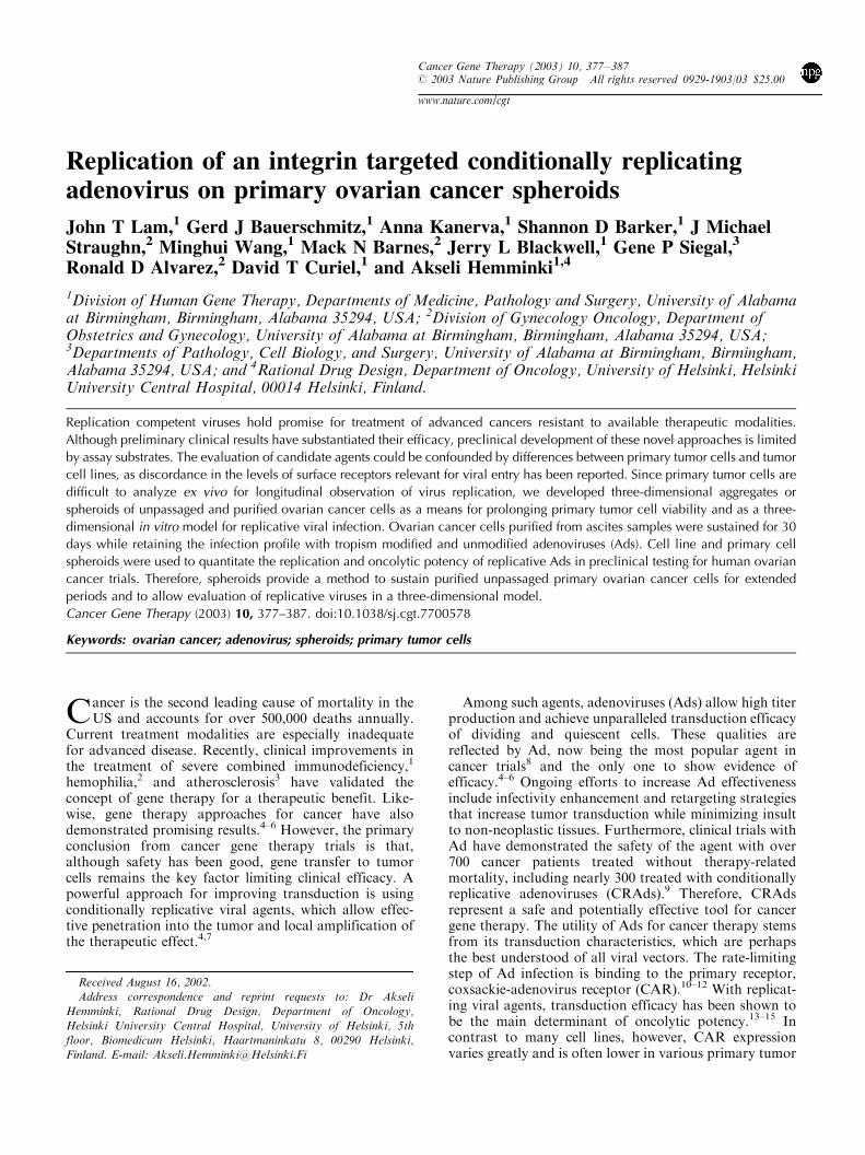
Replication of an integrin targeted conditionally replicating adenovirus on primary ovarian cancer spheroids John T Lam, 1 Gerd J Bauerschmitz, 1 Anna Kanerva, 1 Shannon D Barker, 1 J Michael Straughn, 2 Minghui Wang, 1 Mack N Barnes, 2 Jerry L Blackwell, 1 Gene P Siegal, 3 Ronald D Alvarez, 2 David T Curiel, 1 and Akseli Hemminki 1,4 1 Division of Human Gene Therapy, Departments of Medicine, Pathology and Surgery, University of Alabama at Birmingham, Birmingham, Alabama 35294, USA; 2 Division of Gynecology Oncology, Department of Obstetrics and Gynecology, University of Alabama at Birmingham, Birmingham, Alabama 35294, USA; 3 Departments of Pathology, Cell Biology, and Surgery, University of Alabama at Birmingham, Birmingham, Alabama 35294, USA; and 4 Rational Drug Design, Department of Oncology, University of Helsinki, Helsinki University Central Hospital, 00014 Helsinki, Finland. Replication competent viruses hold promise for treatment of advanced cancers resistant to available therapeutic modalities. Although preliminary clinical results have substantiated their efficacy, preclinical development of these novel approaches is limited by assay substrates. The evaluation of candidate agents could be confounded by differences between primary tumor cells and tumor cell lines, as discordance in the levels of surface receptors relevant for viral entry has been reported. Since primary tumor cells are difficult to analyze ex vivo for longitudinal observation of virus replication, we developed three-dimensional aggregates or spheroids of unpassaged and purified ovarian cancer cells as a means for prolonging primary tumor cell viability and as a three- dimensional in vitro model for replicative viral infection. Ovarian cancer cells purified from ascites samples were sustained for 30 days while retaining the infection profile with tropism modified and unmodified adenoviruses (Ads). Cell line and primary cell spheroids were used to quantitate the replication and oncolytic potency of replicative Ads in preclinical testing for human ovarian cancer trials. Therefore, spheroids provide a method to sustain purified unpassaged primary ovarian cancer cells for extended periods and to allow evaluation of replicative viruses in a three-dimensional model. Cancer Gene Therapy (2003) 10, 377–387. doi:10.1038/sj.cgt.7700578 Keywords: ovarian cancer; adenovirus; spheroids; primary tumor cells C ancer is the second leading cause of mortality in the US and accounts for over 500,000 deaths annually. Current treatment modalities are especially inadequate for advanced disease. Recently, clinical improvements in the treatment of severe combined immunodeficiency, 1 hemophilia, 2 and atherosclerosis 3 have validated the concept of gene therapy for a therapeutic benefit. Like- wise, gene therapy approaches for cancer have also demonstrated promising results. 4–6 However, the primary conclusion from cancer gene therapy trials is that, although safety has been good, gene transfer to tumor cells remains the key factor limiting clinical efficacy. A powerful approach for improving transduction is using conditionally replicative viral agents, which allow effec- tive penetration into the tumor and local amplification of the therapeutic effect. 4,7 Among such agents, adenoviruses (Ads) allow high titer production and achieve unparalleled transduction efficacy of dividing and quiescent cells. These qualities are reflected by Ad, now being the most popular agent in cancer trials 8 and the only one to show evidence of efficacy. 4–6 Ongoing efforts to increase Ad effectiveness include infectivity enhancement and retargeting strategies that increase tumor transduction while minimizing insult to non-neoplastic tissues. Furthermore, clinical trials with Ad have demonstrated the safety of the agent with over 700 cancer patients treated without therapy-related mortality, including nearly 300 treated with conditionally replicative adenoviruses (CRAds). 9 Therefore, CRAds represent a safe and potentially effective tool for cancer gene therapy. The utility of Ads for cancer therapy stems from its transduction characteristics, which are perhaps the best understood of all viral vectors. The rate-limiting step of Ad infection is binding to the primary receptor, coxsackie-adenovirus receptor (CAR). 10–12 With replicat- ing viral agents, transduction efficacy has been shown to be the main determinant of oncolytic potency. 13–15 In contrast to many cell lines, however, CAR expression varies greatly and is often lower in various primary tumor Received August 16, 2002. Address correspondence and reprint requests to: Dr Akseli Hemminki, Rational Drug Design, Department of Oncology, Helsinki University Central Hospital, University of Helsinki, 5th floor, Biomedicum Helsinki, Haartmaninkatu 8, 00290 Helsinki, Finland. E-mail: [email protected] Cancer Gene Therapy (2003) 10, 377–387 r 2003 Nature Publishing Group All rights reserved 0929-1903/03 $25.00 www.nature.com/cgt
-
Upload
independent -
Category
Documents
-
view
3 -
download
0
Transcript of Replication of an integrin targeted conditionally replicating adenovirus on primary ovarian cancer...
Replication of an integrin targeted conditionally replicatingadenovirus on primary ovarian cancer spheroids
John T Lam,1 Gerd J Bauerschmitz,1 Anna Kanerva,1 Shannon D Barker,1 J MichaelStraughn,2 Minghui Wang,1 Mack N Barnes,2 Jerry L Blackwell,1 Gene P Siegal,3
Ronald D Alvarez,2 David T Curiel,1 and Akseli Hemminki1,4
1Division of Human Gene Therapy, Departments of Medicine, Pathology and Surgery, University of Alabamaat Birmingham, Birmingham, Alabama 35294, USA; 2Division of Gynecology Oncology, Department ofObstetrics and Gynecology, University of Alabama at Birmingham, Birmingham, Alabama 35294, USA;3Departments of Pathology, Cell Biology, and Surgery, University of Alabama at Birmingham, Birmingham,Alabama 35294, USA; and 4Rational Drug Design, Department of Oncology, University of Helsinki, HelsinkiUniversity Central Hospital, 00014 Helsinki, Finland.
Replication competent viruses hold promise for treatment of advanced cancers resistant to available therapeutic modalities.
Although preliminary clinical results have substantiated their efficacy, preclinical development of these novel approaches is limited
by assay substrates. The evaluation of candidate agents could be confounded by differences between primary tumor cells and tumor
cell lines, as discordance in the levels of surface receptors relevant for viral entry has been reported. Since primary tumor cells are
difficult to analyze ex vivo for longitudinal observation of virus replication, we developed three-dimensional aggregates or
spheroids of unpassaged and purified ovarian cancer cells as a means for prolonging primary tumor cell viability and as a three-
dimensional in vitro model for replicative viral infection. Ovarian cancer cells purified from ascites samples were sustained for 30
days while retaining the infection profile with tropism modified and unmodified adenoviruses (Ads). Cell line and primary cell
spheroids were used to quantitate the replication and oncolytic potency of replicative Ads in preclinical testing for human ovarian
cancer trials. Therefore, spheroids provide a method to sustain purified unpassaged primary ovarian cancer cells for extended
periods and to allow evaluation of replicative viruses in a three-dimensional model.
Cancer Gene Therapy (2003) 10, 377–387. doi:10.1038/sj.cgt.7700578
Keywords: ovarian cancer; adenovirus; spheroids; primary tumor cells
Cancer is the second leading cause of mortality in theUS and accounts for over 500,000 deaths annually.
Current treatment modalities are especially inadequatefor advanced disease. Recently, clinical improvements inthe treatment of severe combined immunodeficiency,1
hemophilia,2 and atherosclerosis3 have validated theconcept of gene therapy for a therapeutic benefit. Like-wise, gene therapy approaches for cancer have alsodemonstrated promising results.4–6 However, the primaryconclusion from cancer gene therapy trials is that,although safety has been good, gene transfer to tumorcells remains the key factor limiting clinical efficacy. Apowerful approach for improving transduction is usingconditionally replicative viral agents, which allow effec-tive penetration into the tumor and local amplification ofthe therapeutic effect.4,7
Among such agents, adenoviruses (Ads) allow high titerproduction and achieve unparalleled transduction efficacyof dividing and quiescent cells. These qualities arereflected by Ad, now being the most popular agent incancer trials8 and the only one to show evidence ofefficacy.4–6 Ongoing efforts to increase Ad effectivenessinclude infectivity enhancement and retargeting strategiesthat increase tumor transduction while minimizing insultto non-neoplastic tissues. Furthermore, clinical trials withAd have demonstrated the safety of the agent with over700 cancer patients treated without therapy-relatedmortality, including nearly 300 treated with conditionallyreplicative adenoviruses (CRAds).9 Therefore, CRAdsrepresent a safe and potentially effective tool for cancergene therapy. The utility of Ads for cancer therapy stemsfrom its transduction characteristics, which are perhapsthe best understood of all viral vectors. The rate-limitingstep of Ad infection is binding to the primary receptor,coxsackie-adenovirus receptor (CAR).10–12 With replicat-ing viral agents, transduction efficacy has been shown tobe the main determinant of oncolytic potency.13–15 Incontrast to many cell lines, however, CAR expressionvaries greatly and is often lower in various primary tumor
Received August 16, 2002.
Address correspondence and reprint requests to: Dr Akseli
Hemminki, Rational Drug Design, Department of Oncology,
Helsinki University Central Hospital, University of Helsinki, 5thfloor, Biomedicum Helsinki, Haartmaninkatu 8, 00290 Helsinki,
Finland. E-mail: [email protected]
Cancer Gene Therapy (2003) 10, 377–387r 2003 Nature Publishing Group All rights reserved 0929-1903/03 $25.00
www.nature.com/cgt

cells types11,12,16–21 including ovarian cancers.22–25 Con-ceivably, the selection process involved in obtainingimmortalized cell lines from cancer cells could causeother changes also. Therefore, primary tumor cells mightbetter model in vivo tumor behavior for the analysis ofnovel agents.Problems encountered using primary tumor samples
include contaminating normal cells and limited viabilityof primary tumor cells in vitro. To obtain a pure tumorcell population, we have developed a method forpurification of ovarian cancer cells from patient ascites.26
While these cells survive in vitro for only a few dayswithout passaging,27 many CRAd assays require severalweeks for completion. The clonal selection involved inadaptation to replication and growth as a monolayer invitro can cause genotypic and phenotypic changes, whichcould contribute to the discordance seen in expression ofreceptors involved in viral entry. To address this need, wereport here the development of ovarian cancer spheroids,which allow extension of purified unpassaged primarytumor cell viability for analysis of replicative viral agentsin a three-dimensional system.Spheroids are three-dimensional clusters of cells and
have been utilized to study tumor biology, chemotherapy,and radiation therapy.28 Gene therapy studies haveemployed spheroids based on cell lines,29,30 and a recentstudy reports the use of unpurified glioma explants forstudying transduction with nonreplicative Ads.31 Here, wereport the first spheroid model for assessing replicationcompetent viruses on purified primary cancer cells. First,we investigated the viability of primary cancer cells in aspheroid system. Then, Ad infectivity patterns werecompared in spheroid and monolayer cultures. Next,replication competent Ads expressing a reporter genewere used to assess adenovirus replication kinetics inspheroids. Finally, primary tumor cell spheroids wereutilized to evaluate replication of a clinically applicableCRAd in patient samples.
Materials and methods
Cell lines and primary tumor cells
The human ovarian adenocarcinoma cell lines SKO-V3.ip1, Hey, and OV-4 were obtained from Drs JanetPrice and Judy Wolf (both of the University of Texas MDAnderson Cancer Center, Houston, TX), and Dr TimothyJ Eberlein (Harvard Medical School, Boston, MA),respectively. The 293 cell line was purchased fromMicrobix (Toronto, Canada). All cell lines were culturedin conditions recommended by their suppliers. Infectionmedia had 2% fetal bovine serum. RPMI-1640 growthmedium supplemented with 10% fetal bovine serum,2mM l-glutamine, 100 IU/ml penicillin, and 25 mg/mlstreptomycin was used for culture of the primary ovarianadenocarcinoma cells that were purified from malignantascites samples obtained at our institution followinginformed consent from patients with pathologicallyconfirmed ovarian adenocarcinoma. The previouslydescribed purification of the tumor cells26 was utilized
to achieve a 63–96% tumor cell purity. After mosterythrocytes were removed with a lysis buffer, the tumorcells were extracted from benign cells using the CC49monoclonal mouse antibody (Biological ResourcesBranch of the National Cancer Institute, Rockville,MD), Pan Mouse IgG Dynabeads (Dynal AS, Oslo,Norway) coated with anti-mouse IgG antibodies, and amagnetic particle concentrator (Dynal).
Recombinant Ads
Ad5luc1,32 Ad5lucRGD,22 Ad5-Luc3,33 and Ad5-D24RGD34 have been previously reported. The Ad5-D24RGD was propagated on A549 cells. The otherviruses were propagated on 293 cells, and all were purifiedon cesium chloride gradients. The viral particle (VP)concentrations determined at 260 nm were 1.28� 1011,3.25� 1011, 1.1� 1012, and 2.6� 1012VP/ml for Ad5luc1,Ad5lucRGD, Ad5-Luc3, and Ad5-D24RGD, respectively.To determine the functional titer of the viruses, weperformed plaque assays with an overnight infection toallow entry of all functional virions without confoundingby receptor density. The Ad5luc1, Ad5lucRGD, Ad5-Luc3, and Ad5-D24RGD viruses had titers of 4.7� 1010,2.9� 1010, 2� 1010, and 5.2� 1010 plaque-forming units(PFU)/ml, respectively. The E1 region was amplified withPCR to detect the absence of wild-type contamination(Ad5luc1 and Ad5lucRGD), the presence of E1, or thepresence of the 24 bp deletion, respectively.
Culture of spheroids
To prepare agar-coated flasks and plates, agar wasdissolved in deionized water to reach a 3% concentrationand autoclaved. The solution was poured into flasks ormultiwell plates and allowed to solidify at roomtemperature. For culture of spheroids, cells were sus-pended with growth media into concentrations of 1–10million cells/ml, added to the agar-coated containers, andsubjected to intermittent agitation on a rocking platform.
Immunostaining of tumor primary spheroids
Primary tumor cell spheroids were transferred in Hanks’balanced salt solution 1 and 30 days after purificationonto glass slides, air-dried, and fixed with Cell-Fixx(Thermo Shandon, Pittsburgh, PA). Immunohistochem-ical staining with antibodies specific for B72.3 or EMA(epithelial membrane antigen) (both Ventana MedicalSystems, Inc., Tucson, AZ) were performed by usingvendor recommended protocols in an ES-automatedimmunostainer (Ventana). The B72.3 antibody has beenshown to have a high sensitivity and specificity forovarian cancer cells.26,35,36
Infections of spheroids and monolayers
The tumor cell line spheroids composed of SKOV3.ip1,OV-4, or Hey cells were removed from their agar-coatedflasks and filtered through a 200 mesh screen with 75 mmpores (Sigma Chemical Co., St Louis, MO) to separatethe larger spheroids from the smaller ones and single cells.
Adenovirus replication on primary tumor spheroidsJT Lam et al
378
Cancer Gene Therapy

A fraction of the spheroids was used to estimate theaverage number of surface cells within each spheroid bystaining with crystal violet (see below). The remainingspheroids were divided into equal aliquots, resuspended in500ml of infection media with 0, 1, 10, or 100 PFU/surfacecell of Ad5luc1 or Ad5lucRGD, and incubated on arocking platform for 1 hour before transfer in freshgrowth medium into separate agar-coated wells. After 30-hour incubation, Bright-Glo Luciferase Assay Reagent(Promega Corporation, Madison, WI) was used to assaygene transfer. Normalization for variance in cell numberbetween samples was achieved by measuring proteinconcentration with the Bio-Rad DC protein assay (Bio-Rad Laboratories, Hercules, CA) and surface cell staining(see below). Primary ovarian adenocarcinoma cell spher-oids were infected in similar fashion except that nofiltration or staining was performed because of theirsmaller size.Tumor cell line monolayer infections were performed
by seeding 105 SKOV3.ip1, OV-4, or Hey cells into eachwell. After overnight incubation, growth media from eachwell was replaced by 500ml of infection media with either0, 1, 10, or 100PFU/cell of Ad5luc1 or Ad5lucRGD.After 1-hour incubation on a rocking platform, the virusand infection medium solution was replaced by freshgrowth medium. Following a 30-hour incubation, theLuciferase Assay System (Promega Corporation, Madi-son, WI) was used to quantify gene transfer for the tumorcell lines. Variations in cell number were normalized forby the Bio-Rad DC protein assay (Bio-Rad). Infection ofpurified primary ovarian adenocarcinoma cell monolayerswas performed as described above, except that they wereseeded onto Biocoat Cell Environment type I collagen-coated 24-well plates (Becton Dickinson Labware, Bed-ford, MA) instead of standard multiwell plates, and theirluciferase expression was quantified by Bright-Glo Lucifer-ase Assay Reagent (Promega Corporation, Madison, WI).Next, spheroids were infected with replication compe-
tent Ad. After mesh filtering and surface cell staining (seebelow), equal aliquots of Hey spheroids were transferredto 3% agar-coated wells with 2% infection media and 0,10, 100, or 1000 PFU/surface cell of Ad5-Luc3. After 1-hour incubation on a rocking platform, enough fetalbovine serum was added to achieve a 10% concentrationin each well. Beginning 24 hours after the infection anddaily thereafter, the supernatant was entirely extractedfrom each well and replaced with fresh growth media.Supernatant luciferase has been measured to quantifyoncolysis.37 We employed this assay using Bright-GloLuciferase Assay Reagent until all luminometer readingsreached baseline. Mock infection measurements weresubtracted from the readings of the viral-infected groups.Primary ovarian adenocarcinoma cell spheroids wereinfected in similar fashion, but because of their smallsize, no filtration or staining was performed.Primary ovarian adenocarcinoma spheroids were in-
fected with Ad5-D24RGD or the nonreplicative controlAd5lucRGD by resuspension in infection medium with 0,0.2, 2, or 2 PFU/surface cell of either virus in 3% agar-coated wells. At 1 hour after infection, enough fetal
bovine serum was added to achieve a 10% concentrationin each well. At 24 hours after infection, the spheroidsfrom each well were divided into multiple equal aliquots(one for each daily measurement) and each resuspended in200ml of growth medium. One aliquot of spheroids fromeach viral dose was frozen at �201C each day afterinfection. DNA was extracted and purified from thecellular fractions of these samples with the QIAamp DNAMini Kit (Qiagen Inc., Valencia, CA) and from thesupernatant fractions with the QIAamp DNA Blood MiniKit (Qiagen). Quantitative PCR has been employed toanalyze adenovirus replication38–40 and was performed onthese DNA samples with a Roche Lightcycler (RocheDiagnostics Corporation, Indianapolis, IN) as de-scribed.41 The viral DNA copies detected were normalizedby quantitative PCR for actin copies, except for the firstexperiment, which was normalized by cell number. Sincezero cannot be displayed on a logarithmic scale, baselinevalues were set as 1 and sample readings are presentedrelative to the baseline.
Staining of spheroid surface cells
Since replication incompetent Ad transduction wasexpected to be limited to the superficial layers ofspheroids,29 we used the ratio of Ad PFU to spheroidsurface cells rather than the Ad PFU to total cell ratio forinfections. To estimate the proportion of surface cells ineach particular spheroid sample, a fraction of each groupwas mixed with crystal violet and incubated at roomtemperature for 20–60 minutes. Washings with phos-phate-buffered saline removed excess crystal violet, andthe spheroids were resuspended in 2.5% (10� ) trypsinfollowed by disruption into individual cells by repeatedvortexing with intermittent 371C incubation. The numberof stained surface cells and unstained core cells was thendetermined by microscopic examination.
Results
Spheroids provide a novel method for prolonging theviability of purified unpassaged tumor primary samples
Both ovarian cancer cell lines and primary ovarian cancercells spontaneously aggregated to form spheroids within aday of their resuspension in agar-coated containers. Mosttumor cell line spheroids ranged in size from 50 to1000mm in diameter, and primary tumor cell spheroidswere usually smaller, ranging from 10 to 100mm indiameter. Microscopic evaluation showed that the pur-ified primary tumor cell spheroids were morphologicallysimilar to the unpurified primary tumor cell clusters infresh patient ascites (Fig 1a). Spheroids from four of fivepatient samples showed reactive staining (brown) with theovarian adenocarcinoma-specific B72.3 antibody after 1day of culture (Fig 1b). The same four samples alsoshowed the same positive staining after 30 days in culture(Fig 1c). Spheroids from five primary ovarian adenocar-cinoma cell samples showed reactivity with anotheradenocarcinoma-specific antibody, anti-EMA, after 1
Adenovirus replication on primary tumor spheroidsJT Lam et al
379
Cancer Gene Therapy

day in culture. All five of these samples also stainedpositively with EMA after 30 days in culture (data notshown). In all positive samples, neither antibody showedsignificant decrease in staining intensity or cellulardistribution after the 30-day spheroid incubations.
Spheroids provide a valid model for assessment ofadenovirus infectivity
The validity of the surface cell staining method forestimation of surface cell proportion was substantiated bymultiple stainings compared with the following mathe-matical equations: 1) surface area of sphere¼ 4pr2; 2)volume of sphere¼ (4/3) pr3; 3) and area of circle¼ pr2.Measurement of spheroid and individual cell diametersallowed for estimation of spheroid and single cell volumesin addition to exposed surface area of both spheroids andindividual surface cells. Based on these calculations, onegroup of spheroids was mathematically estimated to have16% surface cells. Actual staining of these spheroidsshowed that approximately 23% of all cells were surfacecells.
Since previous reports have shown that the integrinbinding RGD-4C motif in the HI-loop of the serotype 5Ad fiber allows CAR-independent transduction of ovar-ian cancer cells,22 we used Ad5lucRGD and the isogenicAd5-Luc1 (wild-type fiber) to indirectly assess CAR andintegrin expression on spheroids compared to mono-layers. Infections of SKOV3.ip1, OV-4, and Hey mono-layers with Ad5lucRGD produced one to two orders ofmagnitude increased gene expression in comparison toAd5luc1 at doses of 1, 10, and 100PFU/cell. Similartransduction patterns were seen for the respectivespheroid infections (Fig 2a–c).In primary cell spheroids derived from Patient A,
Ad5lucRGD exhibited 7.4-, 10.0-, and 10.0-fold greaterluciferase expression than Ad5luc1 at 1, 10, and 100 PFU/surface cell, respectively. In monolayers derived fromPatient A cells, the respective differences in gene transferwere 15-, 8.3-, and 3.8-fold (Fig 2d). For Patient B,Ad5LucRGD infections resulted in 8.9-, 19-, and 7.9-foldhigher luciferase readings than Ad5luc1 infections inspheroids, while for monolayers these differences were4.8-, 4.0-, and 2.7-fold, respectively (Fig 2e).
100 µm 100 µm
100 µm
c
ab
Figure 1 Spheroids sustain the viability of purified primary tumor cells: unstained viable ovarian adenocarcinoma primary spheroids (a), primary
ovarian adenocarcinoma cell spheroids with reactive tumor-specific B72.3 immunoperoxidase staining (brown) 1 day after purification (the
smaller spheres are magnetic beads used in the purification process) (b), and primary ovarian adenocarcinoma cell spheroids with tumor-specific
B72.3 immunoperoxidase reactivity 30 days after purification (c). Thus, primary tumor cells in spheroids maintain viability for 30 days.
Adenovirus replication on primary tumor spheroidsJT Lam et al
380
Cancer Gene Therapy

Spheroids provide a three-dimensional model forquantifying adenovirus replication and oncolysis in celllines and purified unpassaged primary tumor cells
Spheroids comprised of Hey cells were infected withvarious doses of replication competent Ad5-Luc3 and
showed measurable supernatant luciferase ranging from1� 102 to 1� 107 relative light units (RLU) 9–14 daysafter infection (Fig 3a). Despite complete daily mediumchanges, all three groups showed an overall increase insupernatant luciferase over the first 2–5 days and peaks inluciferase release on days 2, 3, and 5 with doses of 1000,
PFU/surface cell PFU/cell
PFU/surface cell PFU/cell
PFU/surface cell PFU/cell
PFU/surface cell PFU/cell
PFU/surface cell PFU/cell
RL
U/m
g s
urf
ace
cell
pro
tein
OV-4
Spheroids Monolayers
RL
U/m
g s
urf
ace
cell
pro
tein
RL
U/m
g s
urf
ace
cell
pro
tein
RL
U/m
g s
urf
ace
cell
pro
tein
RL
U/m
g s
urf
ace
cell
pro
tein
RL
U/m
g p
rote
inR
LU
/mg
pro
tein
RL
U/m
g p
rote
inR
LU
/mg
pro
tein
RL
U/m
g p
rote
in
Patient B
Patient A
Hey
SKOV3.ip1
1.0E-01
1.0E+01
1.0E+03
1.0E+05
1.0E+07
1 10 100
1.0E-01
1.0E+01
1.0E+03
1.0E+05
1.0E+07
1 10 100
1.0E-01
1.0E+01
1.0E+03
1.0E+05
1.0E+07
1 10 100
1.0E-01
1.0E+01
1.0E+03
1.0E+05
1.0E+07
1 10 100
1.0E-01
1.0E+01
1.0E+03
1.0E+05
1.0E+07
1 10 1001.0E-01
1.0E+01
1.0E+03
1.0E+05
1.0E+07
1 10 100
1.0E-01
1.0E+01
1.0E+03
1.0E+05
1.0E+07
1 10 100
1.0E-01
1.0E+01
1.0E+03
1.0E+05
1.0E+07
1 10 100
1.0E-01
1.0E+01
1.0E+03
1.0E+05
1.0E+07
1 10 100
1.0E-01
1.0E+01
1.0E+03
1.0E+05
1.0E+07
1 10 100
a
b
c
d
e
Figure 2 Spheroids retain an adenovirus infectivity phenotype similar to that of monolayers. Ads with the wild-type fiber of serotype 5 such as
Ad5luc1 (white bars) bind to the coxsackievirus-adenovirus receptor and internalize utilizing cellular integrins. Modification of the fiber with RGD-
4C as in Ad5lucRGD (black bars) allows binding to both CAR and integrins. Therefore, the ratio of transgene expression (RLU) following infection
with these viruses provides an indirect functional estimate of integrin and CAR levels on spheroids (left panel) in comparison to monolayers (right
panel). Similar RLU ratios in monolayers and spheroids suggests similar expression of the receptors relevant for adenovirus binding and entry for
SKOV3.ip1 (a), OV4 (b), and Hey (c) cells and for two patient samples purified from malignant ascites (d, e). Error bars indicate standard deviations.
Adenovirus replication on primary tumor spheroidsJT Lam et al
381
Cancer Gene Therapy

100, and 10 PFU/surface cell, respectively. Purifiedunpassaged primary ovarian cancer cell spheroids infectedwith Ad5-Luc3 showed measurable luciferase expressionranging from 1� 102 to 1� 107 RLU in their supernatants(Fig 3b). Measurable levels of supernatant luciferaseremained up to 17 days after infection.
Spheroids allow use of purified unpassaged primarytumor cells in a three-dimensional model for quantifyingreplication of a clinically applicable CRAd
Quantitative PCR detection of Ad5-D24RGD in infectedprimary ovarian adenocarcinoma cell spheroids gavemeasurable results throughout the duration of all experi-ments (up to 17 days) at all viral doses used (Fig 4).Furthermore, the number of gene copies generallyincreased over the durations of each experiment. Thefirst infection was observed for 5 days during which viralDNA copies increased 2.6� 105-, 1.9� 109-, and3.0� 107-fold for doses of 0.2, 2, and 20PFU/surfacecell, respectively (Fig 4a). The longer duration of thesecond experiment allowed samples with 2 and 20PFU/surface cell doses of Ad5-D24RGD to reach peak virus
production 9 and 11 days after infection, respectively (Fig4b). In comparison to day 1, these samples showed 168-and 708-fold increases in viral copy number, respectively.The two last infections show peaks in viral DNA at 8 and14 days after infection (Fig 4c) and at days 14 and 15 afterinfection (Fig 4d) at doses of 2 and 20 PFU/surface cell,respectively. Increases in viral DNA were 2730- and 3600-fold (Fig 4c) and 4630- and 1150-fold (Fig 4d),respectively. Gene copies of the E1-deleted control virusAd5lucRGD showed 97.6-, 39.5-, and 149-fold increasesin the second, third, and fourth experiments, respectively(Fig 4b–d). To estimate total virus production by thespheroids, cumulative virus copy number was calculated(Fig 4e–h). In all cases, the highest cumulative virus copynumber was detected with 20 PFU/surface cell of Ad5-D24RGD. When compared to the respective dose of theE1-deleted control virus, Ad5-D24RGD produced 9-, 86-,and 25-fold more virus (Fig 4f–h).
Discussion
Gene therapy has recently produced clinical break-throughs in treatment of a heterogeneous group ofhereditary and acquired disorders, including severecombined immunodeficiency,1 hemophilia,2 and vasculardiseases.3 These advances were based on incrementalimprovements in vector systems, and these developmentshave been directly dependent on the availability andutility of appropriate model systems. Adenoviral-basedapproaches have shown some clinical utility for treatmentof advanced malignant disease.4–6 One of the mostpromising areas of cancer gene therapy is CRAds, thesafety of which has been demonstrated for intratumoral,intraperitoneal, intra-arterial and intravenous adminis-tration.42 Although single agent clinical efficacy has beenrelatively modest,42–44 combination with chemotherapyhas shown clinical efficacy.4 To further develop thetumor-penetrating capabilities of CRAds, improved pre-clinical models are required for hypothesis testing inclinically relevant substrates.While cell lines are widely used for analysis of
antitumor agents, recent findings suggest that their levelsof CAR may correspond poorly with those in primarytumor cells obtained from patient specimens.11,12,16–21
This discordance has crucial implications because theCAR level of any tumor may represent the keydeterminant of infectivity for Ad.10,11 In particular,advanced cases of various cancer types seem to expresshighly variable levels of CAR,45,46 and the expression mayinversely correlate with the grade of the tumor.45 CARmay be a part of the tight junction.47 It could function asan adhesion molecule,48 and its downregulation may beassociated with overactivity of the RAS/MAPK path-way.46 Importantly, it has been established that infectivityof target cells determines the oncolytic potential of aCRAd.13–15
Therefore, primary tumor cells could provide a morestringent and clinically relevant model for analysis ofreplication competent viruses.
Hey
Patient C
1.0E+02
1.0E+03
1.0E+04
1.0E+05
1.0E+06
1.0E+07
1.0E+08
1.0E+00
1.0E+01
1.0E+02
1.0E+03
1.0E+04
1.0E+05
1.0E+06
1.0E+07
1 2 3 4 8 95 6 7 10 11 12 13 14 15 16
days after infection
rela
tive
lig
ht
un
its
days after infection
rela
tive
lig
ht
un
its
10 PFU/surface cell
100 PFU/surface cell
1000 PFU/surface cell
10 PFU/surface cell
100 PFU/surface cell
1000 PFU/surface cell
1 2 3 4 8 95 6 7 10 11 12 13 14 15 16 17
a
b
Figure 3 Spheroids allow quantitation of adenovirus replication and
oncolysis. Supernatant was collected daily and luciferase activity
was measured. Since detection of supernatant luciferase requires
lysis of producer cells, these values should correlate with virus
replication-mediated oncolysis. (a) Ad5-Luc3 replication is quantified
in Hey cell line spheroids. (b) Ad5-Luc3 replication is quantified in
primary ovarian adenocarcinoma spheroids. All values have had
mock infection readings subtracted.
Adenovirus replication on primary tumor spheroidsJT Lam et al
382
Cancer Gene Therapy

mock2 PFU/surface cell Ad5lucRGD20 PFU/surface cell Ad5lucRGD0.2 PFU/surface cell Ad5-∆24RGD2 PFU/surface cell Ad5-∆24RGD20 PFU/surface cell Ad5-∆24RGD
1.0E+00
1.0E+02
1.0E+04
1.0E+06
1.0E+08
1.0E+10
1.0E+12
1.0E+14
1 3 4 5days after infection
rela
tive
DN
A c
op
ies
1.0E+00
1.0E+02
1.0E+04
1.0E+06
1.0E+08
1.0E+10
1.0E+12
1.0E+14
1 3 4 5
days after infection
cum
ula
tive
DN
A c
op
ies
1.0E+00
1.0E+02
1.0E+04
1.0E+06
1.0E+08
1.0E+10
1.0E+12
1 2 3 4 5 6 7 8 9 10 11 12 13 14 15
days after infection
rela
tive
DN
A c
op
ies
1.0E+04
1.0E+05
1.0E+06
1.0E+07
1.0E+08
1.0E+09
1.0E+10
1.0E+11
days after infection
cum
ula
tive
DN
A c
opie
s
1.0E+001.0E+011.0E+021.0E+031.0E+041.0E+051.0E+061.0E+071.0E+08
1 2 3 4 5 6 7 8 9 10 11 12 13 14 15
days after infection
rela
tive
DN
A c
op
ies
1.0E+01
1.0E+02
1.0E+03
1.0E+04
1.0E+05
1.0E+06
1 2 3 84 5 6 7 9 10 11 12 13 14 15
days after infection
cum
ula
tive
DN
A c
op
ies
1.0E+00
1.0E+01
1.0E+02
1.0E+03
1.0E+04
1.0E+05
1.0E+06
1.0E+07
1.0E+08
1.0E+09
1 2 3 4 5 6 7 8 9 1011121314151617
days after infection
rela
tive
DN
A c
op
ies
1.0E+04
1.0E+05
1.0E+06
1.0E+07
1.0E+08
1.0E+09
1.0E+10
1 2 3 4 5 6 7 8 9 1011121314151617
1 2 3 4 5 6 7 8 9 10 1112 13 14 15
days after infectioncu
mu
lati
ve D
NA
cop
ies
2 2
a
b
c
d
e
f
g
h
Figure 4 Spheroids of purified primary tumor cells can quantify replication of a clinically applicable conditionally replicating adenovirus (CRAd).
Aliquots of ovarian cancer cell spheroids from four patient ascites samples were infected with various doses of Ad5-D24RGD or the E1-deleted
control, Ad5lucRGD. Viral copy number was measured with quantitative PCR. (a–d) Increase in the CRAd copy number suggests replication of
the agent in the primary tumor cells. (e–h) Presentation of viral gene copies as cumulative values allows comparison of agents with regard to total
virus produced. Mock values were subtracted from all displayed data.
Adenovirus replication on primary tumor spheroidsJT Lam et al
383
Cancer Gene Therapy

The limited viability of unpassaged primary ovariancancer cells in vitro, however, is insufficient to provide auseful substrate for CRAd assays.27 Thus, we developedan approach based on forming spheroids of primarycancer cells purified from patient samples. The purifica-tion step may be crucial since contaminating non-neoplastic cells with genotypes and phenotypes differentfrom tumor cells could confound infectivity data. Primaryovarian adenocarcinoma cells were cultured as spheroidsfor 1 and 30 days. Subsequent immunostaining withantibodies specific for adenocarcinoma, B72.3 and EMA,showed consistent reactivity (Fig 1b, c). The persistentstaining with these antibodies and the identical morphol-ogy after 30 days suggests that the spheroids of primarytumor cells were viable even after this prolonged culture.Furthermore, the consistent degree of staining and thehistological appearance suggests that the proportion oftumor cells in the spheroids remains constant. Therefore,spheroids provide a novel means for retaining the viabilityof purified primary tumor cells.A potential concern pertaining to the spheroid model is
that the expression of surface receptors, which arerequired for viral infectivity and possibly cell adhesion,48
in spheroids might be altered. Compared to monolayers,some spheroids systems have been shown to displayvariations in gene expression,49,50 morphology,51,52 andsurface receptor expression.53–56 Such differences mightimpact the correlation of infectivity data from monolayersto spheroids, and more importantly, to in vivo tumors. Toinvestigate this potential problem, we used an indirectmeans to assess the infectivity of spheroids and mono-layers. Modification of the Ad fiber with an RGD-4Cmotif (Ad5lucRGD) enhances infectivity and improvestransduction by allowing binding to both avb groupintegrins and CAR,22,24,41 whereas Ad with wild-typefiber, such as our isogenic control Ad5luc1, binds only toCAR and then undergoes subsequent integrin-mediatedinternalization. By comparing reporter gene expressionmediated by Ad5lucRGD and Ad5luc1 in monolayersversus spheroids, we can thus indirectly estimate therelative expression levels of CAR and the relevantintegrins. Importantly, the differences in reporter geneexpression resulting from transduction with Ad5luc1 andAd5lucRGD were very similar in monolayers in compar-ison to spheroids (Fig 2a–c). This suggests that expressionof the receptors relevant for Ad binding and entry doesnot markedly differ between monolayers and spheroids.Furthermore, the same was true for purified primarytumor cells (Fig 2d, e).Having established the validity of spheroids as a model
for Ad infection, a replication competent Ad expressingluciferase (Ad5-Luc3) was used to assess replication andoncolysis in spheroids. Since luciferase is an intracellularprotein, it is expected to be present in the supernatantonly if its producer cells undergo lysis37,57 as they doduring Ad replication. In addition, when luciferase isexpressed from a nonreplicating vector, maximum pro-duction is observed between 24 and 72 hours.58 Althoughsupernatant was completely replaced daily for luciferaseactivity assay, it was detectable up to 14 days after
infection (Fig 3a). This persistence and increase ofluciferase measurements up to day 6 (10 PFU/surfacecell) provides preliminary evidence suggesting replication.As expected, infections with higher titers of virus reachedpeak supernatant luciferase levels earlier than those withlower titers. In addition, higher viral titers were associatedwith shorter durations of measurable supernatant lucifer-ase levels.When primary tumor cell spheroids were infected with
Ad5-Luc3, luciferase was detected up to day 16 (Fig 3b).Interestingly, the pattern of luciferase release differedfrom the cell line spheroids as maximum readings weredetected only 1 day after infection. This variation couldbe because of the smaller average size but larger numberof primary cell spheroids resulting in different kinetics ofvirus penetration and dispersion. It can be speculated thatthe transient decrease in luciferase activity on day 2 (100and 1000PFU/surface cell) might correspond with asecond cycle of replication after a collective viral releaseon day 1 and before a second wave of viral dispersion onday 3. The quantitative differences seen in these experi-ments suggest that meaningful comparison of thereplicative and oncolytic potential of different replicativeviral vectors should be possible. Candidate clinical vectorscan be evaluated by comparing their time to reach peakand baseline concentrations of the transgene product inthe supernatant. More potent vectors might be expectedto replicate and lyse tumor cells faster bringing shortertimes to peak and baseline levels.Ad5-D24RGD is a promising novel CRAd34 currently
undergoing preclinical evaluation for treatment of ovar-ian cancer patients.59 Replication of this agent was testedon primary ovarian cancer cell spheroids (Fig 4). At allviral doses, quantitative PCR detected an increase in viralDNA as a function of time, suggesting replication. Thelonger experiments allowed determination of viral DNApeak levels 8–15 days after infection and increases in viralDNA measuring two or three orders of magnitude (Fig4b–d). The first infection (Fig 4a) showed greaterreplication of Ad5-D24RGD ranging from five to nineorders of magnitude during a shorter course of only 5days. This difference could possibly be because of thesmaller size of spheroids from this patient sample, whichmight have led to greater initial infectivity and subse-quently more rapid replication. To demonstrate moreclearly the total replication achieved with each virus,cumulative amounts of virus were calculated (Fig 4e–h).This read-out avoids confusion caused by daily variationsin virus produced, and provides a clear visual end point.These methods can be applied to quantitation andcomparison of other candidate clinical agents.Interestingly, we observed replication of the E1-deleted
control virus (Ad5LucRGD) on all samples tested. Wild-type contamination was not detected with PCR. Further,three ovarian cancer and one teratocarcinoma cell lineswere infected with the same batch of this virus followedby incubation of up to 17 days, without evidence ofcytopathic effect, suggesting lack of wild-type virus.59
Possible reasons for this phenomenon include thepresence of minute amounts of wild-type contaminant
Adenovirus replication on primary tumor spheroidsJT Lam et al
384
Cancer Gene Therapy

not detectable by PCR or on the cell lines. Alternatively,the primary cancer cells might have transcomplementedthe missing E1 proteins. This phenomenon has beenreported before with the presence of IL-6 or papilloma-virus E6 and E7 allowing replication of E1-deletedviruses.60–64 In fact, it was demonstrated that factorsproduced by primary ovarian cancer cells allowed similarreplication of an E1-deleted virus and a wild-typeadenovirus.60
While xenografts have traditionally been utilized forinvestigation of therapeutic efficacy, receptor levels onthese cancer cell lines may not correspond well withprimary tumors.11,12,16–25 In addition, in vitro assays areoften more practical than in vivo analysis because theyallow daily sampling and evaluation of the substrates.Furthermore, improved in vitro models could help reducethe need for animal testing. Importantly, primary tumorspecimens represent the sole means for observing indivi-dual variability between clinical ovarian cancer cases.Thus, by providing a stringent model for assessingreplicative agents on primary tumor substrates, spheroidsshould be useful for complementing therapeutic data fromxenograft models. An interesting concept is a combined invitro–in vivo model. Conceivably, in vitro spheroids couldbe inoculated subcutaneously or intraperitoneally in nudeor SCID mice. This would provide an animal model basedon purified primary tumor cells derived from a patientsample. In this paper, we used spheroids for analysis ofCRAds, but the model should be applicable to any type ofreplicating agent. Replication competent viruses currentlyundergoing preclinical or clinical evaluation for cancertreatment include herpes simplex virus,65,66 vaccinia,67
Newcastle disease virus,68 parvovirus,69 reovirus,70
measles virus,71 retrovirus,72 influenza virus,73 polio-virus,74 vesicular stomatitis virus,75 and avian sarcomaleucosis virus.76 Therefore, replication competent virusesrepresent a dynamic field desperate for practical sub-strates for primary tumor specimen analyses.In summary, we describe a novel approach for
analyzing replication competent anticancer agents. Spher-oids provide a three-dimensional model, which couldrepresent in vivo tumors better than two-dimensionalmonolayers. Importantly, spheroids consisting of purifiedprimary tumor cells would allow analysis of the moststringent available clinical substrates and eliminatepossible confounding variables involved in working withcell lines. This model appears useful for analyzingreplicative viral agents in primary tumor material, therebyproviding crucial preclinical data for translational re-search.
Acknowledgments
We gratefully acknowledge the excellent technical assis-tance of Jan Shultz, Sherri Coffman, and Mitzi Fincher.Gene transfer assays were performed in part at the UABGene Therapy Center Correlative Laboratories for Hu-man Clinical Trials. This study was supported by theUnited States Public Health Service Training Grant T32
CA75930, the University of Alabama Health ServicesFoundation Interdisciplinary Corroborative Laboratoryfor Gene Therapy Clinical Trials, NIH grants CA74243,HL 63736, RO1 CA83821, IT32 CA75930, P50 CA83591,P50 CA89019, PC 99-1018, DAMD 17-00-1-0002, RO1CA94084, RO1 CA93796, DAMD 17-98-1-8571, theLustgarten Foundation LF043, the CapCure Foundation,the Damon Runyon-Walter Winchell Cancer ResearchFund, the Sigrid Juselius Foundation, the Emil AaltonenFoundation, the Maud Kuistila Foundation, and theFinnish Medical Foundation, the Academy of Finland,the Finnisha Cancer Society, Biocentrum Helsinki,University of Helsinki Internal Funds.
References
1. Cavazzana-Calvo MH-BS, de Saint Basile G, Gross F, et al.Gene therapy of human severe combined immunodeficiency(SCID)-X1 disease. Science. 2000;288:669–672.
2. Kay MA, Manno CS, Ragni MV, Larson PJ, et al. Evidencefor gene transfer and expression of factor IX in haemophiliaB patients treated with an AAV vector. Nat Genet.2000;24:257–261.
3. Isner JM, Asahara T. Angiogenesis and vasculogenesis astherapeutic strategies for postnatal neovascularization. JClin Invest. 1999;103:1231–1236.
4. Khuri FR, Nemunaitis J, Ganly I, et al. A controlled trial ofintratumoral ONYX-015 a selectively-replicating adeno-virus, in combination with cisplatin and 5-fluorouracil inpatients with recurrent head and neck cancer. Nat Med.2000;6:879–885.
5. Nemunaitis J, Swisher SG, Timmons T, et al. Adenovirus-mediated p53 gene transfer in sequence with cisplatin totumors of patients with non-small-cell lung cancer. J ClinOncol. 2000;18:609–622.
6. Sandmair AM, Loimas S, Puranen P, et al. Thymidinekinase gene therapy for human malignant glioma, usingreplication-deficient retroviruses or adenoviruses.Hum GeneTher. 2000;11:2197–2205.
7. Kirn D, Martuza RL, Zwiebel J. Replication-selectivevirotherapy for cancer: Biological principles, riskmanagement and future directions. Nat Med. 2001;7:781–787.
8. http: //www#.wiley.co.uk/genmed/clinical. Gene TherapyClinical Trials Website. The Journal of Gene Medicine.Updated September 2001; John Wiley & Sons Ltd.
9. Hemminki A, Alvarez RD. Adenoviruses In Oncology: AViable Option? [In Process Citation]. BioDrugs. 2002;16.
10. Roelvink PW, Mi Lee G, Einfeld DA, et al. Identification ofa conserved receptor-binding site on the fiber proteins ofCAR-recognizing adenoviridae. Science. 1999;286:1568–1571.
11. Li Y, Pong RC, Bergelson JM, et al. Loss of adenoviralreceptor expression in human bladder cancer cells: apotential impact on the efficacy of gene therapy. CancerRes. 1999;59:325–330.
12. Hemmi S, Geertsen R, Mezzacasa A, et al. The presence ofhuman coxsackievirus and adenovirus receptor is associatedwith efficient adenovirus-mediated transgene expression inhuman melanoma cell cultures. Hum Gene Ther.1998;9:2363–2373.
13. Hemminki A, Dmitriev I, Liu B, et al. Targeting oncolyticadenoviral agents to the epidermal growth factor pathway
Adenovirus replication on primary tumor spheroidsJT Lam et al
385
Cancer Gene Therapy

with a secretory fusion molecule. Cancer Res. 2001;61:6377–6381.
14. Shinoura N, Yoshida Y, Tsunoda R, et al. Highlyaugmented cytopathic effect of a fiber-mutant E1B-defectiveadenovirus for gene therapy of gliomas. Cancer Res.1999;59:3411–3416.
15. Douglas JT, Kim M, Sumerel LA, et al. Efficient oncolysisby a replicating adenovirus (ad) in vivo is criticallydependent on tumor expression of primary ad receptors.Cancer Res. 2001;61:813–817.
16. Miller CR, Buchsbaum DJ, Reynolds PN, et al. Differentialsusceptibility of primary and established human glioma cellsto adenovirus infection: targeting via the epidermal growthfactor receptor achieves fiber receptor-independent genetransfer. Cancer Res. 1998;58:5738–5748.
17. Kasono K, Blackwell JL, Douglas JT, et al. Selective genedelivery to head and neck cancer cells via an integrin targetedadenoviral vector. Clin Cancer Res. 1999;5:2571–2579.
18. Fechner H, Wang X, Wang H, et al. Trans-complementa-tion of vector replication versus Coxsackie-adenovirus-receptor overexpression to improve transgene expressionin poorly permissive cancer cells. Gene Ther. 2000;7:1954–1968.
19. Cripe TP, Dunphy EJ, Holub AD, et al. Fiber knobmodifications overcome low, heterogeneous expression ofthe coxsackievirus-adenovirus receptor that limits adeno-virus gene transfer and oncolysis for human rhabdomyo-sarcoma cells. Cancer Res. 2001;61:2953–2960.
20. Heinicke T, Hemmi S, Mauer D, et al. Transductionefficiency of adenoviral vectors in colorectal cancer cells isdetermined by the presence of the coxsackie adenovirusreceptor. Mol. Ther. 2000;1:S126.
21. Dodson J, DeMarzo A, Schoenberg M, et al. Coxsackieadenovirus receptor immunohistochemical staining in super-ficial bladder tumors. Mol. Ther. 2000;1:S123.
22. Dmitriev I, Krasnykh V, Miller CR, et al. An adenovirusvector with genetically modified fibers demonstrates ex-panded tropism via utilization of a coxsackievirus andadenovirus receptor-independent cell entry mechanism. JVirol. 1998;72:9706–9713.
23. Kelly FJ, Miller CR, Buchsbaum DJ, et al. Selectivity ofTAG-72-targeted adenovirus gene transfer to primaryovarian carcinoma cells versus autologous mesothelial cellsin vitro. Clin Cancer Res. 2000;6:4323–4333.
24. Vanderkwaak TJ, Wang M, Gomez-Navarro J, et al. Anadvanced generation of adenoviral vectors selectivelyenhances gene transfer for ovarian cancer gene therapyapproaches. Gynecol Oncol. 1999;74:227–234.
25. Khuu H, Conner M, Vanderkwaak T, et al. Detection ofcoxsackie-adenovirus receptor (CAR) immunoreactivity inovarian tumors of epithelial derivation. Applied Immunohis-tochemistry and Molecular Morphology. 1999;7:266–270.
26. Barker SD, Casado E, Gomez-Navarro J, et al. Animmunomagnetic-based method for the purification ofovarian cancer cells from patient-derived ascites. GynecolOncol. 2001;82:57–63.
27. Casado E, Alemany R, Suzuki K, et al. A ConditionallyReplicative Adenovirus with Enhanced Infectivity (Ad D24-RGD) for Ovarian Cancer Gene Therapy: PreclinicalEvaluation of Selectivity, Oncolytic Potency and Che-motherapy Combination Strategies. Proceedings of theAmerican Society of Clinical Oncology. 2001;20:253a.
28. Kunz-Schughart LA. Multicellular tumor spheroids: inter-mediates between monolayer culture and in vivo tumor. CellBiol Int. 1999;23:157–161.
29. Oyvind PE, Visted T, Thorsen F, et al. Retroviraltransfection of the lacZ gene from Liz-9 packagingcells to glioma spheroids. Int J Dev Neurosci. 1999;17:665–672.
30. Fujiwara T, Grimm EA, Mukhopadhyay T, et al. Aretroviral wild-type p53 expression vector penetrates humanlung cancer spheroids and inhibits growth by inducingapoptosis. Cancer Res. 1993;53:4129–4133.
31. Grill J, Van Beusechem VW, Van Der Valk P, et al.Combined targeting of adenoviruses to integrins andepidermal growth factor receptors increases gene transferinto primary glioma cells and spheroids. Clin Cancer Res.2001;7:641–650.
32. Krasnykh V, Belousova N, Korokhov N, et al. Genetictargeting of an adenovirus vector via replacement of thefiber protein with the phage T4 fibritin. J Virol.2001;75:4176–4183.
33. Mittal SK, McDermott MR, Johnson DC, et al. Monitoringforeign gene expression by a human adenovirus-basedvector using the firefly luciferase gene as a reporter. VirusRes. 1993;28:67–90.
34. Suzuki K, Fueyo J, Krasnykh V, et al. A conditionallyreplicative adenovirus with enhanced infectivity showsimproved oncolytic potency. Clin Cancer Res. 2001;7:120–126.
35. Johnston WW, Szpak CA, Lottich SC, et al. Use of amonoclonal antibody (B72.3) as an immunocytochemicaladjunct to diagnosis of adenocarcinoma in human effusions.Cancer Res. 1985;45:1894–1900.
36. Mansi L, Panza N, Lastoria S, et al. Diagnosis of ovariancancer with radiolabelled monoclonal antibodies: ourexperience using 131I-B72.3. Int J Rad Appl Instrum B.1989;16:127–135.
37. Schafer H, Schafer A, Kiderlen AF, et al. A highly sensitivecytotoxicity assay based on the release of reporter enzymes,from stably transfected cell lines. J Immunol Methods.1997;204:89–98.
38. Wildner O, Morris JC. The role of the E1B 55 kDa geneproduct in oncolytic adenoviral vectors expressing herpessimplex virus-tk: assessment of antitumor efficacy andtoxicity. Cancer Res. 2000;60:4167–4174.
39. Adachi Y, Reynolds PN, Yamamoto M, et al. A midkinepromoter-based conditionally replicative adenovirus fortreatment of pediatric solid tumors and bone marrow tumorpurging. Cancer Res. 2001;61:7882–7888.
40. Haviv YS, Blackwell JL, Kanerva A, et al. Adenoviral genetherapy for renal cancer requires retargeting to alternativecellular receptors. Cancer Res. 2002;62:4273–4281.
41. Hemminki A, Belousova N, Zinn KR, et al. An adenoviruswith enhanced infectivity mediates molecular chemotherapyof ovarian cancer cells and allows imaging of geneexpression. Mol Ther. 2001;4:223–231.
42. Kirn D. Clinical research results with dl1520 (Onyx-015), areplication-selective adenovirus for the treatment of cancer:what have we learned? Gene Ther. 2001;8:89–98.
43. Nemunaitis J, Ganly I, Khuri F, et al. Selective replicationand oncolysis in p53 mutant tumors with ONYX-015, anE1B-55kD gene-deleted adenovirus, in patients with ad-vanced head and neck cancer: a phase II trial. Cancer Res.2000;60:6359–6366.
44. Reid T et al. Hepatic artery infusion of ONYX-015, areplication selective adenovirus, in combination with 5-FU/leucovorin for gastrointestinal carcinoma metastatic to theliver: A Phase I/II clinical trial. Proceedings of the AmericanSociety of Clinical Oncology. 2001;19:953.
Adenovirus replication on primary tumor spheroidsJT Lam et al
386
Cancer Gene Therapy

45. Okegawa T, Li Y, Pong RC, et al. The dual impact ofcoxsackie and adenovirus receptor expression on humanprostate cancer gene therapy. Cancer Res. 2000;60:5031–5036.
46. Anders M, Ding RX, Lipner EM, et al. Inhibition of theMAPK pathway Up-Regulates the Human Coxsackie andAdenovirus Receptor (CAR) and Increases the Infectivity ofCancer Cells with Adenovirus. Proc. Am. Assoc. Cancer Res.2001;42:703.
47. Cohen CJ, Shieh JT, Pickles RJ, et al. The coxsackievirusand adenovirus receptor is a transmembrane component ofthe tight junction. Proc Natl Acad Sci USA. 2001;98:15191–15196.
48. Okegawa T, Pong RC, Li Y, et al. The mechanism of thegrowth-inhibitory effect of coxsackie and adenovirusreceptor (CAR) on human bladder cancer: a functionalanalysis of car protein structure. Cancer Res. 2001;61:6592–6600.
49. Laderoute KR, Murphy BJ, Short SM, et al. Enhancementof transforming growth factor-alpha synthesis in multi-cellular tumour spheroids of A431 squamous carcinomacells. Br J Cancer. 1992;65:157–162.
50. Murphy BJ, Laderoute KR, Vreman HJ, et al. Enhancementof heme oxygenase expression and activity in A431squamous carcinoma multicellular tumor spheroids. CancerRes. 1993;53:2700–2703.
51. Karbach U, Gerharz CD, Groebe K, et al. Rhabdomyo-sarcoma spheroids with central proliferation and differen-tiation. Cancer Res. 1992;52:474–477.
52. Kawata M, Sekiya S, Kera K, et al. Neural rosetteformation within in vitro spheroids of a clonal humanteratocarcinoma cell line, PA-1/NR: role of extracellularmatrix components in the morphogenesis. Cancer Res.1991;51:2655–2669.
53. Hauptmann S, Denkert C, Lohrke H, et al. Integrinexpression on colorectal tumor cells growing as monolayers,as multicellular tumor spheroids, or in nude mice. Int JCancer. 1995;61:819–825.
54. Waleh NS, Gallo J, Grant TD, et al. Selective down-regulation of integrin receptors in spheroids of squamouscell carcinoma. Cancer Res. 1994;54:838–843.
55. Paulus W, Huettner C, Tonn JC. Collagens, integrins andthe mesenchymal drift in glioblastomas: a comparison ofbiopsy specimens, spheroid and early monolayer cultures.Int J Cancer. 1994;58:841–846.
56. Mansbridge JN, Knuchel R, Knapp AM, et al. Importanceof tyrosine phosphatases in the effects of cell-cell contactand microenvironments on EGF-stimulated tyrosine phos-phorylation. J Cell Physiol. 1992;151:433–442.
57. De Wet JR, Wood KV, DeLuca M, et al. Firefly luciferasegene: structure and expression in mammalian cells.Mol CellBiol. 1987;7:725–737.
58. Schwarzenberger P, Hunt JD, Robert E, et al. Receptor-targeted recombinant adenovirus conglomerates: a novelmolecular conjugate vector with improved expressioncharacteristics. J Virol. 1997;71:8563–8571.
59. Bauerschmitz GJ, Lam JT, Kanerva A, et al. Treatment ofovarian cancer with a tropism modified oncolytic adeno-virus. Cancer Res. 2002;62:1266–1270.
60. Rancourt C, Piche A, Gomez-Navarro J, et al. Interleukin-6modulated conditionally replicative adenovirus as an anti-tumor/cytotoxic agent for cancer therapy. Clin Cancer Res.1999;5:43–50.
61. Balague C, Noya F, Alemany R, et al. Human papilloma-virus E6E7-mediated adenovirus cell killing: selectivity ofmutant adenovirus replication in organotypic cultures ofhuman keratinocytes. J Virol. 2001;75:7602–7611.
62. La Thangue NB, Rigby PW. An adenovirus E1A-liketranscription factor is regulated during the differentiationof murine embryonal carcinoma stem cells. Cell.1987;49:507–513.
63. Spergel JM, Chen-Kiang S. Interleukin 6 enhances a cellularactivity that functionally substitutes for E1A protein intransactivation. Proc Natl Acad Sci USA. 1991;88:6472–6476.
64. Imperiale MJ, Kao HT, Feldman LT, et al. Commoncontrol of the heat shock gene and early adenovirus genes:evidence for a cellular E1A-like activity. Mol Cell Biol.1984;4:867–874.
65. Mineta T, Rabkin SD, Martuza RL. Treatment ofmalignant gliomas using ganciclovir-hypersensitive, ribonu-cleotide reductase-deficient herpes simplex viral mutant.Cancer Res. 1994;54:3963–3966.
66. Mineta T, Rabkin SD, Yazaki T, et al. Attenuated multi-mutated herpes simplex virus-1 for the treatment ofmalignant gliomas. Nat Med. 1995;1:938–943.
67. Mastrangelo MJ, Eisenlohr LC, Gomella L, et al. Poxvirusvectors: orphaned and underappreciated. J Clin Invest.2000;105:1031–1034.
68. Lorence RM, Katubig BB, Reichard KW, et al. Completeregression of human fibrosarcoma xenografts after localNewcastle disease virus therapy. Cancer Res. 1994;54:6017–6021.
69. Van Pachterbeke C, Tuynder M, Cosyn JP, et al. ParvovirusH-1 inhibits growth of short-term tumor-derived but notnormal mammary tissue cultures. Int J Cancer. 1993;55:672–677.
70. Coffey MC, Strong JE, Forsyth PA, et al. Reovirus therapyof tumors with activated Ras pathway. Science.1998;282:1332–1334.
71. Peng KW, Ahmann GJ, Pham L, et al. Systemic therapy ofmyeloma xenografts by an attenuated measles virus. Blood.2001;98:2002–2007.
72. Logg CR, Tai CK, Logg A, et al. A uniquely stablereplication-competent retrovirus vector achieves efficientgene delivery in vitro and in solid tumors. Hum Gene Ther.2001;12:921–932.
73. Pavlovic J, Schultz J, Moelling K. Selective lysis of tumorcells with a deficiency in the interferon type I system withinfluenza A virus in vivo. Gene Therapy. 2001;8:S10.
74. Gromeier M, Lachmann S, Rosenfeld MR, et al. Inter-generic poliovirus recombinants for the treatment of malig-nant glioma. Proc Natl Acad Sci USA. 2000;97:6803–6808.
75. Stojdl DF, Lichty B, Knowles S, et al. Exploiting tumor-specific defects in the interferon pathway with a previouslyunknown oncolytic virus. Nat Med. 2000;6:821–825.
76. Barsov E, Oh J, Zheng H, et al. RCAS vectors: newapplications and old problems. Gene Therapy. 2001;8:S2.
Adenovirus replication on primary tumor spheroidsJT Lam et al
387
Cancer Gene Therapy