Endothelin1 Promotes Epithelial-to-Mesenchymal Transition in Human Ovarian Cancer Cells
10
2005;65:11649-11657. Cancer Res Laura Rosanò, Francesca Spinella, Valeriana Di Castro, et al. in Human Ovarian Cancer Cells Endothelin-1 Promotes Epithelial-to-Mesenchymal Transition Updated version http://cancerres.aacrjournals.org/content/65/24/11649 Access the most recent version of this article at: Material Supplementary http://cancerres.aacrjournals.org/content/suppl/2005/12/20/65.24.11649.DC1.html Access the most recent supplemental material at: Cited Articles http://cancerres.aacrjournals.org/content/65/24/11649.full.html#ref-list-1 This article cites by 48 articles, 20 of which you can access for free at: Citing articles http://cancerres.aacrjournals.org/content/65/24/11649.full.html#related-urls This article has been cited by 24 HighWire-hosted articles. Access the articles at: E-mail alerts related to this article or journal. Sign up to receive free email-alerts Subscriptions Reprints and . [email protected] Department at To order reprints of this article or to subscribe to the journal, contact the AACR Publications Permissions . [email protected] Department at To request permission to re-use all or part of this article, contact the AACR Publications Research. on May 21, 2014. © 2005 American Association for Cancer cancerres.aacrjournals.org Downloaded from Research. on May 21, 2014. © 2005 American Association for Cancer cancerres.aacrjournals.org Downloaded from
-
Upload
independent -
Category
Documents
-
view
1 -
download
0
Transcript of Endothelin1 Promotes Epithelial-to-Mesenchymal Transition in Human Ovarian Cancer Cells

2005;65:11649-11657. Cancer Res Laura Rosanò, Francesca Spinella, Valeriana Di Castro, et al. in Human Ovarian Cancer CellsEndothelin-1 Promotes Epithelial-to-Mesenchymal Transition
Updated version
http://cancerres.aacrjournals.org/content/65/24/11649
Access the most recent version of this article at:
Material
Supplementary
http://cancerres.aacrjournals.org/content/suppl/2005/12/20/65.24.11649.DC1.html
Access the most recent supplemental material at:
Cited Articles
http://cancerres.aacrjournals.org/content/65/24/11649.full.html#ref-list-1
This article cites by 48 articles, 20 of which you can access for free at:
Citing articles
http://cancerres.aacrjournals.org/content/65/24/11649.full.html#related-urls
This article has been cited by 24 HighWire-hosted articles. Access the articles at:
E-mail alerts related to this article or journal.Sign up to receive free email-alerts
Subscriptions
Reprints and
To order reprints of this article or to subscribe to the journal, contact the AACR Publications
Permissions
To request permission to re-use all or part of this article, contact the AACR Publications
Research. on May 21, 2014. © 2005 American Association for Cancercancerres.aacrjournals.org Downloaded from Research.
on May 21, 2014. © 2005 American Association for Cancercancerres.aacrjournals.org Downloaded from

Endothelin-1 Promotes Epithelial-to-Mesenchymal Transition in
Human Ovarian Cancer Cells
Laura Rosano,1Francesca Spinella,
1Valeriana Di Castro,
1Maria Rita Nicotra,
2Shoukat Dedhar,
3
Antonio Garcia de Herreros,4Pier Giorgio Natali,
1and Anna Bagnato
1
1Laboratory of Molecular Pathology and Ultrastructure, Regina Elena Cancer Institute; 2Molecular Biology and Pathology Institute, NationalResearch Council, Rome, Italy; 3British Columbia Cancer Research Centre, Vancouver, British Columbia, Canada; and 4Unitat de BiologiaCellular i Molecular, Institut Municipal d’Investigacio Medica, Universitat Pompeu Fabra, Barcelona, Spain
Abstract
Despite considerable efforts to improve early detection andadvances in chemotherapy, metastatic relapses remain amajor challenge in the management of ovarian cancer. Theendothelin A receptor (ETAR)/endothelin-1 (ET-1) axis hasbeen shown to have a significant role in ovarian carcinoma bypromoting tumorigenesis. Here we show that the ET-1/ETARautocrine pathway drives epithelial-to-mesenchymal transi-tion (EMT) in ovarian tumor cells by inducing a fibroblastoidand invasive phenotype, down-regulation of E-cadherin,increased levels of B-catenin, Snail , and other mesenchymalmarkers, and suppression of E-cadherin promoter activity.Activation of ETAR by ET-1 triggers an integrin-linked kinase(ILK)–mediated signaling pathway leading to glycogen syn-thase kinase-3B (GSK-3B) inhibition, Snail and B-cateninstabilization, and regulation of transcriptional programs thatcontrol EMT. Transfection of dominant negative ILK orexposure to an ILK inhibitor suppresses the ET-1-inducedphosphorylation of GSK-3B as well as Snail and B-cateninprotein stability, activity, and invasiveness, indicating that ET-1/ETAR–induced EMT-promoting effects depend on ILK. ETARblockade by specific antagonists or reduction by ETAR RNAinterference reverses EMT and cell invasion by inhibitingautocrine signaling pathways. In ovarian carcinoma xeno-grafts, ABT-627, a specific ETAR antagonist, suppressesEMT determinants and tumor growth. In human ovariancancers, ETAR expression is associated with E-cadherin down-regulation, N-cadherin expression, and tumor grade. Collec-tively, these findings provide evidence of a critical role forthe ET-1/ETAR axis during distinct steps of ovarian carcinomaprogression and identify novel targets of therapeutic inter-vention. (Cancer Res 2005; 65(24): 11649-57)
Introduction
Ovarian cancer, the leading cause of death from gynecologicmalignancies, is a highly metastatic disease characterized bywidespread peritoneal dissemination and ascites (1). Becausetreatment of patients in advanced stages is still penalized by lowsurvival rates, the development of new treatment protocolsdepends on improved knowledge of the molecular mechanismscontrolling metastasis. The endothelin (ET) family is composed of
three isopeptides, ET-1, ET-2, and ET-3, which act through twodistinct subtypes of G-protein coupled receptors (i.e., ETA and ETB).The ETA receptor (ETAR) is highly specific for ET-1 whereas ETBRbinds ET-1, ET-2, and ET-3 with the same affinity (2). ET-1 has beenimplicated in the pathophysiology of a wide range of humantumors (3), including ovarian carcinoma (4, 5). ET-1 and the ETARare overexpressed in primary and metastatic ovarian carcinomasand ET-1 is present at high levels in ovarian tumor ascites (5, 6). Inovarian tumor cells, ET-1 acts as an autocrine growth, survival, andangiogenic factor selectively through the ETAR (4, 6–9), activatingdiverse signaling pathways (10); these include mitogen-activatedprotein kinase (MAPK), phosphoinositide 3-kinase–dependent Aktactivation, src-mediated epidermal growth factor receptor trans-activation (9), which is partly responsible for MAPK phosphoryla-tion (11), and p125 focal adhesion kinase and paxillin activation,which are thought to transduce signals involved in tumor cellinvasion (10). Thus, ET-1, acting through ETAR, consistentlyinduces the activity of multiple metastasis-related proteinases,such as matrix metalloproteinases (MMP) and the urokinase-typeplasminogen activator system (12). Moreover, ET-1 inhibits gapjunction intercellular communication by inducing phosphorylationof connexin 43, allowing tumor cells to escape growth control andinvasion (13).In epithelial cancer, acquisition of invasiveness is often
accompanied by loss of the epithelial features and gain of a mesen-chymal phenotype, a process known as epithelial-to-mesenchymaltransition (EMT).A primary event that governs EMT is the disruption of the
E-cadherin-mediated stable interactions between the cells (14–18).Normal cells of ovarian surface epithelium express little or noE-cadherin. Although many primary ovarian carcinomas expressE-cadherin, its expression is reduced in many advanced carcino-mas, confirming the paradigm of EMT as an integral component ofthe acquisition of the invasive phenotype (19, 20). Interestingly, arecent study shows that E-cadherin expression was significantlyincreased in the metastatic lesions compared with the respectiveprimary ovarian tumors (21), indicating that E-cadherin down-regulation is a dynamic event that is required during the initialinvasion stage whereas the regrowth of the secondary tumor asmetastasis requires the reexpression of E-cadherin.E-cadherin down-regulation can be accompanied by increased
expression of mesenchymal N-cadherin which promotes inappro-priate survival signals and the malignant phenotype throughinteractions with the stromal cells (22–25). Loss of E-cadherin geneexpression is mainly due to up-regulation of the transcriptionfactor Snail , a zinc finger protein that represses E-cadherin bybinding the E-boxes present in its promoter (15). Increasedexpression of Snail has been correlated with loss of E-cadherinexpression in vitro (26, 27) and in vivo (28–31).
Note: Supplementary data for this article are available at Cancer Research Online(http://cancerres.aacrjournals.org/).
Requests for reprints: Anna Bagnato, Laboratory of Molecular Pathology andUltrastructure, Regina Elena Cancer Institute, Via delle Messi D’Oro 156, 00158 Rome,Italy. Phone: 39-06-5266-2565; Fax: 39-06-5266-2600; E-mail: [email protected].
I2005 American Association for Cancer Research.doi:10.1158/0008-5472.CAN-05-2123
www.aacrjournals.org 11649 Cancer Res 2005; 65: (24). December 15, 2005
Research Article
Research. on May 21, 2014. © 2005 American Association for Cancercancerres.aacrjournals.org Downloaded from

The intracellular domain of E-cadherin interacts with cateninproteins, called a-, h- g-, and p120-catenin, to form the cytoplasmicadhesion complex linked to actin filaments (22). Anothercharacteristic cellular event of EMT is an increase in the nuclearamount of h-catenin. In addition to its pivotal role in cadherin-based cell adhesion, h-catenin can act as a transcriptional activatorthrough its interaction with T-cell–specific transcription factor/lymphoid enhancer factors (TCF/LEF). Activity of h-catenin/TCFcomplex is essential for the transcription of genes that direct cellfate, polarity, and proliferation of tumor cells (14). Cytosolich-catenin is normally phosphorylated by glycogen synthase kinase-3h (GSK-3h) at serine and threonine residues in its NH2-terminaldomain. This region is then recognized and ubiquitinated by amultiprotein complex containing the F-box protein h-TrCP withresultant degradation of the polyubiquitinated h-catenin by theproteasome (17). Alternatively, the canonical Wnt signalingpathway can inhibit the ability of GSK-3h to phosphorylate targetsubstrates, with resultant increases in h-catenin levels. Thestabilization of h-catenin consequently leads to enhanced nuclearaccumulation and its transcriptional activity through binding toTCF/LEF complex (32). Different signaling pathways could stabilizethe pool of cytosolic h-catenin that is released from E-cadherin-bound sites as a consequence of Snail-mediated E-cadherinrepression.Recent findings support a model wherein GSK-3h activity also
controls Snail phosphorylation, h-TrCP-directed ubiquitination,and proteasomal degradation. Consistent with this, GSK-3hinhibition induces stability and increased nuclear levels of Snailprotein. Moreover, a block of GSK-3h has been shown to increasethe transcription of Snail gene (33–37).The integrin-linked kinase (ILK), a component of focal adhesion
plaques which interacts directly with the cytoplasmic domains ofh1-integrin subunits, has an essential role in EMT by connectingthe cell-adhesion molecules, integrins, and growth factors to theactin cytoskeleton and to a range of signaling pathways.Overexpression of ILK induces down-regulation of E-cadherinexpression, nuclear translocation of h-catenin, and activation ofh-catenin and Snail transcriptional activity (38–40). Expression ofILK inhibits GSK-3h activity, indicating that GSK-3h is a substrateof ILK (41). Consistent with the premise that GSK-3h regulatesSnail and h-catenin levels and transcriptional programs in acooperative fashion, we showed that activation of the ET-1/ETARpathway results in inhibition of GSK-3h through ILK. This stabilizesSnail and h-catenin proteins which concordantly engage thetranscriptional activities that converge on the EMT process inhuman ovarian cancer cells. A small-molecule ETAR antagonist,ABT-627, suppresses EMT determinants and tumor growth in anovarian xenograft tumor model. These findings indicate that theETAR signaling is essential during distinct steps of tumorprogression by interfering with EMT process.
Materials and Methods
Cell culture. Human ovarian carcinoma cell lines, HEY and OVCA 433,were cultured as previously described (12). All culture reagents were from
Invitrogen (Paisley, Scotland, United Kingdom). Cells were cultured in
serum-free medium for 24 hours before ET-1 (100 nmol/L; Peninsula
Laboratories, Belmont, CA) stimulation. ETAR antagonists, ABT-627(1 Amol/L; kindly provided by Abbott Laboratories, Abbott Park, IL) and
BQ 123 (1 Amol/L; Peninsula Laboratories), were added 15 minutes before
the agonist. Pretreatment with KP-392 (10 Amol/L; Quadra Logic
Technologies QLT, Vancouver, BC), MG132 (10 Amol/L), cycloheximide(20 Amol/L; all from Calbiochem, San Diego, CA), or LiCl (40 mmol/L; Sigma,
St. Louis, MO) was done for 30 to 60 minutes before the addition of ET-1.
ELISA. Subconfluent HEY cells were serum-starved for 24 hours and
incubated for the indicated times. ET-1 release was measured in triplicateon microtiter plates by an ELISA kit (R&D Systems, Minneapolis, MN)
according to the instructions of the manufacturer. ET-1 may be measured in
the range 0 to 120 pg/mL. The sensitivity is <1.0 pg/mL.
Immunoblotting. Mem-PER eukaryotic membrane protein extractionreagent kit (Pierce Biotechnology, Rockford, IL) was used for the
enrichment of membrane protein whereas NE-PER nuclear and cytoplasmic
extraction reagents (Pierce Biotechnology) were used to separate cyto-
plasmic and nuclear fractions. Total cell lysates, membrane, cytoplasmic,or nuclear fractions, or homogenized HEY tumor specimens were subjected
to SDS-PAGE and processed by immunoblotting using antibodies to
E-cadherin, N-cadherin, h-catenin (BD Transduction Laboratories, Heidel-berg, Germany), phosphorylated GSK-3h (pGSK-3h), GSK-3h (Cell Signaling,
Beverly, NA), ILK (Upstate, Charlottesville, VA), h-actin (Oncogene,
Darmstadt, Germany), ETAR (Abbott Laboratories), vimentin (DAKO,
Denmark), and Snail (12). The proteins were visualized by enhancedchemiluminescence (Amersham, Arlington Heights, IL) and quantified using
NIH image (Scion, Frederick, MD).
Reverse transcription-PCR. Total RNA from HEY cells was extracted
using TRIzol (Invitrogen). Reverse transcription-PCR (RT-PCR) was doneusing AccessQuick RT-PCR System (Promega, Madison, WI) according to
the instructions of the manufacturer. The primers sets were as follows:
ETAR , 5V-CACTGGTTGGATGTGTAATC-3Vand 5V-GGAGATCAATGACCACA-TAG-3V; GAPDH , 5V-ACCACAGTCCATGCCATCAC-3V and 5V-TCCACCACC-CTGTTGCTGTA-3V. Thirty-five cycles of amplification were done.
Northern blotting. Total RNA was transferred to a nylon membrane,
which was hybridized in the QuikHyb hybridization Solution (Stratagene,La Jolla, CA). The cDNA probe was prepared using RT-PCR products by
SUPERSCRIPT One-Step RT-PCR System (Invitrogen). Briefly, 1 Ag of
RNA was reverse transcribed using the following primers: E-cadherin ,
5V-AACAGGATGGCTGAAGGTGA-3V and 5V-AAAATCCAAGCCCTTTGCTG-3V; Snail , 5V-TTCCAGCAGCCCTACGACCAG-3V and 5V-GCCTTTCCCAC-TAGTCTCATC-3V. Probes were labeled with [a-32P]dCTP using a random
primer oligolabeling kit (Amersham). The relative intensity of signals wasquantified using NIH image (Scion).
Luciferase reporter gene assay. To measure the transcriptional activity
of Snail , E-cadherin promoter, and h-catenin, 3 � 105 cells per well were
transiently transfected using LipofectAMINE reagent (Invitrogen) with
0.5 Ag of pGL3-SNA (�869/+59) vector containing luciferase (Luc) gene
under the control of the human Snail promoter (40), with 0.5 AgpGL2 Ecad3/luc construct (kindly provided by Dr. E.R. Fearon, University
of Michigan, Ann Arbor, MI), or with a plasmid containing three copies
of the TCF-4 binding site upstream a firefly luciferase reporter gene
(pTOP-Flash, Cell Signaling), respectively, or with empty control vectors
(Promega). Where indicated, HEY cells were transfected overnight with
1 Ag of ILK cDNA (kinase dead) in pUSEamp (E359K mutant) or with
empty vector (Upstate). All constructs were cotransfected with pCMV-h-galactosidase plasmid (Promega). After 24 hours of transfection, serum-
starved cells were treated with ET-1 and/or inhibitors for additional
24 hours. Luciferase activity was determined in the cell lysates using the
Luciferase assay system (Promega) and normalized to h-galactosidaseactivity. The mean of three independent experiments done in sextuplicate
was reported.
Small-interfering RNA treatment. HEY cells were transfected with 100nmol/L small-interfering RNA (siRNA) duplexes against ETAR mRNA
(SMART pool) or scrambled mock siRNA obtained commercially (Dharma-
con, Lafayette, CO). siRNA transfection using LipofectAMINE reagent
(Invitrogen) was done according to the protocol of the manufacturer. Cellswere harvested 48 hours later and ETAR mRNA and protein levels were
determined. In the luciferase assay, cells were transfected with 0.5 Ag of
pGL3-SNA vector or 0.5 Ag of pGL2 Ecad3/luc construct 24 hours after
being transfected with 100 nmol/L ETAR siRNA. Cells were then treatedwith ET-1 (100 nmol/L) and harvested after 24 hours.
Cancer Research
Cancer Res 2005; 65: (24). December 15, 2005 11650 www.aacrjournals.org
Research. on May 21, 2014. © 2005 American Association for Cancercancerres.aacrjournals.org Downloaded from

Immunohistochemistry. Indirect immunoperoxidase staining of tumorxenografts was done on acetone-fixed 4-Am tissue sections as previously
described (43). E-cadherin, N-cadherin, and h-catenin expressions were
detected using antibodies described above with the Vector MOM
immunodetection kit (Vector Laboratories, Burlingame, CA) and 3-amino-9-ethylcarbazole as chromogenic substrate and Mayer’s hematoxylin as
nuclear counterstain. Sections incubated with isotype-matched immuno-
globulins served as negative control.
Chemoinvasion assay. Chemoinvasion assay was done with a 48-wellmodified Boyden chamber (NeuroProbe) and 8-Am pore size polyvinyl
pyrrolidone–free polycarbonate Nucleopore filters (Costar, New York, NY)
as previously described (12). The filters were coated with an even layer of
0.5 mg/mL Matrigel (BD Transduction Laboratories). The lower compart-ment of the chamber was filled with ET-1 (100 nmol/L) and/or ABT-627
(1 Amol/L). Serum-starved HEY cells (5 � 105/mL) were placed in the upper
compartment (55 AL/well). BQ 123 and ABT-627 were previously added tothe cells and preincubated for 15 minutes at 37jC. In the chemoinvasion
assay using ETAR siRNA, HEY cells were transfected for 48 hours and then
incubated in the upper compartment. After 24 hours of incubation at 37jC,the filters were removed, stained with Diff-Quick (Merz-Dade, Dudingen,Switzerland), and the migrated cells in 10 high-power fields were counted.
Each experimental point was analyzed in triplicate.
Xenografts in nude mice. Female athymic (nu+/nu+) mice, 4 to 6 weeks
of age, were used (Charles River Laboratories, Milan, Italy). The treatmentprotocol followed the guidelines for animal experimentation of the Italian
Ministry of Health. Mice were injected s.c. into one flank with 1.5 � 106
viable HEY cells. After 7 days, when tumor reached f0.2 to 0.3 cm indiameter, mice were randomized in two groups (n = 10) to receive different
treatments. One group was treated i.p. for 21 days with 2 mg/kg/d of ABT-
627. Control mice were injected with drug vehicle. On day 40 after tumor
injection, tumors were removed from control and treated mice and snapfrozen for immunohistochemical and immunoblot analysis. Tumor size was
measured with calipers and was calculated using the formula p/6 � largerdiameter � (smaller diameter)2.
Tissue samples. Immunohistochemical analysis of ovarian cancers was
done on archival from 50 tumors collected with informed consent as
indicated by our Institutional Review Board, which were classifiedaccording to WHO criteria. Primary tumors include 36 serous and 14
endometrioid carcinomas. Avidin-biotin indirect immunoperoxidase stain-
ing was done as previously described (5) by using antibodies described
above and monoclonal antibody to ET-1 (clone TR.ET.48.5, AffinityBioreagents, Golden, CO). The avidin-biotin assays were done using the
Vectastain Elite kit (Vector Laboratories). 3-Amino-9-ethylcarbazole was
used as chromogenic substrate and Mayer’s hematoxylin as nuclear
counterstain.Statistical analysis. Statistical analysis was done using m2 test, Student’s
test, or Fisher’s exact test as appropriate. The time course of tumor growth
was compared across the groups with the use of two-way ANOVA withgroup and time as variables (SPSS, Chicago, IL). All statistical tests were
two sided. P < 0.05 was considered statistically significant.
Results
Endothelin-1/endothelin A receptor autocrine pathway isrequired for epithelial-to-mesenchymal transition. ETAR over-expression is often accompanied in ovarian tumor cells byproduction of ET-1, and autocrine stimulation via ET-1 has beenimplicated in tumor progression (4, 5). Because of the unknowninvasive responses elicited by ET-1, such as EMT, we first analyzedcell plasticity (42) of the two human ovarian cancer cell lines, HEYand OVCA 433, which release ET-1 and express functional ETAR (4).The ET-1 levels released from HEY and OVCA 433 cells were withinthe physiologically range needed for activation of ETAR in an
Figure 1. ET-1/ETAR autocrine pathway is required for EMT. A, ET-1 secretion was measured in conditioned medium of serum-starved HEY and OVCA 433 cellsusing ELISA kit. Columns, mean of results from three experiments each done in triplicate; bars, SD. B, effect of ETAR antagonist, ABT-627, on serum-starved HEY cellmorphology evaluated by phase-contrast microscopy after 48 hours of incubation. C, effect of BQ 123 or ABT-627 on the expression of E-cadherin, h-catenin,N-cadherin, and vimentin evaluated by immunoblotting of HEY and OVCA 433 cells. D, HEY cells were transfected for 48 hours with ETAR siRNA or scrambledsiRNA (SCR) and mRNA and protein levels were determined. Effect of ETAR antagonists and ETAR siRNA on basal and ET-1-induced cell invasion. Columns, mean ofthree independent experiments, each done in triplicate; bars, SD. *, P V 0.001, compared with control; **, P V 0.001, compared with ET-1.
ET-1 Is Critical in Ovarian Cancer Progression
www.aacrjournals.org 11651 Cancer Res 2005; 65: (24). December 15, 2005
Research. on May 21, 2014. © 2005 American Association for Cancercancerres.aacrjournals.org Downloaded from

autocrine fashion (Fig. 1A). To test whether the interference withET-1/ETAR autocrine loop would affect EMT, we used twopharmacologic antagonists of ETAR: BQ 123, a selective peptideantagonist, and ABT-627, a small molecule that potently (K I = 34pmol/L) and selectively inhibits ET-1 signaling at the level ofinteraction with ETAR (43). In ovarian cancer primary cultures andcell lines, ABT-627 and BQ 123 cause inhibition of spontaneousgrowth rate. No inhibitory effect is observed in a cell line (C33A)not expressing ETAR, further strengthening the specificity of theseETAR antagonists. Moreover, addition of the ETBR antagonist BQ788 does not affect the basal cell growth rate, showing thatendogenous ET-1 acts as an autocrine modulator of ovarian cancercells selectively through the ETAR (4, 5, 43). Sustained ETAR
signaling caused by an autocrine ET-1/ETAR loop can be requiredfor the maintenance of EMT in HEY cells as shown by a spindle-shaped and motile fibroblastoid phenotype (Fig. 1B). Interestingly,a large percentage of HEY cells reverted to an epithelial phenotypein which the cells formed compact structures in the presence of1 Amol/L ABT-627 for 48 hours (Fig. 1B). To determine whetherEMT-associated molecular alterations have occurred in these cells,we examined the expression of epithelial and mesenchymalmarkers by immunoblotting. Both ETAR antagonists reversed ET-1-induced EMT, in association with repression of N-cadherin andvimentin, and regained expression of endogenous E-cadherin andh-catenin (Fig. 1C). To further verify the role of ETAR in regulatingEMT, we employed RNA interference (siRNA) technology, which
Figure 2. ET-1 down-regulates E-cadherin through Snail and h-catenin. A, time-dependent effect of ET-1 on Snail and E-cadherin mRNA in HEY cells as shown byNorthern blotting analysis. The relative density of mRNA content was analyzed statistically; columns, average value of three independent Northern blots; bars, SD;*, P < 0.01, compared with control (C ); **, P < 0.001, compared with control. B and C, effect of ABT-627 or ETAR siRNA and/or ET-1 on either Snail promoter orE-cadherin promoter activity. D, immunoblotting for h-catenin expression in membrane, cytoplasmic, and nuclear fractions of HEY cells treated with ET-1 and/orABT-627. E, transcriptional activity of h-catenin-TCF/LEF-1 in HEY and OVCA 433 cells. Relative transcriptional activity as a ratio to h-galactosidase activity;columns, mean of three independent experiments each done in sextuplicate; bars, SD. *, P < 0.001, compared with control; **, P < 0.01, compared with control;***, P < 0.001, compared with ET-1.
Cancer Research
Cancer Res 2005; 65: (24). December 15, 2005 11652 www.aacrjournals.org
Research. on May 21, 2014. © 2005 American Association for Cancercancerres.aacrjournals.org Downloaded from

targets ETAR. Transfection with ETAR siRNA, but not with thecontrol scrambled siRNA, markedly reduced both ETAR mRNA andprotein levels (Fig. 1D). ETAR antagonists, as well as ETAR siRNA,resulted in a significant decrease in the basal activity of HEY cellsand ET-1-induced cell invasion whereas the control siRNA wasineffective (Fig. 1D). Taken together, these data indicate that anET-1/ETAR autocrine loop in ovarian carcinoma cells has a criticalrole in inducing EMT morphologic and molecular changes and cellinvasion.Endothelin-1 induces transcriptional up-regulation of Snail.
Because the transcription factor Snail functions as a potentrepressor of E-cadherin, we examined whether ET-1 can regulateSnail at multiple levels. In HEY cells, ET-1 up-regulates Snail mRNAin a time-dependent manner. This effect was paralleled by thedown-regulation of E-cadherin mRNA levels (Fig. 2A). In addition,ET-1 induced a significant increase in the promoter activity ofSnail . The ability of the ETAR antagonist ABT-627 to blockET-1-induced Snail promoter activity indicated that this effect ismediated by the ETAR (Fig. 2B). Because Snail has been implicatedin repression of E-cadherin transcription, we analyzed the effect of
ET-1 on E-cadherin promoter activity. ET-1 treatment suppressedthe transcriptional activity of E-cadherin through ETAR asindicated by the inhibitory effect of ABT-627 (Fig. 2C). Additionally,HEY cells were transiently cotransfected with ETAR siRNA andeither Snail promoter or E-cadherin promoter. As shown in Fig. 2Band C , transfection with ETAR siRNA, but not with control siRNA,reverted the ET-1-induced suppression of E-cadherin promoterand remarkably prevented that induced by endogenous ET-1. Bycontrast, transfection with ETAR siRNA caused a decrease in bothbasal and ET-1-induced Snail promoter activity, resulting inE-cadherin down-regulation at the transcriptional level.Endothelin-1 promotes B-catenin nuclear translocation
and B-catenin-dependent transcriptional activity. Upon Wnt-dependent or Wnt-independent stimulation, h-catenin accumulatesin the cytosol and translocates to the nucleus where it may asso-ciate with the TCF/LEF, thereby activating target gene transcriptionand biological responses (14, 15). To analyze the translocation ofh-catenin in response to activation of the ET-1/ETAR pathway, weseparated membrane, cytoplasmic, and nuclear fractions of HEYcells. ET-1 up-regulated the cytosolic and nuclear accumulation of
Figure 3. ET-1 induces Snail and h-cateninprotein stability and transcriptional activitythrough ILK-dependent GSK-3h inhibition.Immunoblotting for expression of ILK in HEYcells treated with ET-1 and/or ABT-627 (A );of pGSK-3h and total GSK-3h in HEY cellsor cells transfected with kinase-deficientILK (ILK-KD ) treated with ET-1 and/orABT-627 and KP-392 (B ); of pGSK-3h, totalGSK-3h, Snail , and h-catenin in HEY cellsstimulated with LiCl, MG132, or LiCl andMG132, or with ET-1 alone or with MG132(C ); of Snail and h-catenin in HEY cellstreated with cycloheximide (CHX ) fordifferent times in the absence or presenceof ET-1 or LiCl (D ); of Snail and h-cateninin HEY cells or cells transfected withkinase-deficient ILK treated with ET-1 and/orindicated inhibitors (E ). Transcriptionalactivity of Snail promoter (F ) and h-catenin-TCF/LEF-1 (G ) in HEY and OVCA 433 cellstreated with ET-1 and/or KP-392. Columns,mean relative transcriptional activity of threeindependent experiments each done insextuplicate; bars, SD. *, P < 0.001,compared with control; **, P < 0.001,compared with ET-1. H, invasion assay ofHEY cells treated with KP-392 and/or ET-1.Columns, mean of three independentexperiments each done in triplicate; bars,SD. *, P V 0.001, compared with control;**, P V 0.01, compared with ET-1.
ET-1 Is Critical in Ovarian Cancer Progression
www.aacrjournals.org 11653 Cancer Res 2005; 65: (24). December 15, 2005
Research. on May 21, 2014. © 2005 American Association for Cancercancerres.aacrjournals.org Downloaded from
h-catenin, and both were prevented by ABT-627 (Fig. 2D). In HEYand OVCA 433 cells, ET-1 treatment activated transcription of theTCF/LEF reporter. This response was completely blocked by ABT-627, pointing to ETA as the relevant receptor in these events (Fig. 2E).Endothelin-1 induces Snail and B-catenin protein stability
and transcription through integrin-linked kinase–dependentglycogen synthase kinase-3B inhibition. It has been shown thatEMT induced by overexpression of ILK is accompanied by down-regulation of E-cadherin expression, activation of Snail geneexpression, and increased nuclear activity of h-catenin TCF/LEFtranscriptional complex (39, 44, 45). Moreover, ILK expressionincreases during ovarian cancer progression and soluble factors inascitic fluid mediate sustained overexpression of ILK in ovariantumors (46). ILK expression was up-regulated in ET-1-treated cellsand this response was mediated by the ETAR as shown by theblocking activity of ABT-627 (Fig. 3A).ET-1 also promoted a significant increase in the Ser9-phosphor-
ylated GSK-3h (pGSK-3h) through ETAR as shown by abrogation ofthis effect by ABT-627 (Fig. 3B). To establish that GSK-3h is adownstream substrate of ILK in the signaling activated by ET-1/ETAR, we blocked ILK activity by transfecting HEY cells with akinase-deficient, dominant negative form of ILK or with the small-molecule inhibitor of ILK activity, KP-392 (44). Under theseconditions, ET-1 was unable to phosphorylate GSK-3h (Fig. 3B),thus showing the critical role of ILK in GSK-3h inactivationinduced by the ET-1/ETAR axis.The demonstration that GSK-3h inhibition promotes nuclear
localization and transcription of Snail , as well as Snail andh-catenin protein stabilization, adds another level of complexity to
the regulation of EMT (34, 36). The proteasome inhibitor MG132,the GSK-3h inhibitor LiCl, and ET-1 enhanced the expression ofSnail , h-catenin, and p-GSK-3h. These responses were increased incells pretreated in association with lithium or ET-1, indicatingthat pGSK-3h and the proteasome pathway are involved in theET-1-induced regulation of Snail and h-catenin proteins (Fig. 3C).Furthermore, in HEY cells treated with cycloheximide, the half-
lives of Snail and h-catenin were prolonged in the presence of ET-1and were similar to that observed following treatment of HEY cellswith the GSK-3h inhibitor LiCl (Fig. 3D and Supplementary Fig.S1A and B). This further underscores for the first time that ET-1acts through an ETAR-mediated pathway to stabilize Snail andh-catenin proteins in promoting an EMT program through GSK-3h,probably by inhibiting the h-TrCP-mediated ubiquitination of bothproteins. To establish the role of ILK in ET-1-induced Snail orh-catenin stability, we silenced ILK activity by dominant negativeILK and with KP-392. Under these conditions, ET-1 affected neitherSnail nor h-catenin stability (Fig. 3E). Moreover, KP-392 signifi-cantly reduced ET-1-induced Snail and h-catenin/TCF/LEF tran-scriptional activity and cell invasiveness (Fig. 3F-H). Theseobservations clearly place ILK at a crossroad of ET-1/ETARsignaling to activate Snail- and h-catenin-driven transcriptionalprograms that cooperatively support an invasive EMT process.Endothelin A receptor antagonist–induced inhibition of
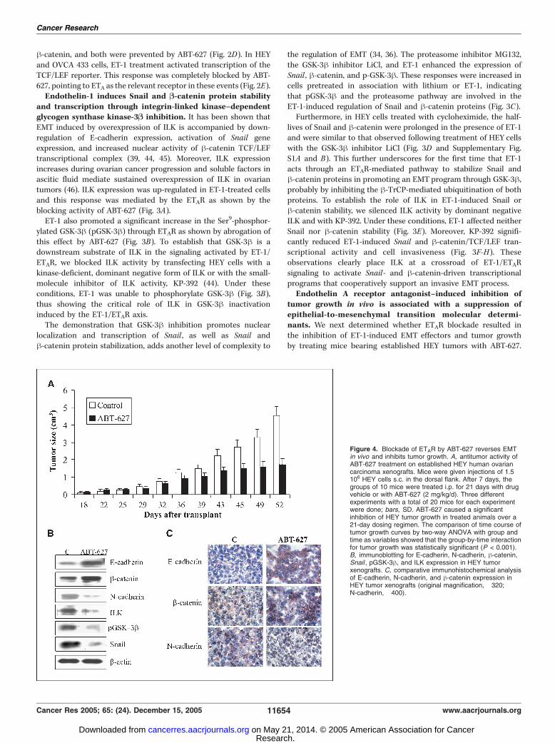
tumor growth in vivo is associated with a suppression ofepithelial-to-mesenchymal transition molecular determi-nants. We next determined whether ETAR blockade resulted inthe inhibition of ET-1-induced EMT effectors and tumor growthby treating mice bearing established HEY tumors with ABT-627.
Figure 4. Blockade of ETAR by ABT-627 reverses EMTin vivo and inhibits tumor growth. A, antitumor activity ofABT-627 treatment on established HEY human ovariancarcinoma xenografts. Mice were given injections of 1.5 �106 HEY cells s.c. in the dorsal flank. After 7 days, thegroups of 10 mice were treated i.p. for 21 days with drugvehicle or with ABT-627 (2 mg/kg/d). Three differentexperiments with a total of 20 mice for each experimentwere done; bars, SD. ABT-627 caused a significantinhibition of HEY tumor growth in treated animals over a21-day dosing regimen. The comparison of time course oftumor growth curves by two-way ANOVA with group andtime as variables showed that the group-by-time interactionfor tumor growth was statistically significant (P < 0.001).B, immunoblotting for E-cadherin, N-cadherin, h-catenin,Snail , pGSK-3h, and ILK expression in HEY tumorxenografts. C, comparative immunohistochemical analysisof E-cadherin, N-cadherin, and h-catenin expression inHEY tumor xenografts (original magnification, �320;N-cadherin, �400).
Cancer Research
Cancer Res 2005; 65: (24). December 15, 2005 11654 www.aacrjournals.org
Research. on May 21, 2014. © 2005 American Association for Cancercancerres.aacrjournals.org Downloaded from

Treatment with ABT-627 produced a 65% inhibition of HEYtumor growth (Fig. 4A). ABT-627 was generally well toleratedwith no detectable signs of acute or delayed toxicity. Analysis ofEMT molecule expression in ovarian carcinoma xenograftsrevealed a marked reduction of N-cadherin, Snail , pGSK-3h,and ILK and an increase of E-cadherin and h-catenin expressionin ABT-627–treated mice compared with controls (Fig. 4B).Immunohistochemical evaluation of tumors confirmed increasedexpressions of E-cadherin and h-catenin and a concomitantdecrease of N-cadherin expression following ABT-627 treatment(Fig. 4C).E-cadherin is down-regulated in endothelin-1/endothelin A
receptor–expressing primary ovarian tumor tissues. To explorethe pathophysiologic function of ETAR in human ovarian cancer, acohort of 50 primary ovarian cancer tissues were stained byimmunohistochemistry for ETAR, E-cadherin, and N-cadherinexpression. Consistent with our observations in cell cultures andHEY xenografts, expression of ETAR was significantly correlatedwith the down-regulation of E-cadherin (P < 0.008) and withenhanced expression of N-cadherin (P < 0.007; Fig. 5A and B). TheETAR expression was grade dependent, with grade 3 and 4 cancersshowing higher levels than early-grade cancers (P < 0.01; Fig. 5B).These results are consistent with our previous findings wherein theexpression of ETAR significantly correlates with neovascularization(P < 0.0002) and vascular endothelial growth factor (VEGF)expression (6). The tumor-staining data indicate the biologicalrelevance of ETAR in the regulation of EMT in vivo , resulting indown-regulation of epithelial marker E-cadherin and concomitantup-regulation of N-cadherin.
Discussion
One hallmark of epithelial cancer progression is EMT in whichtumor cells undergo loss of polarity and cell-cell junctions andacquire a mesenchymal phenotype and the ability to invade theextracellular matrix and to migrate to distant sites (14, 47). In thisstudy, we provide ample evidence that in human ovariancarcinoma cells, ETAR activation by ET-1 contributes to tumorprogression by acting as a crucial mediator of EMT. Thisconclusion is supported by several lines of evidence. First, ET-1through the ETAR induces a spectrum of key events includingdown-regulation of the epithelial adherens, such as E-cadherin andh-catenin, induction of mesenchymal markers, such as N-cadherinand vimentin, and enhanced cell invasiveness. Second, ET-1/ETARregulates Snail and h-catenin, the major contributors to E-cadherinsuppression, at multiple levels. Thus, ET-1 increases both Snail andh-catenin protein stability and transcriptional activity. Third, ILK isa downstream check point of the signaling pathways activated byET-1/ETAR that, via GSK-3h inactivation, promotes Snail andh-catenin signaling and acquisition of an invasive phenotype.Fourth, interruption of ET-1/ETAR autocrine signaling by twoselective ETAR antagonists or by RNA interference reverts EMT,increases the transcription level of E-cadherin, as well as inhibitsSnail transcription activity and basal cell invasion rate. Further-more, pharmacologic blockade of ETAR inhibits tumor xenograftgrowth and EMT molecular determinants. Finally, a significantcorrelation between ETAR and E-cadherin down-regulation andN-cadherin expression was observed in human ovarian cancertissues, indicating the relevant role of ETAR in the EMT process.Loss of normal tissue architecture and microenvironmental
interactions are signatures of epithelial cancer progression (16).
Here we describe a mechanism whereby ET-1/ETAR signalingorchestrates transcriptional programs that regulate E-cadherindown-regulation, EMT, and progression to invasive and metastaticovarian carcinoma.Recent data (34–36) show that inhibition of GSK-3h promotes
nuclear localization and transcription of Snail , as well as Snail andh-catenin protein stabilization, revealing that many oncogenicpathways could control cell adhesion, cell fate, and invasion duringmetastasis. Consistent with these findings, we show that ET-1 actsthrough ETAR-mediated pathway to stabilize Snail and h-cateninproteins and activate a Snail- and h-catenin-driven transcriptionalprogram that regulates EMT determinants. Thus, phosphorylationof GSK-3h by ET-1 resulting in the Snail and h-catenin stabilizationshows that inhibition of GSK-3h remains central to ET-1-inducedkey hallmarks of EMT. These results were consistent with theprevious observation that ET-1 increases cytosolic h-catenin in
Figure 5. Correlation between E-cadherin, N-cadherin, and ETAR expressionin human ovarian tumor tissues. A, immunohistochemical staining ofrepresentative human primary ovarian carcinoma tissue samples for expressionof ET-1, ETAR, E-cadherin, and N-cadherin (original magnification, �200).B, relationship between ETAR expression and E-cadherin and N-cadherin inhuman primary ovarian carcinomas. The expression patterns of ETAR,E-cadherin, and N-cadherin in the 50 ovarian cancer samples were determinedby immunohistochemistry and summarized. The correlation between ETARand E-cadherin and N-cadherin was analyzed using Fisher’s exact test.
ET-1 Is Critical in Ovarian Cancer Progression
www.aacrjournals.org 11655 Cancer Res 2005; 65: (24). December 15, 2005
Research. on May 21, 2014. © 2005 American Association for Cancercancerres.aacrjournals.org Downloaded from

cardiomyocytes to a level equivalent to that induced by the GSK-3hinhibitor (48).In view of its position at the crossroads connecting integrins
and the actin cytoskeleton, ILK was hypothesized to be acandidate signaling molecule that functions at the convergencepoint of cell adhesion– and growth factor–triggered signaltransduction, thus regulating the EMT process (39). Here weshow that ET-1-driven ILK signaling is necessary for GSK-3hphosphorylation and related Snail and h-catenin protein stability,transcriptional activity, and invasion, implicating for the first timeILK and its downstream substrate GSK-3h as check points offinely tuned interconnected signals induced by ET-1/ETAR tomodulate EMT. Thus, we propose a complex scheme whereinactivation of ETAR pathway by ET-1 drives inhibition of GSK-3h byan ILK-mediated signaling pathway to stabilize Snail and h-catenin proteins in a coordinate fashion so as to cooperativelyengage transcriptional programs leading to EMT.Because all the molecular effectors of EMT are triggered by ETAR
activation, blockade of this receptor results in inhibition of tumorgrowth and progression of ovarian carcinoma xenografts. We alsoshow that ETAR blockade restores E-cadherin and h-cateninexpression and suppresses Snail , ILK, and N-cadherin expressionin vivo , further identifying the mechanisms through which theautocrine ET-1/ETAR loop sustains EMT process and, in turn,creates a tumor microenvironment more permissive to progression.In this context, ET-1/ETAR induces the disruption of normal
host-tumor interactions regulating changes in cadherin, connexin,and MMP expression (13, 43). Indeed, the immunohistochemicaland immunoblot analysis of HEY xenografts provides in vivoevidence for this concept showing that treatment with ABT-627induces a significant reduction of microvessel density, expressionof VEGF, cyclooxygenase-2, and MMP-2, and increased tumorapoptosis and connexin 43–based gap junctional intercellularplaques (9, 13, 43).
It is becoming increasingly clear that E-cadherin acts as a late-stage microenvironmental tumor suppressor in ovarian carcinoma(19, 20, 49). This observation was supported by the immunohisto-chemical profile of cadherin phenotype in primary ovarian cancersamples, indicating that expression of ET-1/ETAR axis correlateswith the switch of cadherin expression associated with advanced-stage tumors. These findings complement and extend the recentanalysis of a genome-wide expression profile of late-stage ovariancancer whereby ET-1 has been identified as a key gene that activatescell signaling controlling cell migration, spread, and invasion (50).Although further studies are required to examine the complex
connection network that regulates EMT in tumor metastasis, ascheme is emerging in which we identify for the first time thatmimicry of the Wnt pathway, through ETAR-driven molecularevents, is required for the cooperative regulation of Snail andh-catenin signaling, which occurs via ILK and GSK-3h.Understanding the regulatory loops that allow close coordina-
tion of EMT program may provide a novel strategies aimed atpreventing the development of metastasis. In this regard, the abilityof ETAR antagonists to induce concomitant suppression of tumorcell proliferation and/or survival (7, 43) and simultaneously controlEMT provides a rationale for developing a more effectivetherapeutic intervention in aggressive ovarian cancer.
Acknowledgments
Received 6/17/2005; revised 9/7/2005; accepted 10/10/2005.Grant support: Associazione Italiana per la Ricerca sul Cancro, Ministero della
Salute, and Consiglio Nazionale delle Ricerche-Ministero dell’Istruzione Universita eRicerca.
The costs of publication of this article were defrayed in part by the payment of pagecharges. This article must therefore be hereby marked advertisement in accordancewith 18 U.S.C. Section 1734 solely to indicate this fact.
We thank G. Genovesi, G. Elia, and S. Decandia for excellent technical assistance,M.V. Sarcone for secretarial support, and K.J. Catt for helpful discussion and valuablecomments.
References1. Naora H, Montell DJ. Ovarian Cancer Metastasis:integrating insights from disparate model organisms.Nat Rev Cancer 2005;5:355–66.
2. Rubanyi GM, Polokoff MA. Endothelins: molecularbiology, biochemistry, pharmacology, physiology andpathophysiology. Pharmacol Rev 1994;4:325–415.
3. Nelson J, Bagnato A, Battistini B, Nisen P. Theendothelin axis: emerging role in cancer. Nat RevCancer 2003;3:110–6.
4. Bagnato A, Tecce R, Moretti C, Di Castro V, Spergel D,Catt KJ. Autocrine actions of endothelin-1 as a growthfactor in human ovarian carcinoma cells. Clin CancerRes 1995;1:1059–66.
5. Bagnato A, Salani D, Di Castro V, et al. Expression ofendothelin-1 and endothelin A receptor in ovariancarcinoma: evidence for an autocrine role in tumorgrowth. Cancer Res 1999;59:720–7.
6. Salani D, Di Castro V, Nicotra MR, et al. Role ofendothelin in neovascularization of ovarian carcinoma.Am J Pathol 2000;157:1537–47.
7. Del Bufalo D, Di Castro V, Biroccio A, et al. Endothelin-1 protects against paclitaxel-induced apoptosis: require-ment for Akt activation. Mol Pharmacol 2002;61:524–32.
8. Spinella F, Rosano L, Di Castro V, Natali PG,Bagnato A. Endothelin-1 induces vascular endothelialgrowth factor by increasing hypoxia inducible factor1a in ovarian carcinoma cells. J Biol Chem 2002;277:27850–5.
9. Spinella F, Rosano L, Di Castro V, Nicotra MR, NataliPG, Bagnato A. Inhibition of cyclooxygenase-1 and -2expression by targeting the endothelin A receptor in
human ovarian carcinoma cells. Clin Cancer Res 2004;10:4670–9.
10. Bagnato A, Tecce R, Di Castro V, Catt KJ. Activationof mitogenic signaling by endothelin-1 in ovariancarcinoma cells. Cancer Res 1997;57:1306–11.
11. Vacca F, Bagnato A, Catt KJ, Tecce R. Transactivationof epidermal growth factor receptor in endothelin-1-induced mitogenic signaling in human ovariancarcinoma cells. Cancer Res 2000;60:5310–7.
12. Rosano L, Varmi M, Salani D, et al. Endothelin-1induces tumor proteinase activation and invasiveness ofovarian carcinoma cells. Cancer Res 2001;61:8340–6.
13. Spinella F, Rosano L, Di Castro V, Nicotra MR, NataliPG, Bagnato A. Endothelin-1 decreases gap junctionalintercellular communication by inducing phosphoryla-tion of connexin 43 in human ovarian carcinoma cells.J Biol Chem 2003;278:41294–301.
14. Savagner P. Leaving the neighborhood: molecularmechanisms involved during epithelial-mesenchymaltransitions. BioEssays 2001;23:912–23.
15. Thiery JP. Epithelial-mesenchymal transitions intumor progression. Nat Rev Cancer 2002;2:442–54.
16. Bissell MJ, Radisky D. Putting tumours in context.Nat Rev Cancer 2001;1:46–54.
17. Conacci-Sorrell M, Zhurinsky J, Ben-Ze’ev A. Thecadherin-catenin adhesion system in signalling andcancer. J Clin Invest 2002;109:987–91.
18. Lu Z, Ghosh S, Wang Z, Hunter T. Down-regulationof caveolin-1 function by EGF leads to the loss ofE-cadherin, increased transcriptional activity ofh-catenin, and enhanced tumor cell invasion. CancerCell 2003;4:499–515.
19. Wong AS, Maines-Bandiera SL, Rosen B, et al.
Constitutive and conditional cadherin expression incultured human ovarian surface epithelium: influence offamily history of ovarian cancer. Int J Cancer 1999;81:180–8.
20. Auersperg N, Pan J, Grove BD, et al. E-Cadherininduces mesenchymal-to-epithelial transition in humanovarian surface epithelium. Proc Natl Acad Sci U S A1999;96:6249–54.
21. Imai T, Horiuchi A, Shiozawa T, et al. Elevatedexpression of E-cadherin and a-, h-, and g-catenins inmetastatic lesions compared with primary epithelialovarian carcinomas. Hum Pathol 2004;35:1469–76.
22. Cavallaro U, Christofori G. Cell adhesion andsignalling by cadherins and Ig-CAMs in cancer. NatRev Cancer 2004;4:118–32.
23. Nieman MT, Prudoff RS, Johnson KR, Wheelock MJ.N-Cadherin promotes motility in human breast cancercells regardless of their E-cadherin expression. J Cell Biol1999;147:631–44.
24. Tran NL, Nagle RB, Cress AE, Heimark RL. N-Cadherinexpression in human prostate carcinoma cell lines. Anepithelial-mesenchymal transformation mediating adhe-sion with stromal cells. Am J Pathol 1999;155:787–98.
25. Suyama K, Shapiro I, Guttman M, Hazan RB.A signaling pathway leading to metastasis is controlledby N-cadherin and the FGF receptor. Cancer Cell 2002;2:301–14.
26. Cano A, Perez-Moreno MA, Rodrigo I, et al. Thetranscription factor Snail controls epithelial-mesenchy-mal transitions by repressing E-cadherin expression. NatCell Biol 2000;2:76–83.
27. Batlle E, Sancho E, Franci C, et al. The transcrip-tion factor Snail is a repressor of E-cadherin gene
Cancer Research
Cancer Res 2005; 65: (24). December 15, 2005 11656 www.aacrjournals.org
Research. on May 21, 2014. © 2005 American Association for Cancercancerres.aacrjournals.org Downloaded from

expression in epithelial tumour cells. Nat Cell Biol2000;2:84–9.
28. Blanco MJ, Moreno-Bueno G, Sarrio D, et al.Correlation of Snail expression with histological gradeand lymph node status in breast carcinomas. Oncogene2002;21:3241–6.
29. Rosivatz E, Becker I, Specht K, et al. Differentialexpression of the epithelial-mesenchymal transitionregulators Snail, SIP1, and twist in gastric cancer. AmJ Pathol 2002;161:1881–91.
30. Sugimachi K, Tanaka S, Kameyama T, et al. Transcrip-tional repressor Snail and progression of human hepa-tocellular carcinoma. Clin Cancer Res 2003;9:2657–64.
31. Guaita S, Puig I, Franci C, et al. Snail induction ofepithelial-to-mesenchymal transition in tumor cells isaccompanied by MUC-1 repression and ZEB1 expres-sion. J Biol Chem 2002;277:30209–16.
32. Nelson WJ, Nusse R. Convergence of Wnt, h-catenin,and cadherin pathways. Science 2004;303:1483–7.
33. Domınguez D, Montserrat-Sentis B, Virgos-Soler A,et al. Phosphorylation regulates the subcellular locationand activity of the Snail transcriptional repressor. MolCell Biol 2003;23:5078–89.
34. Zhou BP, Deng J, Xia W, et al. Dual regulation of Snailby GSK-3h-mediated phosphorylation in control ofepithelial-mesenchymal transition. Nat Cell Biol 2004;6:931–40.
35. Bachelder RE, Yoon SO, Franci C, Garcia de HerrerosA, Mercurio AM. Glycogen synthase kinase-3 is an
endogenous inhibitor of Snail transcription: implica-tions for the epithelial-mesenchymal transition. J CellBiol 2005;1681:29–33.
36. Yook JI, Li XY, Ota I, Fearon ER, Weiss SJ. Wnt-dependent regulation of the E-cadherin repressor Snail.J Biol Chem 2005;280:11740–8.
37. Jamora C, DasGupta R, Kocieniewski P, Fuchs E. Linksbetween signal transduction, transcription and adhesionin epithelial bud development. Nature 2003;422:317–22.
38. Novak A, Hsu SC, Leung-Hagesteijn C, et al. Celladhesion and the integrin-linked kinase regulate theLEF-1 and h-catenin signaling pathways. Proc Natl AcadSci U S A 1998;95:4374–9.
39. Oloumi A, McPhee T, Dedhar S. Regulation of E-cadherin expression and h-catenin/Tcf transcriptionalactivity by the integrin-linked kinase. Biochim BiophysActa 2004;1691:1–15.
40. Barbera MJ, Puig I, Dominguez D, et al. Regulation ofSnail transcription during epithelial to mesenchymaltransition of tumor cells. Oncogene 2004;23:7345–54.
41. Delcommenne M, Tan C, Gray V, Rue L, Woodgett J,Dedhar S. Phosphoinositide-3-OH kinase-dependentregulation of glycogen synthase kinase 3 and proteinkinase B/AKT by the integrin-linked kinase. Proc NatlAcad Sci U S A 1998;95:11211–6.
42. Janda E, Lehmann K, Killisch I, et al. Ras and TGFhcooperatively regulate epithelial cell plasticity andmetastasis: dissection of Ras signaling pathways. J CellBiol 2002;156:299–313.
43. Rosano L, Spinella F, Salani D, et al. Therapeutictargeting of endothelin A receptor in human ovariancarcinoma. Cancer Res 2003;63:2447–53.
44. Hannigan G, Troussard AA, Dedhar S. Integrin-linkedkinase: a cancer therapeutic target unique among itsILK. Nat Rev Cancer 2005;5:51–63.
45. Tan C, Costello P, Sanghera J, et al. Inhibition ofintegrin linked kinase (ILK) suppresses h-catenin-Lef/Tcf-dependent transcription and expression of theE-cadherin repressor, snail, in APC�/� human coloncarcinoma cells. Oncogene 2001;20:133–40.
46. Ahmed N, Riley C, Oliva K, et al. Integrin-linkedkinase expression increases with ovarian tumour gradeand is sustained by peritoneal tumour fluid. J Pathol2003;201:229–37.
47. Thiery JP. Epithelial-mesenchymal transitions indevelopment and pathologies. Curr Opin Cell Biol2003;15:740–6.
48. Haq S, Michael A, Andreucci M, et al. Stabilization ofh-catenin by a Wnt-independent mechanism regulatescardiomyocyte growth. Proc Natl Acad Sci U S A 2003;100:4610–5.
49. Roskelley CD, Bissell MJ. The dominance of themicroenvironment in breast and ovarian cancer. SeminCancer Biol 2002;12:97–104.
50. Donninger H, Bonome T, Radonovich M, et al. Wholegenome expression profiling of advance stage papillaryserous ovarian cancer reveals activated pathways.Oncogene 2004;23:8065–77.
ET-1 Is Critical in Ovarian Cancer Progression
www.aacrjournals.org 11657 Cancer Res 2005; 65: (24). December 15, 2005
Research. on May 21, 2014. © 2005 American Association for Cancercancerres.aacrjournals.org Downloaded from