
diagnosis of ovarian factor of sudaneese - SUST Repository
288
SUDAN UNIVERSITY OF SCIENCE & TECHNOLOGY KHARTOUM, SUDAN COLLEGE OF POST GRADUATE STUDIES DIAGNOSIS OF OVARIAN FACTOR OF SUDANEESE FEMALE PRIMARY INFERTILITY USING ENDOVAGINAL ULTRASOUND تشخيص عامل ال المبيضى لعقم ل الأولى لدى نيات السودا ستخدام با مسبار لموجات ا فوق الصوتية مهبلى الA thesis submitted fur fulfillment for PhD degree in medical ultrasound BY:AMNA AHMED MOHMED MOKHTAR Supervisor: Prof. MOTASIM AHMED ALSEED Co-Supervisor: DR. ALWIA MOHMED OSMAN HASSEN MAY, 2015
-
Upload
khangminh22 -
Category
Documents
-
view
0 -
download
0
Transcript of diagnosis of ovarian factor of sudaneese - SUST Repository

SUDAN UNIVERSITY OF SCIENCE & TECHNOLOGY KHARTOUM, SUDAN
COLLEGE OF POST GRADUATE STUDIES
DIAGNOSIS OF OVARIAN FACTOR OF SUDANEESE
FEMALE PRIMARY INFERTILITY USING
ENDOVAGINAL ULTRASOUND
السودانيات لدى الأولى للعقم المبيضى العامل تشخيص
المهبلى الصوتية فوق الموجات مسبار باستخدام
A thesis submitted fur fulfillment for PhD degree in medical ultrasound
BY:AMNA AHMED MOHMED MOKHTAR
Supervisor:
Prof. MOTASIM AHMED ALSEED
Co-Supervisor: DR. ALWIA MOHMED OSMAN HASSEN
MAY, 2015

Dedication
To my FAMILY who always supports me…..
And to the soul of my Mother and Father
God bless them
a

Acknowledgment
I thank God…
My great thanks and appreciation to Dr.Motasim Ahmed Allseed
who is the supervisor of this research and also to Dr.Alwia Mohamed Osman
Hassen who is the co-supervisor.
Great thanks to Dr. Mohamed Omer the principle of the College of
Radiological Sciences and Dr. Mohamed Elfadil Mohamed for their
encouragement.
My great thanks extended to Dr.Mohamed Abd Algafoor who is
general manager of Banoon for Assisted Reproduction Obsfevic and
Gynecology and everybody help and support me.
Great thanks to my husband General Salih Mahmood Alwad, my
daughter Omnia Salih and my son Mahmood Salih.
My great thanks extended to my colleagues who were of great help
and support.
B
ABSTRACT
Endovaginal ultrasound is a test used to look at a woman's reproductive
organs, including the uterus, ovaries, and cervix. The main objective of this
study was to diagnose ovarian factor of Sudanese female primary infertility
using Endovaginal ultrasound scan. The data of this study consisted of 300
random samples of Sudanese female suffering from primary infertility
during the period from 2009 to 2015 in Banoon Center for assisted
reproduction and obstetric and gynecology in Khartoum.
The result of this study showed that 40.7% of the cases had polycystic
ovarian syndrome, 49.7% normal ovary with other infertility remarks e.g.
Tube blocked, fibroids and male factor, 5.6% with unknown reason under
induction treatment and 4% with ovarian cuyst. In conclusion the results
highlights that the increases of female age (average) in relation to the
increases of marriage duration (average) together portrayed the pattern of the
infertility problem in ascending fashion from polycystic ovarian syndrome,
ovarian cyst, unknown obvious cause and normal ovaries with other
infertility causes.
The recommendations were given dealing with all infertility investigation
must be carried out using Endovaginal probe, the duration of marriage and
female age should be taken as indicator for infertility problems and the size
of the follicles in induction treatment should be related to the age of the
female, which gives a good estimate of the time needed to reach a proper
size.
i

الخلصة
الأعضاء فى للنظر يجرى كشف هو المهبل خلل من الصوتيه فوق الموجات استعمال
الرحم. وعنق المبيض ، الرحم ذلك فى بما للمرأه التناسليه
ذات السودانيات للنساء الإبتدائى العقم حالت تشخيص هى الدراسة هذه من الغرض
المهبل. خلل من الصوتيه فوق الموجات باستعمال بالمبيض العلقه
العقم من يعانين سودانيات لسيدات عشوائيه عينه300 من اخذت الدراسة معطيات
والتوليد النساء وأمراض الأنابيب لإطفال بنون بمركز م2015- م2009 من الفتره فى الإبتدائى
بالخرطوم.
، التكيسات متعدد المبيض متلزمة من يعانين% 40.7 أن الى خلصت الدراسة نتيجة
وجود أو فالوب قناة كانسداد للعقم اخرى اسباب وجود مع طبيعيين المبيضين% 49.7
%4 و للتنشيط يخضعن معروف غير سبب نتيجة% 5.6 و بالزوج متعلق عامل أو لحمية
متكيس. مبيض
زيادة فى واضح مؤشر الزواج فترة طول ومتوسط المرأه عمر متوسط أن الى الدراسة خلصت
التكيسات متعدد المبيض متلزمة ذلك فى بما الإبتدائى العقم الى تؤدى التى المشاكل
الأخرى. العقم وأسباب السبب معروفة الغير العقم وحالت المبيض وتكيس
فوق بالموجات الإبتدائى العقم لحالت تجرى التى الكشوفات كل أن الدراسة أوصت
الزواج ومدة المرأة عمر وأن التشخيص لدقة المهبل طريق عن تجرى أن يجب الصوتيه
تحديد عند التنشيط حالت فى. العقم اسباب لتشخيص مهمة كمؤشرات تؤخذ أن يجب
للزمن مؤشر يعطى ذلك أن حيث الإعتبار فى المرأة عمر يوضع أن يجب الحويصلة حجم
المناسب. للحجم الحويصلة فيه تصل الذى

CONTENTS6
Topic Page
Dedication…………………………..………….……….…..…..…. a
Acknowledgement……..……………….………………....………..b
Absrract ( in English ) …………………………………..…….……i
Abstract ( in Arabic ) …………………………..…...…….……….ii
Chapter one : Introduction ………………………………....….….1
1.1 Problem of the study ……………………..…………………2
1.2 Objective of the Study……………………………….……..3
1.3 Significance of the study…………………………………….3
1.4 Overview of the study……………………………………….3
Chapter two : Lierature review ………………………...…….…..4 2 -1
Medical ultrasound…………………...….………………..….5
2-1-1 Medical ultrasound-germination and growth……,,,,,.…5
2-1-2 Gray scale imaging………………………….……….…7
2-1-3 Probes …………………..…………..…...……...9
2-1-3-1 Types of
pobes...................................................9

2-1-3-1-1 Anorectal 3D 2052………………………..9 2-1-3-
1-2 Burr-Hole 8863……………………......…...10
2-1-3-1-3 Curved Array 8823 …...…………….……..11
2-1-3-1- 4 Craniotomy 8862 ………..…..…….…..….12
2-1-3-1-5 Curved Array 8820e …………….…..…….13
2-1-3-1-6 Curved Array 8830 ………………….....….14
2-1-3-1-7 Curved Array 8830 ………....…...…...……15
2-1-3-1-8 Curved Array 8802 ……………..…………16
2-1-3-1-9 Endfire Curved Array 8667 …..………..…17
2-1-3-1-10 End cavity Biplane 8848 ………....……..18
2-1-3-1-11 Endovaginal 8819 …………...……..…….19
2-1-3-1-12 High Frequency Linear Array 8870……...20
2-1-3-1-13 Hockey Stick 8809 ………………...….…21
2-1-3-1-14 Intraoperative 8815 …………..….....……22
2-1-3-1-15 Intraoperative Biplane 8824 ……….…….23
2-1-3-1-16 Linear Array 8811 ………….………....…24
2-1-3-1-17 Linear Array 8670 ……………...…..……25
2-1-3-1-18 ProART™ Robotic Drop In 882..…......…26
2-1-3-1-19 Prostate Biplane 8808e …………..…...….27
2-1-3-1-20 Prostate Triplane 8818 …….………...…..28
2-1-3-1-21 Rigid Laparoscopic 8836 ….……..……...29
2-1-3-1-22 Small Footprint Cardiac 8827 .….….....…30
2-1-3-1-23 Small Footprint Cardiac 8837…....………31
2-1-3-1-24 T-Shaped Intraoperative 8816 ………..….32
2-1-3-1-25 Vascular 8822 …………..…….…………33
2-1-3-1-26 3DART™ 8838 ………..…………...……34
2-1-3-1-27 10L2w Wide Linear ……….……………..35
2-1-3-1-28 13L4w Wide Linear ……………....….…..36
2-1-3-1-29 14L3 Linear ……………………….……..37
2-1-3-1-30 18L5 High Frequency Linear ……....……38
2-1-3-1-31 5P1 Small Footprint Cardiac ……..…..….39
2-1-3-1-32 6C2 Curved ……………..……….....……40
2-1-3-1-33 6C2s Small Curved ………….…....…..…41
2-1-3-1-34 E10C4 End cavity ……….….….……..….42
2-1-3-1-35 E14C4 Endfire Curved ..........................43
2-1-3-1-36 E14C4t Prostate Triplane ……….…...…..44
2-1-3-1-37 E14C4t Prostate Triplane ..…....….…...…45
2-1-3-1-38 E14CL4b Endocavity Biplane ……...……46
2-1-3-1-39 N13C5 Curved …………………..……….47
2-1-3-1-40 4-Way Laparoscopic 8666-RF …….,,.…..48
2-1-4 Biological effects of possible relevance to safety……49
2-1-4-1 Introduction ………………………..….…….49

2-1-4-2 Types of biological effects ……/……..……..…51
2-1-4-2-1 Thermal …………..…….………………….51
2-1-4-2-2 Non-thermal …………….....……...……….……..52
2-1-4-2-3 Cavitations …………………………..…..…...……52
2-1-4-2-4 Other effects ……………..…………....…….……53
2-1-4-3 Measurement of biological effects ……………………….……..53
2-1-5 Guidelines and regulations …………………………..…..…….56
2-1-5-1 Ultrasound Examination Procedures ……………………..59
2-1-5-2 Communication ……………………………….…….……..59
2-2 The ovary ……..……….......................................…....………90
2-2-1 Anatomy……………………………………………..………….91
2-2-1-1 Structure …………………….…………,,,,,,,…………,….91
2-2-2 Physiology ……………………..……….,,…,,,,,,,……….…….…105
2-2-2-1 The major endocrine glands …………..…,,,,,,,,,,,,………...……108
2-2-2-1-1 Pituitary gland …………………………....……..108
2-2-2-1-2 Thyroid gland ………………………….………...,,,…..…….110
2-2-2-1-3 Parathyroid glands………………,,,...…,,,,,,,,…………....…111
2-2-2-1-4 Pancreas ……………………………….……,,,,,,,,,……...….111

2-2-2-1-5 Gonads ………………...…………….…………,,,,,,,,,……...112
2-2-2-1-6 Pineal gland ……………………………….…………..…..112
2-2-1-7 Other hormone-producing structure ………………….
……….112
2-2-2 The female cycle ………………………………….
……………………..……...…..….113
2-2-3 Menstruation ………………………………………….
……………………….…..……….113
2-2-3 Ultrasound appearance of the
ovary…………………………….……..………131
2-2-4 Follicle measurements and tracing ………………………………...
……………150
2-2-4-1 Folliculogenesis …………………………………..
………………….,,,,,…………...169
2-2-4-2 Chronology …………………………………………………..
……,,,,……………..…..154
2-2-4-3 The process ………………………………….…………..
………………….……………155
2-2-4-4 The primordial follicle ……………………………….…….
…….………..…..……156
2-2-4-5 Recruitment……………………………..………………….
………,,……….………….157

2-2-4-6 Mechanism ………………………………………………..
…….……………………….158
2-2-4-7 THE PREANTRAL FOLLICLE ……………………..
………………..,.…..……….160
2-2-4-8 Primary Follicle……………………………..
…………………………………………..161
2-2-4-9 Secondary Follicle
…………………………………………………….….……………165
2-2-4-10 Tertiary Follicle …………………………………………..
………………..………..168
2-2-4-11 THE GRAAFIAN FOLLICLE ………………………..
………………...…..……….170
2-2-4-12 Classification…………………………..
……………………………..…….………….174
2-2-4-13 Selection of the dominant
follicle…………………………..….……….…..176
2-2-4-14 The process…………………………..
…………………………………….……………177
2-2-4-15 Autocrinology and paracrinology………….
………………….………………178
2-2-4-16 Ultrasound monitoring ……………………….
………………………….……..179
2-2-5 Polycystic ovarian syndromes ……………..………………...….
……..182

2-2-5-1 ovarian cyst………………………………………………..…...182
2-2-5-2 Polycystic
ovaries…………………………………………………..
…………………..186
2-2-5-2 ovarian torsion …………………...………………….….….…..187
2-2-6 ovarian masses …………………………………….………..…....187
2-2-6-1 Cystic and semi-cystic ovarian masses...…..…..………..……188
2-2-6-2 Solid ovarian masses ………………….…….……………..…..191
2-3 Feamle infertility….…..……………...…………...……….198
2-3-1 Definition
………………………………………………………………………….
….………199
2-3-1-1 Prevalence
……………………………………………………………….………..
………200
2-3-2 Causes and factors
………………………………………………………….……..
…….201
2-3-2-1 Acquired
………………………………………………………………………………
……201
2-3-2-2 Age and female fertility ………………………..……….….….202
2-3-2-3 Tobacco smoking ………………………………………....……202

2-3-2-4 Sexually transmitted infections ………......……………….…..203
2-3-2-5 Body weight and eating disorders ………….………….......….203
2-3-2-6 Chemotherapy……………………...……………….………….204
2-3-2-7 Other acquired factors ……………………………….......……205
2-3-2-8 Genetic factors ……………………………….………..………205
2-3-2-9 Hypothalamic-pituitary factors……………...………………….210
2-3-2-10 Ovarian factors ………..…..…………...………………….…210
2-3-2-11 Tubal (ectopic)/peritoneal factors …………………….…...…211
2-3-2-12 Uterine factors ……………...…………………….……….…211
2-3-2-13 Cervical factors ………………………………………...……..211
2-3-2-14 Vaginal factors…………………………………………..….…211
2-3-3 Diagnosis ………………………………………………………….211
2 -3-4 Examination and imaging ……………………….……….....…...212
2-3-5 Prevention …………………….................................................213
2-3-6 Social stigma …………..…………………..………………214
2-3-7 Marital role ………………………………………..….….……….215
2-3-8 Domestic abuse …………………………………..……….……...217
2-3-9 Mental and psychological impact ………………….…..…...……217
Chapter three: Methodology……….………………...……….218
3-1 Material ………………………………………….………….…..219

3-2 Population of the study……………..…………….………….….219
3-3 Sample and size of the study…...…………….……….…………219
3-4 Design of the study………………………….…………....………219
3-5 Method of data collection ………………….…………………….219
3-5-1 Techniques………….………….……..………………….……..219
3-6 Protocols…………………………………………………..………222
3-7 Data analysis……………………….…………………………..…..224
4 Chapter four: Results……………………………..…..…….225
5 Chpater five:Discussion, conclusion and ommendation…..235
5-1 Discussion…………………………………………………………….235
5-2 Conclusion ………………………………………….………………..237
5-3 Recommendatios …………………………………………………….238
References …………………………………………………...…………239
Appendix A……………………………………………..………………257
A-1 Form for recording results,,,,,,,,,,,,,,,,,,,,,,,,,,,,,,,,,,………,,,,,,,,,,,,,,,,,,,,,,257
A-2 Master data sheet……………………………………………………..258
Appendix B………………………………………………………….……272
Ultrasound images …………………..……………………………………272

Chapter oneIntroduction

Chapter oneIntroduction
Ultrasound is no longer used simply to distinguish a cystic and solid mass
within the abdomen and pelvis. With improved image resolution and
software, subtle differences in tissue texture can be demarcated and pelvic
organs clearly identified.
Ultrasound is now established as the primary imaging investigation in all
cases of suspected female infertility.
Thus, it has become a significant tool in the diagnosis of female infertility.
Description The definition of infertility varies considerably, particularly in
relation to the length of time of regular unprotected intercourse. It is usually
defined as difficulty in conceiving after 12 months of regular unprotected
intercourse.
The female causes of infertility include ovulatory causes (20%), tubal causes
(14%), endometriosis (6%) and uterine causes (1%).
1-1 Problem of the study
Primary infertility is one of the major problem that encountered by copules
so often and most of the time attributed to female as social trend issue.
Therefore exploring the factors associated with primary infertility can speed
the process of treatment and it can save a lot of money spended in
uneffecient procedures including the traditional one. Ultrasound scaning

using endovaginal probe might provide valuble information regarding this
problem either if it concern the ovary or the related reproductive system or it
might indicate that the problem does not concern the female.
1-2 Objective of the Study:-
The main objective of this study is to evaluate the application of endovaginal
ultrasound in diagnosing ovarian factor of Sudanese female infertility.
Specific objectives:
To find the causes of primary infertility in female using endovadginal
probe .
To find the frequencies of the factors lead to primary infertility
To relate the type of factors behind infertility with female age and
duration of marriage
1-3 Significance of the study
This study will provide rich information about the primary infertility and
highlights the underline causes of this problem safely with minimum
cost. Also it will facilitate the approtch of mangment and follow up of the
treatment process.
1-4 Overview of the study
This study falls into five chapters, with chapter one is an introduction
which includes; problem of the study, objective and significant of the
study. While chapter two deals with background and scolary literature in
previous study format. Chapter three will include material used to collect

the data and the method followed to obtain those data, similarly chapter
four will present the results of this study using tables and figure and
finally chapter five will discuss the result of this study as well as the
conclusion and recommendation
Chapter twoLiterature review

Chapter twoLiterature review
2.1 Medical ultrasound.
2.1.1 Medical ultrasound-germination and growth.
John Eric Edgcumbe Fleming was born in 1934. He started his career with
the EMI Engineering Development Ltd in 1951. Between 1951and 56, he
worked on the development of specialised test equipment for radar and later
on computer development with John Drage and Godfrey Houndsfield; Drage
went on to lead the team which developed the first desk calculator and
Houndsfield to develop Computerised Tomography. During his time at EMI
Fleming obtained a HNC with distinctions in Electrical Engineering.
In 1956 he moved to Ferranti Ltd as a Development Engineer on computer
logic elements and data transmission systems. Then to avoid the increasing
involvement in military projects he moved to Smiths Industries Ltd in
Glasgow, Scotland. There he joined Tom Brown who was working with
Professor Ian Donald. Following Brown's move to another company
Fleming became responsible for the development of medical ultrasonic
products, principally the Diasonograph. This static B-Scan machine was the
first scanner to go into commercial production. However only twelve had
been delivered when Smiths decided to close the factory in Glasgow. After
the sale of the medical ultrasound interest production and further
development continued at Nuclear Enterprises Ltd in Edinburgh under Brian

Fraser, from Smiths, and Tom Brown who had returned to the ultrasound
world.
For some time before Smiths' closure Angus Hall had also been working on
medical ultrasound. Following the closure in 1967 they both joined
Professor Donald in the University of Glasgow at his Department of
Midwifery in the newly built Queen Mother's Hospital. The personal
connection with Fraser and Brown fostered active cooperation between
Professor Donald's department and Nuclear Enterprises. In 1982 Hall moved
to become Head of Medical Physics at St James's University Hospital,
Leeds.
In 1984 partly as a result of having agreed to care for the original contact
scanner (built by Tom Brown in 1954) and used by Ian Donald, Fleming was
asked by the British Medical Ultrasound Society (BMUS) to establish an
Historical Collection. In 1988 following an agreement between the
University of Glasgow's Hunterian Museum and BMUS the Museum
undertook to provide long term care for the Collection. Fleming was
appointed Honorary Assistant Keeper of Ultrasonic Equipment to the
Museum.
The Collection now contains over sixty items of hardware, scanners and
associated equipment, a wide range of manufacturer's literature and a
substantial and increasing archive of unique documents.
Since 1995 Fleming has been working closely with Dr Malcolm Nicolson of
the Wellcome Unit for the History of Medicine, University of Glasgow.
They, together with Dr Ian Spencer, are co-authoring a book on the history
of ultrasound development in Glasgow. Fleming's work in establishing the
BMUS Collection was recognised in 1994 with the award of Honorary

Membership of the Society and the Geddes-Davis Shield, from the British
Institute of Non Destructive testing in 1997.
Mr. Fleming has contributed to over 60 papers on ultrasonics and given
numerus lectures and talks at important International meeings. In addition to
his work in establishing the Historical Collection Fleming contributed to
BMUS as Honorary Treasurer, 1972 - 78, and in the organisation of the
BMUS Annual Meetings in 1972 and 1988. Additionally he was Financial
Director for the World Federation meeting in Brighton, UK (WFUMB
1982). Following retirement in 1995 John Fleming retains a close link with
the University as an Honorary Research Associate. He continues as
Coordinator of the BMUS Historical Collection until 2004.
Fig.2-1 Mr. Fleming with a few of the scanners in the BMUS Historical
Collection c. 1996. In the foreground are machines from KretzTechnik®,
Philips®, Siemens® and ADR®.
2-1-2 Gray scale imaging.
Ultrasound or ultrasonography is a medical imaging technique that uses high
frequency sound waves and their echoes. The technique is similar to the
echolocation used by bats, whales and dolphins, as well as SONAR used by

submarines. In ultrasound, the following events happen: the ultrasound
machine transmits high-frequency (1 to 5 megahertz) sound pulses into your
body using a probe , the sound waves travel into your body and hit a
boundary between tissues (e.g. between fluid and soft tissue, soft tissue and
bone) , some of the sound waves get reflected back to the probe, while some
travel on further until they reach another boundary and get reflected , the
reflected waves are picked up by the probe and relayed to the machine , the
machine calculates the distance from the probe to the tissue or organ
(boundaries) using the speed of sound in tissue (5,005 ft/s or1,540 m/s) and
the time of the each echo's return (usually on the order of millionths of a
second) , and the machine displays the distances and intensities of the echoes
on the screen, forming a two dimensional image like the one shown below.
The Ultrasound Machine:- A basic ultrasound machine has the following
parts: transducer probe - probe that sends and receives the sound waves ,
central processing unit (CPU) - computer that does all of the calculations
and contains the electrical power supplies for itself and the transducer probe
and transducer pulse controls - changes the amplitude, frequency and
duration of the pulses emitted from the transducer probe .(Antiou,2003)

Fig.2-2 Photo courtesy Dynamic Imaging Limited
Ultrasound machine with various transducer probes
2.1.3 Probes.
Transducer probes come in many shapes and sizes, as shown in the
photo above. The shape of the probe determines its field of view, and the
frequency of emitted sound waves determines how deep the sound
waves penetrate and the resolution of the image. Transducer probes may
contain one or more crystal elements; in multiple-element probes, each
crystal has its own circuit. Multiple-element probes have the advantage
that the ultrasounc beam can be "steered" by changing the timing in
which each element gets pulsed; steering the beam is especially
important for cardiac ultrasound. In addition to probes that can be moved
across the surface of the body, some probes are designed to be inserted
through various openings of the body (vagina, rectum, esophagus) so
that they can get closer to the organ being examined (uterus, prostate
gland, stomach); getting closer to the organ can allow for more detailed
views.
2-1-3-1 Types of probes.

2-1-3-1-1 Anorectal 3D 2052
Specification : frequency Range 16 - 6 MHz , transducer Categories
transrectal, transvaginal , focal Range Up to 50 mm , image
field(expanded) 360º and weight 850 g. Detailed, High-Resolution Images
: see all rectal wall layers , evaluate the Radial, Longitudinal Extension of
Sphincter Tears , assess the extent of anal sphincter damage , acquire deep
penetrating, clear dataset images and measure detailed pelvic floor
architecture in all x, y and z planes, accurately.Easy to Use 3D : scan your
patient and examine their data at any time, on any PC , 3D cube provides
accurate distance, area, angle, and volume measurements , reproduce your
work with ease, one-touch operator-independent acquisition and cut
through the data cube to see anatomical details in the best plane.
Fig.2-3

2-1-3-1-2 Burr-Hole 8863
Specification : frequency Range 10 - 3.8 MHz , transducer Categories
Intraoperative, Neurosurgery, Contact Surface 10 x 8.6 mm, .4 x .34
in , Focal Range 5-57 mm, .2-2.2 in and weight 50 g .
High resolution burr-hole transducer for neurosurgical imaging: locate the
ventricular system more easily , insert ventricular drains and implant CSF
shunts. Perform Precise Puncture Procedures: brain lesion biopsy,
intracerebral abscess and intracranial cyst puncture. The 8863 features a
sterile, single use needle guide (UA1346). The needle guide snaps easily on
and off the transducer, so shunts stay in place. The 8863 can also be easily
attached to a LEYLA arm, to stay in place during surgery.
Fig.2-4

2-1-3-1-3 Curved Array 8823
Specification : frequency Range 6 - 1.8 MHz , transducer Categories
Abdominal, Fetal, Pediatric, Urology, contact Surface 31 x 12 mm ,
scanning modes B, M, Doppler, BCFM, Contrast, Tissue Harmonic ,
dimensions 94 x 44 mm and weight 150 g .
Easy Access to Kidney Diagnostics : superior image quality and minimizes
patient discomfort. Exceptional Image Quality: clearly view structures using
deep penetration at higher frequencies obtaining a high image resolution
with coded excitation , measure renal blood flow with superb spectral
Doppler , visualize anatomic variations and residual tumor after RF and
cryoablation with contrast imaging , find kidney stones easily with harmonic
imaging , Ideal for Interventional Procedures and single-use and reusable
needle guides for convenient interventional procedures.
8823 applications: kidney , bladder , pediatric and difficult to access areas .
Fig.2-5

2-1-3-1- 4 Craniotomy 8862
Specification : Frequency Range 10 - 3.8 MHz ,transducer categories
intraoperative, neonatal, neurosurgery, nediatric, contact Surface 29
x10mm/1.1x.4 in , focal Range 5-68mm/.2-2.6 in and weight 50 g. Real
clinical impact with high-resolution neurosurgical imaging : determine the
adequacy of a resection , guide biopsy procedures , differentiate vascular
malformation from adjacent hematoma , the 8862 has a sterile, single-use
needle guide (UA1345) and the transducer can also be easily attached to a
LEYLA arm, to stay in place during surgery.
Fig.2-6

2-1-3-1-5 Curved Array 8820e
Specification: frequency Range 6 - 2 MHz, Transducer Categories
Abdominal, Fetal, Obstetrics, Urology, contact Surface 62.5 x 13 mm,
focal Range 12 - 200 mm, scanning modes B, M, Doppler, BCFM, Tissue
Harmonic, and contrast, dimensions 104 x 77 mm and weight 180 g. Deep
Penetration and High Resolution. Clearly visualize deep anatomical
structures and coded excitation. Comfortable Ergonomic Design.A control
button right on the handle and slim design and rounded handle enables
lighter grip with minimum pressure.3D and harmonic imaging for easier
identification of lesions : use 3D as a diagnostic tool and visualization of
lesions in 3 planes appears to allow improved assessment of capsular
disruption.
8820e Applications : liver , pancreas , bladder , general abdominal and
obstetric scanning and interventional procedures.
Fig.2-7

2-1-3-16 Curved Array 8830
Specification:- frequency Range 6 - 2 MHz , transducer categories
abdominal, fetal, fbstetrics, pediatric , contact Surface 67.5 x 13 mm ,
scanning modes B, M, BCFM, Doppler, Tissue Harmonic , image
field(expanded) 60° and weight 155 g .The 8830 gives you sharp images
and clear details at all depths. Simple and Convenient Interventional
Procedures. Easy-to-use reusable and single-use needle guides. The 8830
needle guides are designed specifically for performing intervention in the
abdominal region. The single-use needle guide is designed to guide ablative
procedures. Continually monitor the path of the needle on the screen.
Comfortable Design: - slim and rounded handle for easy positioning and
start and stop scanning and freeze and unfreeze images with a simple click
on the integrated control button. Superior Image Quality for All-Round
Abdominal Imaging: - ideal for abdominal, urological and OB/GYN
scanning.
Fig.2-8
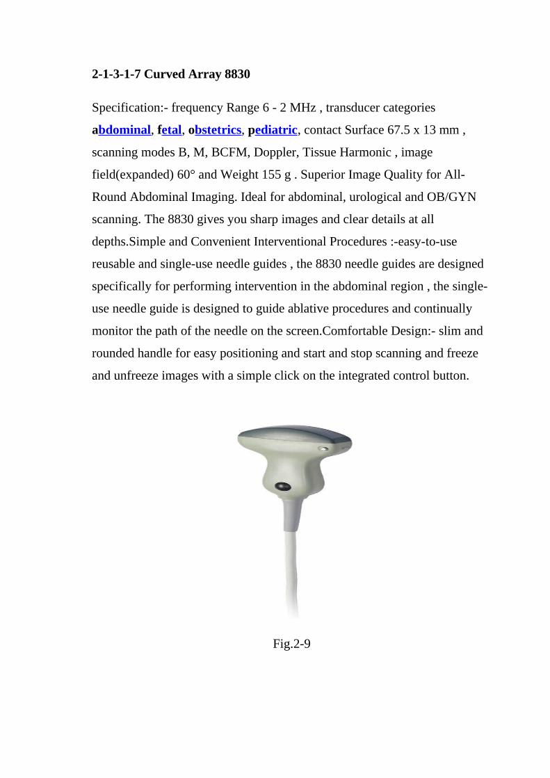
2-1-3-1-7 Curved Array 8830
Specification:- frequency Range 6 - 2 MHz , transducer categories
abdominal, fetal, obstetrics, pediatric, contact Surface 67.5 x 13 mm ,
scanning modes B, M, BCFM, Doppler, Tissue Harmonic , image
field(expanded) 60° and Weight 155 g . Superior Image Quality for All-
Round Abdominal Imaging. Ideal for abdominal, urological and OB/GYN
scanning. The 8830 gives you sharp images and clear details at all
depths.Simple and Convenient Interventional Procedures :-easy-to-use
reusable and single-use needle guides , the 8830 needle guides are designed
specifically for performing intervention in the abdominal region , the single-
use needle guide is designed to guide ablative procedures and continually
monitor the path of the needle on the screen.Comfortable Design:- slim and
rounded handle for easy positioning and start and stop scanning and freeze
and unfreeze images with a simple click on the integrated control button.
Fig.2-9

2-1-3-1-8 Curved Array 8802
Specification:- Frequency Range 6 - 3 MHz , transducer Categories
Abdominal, Fetal, Obstetrics, Pediatric, contact Surface 52 x 8 mm , focal
Range 6-114 mm and Weight 150 g . Excellent Image Quality:- broad
bandwidth gives excellent image quality , deep penetration for clear images
and ergonomic design. Quick and Easy Disinfection. Compatible with
modern sterilization methods. 8802 Applications :- pelvic Floor ,
abdominal , obstetric , pediatric , Interventional procedures and tissue
harmonic imaging
Fig.2-10

2-1-3-1-9 Endfire Curved Array 8667
Specification :- frequency Range 10 - 5 MHz , transducer Categories
Transrectal, Urology , contact Surface 50 mm2 , focal Range 5 - 50 mm ,
scanning modes B, M, BCFM, Doppler, Tissue Harmonic, Power Doppler ,
dimensions 300 x 36 mm and weight 260 g .Endfire prostate imaging. wide
endfire image plane assists in locating lesion. Convenient puncture :- the
needle's path starts at the tip of the probe and monitor the needle from the
start of the puncture to the actual biopsy site. 8667 Applications:- endorectal
scanning , interventional procedures , spectral, CFM and Power Doppler
examinations and tissue harmonic imaging.
Fig. 2-11

2-1-3-1-10 Endocavity Biplane 8848
Specification :- frequency Range 12 - 4 MHz , transducer Categories
Transrectal, Transvaginal, Urology, focal Range 3 - 60 mm , scanning
modes B,M, Doppler, BCFM, Tissue Harmonic Imaging , rame Rate >150 ,
image field(expanded) 180°(transverse) and weight 250 g .Image guided
prostate therapy:- sagittal scanning of any size prostate from base to apex ,
clear and detailed image, for accurate volume studies and source dose
planning , customizable sagittal grids and preferences for brachytherapy ,
clear visualization of seminal vesicles and clear view of needle placement.
Pelvic Floor scanning :- best broad view of anterior and posterior
compartments for functional and anatomical studies , reproducible 3D
studies with external mover and detailed high-resolution biplane with 6.5
cm. linear and convex views
Fig. 2-12

2-1-3-1-11 Endovaginal 8819
Specification: - frequency Range 9 - 5 MHz, transducer categories fetal,
transrectal, transvaginal , contact Surface 26 x 5 mm and weight 140 g.
Versatile Endovaginal Transducer:- ideal all-round ultrasound tool for
routine gynecological investigations , offers good penetration and gives you
detailed images at all depths , simple and convenient needle guide clicks
easily and securely into place and helps simplify interventional procedures ,
easy follicle measurement and visual guidance of aspiration , ergonomic
design makes it comfortable to use and slim design minimizes the
discomfort of a transvaginal examination. 8819 Applications:- endovaginal
scanning , transrectal scanning and spectral and CFM Doppler examinations
Fig. 2-13

2-1-3-1-12 High Frequency Linear Array 8870
Specification :- frequency Range 18 - 6 MHz , transducer Categories
Musculoskeletal, Peripheral Vascular, Small Parts , contact Surface 38.4
x 3.5 mm , focal Range 3 - 60 mm , scanning modes B, M,BCFM ,
doppler,tissue , harmonic imaging , frame Rate >150 Hz and weight 100 g .
High Frequency Imaging with the 8870. Linear array, fine pitch 18 MHz
transducer offers:- high Resolution, Detailed Images of Superficial
Structures , greater confidence during scanning of fingers, toes, wrists,
forefoot, ankles and elbows. High Doppler frequencies allow superb flow
visualization of superficial areas. Easy System Control. Operate system at
the touch of a transducer button – leaves both hands free for scanning.Easy
and Thorough Cleaning and Disinfection. Compatible with modern
disinfection and sterilization techniques.
Fig. 2-14

2-1-3-1-13 Hockey Stick 8809
Specification:- frequency Range 15 - 6 MHz , transducer Categories
Intraoperative, Musculoskeletal, Peripheral Vascular, Small Parts ,
contact Surface 24 x 3.5 mm , focal Range 3 - 55 mm , scanning modes B,
M, Doppler, CFM and weight 80 g . Reach difficult to access areas easily:-
small, flexible tip fits into tight spots and tip can be set at various angles.
Excellent resolution in the extreme near field:- very high frequency (15
MHz) for high resolution and visualize flow with outstanding Doppler
sensitivity.Puncture in tight places :- unique puncture guide that follows
transducers flexible tip and perfect for guiding intraoperative and
percutaneous interventional procedures. 8809 Applications :-intraoperative
vascular , general intraoperative , musculoskeletal , small part and
Interventional,
Fig. 2-15

2-1-3-1-14 Intraoperative 8815
Specification :- frequency Range 10 - 4 MHz , transducer Categories
Intraoperative, Pediatric , contact Surface 14 x 60 mm , focal Range 5 –
95 mm , scanning modes B, M, Doppler , BCFM , contrast Imaging , tissue
Harmonic Imaging , frame Rate (max.) 230 Hz and weight 250 g .
Designed Especially for Interventional Ultrasound During Surgery. I-shaped
intraoperative transducer. Guided intraoperative biopsy with a variety of
angles and positions .Safer intervention with needle guide lock. High image
quality with large footprint and wide near field image .Easily capture 3D
images, review and send to a colleague. Contrast-enhanced* ultrasound for
detection and assisting in characterization of suspicious masses.
Fig. 2-16

2-1-3-1-15 Intraoperative Biplane 8824
Specification :- frequency Range 10 - 3.75 MHz , transducer Categories
Intraoperative, Musculoskeletal, Peripheral Vascular, Small Parts ,
Scanning modes B, M, Doppler, BCFM, Tissue Harmonic, Contrast ,
dimensions 1.4 x 3.3 x 5.1 cm and weight 45 g .8824 - Simultaneous Biplane
Imaging:- choose between the I, T or simultaneous imaging , combined I and
T array for improved orientation , flexibility to take free hand biopsies from
the front, back, left, or right , excellent Images , near field and good
penetration and high frequency coded excitation.Compact Size. Easy access
in tight spaces. Easy to hold in intra0perative condition.
Fig. 2-17

2-1-3-1-16 Linear Array 8811
Specification:- frequency Range 12 - 5 MHz , transducer Categories
Intraoperative, Musculoskeletal, Peripheral Vascular, Small Parts,
Urology , contact Surface 50 x 4 mm , focal Range 2 - 55 mm , dimensions
105 x 64 x 22 mm and weight 98 g . A Clear Choice for Cost-Effective
Imaging. High frequency. Large footprint. Doppler sensitivity. True Echo
Harmonics. Puncture Guides for Convenient Biopsy. Puncture guides
available have:- 30, 45 and 60 angles of insertion and variable diameter,
allowing you to choose the desired needle size. Disposable needle guides
and sterile transducer covers are also available. Fast Lesion Assessment.
Provides image clarity and contrast resolution. Determine necessity for
biopsy on the spot. Perform aspiration or biopsy immediately .
Fig. 2-18

2-1-3-1-17 Linear Array 8670
Specification:-frequency Range 12 - 4 MHz , transducer Categories
Musculoskeletal, Pediatric, Peripheral Vascular, Small Parts, Urology ,
contact Surface 45 x 14 mm , focal Range 0 - 70 mm , scanning modes B,
M, BCFM, Doppler, Tissue Harmonic , dimensions 91 x 52 x 21 mm and
weight 130 g . Quality, Versatility and Convenience in One Transducer.
Small part, musculoskeletal and vascular scanning. The 8670 supports a
number of ultrasound applications, such as small part, breast and
orthopedics, rheumatology and sports medicine.Easy switching between near
and far views. Very high image detail . Excellent choice for penile Doppler
and testis . Optimized for small part, musculoskeletal and vascular
scanning:-easy-to-use puncture and biopsy guide , small part , breast , testis ,
penile Doppler. Musculoskeletal. Peripheral vascular. Interventional
procedures. Contrast Imaging.
Fig. 2-19

2-1-3-1-18 ProART™ Robotic Drop In 8826
Specification: - frequency Range 12 - 5 MHz, transducer Categories
Intraoperative, Urology , image field (expanded) Sector 36°and weight 25
g . Unique specialized ultrasound transducer for robotic-assisted surgery.
Premium Performance and Excellent Image Quality. Curved linear array
with the largest field of view on the market today. High resolution 12–5
MHz transducer. Premium image quality with excellent contrast and detail
resolution. Unique 3D rendering visualization. Designed for Ease of Use:-
"Fingertip" control - fin located directly over transducer array , designed to
fit Prograsp™1 for maximum control , fits through a standard trocar
Fig. 2-20

2-1-3-1-19 Prostate Biplane 8808e
Specification: - frequency Range 10 - 5 MHz , transducer Categories
Transrectal, Urology , scanning modes B, M,BCFM, Doppler, Tissue
Harmonic Imaging , image field(expanded) 126° and weight 250
g.Simultaneous biplane. Efficient and confident prostate examinations.
Precise Biopsies with Simultaneous Biplane. The 8808e gives you real-time
images of both the sagittal and transverse planes which is invaluable for
orientation. Simultaneous images of the sagittal and transverse planes
provide a clear indication of needle placement for quicker and more
confident biopsies.Streamlined Workflow. Save time and effort with the
transducer's one touch operation. Switch between views, freeze, print and
save without touching the scanner. Slender Design. The 8808e's sterile,
single-use and reusable needle guides are an integral part of the transducer
minimizing patient discomfort.
Fig. 2-21

2-1-3-1-20 Prostate Triplane 8818
Specification :- frequency Range 12 - 4 MHz , transducer Categories
transrectal, transvaginal, urology , contact Surface 34.4 x 5.5 mm , focal
Range 3 - 60 mm , scanning modes B, M,BCFM, Doppler, Contrast , tissue
Harmonic , frame Rate 60 Hz , image field(expanded) Triplane / 140° ,
dimensions 36 x 39 x 323 mm and weight 230 g .Triplane – all prostate
zones with one transducer , images in three visionary planes , switch
between prostate zones at the touch of a button and increase diagnostic value
with 3D, Contrast and Doppler. Easy and comfortable to use take confident
apical biopsies with endfire array, biopsy the peripheral, transition and
central zones with simultaneous biplane , one-time insertion and minimal
manipulation using disposable dual guide. 8818 Applications:-transrectal
prostate scanning , transrectal puncture and biopsy , transperineal puncture
and biopsy , transvaginal scanning , spectral and CFM Doppler examinations
, Tissue harmonic imaging and contrast imaging.
Fig. 2-22

2-1-3-1-21 Rigid Laparoscopic 8836
Specification: - frequency Range 12 - 5 MHz, transducer Categories
Intraoperative , scanning modes B, M, Doppler, BCFM, Tissue Harmonic
Imaging and Contrast Imaging. Premium performance, more control, easier
access. Superb detail and contrast resolution of difficult-to-access anatomy.
Curved array giving wide field of view. Durable, easy-grip handle. Fits
through standard 10 mm trocar. Compatible with modern sterilization
methods. Applications:-minimally invasive intraoperative procedures, liver,
gall bladder, kidney and uterus.
Fig. 2-23

2-1-3-1-22 Small Footprint Cardiac 8827
Specification:- frequency Range 4 - 2 MHz , transducer categories cardiac,
transcranial , contact Surface 16 x 13 mm , focal Range 10 - 134 mm ,
frame Rate >200 Hz , image field(expanded) Phased 90°, weight 74 g .
Fig. 2-24

2-1-3-1-23 Small Footprint Cardiac 8837
Specification: - frequency Range 5 – 1 MHz, transducer categories
abdominal, cardiac, transcranial , contact Surface 19.2 x 13.5 mm , focal
Range 10 – 34 mm , scanning modes B, M, Doppler, BCFM, CW Doppler,
Tissue Harmonic Imaging and weight 74 g . Ergonomic Design, Suitable for
All Patients: - easy to hold, ergonomically designed handle, ideal for
intercostal imaging with small footprint, orientation mark on the handle for
imaging plane orientation and dynamic cardiac examinations. Sensitive to
movement with phased array technology and a high frame rate.Single-crystal
transducer technology for optimal imaging conditions with high resolution
and uniform images in all depths. Wide imaging angle (90º) to see entire
myocardium. Super image quality and cardiac calculations .Tissue Harmonic
imaging provides outstanding visualization of cardiac structures.High
Doppler sensitivity for fast flow measurements.
Fig. 2-25

2-1-3-1-24 T-Shaped Intraoperative 8816
Specification:- frequency Range 10 - 4.3 MHz , transducer categories
abdominal, intraoperative , contact Surface 5 x 51 mm , focal Range 5 - 95
mm , scanning modes B, M, Doppler, BCFM, contrast, tissue harmonic
imaging and weight 55 g . Small Intraoperative Transducer with Wide Near
Field View. Small, T-shaped transducer designed primarily for hepatic
surgery. Excellent image quality. Wide near field view. Coded excitation
technology for high resolution with deeper penetration. Contrast Imaging for
improved sensitivity and accuracy of lesion detection and classification.
Compatible with modern sterilization techniques. 8816 Applications: -
intraoperative scanning, abdominal scanning, spectral and CFM Doppler
examinations, coded excitation for deeper penetration and contrast and
harmonic imaging.
Fig. 2-26

2-1-3-1-25 Vascular 8822
Specification: - frequency Range 9 - 3.5 MHz, transducer Categories
Peripheral Vascular , scanning modes B, M, CFM, Doppler, tissue
harmonic imaging and weight 98 g .Peripheral Vascular Imaging. Excellent
image quality in depth. Wide field of view. Broad bandwidth. Tissue
Harmonic Imaging. Easy interventional Procedures and Biopsy. Sterilizable
needle guide with puncture line on image.3 puncture lines: 30º, 45º, 60º.3
needle diameters.Designed for Easy Use:-customizable finger-tip control
button, easy to hold and completely immersible for cleaning and
disinfection.
Fig. 2-27
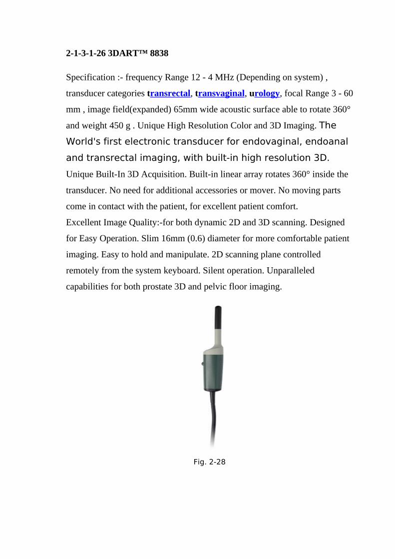
2-1-3-1-26 3DART™ 8838
Specification :- frequency Range 12 - 4 MHz (Depending on system) ,
transducer categories transrectal, transvaginal, urology, focal Range 3 - 60
mm , image field(expanded) 65mm wide acoustic surface able to rotate 360°
and weight 450 g . Unique High Resolution Color and 3D Imaging. The
World's first electronic transducer for endovaginal, endoanal
and transrectal imaging, with built-in high resolution 3D.
Unique Built-In 3D Acquisition. Built-in linear array rotates 360° inside the
transducer. No need for additional accessories or mover. No moving parts
come in contact with the patient, for excellent patient comfort.
Excellent Image Quality:-for both dynamic 2D and 3D scanning. Designed
for Easy Operation. Slim 16mm (0.6) diameter for more comfortable patient
imaging. Easy to hold and manipulate. 2D scanning plane controlled
remotely from the system keyboard. Silent operation. Unparalleled
capabilities for both prostate 3D and pelvic floor imaging.
Fig. 2-28

2-1-3-1-27 10L2w Wide Linear
Specification: - transducer Categories Peripheral Vascular , contact
Surface 57 x 10 mm.
Fig. 2-29
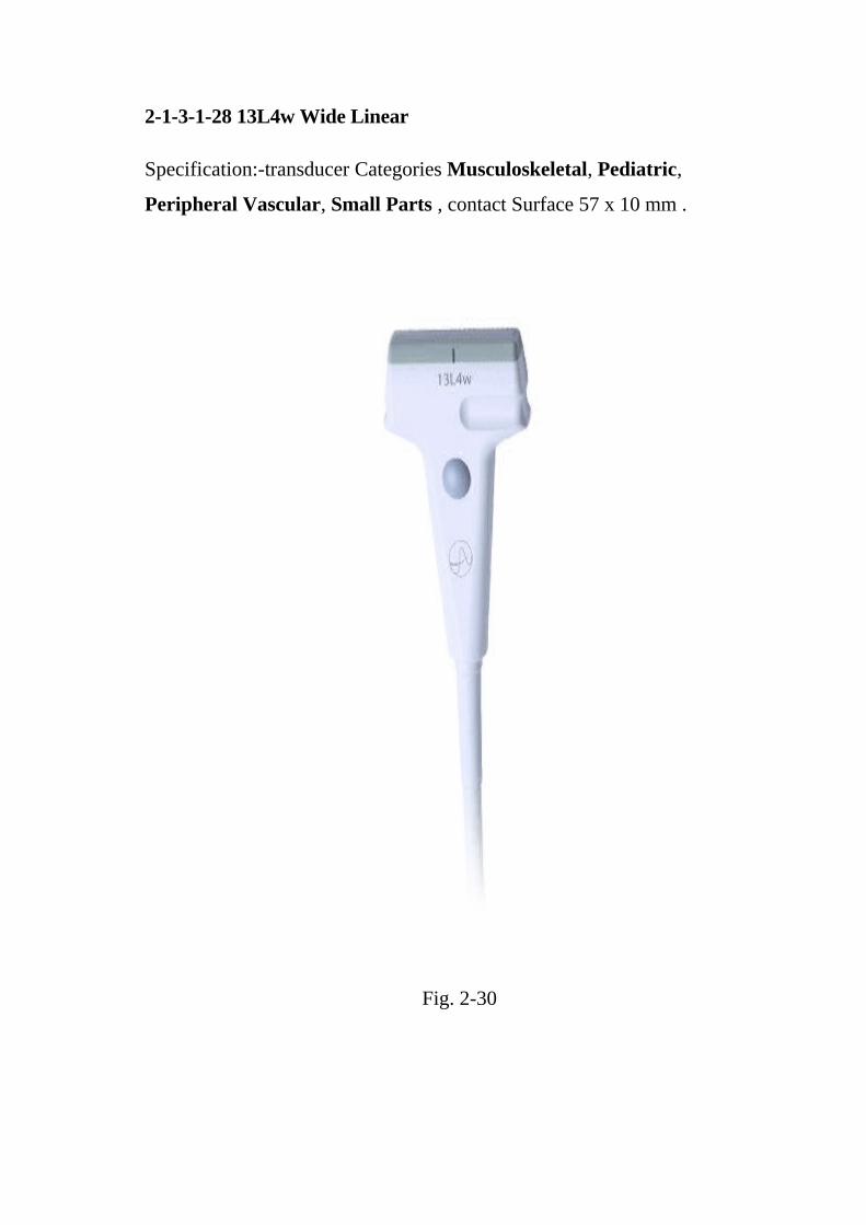
2-1-3-1-28 13L4w Wide Linear
Specification:-transducer Categories Musculoskeletal, Pediatric,
Peripheral Vascular, Small Parts , contact Surface 57 x 10 mm .
Fig. 2-30

2-1-3-1-29 14L3 Linear
Specification: - transducer categories musculoskeletal, pediatric,
peripheral vascular and small Parts and Contact Surface 45 x 14 mm.
Fig. 2-31

2-1-3-1-30 18L5 High Frequency Linear
Specification:- transducer categories musculoskeletal, pediatric,
peripheral vascular and small Parts.
Contact Surface 48 x 13mm.
Fig. 2-32
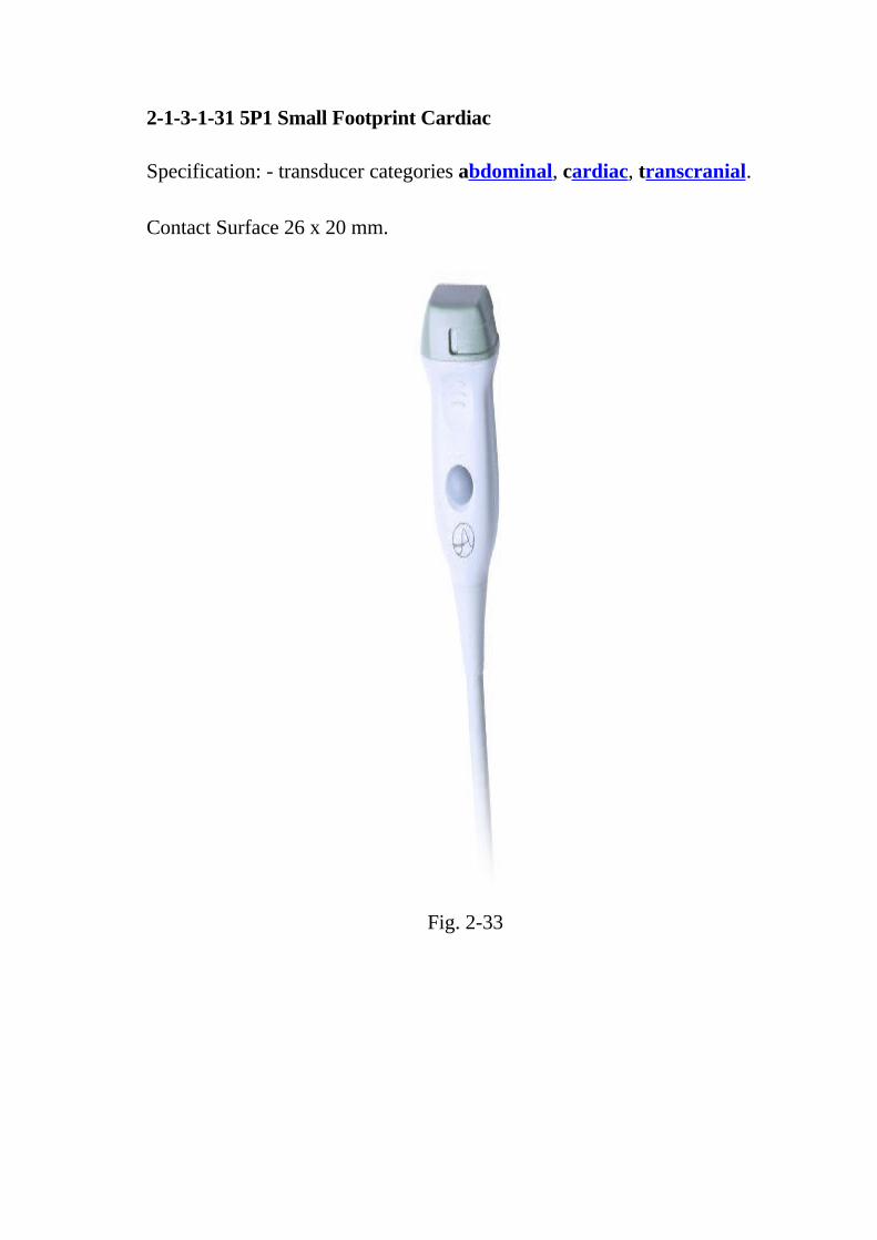
2-1-3-1-31 5P1 Small Footprint Cardiac
Specification: - transducer categories abdominal, cardiac, transcranial.
Contact Surface 26 x 20 mm.
Fig. 2-33

2-1-3-1-32 6C2 Curved
Specification:- transducer categories abdominal, fetal, musculoskeletal,
obstetrics.
Contact Surface 69 x 19 mm.
Fig. 2-34
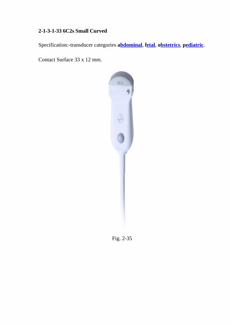
2-1-3-1-33 6C2s Small Curved
Specification:-transducer categories abdominal, fetal, obstetrics, pediatric.
Contact Surface 33 x 12 mm.
Fig. 2-35
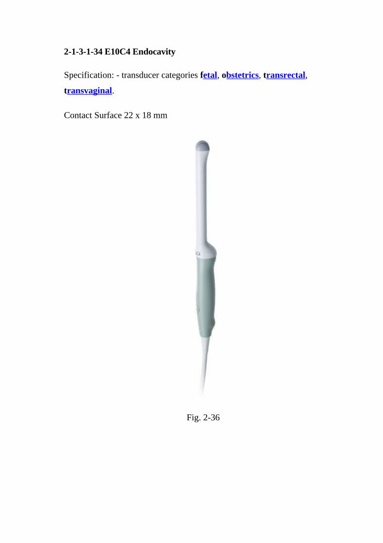
2-1-3-1-34 E10C4 Endocavity
Specification: - transducer categories fetal, obstetrics, transrectal,
transvaginal.
Contact Surface 22 x 18 mm
Fig. 2-36

2-1-3-1-35 E14C4 Endfire Curved
Specification
• Transducer Categories Transrectal, Transvaginal,
• Contact Surface 20 mm
Fig. 2-37
2-1-3-1-36 E14C4t Prostate Triplane
Specification
• Transducer Categories Transrectal, Transvaginal,
• Contact Surface 20 mm

Fig. 2-38
2-1-3-1-37 E14C4t Prostate Triplane
Specification
• Transducer Categories Transrectal, Transvaginal,
• Contact Surface 20 mm

Fig. 2-39
2-1-3-1-38 E14CL4b Endocavity Biplane
Specification
• Transducer Categories Transrectal, Transvaginal,
• Contact Surface 20 mm

Fig. 2-40
2-1-3-1-39 N13C5 Curved
Specification
• Transducer Categories Neonatal,
• Contact Surface 29 x 10 mm

Fig. 2-41

2-1-3-1-40 4-Way Laparoscopic 8666-RF
Specification:- frequency Range 10 - 4.3 MHz , transducer categories
intraoperative, urology, contact Surface 30 x 5 mm , focal Range 5 - 95
mm , scanning modes B, M, Doppler, BCFM, tissue Harmonic Imaging and
Contrast Imaging , dimensions 302 x 178 mm and weight 475 g. The Most
Advanced Laparoscopic Ultrasound Transducer on the Market. Built-in
facilities for LUS-guided biopsies. Ethanol or contrast agent injections
cryoablation. Microwave ablation. Flexible for difficult to reach areas or
rigid for manipulating structures. 8666-RF Applications: - laparoscopic,
intraoperative, radiofrequency tumor ablation (RFA) and biopsy.(Doody
C,1999)
Fig. 2-42
2-1-4 Biological effects of possible relevance to safety.

2-1-4-1 Introduction.
In the 1920s, the availability of piezoelectric materials and electronic
devices made it possible to produce ultrasound (US) in water at high
amplitudes, so that it could be detected after propagation through large
distances. Laboratory experiments with this new mechanical form of
radiation showed that it was capable of producing an astonishing variety of
physical, chemical and biologic effects. In this review, the early findings on
bioeffects are discussed, especially those from experiments done in the first
few decades, as well as the concepts employed in explaining them. Some
recent findings are discussed also, noting how the old and the new are
related. In the first few decades, bioeffects research was motivated partly by
curiosity, and partly by the wish to increase the effectiveness and ensure the
safety of therapeutic US. Beginning in the 1970s, the motivation has come
also from the need for safety guidelines relevant to diagnostic US.
Instrumentation was developed for measuring acoustic pressure in the fields
of pulsed and focused US employed, and standards were established for
specifying the fields of commercial equipment. Critical levels of US
quantities were determined from laboratory experiments, together with
biophysical analysis, for bioeffects produced by thermal and nonthermal
mechanisms. These are the basis for safety advice and guidelines
recommended or being considered by national, international, professional
and governmental organizations.
After the end of World War II, advances in ultrasound (US) technology
brought improved possibilities for medical applications. The first major
efforts in this direction were in the use of US to treat diseases. Medical
studies were accompanied by experiments with laboratory animals and other
model systems to investigate basic biological questions and to obtain better
understanding of mechanisms. Also, improvements were made in methods

for measuring and controlling acoustical quantities such as power, intensity
and pressure. When diagnostic US became widely used, the scope of
biological and physical studies was expanded to include conditions for
addressing relevant safety matters. In this historical review, a major part of
the story is told by 21 investigators who took part in it. Each was invited to
prepare a brief personal account of his/her area(s) of research, emphasizing
the "early days," but including later work, showing how late and early work
are related, if possible, and including anecdotal material about mentors,
colleagues, etc.
We briefly review my early contacts with bioacoustics and the bioacoustic
work at the University of Pennsylvania that took place from the early 1950s
to 1975. It was carried out with E. L. Carstensen, K. Li, A. Smith, H. Pauly,
J. Reid, P. Edmonds and many students. The emphasis was first on basic
biophysical studies. The work with E. Carstensen and H. Pauly was
primarily concerned with the mechanism causing the high absorption typical
for tissues and cell suspensions. Macromolecular content was shown to be
largely responsible for the absorption. Practical applications concerned the
relative merits of electromagnetic and ultrasonic diathermy techniques. P.
Edmonds extended the range of macromolecular studies to 100 MHz and
initiated work on the attenuation in lung tissues. After J. Reid came to
Pennsylvania, the development of echocardiography took place.
Biological effects of ultrasound are the potential biological consequences
due to the interaction between the ultrasound wave and the scanned tissues.
The use of ultrasound for cardiac imaging has not known significant adverse
biological effects. Concern about the safety of ultrasound prompted several
agencies to devise regulatory limits on the machine output intensities. The
visual display of thermal and mechanical indices during ultrasound imaging

provides an aid to limit the output of the machine. Sonographic evaluation of
the human body, including potentially sensitive tissues, such as developing
fetus and the eye, have been performed on millions of patients without
documentation of serious adverse events. However, ultrasound waves have
the potential to cause significant biological effects, depending on ultrasound
wave characteristics and scanned tissues sensitivity. Physicians and
sonographers must be aware of these potential biological effects in assessing
the overall safety of the procedure. (Dalecki,2007)
2-1-4-2 Types of biological effects.
The biological effects of ultrasound depend on the total energy applied to a
given region. Thus, varying duration of exposure to wave emission, intensity
and frequency of the ultrasound beam, pulsed or continuous emission
modality and acoustic power, may lead to significant biological effects, that
are commonly divided in thermal and non-thermal effects.
2-1-4-2-1 Thermal.
The biological effects of ultrasound energy are related primarily to the
production of heat. Heat is generated whenever ultrasound energy is
absorbed, and the amount of heat produced depends on the intensity of the
ultrasound, the time of exposure, and the specific absorption characteristics
of the tissue. As much as 70% of the total temperature increase associated
with ultrasound occurs within the first minute of exposure ], but temperature
continues to rise as exposure time is prolonged. Minimizing the exposure
time is probably the single most important factor for ensuring patient safety
from thermal injury. Other important parameters to be considered are: - The
relative protein content of each tissue, since absorption coefficients of
tissues are directly related to protein content; absorption coefficients vary

between 1 (skin, tendon, spinal cord) and 10 (bone) dB/cm MHz. The
perfusion of the tissue, which has a dampening effect on heat generation and
physically allows heat to be carried away from the point of energy transfer.
Emission modality, since pulsed-wave ultrasound is extremely unlikely to
significantly heated tissues.
Beam width, since a wider beam width reduces the rate and extent of
temperature rise by permitting the energy to be distributed over a larger
perfusion territory.
2-1-4-2-2 Non-thermal
Ultrasound energy creates also mechanical forces independent of thermal
effects, thereby causing biologic effects that are not related to temperature
rise alone, such as cavitations, torque forces, oscillatory shear, radiation,
pressure and micro streaming.
2-1-4-2-3 Cavitations
The interaction of ultrasound with gas bubbles or contrast agents causes
rapid and potentially large changes in bubble size. This process, termed
cavitations, may increase temperature and pressure within the bubble and
thereby cause mechanical stress on surrounding tissues, precipitate fluid
micro jet formation, and generate free radicals . Gas-containing structures
(e.g., lungs, intestines) are most susceptible to the effects of acoustic
cavitations. Ultrasound wavelength has an important role in bubble
formation and growth: short wavelength ultrasound (observed at higher
frequencies) does not provide sufficient time for significant bubble growth;
therefore, cavitations are less likely under these circumstances compared
with long wavelengths. The short half-life of cavitations nuclei prevents

most cavitations-related biological effects, unless ultrasound contrast agents
are also present. Contrast agents markedly reduce the threshold intensity for
cavitations. However, because of the relatively high viscosity of blood and
soft tissue, significant cavitations is unlikely and cavitations has not been
shown to occur with the ultrasound exposure commonly used during a
diagnostic examination.
2-1-4-2-4 Other effects
A variety of other physical forces may also be produced by ultrasound
energy. Although each of these effects can be demonstrated in vitro, there is
no evidence that any of these physical phenomena has a significant
biological effect on patients. (Daleki,2007)
2-1-4-3 Measurement of biological effects
The biological effects of ultrasound are generally discussed in terms of
power (the amount of acoustic energy per unit of time), and the units of
power are in the mill watt range. Intensity (acoustic power per unit of area)
is usually expressed as watts per meter squared (W/m2) or in mill watts per
centimeter squared (mW/cm2). To calculate the energy from a pulsed
ultrasonic beam, it is necessary to know the duty factor, which is a measure
of the fraction of time during which the transducer emits ultrasound. The
maximum overall intensity is then described as the highest exposure within
the beam (spatial peak) averaged over the period of exposure (temporal
average) and is known as the spatial peak temporal average (SPTA)
intensity. Another common measure is the spatial peak pulse average
(SPPA), defined as the average pulse intensity at the spatial location where
the pulse intensity is maximum. Commercial ultrasound instruments
operating in pulsed-wave modality for two-dimensional imaging have spatial

peak, temporal averaged intensities ranging from 0.001 to more than 200
mW/cm2. Pulsed Doppler imaging, however, may have a spatial peak,
temporal average as high as 1900 mW/cm2, considerably greater than 100
mW/cm2 level that has been most extensively studied and has never been
shown to produce a biologic effect. The relatively short periods of pulsing,
coupled with the fact that the transducer is constantly moving so that no
single area is imaged for a long period, contribute to the low likelihood of
delivering significant heat to the tissue.
A major limitation of measuring the intensity of ultrasound exposure is that
estimating the actual tissue exposure is difficult, due to attenuation and other
interactions with the tissue. Furthermore, tissue exposure is limited only to
transmission periods and to the time the ultrasound beam dwells at a specific
point, both of which are considerably shorter than the total examination
time. Other indices that incorporate these factors have been developed to
better define the exposure levels with diagnostic ultrasound. These measures
include the mechanical index (MI) and tissue thermal index (TTI).
The thermal index (TI) and mechanical index (MI) were introduced to
provide the operator with an indication of the potential for ultrasound-
induced bioeffects. The TI provides an on-screen indication of the relative
potential for a tissue temperature rise. MI provides an on-screen indication
of the relative potential for ultrasound to induce an adverse bio-effect by a
non-thermal mechanism such as cavitations. Thermal indices are
conservatively determined to ensure patient safety. Under most clinical
conditions, the thermal index closely approximates or overestimates the
maximum temperature increase for ultrasound exposure. Three different
thermal indices (depending on the structures encountered in the path of the
ultrasound beam, soft tissue or TIs, bone or Tip and cranium or Tic) are used
to estimate temperature increases associated with an ultrasound beam. In
fact, thermal indices in soft tissue or bone provide fairly accurate in vivo

estimates of ultrasound-related temperature rise in the tissue types .
Contemporary ultrasound equipment has the theoretic capability to cause a
tissue temperature increase greater than 4°C at the focal point.The MI
describes the relationship between cavitations formation and acoustic
pressure and is defined as the ratio of the peak rare factional negative
pressure adjusted for tissue attenuation and square root of the frequency. The
MI was originally formulated based on the threshold for acoustic cavitations
in water and blood, and hence may not specifically consider the type of
tissue in which this process occurs.
The American Institute of Ultrasound in Medicine (AIUM) has proposed
guidelines for limits below which ultrasound clearly has been demonstrated
to be safe]. These guidelines include:
A diagnostic exposure that produces a 1°C or less temperature elevation
above normal.
An exposure intensity less than 1 W/cm2 for focused ultrasound beams.
Current diagnostic ultrasound systems have outputs ranging from 10
mW/cm2 (SPTA) for imaging to as high as 430 mW/cm2 (SPTA) for pulsed
Doppler ultrasound. There has been no evidence to date to suggest adverse
effects of echocardiography at these ultrasonic outputs.
During transoesophageal imaging, especially during intraoperative imaging,
the probe may remain nearly stationary for extended periods. The heat
generated by the transducer itself must also be considered. Although there
are no reports of significant injury resulting from even prolonged
intraoperative transoesophageal echocardiography, attention to these issues
is recommended. Limited imaging time, occasional repositioning of the

probe and constant monitoring of the probe temperature will all help to
ensure an impeccable safety record.
All evidence to date suggests that diagnostic ultrasound, particularly which
used in echocardiography, is an extremely safe tool with no demonstrated
adverse effects even with the use of newer technology and more powerful
instrumentation. Although this is reassuring and justifiably inspires
continued confidence in ultrasound imaging, the desire for more and better
diagnostic information should never occur at the expense of patient safety.
Therefore, limiting the scan time to a minimum, knowing the power output
and exposure intensity of different modalities of each instrument, and
keeping up to date on any new scientific findings or data relating to possible
adverse effects, should always be a consideration.(Fowlekes GB,2008)
2-1-5 Guidelines and regulations
The sonographer should:- Recognise his/her scope of practice and work
within its boundaries ensure that a locally agreed written scheme of work is
in place accept properly delegated responsibility, in accordance with local
practice and guidelines An ultrasound examination should not be carried out
unless a valid request has been received. The request should include such
clinical details as are relevant to the examination, clear identification of the
person requesting the examination and to whom the report should be
directed.
The sonographer should be responsive to:- potential bio-effects of ultrasound
and the need to minimise dose at all times , potential hazards arising from
the particular ultrasound equipment , relative risks for each application ,
conditions where current recommendations contra-indicate the use of certain

types of ultrasound equipment and current guidelines regarding replacement
of ultrasound equipment.
The sonographer is expected to: - have detailed knowledge of ultrasound
equipment in order to ensure that it is appropriate for purpose , manipulate
the equipment correctly so that patient diagnosis and management are not
compromised , ensure that an agreed quality assurance programme is in
place that incorporates the regular and inspection of ultrasound machines
and auxiliary equipment.
The stated aim of quality assurance procedures applied to ultrasound
equipment is to ensure consistent and acceptable levels of performance of
the imaging system and image recording facilities. Most quality assurance
protocols focus on the consistency. The acceptability of image quality may
not be apparent from measurable changes in the parameters tested. The issue
of what constitutes unacceptable equipment performance is still very
difficult to assess objectively. In the absence of nationally accepted
performance standards for ultrasound equipment, local and subjective
evaluation is required.
This programme should include a policy on:- electrical safety tests carried
out at least once a year by qualified personnel , baseline/acceptance testing
of all new or upgraded equipment, and following major repair and user tests
including weekly inspection of cables, transducers, monitor and image
recording facilities.
A quality assurance programme should be developed in discussion with
medical physics or service engineers, for each individual machine. This
should be based on its clinical uses, the modes and functions utilized, the
transducer types and frequencies and the auxiliary equipment attached. The

programme should indicate clearly the limits of acceptability for each test,
what and by whom action should be taken when these are exceeded.
The sonographer's responsibilities in relation to the ultrasound equipment
should include:- appropriate selection for the examination and awareness of
its limitations within that clinical context , manipulation of the controls to
maximize the clinical information observed , awareness of system artifacts
and how to interpret their appearances , ensuring that the equipment is
suitably maintained to provide optimal images , ensuring that all transducers
are appropriately prepared and cleaned according to the manufacturers’
guidelines, with especial reference to intra-cavity probes , awareness of and
adherence to local infection control procedures , ensuring that the recorded
image is an accurate record of the displayed real-time information ,
following the proper shut-down procedure for the equipment, so that stored
data and settings are not corrupted or lost , inspection for electrical and
mechanical safety, ensuring that apparently unsafe equipment is not used
until it has been checked and repaired , agreement of equipment performance
criteria for each type of examination undertaken. (This should be updated
regularly, in line with new developments in equipment carry performance),
reporting any concerns in relation to the performance of specific equipment,
and awareness of current guidelines regarding the replacement of ultrasound
equipment.(Fowlekes GB,2008)
2-1-5-1 Ultrasound Examination Procedures.
Relating to all ultrasound examinations, the sonographer should be aware of
locally agreed standards of practice and current guidelines of other
professional bodies and organizations.
The following points should be considered for all ultrasound examinations:-
the clinical details provided are sufficient to carry out the examination
requested and the correct examination has been requested , relevant
information is available from the case notes, previous investigations and
other sources , the role of the ultrasound examination is understood in the
clinical context for the patient , informed consent is obtained before
proceeding with the examination , the necessity for the presence of a
chaperone and/or an interpreter , a systematic scanning approach that can be
modified according to the individual patient , the implications should the
examination be incomplete , the need to extend the ultrasound examination,
and/or proceed to additional imaging techniques .where necessary in
accordance with locally agreed protocol , the after care of the patient , the
potential risks involved in the procedure to the patient and appropriate
national and local Health and Safety regulations including infection control.
2-1-5-2 Communication.
The sonographer should:-obtain sufficient verbal and/or written information
from the referring clinician to undertake correctly the examination
requested , be mindful of the need to use interpreters as and when necessary
to communicate adequately with the patient , greet the patient using his or
her full name and status , be able to discuss the relative risks and benefits of
the examination with the patient , explain the scanning procedure
appropriately to the patient , obtain informed consent* from the patient or
their representative being mindful of his/her capacity to understand , be
aware of the individual patient’s special needs including chaperoning and
privacy during the examination , be professional and understanding
throughout the examination; manage the interaction between the patient and
any accompanying adults and children in a way that enables the examination
to be carried out to a competent standard , explain and discuss the findings
with the patient , Interpret and communicate the appropriately and in a
timely fashion to the referring clinician , ensure appropriate arrangements
have been made for further care before the conclusion of the examination ,
display - displays the image from the ultrasound data processed by the CPU ,
keyboard/cursor - inputs data and takes measurements from the display ,
disk storage device (hard, floppy, CD) - stores the acquired images and
printer prints the image from the displayed data
The transducer probe is the main part of the ultrasound machine. The
transducer probe makes the sound waves and receives the echoes. It is, so to
speak, the mouth and ears of the ultrasound machine. The transducer probe
generates and receives sound waves using a principle called the piezoelectric
(pressure electricity) effect, which was discovered by Pierre and Jacques
Curie in 1880. In the probe, there are one or more quartz crystals called
piezoelectric crystals. When an electric current is applied to these crystals,
they change shape rapidly. The rapid shape changes, or vibrations, of the
crystals produce sound waves that travel outward. Conversely, when sound
or pressure waves hit the crystals, they emit electrical currents. Therefore,
the same crystals can be used to send and receive sound waves. The probe
also has a sound absorbing substance to eliminate back reflections from the
probe itself, and an acoustic lens to help focus the emitted sound waves.

Fig. 2-43 The parts of an ultrasound machine
The CPU is the brain of the ultrasound machine. The CPU is basically a
computer that contains the microprocessor, memory, amplifiers and power
supplies for the microprocessor and transducer probe. The CPU sends
electrical currents to the transducer probe to emit sound waves, and also
receives the electrical pulses from the probes that were created from the
returning echoes. The CPU does all of the calculations involved in
processing the data. Once the raw data are processed, the CPU forms the
image on the monitor. The CPU can also store the processed data and/or
image on disk.
The transducer pulse controls allow the operator, called the
ultrasonographer, to set and change the frequency and duration of the
ultrasound pulses, as well as the scan mode of the machine. The commands
from the operator are translated into changing electric currents that are
applied to the piezoelectric crystals in the transducer probe.

The display is a computer monitor that shows the processed data from the
CPU. Displays can be black-and-white or color, depending upon the model
of the ultrasound machine.
Ultrasound machines have a keyboard and a cursor, such as a trackball, built
in. These devices allow the operator to add notes to and take measurements
from the data.
The processed data and/ or images can be stored on disk. The disks can be
hard disks, floppy disks, compact discs (CDs) or digital video discs (DVDs).
Typically, a patient's ultrasound scans are stored on a floppy disk and
archived with the patient's medical records.
Many ultrasound machines have thermal printers that can be used to capture
a hard copy of the image from the display.
The ultrasound that we have described so far presents a two dimensional
image, or "slice," of a three dimensional object (fetus, organ). Two other
types of ultrasound are currently in use, 3D ultrasound imaging and Doppler
ultrasound.
In the past two years, ultrasound machines capable of three-dimensional
imaging have been developed. In these machines, several two-dimensional
images are acquired by moving the probes across the body surface or
rotating inserted probes. The two-dimensional scans are then combined by
specialized computer software to form 3D images.
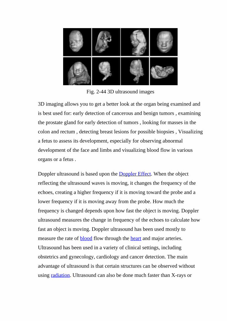
Fig. 2-44 3D ultrasound images
3D imaging allows you to get a better look at the organ being examined and
is best used for: early detection of cancerous and benign tumors , examining
the prostate gland for early detection of tumors , looking for masses in the
colon and rectum , detecting breast lesions for possible biopsies , Visualizing
a fetus to assess its development, especially for observing abnormal
development of the face and limbs and visualizing blood flow in various
organs or a fetus .
Doppler ultrasound is based upon the Doppler Effect. When the object
reflecting the ultrasound waves is moving, it changes the frequency of the
echoes, creating a higher frequency if it is moving toward the probe and a
lower frequency if it is moving away from the probe. How much the
frequency is changed depends upon how fast the object is moving. Doppler
ultrasound measures the change in frequency of the echoes to calculate how
fast an object is moving. Doppler ultrasound has been used mostly to
measure the rate of blood flow through the heart and major arteries.
Ultrasound has been used in a variety of clinical settings, including
obstetrics and gynecology, cardiology and cancer detection. The main
advantage of ultrasound is that certain structures can be observed without
using radiation. Ultrasound can also be done much faster than X-rays or

other radiographic techniques. Here is a short list of some uses for
ultrasound:
Obstetrics and Gynecology : measuring the size of the fetus to determine the
due date , determining the position of the fetus to see if it is in the normal
head down position or breech , checking the position of the placenta to see if
it is improperly developing over the opening to the uterus (cervix) , seeing
the number of fetuses in the uterus , checking the sex of the baby (if the
genital area can be clearly seen) , checking the fetus's growth rate by making
many measurements over time , detecting ectopic pregnancy, the life-
threatening situation in which the baby is implanted in the mother's
Fallopian tubes instead of in the uterus , determining whether there is an
appropriate amount of amniotic fluid cushioning the baby .Monitoring the
baby during specialized procedures - ultrasound has been helpful in seeing
and avoiding the baby during amniocentesis (sampling of the amniotic fluid
with a needle for genetic testing). Years ago, doctors use to perform this
procedure blindly; however, with accompanying use of ultrasound, the risks
of this procedure have dropped dramatically. Seeing tumors of the ovary and
breast.
Cardiology seeing the inside of the heart to identify abnormal structures or
functions and measuring blood flow through the heart and major blood
vessels.
Urology measuring blood flow through the kidney, seeing kidney stones and
detecting prostate cancer early
In addition to these areas, there is a growing use for ultrasound as a rapid
imaging tool for diagnosis in emergency rooms.

There have been many concerns about the safety of ultrasound. Because
ultrasound is energy, the question becomes "What is this energy doing to my
tissues or my baby?" There have been some reports of low birthweight
babies being born to mothers who had frequent ultrasound examinations
during pregnancy. The two major possibilities with ultrasound are as
follows: - development of heat - tissues or water absorb the ultrasound
energy which increases their temperature locally and formation of bubbles
(cavitation) - when dissolved gases come out of solution due to local heat
caused by ultrasound
However, there have been no substantiated ill-effects of ultrasound
documented in studies in either humans or animals. This being said,
ultrasound should still be used only when necessary (i.e. better to be
cautious).
For an ultrasound exam, you go into a room with a technician and the
ultrasound machine. The following happens:- you remove your clothes (all
of your clothes or only those over the area of interest , the ultrasonographer
drapes a cloth over any exposed areas that are not needed for the exam , the
ultrasonographer applies a mineral oil-based jelly to your skin , this jelly
eliminates air between the probe and your skin to help pass the sound waves
into your body , the ultrasonographer covers the probe with a plastic cover ,
he/she passes the probe over your skin to obtain the required images ,
depending upon the type of exam, the probe may be inserted into you , you
may be asked to change positions to get better looks at the area of interest ,
after the images have been acquired and measurements taken, the data is
stored on disk. You may get a hard copy of the images, you are given a
towelette to clean up and you get dressed.

As with other computer technology, ultrasound machines will most likely
get faster and have more memory for storing data. Transducer probes may
get smaller, and more insertable probes will be developed to get better
images of internal organs. Most likely, 3D ultrasound will be more highly
developed and become more popular. The entire ultrasound machine will
probably get smaller, perhaps even hand-held for use in the field (e.g.
paramedics, battlefield triage). One exciting new area of research is the
development of ultrasound imaging combined with heads-up/virtual reality-
type displays that will allow a doctor to "see" inside you as he/she is
performing a minimally invasive or non-invasive procedure such as
amniocentesis or biopsy.
Ultrasound (also termed sonography, ultrasonography, and Doppler study) is
a non-invasive diagnostic medical technique that uses high frequency sound
waves to produce images of the internal structures of the body. These sound
waves are not detectable by human hearing.
Using ultrasonography, a technician or doctor moves a device called a
transducer (probe) over part of your body. The transducer emits sound waves
which bounce off the internal tissues, and creates images from the waves
that bounce back. Different densities of tissues, fluid, and air inside the body
produce different images that can be interpreted by a physician, typically a
radiologist (a physician who specializes in imaging technologies). Many
studies are done by a trained technologist (sonographer) and then interpreted
by a radiologist.
Ultrasound can be used as a diagnostic or screening tool to confirm medical
disorders or to assist in performing medical procedures. It is also used as a
therapeutic tool in treating musculoskeletal problems, renal stones (kidney
stones) and gallstones .

Obstetrics and gynecology: Pregnancy ultrasound (or fetal ultrasound) is
used to assess the progression of the fetus. Vaginal ultrasound, pelvic
ultrasound, or transvaginal ultrasound is used to diagnose growths or tumors
of the ovary, uterus, and Fallopian tubes. It can be used to assess non-
pregnancy related issues as well: - lower abdominal pain , ovarian cysts .
uterine fibroids , uterine growths and endometriosis .
Cardiology: Echocardiography (heart ultrasound) is a common way to
evaluate the overall function of the heart. It is used to evaluate the flow of
blood through the chambers and valves of the heart. It also assesses the
strength of the heart beat and the volume of blood pumped through.
Echocardiography is often used for the following:- heart valve problems,
such as mitral valve prolapsed or aortic stenosis , congestive heart failure ,
blood clots due to irregular heart beats such as in atrial fibrillation ,
abnormal fluid collections around the heart, such as pericardial effusions
and pulmonary artery hypertension.
Blood vessels: Ultrasound is useful in detecting problems with most of the
larger blood vessels in the body. Using Doppler ultrasound technology, the
flow of blood through the vessels can be observed and measured. Narrowing
of vessels (stenosis) or widening of vessels (dilatation, also referred to as
aneurysms) can be detected. Ultrasound testing of blood vessels includes:-
carotid ultrasound , abdominal aorta ultrasound for abdominal aortic
aneurysm, and blood clots in veins (superficial or deep venous thrombosis,
or DVT).
Abdominal structures: Abdominal ultrasound is used to evaluate the solid
organs within the abdominal cavity, including the liver, gallbladder,
pancreas, kidneys, and bladder.

Renal ultrasound is used to evaluate the function and structure of the
kidneys. Swelling around the kidney with blockage in the urinary tract can
be seen with ultrasound, making abdominal ultrasound useful in detecting
kidney stones.
Liver ultrasound is used to find abnormalities in the liver tissue and ducts.
Gallbladder ultrasound can screen for gallstones or an infected gallbladder.
Appendix ultrasound is used in children or pregnant women, where it is
necessary to avoid radiation from aCT scan (computerized tomography).
Testicular ultrasound: Used to diagnose testicular torsion, epididymitis
(testicle infection), and testicular masses.
Neck ultrasound: The thyroid and parathyroid glands can be imaged to
detect nodules, growths, and tumors.
Breast ultrasound: Used to image the breasts and to guide biopsy of breast
masses to evaluate for breast cancer.
Knee ultrasound: Ultrasound can be used to evaluate the structures in the
back of the knee to determine if a Baker's Cyst is present.
Eye ultrasound: An eye ultrasound is used to look at the back of the eye
(retina). It is often used when a patient has cataracts that make looking into
the eye difficult. The test may help diagnose retinal detachment. It can also
assist in cataract surgery.
Skin ultrasound: Ultrasound can be used to help find certain types of foreign
bodies that may become lodged in the skin.

Ultrasound-guided needle biopsy: Ultrasound helps medical professionals
guide needles into specific areas of the body to extract cells for laboratory
testing.
Ultrasound-guided needle aspiration: Ultrasound may be used to guide a
needle into pockets of fluid accumulated in the body that need to be drained
(for example, an abscess, pleural effusions, or ascites).
Ultrasound-assisted intravenous access: When an intravenous (IV) line is
required and veins are difficult to access, ultrasound may be used to assist in
finding larger veins in the neck, chest wall, or groin.
Extracorporeal shock wave lithotripsy (ESWL) is a form of ultrasound used
to break up kidney stones and gallbladder stones. In many cases, the patient
is given some sedation or pain medication as the high intensity waves
needed to fracture the stones can cause discomfort. This technique is used
often to treat stone formation.
A few physicians use HIFU (high intensity focused ultrasound) to treat
cancer, while others use ultrasound for targeted drug delivery, homeostasis
or thrombolytic. However, these techniques are not widely available and are
still being evaluated for efficacy.
Ultrasound is often used to treat musculoskeletal injuries, and is frequently
used to treat sports injuries. For example, plantar fasciitis and tendinitis are
commonly treated using therapeutic ultrasound. It is believed to help reduce
inflammation and increase blood flow to affected areas. However, there is
little evidence that tissue therapeutic ultrasound is effective. More study is
needed

For the most part, ultrasound is considered a painless, non-invasive
diagnostic tool. Most ultrasound scans can be performed with the transducer
placed atop the skin, with the sound waves aimed at the organ or body part
being tested. The patient is usually placed in a comfortable position that
provides the ultrasound technician access to the part of the body being
tested.
The area being studied is covered with a small amount of gel to eliminate air
pockets between the transducer and the skin. The sonographer moves the
transducer across the body part being studied to obtain images.
You may feel pressure as the transducer is moved over an area, and if the
area is sensitive, you may feel pain, but the waves from the transducer do
not cause this pain.
If Doppler ultrasound is used, you may hear pulse-like "whooshing" sounds
that change in pitch as the blood flow is monitored.
Some exams are considered 'invasive ultrasounds,' where the transducer is
attached to a probe and inserted into a natural opening in the body. These
exams may cause some discomfort or pain due to the sensitivity of the tissue
being touched by the probe, not by the ultrasound waves.
Transesophageal echocardiogram: The transducer is inserted into the
esophagus to obtain images of the heart.
Transrectal ultrasound: The transducer is inserted into a man's rectum to
view the prostate.
Transvaginal ultrasound: The transducer is inserted into a woman's vagina to
view the uterus and ovaries.

Once the ultrasound procedure is complete, gel will be wiped off your skin
and you should be able to resume your normal activities immediately in
most cases or within a few hours if a more invasive study is done.
According to the Food and Drug Administration, ultrasound has an excellent
safety record since it was put into regular use more than 30 years ago.
It avoids the use of radiation that is common in other diagnostic procedures
by using harmless sound waves to produce images.
By using ultrasound technology to assist in other procedures, you may also
reduce the risk and increase the effectiveness of those other procedures.
Specialized high intensity ultrasound waves like those used for ESWL and
HIFU have the ability to cause discomfort and potentially serious damage to
tissue nearby the area of treatment. These risks are minimized by good
control of wave intensity exposure timing and focus techniques.
Ultrasound is not the recommended procedure for: - Bowel or organs
obscured by the bowel: Air and gas can disrupt the ultrasound waves, obese
patients and Internal structure of bones or certain joints: Ultrasound has
difficulty penetrating bone.
An abdominal ultrasound uses reflected sound waves to produce a picture of
the organs and other structures in the upper abdomen. Sometimes a
specialized ultrasound is ordered for a detailed evaluation of a specific
organ, such as a kidney ultrasound.
Ultrasound is a test that uses reflected sound waves to produce an image of
organs and other structures in the body. It does not use X-rays or other types
of possibly harmful radiation.

For ultrasound testing, gel or oil is applied to the skin to help transmit the
sound waves. A small, handheld instrument called a transducer is passed
back and forth over the area of the body that is being examined. The
transducer sends out high-pitched sound waves (above the range of human
hearing) that are reflected back to the transducer. A computer analyzes the
reflected sound waves and converts them into a picture that is displayed on a
TV screen. The picture produced by ultrasound is called a sonogram,
echogram, or ultrasound scan. Pictures or videos of the ultrasound images
may be made for a permanent record.
Ultrasound is most useful for looking at organs and structures that are either
uniform and solid (such as the liver) or fluid-filled (such as the gallbladder).
Mineralized structures (such as bones) or air-filled organs (such as the lungs)
do not show up well on a sonogram
An abdominal ultrasound can evaluate the:- abdominal aorta, which is the
large blood vessel (artery) that passes down the back of the chest and
abdomen. The aorta supplies blood to the lower part of the body and the
legs, liver , which is a large dome-shaped organ that lies under the rib cage
on the right side of the abdomen. The liver produces bile (a substance that
helps digest fat), stores sugars, and breaks down many of the body's waste
products, gallbladder, which is a small sac-shaped organ beneath the liver
that stores bile. When food is eaten, the gallbladder contracts, sending bile
into the intestines to help in digesting food and absorbing fat-soluble
vitamins , spleen, which is the soft, round organ that helps fight infection
and filters old red blood cells. The spleen is located to the left of the
stomach, just behind the lower left ribs , pancreas , which is the gland
located in the upper abdomen that produces enzymes that help digest food.
The digestive enzymes are then released into the intestines. The pancreas
also releases insulin into the bloodstream. Insulin helps the body use sugars

for energy and kidneys , which are the pair of bean-shaped organs located
behind the upper abdominal cavity. The kidneys remove wastes from the
blood and produce urine.
A pelvic ultrasound evaluates the structures and organs in the lower
abdominal area (pelvis).
Abdominal ultrasound is done to: find the cause of abdominal pain , find,
measure, or monitor an aneurysm in the aorta( An aneurysm may cause a
large pulsing lump in the abdomen) , check the size, shape, and position of
the liver(An ultrasound may be done to evaluate jaundice and other
problems of the liver, including liver masses, cirrhosis, fat deposits in the
liver (called fatty liver), or abnormal liver function tests , detect gallstones,
inflammation of the gallbladder (cholecystitis), or blocked bile ducts , learn
the size of an enlarged spleen and look for damage or disease , find problems
with the pancreas, such as a pancreatic tumor , look for blocked urine flow in
a kidney. A kidney ultrasound may also be done to find out the size of the
kidneys, detect kidney masses, detect fluid surrounding the kidneys,
investigate causes for recurring urinary tract infections, or check the
condition of transplanted kidneys , find out whether a mass in any of the
abdominal organs (such as the liver) is a solid tumor or a simple fluid-filled
cyst , guide the placement of a needle or other instrument during a biopsy
and Look for fluid buildup in the abdominal cavity (ascites). An ultrasound
also may be done to guide the needle during a procedure to remove fluid
from the abdominal cavity (paracentesis).
Tell your doctor if you have had a barium enema or a series of upper GI
(gastrointestinal) tests within the past 2 days. Barium that remains in the
intestines can interfere with the ultrasound test.

Other preparations depend on the reason for the abdominal ultrasound test
you are having.
For ultrasound of the liver, gallbladder, spleen, and pancreas, you may be
asked to eat a fat-free meal on the evening before the test and then to avoid
eating for 8 to 12 hours before the test.
For ultrasound of the kidneys, you may not need any special preparation.
You may be asked to drink 4 to 6 glasses of liquid (usually juice or water)
about an hour before the test to fill your bladder. You may be asked to avoid
eating for 8 to 12 hours before the test to avoid gas buildup in the intestines.
Gas could interfere with the evaluation of the kidneys, which lay behind the
stomach and intestines.
For ultrasound of the aorta, you may need to avoid eating for 8 to 12 hours
before the test.
This test is done by a doctor who specializes in performing and interpreting
imaging tests (radiologist) or by an ultrasound technologist (sonographer)
who is supervised by a radiologist. It is done in an ultrasound room in a
hospital or doctor's office.
You will need to take off any jewelry that might interfere with the
ultrasound scan. You will need to take off all or most of your clothes,
depending on which area is examined (you may be allowed to keep on your
underwear if it does not interfere with the test). You will be given a cloth or
paper covering to use during the test.
You will lie on your back (or on your side) on a padded exam table. Warmed
gel will be spread on your abdomen (or back) to improve the quality of the

sound waves. A small handheld unit called a transducer is pressed against
your abdomen.
You may be asked to change positions so more scans can be done. For a
kidney ultrasound, you may be asked to lie on your stomach.
You need to lie very still while the ultrasound scan is being done. You may
be asked to take a breath and hold it for several seconds during the scanning.
This lets the sonographer see organs and structures, such as the bile ducts,
more clearly because they are not moving. Holding your breath also
temporarily pushes the liver and spleen lower into the belly so they are not
hidden by the lower ribs, which makes it harder for the sonographer to see
them clearly.
The gel may feel cold when it is put on your skin unless it is first warmed to
body temperature. You will feel light pressure from the transducer as it
passes over your abdomen. The ultrasound usually is not uncomfortable. But
if the test is being done to check damage from a recent injury, the slight
pressure of the transducer may be somewhat painful. You will not hear or
feel the sound waves.
There are no known risks from having an abdominal ultrasound test.
An abdominal ultrasound uses reflected sound waves to produce a picture of
the organs and other structures in the abdomen.
Using abdominal ultrasound, a doctor can usually distinguish among a
simple fluid-filled cyst, a solid tumor, or another type of mass that needs
further evaluation. If a solid tumor is found, abdominal ultrasound cannot
determine whether it is cancerous (malignant) or noncancerous (benign). A

biopsy may be needed if a tumor is found. Ultrasound may be used during
the biopsy to help guide the placement of the needle.
Ultrasound is less expensive than other tests, such as a CT scan or magnetic
resonance imaging (MRI) scan, that also can provide a picture of the
abdominal organs. But for some problems, such as abdominal masses or an
injury, a CT scan or MRI may be a more appropriate test. Also, these tests
may be done if the abdominal ultrasound is normal but abdominal pain
persists.
A pelvic ultrasound will be used to produce a picture of the lower abdominal
(pelvic) organs and other structures inside the pelvis.
A pelvic ultrasound looks at the bladder and The ovaries The ovaries are
two small glands, located on either side of a woman's uterus, that are part of
the female reproductive system. The ovaries produce female sex hormones
and store and release eggs (ova), which can develop into embryos if
fertilized by a male's sperm.
The female sex hormones (estrogen and progesterone) help control the
menstrual cycle, breast development, and other functions. Approximately
once a month, about 2 weeks before a woman's next period is due, one of her
ovaries releases an egg (ovulation).
The uterus is a hollow, pear-shaped organ in a woman's lower abdomen in
which a fetus grows during pregnancy. When a woman is not pregnant, her
monthly menstrual period flows from the uterus.
The lower part of the uterus (cervix) opens into the vagina. The opening to
the cervix is narrow except during labor and birth, when it stretches and
widens to allow the baby's passage out of the uterus. The cervix is the lower

part of the uterus that opens into the vagina. It is part of a woman's
reproductive system.
During a woman's menstrual period, blood flows from her uterus through her
cervix, down her vagina, and out of her body.
During sex, the cervix makes mucus that helps sperm move up into the
uterus.
During pregnancy, the cervix is tightly closed to protect the baby. But later
the cervix opens up so the baby can pass through the vagina during
childbirth.
A woman's two fallopian tubes lead upward from each upper side of the
uterus and end near the ovaries. When an egg is released by an ovary
(ovulation), it travels down a fallopian tube toward the uterus. After
ovulation, egg fertilization usually happens in a fallopian tube. The fertilized
egg then travels to the uterus, where it implants and grows. If a woman's
fallopian tubes are blocked by scar tissue, such as from pelvic inflammatory
disease, she may be unable to become pregnant or may have a tubal
(ectopic) pregnancy.

Fig. 2-45
A woman female organs
Fig. 2-46 Male organs
Pelvic ultrasound can be done three ways: transabdominal, transrectal, and
transvaginal.
Transabdominal ultrasound. A small handheld device called a transducer is
passed back and forth over the lower belly. Transrectal ultrasound. The
transducer is shaped to fit into the rectum. A transrectal ultrasound is the
most common test to look at the male pelvic organs, such as the prostate and
seminal vesicles. Sometimes, a small sample of tissue (biopsy) may be taken
with small tools inserted through the rectum during a transrectal

ultrasound.Transvaginal ultrasound. The transducer is shaped to fit into a
woman's vagina.
Fig. 2-47 A woman's vagina, also called the birth canal, is part of the
reproductive tract and extends from the uterus to outside the body.
The opening to the vagina is located between the bowel opening
(anus) and the opening for the bladder (urethra).
A woman may have both transabdominal and transvaginal ultrasounds to
look at the whole pelvic area. A transvaginal ultrasound is done to look for
problems with fertility. In rare cases, a hysterosonogram is done to look at
the inside of the uterus by filling the uterus with fluid during a transvaginal
ultrasound. Sometimes, a small sample of tissue (biopsy) may be taken with
small tools inserted through the vagina during a transvaginal ultrasound.
In all three types of pelvic ultrasound, the transducer sends the reflected
sound waves to a computer, which makes them into a picture that is shown
on a video screen. Ultrasound pictures or videos may be saved as a
permanent record.

For women, pelvic ultrasound may be done to: find out what is causing
pelvic pain , look for the cause of vaginal bleeding , look for pelvic
inflammatory disease (PID) , find an intrauterine device , look at the size and
shape of the uterus and the thickness of the uterine lining (endometrium) ,
look at the size and shape of the ovaries , check the condition and size of the
ovaries during treatment for infertility , confirm a pregnancy and whether it
is in the uterus , check the cervical length in a pregnant woman at risk for
preterm labor , check a lump found during a pelvic examination , check
uterine fibroids found during a pelvic examination and guide a procedure to
remove an ovarian follicle for in vitro fertilization.
Fig. 48 An intrauterine device (IUD) is a small, plastic, T-shaped
device that is inserted into the uterus to prevent pregnancy.
If you are having a transabdominal ultrasound, your doctor will ask you to
drink 4 to 6 glasses of juice or water about an hour before the test to fill your
bladder. A full bladder pushes the intestines (which contain air) out of the
way of the pelvic organs. This makes the ultrasound picture clearer. If the
ultrasound is being done in an emergency situation, your bladder may be

filled with water through a thin flexible tube (catheter) inserted into your
bladder. If you are having a transvaginal ultrasound, tell the doctor if you are
allergic to latex so that a latex-free cover can be put on the transducer before
it is used.If you are having only a transvaginal ultrasound, do not drink any
fluids for 4 hours before the test. You will not need to drink fluids to fill
your bladder for the test as you do in a transabdominal ultrasound.
If both a transabdominal and transvaginal ultrasound will be done, the
transabdominal ultrasound will usually be done first.You will need to take
off most of your clothes below the waist. You will be given a gown to use
during the test. You will lie on your back (or on your side) on a padded
table. Gel will be put on your belly to improve the quality of the sound
waves. A small, handheld instrument called a transducer is gently moved
over your belly. A picture of the organs and blood vessels can be seen on a
video screen.You need to lie very still while the ultrasound is being done.
You may be asked to take a breath and hold it for several seconds during the
test.When the test is done, the gel is cleaned off your skin. You can urinate
as soon as the test is done.
For transvaginal ultrasound, you will empty your bladder. You will be asked
to lie on your back with your hips slightly raised.A thin, lubricated
transducer probe will be gently inserted into your vagina. Only the tip of the
transducer is put in the vagina. You need to lie very still while the ultrasound
scan is being done.
If you have transabdominal ultrasound, you will likely feel pressure in your
bladder and a strong urge to urinate because your bladder is full.The gel may
feel cold when it is put on your belly. You will feel light pressure from the
transducer as it passes over your belly. If you have an injury or pelvic pain,
the light pressure of the transducer may be painful. You will not hear or feel
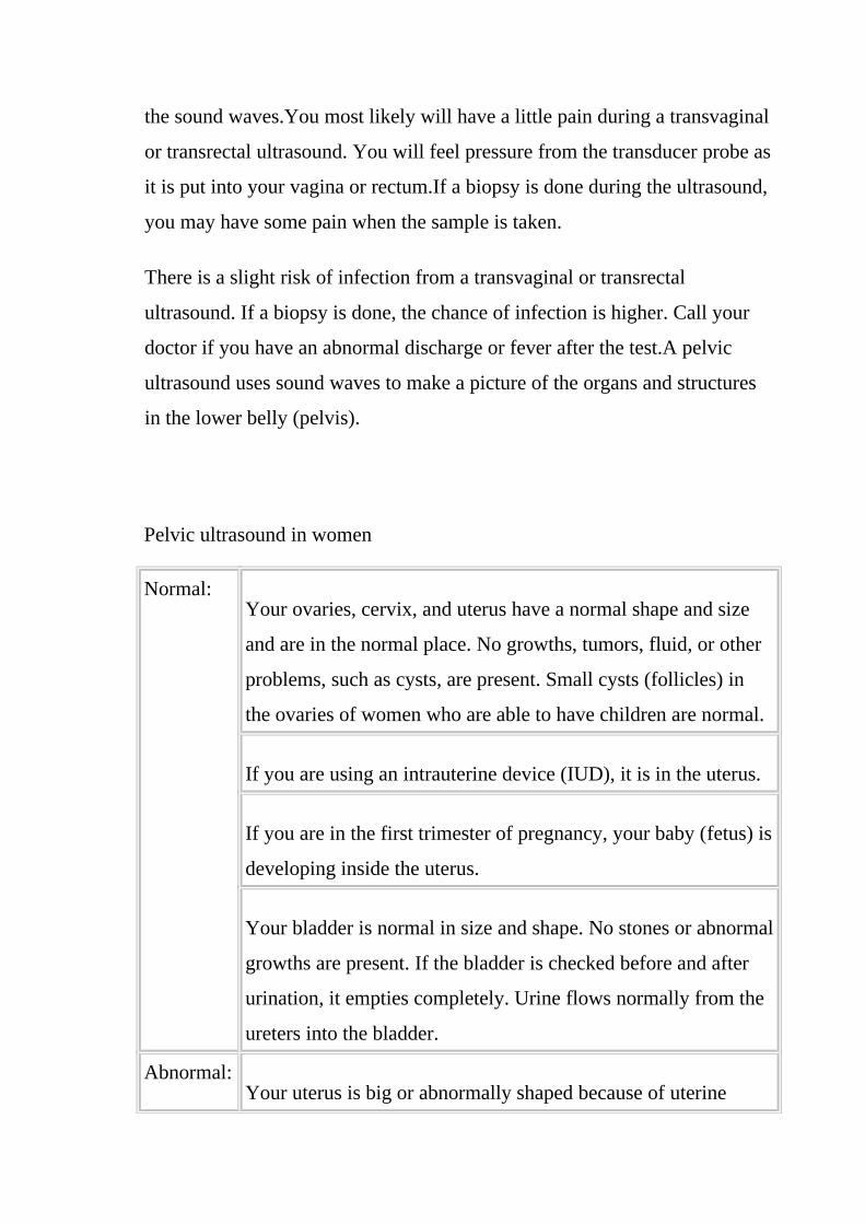
the sound waves.You most likely will have a little pain during a transvaginal
or transrectal ultrasound. You will feel pressure from the transducer probe as
it is put into your vagina or rectum.If a biopsy is done during the ultrasound,
you may have some pain when the sample is taken.
There is a slight risk of infection from a transvaginal or transrectal
ultrasound. If a biopsy is done, the chance of infection is higher. Call your
doctor if you have an abnormal discharge or fever after the test.A pelvic
ultrasound uses sound waves to make a picture of the organs and structures
in the lower belly (pelvis).
Pelvic ultrasound in women
Normal: Your ovaries, cervix, and uterus have a normal shape and size
and are in the normal place. No growths, tumors, fluid, or other
problems, such as cysts, are present. Small cysts (follicles) in
the ovaries of women who are able to have children are normal.
If you are using an intrauterine device (IUD), it is in the uterus.
If you are in the first trimester of pregnancy, your baby (fetus) is
developing inside the uterus.
Your bladder is normal in size and shape. No stones or abnormal
growths are present. If the bladder is checked before and after
urination, it empties completely. Urine flows normally from the
ureters into the bladder.
Abnormal: Your uterus is big or abnormally shaped because of uterine
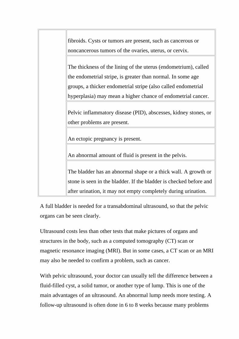
fibroids. Cysts or tumors are present, such as cancerous or
noncancerous tumors of the ovaries, uterus, or cervix.
The thickness of the lining of the uterus (endometrium), called
the endometrial stripe, is greater than normal. In some age
groups, a thicker endometrial stripe (also called endometrial
hyperplasia) may mean a higher chance of endometrial cancer.
Pelvic inflammatory disease (PID), abscesses, kidney stones, or
other problems are present.
An ectopic pregnancy is present.
An abnormal amount of fluid is present in the pelvis.
The bladder has an abnormal shape or a thick wall. A growth or
stone is seen in the bladder. If the bladder is checked before and
after urination, it may not empty completely during urination.
A full bladder is needed for a transabdominal ultrasound, so that the pelvic
organs can be seen clearly.
Ultrasound costs less than other tests that make pictures of organs and
structures in the body, such as a computed tomography (CT) scan or
magnetic resonance imaging (MRI). But in some cases, a CT scan or an MRI
may also be needed to confirm a problem, such as cancer.
With pelvic ultrasound, your doctor can usually tell the difference between a
fluid-filled cyst, a solid tumor, or another type of lump. This is one of the
main advantages of an ultrasound. An abnormal lump needs more testing. A
follow-up ultrasound is often done in 6 to 8 weeks because many problems

go away on their own within that time. Pelvic ultrasound cannot determine
whether a lump is cancerous (malignant) or noncancerous (benign). A
biopsy may have to be done for this.Transvaginal ultrasound is used during
fertility checks to help guide the removal of ovarian follicles for in vitro
fertilization.
The words diagnostic medical ultrasound, ultrasound, and ultrasonography
have all been used to describe the instrumentation utilized in ultrasound.
Sonography is the term used to describe a pecializedimaging technique used
to visualize soft tissue structures of the body. Extensive research has verified
the safety of ultrasound as a diagnostic procedure.
Diagnostic ultrasound has come to be such a valuable diagnostic imaging
technique for so many different body structures for many reasons, but two
are especially significant. First is the lack of ionizing radiation for
ultrasound as compared with the other imaging modalities of magnetic
resonance imaging (MRI), computed tomography (CT), or nuclear medicine.
The second reason is the portability of the ultrasound equipment. An
ultrasound may be done to evaluate jaundice and other problems of the liver,
including liver masses, cirrhosis, fat deposits in the liver (called fatty liver),
or abnormal liver function tests , Detect gallstones, inflammation of the
gallbladder, or blocked bile ducts , Learn the size of an enlarged spleen and
look for damage or disease , Find problems with the pancreas, such as a
pancreatic tumor , Look for blocked urine flow in a kidney. A kidney
ultrasound may also be done to find out the size of the kidneys, detect
kidney masses, detect fluid surrounding the kidneys, investigate causes for
recurring urinary tract infections, or check the condition of transplanted
kidneys , Find out whether a mass in any of the abdominal organs is a solid
tumor or a simple fluid-filled cyst , Guide the placement of a needle or other
instrument during a biopsy and Look for fluid buildup in the abdominal

cavity (ascites). An ultrasound also may be done to guide the needle during a
procedure to remove fluid from the abdominal cavity (paracentesis).Tell
the doctor if you have had a barium enema or a series of
upper GI (gastrointestinal) tests within the past 2 days.
Barium that remains in the intestines can interfere with the
ultrasound test.Other preparations depend on the reason for
the abdominal ultrasound test you are having.
For ultrasound of the liver, gallbladder, spleen, and pancreas, you may be
asked to eat a fat-free meal on the evening before the test and then to avoid
eating for 8 to 12 hours before the test.For ultrasound of the kidneys, you
may not need any special preparation. You may be asked to drink 4 to 6
glasses of liquid (usually juice or water) about an hour before the test to fill
your bladder. You may be asked to avoid eating for 8 to 12 hours before the
test to avoid gas buildup in the intestines. Gas could interfere with the
evaluation of the kidneys, which lay behind the stomach and intestines.For
ultrasound of the aorta, you may need to avoid eating for 8 to 12 hours
before the test.
This test is done by a doctor who specializes in performing and interpreting
imaging tests (radiologist) or by an ultrasound technologist (sonographer)
who is supervised by a radiologist. It is done in an ultrasound room in a
hospital or doctor's office.You will need to take off any jewelry that might
interfere with the ultrasound scan. You will need to take off all or most of
your clothes; depending on which area is examined (you may be allowed to
keep on your underwear if it does not interfere with the test). You will be
given a cloth or paper covering to use during the test.You will lie on your
back (or on your side) on a padded exam table. Warmed gel will be spread
on your abdomen (or back) to improve the quality of the sound waves. A
small handheld unit called a transducer is pressed against your abdomen.You
may be asked to change positions so more scans can be done. For a kidney
ultrasound, you may be asked to lie on your stomach.
You need to lie very still while the ultrasound scan is being done. You may
be asked to take a breath and hold it for several seconds during the scanning.
This lets the sonographer see organs and structures, such as the bile ducts,
more clearly because they are not moving. Holding your breath also
temporarily pushes the liver and spleen lower into the belly so they are not
hidden by the lower ribs, which makes it harder for the sonographer to see
them clearly.You may be asked to wait until the radiologist has reviewed the
information. The radiologist may want to do more ultrasound views of some
areas of your abdomen.
The gel may feel cold when it is put on your skin unless it is first warmed to
body temperature. You will feel light pressure from the transducer as it
passes over your abdomen.
The ultrasound usually is not uncomfortable. But if the test is being done to
check damage from a recent injury, the slight pressure of the transducer may
be somewhat painful. You will not hear or feel the sound waves.There are no
known risks from having an abdominal ultrasound test.
An abdominal ultrasound uses reflected sound waves to produce a picture of
the organs and other structures in the abdomen.
Abdominal ultrasound
Normal: The size and shape of the abdominal organs appear normal.
The liver, spleen, and pancreas appear normal in size and

texture. No abnormal growths are seen. No fluid is found in
the abdomen.
The diameter of the aorta is normal, and no aneurysms are
seen.
The thickness of the gallbladder wall is normal. The size of
the bile ducts between the gallbladder and the small intestine
is normal. No gallstones are seen.
The kidneys appear as sharply outlined bean-shaped organs.
No kidney stones are seen. No blockage to the system
draining the kidneys is present.
Abnormal: An organ may appear abnormal because of inflammation,
infection, or other diseases. An organ may be smaller than
normal because of an old injury or past inflammation. An
organ may be pushed out of its normal location because of
an abnormal growth pressing against it. An abnormal growth
(such as a tumor) may be seen in an organ. Fluid in the
abdominal cavity may be seen.
The aorta is enlarged, or an aneurysm is seen.
The liver may appear abnormal, which may point to liver
disease (such as cirrhosis or cancer).
The walls of the gallbladder may be thickened, or fluid may
be present around the gallbladder, which may point to
inflammation. The bile ducts may be enlarged because of

blockage (from a gallstone or an abnormal growth in the
pancreas). Gallstones may be seen inside the gallbladder.
The kidneys or the tubes that drain the kidneys (ureters) may
be enlarged because of urine that is not draining properly.
Kidney stones are seen within the kidneys (not all stones can
be seen with ultrasound).
An area of infection (abscess) or a fluid-filled cyst may
appear as a round, hollow structure inside an organ. The
spleen may be ruptured (if an injury to the abdomen has
occurred).
Reasons you may not be able to have the test or why the results may not be
helpful include: , Stool, air (or other gas), or contrast material (such as
barium) in the stomach or intestines , Not being able to remain still during
the test , Extreme obesity and Having an open or bandaged wound in the
area being viewed.
Other tests, such as a computed tomography (CT) scan, may be needed to
follow up abnormal ultrasound results. To learn more, see the topic
Computed Tomography (CT) Scan of the Body.
X-rays are not recommended during pregnancy because of the risk of
damage to the fetus. Because ultrasound is safe during pregnancy, it
generally is used instead of an abdominal X-ray if a pregnant woman's
abdomen needs to be checked.
In rare cases, gallstones may not be found by ultrasound. Other imaging tests
may be done if gallstones are suspected but not seen on the ultrasound:

Gallbladder Scan , Endoscopic Retrograde Cholangiopancreatogram (ERCP)
and Ray. Bones or air-filled organs, such as the intestines, do
not show up well on an ultrasound and may keep other
organs from being seen clearly.Pelvic ultrasound can be
done three ways: transabdominal, transrectal, and
transvaginal.
Transabdominal ultrasound. A small handheld device called a transducer is
passed back and forth over the lower belly. A transabdominal ultrasound is
commonly done in women to look for large uterine fibroids (are growths on
or in your uterus. Although they are sometimes called fibroid tumors, they
aren't cancer. You don't need to do anything about them unless they are
causing problems). or other problems.Tran rectal ultrasound. The transducer
is shaped to fit into the rectum. A transrectal ultrasound is the most common
test to look at the male pelvic organs, such as the prostate and seminal
vesicles. Sometimes, a small sample of tissue (biopsy) may be taken with
small tools inserted through the rectum during a transrectal
ultrasound.Transvaginal ultrasound. The transducer is shaped to fit into a
woman's vagina.
A woman may have both transabdominal and transvaginal ultrasounds to
look at the whole pelvic area. A transvaginal ultrasound is done to look for
problems with fertility. In rare cases, a hysterosonogram is done to look at
the inside of the uterus by filling the uterus with fluid during a transvaginal
ultrasound. Sometimes, a small sample of tissue (biopsy) may be taken
with small tools inserted through the vagina during a transvaginal
ultrasound.
In all three types of pelvic ultrasound, the transducer sends the reflected
sound waves to a computer, which makes them into a picture that is shown

on a video screen. Ultrasound pictures or videos may be saved as a
permanent record. For women, pelvic ultrasound may be done to: Find out
what is causing pelvic pain , Look for the cause of vaginal bleeding and
Look for pelvic inflammatory disease (PID).
Confirm a pregnancy and whether it is in the uterus. Pelvic ultrasound may
be used early in pregnancy to check the age of the pregnancy or to find a
tubal pregnancy (ectopic pregnancy) or multiple pregnancy. Check uterine
fibroids found during a pelvic examination. Pelvic ultrasound may also be
done to check the growth of uterine fibroids.Guide a procedure to remove an
ovarian follicle for in vitro fertilization.(M. Bazot,2004)
2-2 The ovary.
The ovaries are two almond shaped glands located on either side of the
uterus, which develop ova or eggs and produce the hormones estrogen and
progesterone. The fallopian tubes transport the eggs from the ovaries to the
uterus. It is usually in the fallopian tubes that fertilization takes place.They
are almond-shaped and about 3.5 cm (1.5 inches) long. The ovaries are deep
in a woman’s pelvis, on both sides of the uterus (womb), close to the ends of
the Fallopian tubes.
In a fertile woman an egg is usually released once a month from one of her
two ovaries. This is called ovulation and usually occurs about 14 days before
the first day of menses. Two to five days before and after ovulation is when
pregnancy can occur.
If unprotected intercourse takes place during this time, the sperm moves up
the uterus into the tubes within minutes.If a single sperm joins with the egg,
the egg becomes fertilized. This fertilized egg travels down the tube and
attaches to the wall of the uterus, which has increased its blood supply in

preparation for pregnancy. If the egg is not fertilized, in about 14 days, the
lining of the uterus is shed as menstrual flow.(N. Pierce,2008)
2-2-1 Anatomy.
2-2-1-1 Structure.
The ovaries are made up of 3 different types of cells:- epithelial cells make
up the outer layer covering the ovary (epithelium) and germ cells are inside
the ovary. They develop into eggs and stromal cells form the supportive or
connective tissues of the ovary (stroma).Each ovary is surrounded by a thin
layer of tissue called the capsule.
Fig. 2-49 Cross-section of the ovary
The ovaries play two central roles in the female reproductive system by
acting as both glands and gonads. Acting as glands, the ovaries produce
several female sex hormones including estrogens and progesterone. Estrogen
controls the development of the mammary glands and uterus during puberty
and stimulates the development of the uterine lining during the menstrual
cycle. Progesterone acts on the uterus during pregnancy to allow the embryo
to implant and develop in the womb.

At birth the ovaries contain anywhere from several hundred thousand to
several million circular bundles of cells known as follicles. Each follicle
surrounds and supports a single oocyte that has the ability to mature into an
ovum, the female gamete. Despite this large number of potential ova, only
around 4,000 oocytes survive to puberty and only 400 oocytes mature into
ova in a woman’s lifetime. During each menstrual cycle around 10-20
follicles and their oocytes begin to develop under the influence of the
pituitary hormone follicle-stimulating hormone (FSH). Of these follicles,
only one cell completes its development and becomes a mature ovum.
Fig. 2-50 Ovary
The uterus, also commonly known as the womb, is a hollow muscular organ
of the female reproductive system that is responsible for the development of
the embryo and fetus during pregnancy. An incredibly distensible organ, the
uterus can expand during pregnancy from around the size of a closed fist to
become large enough to hold a full term baby. It is also an incredibly strong
organ, able to contract forcefully to propel a full term baby out of the body
during childbirth.

The uterus is approximately the shape and size of a pear and sits in an
inverted position within the pelvic cavity of the torso. It is located along the
body’s midline posterior to the urinary bladder and anterior to the rectum.
The narrow inferior region of the uterus, known as the cervix, connects the
uterus to the vagina below it and acts as a sphincter muscle to control the
flow of material into and out of the uterus.
The body (or corpus) of the uterus is the wider region of the uterus superior
to the cervix. The body is an open and hollow region where the fertilized
egg, or zygote, implants itself and develops during pregnancy. The walls of
the body are much thicker than those of the cervix as they provide for the
protection and support of the developing fetus and contain the muscles that
propel the fetus out of the mother’s body during childbirth.Superior to the
body is a domed region known as the fundus of the uterus. The fallopian
tubes extend laterally from the corners of the fundus.
Three distinct tissue layers make up the walls of the uterus:- the perimetrium
is the outermost layer that forms the external skin of the uterus. It is a serous
membrane continuous with the peritoneum that covers the major organs of
the abdominopelvic cavity. The perimetrium protects the uterus from friction
by forming a smooth layer of simple squamous epithelium along its surface
and by secreting watery serous fluid to lubricate its surface.Deep to the
perimetrium layer, the myometrium forms the middle layer of the uterus and
contains many layers of visceral muscle tissue. During pregnancy the
myometrium allows the uterus to expand and then contracts the uterus
during childbirth.Inside the myometrium is the endometrium layer that
borders the hollow lumen of the uterus. The endometrium is made of simple
columnar epithelial tissue with many associated exocrine glands and a
highly vascular connective tissue that provides support to the developing
embryo and fetus during pregnancy.

Around the time of ovulation the uterus builds a thick layer of vascular
endometrial tissue in preparation to receive a zygote, or fertilized egg cell. If
the egg cell does not become fertilized by the time it reaches the uterus, it
will pass through the uterus and trigger the blood vessels of the
endometrium to atrophy and the uterine lining to be shed. The shedding of
the egg cell and uterine lining is known as menstruation and occurs
approximately every 28 days for most women.
In the case of successful fertilization of the ova, a zygote will implant itself
into the endometrial lining, where it begins to develop over many weeks into
an embryo and eventually a fetus. As the embryo develops into a fetus, it
triggers changes within the endometrium that lead to the formation of the
placenta. The placenta provides the developing fetus with vital nutrients and
oxygen from the mother’s blood, while transferring carbon dioxide and
metabolic waste products to the mother’s blood for disposal.
At the end of pregnancy, the uterus plays a critical role in the process of
childbirth. Prior to delivery, hormones trigger waves of smooth muscle
contraction in the myometrium that slowly increase in strength and
frequency. At the same time, the smooth muscle tissue of the cervix begins
to efface, or thin, and dilate from less than a centimeter in diameter to
around ten centimeters at full dilation. Once the cervix is fully dilated, the
uterine contractions drastically increase in intensity and duration until the
fetus is pushed out of the uterus, through the vagina, and out of the mother’s
body.
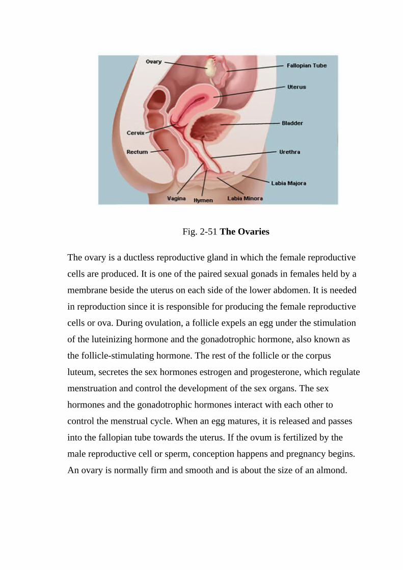
Fig. 2-51 The Ovaries
The ovary is a ductless reproductive gland in which the female reproductive
cells are produced. It is one of the paired sexual gonads in females held by a
membrane beside the uterus on each side of the lower abdomen. It is needed
in reproduction since it is responsible for producing the female reproductive
cells or ova. During ovulation, a follicle expels an egg under the stimulation
of the luteinizing hormone and the gonadotrophic hormone, also known as
the follicle-stimulating hormone. The rest of the follicle or the corpus
luteum, secretes the sex hormones estrogen and progesterone, which regulate
menstruation and control the development of the sex organs. The sex
hormones and the gonadotrophic hormones interact with each other to
control the menstrual cycle. When an egg matures, it is released and passes
into the fallopian tube towards the uterus. If the ovum is fertilized by the
male reproductive cell or sperm, conception happens and pregnancy begins.
An ovary is normally firm and smooth and is about the size of an almond.

Fig. 2-52 Overview of the female reproductive tract.
In both the males and females, the gonads develop within the mesonephric
ridge and descend through the abdomen. However, unlike the testes, the
ovaries stop in the pelvis.
The ovaries are paired, oval organs attached to the posterior surface of the
broad ligament of the uterus by the mesovarium (a fold of peritoneum,
continuous with the outer surface of the ovaries).
Neurovascular structures enter the hilum of the ovary via the mesovarium.
The ovary has 3 components:- surface: The surface layer of the ovary is
formed by simple cuboidal epithelium, known as germinal epithelium ,
cortex: The cortex (outer part) of the ovary is largely comprised of
a connective tissue stroma. It supports thousands of follicles. Each
primordial follicle contains an oocyte surrounded by a single layer of
follicular cells and medulla.

The medulla (inner part) is composed of supporting stroma and contains a
rich neurovascular network which enters the hilum of ovary from the
mesovarium.
Fig. 2-53 Cross section of an ovary. Shows the three major components of
the ovary. Also shows the follicles at various stages of development.
In both the males and females, the gonads develop within the mesonephric
ridge and descend through the abdomen. However, unlike the testes, the
ovaries stop in the pelvis.
The ovaries are paired, oval organs attached to the posterior surface of the
broad ligament of the uterus by the mesovarium (a fold of peritoneum,
continuous with the outer surface of the ovaries).
Neurovascular structures enter the hilum of the ovary via the mesovarium.
The ovary has 3 components:- surface: The surface layer of the ovary is
formed by simple cuboidal epithelium, known as germinal epithelium ,
cortex: The cortex (outer part) of the ovary is largely comprised of
a connective tissue stroma. It supports thousands of follicles. Each
primordial follicle contains an oocyte surrounded by a single layer of

follicular cells and medulla: the medulla (inner part) is composed of
supporting stroma and contains a rich neurovascular network which enters
the hilum of ovary from the mesovarium.
Fig. 2-54 The major ligaments of the ovary.
Two peritoneal ligaments attach to the ovary:- suspensory ligament of
ovary: fold of peritoneum extending from the mesovarium to the pelvic wall.
Contains neurovascular structures and ligament of ovary: extends from
the ovary to the fundus of the uterus. It then continues from the uterus to the
connective tissue of the labium majus, as the round ligament of uterus.
The ovaries receive blood from paired ovarian arteries, which arise directly
from the abdominal aorta, below the renal artery.
Venous drainage is achieved by a pair of ovarian veins. The left ovarian vein
drains into the left renal vein, and the right ovarian vein drains directly into
the inferior vena cava.
Lymph from the ovaries drains into the para-aortic nodes.

Fig. 2-55 Posterior view of the arterial supply to the female reproductive
tract.
Fig. 2-56 Vessels of the uterus and its appendages, rear view. (Fallopian
tubes visible at top right and top left.)
The nerve supply to ovaries runs via the suspensory ligament of the ovary
with the vasculature, to enter the ovary at the hilum. The ovaries receive
sympathetic and parasympathetic nerve fibres from the ovarian and uterine
(pelvic) plexuses, respectively. The uterus and ovaries are the most vital
organs of the female reproductive system. These organs work together to

produce female sex hormones, produce and develop ova (egg cells), and
support the development of a fetus during pregnancy. The uterus, also
known as the womb, is a hollow, muscular, pear-shaped organ found in the
pelvic region of the abdominopelvic cavity. It is located posterior to the
urinary bladder and is connected via the cervix to the vagina on its inferior
border and the fallopian tubes along its superior end. Many smooth muscle
cells in the walls of the uterus provide it with great extensibility and
contractile strength.
The endometrium, or lining of the uterus, consists of a thick layer of
epithelial and connective tissues that are shed and regrown periodically
during the menstrual cycle. Female hormones encourage the growth of the
endometrium to support the potential implantation of an embryo in the event
of a successful fertilization of an ovum. At the end of the menstrual cycle, if
a fertilized ovum has not implanted into the endometrium, the endometrium
is cut off from its blood supply and is shed along with the ovum as menstrual
flow. Menstrual flow then exits the body through the cervix and vagina so
that the uterus can grow a new endometrium for the next ovum. During
pregnancy, the tiny embryo implants itself into the tissues of the
endometrium and begins to grow inside of the uterine lining. Tissue from the
embryo begins to merge with the tissues of the uterus to form the placenta
and umbilical cord that allow for the exchange of respiratory gases,
nutrients, and wastes between the mother and the developing embryo. The
walls of the uterus extend and grow to accommodate the growing embryo as
it enters the fetal stage of development. At the end of pregnancy the cervix
dilates and the muscles of the myometrium contract to push the fetus into the
birth canal to initiate childbirth.The ovaries are small, oval-shaped, and
grayish in color, with an uneven surface. The actual size of an ovary depends
on a woman’s age and hormonal status; the ovaries, covered by a modified

peritoneum, are approximately 3-5 cm in length during childbearing years
and become much smaller and then atrophic once menopause occurs. A
cross-section of the ovary reveals many cystic structures that vary in size.
These structures represent ovarian follicles at different stages of
development and degeneration.
Several paired ligaments support the ovaries. The ovarian ligament connects
the uterus and ovary. The posterior portion of the broad ligament forms the
mesovarium, which supports the ovary and houses its arterial and venous
supply. The suspensory ligament of the ovary (infundibular pelvic ligament)
attaches the ovary to the pelvic sidewall. This larger structure also contains
the ovarian artery and vein, as well as nerve supply to the ovary.
Blood supply to the ovary is via the ovarian artery; both the right and left
arteries originate directly from the descending aorta. The ovarian artery and
vein enter and exit the ovary at the hilum. The left ovarian vein drains into
the left renal vein, and the right ovarian vein empties directly into the
inferior vena cava. Nerve supply to the ovaries runs with the vasculature via
the suspensory ligament of the ovary, entering the ovary at the hilum.
Supply is through the ovarian, hypogastric, and aortic plexuses.Lymph
drainage of the ovary is primarily to the lateral aortic nodes; however, the
iliac nodes are also involved.
Histologically, the ovary has 2 main sections: the outer cortex and inner
medulla. A germinal layer coats the entire ovary, made of cuboidal epithelial
cells.The cortex is where the follicles and oocytes are found at various stages
of development and degeneration. The cortex is made of tightly packed
connective tissue. The stroma of this cortical connective tissue is composed
of spindle-shaped fibroblasts that respond to hormonal stimulation in a way

different from that of other fibroblasts in the body.The medulla is where the
ovarian vasculature is found and is primarily loose stromal tissue.
The ovarian follicles are found within the stroma of the ovarian cortex. A
follicle consists of an oocyte surrounded by follicular cells called granulosa
cells. Follicles go through stages of development each month, with the goal
of their maturation to release the oocyte for the purpose of fertilization and
reproduction. If the follicle fails to release the egg, it goes through
degeneration.
Fig. 2-57 Ovaries.

Fig. 2-58 Internal sexual &reproductive organs (A)
Fig. 2-59 Internal sexual &reproductive organs (B)

Fig. 2-60 Female reproductive anatomy
Fig. 2-61 Female reproductive system
2-2-2 Physiology

The ovaries have 2 main functions. They produce mature eggs. They also
make the female sex hormones, which control reproduction and sexual
development. Estrogen is responsible for the development of secondary sex
characteristics, such as the growth of breasts.
Progesterone prepares the body for conception by causing the buildup of the
uterine lining (endometrium) and other changes. The ovaries are the main
source of estrogen in sexually mature women.
Each month during ovulation, an ovary releases a mature egg. The egg
travels down the Fallopian tube to the uterus. If it is fertilized by a sperm,
the egg implants into the lining of the uterus and begins to develop into a
fetus. If the egg is not fertilized, it is shed from the body along with the
lining of the uterus during menstruation. During menopause, the ovaries stop
releasing eggs and producing sex hormones. The uterus and ovaries
are the most vital organs of the female reproductive system.
These organs work together to produce female sex
hormones, produce and develop ova (egg cells), and support
the development of a fetus during pregnancy. The uterus,
also known as the womb, is a hollow, muscular, pear-shaped
organ found in the pelvic region of the abdominopelvic
cavity. It is located posterior to the urinary bladder and is
connected via the cervix to the vagina on its inferior border
and the fallopian tubes along its superior end. Many smooth
muscle cells in the walls of the uterus provide it with great
extensibility and contractile strength.
The endometrium, or lining of the uterus, consists of a thick layer of
epithelial and connective tissues that are shed and regrown periodically
during the menstrual cycle. Female hormones encourage the growth of the

endometrium to support the potential implantation of an embryo in the event
of a successful fertilization of an ovum. At the end of the menstrual cycle, if
a fertilized ovum has not implanted into the endometrium, the endometrium
is cut off from its blood supply and is shed along with the ovum as menstrual
flow. Menstrual flow then exits the body through the cervix and vagina so
that the uterus can grow a new endometrium for the next ovum.
During pregnancy, the tiny embryo implants itself into the tissues of the
endometrium and begins to grow inside of the uterine lining. Tissue from the
embryo begins to merge with the tissues of the uterus to form the placenta
and umbilical cord that allow for the exchange of respiratory gases,
nutrients, and wastes between the mother and the developing embryo. The
walls of the uterus extend and grow to accommodate the growing embryo as
it enters the fetal stage of development. At the end of pregnancy the cervix
dilates and the muscles of the myometrium contract to push the fetus into the
birth canal to initiate childbirth.
The ovaries are the female pelvic reproductive organs that house the ova and
are also responsible for the production of sex hormones. They are paired
organs located on either side of the uterus within the broad ligament below
the uterine (fallopian) tubes. The ovary is within the ovarian fossa, a space
that is bound by the external iliac vessels, obliterated umbilical artery, and
the ureter. The ovaries are responsible for housing and releasing ova, or
eggs, necessary for reproduction. At birth, a female has approximately 1-2
million eggs, but only 300 of these eggs will ever become mature and be
released for the purpose of fertilization.
The ovary (From Latin: ovarium, literally "egg" or "nut") is an ovum-
producing reproductive organ, often found in pairs as part of the vertebrate
female reproductive system. Ovaries in female individuals are analogous to

testes in male individuals, in that they are both gonads and endocrine glands.
Endocrine glands are glands of the endocrine system that secrete their
products, hormones, directly into the blood rather than through a duct. The
major glands of the endocrine system include the pineal gland, pituitary
gland, pancreas, ovaries, testes, thyroid gland, parathyroid gland,
hypothalamus and adrenal glands. The hypothalamus and pituitary gland are
neuroendocrine organs. Local chemical messengers, not generally
considered part of the endocrine system, include autocrines, which act on the
cells that secrete them, and paracrines, which act on a different cell type
nearby.
The ability of a target cell to respond to a hormone depends on the presence
of receptors, within the cell or on its plasma membrane, to which the
hormone can bind.Hormone receptors are dynamic structures. Changes in
number and sensitivity of hormone receptors may occur in response to high
or low levels of stimulating hormones. Blood levels of hormones reflect a
balance between secretion and degradation/excretion. The liver and kidneys
are the major organs that degrade hormones; breakdown products are
excreted in urine and feces. Hormone half-life and duration of activity are
limited and vary from hormone to hormone.(Outwater EK,1997)
2-2-2-1 The major endocrine glands:

Fig. 2-62 The major endocrine glands
2-2-2-1-1 Pituitary gland (hypophysis)
Fig. 2-63 Endocrine glands in the human head and neck and their hormones
Pituitary gland
The pituitary gland hangs from the base of the brain by a stalk and is
enclosed by bone. It consists of a hormone-producing glandular portion

(anterior pituitary) and a neural portion (posterior pituitary), which is an
extension of the hypothalamus. The hypothalamus regulates the hormonal
output of the anterior pituitary and synthesizes two hormones that it exports
to the posterior pituitary for storage and later release.Four of the six
adenohypophyseal hormones are tropic hormones that regulate the function
of other endocrine organs. Most anterior pituitary hormones exhibit a diurnal
rhythm of release, which is subject to modification by stimuli influencing
the hypothalamus.
Somatotropic hormone or Growth hormone (GH) is an anabolic hormone
that stimulates growth of all body tissues but especially skeletal muscle and
bone. It may act directly, or indirectly via insulin-like growth factors (IGFs).
GH mobilizes fats, stimulates protein synthesis, and inhibits glucose uptake
and metabolism. Secretion is regulated by growth hormone releasing
hormone (GHRH) and growth hormone inhibiting hormone (GHIH), or
somatostatin. Hypersecretion causes gigantism in children and acromegaly
in adults; hyposecretion in children causes pituitary dwarfism.Thyroid-
stimulating hormone (TSH) promotes normal development and activity of
the thyroid gland. Thyrotropin-releasing hormone (TRH) stimulates its
release; negative feedback of thyroid hormone inhibits it.
Adrenocorticotropic hormone (ACTH) stimulates the adrenal cortex to
release corticosteroids. ACTH release is triggered by corticotropin-releasing
hormone (CRH) and inhibited by rising glucocorticoid levels.The
gonadotropins—follicle-stimulating hormone (FSH) and luteinizing
hormone (LH) regulate the functions of the gonads in both sexes. FSH
stimulates sex cell production; LH stimulates gonadal hormone production.
Gonadotropin levels rise in response to gonadotropin-releasing hormone
(GnRH). Negative feedback of gonadal hormones inhibits gonadotropin
release.Prolactin (PRL) promotes milk production in humans females. Its

secretion is prompted by prolactin-releasing hormone (PRH) and inhibited
by prolactin-inhibiting hormone (PIH).
The neurohypophysis stores and releases two hypothalamic hormones:-
Oxytocin stimulates powerful uterine contractions, which trigger labor and
delivery of an infant, and milk ejection in nursing women. Its release is
mediated reflexively by the hypothalamus and represents a positive feedback
mechanism and Antidiuretic hormone (ADH) stimulates the kidney tubules
to reabsorb and conserve water, resulting in small volumes of highly
concentrated urine and decreased plasma osmolarity. ADH is released in
response to high solute concentrations in the blood and inhibited by low
solute concentrations in the blood. Hyposecretion results in diabetes
insipidus.
2-2-2-1-2 Thyroid gland
The thyroid gland is located in the anterior throat. Thyroid follicles store
colloid containing thyroglobulin, a glycoprotein from which thyroid
hormone is derived.
Thyroid hormone (TH) includes thyroxine (T4) and triiodothyronine (T3),
which increase the rate of cellular metabolism. Consequently, oxygen use
and heat production rise.
Secretion of thyroid hormone, prompted by TSH, requires reuptake of the
stored colloid by the follicle cells and splitting of the hormones from the
colloid for release. Rising levels of thyroid hormone feed back to inhibit the
pituitary and hypothalamus. Most T4 is converted to T3 (the more active
form) in the target tissues. These hormones act by turning on gene and
protein synthesis. Graves' disease is the most common cause of

hyperthyroidism; hyposecretion causes cretinism in infants and myxoedema
in adults.
Calcitonin, produced by the parafollicular cells of the thyroid gland in
response to rising blood calcium levels, depresses blood calcium levels by
inhibiting bone matrix resorption and enhancing calcium deposit in bone.
2-2-1-3 Parathyroid glands
The parathyroid glands, located on the dorsal aspect of the thyroid gland,
secrete parathyroid hormone (PTH),[1] which causes an increase in blood
calcium levels by targeting bone, the intestine, and the kidneys. PTH is the
antagonist of calcitonin. PTH release is triggered by falling blood calcium
levels and is inhibited by rising blood calcium levels.
Hyperparathyroidism results in hypercalcaemia and all its effects and in
extreme bone wasting. Hypoparathyroidism leads to hypocalcaemia,
evidenced by tetany and respiratory paralysis.
2-2-1-4 Pancreas
The pancreas, located in the abdomen close to the stomach, is both an
exocrine and an endocrine gland. The endocrine portion (islets of
langerhans) releases insulin and glucagon and smaller amounts of other
hormones to the blood. Glucagon, released by alpha (α) cells when glucose
level in blood are low, stimulates the liver to release glucose to the blood.
Insulin is released by beta (β) cells when blood levels of glucose (and amino
acids) are rising. It increases the rate of glucose uptake and metabolism by
most body cells. Hyposecretion of insulin results in diabetes mellitus;
cardinal signs are polyuria, polydipsia, and polyphagia.

2-2-1-5 Gonads
The ovaries of the female, located in the pelvic cavity, release two main
hormones. Secretion of estrogens by the ovarian follicles begins at puberty
under the influence of FSH. Estrogens stimulate maturation of the female
reproductive system and development of the secondary sexual
characteristics. Progesterone is released in response to high blood levels of
LH. It works with estrogens in establishing the menstrual cycle.The testes of
the male begin to produce testosterone at puberty in response to LH.
Testosterone promotes maturation of the male reproductive organs,
development of secondary sex characteristics, and production of sperm by
the testes.
2-2-1-6 Pineal gland
The pineal gland is located in the diencephalon. Its primary hormone is
melatonin, which influences daily rhythms and may have an
antigonadotropic effect in humans.
2-2-1-7 Other hormone-producing structures
Many body organs not normally considered endocrine organs contain
isolated cell clusters that secrete hormones. Examples include the heart
(atrial natriuretic peptide); gastrointestinal tract organs (gastrin, secretin, and
others); the placenta (hormones of pregnancy—estrogen, progesterone, and
others); the kidneys (erythropoietin and renin); the thymus; skin
(cholecalciferol); and adipose tissue (leptin and resistin).
2-2-2 The female cycle.
Each month, the ovaries go through a series of stages, depending on
stimulation by the anterior pituitary hormones the follicle stimulating

hormone (FSH) and the luteinizing hormone (LH). A typical female cycle
lasts 28 days; however, this can range from 21-35 days.The ovarian cycle
has 2 distinct phases: the follicular phase (days 1-14) and the luteal phase
(days 14-28). The follicular phase is characterized by follicle development
and growth, the goal being that one follicle matures and releases an egg at
the time of ovulation, around day 14 of the female cycle. The remaining
immature follicles go through stages of degeneration up until day 28, when
the cycle repeats itself. The egg that is released is picked up by the fimbriae
of the uterine tube, and the egg is transported toward the uterus. If
fertilization does not occur, the egg degenerates, and menstruation occurs.
2-2-3 Menstruation.
At the time the ovaries are formed in the fetus, there are approximately
6000000 primordial follicles, which decrease to about 600000 at birth, to
300000 at the first menstrual cycle, to about 10000 at the time of
menopause.The average cycle is 28 days and has two distinct phases. The
follicular phase starts on day one of the menstrual cycle .The hypothalamus
in the brain releases gonadotropin-releasing hormone (GnRH). GnRH
signals the pituitary gland to release follicle stimulating hormone (FSH).
FSH stimulates the eggs inside the ovaries to grow. About 20 immature eggs
response and begin to develop within sacs known as follicles. Follicles
provide nourishment to the eggs. As the eggs develop, the ovaries release
estrogen. Estrogen signals the pituitary gland to reduce FSH production.
Only enough RSH is now released to stimulate one egg to continue
developing, the rest of the eggs shrivel away. Estrogen stimulates the lining
of the uterus to thicken. The primary follicle contains the contains the egg
that has grown the most rapidly. Estrogen continues to rise until it triggers a
surge of luteinizing hormone (LH) from the pituitary gland. LH stimulates
ovulation. The follicle ruptures and the egg is released along with the
follicular fluid onto the surface of the ovary.The ruptured follicle continues
to receive LH. The LH enables the follicle to turn into a small cyst known as
the corpus luteum. The corpus luteum produces progesterone.
Progesterone :- builds and thickens the endometrium, developing glandular
structures and blood vessels that supply nutrients to the developing embryo ,
it switches off FSH an LH and it raises the basal body temperature (BBT)
by half a degree, warming the uterus and fertilized egg. The egg is
surrounded by the zona pellucida, a protective shell. The shell is surrounded
by a mass of sticky cells called the cumulus oophorus. These sticky cells
allow the finger like projections at the end of each fallopian tube, to pick up
the egg and sweep it into the tube. The channel from the fimbriae to the
uterus is lined with cilia, which together with muscular contractions move
the ovum along the tube to the uterus.The journey from the ovary to the
uterus take about 6 days. If the egg is not fertilized it will disintegrate and is
absorbed
One of the most critical periods of pregnancy is from approximately day 11-
16 after mating. The blastocysts, elongate into long (2-3 feet), stringy
masses, and they begin to attach to the uterine wall. The greatest potential
loss in litter size may occur during this stage of development because of
attachment failures. On the average, 17 ova are shed at estrus yet only about
12 are accounted for as blastocysts when attachment complete. There is
thought to be some factor(s) which limit the number of blastocysts that can
attach in a given uterus. Research has shown that overcrowding is not a
major limiting factor. Embryonic survival may be related to certain uterine
secretions which have not presently been well defined. Environmental
stressors such as high temperatures and fighting as a result of mixing or
regrouping animals also adversely influence implantation and embryo
survival. Generally, the presence of at least four blastocysts are required in

order for pregnancy to continue.By days 25-35, the embryo is about 1 to 1
1/2 inches long, and major body systems and appendages are well formed.
Each embryo is surrounded by a separate series of fluid-filled membranes,
the amnion and chorion, which are comprise the placenta. These membranes
help to protect and nourish the growing embryo. Nutrients, waste, gases and
certain antibodies cross the membranes between the dam and embryos blood
systems. It is the presence or absence of this fluid that is detected by the
commonly used "ultrasound" pregnancy diagnosis equipment.
The fetal period begins at approximately day 36. Sexes may be easily
determined by external examination and the main systems of the body are
more well defined. The fetus can now be called a miniature adult. Fetal
orientation is random; some are head to head, some are tail to tail and some
are head to tail. At farrowing, about half are born tail first and half are born
head first. Embryos which die before day 35-40 are usually reabsorbed by
the dam. However, progressive calcification of the skeleton begins to occur
from day 36 onward and deaths occurring after this point will result in
mummification.
By day 109 the fetus weighs about 1½ to 2 pounds. Hair shafts begin to
emerge but remain trapped under a skin layer until close to birth.
Throughout gestation, the uterus gradually enlarges from about 2 to 3
pounds at mating to up to 60 pounds, including fetal contents, during the last
week. Some females will lose up to 10 to 11% of their body weight at
parturition or farrowing.The average gestation length or time from
conception until farrowing is 114 days. During gestation, the piglet grows
from the union of a microscopic sperm and ovum into a fully formed
individual weighing from 3 to 3½ pounds. Several factors can influence
ovulation rate age, nutrition and breed .Semen is deposited into the cervix
during mating or insemination. Muscular contractions of the cervix stimulate

ejaculation of the boar and is the basis of the boar’s ejaculatory response to
the "gloved hand" or "digital pressure" technique of semen collection.
Thirty to sixty billion sperm are deposited by the boar during natural service
or 2 to 6 billion during insemination but only a small fraction of these reach
the oviduct and the vicinity of the ova. Even though sperm are motile, they
do not propel themselves through the female’s tract. Sperm transport results
from uterine contractions. These contractions are stimulated by oxytocin
released from the pituitary gland. Its release is mediated by the stimuli from
mating behavior and copulation. Keep this in mind when inseminating a sow
or gilt. Face-to-face contact with a boar, plus tactile stimulus during and
after insemination may improve sperm transport. Sperm must reside in the
female from 6-10 hours before they are capable of fertilization. This process
is called capacitation and it includes both physical and biochemical changes
within the sperm.
Fertilization or union of sperm and ova occurs in the upper 1/3 of the
oviduct. Fertilization rate is virtually 100% if the female is mated to a fertile
boar at the correct time relative to ovulation. About 2 days after fertilization,
the blastocysts enter the uterus. They are in the 4-8 cell stage and begin to
space themselves equal distances apart in the uterine horns. The blastocysts
can migrate from one horn to another during this stage.(P. B. J.
VanVierzen,1998)
2-3 Ultrasound appearance of the ovary.
Ultrasound is safe and painless, and produces pictures of the inside of the
body using sound waves. Ultrasound imaging, also called ultrasound
scanning or sonography, involves the use of a small transducer (probe) and
ultrasound gel placed directly on the skin. High-frequency sound waves are

transmitted from the probe through the gel into the body. The transducer
collects the sounds that bounce back and a computer then uses those sound
waves to create an image. Ultrasound examinations do not use ionizing
radiation (as used in x-rays), thus there is no radiation exposure to the
patient. Because ultrasound images are captured in real-time, they can show
the structure and movement of the body's internal organs, as well as blood
flowing through blood vessels.
Ultrasound imaging is a noninvasive medical test that helps physicians
diagnose and treat medical conditions. A pelvic ultrasound provides pictures
of the structures and organs in the lower abdomen and pelvis. A Doppler
ultrasound exam may be part of a pelvic ultrasound examination. Doppler
ultrasound is a special ultrasound technique that evaluates blood flow
through a blood vessel, including the body's major arteries and veins in the
abdomen, arms, legs, neck and head (in infants and children).
In women, a pelvic ultrasound is most often performed to evaluate the
uterus , cervix , ovaries , fallopian tubes and bladder.Pelvic ultrasound
exams are also used to monitor the health and development of an embryo or
fetus during pregnancy. Ultrasound examinations can help diagnose
symptoms experienced by women such as pelvic pain , abnormal bleeding ,
other menstrual problems and help identify palpable masses such as ovarian
cysts and uterine fibroids.
A transvaginal ultrasound is usually performed to view the endometrium, the
lining of the uterus, and the ovaries. Transvaginal ultrasound also provides a
good way to evaluate the muscular walls of the uterus, called the
myometrium. Sonohysterography allows for a more in-depth investigation of
the uterine cavity. Three-dimensional (3-D) ultrasound permits evaluation of
the uterus and ovaries in planes that cannot be imaged directly.These exams

are typically performed to detect uterine anomalies , uterine scars ,
endometrial polyps , fibroids and cancer, especially in patients with
abnormal uterine bleeding. Some physicians also use 3-D ultrasound or
sonohysterography for patients with infertility. Three-dimensional
ultrasound provides information about the outer contour of the uterus and
about uterine irregularities.
Doppler ultrasound images can help the physician to see and evaluate
blockages to blood flow (such as clots) , narrowing of vessels , tumors and
congenital vascular malformations.You should wear comfortable, loose-
fitting clothing for your ultrasound exam. You may need to remove all
clothing and jewelry in the area to be examined. You may be asked to wear a
gown during the procedure.
ROLE OF ULTRASOUND is To examine the uterus, ovaries cervix vagina and
adnexae, Classification of a mass identified on other modalities eg solid,
cystic, mixed, Post surgical complications eg abscess, oedema , Guidance of
injections, aspiration or biopsy , Assistance with IVFand To identify the
relationship of normal anatomy and pathology to each other. INDICATIONS :
P/V bleeding/discharge , Menorrhagia , Polymenorrhea , Amenorrhea ,
Irregular periods , Pelvic pain , F/H uterine or ovarian Cancer , Palpable
lump , Infertility- primary or secondary and Anomalies .
LIMITATIONS: - transvaginal scanning is contra-indicated if the patient is not
yet sexually active, or cannot provide informed consent , large patient
habitus will reduce detail, particularly via the transabdominal approach and
excessive bowel gas can obscure the ovaries.
EQUIPMENT SELECTION AND TECHNIQUE: - use of a curvilinear 3-6 MHz
probe and a 6-10 MHz endovaginal probe. Low dynamic range.

PATIENT PREPARATION: - If possible, scan the patient in the first 10 days of
the cycle. Preferably Day 5-10 for improved diagnostic accuracy in the
assessment of the endometrium and ovaries and a full bladder is required .
Instruct the patient to drink 1 Litre of water to be finished 1 hour prior to
their appointment. They cannot empty their bladder until after the
scan.Transabdominal approach is a generalised overview to identify the
cervix, uterus and ovaries.
Scan sagitally in the midline immediately above the pubis. In this plane you
should be able to assess the uterus, vagina and cervix. Zoom the image to
assess and measure the endometrial thickness. Rotate into transverse and
angle slightly cranially to be perpendicular to the uterus. Whilst in transverse
and slightly right of midline, angle left laterally to identify the left ovary
using the full bladder as an acoustic window. Examine this ovary in two
planes. Now repeat this for the right ovary.
Endovaginal approach:-before letting the patient empty their bladder, show
them the TV probe and explain the procedure. Indicate the length that is
inserted which is approximately the length of a standard tampon. Explain
there is no speculum used. Explain the importance of a TV scan because it is
the gold standard in gynaecological ultrasound because of its superior
accuracy and improved diagnostic resolution.
Cover the probe with a latex free Transvaginal sheath and lubricate with
sterile gel on the outside.Elevate the patients bottom on a thick
sponge/pillow to assist the scan. A gynaecological ultrasound couch which
drops down is ideal so that a better angulation is achieved for an anteverted
uterus.Ensure the patient is ready and get permission before inserting the
probe.
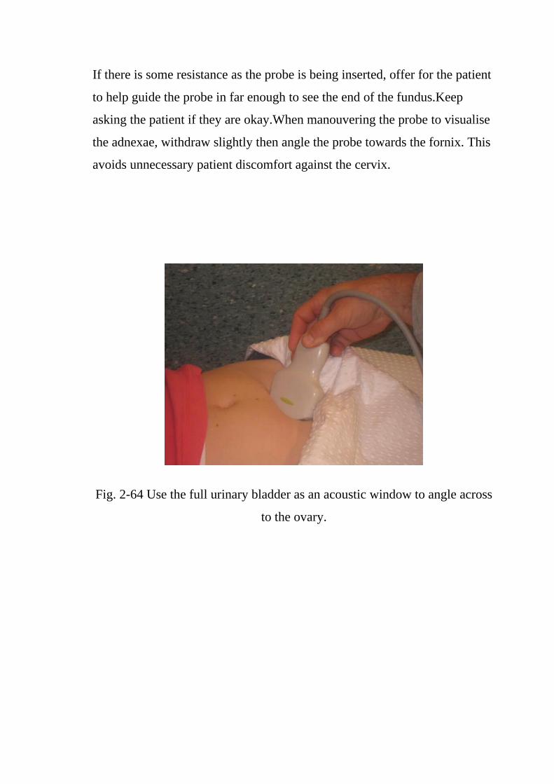
If there is some resistance as the probe is being inserted, offer for the patient
to help guide the probe in far enough to see the end of the fundus.Keep
asking the patient if they are okay.When manouvering the probe to visualise
the adnexae, withdraw slightly then angle the probe towards the fornix. This
avoids unnecessary patient discomfort against the cervix.
Fig. 2-64 Use the full urinary bladder as an acoustic window to angle across
to the ovary.

Fig. 2-65 Axial trans-abdominal image with the ovary lateral to the uterus.
Fig. 2-66Axial trans-vaginal scan plane

Fig. 2-67 Axial trans-vaginal image
Fig. 2-68 Normal transvaginal ovary demonstration normal peripheral
follicles.

Fig. 2-69 The uterus may be oblique and squash the ovary giving it a
flattened ovoid shape.
Fig. 2-70 Uterus with gestation sac and embryo

Fig. 2-71 Lt. adnexal cystic mass- Luteal cyst (Lt. ovary)
Fig. 2-72 Rt. ovarian simple cyst
This patient had an early gestation with embryo and intrauterine gestation
sac. Sonography of the adnexal regions also showed a cystic lesion of the

right ovary, which was thin walled and showed no septae or nodules within
it, suggestive of a simple cyst (functional) of the right ovary. However, the
left adnexal region showed a thick walled cystic lesion with echogenic walls.
This appearance can easily be due to an ectopic pregnancy. Both ectopic
gestations and corpus luteal cysts show similar features including the
presence of "ring of fire" or ring of vessels around the lesion (on Color
Doppler imaging). The left ovary was not seen separate from the left adnexal
cyst; also there was no evidence of significant fluid in the cul de sac;
besides, the presence of intrauterine pregnancy lead to the diagnosis of a left
ovarian Corpus Luteal cyst. Ultrasound images are courtesy of Dr. V.
Ganesan, MD, India.
Hemorrhagic cysts of the ovary are the result of bleeding from vascular
tissues within the walls of the freshly formed corpus luteum or luteal cyst.
The hemorrhagic cyst of ovary can present in various ways, mimicking a
number of more ominous diseases or cysts of the ovary. These include
ovarian cancers to serous or mucinous cystadenomas. Sometimes, rupture of
the hemorrhagic ovarian cyst can result in a medical emergency .

Fig. 2- 73 Hemorrhagic cyst of ovary resulting from Ovulation induction
Fig. 2-74 Polycystic ovary

Fig. 2-75 Polycystic ovary with doppler
Fig. 2-76This young nulliparous female patient undwerwent ultrasonography
following ovulation induction.
The right ovary shows a typical hemorrhagic cyst formed from the corpus
luteum. The first image (top row- left) is a transabdominal ultrasound image
showing fine fibrinous strands within the cystic mass in the right ovary.
Transvaginal ultrasound and color Doppler images confirm these findings.
The uterus shows typical secretory changes in the endometrium suggesting
post ovulatory phase.

Fig. 2-77 Chocolate cyst
This patient has a co-existing chocolate cyst with a hemorrhagic cyst in the
same (right) ovary. The cyst on the left half of the ultrasound image is a
hemorrhagic cyst. Note the fine fibrinous strands within the cyst suggesting
clot formation. The cyst on the right half of the image is homogenous with
fine echoes throughout the ovarian cyst. This is a typical appearance of an
endometrioma (chocolate cyst). Ultrasound image is courtesy of Dr. Gunjan
Puri, MD, India.

Fig. 2-78 Chocolate cyst
Fig. 2-79 This female patient has a left ovarian hemorrhagic cyst (see
ultrasound image above-left). In addition, there is a large collection of free
fluid with particulate matter in the pelvis. The right fallopian tube is
thickened with a ring shaped mass. This suggests that there is significant
hemorrhage into the pelvis due to a ruptured ectopic pregnancy (right tubal
ectopic gestation). The left ovarian hemorrhagic cyst appears intact, ruling
out ruptured hemorrhagic cyst. Both above ultrasound images are courtesy
of Gunjan Puri, MD, India.

Fig. 2-80 Lt. ovarian cyst
Fig, 2-81 bilateral ovarian complex masses

Fig. 2-82solid nodule containing fat and various tissues including hair
Fig. 2-83 Left ovarian dermoid cyst

Fig. 2-84 Echogenic mass in left ovary
Fig. 2-85 Normal right ovary

Fig. 2-86 Left ovarian dermoid
These ultrasound images show a typical dermoid cyst of the left ovary.
Transvaginal images of the left ovary show an echogenic mass of the left
ovary with attenuation of the ultrasound beam posteriorly. The echogenic
mass contains sebum and other fatty material giving rise to hyperechoic
appearance of the lesion. The differential diagnosis in this case include-
hemorrhagic cyst of the ovary with organized hematoma. Here the
hematoma would show acoustic enhancement posterior to the lesion, rather
than the attenuation (shadowing) seen in dermoid cyst. Bowel gas can also
produce the appearance of an echogenic lesion in the ovary. However, the
dirty shadowing seen in bowel gas and also peristalsis would help
distinguish bowel gas. Images are courtesy of Dr. Jaydeep Gandhi, MD,
India.

Fig. 2-87 Endometrioma or Endometriotic cyst of ovary or Chocolate cyst of
the ovary
Fig. 2-88 Chocolate cyst

Fig. 2-89 Ultrasound images of chocolate cysts
Fig. 2-90 Ovarian cyst

Fig. 2-91 Sonographic images of endometrioma
Fig. 2-92 Ultrasound image of endometrioma
Chocolate cysts or endometrioma of the ovary are caused by bleeding from
ectopic endometrial tissue within the ovary. Some researchers believe that
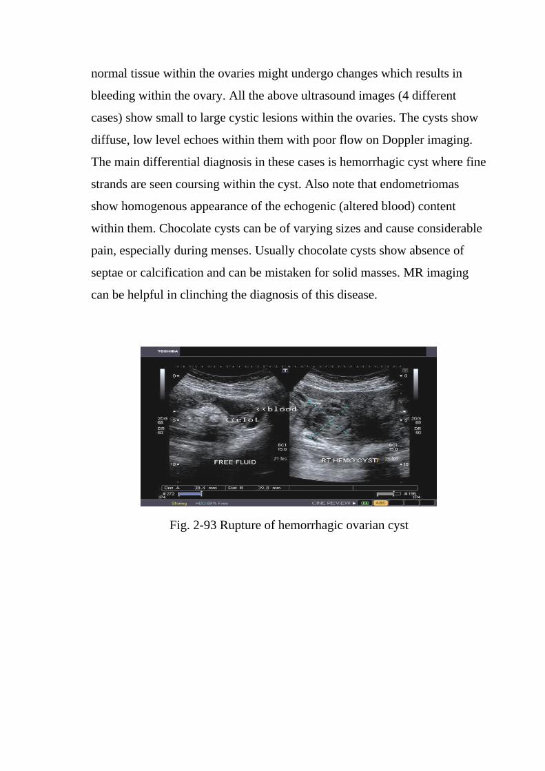
normal tissue within the ovaries might undergo changes which results in
bleeding within the ovary. All the above ultrasound images (4 different
cases) show small to large cystic lesions within the ovaries. The cysts show
diffuse, low level echoes within them with poor flow on Doppler imaging.
The main differential diagnosis in these cases is hemorrhagic cyst where fine
strands are seen coursing within the cyst. Also note that endometriomas
show homogenous appearance of the echogenic (altered blood) content
within them. Chocolate cysts can be of varying sizes and cause considerable
pain, especially during menses. Usually chocolate cysts show absence of
septae or calcification and can be mistaken for solid masses. MR imaging
can be helpful in clinching the diagnosis of this disease.
Fig. 2-93 Rupture of hemorrhagic ovarian cyst

Fig. 2-94 This female patient presented with pain in the pelvis and
tenderness. Transabdominal sonography of the pelvis showed a large amount
of free fluid with particulate matter in the cul de sac and around the right
adnexal region. The right ovary showed remnants of what appeared to be a
ruptured hemorrhagic corpus luteal cyst. Endovaginal imaging showed large
amount of fluid with debris posterior to the uterus. The left ovary appeared
normal. Lab tests and patient history excluded the possiblity of ectopic
pregnancy. It was hence concluded that this was a ruptured right
hemorrhagic ovarian cyst with free blood in the pelvis. The rounded
echogenic mass seen near the fluid appears to be an organized blood clot.

Fig. 2-95 Ovarian hyperstimulation syndrome (OHSS)
Fig. 2-96 This young adult female patient was examined to evaluate the
uterus and ovaries. She was under treatment for infertility and was using
gonadotropins. Ultrasound images of the ovaries show grossly enlarged
ovaries with large cysts (measuring 2.6 to 3 cms.) in both ovaries. These
ultrasound findings are diagnostic of OHSS or ovarian hyperstimulation
syndrome.

Fig. 2-97 a
Fig. 2-97 b
The above (Fig. 2-97a &Fig. 2-97b) ultrasound images again show
hyperstimulated ovaries. Both ovaries are grossly enlarged and cystic.

Fig. 2-98 a
Fig. 2-98 b
The above images(Fig. 2-98 a& Fig. 2-98 b) are courtesy of Dr. Dilraj
Gandhi, MD, Delhi, India. Grades of OHSS: There are 3 grades of ovarian
hyperstimulation based on sonography and clinical features: 1) mild OHSS-
ovaries are less than 5 cms. in diameter and patient has mild abdominal
dsicomfort. 2) moderate OHSS- ovaries measure 5 to 10 cms. in diameter 3)
severe OHSS- ovaries measure greater than 10 cms. in diameter.
:

Fig. 2-99 Transvaginal ultrasound images of ovarian hyperstimulation
syndrome
Fig. 2-100 a

Fig. 2-100 b
Fig. 2-100 c
(Fig. 2-100 a, Fig. 2-100 b & Fig. 2-100 c)This young adult female patient
showed multiple large theca lutein cysts of both ovaries, arranged in spoke-
wheel pattern (ultrasound images above) which were the result of use of
gonadotropins in the management of infertility. The cysts vary in size from 2
to 4 cms. with the ovaries massively enlarged (each ovary measures up to 7

cms. in size). This can be classified as grade-2 hyperstimulation of the
ovaries (ovarian diameter from 5 to 10 cms.). There is not evidence of
ascites. The color Doppler image of the ovaries shows vessels passing along
the margins of the cysts. One of the complications of such enlarged ovaries
in OHSS is torsion and in certain cases rupture of the ovaries, both of which
are medical emergencies. Ovarian hyperstimulation syndrome is known to
occur more frequently in patients of pre-existing Polycystic ovaries (PCO).
Fig. 2-101 pre-existing Polycystic ovaries (PCO).

Fig. 2-102 a Enlarged ovary
Fig. 2-102 b Enlarged ovary
Ultrasound shows enlarged ovaries on both sides. However, no definite
follicles were visualized. The uterus was normal in size. The transvaginal
ultrasound images show surprising details of the affected ovaries. The
sonographic findings include: a) Enlarged ovaries- the volume of these

ovaries ranged from 12 to 15 cc. This is due to stromal proliferation. b) The
ovarian stroma (parenchyma) appears echogenic. c) Multiple follicles of
small size are seen along the rim of the ovaries. d) The ovarian follicles are
less than 10 mm. in size (each of the follicles averaged at 4 to 5 mm. in
size). e) There are more than 10 follicles per ovary (here we could count at
least 12 to 15 follicles per ovary). The arrangement of the follicles along the
rim of the ovary is called a necklace sign and is iagnostic of PCOD
or polycystic ovary disease . Usually, clinical findings associated with PCOD
include- hirsuitism, obesity, irregular menses and infertility. These
conditions associated with polycystic ovaries are together labelled PCOS
(polycystic ovarian syndrome).
Fig. 2-103 ovarian torsion

Fig. 2-104 Enlarged lt. ovary
Fig. 2-105 Enlarged lt. ovary
This young girl (6 years age) had severe left pelvic pain. Ultrasound images
show an enlarged left ovary with lack of significant blood flow on power
Doppler imaging. The right ovary appeared normal. These ultrasound
images suggest torsion of the left ovary. This was surgically confirmed, with

the left ovary seen to be gangrenous. The normal right ovary is seen as a
pink healthy organ (see snapshot) compared to the gangrenous left ovary.
These images of torsion of ovary are courtesy of Dr. Jaydeep Gandhi, MD,
India.
Fig. 2-106 peritoneal inclusion cyst
Fig. 2-107 Ectobic pregnancy

Fig. 2-108 Ectobic pregnancy
This young adult female patient has a past history of ectopic pregnancy for
which she underwent surgery. she presented with symptoms of early
pregnancy with pelvic pain. Ultrasound shows presence of intrauterine
gestation sac with multiple adhesions within the pelvis with pockets of
ascitic fluid. The right ovary appears to be surrounded by multiple adhesions
and fluid resulting in an appearance like a spider in a web. These ultrasound
image findings are diagnostic of peritoneal inclusion cyst. This is a condition
resulting from trauma to the peritoneum usually the result of surgery with
resultant accumulation of peritoneal fluid and multiple adhesions. The
ovaries appear normal in structure and are well preserved. These ultrasound
images of peritoneal inclusions cyst are courtesy of Ajay Garg, MD.
The principal differential diagnosis in such cases of peritoneal inclusion
cysts includes ovarian cystic masses such as serous or mucinous
cystadenoma of the ovaries. Also, peritoneal inclusion cysts must be
differentiated from more ominous conditions such as ovarian malignancies.
Careful study of the ovaries, usually via the transvaginal ultrasound scan
will help differentiate peritoneal inclusion cysts from ovarian lesions
mentioned above. (Ross, E.K.,2013)
2-2-4 Follicle measurements and tracing.
The size of the leading follicles in conjunction with serum hormone levels
are used to determine the optimal time for giving the HCG trigger for IVF.
Follicles have traditionally been measured manually with two-dimensional
(2D) ultrasound by calculating the mean of the 2 largest follicle
diameters.Because follicles are often quite irregular in shape, measuring the
follicles in the traditional fashion can result in significant inaccuracy.
Another issue with manual measurements is that it is time consuming for the
staff and the patients.
General Electric (GE) has software (Automated Volume Count, SonoAVC)
for three-dimensional (3D) ultrasound machines that can identify follicles
and automatically calculate their volumes. It then converts the measured
volumes to diameters by using the sphere formula. It is possible that this
technology could give more accurate follicle measurements and also could
increase the efficiency of the ultrasound monitoring process.
This study was a retrospective review of ultrasound follicle measurements
during ovarian stimulation for IVF and the outcome parameters for those
cycles. The purpose was to study effectiveness and efficiency of 3D
ultrasound assessment of follicle size during ovarian stimulation for IVF
using SonoAVC. Clinical pregnancy rates, total number of eggs retrieved,
mature eggs retrieved and percentage of mature eggs were compared in 2
groups.
Group 1 included all women using own eggs under age 35 having egg
retrievals during the 9 month period of October 2007 through June 2008.
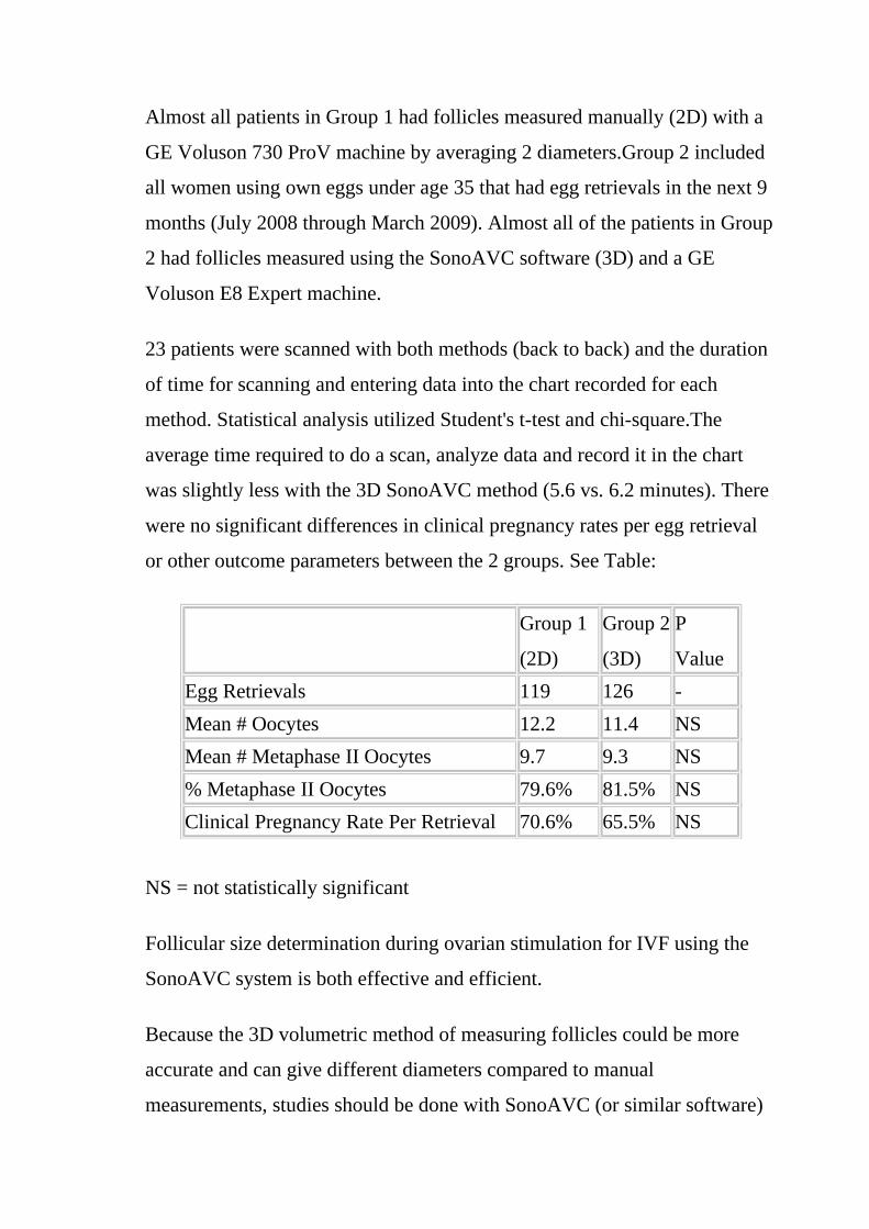
Almost all patients in Group 1 had follicles measured manually (2D) with a
GE Voluson 730 ProV machine by averaging 2 diameters.Group 2 included
all women using own eggs under age 35 that had egg retrievals in the next 9
months (July 2008 through March 2009). Almost all of the patients in Group
2 had follicles measured using the SonoAVC software (3D) and a GE
Voluson E8 Expert machine.
23 patients were scanned with both methods (back to back) and the duration
of time for scanning and entering data into the chart recorded for each
method. Statistical analysis utilized Student's t-test and chi-square.The
average time required to do a scan, analyze data and record it in the chart
was slightly less with the 3D SonoAVC method (5.6 vs. 6.2 minutes). There
were no significant differences in clinical pregnancy rates per egg retrieval
or other outcome parameters between the 2 groups. See Table:
Group 1
(2D)
Group 2
(3D)
P
Value
Egg Retrievals 119 126 -
Mean # Oocytes 12.2 11.4 NS
Mean # Metaphase II Oocytes 9.7 9.3 NS
% Metaphase II Oocytes 79.6% 81.5% NS
Clinical Pregnancy Rate Per Retrieval 70.6% 65.5% NS
NS = not statistically significant
Follicular size determination during ovarian stimulation for IVF using the
SonoAVC system is both effective and efficient.
Because the 3D volumetric method of measuring follicles could be more
accurate and can give different diameters compared to manual
measurements, studies should be done with SonoAVC (or similar software)

to further evaluate the ideal leading follicle size for HCG trigger in order to
optimize.Typically, the human ovaries produce a single dominant follicle
that results in a single ovulation each menstrual cycle . In any given cycle,
the dominant follicle must complete all the steps in folliculogenesis in a
timely manner. In this capacity, it survives the negative events that operate
to destroy the other follicles by atresia. Recognition that only a few follicles
become dominant beautifully demonstrates the fundamental principle that
folliculogenesis in mammals is a highly selective process. This chapter
considers what is known about the process underlying the expression of the
structural and functional organization of developing follicles and how they
are controlled.
Fig. 2-10. Photomicrograph of ovulation shows the expanded egg-cumulus
complex leaving the follicle through the stigma. The remaining cells in the
follicle wall ( i.e. granulosa, membrana and periantral, theca, the theca
interna and externa) develop into the corpus luteum.(From Blandau RJ:
Growth of the ovarian follicle and ovulation. Prog Gynecol 5:58, 1970.)
2-2-4-1 Folliculogenesis.
Folliculogenesis is the process in which a recruited primordial follicle grows
and develops into a specialized graafian follicle with the potential to either

ovulate its egg into the oviduct at mid-cycle to be fertilized or to die by
atresia. In women, the process is long, requiring almost 1 year for a
primordial follicle to grow and develop to the ovulatory stage. During the
course of folliculogenesis, growth is achieved by cell proliferation and
formation of follicular fluid, whereas development involves
cytodifferentiation of all the cells and tissues in the follicle. Only a few
follicles in the human ovary survive to complete the cytodifferentiation
process, with 99.9% dying by a programmed cell death mechanism called
apoptosis.
The mechanisms regulating follicle growth and development are under the
control of changing concentrations of ligands (i.e. hormones and growth
factors). At the endocrine level, folliculogenesis is regulated by a central
nervous system, anterior pituitary, and ovary cascade mechanism.
Specialized hypothalamic neurons secrete pulses of gonadotropin-releasing
hormone (GnRH) into the portal blood vessels, which acts on the
gonadotrophs to cause a pulsatile release of follicle-stimulating hormone
(FSH) and luteinizing hormone (LH), which act on ovarian follicle cells to
control folliculogenesis. Although GnRH, FSH, and LH are critically
important in regulating folliculogenesis, hormones and growth factors,
which are themselves products of the follicle, can act locally to modulate
(amplify or attenuate) FSH and LH action. This is the autocrine/paracrine
system of developing follicles. It is believed that this local regulatory system
plays an important role in the complex mechanisms governing the timing of
folliculogenesis and whether a follicle becomes dominant or atretic.
2-2-4-2 Chronology.
In women, folliculogenesis is a long process. In each menstrual cycle, the
dominant follicle that ovulates its egg originates from a primordial follicle

that was recruited to initiate growth almost 1 year earlier. In a broad sense,
there are two types of follicles : preantral (primordial, primary, secondary ,
tertiary and antral (graafian, small , medium , large , preovulatory . The
development of preantral and antral follicles is gonadotropin independent
and gonadotropin dependent, respectively.
The rate of preantral follicle development is slow, requiring about 300 days
for a recruited primordial follicle to complete the whole preantral period . A
long doubling time (about 10 days) for the granulosa cells is responsible for
the slow growth rate. After antrum formation in the class 3 follicle (about
0.4 mm in diameter), the rate of growth accelerates. The time interval
between antrum formation and the development of a 20-mm preovulatory
follicle is about 50 days. The dominant follicle appears to be selected from a
cohort of class 5 follicles at the end of the luteal phase of the menstrual
cycle. About 15 to 20 days are required for a dominant follicle to grow and
develop to the preovulatory stage Atresia can occur in all follicles (preantral
and antral) after the class 1 or secondary follicle stage; however, the highest
incidence is seen in the antral follicles that are more than 2 mm in diameter .

Fig. 2-111 Folliculogenesis
2-2-4-3 The process.Folliculogenesis occurs within the cortex of the ovary.
The follicles in the cortex are present in a wide range of sizes representing
various stages of folliculogenesis. The goal of folliculogenesis is to produce
a single dominant follicle from a pool of growing follicles. There are four
major regulatory events involved in this process: recruitment, preantral
follicle development, selection, and atresia.

Fig. 2-112 Photomicrograph of an adult primate ovary. Follicular and luteal
units are seen in the cortex and large blood vessels and nerves in the
medulla. Serous or surface epithelium.
2-2-4-4 The primordial follicle.
All primordial follicles are composed of a small primary oocyte (about 25
μm in diameter) arrested in the diplotene (or dictyate) stage of meiosis, a
single layer of flattened (squamous) granulosa cells, and a basal lamina. The
mean diameter of the human primordial follicle is 29 μm.By virtue of the
basal lamina; the granulosa and oocyte exist within a microenvironment in
which direct contact with other cells does not occur. The primordial follicles
do not have an independent blood supply, it follows that primordial follicles
have limited access to the endocrine system.

Fig. 2- 113 Electron micrograph of a human primordial follicle shows the
flattened granulosa cells (GC), the oocyte with its germinal vesicle (GV) or
nucleus, the Balbiani body (BB), with all the oocyte organelles gathered at
one pole of the GV, and basal lamina (BL).(From Erickson GF: The ovary:
Basic principles and concepts.
2-2-4-5 Recruitment.
The first major event in folliculogenesis is recruitment. Recruitment is the
process by which an arrested primordial follicle is triggered to reinitiate
development and enter the pool of growing follicles. All primordial follicles
(oocytes) present in the human ovaries are formed in the fetus between the
sixth and the ninth month of gestation. Because the entire stock of oocytes in
primordial follicles is in meiotic prophase, none is capable of dividing
mitotically. All oocytes (primordial follicles) capable of participating in
reproduction during a woman's life are present in the ovaries at birth. The
total number of primordial follicles in the ovaries at any given moment of
time is called the ovary reserve (OR). The process of recruitment begins
soon after the formation of the primordial follicles in the fetus, and it

continues throughout the life of the female until the pool of primordial
follicles is exhausted at the menopause. There is a bi-exponential decrease in
OR during aging. The number of primordial follicles falls steadily for more
than three decades, but when the OR reaches a critical number of about
25,000 at 37.5 ± 1.2 years of age, the rate of loss of primordial follicles
accelerates about twofold .This change in OR is associated in an age-related
decrease in fecundity, perhaps causal to the age-related increase in FSH that
occurs in women after 36 years of age.
2-2-4-6 Mechanism.
The first visible sign that a primordial follicle is being recruited is that some
granulosa cells begin to change from a squamous to a cuboidal shape. The
first cuboidal cell is seen when the primordial follicle contains 8 granulosa
cells, and the process is complete when the granulosa number reaches 19.
The shape change is followed by the onset, albeit slow, of DNA synthesis
and mitosis in the granulosa cells. A change in shape and acquisition of
mitotic potential in the granulosa cells are the hallmarks of recruitment.
Such observations suggest that the mechanisms governing recruitment may
involve a regulatory response at the level of the granulosa cell. Recruitment
is pituitary independent, and it probably is controlled by autocrine/paracrine
mechanisms. Whether it is affected by a stimulator or the loss of an inhibitor
is uncertain; however, primordial follicles undergo rapid recruitment when
removed from the ovary and cultured in vitro. These observations support
the inhibitor idea.

Fig. 2-114 Photomicrograph of nongrowing primordial and a newly
recruited follicle in the human ovary.
Fig, 2-115 Relation between granulosa number in the largest cross section of
the follicle and the distribution of flattened and cuboidal cells
Several different hypotheses have been put forth to explain the mechanism
of recruitment. First, the process appears to occur in primordial follicles
nearest the medulla where blood vessels are prominent. This supports the
hypothesis that exposure to nutrients or blood-borne regulatory molecules
could play a role in the control of recruitment. Second, an internal oocyte
clock mechanism has been proposed to control recruitment. In this
hypothesis, the clock is related to the time that the oocyte initiates meiosis in
the embryo. It is noteworthy that recruitment can be modulated. In rodents,

the rate of recruitment can be attenuated by removing the neonatal thymus
gland, starvation, or treatment with exogenous opioid peptides. These are
important observations, because they argue that ligand-receptor signaling
pathways are likely to regulate recruitment. Understanding the regulatory
mechanisms underlying recruitment remains a major task in reproductive
biology.
2-2-4-7 THE PREANTRAL FOLLICLE.
The early stages of folliculogenesis can be divided into three classes based
on the number of layers of granulosa cells, the development of theca tissue,
and the expression of a small cavity or antrum. The classes are the primary,
secondary, and early tertiary follicles. As the morphologic complexity
increases, important cellular and physiologic changes occur in the follicle
that renders it competent to respond to gonadotropins. The following
sections examine the structure and function changes that accompany
preantral follicle growth and development.

Fig. 2-116 Diagram illustrating the size and histologic organization of early
developing human follicles during the gonadotropin-independent period of
folliculogenesis
2-2-4-8 Primary Follicle.
A primary follicle consists of one or more cuboidal granulosa cells that are
arranged in a single layer surrounding the oocyte. Simultaneous with the
shape change and mitotic activities that accompany recruitment, the cuboidal
granulosa cells begin to express FSH receptors. The mechanism underlying
this critical event in folliculogenesis remains uncertain, but there is evidence
in rodents that granulosa-derived activin may play an important role in the
expression of FSH receptor by autocrine/paracrine mechanisms . Although

the granulosa cells express FSH receptors at this very early stage in
folliculogenesis, it is believed that the physiologic levels of plasma FSH
during the normal menstrual cycle do not influence granulosa responses
because primary follicles lack an independent vascular system. Nevertheless,
because there are blood vessels in the vicinity , FSH-induced changes in
primary follicle function may occur in response to abnormally high levels of
plasma FSH, such as those that occur during ovulation induction or aging.
Fig. 2-117 Drawing of a developing primary follicle embedded in the
connective tissue or stroma of the ovary cortex. A nucleolus and meiotic
chromosomes are evident in the oocyte nucleus. The mitochondria appear
aggregated at one pole of the oocyte nucleus

Fig. 2-118 Diagram of the proposed mechanism for the autocrine control of
follicle-stimulating hormone receptor expression in granulosa cells of
preantral follicles.(From Erickson GF: Dissociation of endocrine and
gametogenic ovarian function.
Beginning approximately at the time of recruitment, the oocyte begins to
grow and differentiate. This period is marked by a progressive increase in
the level of oocyte RNA synthesis. A number of important oocyte genes are
turned on at this time. For example, the genes encoding the zona pellucida
(ZP) proteins (i.e. ZP-1, ZP-2, and ZP-3) are transcribed and translated. The
secreted ZP proteins begin to polymerize near the oocyte surface, forming an
extracellular matrix coat (the zona pellucida) that eventually encapsulates
the egg. The importance of the zona pellucida is emphasized by the fact that
the carbohydrate moiety of ZP-3 is the species-specific sperm-binding

molecule. It is responsible for initiating the acrosome reaction in capacitated
sperm. During primary follicle development, the granulosa cells send
processes through the zona layer, where they form gap junctions with the
oocyte cell membrane, or oolemma .Gap junctions are intercellular channels
composed of proteins called connexins. There are at least 13 members of the
connexin family that directly couple adjacent cells to allow the diffusion of
ions, metabolites, and other low-molecular-weight signaling molecules such
as cAMP and calcium. Connexin 37 (C×37) is an oocyte-derived connexin
that forms gap junctions between the oocyte and surrounding granulosa
cells. Evidence from C×37-deficient mice assigns C×37 an obligatory role
for folliculogenesis, ovulation, and fertility.Large gap junctions are also
present between the granulosa cells themselves (Fig. 12). C×43 is a major
gap junction protein expressed in the granulosa cells. As a consequence of
gap junctions, the primary follicle becomes a metabolically and electrically
coupled unit. This communication between the granulosa and oocyte
remains throughout folliculogenesis and is responsible for the synchronous
expression of important activities (positive and negative).
Fig. 2-119 Electron micrograph of the oocyte-corona radiata granulosa cells
in a preantral follicle. The granulosa cell processes traversing the zona

pellucida (ZP) make small gap junctions (arrowheads) with the oocyte
plasma membrane. Larger gap junctions (arrows) are evident between
corona radiata cells.
2-2-4-9 Secondary Follicle.
A secondary follicle is a preantral follicle with 2 to 10 layers of cuboidal or
low columnar cells that form a stratified epithelium. As seen in, the
transition from a primary to a secondary follicle involves the acquisition of a
second layer of granulosa cells. This transition is accomplished by the
continuing division of the granulosa cells. The mechanisms regulating
granulosa mitosis are poorly understood. However, exciting research in
rodents has provided compelling evidence for the involvement of an
oocytederived growth factor, called growth differentiation factor-9 (GDF-9).
GDF-9 is a novel member of the transforming growth factor-β (TGF-β)
superfamily. GDF-9 is strongly expressed in the ovary; it is localized only in
oocytes of recruited follicles. In GDF-9 deficient mice, follicle growth and
development stop at the primary stage; consequently no dominant follicles
form, and the females are infertile. Accordingly, GDF-9 is obligatory for
folliculogenesis after the primary stage, presumably because it is an
obligatory mitogen for granulosa cells. A fundamental concept that emerges
from this work is that the oocyte plays a pivotal role in regulating
folliculogenesis through its ability to produce novel regulatory ligands ,
which are crucial for folliculogenesis.

Fig. 2-120 A typical healthy secondary follicle contains a fully grown oocyte
surrounded by the zona pellucida, five to eight layers of granulosa cells, a
basal lamina, and developing theca tissue with numerous blood vessels
One of the most important changes that occur in the development of a
secondary follicle is the acquisition of a theca layer. This tissue, which
consists of a layer of stroma-like cells around the basal lamina, subsequently
differentiates into the inner theca interna and outer theca externa. Theca
development is accompanied by the neoformation of numerous small
vessels, presumably through angiogenesis. This is a critical event because
blood circulates around the follicle, bringing nutrients and hormones to and
waste and secretory products from the secondary follicle. In this regard,
some stromal cells in the inner layer express LH receptors. These cells
subsequently differentiate into steroidogenic cells called theca interstitial
cells (TICs), most likely in response to the plasma LH delivered by the theca
vascular system. All the granulosa cells in secondary follicles express FSH
receptors. It seems likely that diffusion of plasma FSH into the secondary
follicle may evoke FSH-dependent granulosa responses. The outer layer of
stroma cells subsequently differentiates into smooth muscle cells called the

theca externa. These smooth muscle cells are innervated by the autonomic
nervous system. In the secondary follicle, the oocyte completes its growth.
When the follicle is about 200 μm in diameter, the oocyte has attained its
maximum size and grows no more, despite the fact that the human follicle
enlarges to a diameter of 2 cm or more (Fig. 14). It is well documented in
rodents that granulosa cells play an obligatory role in the growth and
differentiation of the oocyte. An important differentiation event that occurs
when the oocyte completes its growth is acquisition of the capacity to
resume meiosis. Oocytes normally do not resume meiosis during
folliculogenesis, and a mechanism must operate to inhibit this process (i.e.
germinal vesicle breakdown [GVBD]) and the resumption of meiosis. The
underlying mechanism for the inhibition remains unknown; however, there
is evidence to support the concept that granulosa derived cAMP may play an
important role in inhibiting the resumption of meiosis. In such a mechanism,
FSH-induces cAMP in the granulosa cells, which diffuses into the oocyte
through the C×37 gap junction, where it proceeds to inhibit GVBD .
Fig. 2-121 Diagram showing the relation between the size of the oocyte and
the size of the follicles in the human infant ovary
2-2-4-10 Tertiary Follicle.
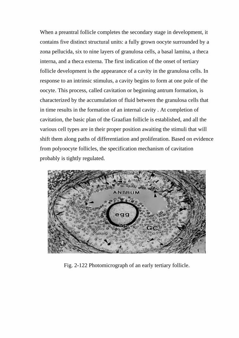
When a preantral follicle completes the secondary stage in development, it
contains five distinct structural units: a fully grown oocyte surrounded by a
zona pellucida, six to nine layers of granulosa cells, a basal lamina, a theca
interna, and a theca externa. The first indication of the onset of tertiary
follicle development is the appearance of a cavity in the granulosa cells. In
response to an intrinsic stimulus, a cavity begins to form at one pole of the
oocyte. This process, called cavitation or beginning antrum formation, is
characterized by the accumulation of fluid between the granulosa cells that
in time results in the formation of an internal cavity . At completion of
cavitation, the basic plan of the Graafian follicle is established, and all the
various cell types are in their proper position awaiting the stimuli that will
shift them along paths of differentiation and proliferation. Based on evidence
from polyoocyte follicles, the specification mechanism of cavitation
probably is tightly regulated.
Fig. 2-122 Photomicrograph of an early tertiary follicle.

Fig. 2-123 Photomicrograph of a polyovular follicle at the early tertiary
stage shows the sites of cavitation or early antrum formation (clear spaces)
just above oocytes (asterisk). This event, which is under intraovarian control,
seems to arise in a specific synchronized manner and establishes the polarity
of the follicle.
It is well known that cavitation occurs in hypophysectomized animals,
demonstrating that pituitary hormones such as FSH are not required for this
morphogenetic event. Consistent with this concept is the observation that
cavitation occurs in FSH-β-deficient mice. It seems reasonable to conclude
that cavitation is controlled by autocrine/paracrine mechanisms. Two growth
factors expressed in the follicle itself have been implicated in cavitation:
activin and KIT ligand. Treating cultured granulosa cells with activin causes
morphogenetic changes that result in the formation of a histologic unit with
an antrum-like cavity. Blocking the action of the KIT ligand in the ovary
prevents the formation of antral follicles; consequently, there are no
ovulations, and the female is infertile. In this regard, evidence supports the

concept that the oocyte gap junctions are also important for cavitation. Gap
junctions are intercellular channels composed of proteins called connexins.
There are at least 13 members of the connexin family that directly couple
adjacent cells, allowing diffusion of ions, metabolites, and other low-
molecular-weight signaling molecules such as cAMP, C×37 appears to be an
oocyte-derived connexin that forms gap junctions between the oocyte and
surrounding granulosa cells. Evidence from C×37-deficient mice assigns to
C×37 an obligatory role in Graafian follicle formation, ovulation, and
fertility. Collectively, all this evidence suggests that follicle-derived activin,
KIT, and C×37 are involved in the autocrine/paracrine mechanisms that
control cavitation.
2-2-4-11 THE GRAAFIAN FOLLICLE.
A graafian follicle can be defined structurally as a heterogeneous family of
relatively large follicles (0.4 to 23 mm) characterized by a cavity or antrum
containing a fluid called follicular fluid or liquor folliculi. The characteristic
structural unit of all Graafian follicle is the antrum. For this reason, the term
antral follicle is used correctly as a synonym for Graafian follicle. The
follicular fluid is the medium in which the granulosa cells and oocyte are
found and through which regulatory molecules must pass on their way to
and from this microenvironment. Surprisingly, we know almost nothing
about the physiologic significance of the antrum and follicular fluid in
folliculogenesis. It is clear that follicle development and ovulation occur in
birds and amphibians despite the absence of an antrum and follicular fluid.
Nonetheless, its presence in all mammalian species testifies to its
physiologic importance.
A graafian follicle is a three-dimensional structure with a central antrum
surrounded by a variety of different cell types .There are six distinct

histologic components in the graafian follicle, including the theca externa,
theca interna, basal lamina, granulosa cells, oocyte, and follicular fluid . A
graafian follicle does not change its morphologic complexity as growth
proceeds. All graafian follicles have this same basic architecture; even
though there are dramatic changes in Graafian follicle size, their appearance
remains more or less the same.
Fig. 2-124 Diagram of the architecture of a typical class 5 Graafian follicle.
The theca externa is characterized by the presence of smooth muscle cells,
which are innervated by autonomic nerves. Although the physiologic
significance of the theca externa remains unclear, there is evidence that it
contracts during ovulation and atresia. Changes in the contractile activity of
the theca externa may be involved in atresia and ovulation; however, this has
not been rigorously proved. The corpus luteum retains a theca externa
throughout its life, but the significance during luteinization and luteolysis is
not known.

Fig. 2-125 Drawing of the wall of a graafian follicle.
The differentiation of a granulosa cell can be traced to its position within the
cellular mass. For example, cells in the membrana domain stop proliferating
before those in central domain. The ability of the granulosa cells in the inner
domains to continue dividing throughout graafian follicle development
suggests they may be precursor cells. The cessation of mitosis in the
membrana domain is characterized by the progressive expression of overt
differentiation in which they assume the functional phenotype of fully
differentiated cells. This process requires the temporal and coordinate
expression of genes that form the basis of granulosa cytodifferentiation. The
mechanism by which this occurs involves ligand-dependent signaling
pathways that are coupled to the activation and inhibition of specific genes.
For example, normal differentiation of the membrana granulosa cells
requires the activation of specific genes, including those for cytochrome
P450 aromatase (P450arom) and the LH receptor, and the inhibition of
structural genes in the apoptotic pathway. In contrast, the granulosa cells in

the periantral, cumulus, and corona radiata domains proliferate, but they fail
to express the genes that are involved in a terminal differentiation.
All the granulosa cells in the healthy Graafian follicle express FSH receptor,
and it has been shown that murine granulosa cells in the membrana and
cumulus domains produce cAMP in response to FSH stimulation. These
observations argue that post cAMP regulatory events are involved in the
aspects of granulosa heterogeneity. The idea that the oocyte plays a key role
in causing the different patterns of granulosa cytodifferentiation during
Graafian follicle development is supported by studies in rodents. A dialogue
takes place between the oocyte and granulosa cells that has a great impact on
folliculogenesis. In developing murine graafian follicles, the differential
pattern of proliferation and differentiation between the granulosa in the
membrana and cumulus domains are under the control of secreted oocyte
morphogens.A novel TGF-β family member, GDF-9, was discovered in the
mouse. Definitive evidence that GDF-9 is obligatory for folliculogenesis
came from studies of GDF-9-deficient mice.In these animals, the absence of
GDF-9 resulted in the arrest of follicle growth and development at primary
stage and the females are infertile. These data support the idea that GDF-9
secreted by the egg is obligatory for Graafian follicle development,
granulosa cytodifferentiation and proliferation, and female fertility. The
clinical relevance of this new concept is demonstrated by the presence of
GDF-9 mRNA in the human ovary. The current challenges are to elucidate
the mechanisms controlling GDF-9 expression and to identify the target cells
for GDF-9 and the biologic processes that GDF-9 regulates. The concept
that oocyte-derived growth factors control folliculogenesis and fertility
could have important implications for human physiology and
pathophysiology.
2-2-4-12 Classification.

All graafian follicles can be divided broadly into two groups: healthy and
atretic. The main difference between these two groups is whether apoptosis
is occurring in the granulosa cells. The development of a graafian follicle
(healthy or atretic) follows a progressive course over time. This implies that
variability or heterogeneity is a normal consequence of folliculogenesis. A
healthy graafian follicle becomes progressively more differentiated with
increasing time until it attains the preovulatory stage. The time for this
process is about 2 months in women. As this occurs, there is a temporal and
spatial pattern of expression of large numbers of genes. In healthy follicles,
these genes direct cytodifferentiation, proliferation, and follicular fluid
formation. In atretic follicles, the time-dependent changes in gene expression
cause the cessation of mitosis and the expression of apoptosis (i.e. follicle
atresia). During atresia, the oocyte and granulosa cells become committed to
express genes that lead to apoptosis. In healthy and atretic graafian follicles,
the control mechanisms involve ligand-dependent signaling pathways that
inhibit or stimulate the expression of differentiation and apoptosis.
Understanding the molecular mechanisms and cellular consequences of the
ligand-receptor signaling pathways that control Graafian follicle fate is a
major goal of reproductive research.
The process of graafian follicle growth and development can be arbitrarily
divided into several stages based on follicle size . It is convenient and
important for clinicians and researchers to identify the physiologic function
of various types or classes of follicles over the cycle. The healthy human
Graafian follicle has a destiny to complete the transition from the small (1 to
6 mm), medium (7 to 11 mm), and large (12 to 17 mm) to the fully
differentiated preovulatory state (18 to 23 mm). The atretic Graafian follicle
has a destiny to complete the transition from the small to the medium stage
(1 to 10 mm) but appears incapable of growing to the large size under

normal physiologic conditions. Because the process of graafian follicle
development is asynchronous, it produces a large, heterogeneous population
of graafian follicles in the ovaries at any moment in time. Each of these
morphologically distinct graafian follicles is a dynamic structure undergoing
a flow or progression of developmental change on its way to becoming more
differentiated or more atretic. It should be kept in mind that this results in the
presence of an extremely heterogeneous pool of graafian follicles. It is the
heterogeneity that makes it difficult to come to grips with a simple
functional definition for the Graafian follicle.
The size of a graafian follicle is determined largely by the size of the antrum,
which is determined by the volume of follicular fluid, which is determined
by the bioavailability of FSH in the fluid. FSH is obligatory for Graafian
follicle development, and no other ligand by itself has the ability to induce
follicular fluid formation. In the absence of FSH, follicular fluid is not
produced, and no graafian follicles develop. The proliferation of the follicle
cells also contributes to Graafian follicle growth; in healthy follicles, the
granulosa and theca cells proliferate extensively (as much as 100-fold),
concomitant with the antrum becoming filled with follicular fluid. These
events (i.e. increased follicular fluid accumulation and cell proliferation) are
responsible for the tremendous growth of healthy graafian follicles. In
contrast, it is the cessation of mitosis and follicular fluid formation that
determines the size of the atretic Graafian follicle.(K. Kinkle,2006)
2-2-4-13 Selection of the dominant follicle.

In each menstrual cycle, the ovaries normally produce a single dominant
follicle that participates in a single ovulation. Morphometric analysis of
normal human ovaries indicates that the dominant follicle that will ovulate in
the subsequent cycle is selected from a cohort of healthy, class 5 follicles
measuring 4.7 ± 0.7 mm in diameter at the end of the luteal phase of the
menstrual cycle. At the time of selection, each cohort follicle contains a fully
grown oocyte, about 1 million granulosa cells, a theca interna containing
several layers of TICs, and theca externa composed of smooth muscle cells.
A characteristic feature of a dominant follicle is a high rate of mitosis in the
granulosa cells. The evidence suggests that shortly after the mid-luteal
phase, the rate of granulosa mitosis increases sharply (about twofold) in the
granulosa cells within all cohort follicles. This suggests that luteolysis may
be accompanied by a burst of mitosis in the granulosa of the cohort of class
5 follicles. The first indication that one follicle has been selected appears to
be that the granulosa cells in the chosen follicle continue dividing at a
relatively fast rate while proliferation slows in the granulosa of the other
cohort follicles. Because this difference becomes apparent at the end of the
luteal phase, it has been argued that selection occurs at the late luteal phase
of the menstrual cycle. As a consequence of increased mitosis, the dominant
follicle continues to grow rapidly.during the follicular phase, reaching 6.9 ±
0.5 mm at days 1 to 5, 13.7 ± 1.2 mm at days 6 to 10, and 18.8 ± 0.5 mm at
days 11 to 14. Conversely, growth proceeds more slowly in the cohort
follicles, and with time, atresia becomes increasingly more evident in the
nondominant cohort follicles, presumably because of the expression of
specific genes in the apoptotic pathway. Rarely does an atretic follicle reach
more than 10 mm in diameter, regardless of the stage in the cycle.

2-2-4-14 The process.
There is compelling evidence from laboratory animal and primate
experiments, that a secondary rise in plasma FSH must be attained for a
follicle to achieve dominance. As shown in Figure 24, the secondary FSH
rise in women begins a few days before the progesterone levels fall to basal
levels at the end of luteal phase, and the FSH levels remain elevated during
the first week of the follicular phase of the cycle. Experiments using
monkeys have demonstrated that the dominant follicle undergoes atresia if
the secondary rise in FSH is prevented by treatment with exogenous
estradiol. An important concept in reproductive biology is that an increase in
bioactive FSH is obligatory for follicle selection and fertility. It appears that
decreased estradiol production by the corpus luteum is the principal cause
for the secondary rise in FSH rather than the fall in corpus luteum-derived
inhibin A.
The results from studies of human follicular fluid support the conclusion that
the rise in plasma FSH leads to a progressive accumulation of relatively high
concentrations of FSH in the microenvironment of one follicle in the cohort;
this follicle is destined to become dominant. In developing healthy
(dominant) follicles (class 5 to 8 follicles), the mean concentration of
follicular fluid FSH increases from about 1.3 mIU/ml (about 58 ng/ml) to
about 3.2 mIU/ml (about 143 ng/ml) through the follicular phase.In contrast,
the levels of FSH are low or undetectable in the microenvironment of the
nondominant cohort follicles.
The entry of FSH into follicular fluid at cavitation is believed to provide the
induction stimulus that initiates the process of Graafian follicle growth and
development. At the cellular level, it is the FSH receptor on the granulosa
cell that is the fundamental player in this process. When an appropriate high

FSH threshold is reached in one Graafian follicle, it is selected to become
dominant. In contrast, the small graafian follicles in the cohort with
subthreshold levels of FSH become nondominant. The mechanism whereby
one small Graafian follicle in a cohort is able to concentrate high levels of
FSH in its microenvironment remains one of the mysteries in ovary
physiology. An important point is that estradiol produced by the dominant
follicle inhibits the secondary rise in FSH by a negative feedback
mechanism (Figs. 24 and 26). This is believed to ensure a subthreshold level
of FSH in the nondominant cohort follicles, which then leads to atresia.
Mitosis in granulosa cells of atretic cohort follicles can be stimulated by
treatment with human menopausal gonadotropin (hMG) during the early
follicular phase. If FSH levels are increased to threshold levels within the
microenvironment, then nondominant follicles may be rescued from atresia.
This phenomenon could have implications for the way in which exogenous
FSH or hMG triggers the formation of multiple dominant follicles in women
undergoing ovulation induction.
2-2-4-15 Autocrinology and paracrinology.
There is no doubt that the ability of the ovary to produce a dominant follicle,
which ovulates a fertilizable egg, is under the control of the endocrine
system, most notably by the hormones FSH and LH. Anything that interferes
directly or indirectly with the normal action of the gonadotropins can be
expected to produce a condition leading to apoptosis and infertility.
Research in the past decade has established the concept that FSH and LH
signal transduction can be modulated by proteins with growth factor activity.
All growth factors are ligands that act locally to amplify or attenuate cellular
responses. The autocrine concept is that ligands (e.g. hormones, growth
factors, neurotrophins, cytokines) produced by a cell act on the cell itself to

modulate cellular activities (e.g. growth, differentiation, apoptosis). The
paracrine concept is that ligands produced by one cell act on adjacent cells to
modify or modulate cell functions.
All five major families of growth factors are expressed within developing
follicles of rats and humans. The principle emerging from an enormous
amount of in vivo and in vitro research is that intrinsic growth factors
interact with the endocrine system to evoke the physiologic control of all
aspects of folliculogenesis, including recruitment, preantral follicle growth,
selection, atresia, and ovulation. Two growth factors, oocyte-derived GDF-9
and granulosa-derived IGF-I, are obligatory for folliculogenesis and fertility
in female mice.
The probability that new ovarian growth factor systems will be discovered in
the future is high. Definitive evidence that local growth factors are
obligatory for folliculogenesis and fertility in women is lacking, and the
physiologic significance of the autocrine/paracrine concept in human ovaries
remains to be established. The current challenges are to understand how
specific autocrine/paracrine regulatory molecules control folliculogenesis
and how these controls are integrated into the overall physiologic and
pathophysiologic mechanisms.
2-2-4-16 Ultrasound monitoring.
Transvaginal ultrasound is preferred and usually mandatory modality for
monitoring follicles. Ultrasound monitoring may begin on day 3 of the cycle,
to assess a baseline size, as well as exclude if any cyst remains from
previous hyperstimulation or otherwise. It's important to count the number of
existing follicles, document two/three dimensions of each follicle, and also
comment on shape (round/oval/rectangular/triangular), echogenicity

(echogenic/hypoechoic/anechoic) and antral edges
(smooth/intermediate/rough) if possible.
As the study progresses on day 7, we should start guessing the ovulatory
dominant follicle i.e. dominant follicle which is destined to ovulate.
Basically, there are three varieties of eligible follicles:- atretic dominant
follicle: This follicle is usually largest follicle on day 3, but it is not destined
to ovulate. It has an irregular shape, rough edges, and may be little
echogenic , ovulatory dominant follicle: This follicle is typically round, with
smooth borders, and usually hypoechoic and anovulatory-luteinizing
dominant follicle: This dominant follicle grows at a good pace but fails to
ovulate, and later becomes a cyst or luteinizes. These are also round and
smooth, however anechoic. This subtle recognition of echogenicity
difference between hypoechoic and anechoic follicle can help determine
whether a follicle is growing to ovulate. Once the follicle reaches 16 mm
size, a daily monitoring of follicle is recommended. Next step is
documentation of ovulation. Ovulation is sonographically determined by
following sonographic signs:- follicle suddenly disappears or regresses in
size , irregular margins , intra-follicular echoes. Follicle suddenly becomes
more echogenic , free fluid in the pouch of Douglas and increased
perifollicular blood flow velocities, on doppler

Ultrasound monitoring in induced cycles, and predicting success of IVF.
Most of the IVF studies are conducted after induction of
ovaries with help of ovulation inducing agents like
Clomiphene citrate. In such induced cycle, primary
determinants of success are:- ovarian volume , antral follicle
number and ovarian stromal blood flow.
Ovarian volume is easy to measure, although not a good predictor of IVF
outcome. Now, it is documented, that a low ovarian volume does not always
lead to anovulatory cycle. But, it's important to recognize a polycystic
ovarian pattern and differentiate it from post-induction multicystic ovaries.
Follicles arranged in the periphery forming a 'necklace sign', echogenic
stroma, and more than 10 follicles of less than 9 mm size, signify a
polycystic pattern in induced cycle. While, follicles in the center as well as
the periphery, are seen in normal induced multicystic ovaries.
Antral follicle number of less than three, usually signify possible failure of
assisted reproductive therapy (ART).Ovarian stromal blood flow has been
recommended as a good predictor of ART success. Increased peak systolic
velocity (>10 cm/sec) is one of such parameters which has been advocated.
Although, its a matter of choice, based on experience of individual IVF
specialists, there are certain parameters which may be considered. Minimal
criteria6 suggested is a follicle size of atleast 15 mm, and serum estradiol
level of 0.49 nmol/L. Better prospects are at follicle size of 18 mm, and
serum estradiol level of 0.91 nmol/L.
Random HCG administration should be avoided3, to prevent a risk of
ovarian hyperstimulation syndrome (OHSS).(Joja I,1998)

2-2-5 Polycystic ovarian syndromes.
2-2-5-1 ovarian cyst.
An ovarian cyst is a fluid-filled sac which develops in an ovary. Most
ovarian cysts are non-cancerous (benign) and cause no symptoms. Some
cause problems such as pain and irregular bleeding. No treatment may be
needed for certain types of ovarian cysts which tend to go away on their
own. For other types, an operation may be advised to remove the
cyst.Ovarian cysts are fluid-filled sacs in the ovary. They are common and
usually form during ovulation. Ovulation happens when the ovary releases
an egg each month. Many women with ovarian cysts don’t have symptoms.
The cysts are usually harmless.
A cyst is a fluid-filled sac. It can form in many places in the body. Ovarian
cysts form in or on the ovaries.The most common types of ovarian cysts
(called functional cysts) form during the menstrual cycle. They are usually
benign (not cancerous).
The two most common types of cysts are: - follicle cysts. In a normal
menstrual cycle, the ovaries release an egg each month. The egg grows
inside a tiny sac called a follicle. When the egg matures, the follicle breaks
open to release the egg. Follicle cysts form when the follicle doesn’t break
open to release the egg. This causes the follicle to continue growing into a
cyst. Follicle cysts often have no symptoms and go away in one to three
months and corpus luteum cysts. Once the follicle breaks open and releases
the egg, the empty follicle sac shrinks into a mass of cells called corpus
luteum. Corpus luteum makes hormones to prepare for the next egg for the
next menstrual cycle. Corpus luteum cysts form if the sac doesn’t shrink.
Instead, the sac reseals itself after the egg is released, and then fluid builds
up inside. Most corpus luteum cysts go away after a few weeks. But, they

can grow to almost four inches wide. They also may bleed or twist the ovary
and cause pain. Some medicines used to cause ovulation can raise the risk of
getting these cysts.
Other types of benign ovarian cysts are less common: - endometriomas are
caused by endometriosis. Endometriosis happens when the lining of the
uterus grows outside of the uterus, dermoids come from cells present from
birth and do not usually cause symptoms and cystadenomas are filled with
watery fluid and can sometimes grow large.In some women, the ovaries
make many small cysts. This is called polycystic ovary syndrome (PCOS).
PCOS can cause problems with the ovaries and with getting pregnant.
Malignant (cancerous) cysts are rare. They are more common in older
women. Cancerous cysts are ovarian cancer. For this reason, ovarian cysts
should be checked by your doctor. Most ovarian cysts are not cancerous.
Ovarian cysts are common in women with regular periods. In fact, most
women make at least one follicle or corpus luteum cyst every month. You
may not be aware that you have a cyst unless there is a problem that causes
the cyst to grow or if multiple cysts form. About 8% of premenopausal
women develop large cysts that need treatment. Ovarian cysts are less
common after menopause. Postmenopausal women with ovarian cysts are at
higher risk for ovarian cancer.
At any age, see your doctor if you think you have a cyst. See your doctor
also if you have symptoms such as bloating, needing to urinate more often,
pelvic pressure or pain, or abnormal (unusual) vaginal bleeding. These can
be signs of a cyst or other serious problem.
The most common causes of ovarian cysts include:- hormonal problems.
Functional cysts usually go away on their own without treatment. They may

be caused by hormonal problems or by drugs used to help you ovulate,
endometriosis. Women with endometriosis can develop a type of ovarian
cyst called an endometrioma. The endometriosis tissue may attach to the
ovary and form a growth. These cysts can be painful during sex and during
your period, pregnancy. An ovarian cyst normally develops in early
pregnancy to help support the pregnancy until the placenta forms.
Sometimes, the cyst stays on the ovary until later in the pregnancy and may
need to be removed and severe pelvic infections. Infections can spread to the
ovaries and fallopian tubes and cause cysts to form.
Most ovarian cysts are small and don’t cause symptoms. If a cyst does cause
symptoms, you may have pressure, bloating, swelling, or pain in the lower
abdomen on the side of the cyst. This pain may be sharp or dull and may
come and go. If a cyst ruptures, it can cause sudden, severe pain. If a cyst
causes twisting of an ovary, you may have pain along with nausea and
vomiting.
Less common symptoms include:- pelvic pain , dull ache in the lower back
and thighs , problems emptying the bladder or bowel completely , pain
during sex , unexplained weight gain , pain during your period , unusual (not
normal) vaginal bleeding , breast tenderness and needing to urinate more
often.
If you have symptoms of ovarian cysts, talk to your doctor. Your doctor may
do a pelvic exam to feel for swelling of a cyst on your ovary.If a cyst is
found, your doctor will either watch and wait or order tests to help plan
treatment.
Tests include: - ultrasound test uses sound waves to create images of the
body. With ultrasound, your doctor can see the cyst’s shape, size, location
and mass (whether it is fluid-filled, solid, or mixed), pregnancy test to rule
out pregnancy, hormone level tests to see if there are hormone-related
problems and blood test.
If you are past menopause, your doctor may give you a test to measure the
amount of cancer-antigen 125 (CA-125) in your blood. The amount of CA-
125 is higher with ovarian cancer. In premenopausal women, many other
illnesses or diseases besides cancer can cause higher levels of CA-125.If
your doctor told you that you have an ovarian cyst and you have any of the
following symptoms, get medical help right away:- pain with fever and
vomiting , sudden, severe abdominal pain , faintness, dizziness, or weakness
and rapid breathing.
These symptoms could mean that your cyst has broken open, or ruptured.
Sometimes, large, ruptured cysts can cause heavy bleeding.The National
Institutes of Health estimates that 5% to 10% of women have surgery to
remove an ovarian cyst. Only 13% to 21% of these cysts are cancerous.Your
cyst may require surgery if you are past menopause or if
your cyst:- does not go away after several menstrual cycles ,
gets larger , looks unusual on the ultrasound and causes
pain
If your cyst does not require surgery, your doctor may:- talk to you about
pain medicine. Your doctor may recommend over-the-counter medicine or
prescribe stronger medicine for pain relief , prescribe hormonal birth control
if you have cysts often. Hormonal birth control, such as the pill, vaginal ring,
shot, or patch, help prevent ovulation. This may lower your chances of
getting more cysts. If your cyst requires surgery, your doctor will either
remove just the cyst or the entire ovary. Surgery can be done in two different
ways:- laparoscopy or laparotomy.Some ovarian cysts can become

cancerous. But most ovarian cysts are not cancerous. The risk for ovarian
cancer increases as you get older. Women who are past menopause with
ovarian cysts have a higher risk for ovarian cancer. Talk to your doctor about
your risk for ovarian cancer. Screening for ovarian cancer is not
recommended for most women. This is because testing can lead to “false
positives.” A false positive is a test result that says a woman has ovarian
cancer when she does not.
Most ovarian cysts do not affect the chances of getting pregnant. Sometimes,
though, the illness causing the cyst can make it harder to get pregnant. Two
conditions that cause ovarian cysts and affect fertility are:-endometriosis,
which happens when the lining of the uterus grows outside of the uterus.
Cysts caused by endometriosis are called endometriomas and polycystic
ovary syndrome (PCOS), one of the leading causes of infertility (problems
getting pregnant). Women with PCOS often have many small cysts on their
ovaries.
Ovarian cysts are common during pregnancy. Typically, these cysts are
benign (not cancerous) and harmless. Ovarian cysts that continue to grow
during pregnancy can rupture or twist or cause problems during childbirth.
Your doctor will monitor any ovarian cyst found during pregnancy.
2-2-5-2 Polycystic ovaries.
Are characterized by hormone dysfunction and multiple (over 10) ovarian
cysts. It is associated with infertility.

Fig. 2-126 Ultrasound image of a polycystic ovary
2-2-5-2 ovarian torsion.
Ovarian torsion is an ovarian cyst that has grown in size to the point at
which it turns over on itself, twisting the suspensory ligament of the ovary
and cutting off blood supply. The typical presentation of a woman with
ovarian torsion is intense, severe, sudden-onset pain in the right or left lower
quadrant. Ultrasound evaluation reveals decreased or absent Doppler flow to
the ovary on the affected side. The diagnosis of ovarian torsion warrants
emergency surgery to reverse the torsion, hopefully in time to avoid necrosis
of the tissue.
2-2-6 ovarian masses
Ovarian masses is detected physically in the same manner as an ovarian cyst,
by bimanual or pelvic examination. Confirmation is then obtained by
ultrasound and further workup as necessary.Ovarian tumours are another
serious disorder. The most common cancers arise from epithelial
components or germ cells. 90% of ovarian cancers are derived from
epithelium, these are termed ovarian adenocarcinomas. Most germ cell
tumours are teratomas, which comprise cells from all 3 germ cell layers and

are usually benign.Suspicion of an ovarian carcinoma on ultrasound
examination includes characteristics such as complex, multiloculated,
septated masses. The tumor marker CA-125 may be tested serologically, and
an elevated level may support the diagnosis of ovarian cancer. This tumor
marker is not always helpful, as it can be elevated in noncancerous
conditions such as endometriosis, peritonitis, pregnancy, and liver disease.
Ovarian cancer is an aggressive disease that is often not detected until late
stages.
2-2-6-1 Cystic and semi-cystic ovarian masses.
Tumors can form in the ovaries, just as they form in other parts of the body.
If tumors are non-cancerous, they are said to be benign. If they are
cancerous, they are called malignant. The three types of ovarian tumors are:-
epithelial cell tumors start from the cells on the surface of the ovaries. These
are the most common type of ovarian tumors, germ cell tumors start in the
cells that produce the eggs. They can either be benign or cancerous. Most are
benign and stromal tumors originate in the cells that produce female
hormones.
Doctors aren’t sure what causes ovarian cancer. They have identified,
though, several risk factors, including:- age specifically women who have
gone through menopause , smoking , obesity , not having children or not
breastfeeding (however, using birth control pills seems to lower the risk) ,
taking fertility drugs (such as Clomid) , hormone replacement therapy and
family or personal history of ovarian, breast, or colorectal cancer (having the
BRCA gene can increase the risk)
Often, ovarian cysts don't cause any symptoms. You may not realize you
have one until you visit your health care provider for a routine pelvic exam.

Ovarian cysts can, however, cause problems if they twist, bleed, or rupture.If
you have any of the symptoms below, it's important to have them checked
out. That's because they can also be symptoms of ovarian tumors. Ovarian
cancer often spreads before it is detected.
Symptoms of ovarian cysts and tumors include:- pain or bloating in the
abdomen , difficulty urinating, or frequent need to urinate , dull ache in the
lower back , pain during sexual intercourse , painful menstruation and
abnormal bleeding , weight gain , nausea or vomiting and loss of appetite,
feeling full quickly
The obstetrician/gynecologist or your regular doctor may feel a lump while
doing a routine pelvic exam. Most ovarian growths are benign. But a small
number can be cancerous. That’s why it’s important to have any growths
checked. Postmenopausal women in particular should get examined. That's
because they face a higher risk of ovarian cancer.
Tests that look for ovarian cysts or tumors include:- ultrasound which uses
sound waves to create an image of the ovaries , other imaging tests.
Computed tomography (CT), magnetic resonance imaging (MRI), and
positron emission tomography (PET) are highly detailed imaging scans. The
doctor can use them to find ovarian tumors and see whether and how far they
have spread , hormone levels. The doctor may take a blood test to check
levels of several hormones. These may include luteinizing hormone (LH),
follicle stimulating hormone (FSH), estradiol, and testosterone , laparoscopy
and CA-125. If the doctor thinks the growth may be cancerous, he might
take a blood test to look for a protein called CA-125. Levels of this protein
tend to be higher in some -- but not all -- women with ovarian cancer. This
test is mainly used in women over age 35, who are at slightly higher risk for
ovarian cancer.

If the diagnosis is ovarian cancer, the doctor will use the diagnostic test
results to determine whether the cancer has spread outside of the ovaries. If
it has, the doctor will also use the results to determine how far it has spread.
This diagnostic procedure is called staging. This helps the doctor plan your
treatment.
Most ovarian cysts will go away on their own. If you don't have any
bothersome symptoms, especially if you haven’t yet gone through
menopause, your doctor may advocate ''watchful waiting.'' The doctor won’t
treat you. But the doctor will check you every one to three months to see if
there has been any change in the cyst. Birth control pills may relieve the pain
from ovarian cysts. They prevent ovulation, which reduces the odds that new
cysts will form.
Surgery is an option if the cyst doesn’t go away, grows, or causes you pain.
There are two types of surgery: - laparoscopy uses a very small incision and
a tiny, lighted telescope-like instrument. The instrument is inserted into the
abdomen to remove the cyst. This technique works for smaller cysts and
laparotomy involves a bigger incision in the stomach. Doctors prefer this
technique for larger cysts and ovarian tumors. If the growth is cancerous, the
surgeon will remove as much of the tumor as possible. This is called
debulking. Depending on how far the cancer has spread, the surgeon may
also remove the ovaries, uterus, fallopian tubes, omentum fatty tissue
covering the intestines and nearby lymph nodes.
Other treatments for cancerous ovarian tumors include:- chemotherapy drugs
given through a vein (IV), by mouth, or directly into the abdomen to kill
cancer cells. Because they kill normal cells as well as cancerous ones,
chemotherapy medications can have side effects, including nausea and
vomiting, hair loss, kidney damage, and increased risk of infection. These

side effects should go away after the treatment is done and radiation -- high-
energy X-rays that kill or shrink cancer cells. Radiation is either delivered
from outside the body, or placed inside the body near the site of the tumor.
This treatment also can cause side effects, including red skin, nausea,
diarrhea, and fatigue. Radiation is not often used for ovarian cancer.
Surgery, chemotherapy, and radiation may be given individually or together.
It is possible for cancerous ovarian tumors to return. If that happens, you
will need to have more surgery, sometimes combined with chemotherapy or
radiation.
2-2-6-2 Solid ovarian masses.
Cystadenofibroma is a subset of epithelial ovarian neoplasms that are
usually benign. A lesion with a solid portion that exhibits intense contrast
enhancement is the prominent feature of cystadenofibroma that mimics
malignancy. The presence of rims, plaques, or nodules that have low signal
intensity on T2-weighted images and that range from 2 mm to 4 cm in a
multiloculated cystic ovarian mass can suggest the diagnosis . The low-
signal-intensity foci correspond to intratumoral regions of dense fibrous
tissue .

Fig. 2-127 38-year-old woman with ovarian mucinous cystadenofibroma.
CT scan shows large cystic mass with enhancing solid portion ( arrows ).
Fig. 2-128 43-year-old woman with ovarian serous cystadenofibroma. Axial
T2-weighted (TR/TE, 2,000/80) (A), axial T1-weighted (600/15) (B), and
axial contrast-enhanced T1-weighted (C) MR images show cystic lesion
with solid nodular area ( thin arrows ) surrounded by thickened septa and
wall with moderate enhancement ( thick arrows ).
Fig. 2-129 43-year-old woman with ovarian serous cystadenofibroma. Axial
T2-weighted (TR/TE, 2,000/80) (A), axial T1-weighted (600/15) (B), and
axial contrast-enhanced T1-weighted (C) MR images show cystic lesion
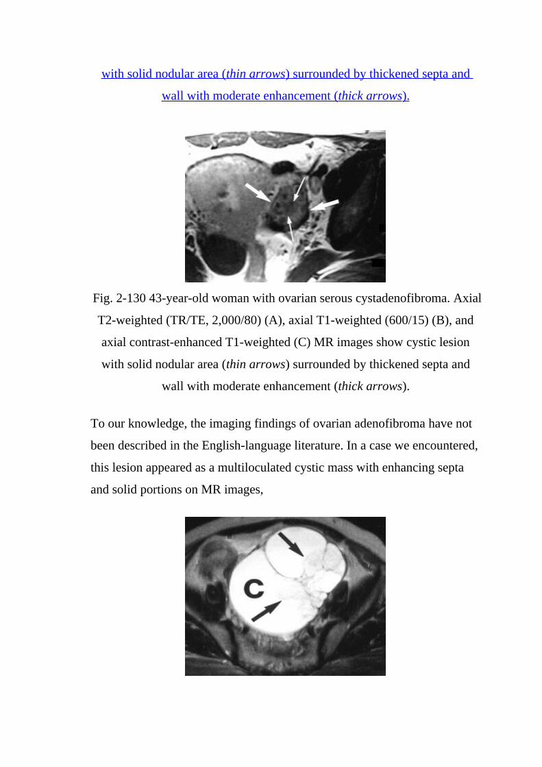
with solid nodular area ( thin arrows ) surrounded by thickened septa and
wall with moderate enhancement ( thick arrows ).
Fig. 2-130 43-year-old woman with ovarian serous cystadenofibroma. Axial
T2-weighted (TR/TE, 2,000/80) (A), axial T1-weighted (600/15) (B), and
axial contrast-enhanced T1-weighted (C) MR images show cystic lesion
with solid nodular area (thin arrows) surrounded by thickened septa and
wall with moderate enhancement (thick arrows).
To our knowledge, the imaging findings of ovarian adenofibroma have not
been described in the English-language literature. In a case we encountered,
this lesion appeared as a multiloculated cystic mass with enhancing septa
and solid portions on MR images,

Fig. 2-131 62-year-old woman with ovarian clear cell adenofibroma. Axial
T2-weighted (TR/TE, 2,000/80) (A), axial T1-weighted (600/15) (B), and
axial contrast-enhanced T1-weighted (C) MR images show large cystic mass
with enhancing multiple septa and solid portions ( arrows ). C = cystic
portions of mass.
Fig. 2-132 62-year-old woman with ovarian clear cell adenofibroma. Axial
T2-weighted (TR/TE, 2,000/80) (A), axial T1-weighted (600/15) (B), and
axial contrast-enhanced T1-weighted (C) MR images show large cystic mass
with enhancing multiple septa and solid portions ( arrows ). C = cystic
portions of mass.
Fig. 2-133 62-year-old woman with ovarian clear cell adenofibroma. Axial

T2-weighted (TR/TE, 2,000/80) (A), axial T1-weighted (600/15) (B), and
axial contrast-enhanced T1-weighted (C) MR images show large cystic mass
with enhancing multiple septa and solid portions (arrows). C = cystic
portions of mass.
Benign transitional cell (Brenner) tumors of the ovary compose
approximately 2% of epithelial ovarian neoplasms. Brenner tumors are often
discovered incidentally at surgery or pathologic examination. Extensive
amorphous calcification in a solid mass or a solid component in a
multilocular cystic mass is a characteristic finding. Low signal intensity on
T2-weighted MR images may result from the abundant fibrous stroma.
(Kimura, K.,1996)
Fig. 2-134 68-year-old woman with Brenner tumor. Radiograph shows
dense calcification ( arrows ) in pelvis.

Fig. 2-135 68-year-old woman with Brenner tumor. CT scan shows solid
pelvic mass ( white arrows ) with calcifications ( black arrows ).
Fig. 2-136 68-year-old woman with Brenner tumor. Axial T2-weighted
(TR/TE, 2,000/80) (C), axial T1-weighted (600/15) (D), and axial contrast-
enhanced T1-weighted (E) MR images show enhancing ovarian solid mass
( arrows ). Low signal intensity on T2-weighted image is due to abundant
fibrous stroma.

Fig 2-137 68-year-old woman with Brenner tumor. Axial T2-weighted
(TR/TE, 2,000/80) (C), axial T1-weighted (600/15) (D), and axial contrast-
enhanced T1-weighted (E) MR images show enhancing ovarian solid mass
( arrows ). Low signal intensity on T2-weighted image is due to abundant
fibrous stroma.
Fig. 2-138 68-year-old woman with Brenner tumor. Axial T2-weighted
(TR/TE, 2,000/80) (C), axial T1-weighted (600/15) (D), and axial
contrast-enhanced T1-weighted (E) MR images show enhancing
ovarian solid mass ( arrows ). Low signal intensity on T2-weighted
image is due to abundant fibrous stroma.

2-3 Feamle infertility.
Female infertility refers to infertility in female humans. It affects an
estimated 48 million women with the highest prevalence of infertility
affecting people in South Asia, Sub-Saharan Africa, North
Africa/Middle East, and Central/Eastern Europe and Central Asia.
Infertility is caused by many sources, including nutrition, diseases, and
other malformations of the uterus. Infertility affects women from
around the world, and the cultural and social stigma surrounding it
varies.Fertility awareness is a type of natural family planning based on
a woman's physical signs of ovulation, such as changes in cervical
mucus consistency and basal (resting) body temperature. Keeping track
of these symptoms may be helpful if a couple is trying to become
pregnant or trying to avoid pregnancy.
If a woman is trying to become pregnant, having sex during the 5 days
before she ovulates and the day she ovulates increases her chances of
success.If a woman is trying to avoid pregnancy, avoiding sex until
several days after she has ovulated helps prevent pregnancy (the human
egg is typically fertile for only 12 to 24 hours after ovulation).Because
intercourse before ovulation can lead to pregnancy, and ovulation does
not always occur at the same time every month, fertility awareness is
not the most reliable method of avoiding pregnancy.
2-3-1 Definition
There is no unanimous definition of female infertility, because the
definition depends on social and physical characteristics which may
vary by culture and situation. NICE guidelines state that: "A woman of
reproductive age who has not conceived after 1 year of unprotected

vaginal sexual intercourse, in the absence of any known cause of
infertility, should be offered further clinical assessment and
investigation along with her partner. It is recommended that a
consultation with a fertility specialist should be made earlier if the
woman is aged 36 years or over, or there is a known clinical cause of
infertility or a history of predisposing factors for infertility. According
to the World Health Organization (WHO), infertility can be described
as the inability to become pregnant, maintain a pregnancy, or carry a
pregnancy to live birth. A clinical definition of infertility by the WHO
and ICMART is “a disease of the reproductive system defined by the
failure to achieve a clinical pregnancy after 12 months or more of
regular unprotected sexual intercourse. Infertility can further be broken
down into primary and secondary infertility. Primary infertility refers to
the inability to give birth either because of not being able to become
pregnant, or carry a child to live birth, which may include miscarriage
or a stillborn child. [5][6] Secondary infertility refers to the inability to
conceive or give birth when there was a previous pregnancy or live
birth.
2-3-1-1 Prevalence
Female infertility varies widely by geographic location around the
world. In 2010, there was an estimated 48.5 million infertile couples
worldwide, and from 1990 to 2010 there was little change in levels of
infertility in most of the world. In 2010, the countries with the lowest
rates of female infertility included the South American countries of
Peru, Ecuador and Bolivia, as well as in Poland, Kenya, and Republic
of Korea. The highest rate regions included Eastern Europe, North
Africa, the Middle East, Oceania, and Sub-Saharan Africa. The

prevalence of primary infertility has increased since 1990, but
secondary infertility has decreased overall. Rates decreased (although
not prevalence) of female infertility in high-income, Central/Eastern
Europe, and Central Asia regions.Sub-Saharan Africa has had
decreasing levels of primary infertility from 1990 to 2010. Within the
Sub-Saharan region, rates were lowest in Kenya, Zimbabwe, and
Rwanda, while the highest rates were in Guinea, Mozambique, Angola,
Gabon, and Cameroon along with Northern Africa near the Middle
East. According to a 2004 DHS report, rates in Africa were highest in
Middle and Sub-Saharan Africa, with East Africa’s rates close behind.
In Asia, the highest rates of combined secondary and primary infertility
was in the South Central region, and then in the Southeast region, with
the lowest rates in the Western areas. The prevalence of female
infertility in the Latin America/Caribbean region is typically lower than
the global prevalence. However, the greatest rates occurred in Jamaica,
Suriname, Haiti, and Trinidad and Tobago. Central and Western Latin
America has some of the lowest rates of prevalence. The highest
regions in Latin America and the Caribbean was in the Caribbean
Islands and in less developed countries.
2-3-2 Causes and factors
Causes or factors of female infertility can basically be classified
regarding whether they are acquired or genetic, or strictly by
location.Although factors of female infertility can be classified as either
acquired or genetic, female infertility is usually more or less a
combination of nature and nurture. Also, the presence of any single risk
factor of female infertility (such as smoking, mentioned further below)

does not necessarily cause infertility, and even if a woman is definitely
infertile, the infertility cannot definitely be blamed on any single risk
factor even if the risk factor is present.
2-3-2-1 Acquired
According to the American Society for Reproductive Medicine
(ASRM), Age, Smoking, Sexually Transmitted Infections, and Being
Overweight or Underweight can all affect fertility. In broad sense,
acquired factors practically include any factor that is not based on a
genetic mutation, including any intrauterine exposure to toxins during
fetal development, which may present as infertility many years later as
an adult.
2-3-2-2 Age and female fertility
A woman's fertility is affected by her age. The average age of a girl's
first period (menarche) is 12-13 (12.5 years in the United States, 12.72
in Canada, 12.9 in the UK), but, in postmenarchal girls, about 80% of
the cycles are anovulatory in the first year after menarche, 50% in the
third and 10% in the sixth year. A woman's fertility peaks in the early
and mid 20s, after which it starts to decline, with this decline being
accelerated after age 35. However, the exact estimates of the chances of
a woman to conceive after a certain age are not clear, with research
giving differing results. The chances of a couple to successfully
conceive at an advanced age depend on many factors, including the
general health of a woman and the fertility of the male partner.
2-3-2-3 Tobacco smoking
See also: Women and smoking § Unique gender differences and health

effects for Females. Tobacco smoking is harmful to the
ovaries, and the degree of damage is dependent upon
the amount and length of time a woman smokes or is
exposed to a smoke-filled environment. Nicotine and
other harmful chemicals in cigarettes interfere with the
body’s ability to create estrogen, a hormone that
regulates folliculogenesis and ovulation. Also, cigarette
smoking interferes with folliculogenesis, embryo
transport, endometrial receptivity, endometrial
angiogenesis, uterine blood flow and the uterine
myometrium. Some damage is irreversible, but stopping
smoking can prevent further damage. Smokers are 60%
more likely to be infertile than non-smokers. Smoking
reduces the chances of IVF producing a live birth by 34%
and increases the risk of an IVF pregnancy miscarrying
by 30%. Also, female smokers have an earlier onset of
menopause by approximately 1–4 years.
2-3-2-4 Sexually transmitted infections
Sexually transmitted infections are a leading cause of infertility. They
often display few, if any visible symptoms, with the risk of failing to
seek proper treatment in time to prevent decreased fertility.
2-3-2-5 Body weight and eating disorders
Twelve percent of all infertility cases are a result of a woman either
being underweight or overweight. Fat cells produce estrogen, in
addition to the primary sex organs. Too much body fat causes
production of too much estrogen and the body begins to react as if it is

on birth control, limiting the odds of getting pregnant. Too little body
fat causes insufficient production of estrogen and disruption of the
menstrual cycle. Both under and overweight women have irregular
cycles in which ovulation does not occur or is inadequate. Proper
nutrition in early life is also a major factor for later fertility. A study in
the US indicated that approximately 20% of infertile women had a past
or current eating disorder, which is five times higher than the general
lifetime prevalence rate. A review from 2010 concluded that
overweight and obese subfertile women have a reduced probability of
successful fertility treatment and their pregnancies are associated with
more complications and higher costs. In hypothetical groups of 1000
women undergoing fertility care, the study counted approximately 800
live births for normal weight and 690 live births for overweight and
obese anovulatory women. For ovulatory women, the study counted
approximately 700 live births for normal weight, 550 live births for
overweight and 530 live births for obese women. The increase in cost
per live birth in anovulatory overweight and obese women were,
respectively, 54 and 100% higher than their normal weight
counterparts, for ovulatory women they were 44 and 70% higher,
respectively.
2-3-2-6 Chemotherapy
Chemotherapy poses a high risk of infertility. Chemotherapies with
high risk of infertility include procarbazine and other alkylating drugs
such as cyclophosphamide, ifosfamide, busulfan, melphalan,
chlorambucil and chlormethine. Drugs with medium risk include
doxorubicin and platinum analogs such as cisplatin and carboplatin. On
the other hand, therapies with low risk of gonadotoxicity include plant
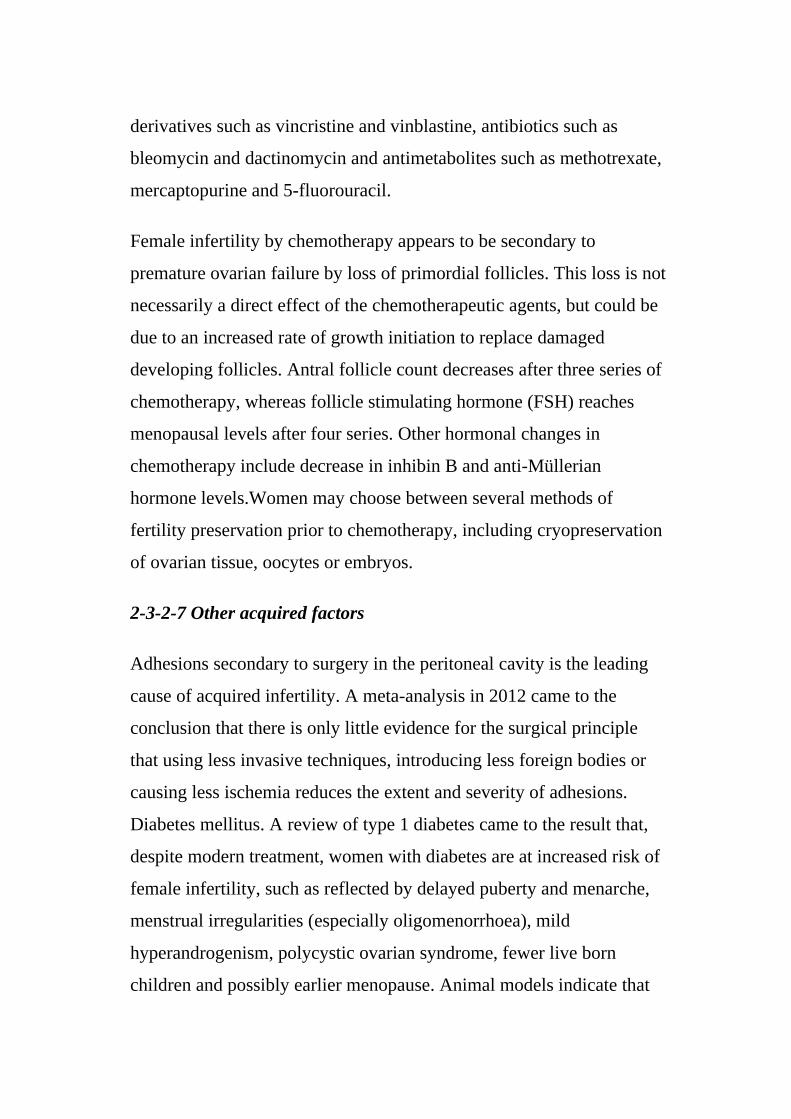
derivatives such as vincristine and vinblastine, antibiotics such as
bleomycin and dactinomycin and antimetabolites such as methotrexate,
mercaptopurine and 5-fluorouracil.
Female infertility by chemotherapy appears to be secondary to
premature ovarian failure by loss of primordial follicles. This loss is not
necessarily a direct effect of the chemotherapeutic agents, but could be
due to an increased rate of growth initiation to replace damaged
developing follicles. Antral follicle count decreases after three series of
chemotherapy, whereas follicle stimulating hormone (FSH) reaches
menopausal levels after four series. Other hormonal changes in
chemotherapy include decrease in inhibin B and anti-Müllerian
hormone levels.Women may choose between several methods of
fertility preservation prior to chemotherapy, including cryopreservation
of ovarian tissue, oocytes or embryos.
2-3-2-7 Other acquired factors
Adhesions secondary to surgery in the peritoneal cavity is the leading
cause of acquired infertility. A meta-analysis in 2012 came to the
conclusion that there is only little evidence for the surgical principle
that using less invasive techniques, introducing less foreign bodies or
causing less ischemia reduces the extent and severity of adhesions.
Diabetes mellitus. A review of type 1 diabetes came to the result that,
despite modern treatment, women with diabetes are at increased risk of
female infertility, such as reflected by delayed puberty and menarche,
menstrual irregularities (especially oligomenorrhoea), mild
hyperandrogenism, polycystic ovarian syndrome, fewer live born
children and possibly earlier menopause. Animal models indicate that

abnormalities on the molecular level caused by diabetes include
defective leptin, insulin and kisspeptin signalling. Significant liver or
kidney disease. Thrombophilia. Cannabis smoking, such as of
marijuana causes disturbances in the endocannabinoid system,
potentially causing infertility. Radiation, such as in radiation therapy.
The radiation dose to the ovaries that generally causes permanent
female infertility is 20.3 Gy at birth, 18.4 Gy at 10 years, 16.5 Gy at 20
years and 14.3 Gy at 30 years.[30] After total body irradiation, recovery
of gonadal function occurs in 10−14% of cases, and the number of
pregnancies observed after hematopoietic stem cell transplantation
involving such as procedure is lower than 2%.
2-3-2-8 Genetic factors
There are many genes wherein mutation causes female infertility, as
shown in table below. Also, there are additional conditions involving
female infertility which are believed to be genetic but where no single
gene has been found to be responsible, notably Mayer-Rokitansky-
Küstner-Hauser Syndrome (MRKH). Finally, an unknown number of
genetic mutations cause a state of subfertility, which in addition to other
factors such as environmental ones may manifest as frank
infertility.Chromosomal abnormalities causing female infertility include
Turner syndrome.Some of these gene or chromosome abnormalities
cause intersexed conditions, such as androgen insensitivity syndrome.
Genes wherein mutation causes female infertility
Gene Encoded protein Effect of deficiency
BMP15Bone morphogenetic
protein 15
Hypergonadotrophic ovarian
failure (POF4)

BMPR1BBone morphogenetic
protein receptor 1B
Ovarian dysfunction,
hypergonadotrophic
hypogonadism and
acromesomelic
chondrodysplasia
CBX2;
M33
Chromobox protein
homolog 2 ; Drosophila
polycomb class
Autosomal 46,XY, male-to-
female sex reversal
(phenotypically perfect
females)
CHD7Chromodomain-helicase-
DNA-binding protein 7
CHARGE syndrome and
Kallmann syndrome (KAL5)
DIAPH2 Diaphanous homolog 2
Hypergonadotrophic,
premature ovarian failure
(POF2A)
FGF8 Fibroblast growth factor 8
Normosmic
hypogonadotrophic
hypogonadism and Kallmann
syndrome (KAL6)
FGFR1Fibroblast growth factor
receptor 1Kallmann syndrome (KAL2)
HFM1 Primary ovarian failure[
FSHR FSH receptor
Hypergonadotrophic
hypogonadism and ovarian
hyperstimulation syndrome
FSHB Follitropin subunit beta
Deficiency of follicle-
stimulating hormone, primary
amenorrhoea and infertility

FOXL2 Forkhead box L2
Isolated premature ovarian
failure (POF3) associated with
BPES type I; FOXL2
402C --> G mutations
associated with human
granulosa cell tumours
FMR1 Fragile X mental retardation
Premature ovarian failure
(POF1) associated with
premutations
GNRH1Gonadotropin releasing
hormone
Normosmic
hypogonadotrophic
hypogonadism
GNRHR GnRH receptorHypogonadotrophic
hypogonadism
KAL1 Kallmann syndrome
Hypogonadotrophic
hypogonadism and insomnia,
X-linked Kallmann syndrome
(KAL1)
KISS1R ;
GPR54KISS1 receptor
Hypogonadotrophic
hypogonadism
LHBLuteinizing hormone beta
polypeptide
Hypogonadism and
pseudohermaphroditism
LHCGRLH/choriogonadotrophin
receptor
Hypergonadotrophic
hypogonadism (luteinizing
hormone resistance)
DAX1 Dosage-sensitive sex
reversal, adrenal hypoplasia
X-linked congenital adrenal
hypoplasia with
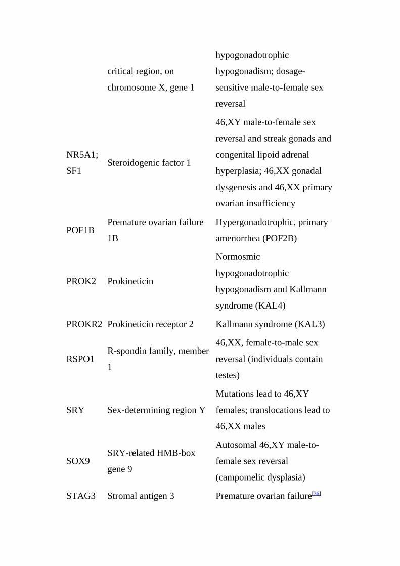
critical region, on
chromosome X, gene 1
hypogonadotrophic
hypogonadism; dosage-
sensitive male-to-female sex
reversal
NR5A1;
SF1Steroidogenic factor 1
46,XY male-to-female sex
reversal and streak gonads and
congenital lipoid adrenal
hyperplasia; 46,XX gonadal
dysgenesis and 46,XX primary
ovarian insufficiency
POF1BPremature ovarian failure
1B
Hypergonadotrophic, primary
amenorrhea (POF2B)
PROK2 Prokineticin
Normosmic
hypogonadotrophic
hypogonadism and Kallmann
syndrome (KAL4)
PROKR2 Prokineticin receptor 2 Kallmann syndrome (KAL3)
RSPO1R-spondin family, member
1
46,XX, female-to-male sex
reversal (individuals contain
testes)
SRY Sex-determining region Y
Mutations lead to 46,XY
females; translocations lead to
46,XX males
SOX9SRY-related HMB-box
gene 9
Autosomal 46,XY male-to-
female sex reversal
(campomelic dysplasia)
STAG3 Stromal antigen 3 Premature ovarian failure[36]

TAC3 Tachykinin 3
Normosmic
hypogonadotrophic
hypogonadism
TACR3 Tachykinin receptor 3
Normosmic
hypogonadotrophic
hypogonadism
ZP1zona pellucida glycoprotein
1
Dysfunctional zona pellucida
formation[37]
By location
2-3-2-9 Hypothalamic-pituitary factors.
Hypothalamic dysfunction and hyperprolactinemia.
2-3-2-10 Ovarian factors
Chemotherapy (as detailed previously) with certain agents have a high
risk of toxicity on the ovaries , many genetic defects (as also detailed
previously) also disturb ovarian function , polycystic ovary syndrome
(also see infertility in polycystic ovary syndrome) , anovulation. Female
infertility caused by anovulation is called "anovulatory infertility", as
opposed to "ovulatory infertility" in which ovulation is present ,
diminished ovarian reserve, also see Poor Ovarian Reserve , premature
menopause , menopause , luteal dysfunction , gonadal dysgenesis and
ovarian cancer.
2-3-2-11 Tubal (ectopic)/peritoneal factors
Endometriosis (also see endometriosis and infertility) , pelvic adhesions
, Pelvic inflammatory disease (PID, usually due to chlamydia) , tubal

occlusion , tubal dysfunction and Previous ectopic pregnancy. A
randomized study in 2013 came to the result that the rates of
intrauterine pregnancy 2 years after treatment of ectopic pregnancy are
approximately 64% with radical surgery, 67% with medication, and
70% with conservative surgery.[43] In comparison, the cumulative
pregnancy rate of women under 40 years of age in the general
population over 2 years is over 90%.[2]
2-3-2-12 Uterine factors
Uterine malformations , Uterine fibroids , asherman's Syndrome ,
tmplantation failure without any known primary cause. It results in
negative pregnancy test despite having performed e.g. embryo transfer
and previously, a bicornuate uterus was thought to be
associated with infertility but recent studies have not
confirmed such an association.
2-3-2-13 Cervical factors
Cervical stenosis , antisperm antibodies and non-receptive cervical
mucus.
2-3-2-14 Vaginal factors
Vaginismus and vaginal obstruction.
2-3-3 Diagnosis
Diagnosis of infertility begins with a medical history and physical
exam. The healthcare provider may order tests, including the
following:Lab tests:- hormone testing, to measure levels of female

hormones at certain times during a menstrual cycle , day 2 or 3 measure
of FSH and estrogen, to assess ovarian reserve , measurements of
thyroid function (a thyroid stimulating hormone (TSH) level of between
1 and 2 is considered optimal for conception) , measurement of
progesterone in the second half of the cycle to help confirm ovulation
and anti-Müllerian hormone to estimate ovarian reserve.
2.3.4 Examination and imaging
An endometrial biopsy, to verify ovulation and inspect the lining of the
uterus , laparoscopy, which allows the provider to inspect the pelvic
organs , fertiloscopy, a relatively new surgical technique used for early
diagnosis (and immediate treatment) , Pap smear, to check for signs of
infection , pelvic exam, to look for abnormalities or infection , a
postcoital test, which is done soon after intercourse to check for
problems with sperm surviving in cervical mucous (not commonly used
now because of test unreliability) , hysterosalpingography or
sonosalpingography, to check for tube patency and Sonohysterography
to check for uterine abnormalities.There are genetic testing
techniques under development to detect any mutation in
genes associated with female infertility.[34]
Initial diagnosis and treatment of infertility is usually made by
obstetrician/gynecologists or women's health nurse practitioners. If
initial treatments are unsuccessful, referral is usually made to
physicians who are fellowship trained as reproductive endocrinologists.
Reproductive endocrinologists are usually obstetrician/gynecologists
with advanced training in reproductive endocrinology and infertility (in
North America). These physicians treat reproductive disorders affecting

not only women but also men, children, and teens.
Usually reproductive endocrinology & infertility medical practices do
not see women for general maternity care. The practice is primarily
focused on helping their women to conceive and to correct any issues
related to recurring pregnancy loss.
2-3-5 Prevention
Acquired female infertility may be prevented through identified
interventions:Maintaining a healthy lifestyle. Excessive exercise,
consumption of caffeine and alcohol, and smoking have all been
associated with decreased fertility. Eating a well-balanced, nutritious
diet, with plenty of fresh fruits and vegetables, and maintaining a
normal weight, on the other hand, have been associated with better
fertility prospects.
Treating or preventing existing diseases. Identifying and controlling
chronic diseases such as diabetes and hypothyroidism increases fertility
prospects. Lifelong practice of safer sex reduces the likelihood that
sexually transmitted diseases will impair fertility; obtaining prompt
treatment for sexually transmitted diseases reduces the likelihood that
such infections will do significant damage. Regular physical
examinations (including pap smears) help detect early signs of
infections or abnormalities.
Not delaying parenthood. Fertility does not ultimately cease before
menopause, but it starts declining after age 27 and drops at a somewhat
greater rate after age 35. Women whose biological mothers had unusual
or abnormal issues related to conceiving may be at particular risk for

some conditions, such as premature menopause, that can be mitigated
by not delaying parenthood.
Egg freezing. A woman can freeze her eggs preserve her fertility. By
using egg freezing while in the peak reproductive years, a woman's
oocytes are cryogenicaly frozen and ready for her use later in life,
reducing her chances of female infertility.
2-3-6 Social stigma
Social stigma due to infertility is seen in many cultures throughout the
world in varying forms. Often, when women cannot conceive, the
blame is put on them, even when approximately 50% of infertility
issues come from the man . In addition, many societies only tend to
value a woman if she is able to produce at least one child, and a
marriage can be considered a failure when the couple cannot conceive.
The act of conceiving a child can be linked to the couple’s
consummation of marriage, and reflect their social role in society. This
is seen in the "African infertility belt", where infertility is prevalent in
Africa which includes countries spanning from Tanzania in the east to
Gabon in the west In this region, infertility is highly stigmatized and
can be considered a failure of the couple to their societies. This is
demonstrated in Uganda and Nigeria where there is a great pressure put
on childbearing and its social implications. This is also seen in some
Middle Eastern societies including Egypt and Pakistan.
Wealth is sometimes measured by the number of children a woman has,
as well as inheritance of property. Children can influence financial
security in many ways. In Nigeria and Cameroon, land claims are
decided by the number of children. Also, in some Sub-Saharan

countries women may be neglected inheritance if she did not bear and
children In some African and Asian countries a husband can neglect
his infertile wife of food, shelter and other basic necessities like
clothing. In Cameroon, a woman may lose access to land from her
husband and left on her own in old age.
In many cases, a woman who cannot bear children is excluded from
social and cultural events including traditional ceremonies. This
stigmatization is seen in Mozambique and Nigeria where infertile
women have been treated as outcasts to society. This is a humiliating
practice which devalues infertile women in society. In the Macua
tradition, pregnancy and birth are considered major life events for a
woman, with the ceremonies of nthaa´ra and ntha´ara no mwana, which
can only be attended by women who have been pregnant and have had a
baby.
The effect of infertility can lead to social shaming from internal and
social norms surrounding pregnancy, which affects women around the
world. When pregnancy is considered such an important event in life,
and considered a “socially unacceptable condition”, it can lead to a
search for treatment in the form of traditional healers and expensive
Western treatments. The limited access to treatment in many areas can
lead to extreme and sometimes illegal acts in order to produce a child.
2-3-7 Marital role
Men in some countries may find another wife when their first cannot
produce a child, hoping that by sleeping with more women he will be
able to produce his own child. This can be prevalent in some societies,
including Cameroon, Nigeria, Mozambique, Egypt, Botswana, and

Bangladesh, among many more where polygamy is more common and
more socially acceptable.In some cultures, including Botswana and
Nigeria, women can select a woman with whom she allows her husband
to sleep with in hopes of conceiving a child. Women who are desperate
for children may compromise with her husband to select a woman and
accept duties of taking care of the children to feel accepted and useful
in society.
Women may also sleep with other men in hopes of becoming pregnant.
This can be done for many reasons including advice from a traditional
healer, or finding if another man was "more compatible". In many
cases, the husband was not aware of the extra sexual relations and
would not be informed if a woman became pregnant by another man.
This is not as culturally acceptable however, and can contribute to the
gendered suffering of women who have fewer options to become
pregnant on their own as opposed to men.
Men and women can also turn to divorce in attempt to find a new
partner with whom to bear a child. Infertility in many cultures is a
reason for divorce, and a way for a man or woman to increase his/her
chances of producing an heir. When a woman is divorced, she can lose
her security that often comes with land, wealth, and a family. This can
ruin marriages and can lead to distrust in the marriage. The increase of
sexual partners can potentially result with the spread of disease
including HIV/AIDS, and can actually contribute to future generations
of infertility.
2-3-8 Domestic abuse
The emotional strain and stress that comes with infertility in the

household can lead to the mistreatment and domestic abuse of a
woman. The devaluation of a wife due to her inability to conceive can
lead to domestic abuse and emotional trauma such as victim blaming.
Women are sometimes or often blamed as the cause of a couples'
infertility, which can lead to emotional abuse, anxiety, and shame. In
addition, blame for not being able to conceive is often put on the
female, even if it is the man who is infertile. Women who are not able
to conceive can be starved, beaten, and may be neglected financially by
her husband as if she had no child bearing use to him. The physical
abuse related to infertility may result from this and the emotional stress
that comes with it. In some countries, the emotional and physical
abuses that come with infertility can potentially lead to assault, murder,
and suicide.[63]
2-3-9 Mental and psychological impact
Many infertile women tend to cope with immense stress and social
stigma behind their condition, which can lead to considerable mental
distress The long-term stress involved in attempting to conceive a child
and the social pressures behind giving birth can lead to emotional
distress that may manifest as mental disease. Women who suffer from
infertility might deal with psychological stressors such as denial, anger,
grief, guilt, and depression. There can be considerable social shaming
that can lead to intense feelings of sadness and frustration that
potentially contribute to depression and suicide. (Legendre G,2012)

Chapter three
Methodology

Chapter threeMethodology
3-1 Material
The data of this study collected by Aloka SSD 500 with convex 3.5 MHRz and
endovaginal probs.
3-2 Population of the study
This study consisted of female complaining from primary infertility with
different clinical symptoms and manifestation, visited the ultrasound
clinic for examination.
3-3 Sample and size of the study
The data of this study collected from 300 patients selected conveniently
3-4 Design of the study
This study is a corss-sectional, descriptive study
3-5 Method of data collection
3-5-1 Techniques
A pelvic ultrasound is a noninvasive diagnostic exam that produces images
that are used to assess organs and structures within the female pelvis. A
pelvic ultrasound allows quick visualization of the female pelvic organs and
structures including the uterus, cervix, vagina, fallopian tubes and ovaries.
Ultrasound uses a transducer that sends out ultrasound waves at a frequency
too high to be heard. The ultrasound transducer is placed on the skin, and the
ultrasound waves move through the body to the organs and structures within.
The sound waves bounce off the organs like an echo and return to the
transducer. The transducer processes the reflected waves, which are then
converted by a computer into an image of the organs or tissues being
examined.The sound waves travel at different speeds depending on the type
of tissue encountered - fastest through bone tissue and slowest through air.
The speed at which the sound waves are returned to the transducer, as well
as how much of the sound wave returns, is translated by the transducer as
different types of tissue.
An ultrasound gel is placed on the transducer and the skin to allow for
smooth movement of the transducer over the skin and to eliminate air
between the skin and the transducer for the best sound conduction.
Transvaginal ultrasound is a test used to look at a woman's reproductive
organs, including the uterus, ovaries, and cervix.
Transvaginal means across or through the vagina. The ultrasound probe will
be placed inside the vagina.You will lie down on a table with your knees
bent. Your feet may be held in stirrups. You will be given a probe, called a
transducer, to place into the vagina. The probe is covered with a condom and
a gel. The probe sends out sound waves, which reflect off body structures. A
computer receives these waves and uses them to create a picture.
The ultrasound technician or doctor can see the picture on a TV monitor.The
health care provider will move the probe around the area to see the pelvic
organs.In some cases, a special transvaginal ultrasound method called saline
infusion sonography (SIS) may be needed to more clearly view the uterus.
You will be asked to undress, usually from the waist down. A transvaginal
ultrasound is done with your bladder empty or partly filled.

The test is usually painless, although some women may have mild
discomfort from the pressure of the probe. Only a small part of the probe is
placed into the vagina.Transvaginal ultrasound may be done for the
following problems: Abnormal findings on a physical exam, such as cysts,
fibroid tumors, or other growths , Abnormal vaginal bleeding and menstrual
problems , Certain types of infertility , Ectopic pregnancy , Pelvic pain and
Transvaginal ultrasound is also used during pregnancy.
The pelvic structures or fetus is normal. An abnormal result may be due to
many conditions. Some problems that may be seen include: Birth defects,
Cancers of the uterus, ovaries, vagina, and other pelvic structures, Infection,
including pelvic inflammatory disease , Growths in or around the uterus
and ovaries (such as cysts or fibroids) and Twisting of the ovaries.There are
no known harmful effects of transvaginal ultrasound on humans.Unlike
traditional x-rays, there is no radiation exposure with this test.
A special type of transvaginal ultrasound is called a saline infusion
sonography (SIS). This procedure involves inserting sterile salt water into
the uterus before the ultrasound to help identify any possible masses. The
saline solution stretches the uterus slightly, providing a more detailed picture
of the inside of the uterus than a conventional ultrasound.
3-6 Protocols
The following points should be considered for all ultrasound examinations:-
the clinical details provided are sufficient to carry out the examination

requested and the correct examination has been requested , relevant
information is available from the case notes, previous investigations and
other sources , the role of the ultrasound examination is understood in the
clinical context for the patient , informed consent is obtained before
proceeding with the examination , the necessity for the presence of a
chaperone and/or an interpreter , a systematic scanning approach that can be
modified according to the individual patient , the implications should the
examination be incomplete , the need to extend the ultrasound examination,
and/or proceed to additional imaging techniques .where necessary in
accordance with locally agreed protocol , the after care of the patient , the
potential risks involved in the procedure to the patient and appropriate
national and local Health and Safety regulations including infection control.
The sonographer should:- Recognise his/her scope of practice and work
within its boundaries ensure that a locally agreed written scheme of work is
in place accept properly delegated responsibility, in accordance with local
practice and guidelines An ultrasound examination should not be carried out
unless a valid request has been received. The request should include such
clinical details as are relevant to the examination, clear identification of the
person requesting the examination and to whom the report should be
directed.
The sonographer should be responsive to:- potential bio-effects of ultrasound
and the need to minimise dose at all times , potential hazards arising from
the particular ultrasound equipment , relative risks for each application ,
conditions where current recommendations contra-indicate the use of certain
types of ultrasound equipment and current guidelines regarding replacement
of ultrasound equipment.
The sonographer is expected to:- have detailed knowledge of ultrasound
equipment in order to ensure that it is appropriate for purpose , manipulate
the equipment correctly so that patient diagnosis and management are not
compromised , ensure that an agreed quality assurance programme is in
place that incorporates the regular and inspection of ultrasound machines
and auxiliary equipment.
The stated aim of quality assurance procedures applied to ultrasound
equipment is to ensure consistent and acceptable levels of performance of
the imaging system and image recording facilities. Most quality assurance
protocols focus on the consistency. The acceptability of image quality may
not be apparent from measurable changes in the parameters tested. The issue
of what constitutes unacceptable equipment performance is still very
difficult to assess objectively. In the absence of nationally accepted
performance standards for ultrasound equipment, local and subjective
evaluation is required.
We usually get better images during transabdominal ultrasound if the
bladder is partially filled, so to help your examination we ask you to drink
water prior to the assessment. Please empty your bladder 1 hour before your
appointment, drink 2 glasses of water and try not to empty your bladder
again until after your appointment. A full bladder moves bowel out from the
pelvis into the abdomen, helping visualisation of the uterus and ovaries.
The bladder should not be so full that it causes pain. If your bladder is very
full and painful, you should empty a small amount so you are more
comfortable.You will be able to empty your bladder after the transabdominal
ultrasound is completed and before the transvaginal ultrasound begins.
A pelvic ultrasound can be performed at any time during the menstrual
cycle. Your referring doctor will let you know if it is more appropriate to

have your pelvic ultrasound at a particular time during your cycle. Most
pelvic conditions can be adequately assessed at any time of the menstrual
cycle.
The best time to have your pelvic scan is usually just after your period has
finished. The timing of your scan may be more important if your doctor is
concerned about an endometrial problem such as endometrial polyps
(growths of the endometrium). The endometrial lining becomes thin just
after the period has ceased, making polyps easier to view.
You will be asked to remove your clothes and put on a gown or cover for the
procedure. Depending on your doctor’s instructions and the reasons for the
ultrasound, you might need to either have your bladder empty or partially
filled. When your bladder is full, it actually lifts the intestines away and
allows for a clearer picture of your pelvic organs. You can have this
procedure performed during your period or if you’re spotting, but if you are
wearing a tampon, you will have to take it out before the ultrasound.
3-7 Data analysis:-
The collected data will be analyzed through statistical procedures, which
include quantitative and descriptive analysis such as percentage, mean and
standard deviation to obtain the results of this study.

Chapter fourResults
Chapter fourResults

The main objective of this study was to investigate the potential of
endovaginal probe in exploring the factors behind the primary infertility in
Sudanese females. The study consisted of 300 female suffers from primary
infertility visits the ultrasound clinic for diagnosis purposes.
Diagnosis Frequency
Polycystic ovarian syndrome 122
Normal ovary 149
Follicle size 17
Cyst 12
total 300Table 4-1 a frequency distribution table, shows the distribution of ultrasound
diagnosis of ovaries using Transvaginal probe
Table 4-1 a bar plot, shows the % distribution of ovaries diagnosis by
ultrasound using Transvaginal probe
Remarks of infertility FrequencyFibroid 40Male factor 83Tube blocked 26
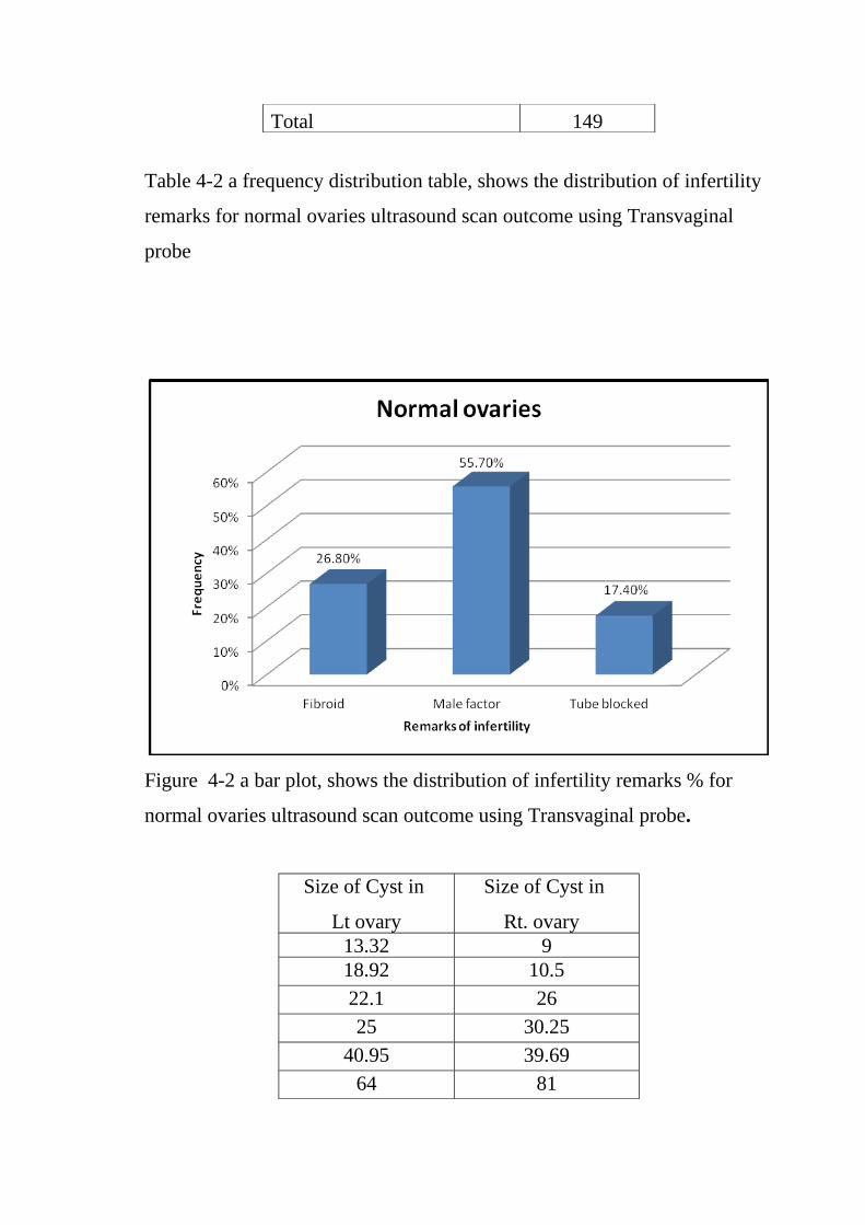
Total 149
Table 4-2 a frequency distribution table, shows the distribution of infertility
remarks for normal ovaries ultrasound scan outcome using Transvaginal
probe
Figure 4-2 a bar plot, shows the distribution of infertility remarks % for
normal ovaries ultrasound scan outcome using Transvaginal probe.
Size of Cyst in
Lt ovary
Size of Cyst in
Rt. ovary 13.32 918.92 10.5
22.1 26
25 30.25
40.95 39.69
64 81

Table 4-3 the sizes of the diagnosed cyst in the Rt or Lt ovary
Figure 4-3 a bar plot show the sizes distribution of cyst in the Rt or Lt ovary
diagnosed using transvaginal probe
Follicle size (cm) Frequency
13-14 1
15-16 4
16-17 5
18-19 6Table 4-4 a frequency distribution table, shows the distribution of follicles
sizes for female in induction treatment using Transvaginal probe

Figure 4-4 a bar plot shows % distribution of follicles sizes for female in
induction treatment using Transvaginal probe
Figure 4-5 scatter plot shows the relationship between the female age and
the size of the follicles in the induction treatment, follicle size decreases by
0.044 mm/year of age.

Figure 4-6 scatter plot shows the relationship between the female age and
the size of the ovarian cyst, cyst size increases by 0.21 cm/year of age.
Figure 4-7 scatter plot shows the relationship between the female age and
marriage duration in female with polycystic ovarian syndrome, marriage
duration increases by 1.5 month/year of age.

Figure 4-8 scatter plot shows the relationship between the female age and
marriage duration in female with normal ovaries and other remarks of
infertility, marriage duration increases by 2.2 month/year of age.
Figure 4-9 scatter plot shows the relationship between the female age and
marriage duration in female with normal ovaries and under induction
treatment for infertility, marriage duration increases by 5.8 month/year of
age.

Figure 4-10 scatter plot shows the relationship between the female age and
marriage duration in female with ovarian cyst, marriage duration increases
by 5.6 month/year of age.
Figure 4-11 a bar blot shows the distribution of female infertility problems
in respect average female age and marriage duration. With the lowest
average age and marriage duration recorded by polycystic disease and
relatively highest average female age and marriage duration scored by group
with normal ovaries with others infertility factors.

Chpater fiveDiscussion, conclusion and recommendation

Chpater fiveDiscussion, conclusion and recommendation
5-1 Discussion
The main objective of this study was to investigate the potential of
endovaginal probe in exploring the factors behind the primary infertility in
Sudanese females. The study consisted of 300 female suffers from primary
infertility visits the ultrasound clinic for diagnosis purposes.
The mean age of the female enrolled in this study was 31.4±7.2 years which
is range from 18 to 50 years old. Their mean marriage duration period was
55.2±45 month, which is ranged from 12 to 300 month. The diagnosis of
these cases using endovaginal probe reveals that 122 (40.7%) females
suffering from polycystic ovarian syndrome, 149 (49.7%) had normal
ovaries but with other infertility remarks, 12 (4%) had ovarian cyst and 17
(5.7%) had no obvious reasons fro infertility and they are under stimulation
(induction) treatment of ovulation (Table and Figure 4-1). This result

implicate that the most obvious reasons for infertility were might not
attributed to ovaries problems directly, although direct ovaries abnormality
has mostly 40% of the underlying problem which represented mostly by
polycystic ovarian syndrome and in small scale by ovarian cyst.
The presence of polycystic ovarian syndrome can be mostly observed
clinically and confirmed by ultrasound or laboratory test. But other causes
which is attributed to other factors than ovaries makes the ultrasound one of
the potential tool for exploring the causes of primary infertility. The results
of this study as mentioned earlier emphasizes that almost 50% of primary
infertility attributed to other factors than ovarian problem (i.e. normal
ovaries). This study concerning the category of normal ovaries found that in
83 cases (55.7%) out of 149 the causes of infertility were male problem and
in 26.8% were attributed to the presence of fibroid and in 17.4% of this
category attributed to blocked tubes. This result dictate that, investigation of
infertility problems should start by excluding ovarian problem, then both
partner should be enrolled in the investigation after that since after the
exclusion of ovarian problem the causes might be anywhere including the
male partner (Figure and Table (4-2).
The presence of ovarian cysts were unilaterally 6 in each ovary fro 12
females with average size of 30.7 and 32.7 cm for the Lt and Rt ovary
respectively with minor increases of the ovarian cyst in the Rt one (Figure
and Table 4-3). Also as mentioned before there is 17 female in induction
treatment their follicular size ranged 14 to 18 mm (Figure and Table 4-4).
The size of the follicles indicates the success of induction treatment and this
size taken for several days for fertilization reasons. In this study it has been
noticed that the size of the follicle regardless the day of scanning inversely
proportion with the female age where the size of the follicle decreases by

0.044 mm/year. This means younger females will have optimum follicle size
in a shorter period than older in respect to the decreases coefficient
mentioned earlier (Figure 4-5). Also it has been found that the ovarian cyst
in respect to female age had a direct relationship i.e. as the age of the female
increase the cyst size increases; where it increases by 0.21cm/year. This
means older women developed larger ovarian cyst than the youngest one
(Figure 4-6).
Concerning the relationship of the female age with the duration of marriage
in connection with the causes of infertility like in polycystic ovarian
syndrome, those with normal ovaries but with other factors of infertility
problem, those in induction treatment without known causes and ovarian
cyst; in all of these situation the duration of marriage period directly
increases with the female age which is equal to 1.5 month/year, 2.2
month/year, 5.8/year and 5.6/year for the mentioned causes respectively
(Figure 4-7 to 4-10). This results implicate that the polycystic ovarian
syndrome discovered very early within a few months while ovarian cyst take
respectively longer time to be prevail mostly as the results of treatment or
irregular cycle and so one. In case of polycystic ovarian syndrome the
average marriage duration to average female age was 28.2 month to 44.2
years old. While for normal ovaries (other factors of infertilities) it was ≈ 40
month to 63.3 year. These results in summary indicates that the increases in
female age in (average) connection with the increases of marriage duration
in month (average) characterizes the pattern of the infertility problem
ascendingly from polycystic ovarian syndrome (28.2 month, 44.5 years),
ovarian cyst (30.3 month, 52.6 years), no obvious cause (32.0 month, 62.4
years) and normal ovaries with other infertility causes (32.9 month, 63,2
years) Figure 4-11.

5-2 Conclusion
This study was intended to explore and emphasis the ability of the
endovaginal probe to investigate the causes of primary infertility in
Sudanese females. The infertility problem considers one of the major health
conditions, mostly can be treated if the underlying causes were diagnosed as
soon as possible. Endovaginal ultrasound is considered as one of the
important tool in revealing the mystery behind the primary infertility in most
cases weather the problem confined to the ovary or other reproductive
system regions including the husband.
Three hundred females were enrolled in this study all of them suffering from
primary infertility for variable periods of time. All female underwent
endovaginal ultrasound exam under by expert gynecologist with the
presence and contribution of the researcher in the examination. The duration
of marriage in moth and the age of the patient were recorded including the
diagnosis, clinical remarks and size of the follicles and cyst if present.
The result of this study showed that ≈50% had normal ovaries but with other
infertility remarks, which includes: fibroid, male factor and blocked tubes,
with the male factor represent the dominant factor in this category (55.7%).
The results also highlights that the increases of female age (average) in
relation to the increases of marriage duration (average) together portrayed
the pattern of the infertility problem in ascending fashion from polycystic
ovarian syndrome, ovarian cyst, unknown obvious cause and normal ovaries
with other infertility causes.
5-3 Recommendatios
• All infertility investigation must be carried out using endovaginal
probe

• The duration of marriage and female age should be taken as indicator
for infertility problems.
• The size of the follicles in induction treatment should be related to the
age of the female, which gives a good estimate of the time needed to
reach a proper size.
• For further studies a large sample could be used where each category
of primary infertility factor can be represented by sufficient data to
generate an empirical equation that included the period of marriage,
female age and the causes of infertility.
References
Aarnio M, Sankila R, Pukkala E, et al. Cancer risk in mutation
carriers of DNA-mismatch-repair genes. Int J Cancer. 1999; 81:214-8.
[PubMed Abstract]
AJR Am J Roentgenol ,emergencies. 2000; 174:651–656.
American Cancer Society. (2011, December 5). Ovarian Cancer.
Atlanta, GA:
American Cancer Society. Ovarian Cancer: Can Ovarian Cancer Be
Found Early? Atlanta, GA: American Cancer Society 2006.
American College of Obstetricians and Gynecologists. ACOG
Committee Opinion: The role of the generalist obstetrician in the early
detection of ovarian cancer. Obstet Gynecol 2002; 100:1413-6.
[PubMed Abstract]
American College of Obstricians and Gynecologists Practice Bulletin:
Alternatives to hysterectomy in the management of leiomyomas.
Number 96, August 2008.Obstet Gynecol

AndZiskin, M. C.372 G ter Haar Proc. IMechE Vol. 224 Part H: J.
Engineering in Medicine JEIM613 F IMechE 2010 And Zachary, J.
F. and umbilicoplacental hemodynamics during early
Antoniou A, Pharoah PD, Narod S, et al. Average risks of breast and
ovarian cancer associated with BRCA1 or BRCA2 mutations detected
in Series unselected for family history: a combined analysis of 22
studies. Am J Hum Genet. 2003; 72:1117-30. [PubMed Abstract]
Atiomo WU, Pearson S, Shaw S et-al. Ultrasound criteria in the
diagnosis of polycystic ovary syndrome (PCOS). Ultrasound Med
Biol. 2000;26 (6): 977-80. Ultrasound Med Biol (link) - Pubmed
citation
Azziz R, Carmina E, Dewailly D et-al. The Androgen Excess and
PCOS Society criteria for the polycystic ovary syndrome: the
complete task force report. Fertil. Steril. 2009;91 (2): 456-88.
doi:10.1016/j.fertnstert.2008.06.035 - Pubmed citation
Balen A. Polycystic ovary syndrome and cancer. Hum. Reprod.
Update. 7 (6): 522-5. doi:10.1093/humupd/7.6.522 - Pubmed citation
Balen AH, Laven JS, Tan SL et-al. Ultrasound assessment of the
polycystic ovary: international consensus definitions. Hum. Reprod.
Update. 9 (6): 505-14. doi:10.1093/humupd/dmg044 - Pubmed
citation
Balen and K. Michelmore, “What is polycystic ovary syndrome? Are
national views important?” Human Reproduction, vol. 17, no. 9, pp.
2219–2227, 2002. View at Google Scholar · View at Scopus
Bell R, Petticrew M, Luengo S, Sheldon TA. Screening for ovarian
cancer: a systematic review. Health Technology Assessment. 1998;
2:1-84. [PubMed Abstract]

Bergfeldt K, Rydh B, Granath F, et al. Risk of ovarian cancer in
breast-cancer patients with a family history of breast or ovarian
cancer: a population-based cohort study. Lancet. 2002; 360:891-4.
[PubMed Abstract]
Brinton, LA, Lamb EJ, Moghissi KS, et al. Ovarian cancer risk
associated with varying causes of infertility. Fertil Steril. 2004;
82:405-14. [PubMed Abstract]
Brown DL, Zou KH, Tempany CM, et al. Primary versus secondary
ovarian malignancy: imaging findings of adnexal masses in the
Radiology Diagnostic Oncology Group Study.Radiology 2001;
219:213–218.
Buys SS, Partridge E, Greene MH, et al. Ovarian cancer screening in
the Prostate, Lung, Colorectal and Ovarian (PLCO) cancer screening
trial: findings from the initial screen of a randomized trial. Am J
Obstet Gynecol. 2005; 193:1630-9. [PubMed Abstract]
C. A. Gougoutas, E. S. Siegelman, J. Hunt, and E. K. Outwater,
“Pelvic endometriosis: various manifestations and MR imaging
findings,” American Journal of Roentgenology, vol. 175, no. 2, pp.
353–358, 2000. View at Google Scholar · View at Scopus
Chernecky CC, Berger BJ (2013). Laboratory Tests and Diagnostic
Procedures, 6th ed. St. Louis: Saunders.
Chi DS, Eisenhauer EL, Zivanovic O, Sonoda Y, Abu-Rustum NR,
Levine DA, Guile MW, Bristow RE, Aghajanian C, Barakat
Christopher T: Computing the mechanical index. J Ultrasound Med
1999; 18:63– 8

Cohen JG, White M, Cruz A, et al ; In 2014, can we do better than
CA125 in the early detection of ovarian cancer? World J Biol Chem.
2014 Aug 26;5(3):286-300. doi: 10.4331/wjbc.v5.i3.286.
Coleman RL, Ramirez PT, Gershenson DM. Neoplastic diseases of
the ovary: Screening, benign and malignant epithelial and germ cell
neoplasms, sex-cord stromal tumors. In: Lentz GM, Lobo RA,
Gershenson DM, Katz VL, eds.Comprehensive Gynecology
Dalecki D: Mechanical bioeffects of ultrasound. Annu Rev Biomed
Eng 2004; 6:229 – 48.
Deane C, Lees C: Doppler obstetric ultrasound: A graphical display of
temporal changes in safety indices. Ultrasound Obstet Gynecol 2000;
15:418–23
Dodge JE, Covens AL, Lacchetti C, et al ; Preoperative identification
of a suspicious adnexal mass: a systematic review and meta-analysis.
Gynecol Oncol. 2012 Jul;126(1):157-66. doi:
10.1016/j.ygyno.2012.03.048. Epub 2012 Apr 6.
Doody C, Porter H, Duck FA, Humphrey VF: In vitro heating
of human fetal vertebra by pulsed diagnostic ultrasound.
Ultrasound Med Biol 1999; 25:1289–94.
Dunlop MG, Farrington SM, Carothers AD, et al. Cancer risk
associated with germline DNA mismatch repair gene mutations. Hum
Mol Genet. 1997; 6:105-10. [PubMed Abstract]
E. Bouic-Pagès, H. Perrochia, S. Mérigeaud, P. Y. Giacalone, and P.
Taourel, “Corrélations anatomopathologiques: IRM des tumeurs
ovariennes primitives,” Journal de Radiologie, vol. 90, no. 7-8, pp.
787–802, 2009. View at Publisher · View at Google Scholar

E. K. Outwater and D. G. Mitchell, “Magnetic resonance imaging
techniques in the pelvis,” Magnetic Resonance Imaging Clinics of
North America, vol. 2, no. 2, pp. 161–188, 1994. View at Google
Scholar · View at Scopus
E. Outwater, M. L. Schiebler, R. S. Owen, and M. D. Schnall,
“Characterization of hemorrhagic adnexal lesions with MR imaging:
blinded reader study,” Radiology, vol. 186, no. 2, pp. 489–494, 1993.
View at Google Scholar · View at Scopus
E. S. Siegelman and E. K. Outwater, “Tissue characterization in the
female pelvis by means of MR imaging,” Radiology, vol. 212, no. 1,
pp. 5–18, 1999. View at Google Scholar · View at Scopus
Edmondson RJ, Monaghan JM. The epidemiology of ovarian cancer.
Int J Gynecol Cancer. 2001; 11:423-9. [PubMed Abstract]
F. Minutoli, A. Blandino, M. Gaeta, M. Lentini, and I. Pandolfo,
“Twisted ovarian fibroma with high signal intensity on T1-weighted
MR image: a new sign of torsion of ovarian tumors?” European
Radiology, vol. 11, no. 7, pp. 1151–1154, 2001. View at Publisher ·
View at Google Scholar · View at Scopus
Fischbach FT, Dunning MB III, eds. (2009). Manual of Laboratory
and Diagnostic Tests, 8th ed. Philadelphia: Lippincott Williams and
Wilkins.
Ford D, Easton DF, Stratton M, et al. Genetic heterogeneity and
penetrance analysis of the BRCA1 and BRCA2 genes in breast cancer
families. The Breast Cancer Linkage Consortium. Am J Hum Genet.
1998; 62:676-89. [PubMed Abstract]
Fowlkes JB, Bioeffects Committee of the American Institute of
Ultrasound in Medicine: American Institute of Ultrasound in

Medicine consensus report on potential bioeffects of diagnostic
ultrasound: Executive summary. J Ultrasound Med 2008; 27:503–15
Franks S. Controversy in clinical endocrinology: diagnosis of
polycystic ovarian syndrome: in defense of the Rotterdam criteria. J.
Clin. Endocrinol. Metab. 2006;91 (3): 786-9. doi:10.1210/jc.2005-
2501 - Pubmed citation
Funt SA, Hann LE. Detection and characterization of adnexal masses.
Radiol Clin North Am 2002; 40:591–608.
G. M. Kalish, M. D. Patel, M. L. D. Gunn, and T. J. Dubinsky,
“Computed tomographic and magnetic resonance features of
gynecologic abnormalities in women presenting with acute or chronic
abdominal pain,” Ultrasound Quarterly, vol. 23, no. 3, pp. 167–175,
2007. View at Publisher · View at Google Scholar · View at PubMed ·
View at Scopus
Garel L, Dubois J, Grignon A, Filiatrault D, Van Vliet G. US of the
pediatric female pelvis: a clinical perspective. Radiographics 2001;
21:1393–1407.
Gertig DM, Hunter DJ, Cramer DW, et al. Prospective study of talc
use and ovarian cancer. J Natl Cancer Inst. 2000; 92:249-52. [PubMed
Abstract]
Ghossain MA, Buy JN, Ligneres C, et al. Epithelial tumors of the
ovary: comparison of MR and CT findings. Radiology 1991; 181:863
–870 [CrossRef] [Medline]
Goh W, Bohrer J, Zalud I ; Management of the adnexal mass in
pregnancy. Curr Opin Obstet Gynecol. 2014 Apr;26(2):49-53. doi:
10.1097/GCO.0000000000000048.

Goodwin SC, Spies JB, Worthington-Kirsch R et al.
Uterine artery embolization for treatment of
leiomyomata: long-term outcomes from the FIBROID
registry.Obstet Gynecol
Grimes DA, Jones LB, Lopez LM, et al ; Oral contraceptives for
functional ovarian cysts. Cochrane Database Syst Rev. 2014 Apr
29;4:CD006134. doi: 10.1002/14651858.CD006134.pub5.
Hernon M, McKenna J, Busby G, et al ; The histology and
management of ovarian cysts found in children and adolescents
BJOG. 2010 Jan;117(2):181-4.
Horder MM, Barnett SB, Vella GJ, Edwards MJ, Wood AK: In vivo
heating of the guinea-pig fetal brain by pulsed ultra- sound and
estimates of thermal index. Ultrasound Med Biol 1998; 24:1467–74
Horowitz, N.S. (2011). Management of adnexal masses in pregnancy.
Clinical Obstetrics & Gynecology; 54: 519–527
http://drjoea.googlepages.com/ovary
http://emedicine.medscape.com/article/1343572-overview (free article
and grades of OHSS)
http://emedicine.medscape.com/article/255865-diagnosis
http://emedicine.medscape.com/article/255865-overview
http://emedicine.medscape.com/article/403435-media
http://en.wikipedia.org/wiki/Main_Page
http://images.medicinenet.com/images/medicinenet/header/medicinen
et_logo.png

http://nscfa.web.its.manchester.ac.uk/images/Fetal/Publications/anten
atal_working_standards.pdf
http://radiopaedia.org/articles/ovarian-hyperstimulation-syndrome-1(free
images/ radiology of OHSS)
http://teachmeanatomy.info/
http://techterms.com
http://www.advancedfertility.com/
http://www.ajronline.org/cgi/reprint/173/5/1301.pdf (free
article and images)
http://www.babble.com/
http://www.babycentre.co.uk/a7432/polycystic-ovary-syndrome-
pcos#ixzz3Z3yVBipL
http://www.babycentre.co.uk/a7432/polycystic-ovary-syndrome-
pcos#ixzz3Z3zH4wfQ
http://www.bma.org.uk/ap.nsf/Content/Hubethicsconsentandcapacity
http://www.cancer.ca/en/?region=on
http://www.cancerresearchuk.org/home/
http://www.drspock.com/article/0,1510,5335,00.html
http://www.emedicinehealth.com/ovarian_cysts/article_em
.htm
http://www.glowm.com/
http://www.innerbody.com/
http://www.jultrasoundmed.org/cgi/reprint/21/8/879 (free
article and images)

http://www.jultrasoundmed.org/content/21/8/879.full.pdf(f
ree article on hemorrhagic cysts of ovary)
http://www.mayoclinic.org/~/media/images/mayologo.ashx
http://www.medscape.com
http://www.merckmanuals.com/professional
http://www.mskcc.org/
http://www.ncbi.nlm.nih.gov/pubmed
http://www.nlm.nih.gov/medlineplus/medlineplus.html
http://www.patient.co.uk/
http://www.radiologyinfo.org/
http://www.rcog.org.uk/resources/Public/pdf/CGA_No6.pdf
http://www.rcr.ac.uk/index.asp?PageID=310&PublicationID=73
http://www.thefreedictionary.co/
http://www.uptodate.com/index
http://www.womenshealth.gov/index.html
https://www.womentowomen.com/
Imaoka, A. Wada, Y. Kaji et al., “Developing an MR imaging strategy
for diagnosis of ovarian masses,” Radiographics, vol. 26, no. 5, pp.
1431–1448, 2006. View at Publisher · View at Google Scholar · View
at PubMed · View at Scopus
J. A. Spencer and S. Ghattamaneni, “MR imaging of the
sonographically indeterminate adnexal mass,” Radiology, vol. 256,
no. 3, pp. 677–694, 2010. View at Publisher · View at Google Scholar
· View at PubMed · View at Scopus

J. A. Spencer, R. Forstner, T. M. Cunha, and K. Kinkel, “ESUR
guidelines for MR imaging of the sonographically indeterminate
adnexal mass: an algorithmic approach,” European Radiology, vol.
20, no. 1, pp. 25–35, 2010. View at Publisher · View at Google
Scholar · View at Scopus
J. Acoust. Soc. Am. , 2004, 113 , 2912–2926.15 Tarantal, A. F. and
Canfield, D. R.
J. M. Hawnaur, K. Reynolds, and C. McGettigan, “Magnetic
resonance imaging of actinomycosis presenting as pelvic
malignancy,” The British Journal of Radiology, vol. 72, pp. 1006–
1011, 1999. View at Google Scholar · View at Scopus
J. Ultrasound Med. , 2008, 27 , 565–592. 21 Fatemi, M., Ogburn, P.
L., and Greenleaf, J. F.J. Thermal Biol.
J.E.E. Fleming, I.H. Spencer and M. Nicholson ., in Clinical
Diagnostic Ultrasound, Baxter G M, Paul L P Allan and Patricia
Morley, Eds. Blackwell Publishing. 2nd Edition, 1999.
Jacobs IJ, Skates SJ, MacDonald N, et al. Screening for ovarian
cancer: a pilot randomized controlled trial. Lancet. 1999; 353:1207-
10. [PubMed Abstract]
Jarvela IY, Sladkevicius P, Kelly S, Ojha K, Nargund G, Campbell S.
Three-dimensional sonographic and power Doppler characterization
of ovaries in late follicular phase.
Joja I, Asakawa T, Mitsumori A, et al. Struma ovarii: appearance on
MR images. Abdom Imaging 1998; 23:652 –656 [CrossRef] [Medline]
Joja I, Okuno K, Tsunoda M, et al. Sclerosing stromal tumor of the
ovary: US, MR, and dynamic MR findings. J Comput Assist Tomogr
2001; 25:201 –206 [CrossRef] [Medline]

K. Kinkel, K. A. Frei, C. Balleyguier, and C. Chapron, “Diagnosis of
endometriosis with imaging: a review,” European Radiology, vol. 16,
no. 2, pp. 285–298, 2006. View at Publisher · View at Google Scholar
· View at PubMed · View at Scopus
K. Tamai, T. Koyama, T. Saga et al., “MR features of physiologic and
benign conditions of the ovary,” European Radiology, vol. 16, no. 12,
pp. 2700–2711, 2006. View at Publisher · View at Google Scholar ·
View at PubMed · View at Scopus
K. Togashi, K. Nishimura, I. Kimura et al., “Endometrial cysts:
diagnosis with MR imaging,” Radiology, vol. 180, no. 1, pp. 73–78,
1991. View at Google Scholar · View at Scopus
Kaakaji Y, Nghiem HV, Nodell C, Winter TC. Sonography of
obstetric and gynecologic emergencies, part II: gynecologic
emergencies. AJR Am J Roentgenol 2000; 174:651–656.
Katz VL. Benign gynecologic lesions: vulva, vagina, cervix, uterus,
oviduct, ovary, ultrasound imaging of pelvic structures. In: Lentz GM,
Lobo RA, Gershenson DM, Katz VL, eds.Comprehensive
Gynecology
Kauff ND, Mitra M, Robson ME, et al. Risk of Ovarian Cancer in
BRCA1 and BRCA2 Mutation Negative Hereditary Breast Cancer
Families. Journal of the National Cancer Institute. 2005; 97:1382-4.
[PubMed Abstract]
Kerlikowske K, Brown JS, Grady DG. Should women with familial
ovarian cancer undergo prophylactic oophorectomy? Obstet Gynecol.
1992; 80:700-7. [PubMed Abstract]

Kimura, K. Togashi, S. Kawakami et al., “Polycystic ovaries:
implications of diagnosis with MR imaging,” Radiology, vol. 201, no.
2, pp. 549–552, 1996. View at Google Scholar · View at Scopus
Kjaer SK, Mellemkjaer L, Brinton LA, Johansen C, Gridley G, Olsen
JH. Tubal sterilization and risk of ovarian, endometrial and cervical
cancer. A Danish population-based follow-up study of more than
65,000 sterilized women. Int J Epidemiol. 2004; 33:596-602.
[PubMed Abstract]
Legendre G, Catala L, Moriniere C, et al ; Relationship between
ovarian cysts and infertility: what surgery and when? Fertil Steril.
2014 Mar;101(3):608-14. doi: 10.1016/j.fertnstert.2014.01.021.
Levine D, Brown DL, Andreotti RF, et al ; Management of
asymptomatic ovarian and other adnexal cysts imaged at US: Society
Radiology. 2010 Sep;256(3):943-54. Epub 2010 May 26.
Lu Y, Tsuda K, Filly RA. US characterization of ovarian masses: a
meta-analysis. Radiology 2000; 217:803–811.
M. Bazot, I. Thomassin, R. Hourani, A. Cortez, and E. Darai,
“Diagnostic accuracy of transvaginal sonography for deep pelvic
endometriosis,” Ultrasound in Obstetrics and Gynecology, vol. 24, no.
2, pp. 180–185, 2004. View at Publisher · View at Google Scholar ·
View at PubMed · View at Scopus
M. E. Heilbrun, J. Olpin, and A. Shaaban, “Imaging of benign adnexal
masses: characteristic presentations on ultrasound, computed
tomography, and magnetic resonance imaging,” Clinical Obstetrics
and Gynecology, vol. 52, no. 1, pp. 21–39, 2009. View at Publisher ·
View at Google Scholar · View at PubMed · View at Scopus

Management of epithelial ovarian cancer ; Scottish Intercollegiate
Guidelines Network - SIGN (Nov 2013)
Management of Suspected Ovarian Masses in Premenopausal
Women; Royal College of Obstetricians and Gynaecologists
(December 2011)
Margaret B McNay and John E.E. Fleming. "Forty Years of Obstetric
Ultrasound 1957-1997: From A-scope to Three Dimensions".
Ultrasound in Med. & Biol., Vol. 25, No. 1, pp. 3 - 56, 1999.
Martini, F. H., Timmons, M. J., & Tallitsch, R. B. (2012). Human
Anatomy. (7th Edition). San Francisco: Pearson Benjamin Cummings.
McCartney CR, Eagleson CA, Marshall JC. Regulation of
gonadotropin secretion: implications for polycystic ovary syndrome.
Semin. Reprod. Med. 2002;20 (4): 317-26. doi:10.1055/s-2002-36706
- Pubmed citation
MHRA Device Bulletin DB2006(05) “Managing Medical Devices:
Guidance for healthcare and social services organizations”, Nov.
2006. Available at www.mhra.gov.uk/Home Publications Safety
guidance Device Bulletins
Moon WJ, Koh BH, Kim SK, et al. Brenner tumor of the ovary: CT
and MR findings. J Comput Assist Tomogr 2000; 24:72 –76
[CrossRef] [Medline]
N. Pierce, P. Narayanan, A. Sahdev, R. Reznek, and A. Rockall,
“Ovarian lesions pose diagnostic dilemmas,” Diagnostic Imaging
Europe, vol. 24, no. 3, pp. 14–18, 2008. View at Google Scholar .
National Cancer Institute. (2012, March 2). Ovarian Epithelial
CancerTreatment (PDQ®) Patient Version. Bethesda, MD: National
Cancer Institute.

Ness RB, Cramer DW, Goodman MT, et al. Infertility, fertility drugs,
and ovarian cancer: a pooled analysis of case-control studies. Am J
Epidemiol. 2002; 155:217-24. [PubMed Abstract]
NIH consensus conference (1995). Ovarian cancer: screening,
treatment, and follow-up. NIH Consensus Development Panel on
Ovarian Cancer. JAMA; 273: 491–497. Retrieved from 2013 article.
O’Brien WD Jr: Ultrasound-biophysics mechanisms. Prog Biophys
Mol Biol 2007; 93:212–55 NSC (2007) Informed Consent n-invasive
low-intens Neuroimage
Obeidat BR, Amarin ZO, Latimer JA, et al ; Risk of malignancy index
in the preoperative evaluation of pelvic masses. Int J Gynaecol Obstet.
2004 Jun;85(3):255-8.
Outwater EK, Siegelman ES, Talerman A, Dunton C. Ovarian
fibromas and cystadenofibromas: MRI features of the fibrous
component. J Magn Reson Imaging 1997; 7:465 –471 [CrossRef]
[Medline]
P. B. J. Van Vierzen, L. F. A. G. Massuger, S. H. J. Ruys, and J. O.
Barentsz, “Borderline ovarian malignancy: ultrasound and fast
dynamic MR findings,” European Journal of Radiology, vol. 28, no.
2, pp. 136–142, 1998. View at Publisher · View at Google Scholar ·
View at Scopus
P. J. Woodward and M. Gilfeather, “Magnetic resonance imaging of
the female pelvis,” Seminars in Ultrasound CT and MRI, vol. 19, no.
1, pp. 90–103, 1998. View at Publisher · View at Google Scholar ·
View at Scopus

Pagana KD, Pagana TJ (2010). Mosby’s Manual of
Diagnostic and Laboratory Tests, 4th ed. St. Louis:
Mosby Elsevier.
Pal T, Permuth-Wey J, Betts JA, et al. BRCA1 and BRCA2 mutations
account for a large proportion of ovarian carcinoma cases. Cancer.
2005; 104:2807-16. [PubMed Abstract]
Paradigm. Gynecol Oncol. 2009 Jul;114(1):26-31. Epub 2009 Apr 23.
Patel MD ; Pitfalls in the sonographic evaluation of adnexal masses.
Ultrasound Q. 2012 Mar;28(1):29-40. doi:
10.1097/RUQ.0b013e31823c22a4.
Petricoin EF, Ardekani AM, Hitt BA, et al. Use of proteomic patterns
in serum to identify ovarian cancer. Lancet. 2002; 359:572-7.
[PubMed Abstract]
R. D. Bellah and N. T. Griscom, “Torsion of normal uterine adnexa
before menarche: CT appearance,” American Journal of
Roentgenology, vol. 152, no. 1, pp. 123–124, 1989. View at Google
Scholar · View at Scopus
R. N. Low and J. S. Sigeti, “MR imaging of peritoneal disease:
comparison of contrast-enhanced fast multiplanar spoiled gradient-
recalled and spin-echo imaging,” American Journal of
Roentgenology, vol. 163, no. 5, pp. 1131–1140, 1994. View at Google
Scholar · View at Scopus
R. N. Troiano and S. McCarthy, “Magnetic resonance imaging
evaluation of adnexal masses,” Seminars in Ultrasound CT and MRI,
vol. 15, no. 1, pp. 38–48, 1994. View at Google Scholar · View at
Scopus.

RCOG (2004) Obtaining valid consent. Clinical Governance Advice
No 6. London, Royal College of Obstetricians .
Ries LAG, Harkins D, Krapcho M, et al. (eds). SEER Cancer
Statistics Review, 1975-2003, National Cancer Institute.
Bethesda, MD.
Risch HA, McLaughlin JR, Cole DE, et al. Prevalence and penetrance
of germline BRCA1 and BRCA2 mutations in a population series of
649 women with ovarian cancer. Am J Hum Genet. 2001; 68: 700-
710. [PubMed Abstract]
Rodriguez MI, Warden M, Darney PD. Intrauterine
progestins, progesterone antagonists, and receptor
modulators: a review of gynecologic applications.Am J
Obstet Gynecol.
Ross, E.K. (2013). Incidental Ovarian Cysts: When to Reassure,
When to Reassess, When to Refer. Cleveland Clinic Journal of
Medicine; 80(8): 503–514. Retrieved from 2013 article.
Rossing MA,Tang MC, Flagg EW, Weiss LK. Wicklund KG. A case-
control study of ovarian cancer in relation to infertility and the use of
ovulation-inducing drugs. Am J Epidemiol. 2004 Dec
1;160(11):1070-8. [PubMed Abstract]
Rossouw JE, Anderson GL, Prentice RL, et al. Risks and benefits of
estrogen plus progestin in healthy postmenopausal women: principal
results From the Women’s Health Initiative randomized controlled
trial. JAMA. 2002; 288:321-33. [PubMed Abstract]
RR. Improved progression-free and overall survival in advanced
ovarian cancer as a result of a change in surgical

Rubin SC, Blackwood MA, Bandera C, et al. BRCA1, BRCA2 and
hereditary nonpolyposis colorectal cancer gene mutations in an
unselected ovarian cancer population: relationship to family history
and implications for genetic testing. Am J Obstet Gynecol. 1998; 178:
670-677. [PubMed Abstract]
S. A. A. Sohaib and R. H. Reznek, “MR imaging in ovarian cancer,”
Cancer Imaging, vol. 7, pp. S119–S129, 2007. View at Publisher ·
View at Google Scholar · View at PubMed · View at Scopus
S. E. Rha, J. Y. Byun, S. E. Jung et al., “CT and MR imaging features
of adnexal torsion,” Radiographics, vol. 22, no. 2, pp. 283–294, 2002.
View at Google Scholar · View at Scopus
S. H. Kim, D. M. Yang, and K. A. Kim, “Unusual causes of tubo-
ovarian abscess: CT and MR imaging findings,” Radiographics, vol.
24, no. 6, pp. 1575–1793, 2004. View at Google Scholar · View at
Scopus
S. K. Stevens, H. Hricak, and Z. Campos, “Teratomas versus cystic
hemorrhagic adnexal lesions: differentiation with proton-selective fat-
saturation MR imaging,” Radiology, vol. 186, no. 2, pp. 481–488,
1993. View at Google Scholar · View at Scopus
Sato S, Yokoyama Y, Sakamoto T, Futagami M, Saito Y. Usefulness
of mass screening for ovarian carcinoma using transvaginal
ultrasonography. Cancer 2000; 89:582–588.
Shankar H, Pagel PS. Potential adverse ultrasound-related
biological effects: a critical review. Anesthesiology. 2011
Nov;115(5):1109-24.
Shapiro HM. Practical flow cytometry. New York: Alan R. Liss,
1988.

Smorgick N, Maymon R ; Assessment of adnexal masses using
ultrasound: a practical review. Int J Womens Health. 2014 Sep
23;6:857-63. doi: 10.2147/IJWH.S47075. eCollection 2014.
Solnik MJ, Alexander C ; Ovarian incidentaloma. Best Pract Res Clin
Endocrinol Metab. 2012 Feb;26(1):105-16. doi:
10.1016/j.beem.2011.07.002.
Stein IF, Leventhal ML. Amenorrhoea associated with bilateral
polycystic ovaries. Am J Obstet Gynecol 1935;29:181–91
Stevens SK, Hricak H, Stern JL. Ovarian lesions: detection and
characterization with gadolinium-enhanced MR imaging at 1.5 T.
Radiology 1991; 181:481 –488 [Medline]
Stratton JF, Pharoah P, Smith SK, Easton D, Ponder BA. A systematic
review and meta-analysis of family history and risk of ovarian cancer.
Br J Obstet Gynaecol. 1998; 105:493-9. [PubMed Abstract]
Struewing JP, Hartge P, Wacholder S, et al. The risk of cancer
associated with specific mutations of BRCA1 and BRCA2 among
Ashkenazi Jews. N Engl J Med. 1997; 336:1401-8. [PubMed
Abstract]
T. A. Tukeva, H. J. Aronen, P. T. Karjalainen, P. Molander, T.
Paavonen, and J. Paavonen, “MR imaging in pelvic inflammatory
disease: comparison with laparoscopy and US,” Radiology, vol. 210,
no. 1, pp. 209–216, 1999. View at Google Scholar · View at Scopus
Thomassin-Naggara, G. Dubernard, C. Lafont, J. Chopier, E. Daraï,
and M. Bazot, “Imaging in pelvic inflammatory disease,” Journal de
Radiologie, vol. 89, no. 1, pp. 134–141, 2008. View at Publisher ·
View at Google Scholar · View at Scopus.
Ueda J, Furukawa T, Higashino K, et al. Ovarian fibroma of high
signal intensity on T2-weighted MR image. Abdom Imaging 1998;
23:657 –658 [CrossRef] [Medline] .
Van Nagell JR Jr, DePriest PD, Reedy MB, et al. The efficacy of
transvaginal sonographic screening in asymptomatic women at risk
for ovarian cancer. Gynecol Oncol. 2000; 77:350-6. [PubMed
Abstract]
Y. O. Tanaka, T. Yoshizako, M. Nishida, M. Yamaguchi, K.
Sugimura, and Y. Itai, “Ovarian carcinoma in patients with
endometriosis: MR imaging findings,” American Journal of
Roentgenology, vol. 175, no. 5, pp. 1423–1430, 2000. View at Google
Scholar · View at Scopus
Y. Y. Jeong, E. K. Outwater, and H. K. Kang, “From the RSNA
refresher courses: imaging evaluation of ovarian masses,”
Radiographics, vol. 20, no. 5, pp. 1445–1470, 2000. View at Google
Scholar · View at Scopus
Yamashita Y, Torashima M, Hatanaka Y, et al. Adnexal masses:
accuracy of characterization with transvaginal US and precontrast and
postcontrast MR imaging. Radiology 1995; 194:557 –565 [Medline].

Appendix A
A-1 Form for recording results
DATA SHEET
NO. .. , Female Age:- …….years , Marriage Age :-………Months
ENDOVAGINAL U/S
TRANSABDOMINAL U/S
OVARYDIGNOSING
NormalRt. OvaryFollicle MeasurementsLt. OvaryRt. OvaryCystLt. Ovary
Polycystic

Rt. OvaryDermoid CystLt. Ovary
Rt. OvaryCystic Tertomas Lt. OvaryRt. OvaryFibromas Lt. OvaryRt. Ovary CystadenomasLt. OvaryRt. OvarySolid Ovarian
Masses Lt. Ovary
Rt. OvaryOvarian CancerLt. Ovary
A-2 Master data sheet
No. Female Age
(years)
Marriage
Age
(months)
Diagnosis Remarks
1. 39 48 pcos2. 39 72 pcos
3. 39 42 pcos
4. 38 96 pcos
5. 38 72 pcos
6. 37 30 pcos
7. 37 36 pcos
8. 37 48 pcos
9. 37 30 pcos

10. 36 24 pcos
11. 36 19 pcos
12. 36 36 pcos
13. 36 79 pcos
14. 35 24 pcos
15. 35 30 pcos
16. 35 81 pcos
17. 35 48 pcos
18. 34 72 pcos
19. 34 24 pcos
20. 34 60 pcos
21. 34 30 pcos
22. 34 89 pcos
23. 34 58 pcos
24. 34 120 pcos
25. 33 120 pcos
26. 33 24 pcos
27. 33 74 pcos
28. 32 20 pcos
29. 32 98 pcos
30. 32 96 pcos
31. 32 45 pcos
32. 32 62 pcos
33. 32 86 pcos
34. 31 15 pcos
35. 31 19 pcos

36. 31 48 pcos
37. 31 85 pcos
38. 31 55 pcos
39. 30 15 pcos
40. 30 18 pcos
41. 30 13 pcos
42. 30 42 pcos
43. 30 21 pcos
44. 30 20 pcos
45. 30 53 pcos
46. 30 pcos
47. 30 144 pcos
48. 29 21 pcos
49. 29 27 pcos
50. 29 15 pcos
51. 29 24 pcos
52. 29 21 pcos
53. 29 55 pcos
54. 29 67 pcos
55. 29 63 pcos
56. 29 25 pcos
57. 29 59 pcos
58. 29 88 pcos
59. 28 24 pcos
60. 28 15 pcos
61. 28 99 pcos

62. 28 24 pcos
63. 28 72 pcos
64. 28 67 pcos
65. 28 60 pcos
66. 28 108 pcos
67. 28 72 pcos
68. 27 36 pcos
69. 27 60 pcos
70. 27 30 pcos
71. 27 20 pcos
72. 27 50 pcos
73. 26 39 pcos
74. 26 15 pcos
75. 26 60 pcos
76. 26 14 pcos
77. 26 48 pcos
78. 26 30 pcos
79. 26 15 pcos
80. 26 18 pcos
81. 26 36 pcos
82. 26 84 pcos
83. 25 24 pcos
84. 25 18 pcos
85. 25 60 pcos
86. 25 48 pcos
87. 25 14 pcos

88. 25 15 pcos
89. 25 84 pcos
90. 25 17 pcos
91. 25 60 pcos
92. 24 60 pcos
93. 24 15 pcos
94. 24 24 pcos
95. 24 15 pcos
96. 23 13 pcos
97. 23 15 pcos
98. 23 60 pcos
99. 23 49 pcos
100. 23 60 pcos
101. 23 50 pcos
102. 23 60 pcos
103. 23 72 pcos
104. 23 13 pcos
105. 22 18 pcos
106. 22 18 pcos
107. 22 60 pcos
108. 22 15 pcos
109. 22 24 pcos
110. 22 32 pcos
111. 22 14 pcos
112. 21 20 pcos
113. 21 24 pcos

114. 21 38 pcos
115. 21 25 pcos
116. 20 24 pcos
117. 20 24 pcos
118. 20 36 pcos
119. 19 36 pcos
120. 19 15 pcos
121. 19 14 pcos
122. 18 96 pcos
123. 50 48 Normal Fibroid
124. 48 16 Normal Fibroid125. 46 24 Normal Fibroid126. 46 132 Normal Fibroid
127. 45 24 Normal Fibroid128. 45 60 Normal Fibroid129. 45 288 Normal Fibroid
130. 44 84 Normal Fibroid131. 44 48 Normal Fibroid132. 44 33 Normal Fibroid
133. 43 36 Normal Fibroid134. 43 84 Normal Fibroid
135. 42 72 Normal Fibroid136. 42 96 Normal Fibroid137. 42 48 Normal Fibroid138. 41 72 Normal Fibroid139. 40 36 Normal Fibroid140. 40 48 Normal Fibroid141. 40 96 Normal Fibroid142. 40 138 Normal Fibroid143. 40 122 Normal Fibroid144. 40 13 Normal Fibroid145. 40 96 Normal Fibroid146. 40 24 Normal Fibroid

147. 39 36 Normal Fibroid148. 39 60 Normal Fibroid149. 39 51 Normal Fibroid150. 39 48 Normal Fibroid151. 38 48 Normal Fibroid152. 38 77 Normal Fibroid153. 38 48 Normal Fibroid154. 38 37 Normal Fibroid155. 38 144 Normal Fibroid
156. 37 13 Normal Fibroid
157. 36 51 Normal Fibroid158. 34 180 Normal Fibroid
159. 33 24 Normal Fibroid160. 33 15 Normal Fibroid161. 32 14 Normal Fibroid
162. 27 36 Normal Fibroid163. 44 84 Normal Male factor164. 44 228 Normal Male factor
165. 43 96 Normal Male factor166. 43 158 Normal Male factor167. 43 108 Normal Male factor168. 40 132 Normal Male factor169. 40 42 Normal Male factor
170. 39 156 Normal Male factor171. 39 84 Normal Male factor172. 39 204 Normal Male factor
173. 38 72 Normal Male factor174. 38 24 Normal Male factor
175. 37 36 Normal Male factor176. 37 50 Normal Male factor177. 37 108 Normal Male factor178. 36 48 Normal Male factor179. 36 96 Normal Male factor180. 36 120 Normal Male factor181. 36 48 Normal Male factor

182. 35 15 Normal Male factor183. 35 18 Normal Male factor184. 35 24 Normal Male factor185. 35 12 Normal Male factor186. 35 33 Normal Male factor187. 35 180 Normal Male factor
188. 34 121 Normal Male factor189. 34 75 Normal Male factor190. 33 62 Normal Male factor191. 33 168 Normal Male factor
192. 33 168 Normal Male factor
193. 32 54 Normal Male factor194. 32 18 Normal Male factor195. 32 72 Normal Male factor196. 32 21 Normal Male factor197. 32 120 Normal Male factor
198. 32 108 Normal Male factor
199. 31 96 Normal Male factor
200. 30 16 Normal Male factor201. 30 96 Normal Male factor202. 30 72 Normal Male factor203. 30 36 Normal Male factor204. 30 120 Normal Male factor205. 29 36 Normal Male factor206. 29 15 Normal Male factor207. 29 108 Normal Male factor208. 29 60 Normal Male factor
209. 29 60 Normal Male factor
210. 28 18 Normal Male factor211. 28 108 Normal Male factor
212. 27 19 Normal Male factor213. 27 60 Normal Male factor214. 27 84 Normal Male factor
215. 26 60 Normal Male factor216. 26 84 Normal Male factor

217. 25 72 Normal Male factor218. 25 18 Normal Male factor219. 25 21 Normal Male factor220. 25 36 Normal Male factor221. 25 60 Normal Male factor
222. 24 24 Normal Male factor223. 24 18 Normal Male factor224. 24 27 Normal Male factor225. 24 84 Normal Male factor226. 23 18 Normal Male factor227. 23 29 Normal Male factor228. 23 27 Normal Male factor229. 23 14 Normal Male factor230. 23 38 Normal Male factor231. 23 51 Normal Male factor232. 23 60 Normal Male factor233. 22 72 Normal Male factor234. 22 16 Normal Male factor235. 22 38 Normal Male factor236. 22 24 Normal Male factor
237. 22 24 Normal Male factor
238. 21 48 Normal Male factor239. 21 24 Normal Male factor240. 21 48 Normal Male factor
241. 20 15 Normal Male factor242. 19 15 Normal Male factor243. 19 14 Normal Male factor244. 19 24 Normal Male factor245. 19 23 Normal Male factor246. 49 108 Normal Tube blocked
247. 43 60 Normal Tube blocked
248. 42 111 Normal Tube blocked
249. 42 151 Normal Tube blocked
250. 42 25 Normal Tube blocked
251. 40 84 Normal Tube blocked
252. 40 24 Normal Tube blocked
253. 40 36 Normal Tube blocked
254. 40 126 Normal Tube blocked
255. 40 48 Normal Tube blocked

256. 40 54 Normal Tube blocked
257. 39 72 Normal Tube blocked
258. 38 24 Normal Tube blocked
259. 36 30 Normal Tube blocked
260. 36 48 Normal Tube blocked
261. 35 84 Normal Tube blocked
262. 35 24 Normal Tube blocked
263. 35 14 Normal Tube blocked
264. 34 36 Normal Tube blocked
265. 34 37 Normal Tube blocked
266. 33 36 Normal Tube blocked
267. 32 24 Normal Tube blocked
268. 32 99 Normal Tube blocked
269. 32 72 Normal Tube blocked
270. 31 60 Normal Tube blocked
271. 25 24 Normal Tube blocked
272. 38 74 Follicle measurement 18mm
273. 37 44 Follicle measurement 18mm
274. 29 18 Follicle measurement 18mm
275. 29 41 Follicle measurement 18mm
276. 23 48 Follicle measurement 18mm
277. 22 33 Follicle measurement 18mm
278. 45 288 Follicle measurement 18 mm
279. 37 24 Follicle measurement 17mm
280. 33 96 Follicle measurement 17mm
281. 29 63 Follicle measurement 17mm
282. 22 24 Follicle measurement 17 mm
283. 40 14 Follicle measurement 16mm
284. 29 15 Follicle measurement 16.7mm
285. 34 72 Follicle measurement 15mm
286. 33 30 Follicle measurement 15mm

287. 25 32 Follicle measurement 15 mm
288. 39 144 Follicle measurement 14mm
289. 45 300 Cyst Lt. ovary
4.4x4.3cm290. 40 16 Cyst Rt. ovary 9x9cm
291. 39 72 Cyst Rt. ovary
6.3x6.3cm292. 36 18 Cyst Rt. ovary
5.5x5.5cm293. 32 24 Cyst Rt. ovary 3x3cm
294. 28 36 Cyst Lt. ovary
3.7x3.6cm295. 28 33 Cyst Rt. ovary
3.5x3cm296. 28 18 Cyst Rt. ovary 5.2x5
cm297. 26 36 Cyst Lt. ovary 5x5cm
298. 23 40 Cyst Lt. ovary
6.5x6.3cm299. 20 24 Cyst Lt. ovary 8x8cm
300. 18 14 Cyst Lt. ovary
4.8x4.6cm
Appendix BUltrasound images

Fig. B-1 Lt. ovary
Fig, B-2 Normal Lt. ovary & uterus

Fig. B-3 Follicle measuring 1.44cm
Fig. B-4 polyscestic ovary

Fig. B-5 Two follicles one measuring 1.92cm the other measuring 1.48cm
Fig. B-6 Follicle measuring 1.44cm

Fig. B-7 Follicle measuring 1.44cm
Fig. B-8 Lt. ovary

Fig. B-9 Fibroid
Fig. B-10 A folliclemeasuring 1.23cm

Fig. B-11 Follicle measuring 1.25 cm
Fig. B-12 Follicle measuring 1.43cm