
Chapter One Introduction - SUST Repository
77
Chapter One Introduction 1
-
Upload
khangminh22 -
Category
Documents
-
view
1 -
download
0
Transcript of Chapter One Introduction - SUST Repository

Chapter OneIntroduction
1
1.1 Introduction:
Threatened Abortion is the most common complication in the first
half of pregnancy. Its incidence varies between 20-25%.
The main reasons for vaginal bleeding in early pregnancy are
subchorionic haemorrhage, subchorionic haematoma and rupture of a
marginal placental sinus.( Saurbrei1986)
In majority of the cases of threatened abortion the bleeding is of
unknown origin and usually slight. Most of these pregnancies continue
to term with or without treatment. Spontaneous abortion occurs in less
than 30% of the women who experience threatened abortion .(Arias
F.1997)
The symptoms and signs of threatened abortion are so variable
that the outcome of the pregnancy cannot be reliably predicted by
clinical features at presentation. Thus various biochemical and
biophysical tests have been applied extensively in attempts to improve
the accuracy of predicting the outcome of these pregnancies(Stabile
1989 .) Threatened abortions have been shown to be associated with
increased incidence of antepartum haemorrhage, preterm labor and
intra uterine growth retardation.( Baztofen 1984 .)
The clinical features of threatened abortion are vaginal bleeding
before 20 weeks of gestation, accompanied by cramping pain, and
sometimes without changes of the cervix .(Sotiriadis A 2004 ),Other than
clinical symptoms, ultrasound examination is an important auxiliary
procedure for diagnosis of abortion.The sonographic findings using
conventional ultrasound have been assessed and are considered to
have a prognostic value that interacts with other clinical and maternal
factors analyzed .
Diagnostic ultrasound is the most common imaging technique
used to supplement the physical and clinical examination of the
threatened abortion and is an accurate means of evaluating many
associated sonographic findings Of the sonographic factors used in the
assessment and prognosis of fetal viability in early pregnancy, the
presence and significance of subchorionic bleeding have received scant
attention. ( Carol M. Rumack 2011.)
A cross-sectional study was conducted to evaluate the threatened
abortion in first half of pregnancy, sonographic appearance, and natural
history of subchorionic bleeding on sequential scans, correlating it with
fetal motion, cardiac activity, and the long-term prognosis.
1.2 Problem:
Abortion is the most common complication in the first half of
pregnancy and threatened abortion had been shown to be associated
with increased incidence of antepartum haemorrhage, preterm labour
and intra uterine growth retardation. So this study was to asses the role
of ultrasound in threatened abortion to prevent spontaneous
pregnancy loss.
1.3 Objective:
1.3.1 General objective:
The purpose of this study is to prospectively investigate the role of
ultrasound in characterization of threatened abortion .
1.3.2 Specific objective:
To determine the ultrasound findings that associated with the
threatened abortion.
To asses of ultrasound findings in hematoma .
To examine the possible relationship of duration of vaginal bleeding,
sub-chorionic hematoma size, and gestational age.
.1.4 Overview of study:
3

This study consist of five chapters , Chapter One contents
Introduction, hypothesis, objectives and overview of the study. Chapter
tow deals with Literature reviewwhich includes anatomy, physiology
and pathology of female genital tract, ultrasound physics and normal
and abnormal sonographic features of threatened abortion and previous
studies. Chapter Three contains methodology of the study. Chapter
four contains results of the study. Chapter five contains Discussion of
the results, Conclusion and recommendation, finally there are list of
refrences and appendices which include ultrasound images
Chapter TwoLiterature Review
2.1 Embryologyof the female reproductive tract:-
In females the genital organs comprise of gonads, reproductive
ducts and external genitalia. Gonadal differentiation occurs before the
end of the embryonic period, Both the reproductive ducts and external
genitalia differentiate before the end of the first trimester (Moore KL,
Persaud 2012)
Development of the female genital tract continues in utero,
Maturation of the genital tract is continuous during childhood through to
puberty. The postnatal development of the reproductive tract is
discussed in Normal Pubertal Development and Growth. (Moore KL,
Persaud 2012)
2.2 Indifferent gonadal phase:-
The gonads develop from primitive germ cells, the mesothelium
of the posterior abdominal wall and adjacent mesenchyme. Gonadal
development begins in the fifth fetal week. The mesothelium medial to
the mesonephros of the developing kidneys thickens, yielding the
paired gonadal (urogenital) ridges, transient epithelial finger-like
structures, referred to as the primary sex cords, form and extend into
the supporting mesenchyme. The gonadal ridges remain similar in both
male and female fetuses until the seventh week. The indifferent
(undifferentiated) gonads are located inside the Wolffian body on the
medial aspect of the urogenital ridge, either side of the spine. (Moore KL,
Persaud 2012)
5
2.3 The ovary:-
In female embryos with an XX sex chromosome complement
and no Y chromosome, primitive sex cords dissociate into irregular cell
clusters, these clusters containing groups of primitive germ cells,
occupy the medullary part of the ovary. Later, they disappear and are
replaced by a vascular stroma that forms the ovarian medulla (Fig. 2.1).
The surface epithelium of the female gonad, unlike that of the male,
continues to proliferate.
In the seventh week, it gives rise to a second generation of cords,
cortical cords, which penetrate the underlying mesenchyme but remain
close to the surface (Fig. 2.1). In the third month, these cords split into
isolated cell clusters.
Cells in these clusters continue to proliferate and begin to surround
each oogonium with a layer of epithelial cells called follicular cells.
Together, the oogonia and follicular cells constitute a primordial follicle
(Fig. 2.1), It may thus be stated that the genetic sex of an embryo is
determined at the time of fertilization, depending on whether the
spermatocyte carries an X or a Y chromosome. In embryos with an XX
sex chromosome configuration, medullary cords of the gonad regress,
and a secondary generation of corticalcords develops . In embryos with
an XY sex chromosome complex, medullary cords develop into testis
cords, and secondary cortical cords fail to develop . (Moore KL, Persaud
2012) .
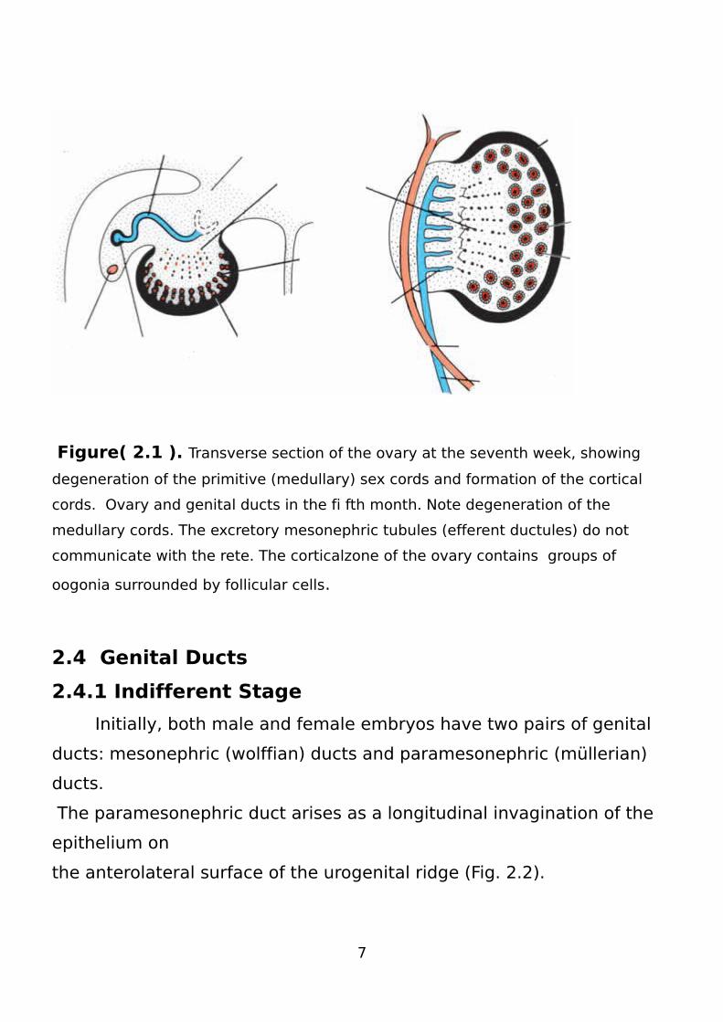
Figure( 2.1 ). Transverse section of the ovary at the seventh week, showing
degeneration of the primitive (medullary) sex cords and formation of the cortical
cords. Ovary and genital ducts in the fi fth month. Note degeneration of the
medullary cords. The excretory mesonephric tubules (efferent ductules) do not
communicate with the rete. The corticalzone of the ovary contains groups of
oogonia surrounded by follicular cells.
2.4 Genital Ducts
2.4.1 Indifferent Stage
Initially, both male and female embryos have two pairs of genital
ducts: mesonephric (wolffian) ducts and paramesonephric (müllerian)
ducts.
The paramesonephric duct arises as a longitudinal invagination of the
epithelium on
the anterolateral surface of the urogenital ridge (Fig. 2.2).
7
Cranially, the duct opens into the abdominal cavity with a funnel-like
structure. Caudally, it first runs lateral to the mesonephric duct, then
crosses it ventrally to grow caudomedially (Fig. 2.2) .
In the midline, it comes in close contact with the paramesonephric duct
from the opposite side. The two ducts are initially separated by a
septum but later fuse to form the uterine canal (Fig. 2.3). The caudal tip
of the combined ducts projects into the posterior wall of the urogenital
sinus, where it causes a small swelling, the paramesonephric or
müllerian
tubercle (Fig.2.3). The mesonephric ducts open into the urogenital
sinus on either side of the müllerian tubercle. (Moore KL, Persaud 2012) .
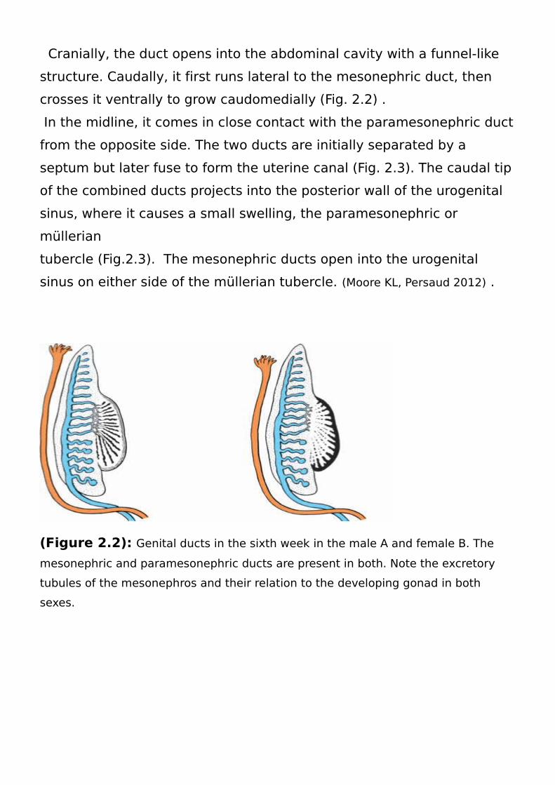
(Figure 2.2): Genital ducts in the sixth week in the male A and female B. The
mesonephric and paramesonephric ducts are present in both. Note the excretory
tubules of the mesonephros and their relation to the developing gonad in both
sexes.
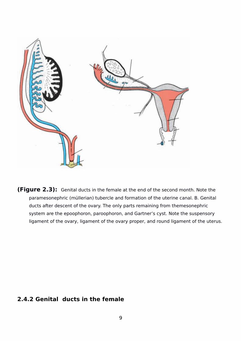
(Figure 2.3): Genital ducts in the female at the end of the second month. Note the
paramesonephric (müllerian) tubercle and formation of the uterine canal. B. Genital
ducts after descent of the ovary. The only parts remaining from themesonephric
system are the epoophoron, paroophoron, and Gartner’s cyst. Note the suspensory
ligament of the ovary, ligament of the ovary proper, and round ligament of the uterus.
2.4.2 Genital ducts in the female
9
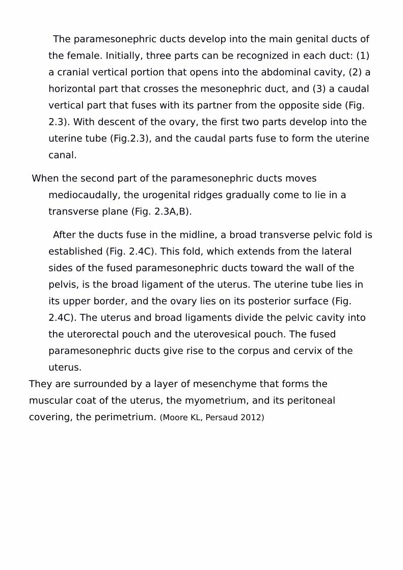
The paramesonephric ducts develop into the main genital ducts of
the female. Initially, three parts can be recognized in each duct: (1)
a cranial vertical portion that opens into the abdominal cavity, (2) a
horizontal part that crosses the mesonephric duct, and (3) a caudal
vertical part that fuses with its partner from the opposite side (Fig.
2.3). With descent of the ovary, the first two parts develop into the
uterine tube (Fig.2.3), and the caudal parts fuse to form the uterine
canal.
When the second part of the paramesonephric ducts moves
mediocaudally, the urogenital ridges gradually come to lie in a
transverse plane (Fig. 2.3A,B).
After the ducts fuse in the midline, a broad transverse pelvic fold is
established (Fig. 2.4C). This fold, which extends from the lateral
sides of the fused paramesonephric ducts toward the wall of the
pelvis, is the broad ligament of the uterus. The uterine tube lies in
its upper border, and the ovary lies on its posterior surface (Fig.
2.4C). The uterus and broad ligaments divide the pelvic cavity into
the uterorectal pouch and the uterovesical pouch. The fused
paramesonephric ducts give rise to the corpus and cervix of the
uterus.
They are surrounded by a layer of mesenchyme that forms the
muscular coat of the uterus, the myometrium, and its peritoneal
covering, the perimetrium. (Moore KL, Persaud 2012)
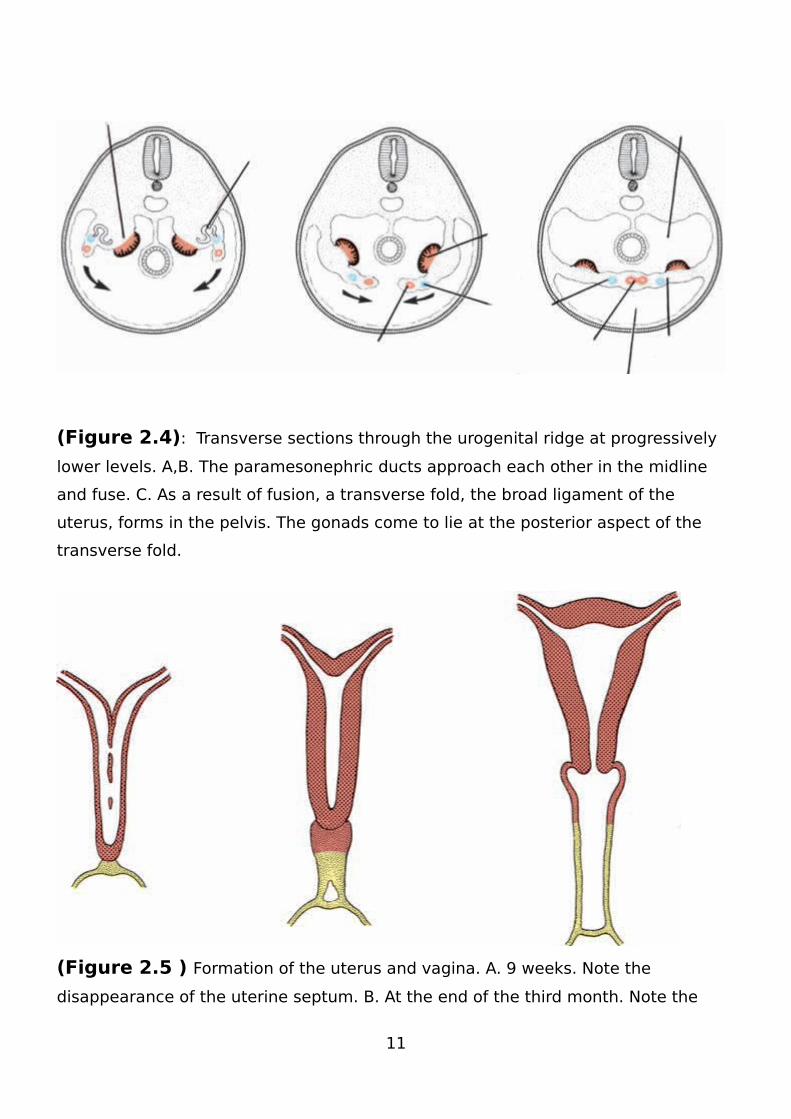
(Figure 2.4): Transverse sections through the urogenital ridge at progressively
lower levels. A,B. The paramesonephric ducts approach each other in the midline
and fuse. C. As a result of fusion, a transverse fold, the broad ligament of the
uterus, forms in the pelvis. The gonads come to lie at the posterior aspect of the
transverse fold.
(Figure 2.5 ) Formation of the uterus and vagina. A. 9 weeks. Note the
disappearance of the uterine septum. B. At the end of the third month. Note the
11
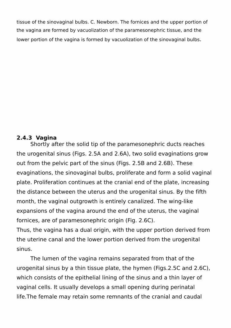
tissue of the sinovaginal bulbs. C. Newborn. The fornices and the upper portion of
the vagina are formed by vacuolization of the paramesonephric tissue, and the
lower portion of the vagina is formed by vacuolization of the sinovaginal bulbs.
2.4.3 Vagina Shortly after the solid tip of the paramesonephric ducts reaches
the urogenital sinus (Figs. 2.5A and 2.6A), two solid evaginations grow
out from the pelvic part of the sinus (Figs. 2.5B and 2.6B). These
evaginations, the sinovaginal bulbs, proliferate and form a solid vaginal
plate. Proliferation continues at the cranial end of the plate, increasing
the distance between the uterus and the urogenital sinus. By the fifth
month, the vaginal outgrowth is entirely canalized. The wing-like
expansions of the vagina around the end of the uterus, the vaginal
fornices, are of paramesonephric origin (Fig. 2.6C).
Thus, the vagina has a dual origin, with the upper portion derived from
the uterine canal and the lower portion derived from the urogenital
sinus.
The lumen of the vagina remains separated from that of the
urogenital sinus by a thin tissue plate, the hymen (Figs.2.5C and 2.6C),
which consists of the epithelial lining of the sinus and a thin layer of
vaginal cells. It usually develops a small opening during perinatal
life.The female may retain some remnants of the cranial and caudal
excretory tubules in the mesovarium, where they form the epoophoron
and paroophoron, respectively (Fig. 2.3B).
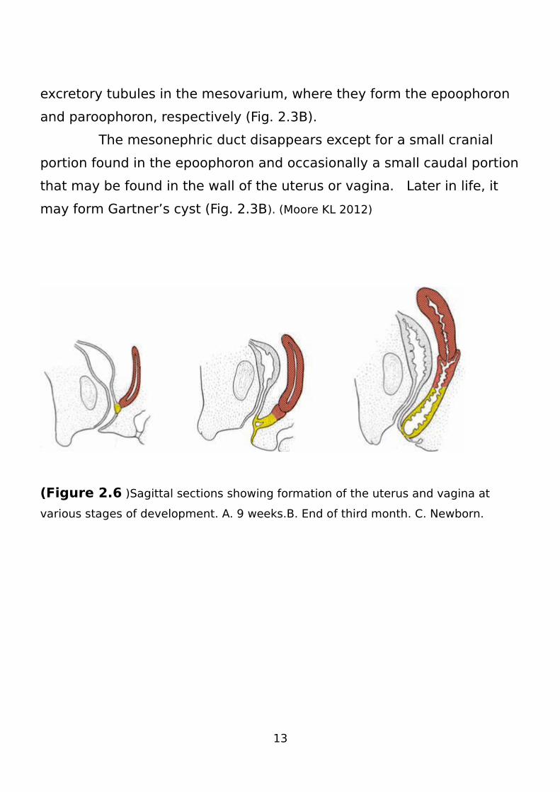
The mesonephric duct disappears except for a small cranial
portion found in the epoophoron and occasionally a small caudal portion
that may be found in the wall of the uterus or vagina. Later in life, it
may form Gartner’s cyst (Fig. 2.3B). (Moore KL 2012)
(Figure 2.6 )Sagittal sections showing formation of the uterus and vagina at
various stages of development. A. 9 weeks.B. End of third month. C. Newborn.
13
2.5 Anatomy:
2.5.1 Uterus:
Is a small, thick-walled, hollow, muscular orange, shaped very like
a pear, placed in the pelvis between the bladder and rectum, and
laving with its long axis forwards, so that when the women is
standing upright uterus is almost horizontal.For convenience, the
orange is considered as consisting of two portions, a corpus or
body, and a cervix or neck. The junction of the two is marked upon
the outer surface by a slight body is slightly bent forwards on the
cervix. The whole uterus, though best described as pear-shaped, is
distinctly flattened anteroposteriorly in the body.
There is also a slight rotation of the organ to the right so that the
left edge is slightly nearer to the front of the pelvis than the right
edge. The cervix is practically cylindrical shape. Although the uterus
is hollow its walls are normally almost in contact. When these are
separated the cavity of the body is about 1 inch (2.5 centimeter)
wide at the fundus (the uppermost and widest part), by 1.5 inch
(3.5 centimeter) long; it contracts at the level of the isthmus to
from the internal os, continues in the cervix as a narrow spindle-
shaped tube, and contracts again at the lower end of the cervix to
from external os .The cervical cavity is about 1 inch long, so that
the total length of the uterine cavity is about 2.5 inches (6.25
centimeter).
The fallopian or uterine tubes join the uterus at the extremities of its
greatest transverse diameter (the cornua or angles of the uterus).
The external measurements of the uterus are Length, 3 inches, or
7.5 centimeters. Breadth, 2 inches, or 5 centimeters (measured
between fallopian tubes).Thickness, 1.5 inches, or 3.75 centimeter
(measured between fallopian tubes). The uterus weighs about 1.5
ounces, or 42 grams. The uterine walls are about 0.5 inch (1.25
centimeters) thick. All the above-mentioned measurements and
weight refer to the normal adult virgin uterus. After pregnancy they
are increased and in old age often much diminished. In childhood
they are naturally much less. The cavity of the uterus is lined with a
special kind of mucous membrane, which is called the
endometrium, and which is actively concerned in the phenomena of
menstruation and pregnancy. The uterine cavity communicates
through the external os with the peritoneal cavity or coelom . That
is called the vaginal cervix or portiovaginalis; the upper part of the
neck of the uterus which lies above the cervico-vaginal attachment
is called the supravaginal cervix or the portio supravaginalis. In
certain forms of abnormal elongation of the cervix an intermediate
portion is also referred to for descriptive purposes. In multiporous
women the vaginal cervix is cone-shaped with the apex of the cone
directed downwards, and the external os presents as a small
rounded opening. After pregnancy the opening is a transverse site,
and definite anterior and posterior lips can be noted (often scars
and laceration also).The portiovaginal is covered by a reflection of
the vaginal mucous membrane, which is firmly a bed to it. The
cervix is loosely united to the bladder above the vagina by cellular
15
tissue; laterally it forms attachments to the pelvic connective
tissue. The uterus is almost entirely covered with peritoneum, back
and front, but at the sides there are small portions without any such
covering. The cervix is not covered at the front and sides, LMT
posteriorly there is peritoneum over the supravaginal cervix
forming part of the pouch of Douglas. The abdominal peritoneum
passing down the anterior abdominal wall onto the bladder forms a
pelvic fold between the bladder and the uterus, which is known as
the utero vesical pouch. This pouch is bounded by two thickened
edges, the utero-vesical ligaments. The pelvic peritoneum up joins
the uterus at the is thmus and covers the anterior surface, the
fundus, and the posterior surface of that organ. At the point where
the cervix meets the vagina the peritoneum is continued onto the
posterior vaginal wall, dips down for inch or so, and then becomes
reflected onto the anterior wall of the rectum, thus forming the
utero-vagino-rectal pouch of Douglas. As the peritoneum passes
up wards from Douglas' pouch posteriorly, it gradually surrounds
the bowel more and more until it finally completely encloses it and
forms its mesentery.
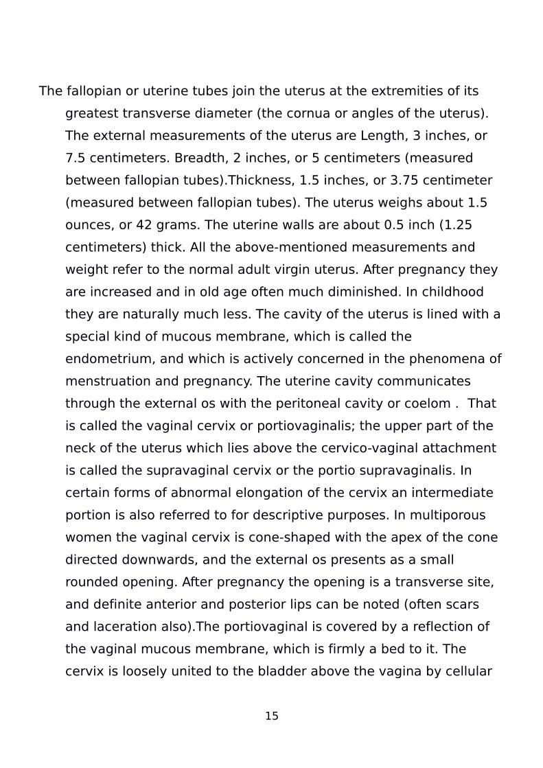
figure 2.7 shows the anatomy of utrus ovaries and fallopian tubes .

17
2.5.2 The fallopian tubes :
Uterine tubes, oviducts connect the ovary with the uterus. They also
connect the uterus with the peritoneal cavity, so that in the female
it may be said that there is a direct passage from the exterior of the
body to the peritoneum. It is true that under ordinary conditions this
passage is not actually patent, but it is a very important factor to
bear well in mind with regard to the spread of infective
inflammations.The Fallopian tubes are hollow muscular tubes,
about 4 inches Io centimeters) long, and varying from to inch 3 to 6
millimetres) in thickness. They extend out wards from the uterus,
and arch over the ovaries to which they are usually connected by
one firming or firming which surround the abdominal opening so
tubes. Morphologically, the Fallopian tubes represent the upper
portions of the Millerian ducts. For purposes of description four
parts of the tube are referred to. They are the uterine or interstitial
portion (one inch long), the isthmus or inner third. (Note.-{I)
and(2)portions willadmit abristle.) The ampulla, the widest portion,
which will admit a uterine sound. The infundibulum, the firmbrate
extremity of the ampulla which opens into the coelom.
2.5.3 The ovaries:
Are the essential organs of the generative system of the female.
They are ductless glands attached to the posterior layer of the broad
ligament one a chside of the uterus. Though attached to the
posterior layer they are actually situate between the two layers, i.e.
Intraligamentous. Their level in the pelvis is about that of the pelvic
brim, but the position of their long axis differs so much even in perfectly
normal and healthy women, that it can only be said to vary from
horizontal to vertical. The average ovary is aboutit inch slong,1inch
wide, and inch thick; its average weightis! Drachms. It is held in
position (I}by the attachment of its anterior edge to the broad
ligament;
(2) by at hick band running from its upper and inner end to the cornu of
the uterus = the ovarian ligament; (3) by the ovarian fimbria of the
Fallopian tube, which is attached to the lower and outer end of the
ovary; (4) by the so-called" suspensory ligament 11 of the ovary
(infundibulo-pelvic ligament), which Is attached to the side wall of the
pelvis and is really the free edge of the broad Ligament.
19
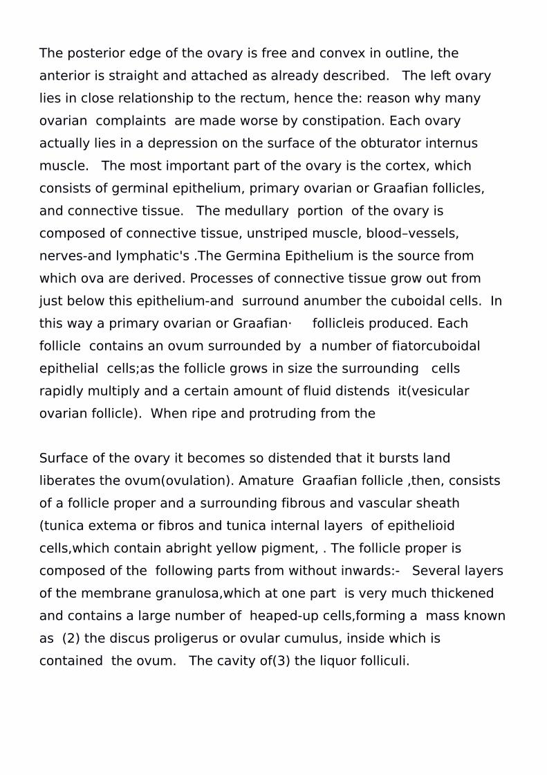
The posterior edge of the ovary is free and convex in outline, the
anterior is straight and attached as already described. The left ovary
lies in close relationship to the rectum, hence the: reason why many
ovarian complaints are made worse by constipation. Each ovary
actually lies in a depression on the surface of the obturator internus
muscle. The most important part of the ovary is the cortex, which
consists of germinal epithelium, primary ovarian or Graafian follicles,
and connective tissue. The medullary portion of the ovary is
composed of connective tissue, unstriped muscle, blood–vessels,
nerves-and lymphatic's .The Germina Epithelium is the source from
which ova are derived. Processes of connective tissue grow out from
just below this epithelium-and surround anumber the cuboidal cells. In
this way a primary ovarian or Graafian· follicleis produced. Each
follicle contains an ovum surrounded by a number of fiatorcuboidal
epithelial cells;as the follicle grows in size the surrounding cells
rapidly multiply and a certain amount of fluid distends it(vesicular
ovarian follicle). When ripe and protruding from the
Surface of the ovary it becomes so distended that it bursts land
liberates the ovum(ovulation). Amature Graafian follicle ,then, consists
of a follicle proper and a surrounding fibrous and vascular sheath
(tunica extema or fibros and tunica internal layers of epithelioid
cells,which contain abright yellow pigment, . The follicle proper is
composed of the following parts from without inwards:- Several layers
of the membrane granulosa,which at one part is very much thickened
and contains a large number of heaped-up cells,forming a mass known
as (2) the discus proligerus or ovular cumulus, inside which is
contained the ovum. The cavity of(3) the liquor folliculi.
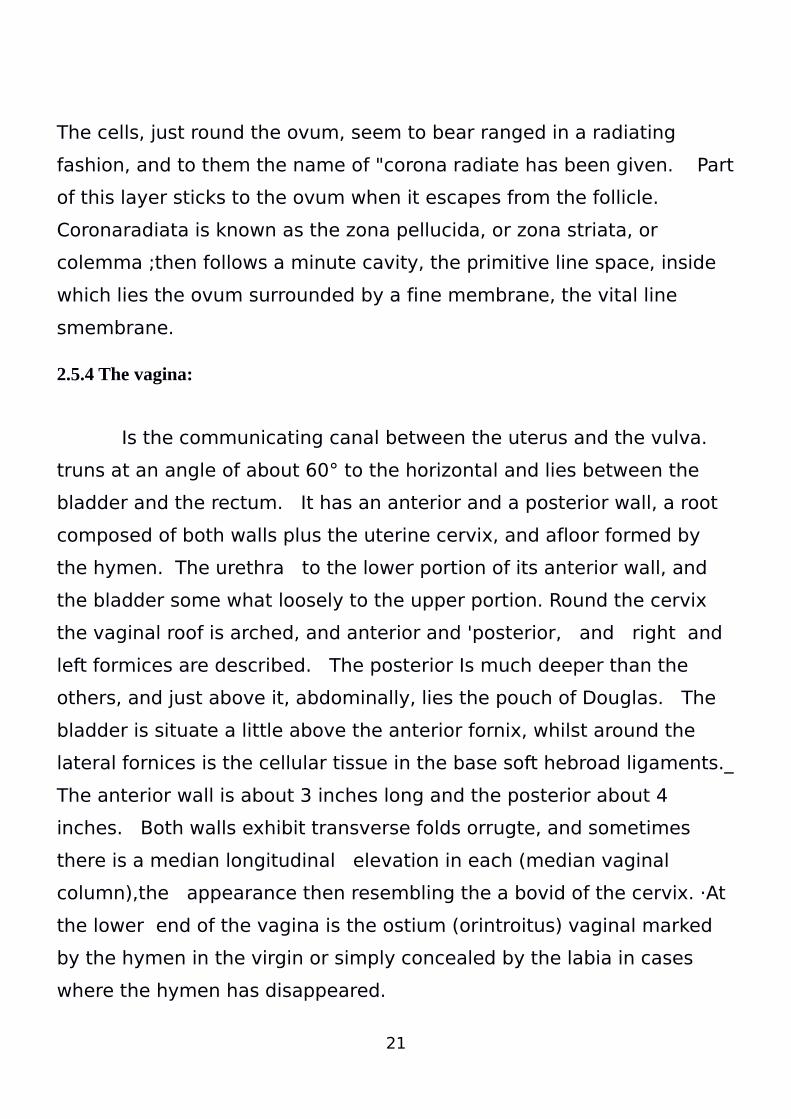
The cells, just round the ovum, seem to bear ranged in a radiating
fashion, and to them the name of "corona radiate has been given. Part
of this layer sticks to the ovum when it escapes from the follicle.
Coronaradiata is known as the zona pellucida, or zona striata, or
colemma ;then follows a minute cavity, the primitive line space, inside
which lies the ovum surrounded by a fine membrane, the vital line
smembrane.
2.5.4 The vagina:
Is the communicating canal between the uterus and the vulva.
truns at an angle of about 60° to the horizontal and lies between the
bladder and the rectum. It has an anterior and a posterior wall, a root
composed of both walls plus the uterine cervix, and afloor formed by
the hymen. The urethra to the lower portion of its anterior wall, and
the bladder some what loosely to the upper portion. Round the cervix
the vaginal roof is arched, and anterior and 'posterior, and right and
left formices are described. The posterior Is much deeper than the
others, and just above it, abdominally, lies the pouch of Douglas. The
bladder is situate a little above the anterior fornix, whilst around the
lateral fornices is the cellular tissue in the base soft hebroad ligaments._
The anterior wall is about 3 inches long and the posterior about 4
inches. Both walls exhibit transverse folds orrugte, and sometimes
there is a median longitudinal elevation in each (median vaginal
column),the appearance then resembling the a bovid of the cervix. ·At
the lower end of the vagina is the ostium (orintroitus) vaginal marked
by the hymen in the virgin or simply concealed by the labia in cases
where the hymen has disappeared.
21
2.6 Reproductive physiology:
In the normal female between the age of 9 and 16, cyclic changes
occur in the ovaries and the uterus in response to endocrinologic
activities. These cyclic changes are known as the menstrual cycle and
represent the reproductive phase of a female's life cycle. The changes
associated with the ovary are known as the ovarian cycle whereas those
associated with the endometrium are known as the endometrial cycle.
The purpose of the ovarian cycle is to provide a suitable ovum for
fertilization, whereas that of the endometrial cycle is to provide a
suitable site in which the blastocyst can implant and develop properly.
Since the endometrial changes are regulated by the ovarian hormones,
the two cycles are intimately related.The typical menstrual cycle is 28
days however variations are very common and normal.For the purpose
of description, the 28 day "idealized" cycle is used. The cycle is divided
into four or five phases. It is customary to assign the first day of
menstruation as the first day of the cycle.(Devin ,etal,1992)
2.6.1 Ovarian cycle:
Throughout the reproductive years, at the onset of each menstrual
cycle, a number of small, immature follicles known as primary or
primordial follicles, undergo growth and development. The hormonal
stimulus that activates the follicular process is mediated by follicle-
stimulating hormone or FSH which is secreted by the anterior pituitary
gland.With each menstrual cycle, there is usually only one mature
follicle, known as the dominant or Graafian follicle, which makes its way
to the surface of the ovary where it appears as a transparent cyst. The
mature preovulatory follicle contains the ovum at one end and a cystic
cavity or antrum at the other. There are several layers of specialized
cells known as theca and granulosa cells which secrete
estrogenprogesterone and luteinizing substances. The ovum is
released from the mature follicle during ovulation. Ovulation normally
occurs on day 14 which is the mid-point of the idealized cycle.Following
ovulation, the ruptured dominant follicle becomes the corpus
hemorrhagicum which is then followed by the corpus luteum. The
corpus luteum (CL) secretes progesterone (as well as estrogen) which is
absolutely necessary to maintain the endometrium for successful
implantation.If fertilization does not occur, the CL undergoes regressive
changes, progesterone output is diminished, and by the end of the cycle
complete regression occurs. The failing CL triggers endometrial
sloughing, and menstrual bleeding ensues. The end point of the
regressing CL is the corpus albicans, which is a small fibrous area in the
cortex of the ovary.the theca interna cells of multiple secondary follicles
full fill an endocrine function as they differentiate into
estrogen.secreting cells the hormone estrogen promotes proliferation of
the endometrium while many follieles develop in the ovaries in
response to follicle stamulating hormone (FSH), only one follicle
matures completely to be released at avulation.Most of the follicles
undergo follicular atresia beyond the stage of secondary follicle .one
secondary follicle continues to mature to become agraafian follicle prior
to ovulation. The ovum continues to mature through meiotic divition,
forming the secondary oocyte .Now the oocyte floats freely with in the
enlarged follicular antrum of the graafianfollicle .
23
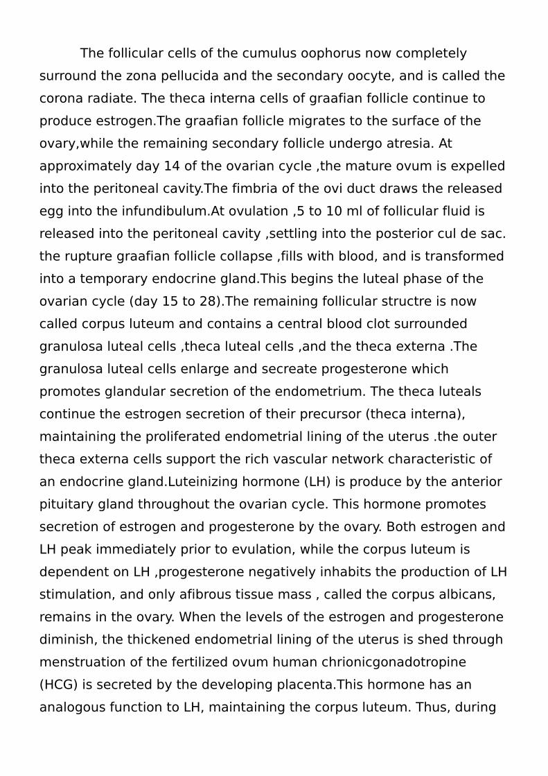
The follicular cells of the cumulus oophorus now completely
surround the zona pellucida and the secondary oocyte, and is called the
corona radiate. The theca interna cells of graafian follicle continue to
produce estrogen.The graafian follicle migrates to the surface of the
ovary,while the remaining secondary follicle undergo atresia. At
approximately day 14 of the ovarian cycle ,the mature ovum is expelled
into the peritoneal cavity.The fimbria of the ovi duct draws the released
egg into the infundibulum.At ovulation ,5 to 10 ml of follicular fluid is
released into the peritoneal cavity ,settling into the posterior cul de sac.
the rupture graafian follicle collapse ,fills with blood, and is transformed
into a temporary endocrine gland.This begins the luteal phase of the
ovarian cycle (day 15 to 28).The remaining follicular structre is now
called corpus luteum and contains a central blood clot surrounded
granulosa luteal cells ,theca luteal cells ,and the theca externa .The
granulosa luteal cells enlarge and secreate progesterone which
promotes glandular secretion of the endometrium. The theca luteals
continue the estrogen secretion of their precursor (theca interna),
maintaining the proliferated endometrial lining of the uterus .the outer
theca externa cells support the rich vascular network characteristic of
an endocrine gland.Luteinizing hormone (LH) is produce by the anterior
pituitary gland throughout the ovarian cycle. This hormone promotes
secretion of estrogen and progesterone by the ovary. Both estrogen and
LH peak immediately prior to evulation, while the corpus luteum is
dependent on LH ,progesterone negatively inhabits the production of LH
stimulation, and only afibrous tissue mass , called the corpus albicans,
remains in the ovary. When the levels of the estrogen and progesterone
diminish, the thickened endometrial lining of the uterus is shed through
menstruation of the fertilized ovum human chrionicgonadotropine
(HCG) is secreted by the developing placenta.This hormone has an
analogous function to LH, maintaining the corpus luteum. Thus, during
pregnancy, the corpus luteum continues to secrete estrogen and
progestore throughout the first trimester.The placenta ultimately lakes
over this endocrine function and the corpus luteum regresses, forming
the corpus albicans .
2.6.2 Endometrial cycle
With each menstrual cycle, and in step with ovarian activity, the
functional layer of the endometrium undergoes changes characterized
by regeneration, proliferation, secretory activity, necrosis, and
sloughing.During menstruation, the functional layer of the endometrium
is sloughed off and along with blood, passes into the vagina. Following
menstruation, new functional layer begins to form from the basal layer.
Primed by estrogen secreted by the ovary, the endometrium
progressively thickens throughout the proliferative and secretory
phases.
Following ovulation and the formation of the CL, the endometrial
glands exhibit secretory activity. If fertilization does not occur, the
corpus luteum undergoes regressive changes, and the endometrium,
supported by the hormonal output of the ovary, begins to "shrink". The
shrinking is due to the loss of tissue fluids and secretions which occurs
secondary to the drop in estrogen. Estrogen has a "water-retaining"
effect on tissues whereas progesterone is a factor in the secretory
activity of the gland.As the endometrium shrinks, the spiral arteries kink
resulting in vascular stasis followed by ischemia, necrosis, sloughing
and bleeding.The menstrual cycle is a continuous ongoing cycle but for
descriptive purposes it is divided into specific phases based on
hormonal levels, and events occurring in the ovary and endomsetrium.
The hormonal relationships and the effects of these hormones on the
25
receptor tissues and organs are considered with these phases in mind.
The "ideal" 28 day cycle will be considered although in reality the length
of the normal menstrual cycle may vary .(Devin ,1992 )
Figure 2.9 shows the physiological change In ovarian and endometrial cycles.
2.7 Pathology
Miscarriage is classified as threatened, missed incomplete and complete based on
the ultrasound findings.
2.7.1 Threatened abortion
Threatened abortion defines a woman who presents during the
first half of pregnancy
with mild vaginal bleeding and uterine cramping without cervical
dilatation. Threatened
abortion is the most common clinical indication for ultrasound
evaluation of an early
pregnancy. Threatened abortion occurs in about one-quarter (25%) of all pregnancies;
about one-half (50%) of women presenting with threatened abortion have a poor
outcome (e.g. go on to abort). In the group of patients presenting with threatened
abortion who subsequently abort, the embryo (fetus) is usually already dead at the time
of ultrasounsd evaluation. Spontaneous expulsion of the pregnancy (spontaneous
abortion) normally occurs in patients who present with TA but may be delayed by as
much as two weeks.
All pregnant women who present with vaginal bleeding during the first trimester of
pregnancy are also potentially “rule out ectopic pregnancy” therefore a primary goal of
ultrasound evaluation is to determine if the pregnancy is intrauterine or ectopic.
Assuming an intrauterine pregnancy (IUP) is diagnosed, the second goal of ultrasound
evaluation is to identify an embryo(s) and determine if it is alive or dead.
If an embryo is not seen, the sonographer should determine if one should be based on
the size of the gestational sac or other sonographic criteria.
If a live embryo is detected, an ancillary goal of the ultrasound study is to search for
sonographic findings that are associated with a high risk of subsequent demise or fetal
anomaly. If a live intrauterine embryo is detected, the pregnancy can be managed
expectantly.
If a nonviable IUP is indicated with certainty, the uterus may be evacuated (dilatation
and evacuation or D&E). If the sonographic findings are inconclusive, a follow up study
at an appropriate interval (7 to 14 days depending on the findings of the initial
ultrasound study).
In many women, because the embryo is not detected at the time of the
initial
sonographic study, the diagnosis of early embryonic demise cannot be
made on the
basis of embryonic cardiac activity; in these women, it may still be
possible to make the
diagnosis of early pregnancy failure on a single study by assessing the
gestational sac.
27
A small number of pregnant women have some vaginal bleeding, with
or without
abdominal cramps, during the first trimester of pregnancy. This is
known as a threatened abortion. Most of these pregnancies go on to
term with or without treatment. Spontaneous
abortion occurs in just a small percentage of women who have vaginal
bleeding during
pregnancy.
When spontaneous abortion occurs, the usual cause is fetal death. Such
death is typically
the result of a chromosomal or developmental problem.
2.7.1.1 Other possible causes include:
Defects in the mother's anatomy�
Endocrine factors�
Immune system factors�
Infection�
Systemic disease in the mother�
About half of all fertilized eggs abort on their own,usually before the
woman knows she is pregnant. The rate of spontaneous abortion is very
low among known pregnancies. These usually occur 7 - 12 weeks into
the pregnancy
2.7.1.2 Risks of threatened abortion are higher in:
Women over age 35�
Women with a history of 3 or more spontaneous abortions�
Women with systemic disease (such as diabetes or thyroid �
dysfunction)
2.7.1.3 Symptoms:
Abdominal cramps with or without vaginal bleeding�
Vaginal bleeding during the first 20 weeks of pregnancy (last �
menstrual period
was less than 20 weeks ago)
Note: With true miscarriage, low back pain or abdominal pain (dull to
sharp, constant to
intermittent) typically occurs and tissue or clot-like material may pass
from the vagina.
2.7.1.4 Exams and Tests:
Pelvic exam shows a cervix that isn't thinned (effaced) or open (dilated).
Either of these
can suggest that a miscarriage will soon occur.
2.7.1.5 Other tests include:
Beta HCG (quantitative) test over a period of days or weeks to confirm �
whether
the pregnancy is continuing or the fetus has died
CBC to find out the amount of blood loss�
Serum HCG to confirm that the woman is pregnant�
Ultrasound to detect fetal heartbeat�
WBC with differential to rule out infection�
This disease also can change the results of the serum progesterone
test.
2.7.1.6 Treatment
Bed rest or pelvic rest (not having intercourse, douching, or using
tampons) may be
recommended, but there is no evidence to show that these actually
reduce the miscarriage
29
rate.
The use of progesterone is controversial. It might relax smooth muscles,
including the
muscles of the uterus. However, it also might increase the risk of an
incomplete abortion
or an abnormal pregnancy. Unless there is a luteal phase defect,
progesterone should not
be used.
2.7.1.7 Outlook (Prognosis)
The outcome is good when the pregnancy continues to progress and all
the symptoms
disappear.
2.7.1.8 Possible Complications
Anemia�
Dead fetus syndrome�
Infection�
Moderate-to-heavy blood loss�
Spontaneous abortion�
2.8 Subchorionic Hesmatoma (SCH)
Subchorionic Hematoma is a hematoma that forms beneath the
chorion and expands in the uterine cavity. Partial separation of the
chorion frondosum is indicated when the
hematoma is seen behind the chorion frondosum. SCH is also known as
intrauterine hematoma, retrochorionic hematoma, and
submembranous hematoma. The exact etiology of SCH is uncertain
but is postulated to be related to rupture of basal veins at the edge
of the chorion frondosum (may be referred to as marginal sinus
rupture). SCH may be asymptomatic however in most cases is
associated with vaginal bleeding. The prognosis associated with
SCH is questionable however small collections are generally
associated with a better prognosis.
Generally, larger SCH are associated with a worse prognosis.
Ultrasound/Doppler - The echogenicity of a SCH depends on the age of
the
hematoma and also on technical factors such as transducer frequency
and gain setting. Echogenicity decreases with the age of the
hematoma; a fresh SCH may appear anechoic at low gain settings
and when viewed with a low frequency TAS transducer; the same
SCH may appear diffusely echogenic when evaluated with a higher
frequency EVS transducer. The shape of a SCH varies with the
scanning plane and on the relative size of the hematoma; in
general, a SCH has a crescent or wedge shape consistent with the
uterine cavity.
31
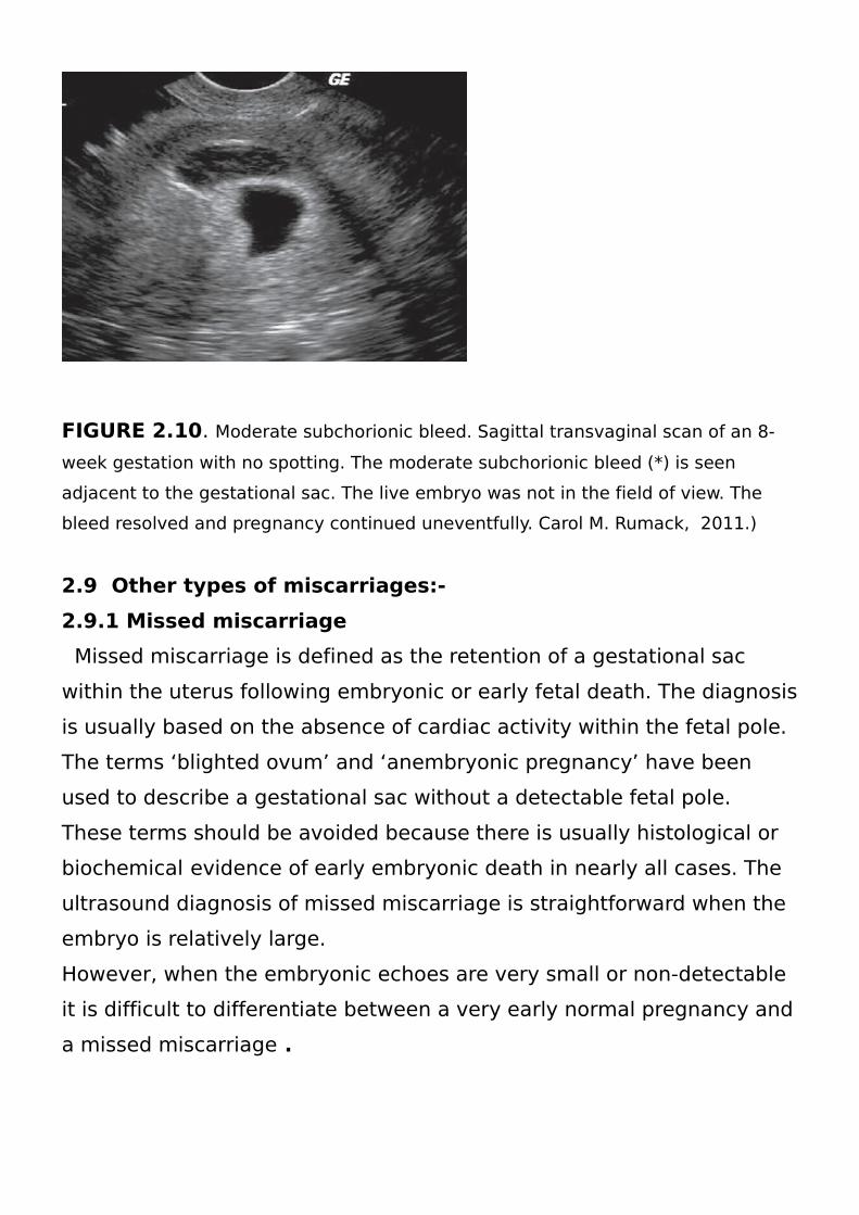
FIGURE 2.10. Moderate subchorionic bleed. Sagittal transvaginal scan of an 8-
week gestation with no spotting. The moderate subchorionic bleed (*) is seen
adjacent to the gestational sac. The live embryo was not in the field of view. The
bleed resolved and pregnancy continued uneventfully. Carol M. Rumack, 2011.)
2.9 Other types of miscarriages:-
2.9.1 Missed miscarriage
Missed miscarriage is defined as the retention of a gestational sac
within the uterus following embryonic or early fetal death. The diagnosis
is usually based on the absence of cardiac activity within the fetal pole.
The terms ‘blighted ovum’ and ‘anembryonic pregnancy’ have been
used to describe a gestational sac without a detectable fetal pole.
These terms should be avoided because there is usually histological or
biochemical evidence of early embryonic death in nearly all cases. The
ultrasound diagnosis of missed miscarriage is straightforward when the
embryo is relatively large.
However, when the embryonic echoes are very small or non-detectable
it is difficult to differentiate between a very early normal pregnancy and
a missed miscarriage .
2.9.2 Diagnostic errors have been reported in such cases.
The Royal College of Obstetricians and Gynaecologists (RCOG) has
proposed a set ofguidelines to establish embryonic death by
ultrasound.According to these guidelines, the absence of cardiac
activity in an embryo of crown–rump length (CRL) > 6 mm, or the
absence of a yolk sac or embryo in a gestation sac of mean
diameter > 20 mm, enables conclusive diagnosis of a missed
miscarriage.
In pregnancies in which the embryo and sac are smaller than 6 mm or
20 mm,respectively, a repeat ultrasound examination 1 week later
is necessary to clarify the diagnosis (RCOG 1995).
2.9.3 Complete and incomplete miscarriage
The diagnosis of complete and incomplete miscarriage depends
even more on the experience and skill of the operator than the
diagnosis of missed ssmiscarriage.Complete miscarriage is usually
diagnosed when the endometrium is very thin and regular. The
ultrasound appearances are therefore comparable to those of the non-
pregnant uterus in the early proliferative phase.
33
The diagnosis of incomplete miscarriage is more controversial and
diagnostic criteria ofendometrial thickness vary between 5 and 15 mm.
The main difficulty with using predefined cut off levels is the inability to
differentiate between blood clots, which are often seen within the
uterine cavity at the time of miscarriage and retained products. We
therefore favor subjective assessment of the endometrium in
preference to quantitative criteria.
Retained products are usually seen as a well-defined area of
hyperechoic tissue within the uterine cavity as opposed to blood clots
that are more irregular. Blood clots will be also seen sliding within the
uterine cavity when pressure is applied on the uterus by the
transvaginal probe. However,even with a Doppler examination the
diagnosis of incomplete miscarriage is difficult and ultrasound should
always be combined with clinical and biochemical assessment to rule-
out the possibility of ectopic in these cases.
C D
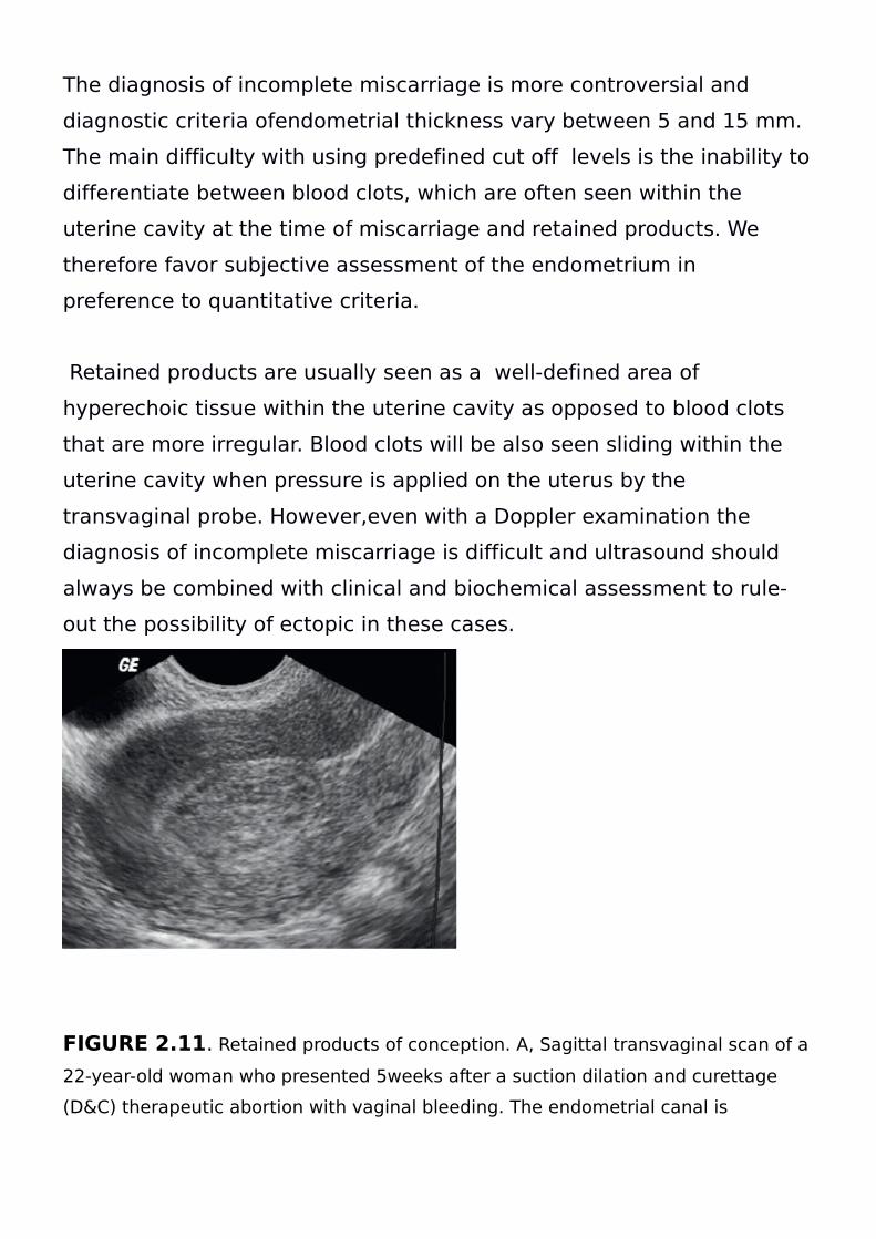
FIGURE 2.11. Retained products of conception. A, Sagittal transvaginal scan of a
22-year-old woman who presented 5weeks after a suction dilation and curettage
(D&C) therapeutic abortion with vaginal bleeding. The endometrial canal is
distended with a 1.8 2.5–cm echogenic mass (arrows). Carol M. Rumack, 2011.)
.
2.9.4 Management of miscarriage
Surgical evacuation of retained products has become universally
accepted as the method of choice for the management of miscarriage.
When it was introduced (in the 1960s) the rationale for the use of
curettage was a perceived risk of sepsis and hemorrhage associated
with spontaneous abortion. It is likely that a number of complicated
miscarriages at that time represented retained products following illegal
abortions, which contributedto the severity of clinical presentation.
Women’s general health has improved considerably in the intervening
50 years and most infections can now be treated effectively using
antibiotics. Legalisation of abortion has eliminated problems caused by
criminal abortion in many developed countries.
However, there is now a growing concern about the unconditional and
non-selective use of surgery for the treatment of miscarriage. There is
also concern about morbidity caused by surgical and anesthetic
complications. Expectant management of incomplete miscarriage is an
attractive option in this context. It follows the natural history of the
disease, avoids iatrogenic problems associated with both medical and
surgical treatment and, as such,
is likely to be cost effective. In cases of missed miscarriage, both
expectant and medical management are relatively ineffective and are
suitable only for individual, highly motivated women or those who have
difficulty in accepting the diagnosis of a failed
pregnancy and feel unable to make a rapid decision about surgical
treatment.( Trish chudleigh 2004 ).
35

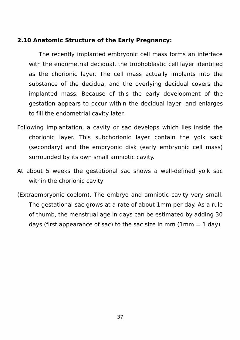
2.10 Anatomic Structure of the Early Pregnancy:
The recently implanted embryonic cell mass forms an interface
with the endometrial decidual, the trophoblastic cell layer identified
as the chorionic layer. The cell mass actually implants into the
substance of the decidua, and the overlying decidual covers the
implanted mass. Because of this the early development of the
gestation appears to occur within the decidual layer, and enlarges
to fill the endometrial cavity later.
Following implantation, a cavity or sac develops which lies inside the
chorionic layer. This subchorionic layer contain the yolk sack
(secondary) and the embryonic disk (early embryonic cell mass)
surrounded by its own small amniotic cavity.
At about 5 weeks the gestational sac shows a well-defined yolk sac
within the chorionic cavity
(Extraembryonic coelom). The embryo and amniotic cavity very small.
The gestational sac grows at a rate of about 1mm per day. As a rule
of thumb, the menstrual age in days can be estimated by adding 30
days (first appearance of sac) to the sac size in mm (1mm = 1 day)
37
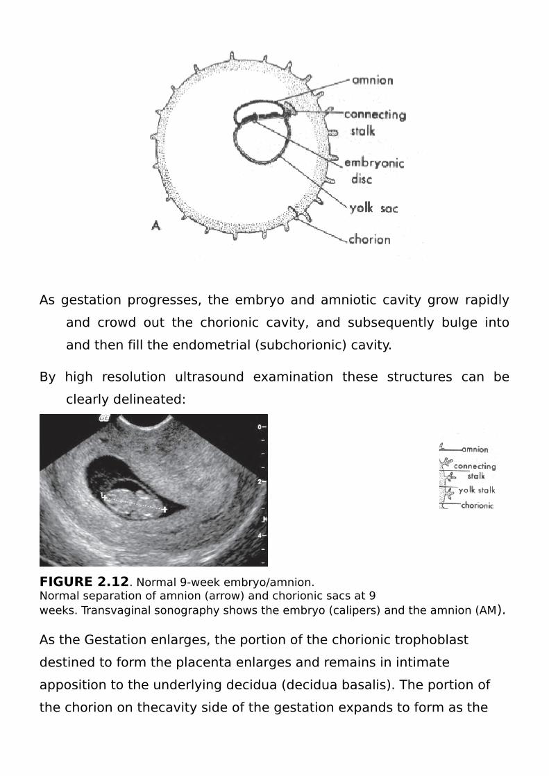
As gestation progresses, the embryo and amniotic cavity grow rapidly
and crowd out the chorionic cavity, and subsequently bulge into
and then fill the endometrial (subchorionic) cavity.
By high resolution ultrasound examination these structures can be
clearly delineated:
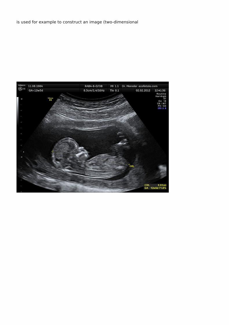
FIGURE 2.12. Normal 9-week embryo/amnion.Normal separation of amnion (arrow) and chorionic sacs at 9weeks. Transvaginal sonography shows the embryo (calipers) and the amnion (AM).
As the Gestation enlarges, the portion of the chorionic trophoblast
destined to form the placenta enlarges and remains in intimate
apposition to the underlying decidua (decidua basalis). The portion of
the chorion on thecavity side of the gestation expands to form as the
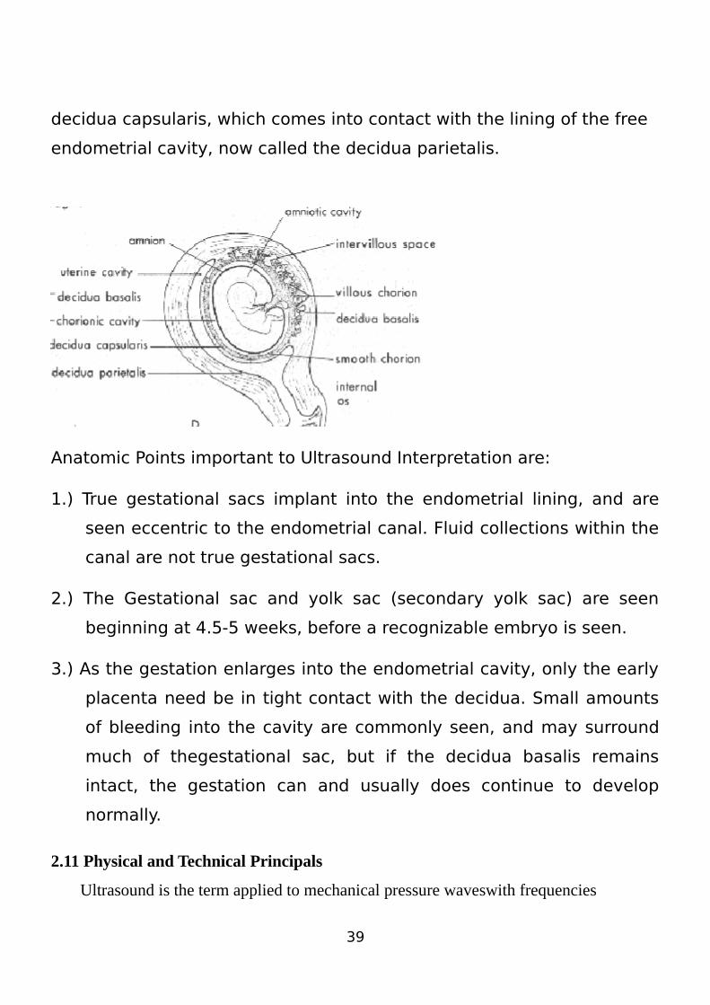
decidua capsularis, which comes into contact with the lining of the free
endometrial cavity, now called the decidua parietalis.
Anatomic Points important to Ultrasound Interpretation are:
1.) True gestational sacs implant into the endometrial lining, and are
seen eccentric to the endometrial canal. Fluid collections within the
canal are not true gestational sacs.
2.) The Gestational sac and yolk sac (secondary yolk sac) are seen
beginning at 4.5-5 weeks, before a recognizable embryo is seen.
3.) As the gestation enlarges into the endometrial cavity, only the early
placenta need be in tight contact with the decidua. Small amounts
of bleeding into the cavity are commonly seen, and may surround
much of thegestational sac, but if the decidua basalis remains
intact, the gestation can and usually does continue to develop
normally.
2.11 Physical and Technical Principals
Ultrasound is the term applied to mechanical pressure waveswith frequencies
39
above 20,000 Hz (beyond the audible range).
A medium must be present for ultrasound propagation to occur. In biological tissues,
ultrasonic energy is propagated mainly in the form of longitudinal waves, as it is in fluid.
The ultrasound wave can be both emitted and received by a piezoelectric transducer. The
piezoelectric transducer is able to change electrical signals
into mechanical waves, that is, transmitting ultrasound (= reverse piezoelectric
effect), and vice versa to change mechanical pressure (reflected ultrasound waves,
“echoes”) into electrical signals (= direct piezoelectric effect).
Ultrasound in the MHz range (high-frequency) can be emitted as a directional
beam, comparable to a light beam, from transducers of practical size.
Ultrasound waves propagate in biological tissue at an average speed of
1540 meters (m) per second, with the exception of bones, where the waves
move at more than 3000m per second. Ultrasound waves interact with biological tissue in
various ways; they are partially absorbed by the tissue, which means that their energy is
converted into heat. This is important for safety reasons.
Ultrasound waves can also be reflected (interference > beam diameter) or (back-) scattered
on their way through the tissue.Whether reflected or back-scattered, echoes are received
by the transducer.
These echoes are the source of the diagnostic information.
The echoes are analyzed first with regard to their site of origin (time– distance principle),
and secondly with regard to their intensity.
This information B-scan technique). One of the preconditions is that only a small part of
the ultrasound is reflected at each interface, and most of the ultrasound is transmitted to
deeper levels. Only bones, gas, and foreign bodies (metallic or nonmetallic) cause a very
strong reflection (acoustic shadow); thus no information is obtainable from regions behind
such obstacles.
Absorption, reflection, and scattering cause a permanent attenuation of ultrasound energy
of approx. 1 decibel per cm of propagation in the tissue traversed per 1MHz of frequency.
The ultrasound attenuation must be corrected by amplifying echoes as a function of
distance from the transducer (TGC), in order to get a homogeneous display of the echoes.
Nevertheless this attenuation can seriously limit the depth of penetration of higher
frequencies (the so-called small part transducers are suitable for small and superficial
organs only!).
The ultrasonic field is a geometric description of the region encompassed by the
ultrasound beam. There are two main sectors, the near field (interference field), located
between the ultrasound transducer and the (natural) focus, and the far field.
The lateral boundary of the ultrasound field is not sharp, because the beam intensity falls
off continuously with distance from the center.
The lateral resolution depends on the diameter of the ultrasound beam:
the smaller the diameter, the better the resolution. The resolution therefore
is best in the focal area.
The ultrasound beams are focused (mainly electronically by modern techniques), enabling
the clinician to always focus on the region of interest.
The axial resolution depends on the length of the emitted ultrasound pulses and finally on
the wave length, i.e., the frequency.
These basic physical principles are still important in regard to the quality of ultrasound
equipment despite the advances in electronic techniques:
the higher the frequency, the better the resolution on the one hand, but the
higher the attenuation in the tissue on the other hand, whichmeans a limited
penetration depth. For small and superficial parts, therefore, highfrequency
transducers (5–10 MHz) should be used, whereas for the abdomenor in latepregnancy,
transducerswith lower frequencies (2–5MHz) are necessary.
2.11.1 Imaging Techniques
The echo principle forms the basis of all of the commonly used diagnostic
ultrasound techniques. These are:
A-scan
M-scan
B-scan
41
2.11.2 Doppler techniques
A-scan (amplitude modulation) is a one-dimensional technique. The echoes received are
displayed on a screen as vertical deflections. This technique is rarely used today except for
measurements.
B-scan (brightness modulation) is a technique in which the echo
amplitude
is depicted as dots of different brightness (gray scale). It is mostly
used as a two-dimensional B-scan to form a two-dimensional ultrasound
image by multiple ultrasound beams, arranged successively in one
plane.
The images are built up by mechanically or electronically regulated
scanning
in a fraction of a second. The image rate of more than 15 per second
enables an impression of “permanent” imaging during the examination
(real time).
M-scan (also sometimes referred to as TM-scan) is a way to display
motion, e.g. of parts of the heart. The echoes produced by a stationary
ultrasound beam are recorded over time, continuously.
Doppler techniques use the Doppler effect as a further source of
information:
if the ultrasound waves are reflected by an interface moving towards
the transducer or away from it, the reflected frequency will be higher or
lower respectively than the transmitted frequency.
The difference between the emitted and received frequencies is
proportional to the speed of the moving reflector. This phenomenon is
called the Doppler effect, and the difference is called the Doppler
frequency or Dopplershift.
The Doppler shift depends on the ultrasonic frequency (f ), the velocity
of the reflector (v), and the angle between the ultrasound beam and the
blood stream. Information can only achieved if the angle is less than
60◦. An angle of 90◦ has the cosine α = 0, which means no Doppler
shift = no signal.
Doppler Formula: Δf = 2f /c ・ v ・ cos α .
There are various Doppler techniques:
Continuous wave Doppler (cw Doppler): the transducer is divided in two
parts: one crystal transmits ultrasound permanently, the other crystal
receives all the echoes. There is no information about the distance ofthe
reflector(s), but only about the velocity, at which the reflector (theblood
stream) moves.
Pulsed Doppler: Ultrasound is emitted in very short pulses (as in the
A-,B-, and M-scan techniques). Between the pulses the echoes reaching
the transducer in a certain time interval are received and analyzed.
In this way, the movement of the reflectors in a partiscular distance
(gate, selected by the operator) can be displayednand analyzed.(H.T.Lutz
etal. 2006)
43
is used for example to construct an image (two-dimensional
Ultrasonic's is the application of ultrasound. Ultrasound can be used
for medical imaging, detection, measurement and cleaning. At
higher power levels, ultrasonic's is useful for changing the chemical
properties of substances.
2.11.3 Early ultrasound findings in normal pregnancy:
The visualization of early structures benefits from high resolution
technique. In most cases, ultrasound probesdesigned to operate in
the vagina provide the best resolution, and a necessary whenever
definitive diagnosis cannot be made by standard scanning.
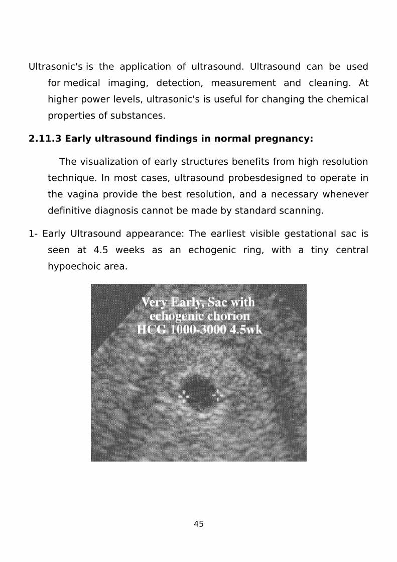
1- Early Ultrasound appearance: The earliest visible gestational sac is
seen at 4.5 weeks as an echogenic ring, with a tiny central
hypoechoic area.
45
The Nearly horizontal line beneath the sac is the endometrial cavity.
Note the gestational sac lies outside the cavity, embedded in the
decidua (lining).
This eccentric position is called the intradecidual sign, seen in
intrauterine implantations, and different from fluid collections in the
endometrial cavity which can be seen in both intrauterine and
ectopic pregnancies.Pseudosacs never show the intradecidual sign
however. Gestational Age Estimate: Measurement of the mean
gestational sac diameter is an effective estimate of gestational age,
used between 5 and 5.7-6weeks. The accuracy in this period is
about +/-5 days. As soon as an identifiable embryo crown-rump
length (CRL) is measurable (5.7-6 weeks), it should be used. This is
because later gestational sac measurements may not reflect the
embryonic size (or even its presence), but the embryonic CRL
directly reflects embryonic growth. Tables of Mean Sac Size may be
used, or as a rule of
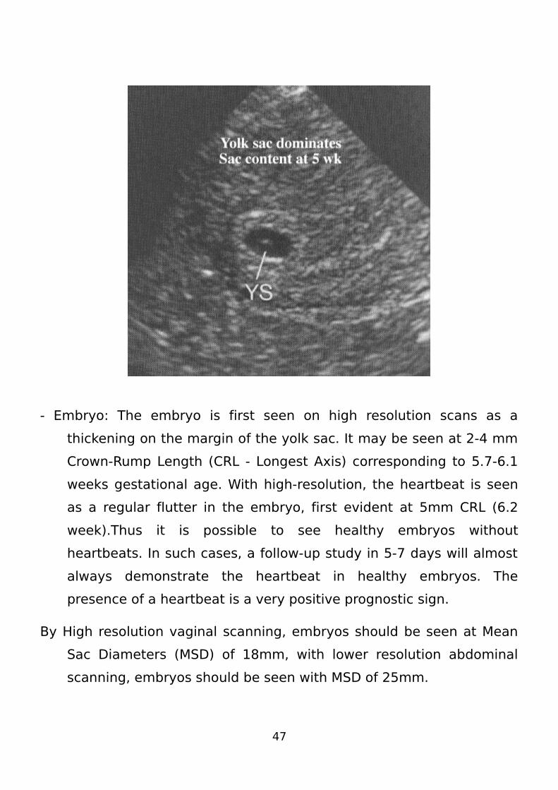
- Yolk Sac: The secondary yolk sac is the first element seen in the
gestational sac. Because it is reliably seen early, usually be 5
weeks, it is a critical landmark identifying a true gestation sac. Yolk
sac should be seen in normal pregnancy when Mean Sac Diameter
is 20 mm by transabdominal scan, and 8- mm by high resolution
vaginal imaging. It is a spherical membrane, quite echogenic and
readily seen.
- Embryo: The embryo is first seen on high resolution scans as a
thickening on the margin of the yolk sac. It may be seen at 2-4 mm
Crown-Rump Length (CRL - Longest Axis) corresponding to 5.7-6.1
weeks gestational age. With high-resolution, the heartbeat is seen
as a regular flutter in the embryo, first evident at 5mm CRL (6.2
week).Thus it is possible to see healthy embryos without
heartbeats. In such cases, a follow-up study in 5-7 days will almost
always demonstrate the heartbeat in healthy embryos. The
presence of a heartbeat is a very positive prognostic sign.
By High resolution vaginal scanning, embryos should be seen at Mean
Sac Diameters (MSD) of 18mm, with lower resolution abdominal
scanning, embryos should be seen with MSD of 25mm.
47
2.11.4 Previous Studies:-
Study one :-Tannirandeny et al Saied:
Pregnant women of under20 gestational weeks diagnosed clinically
as threatened a portion Were eructed for ultrasound seen - the
sonographic were reported as viable pregnancy, incomplete abortion
and inclusive finding. 150 pregnant patients enrolled. Ultrasound cams
demonstrate 75 viable fetus 50%. In compel abortion 33.3% inclusive
finding reveal 16.7% the viable pregnancy rate according to maternal
age was night at the maternal ag 025% 29 years old whereas it was
lowest at the maternal and age of 40% 44 years old 2%.
The viable pregnancy rate according to gestational age was highest at 6
to 8 weeks (6) 2% where it was lowest at 11 to 13 weeks 20%.
Sonographic findings in patients with Uinically throated abortion
demonstrate viable pregnancies in around wall of the cuscus. Use of
ultrasound in clinically diagnosed threaded abortion may assist
clinicians in
establishing a definite diagnosis so that appropriate cane could be
offered to the patients.(may 2001)
study two:- Daiter .E, who showed that the most widely accepted rate of loss for a
single sponteious abortion in an unselected population of couples that is
( regardless of characteristic associated with pregnancy loss ) was
about 15%-20% ( 1 in 6) of clinically detected pregnancies ( where the
woman missed menses or otherwise know that she was pregnant )
Study three:-
Elizabeth study showed that the over all miscarriage rate is reported as
15-20% which mean -20% of recognized pregnancies result in
miscarriage, the frequency of sponteious miscarrge increase with the
maternal age .
About 80%of miscarriage ocuur within the first trimester ,and the
frequency of miscarriage decrease with an increasing GA.
Study four :-
Millie A.Behera et al reported that sponteious abortion with is loss of
pregnancy witout outside intervention before 20 weeks GA affect up to
20%of rcognized pregnancies.
Study five :-
Stopplar ,M. stated that miscarriage occurs on about 15-20% of all
recognized pregnancies ,and usually occur before 13th week of
pregnancy, the acuall percentage of miscarriage is estimated to as high
as 50% of all pregnancies, since many miscarge occur without the
women ever having known she had pregnant.
Study six
Sandor Nagy et al Saied:187 pregnant women with intra uterine hematomas were detected at
first trimester ultrasonographic examination. The presents of a viable,
singleton gestational between 5 and 13 weeks. Gestational age was
calculalid based on thelast menstrual period. Or was corrected when the
crown. Rump length measurement were more than 5 days different
from the last menstrual period. The following sunographic factors were
evaluated: crown – rumplength, in complete abortion fetal heart rate.
The site of the gestational sac A hemamatoma was defined as a
crescent – shape sonolucent Fluid collection behind the fetal membrane
49
sub chorionic was defined us being located between the chorion and the
ulterine wall. The location of the gestation sac was marked as fundal,
eccentric – lower. The sonographic evaluation also include the shape of
the gestational sac. 230 patients with hematoma with singleton
intrauterine pregnancies. Under routine first trimester obstetric
ultrasonographic the hematoma was subchorinic in 91 patient 57%, it
was not possible to localize the hematoma in 28 cases because of early
gestational age. Less than 7 weak. The most gestational age were 8-9
weak at the first ultrasound scan, the most frequent incident were
advance maternal age. The gestational shape were regular 207 92% the
most location of gestational sac eccentric 120 patents 52.1% fundal 64
patient 27.9% lower 56 patient 24%. In complete abortion 23 patient
10%.
(1999- 2001)
Study seven
Robertson A, estimated that up to 50% of all fertilized eggs die and are
lost (aborted ) spontaneously ,usually before the women knows she is
pregnant , among known pregnensies the rate of miscarriage is
approximately 10% and usually occurs between 7th and 12th weeks of
pregnancy .

Chapter Three
Material and Methods
3. Material and Methods
By a sampling of consecutive cases, we included patients who
presented to our hospital between 5 to 13 weeks of pregnancy and
51
were diagnosed with threatened abortion. The patients were recruited
according to the following criteria:
Inclusion criteria: patients with pregnancy between 5 to 13 weeks of
gestation, who were diagnosed with threatened abortion according to
the institutional standards of our hospital. Exclusion criteria: patients
with multiple pregnancies,
molar pregnancy, ectospic pregnancy, amenorrhea with different
etiologies of pregnancy, maternal history of systemic diseases and
uterine anatomic abnormalities. Elimination criteria: patients who did
not have their outcome
data through week 20 of gestation due to relocation. Also clinical
factors such as medical conditions and obstetric complications were
included such as the presence of pelvic pain, vaginal bleeding and
cervical features (closed or dilated). These parameters were coded as 1
when present and 0 when absent. Afterward, a transvaginal ultrasound
examination was carried out with a high color Doppler
3.1 Methodology
3.1.1 Type of the study
This is cross sectional, descriptive study dialed with pregnant
patients who were hospitalized with clinical diagnosis of threatened
abortion (with vaginal bleeding, lower abdominal pain , cramping and
closed os).
3.1.2 Area of the study
El-Shaikh Mohammed Ali Fadoul Hospital - Omdurman .
3.1.3 duration of the study
This study was carried out during a period from 2014 – 2015 .
3.1.4 Population of the study
Pregnant patients who were hospitalized with clinical diagnosis of
threatened abortion and referred to ultrasound department of the study
areas at the time of study.
3.1.5 Study sample
They were 50 pregnant patients who were hospitalized with clinical
diagnosis of threatened abortion and they selected randomly.
3.1.6 Inclusion criteria:
patients age of 18-46 years old with pregnancy between 5 to 13 weeks
of gestation, who were diagnosed with threatened abortion and referred
to the ultrasound department.
3.1.7Exclusion criteria
Pregnant women less than 18 and more than 46 year old with GA
less than 5 and more than 13 weeks.
3.1.8Study variables
3.1.8.1 Independent variables: Patient age, gestational ages .
3.1.8.2 Dependent variables: gestational sac position,
gestational sac shape, presence of subchorionic and yolk sac.
3.2 Instrumentations and equipments
1- fukuda Danish 4000 Diagnostic Ultrasound System, manufactured in
Japan With 3.5MHz curve linear probe.
2. Coupling gel.
3- Towel sheath.
4- Thermal paper printer.
5- Digital camera.
6- Data collecting sheets.
3.3 data collection
53
The data were collected by clinical data sheets, ultrasound imaging
and interview with the patients.
3.4 Method
pregnant patients who were hospitalized with clinical diagnosis of
threatened abortion. Between 2014 and 2015 came to the ultrasound
department of the study area hospital and underwent pelvic
ultrasonograph (US), were cross-sectionally reviewed. Evaluation
Includes the grey scale ultrasound.
History findings include patient age and GA.
3.5 Scanning technique
Pelvic sonography was performed with the patient lying in a supine
position and the towel placed on the lower pelvis . Optimal results were
obtained with 3.5 MHz high-frequency curve linear-array transducers.
The pelvis were studied with full bladder in two planes (i.e, longitudinal
and transverse axes).
The following songraphic findings evaluated:
size, shape, outline, and contents of the gestational sac; the presence
or absence of sub chorionic hematoma fetal cardiac activity.
3.6 Data analysis
The Data were analyzed using SPSS program
3.7 Data Representation
The Data were represented inform of cross tabulation and graphs,
using Microsoft office Word and Excel programs.
3.8 Ethical consideration
1. The data were collected and used upon patient consent.
2. No identification or individual details were published.
3. No information or patient details were disclosed or used for other
reasons than the study.
4. The study was approved by the ethics of the hospital
administration.

Chapter Four
Results
55
4- Result:
This study was carried out on 50 patients with threatened abortion, the
following as the results of this study patients collecting data is
presented in:
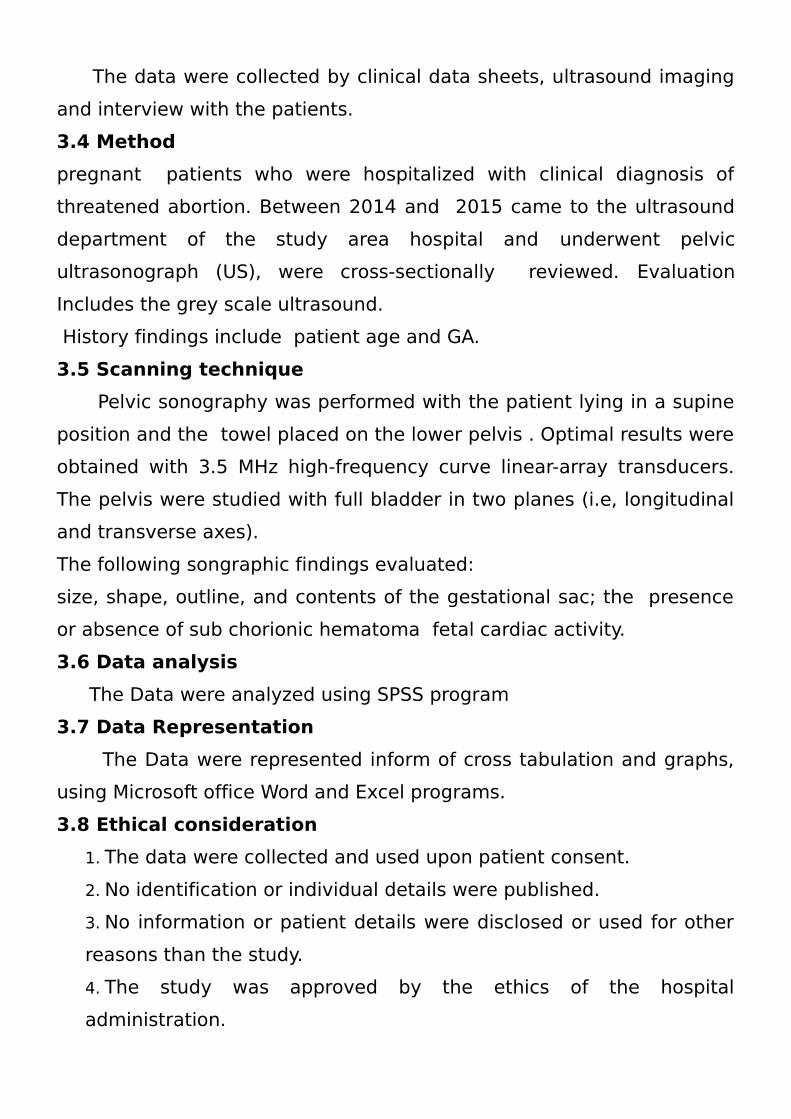
Table 4-1 show the Maternal Age distribution :
Maternal
age Frequency Percent
18-25 20 40.0
26-35 5 10.0
36-45 25 50.0
Total 50 100.0
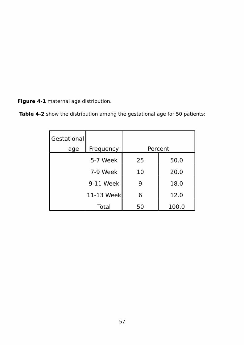
Figure 4-1 maternal age distribution.
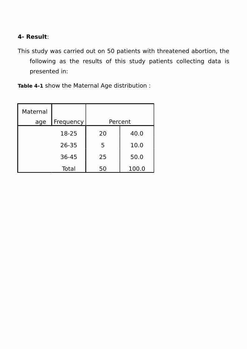
Table 4-2 show the distribution among the gestational age for 50 patients:
Gestational
age Frequency Percent
5-7 Week 25 50.0
7-9 Week 10 20.0
9-11 Week 9 18.0
11-13 Week 6 12.0
Total 50 100.0
57
Figure 4-2 shows GA distribution.
Table 4-3 show the gestational sac shape disribusion:
Gestational
shape Frequency Percent
Regular 45 90.0
Irregular 5 10.0
Total 50 100.0
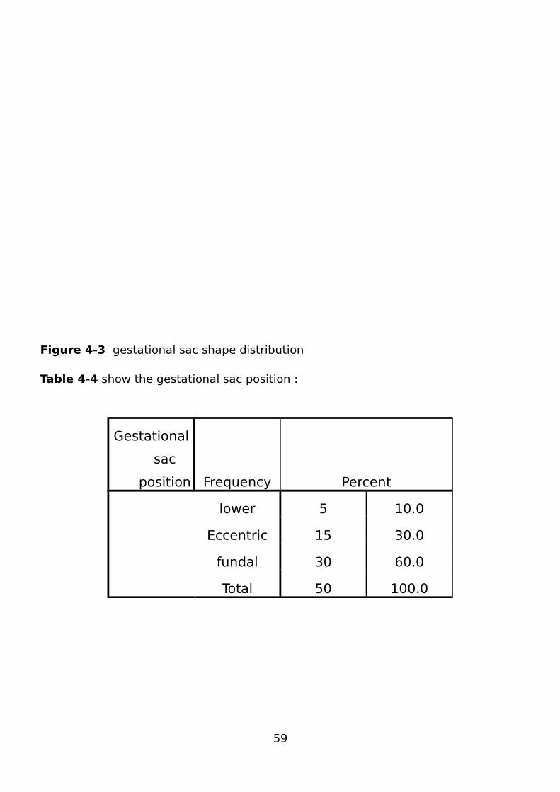
Figure 4-3 gestational sac shape distribution
Table 4-4 show the gestational sac position :
Gestational
sac
position Frequency Percent
lower 5 10.0
Eccentric 15 30.0
fundal 30 60.0
Total 50 100.0
59
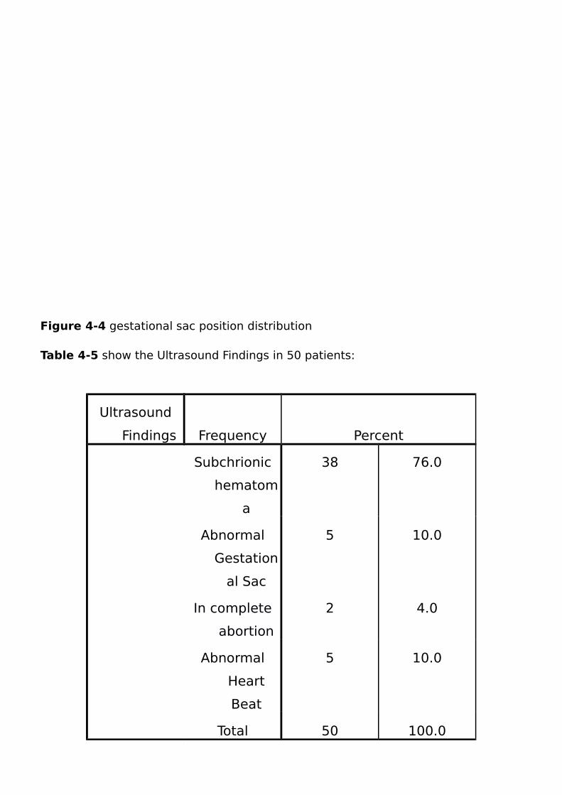
Figure 4-4 gestational sac position distribution
Table 4-5 show the Ultrasound Findings in 50 patients:
Ultrasound
Findings Frequency Percent
Subchrionic
hematom
a
38 76.0
Abnormal
Gestation
al Sac
5 10.0
In complete
abortion
2 4.0
Abnormal
Heart
Beat
5 10.0
Total 50 100.0
Figure 4-5 ultrasound finding distribution
Table 4-6 show the correlation between Maternal Age and Ultrasound Findings :
61
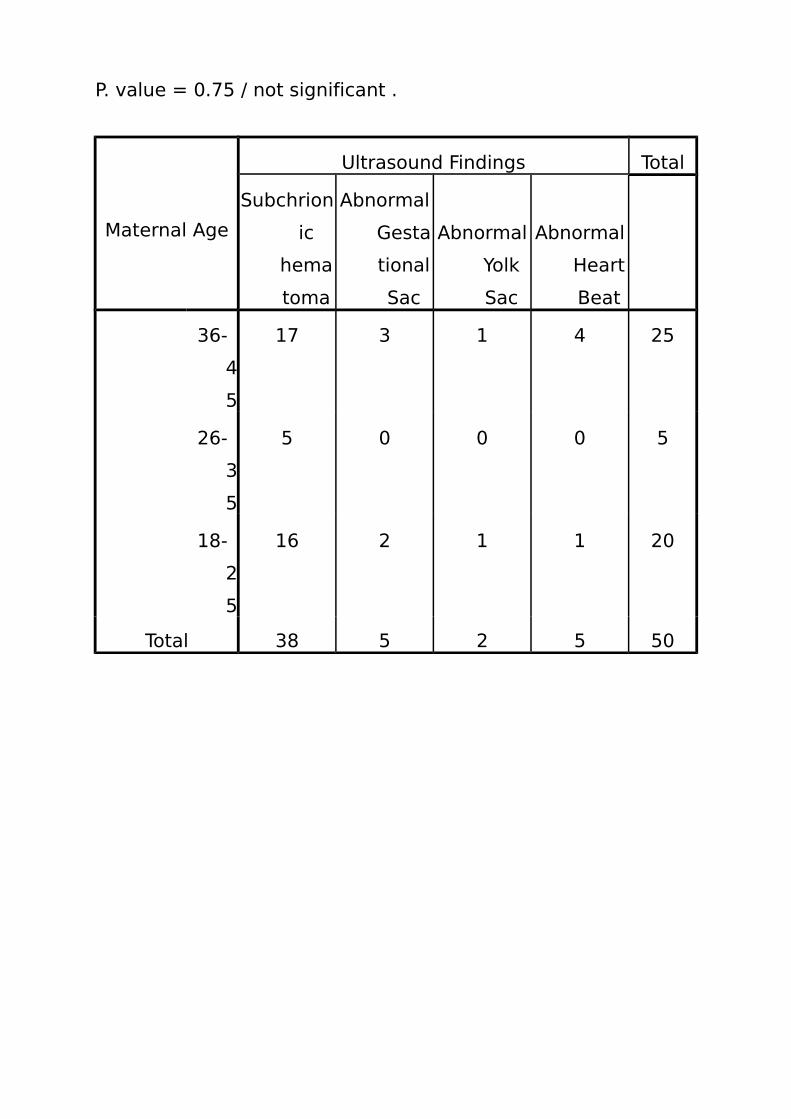
P. value = 0.75 / not significant .
Maternal Age
Ultrasound Findings Total
Subchrion
ic
hema
toma
Abnormal
Gesta
tional
Sac
Abnormal
Yolk
Sac
Abnormal
Heart
Beat
36-
4
5
17 3 1 4 25
26-
3
5
5 0 0 0 5
18-
2
5
16 2 1 1 20
Total 38 5 2 5 50
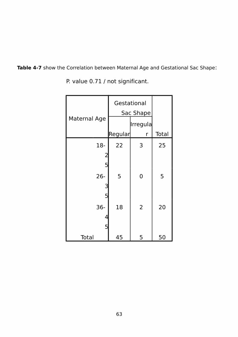
Table 4-7 show the Correlation between Maternal Age and Gestational Sac Shape:
P. value 0.71 / not significant.
Maternal Age
Gestational
Sac Shape
TotalRegular
Irregula
r
18-
2
5
22 3 25
26-
3
5
5 0 5
36-
4
5
18 2 20
Total 45 5 50
63

Table 4-8 show the Correlation between Maternal Age and Gestational Sac
Position:
P. value 0.22
Maternal Age
Gestational Sac Position
Total
LOWE
R
INBETWE
EN
HIGH
FUND
AL
36-
4
5
1 6 18 25
26-
3
5
1 3 1 5
18-
2
5
3 6 11 20
Total 5 15 30 50

Table 4-9 show the Correlation between Gestational Age and Ultrasound Findings:
65
P. value 0.11/not significant
Gestational Age
Ultrasound Findings
Total
Subchrion
ic
hema
toma
Abnormal
Gesta
tional
Sac
Abnormal
Yolk
Sac
Abnormal
Heart
Beat
5-7
Wee
k
16 5 2 2 25
7-9
Wee
k
7 0 0 3 10
9-11
Wee
k
9 0 0 0 9
11-13
Wee
k
6 0 0 0 6
Total 38 5 2 5 50

Chapter five
Discussion, Conclusion and Recommendation
5.1 Discussion:
During the period of the study 50 pregnant attended to ultrasound
department for investigation to determine the ultrasound finding of
threatened abortion, the sonographic findings were documented and
analyzed.
In table 4-1: The most common represented ages were women between
(36-45) years old (50%), followed by age groups (18-25) years and (26-
67

35) years, (40%) and (10%) respectively, which agreed with
Tannirandeny et al study done in 2001.
In table 4-2: The study showed that The threatened abortion were most
common among the gestational age between 5-7 weeks (50%)
followed by gestational age 7-9 weeks (20%), 9-11 weeks (18%) and 11-
13 weeks (12%) respectively, which agreed with rearch result of
Elizabeth Sep 2011, Tannirandeny et al 2001 and Millie A.Behera et al
Oct 2011.
In table 4-3: regular , and irregular shape gestational sac were
presented in most cases of the threatened abortion; (90%) and
(10%) respectively, agreed with study of Sandor Nagy et al 1999-
2001.
In table 4-4: 60% of the represented cases were presented with
high fundal gestional sac position, 30% were presented with eccentric
gestational sac position and 10% with lower as shown in table (4-4).
In table 4-5: The study showed that Sonographic feature of threatened
abortion were Sub chorionic hematoma which is the most
significant finding 76% , presence of irregular heart beat 10% ,
irregular thin walled gestational sac 10% and abnormal yolk sac in
4% which agreed with study of stannirandeny et al 2001.
(Table (4-6) Chi square test shows that the P-value is (0.75), means that
there is no significant relationship between the diagnosis and patient
age.
5.2 Conclusion:
Diagnostic ultrasound is the most common imaging technique used
to diagnose the threatened abortion and is an accurate means of
evaluating many associated sonographic findings.
because Sonographic findings are operator-dependent have to be
supported by the history and physical exam of the patient.
Most common represented ages were women between (36-45) years
old and The threatened abortion were most common among the
gestational age between 5-7 weeks (50%) .
The Sonographic features of threatened abortion were Sub chorionic
hematoma which is the most significant finding 76% and regular shape
gestational sac presented in most cases of the threatened abortion
(90%) containing fetus with irregular rapid heart beat 10% and
abnormal yolk sac of 4% .
5.3 Recommendation:
1. The study recommended that real time ultrasonography should be
the first choice modality for the diagnosis of threatened abortion.
2. All obstetrical ER rooms must include US imaging machine.
3. US request should included brief clinical and historical background
which may add in narrowing the deferential diagnosis.
4. ER sinologists have to be adapted and continuously educated to
increase their knowledge. Familiarity with the sonographic features
of threatened abortion.
5. Women with risk factors may take care and bed rest.
69

Refrences :-
1- Arias F. Early pregnancy loss. In practical guide to high risk pregnancy and delivery; 2nd ed. Missouri 1997; 62-7
2- Baztofen JH, Fielding WL, Friedman EA. Effect of vaginal bleeding in early pregnancy outcome. Obstet Gynecol 1984;63:515-8.)
3- Carol M. Rumack, Stephamie R. Wilson, J. William Charboneau and Deborah Levine. Diagnostic Ultrasound.4th edition Volume one. Elsevier Mosby; Philadelphia: 2011. P.840
4- Cunningham FG, GantNF, Leveno KJ, Gilstrap LC III, Health JC, Wenstrom KD. 21st ed. Williams obstetrics. New York: McGraw-Hill,. P. 866-67
5- David Deen ,gynecology obstetric ,burwin institude of diagnostic medical ultrasound 1992
6- David Shier, Jackie Butler, and Richi Lewis. Human Anatomy and Physiology. 10thed.The Mc Graw-Hill Companies; New York: 2004.P 835
7- Elsevier Mosby; basic of obstetrical and gynecological ultrasonography ,3rd edition , Philadelphia: 2011. P.840-866-869.
8- H.T.Lutz H.A.Gharbi , manual of diagnostic ultrasound in infectious tropical disease ,verlage berlin ,germany 2006 P.1-7
9- Micael Mc Kinley, and Valerie Dean O’Louchlin. Human Anatomy.Higher Education; New York: 2006.P.822
10- Moore KL, Persaud TVN (1998) The urogenital system. In: Moore KL, Persaud TVN (eds) The developing human. Clinically oriented embryology, ed 6. WB Saunders, Philadelphia, pp 303–347.
11- obstetrical u\s 3rd edition ,Trish chudleigh and Basky Thilaganathan Elsevier Limited 2004 london p.52-55
12- Saurbrei EE, Pham DH. Placental abruption and subchorionic hemorrhage in the first half of pregnancy: US appearance and clinical outcome. Radiology 1986. P.160
13- Sotiriadis A, Papatheodorou S, Makrydimas G. Threatenedmiscarriage: evaluation and management. BMJ 2004;329:152-155
14- Stabile I, Campbell S, Grudzinskas JG. Threatened miscarriage and intrauterine haematomas: Sonographic and biochemical studies. J Ultrasound Med 1989;8:289-92
The appendices :-
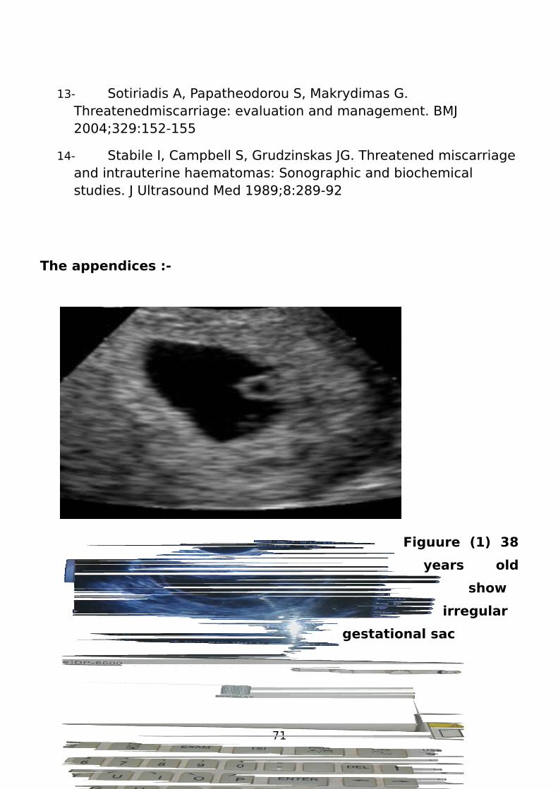
Figuure (1) 38
years old
show
irregular
gestational sac
71
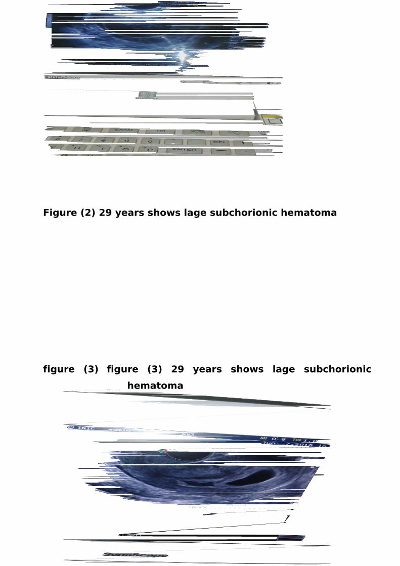
Figure (2) 29 years shows lage subchorionic hematoma
figure (3) figure (3) 29 years shows lage subchorionic
hematoma
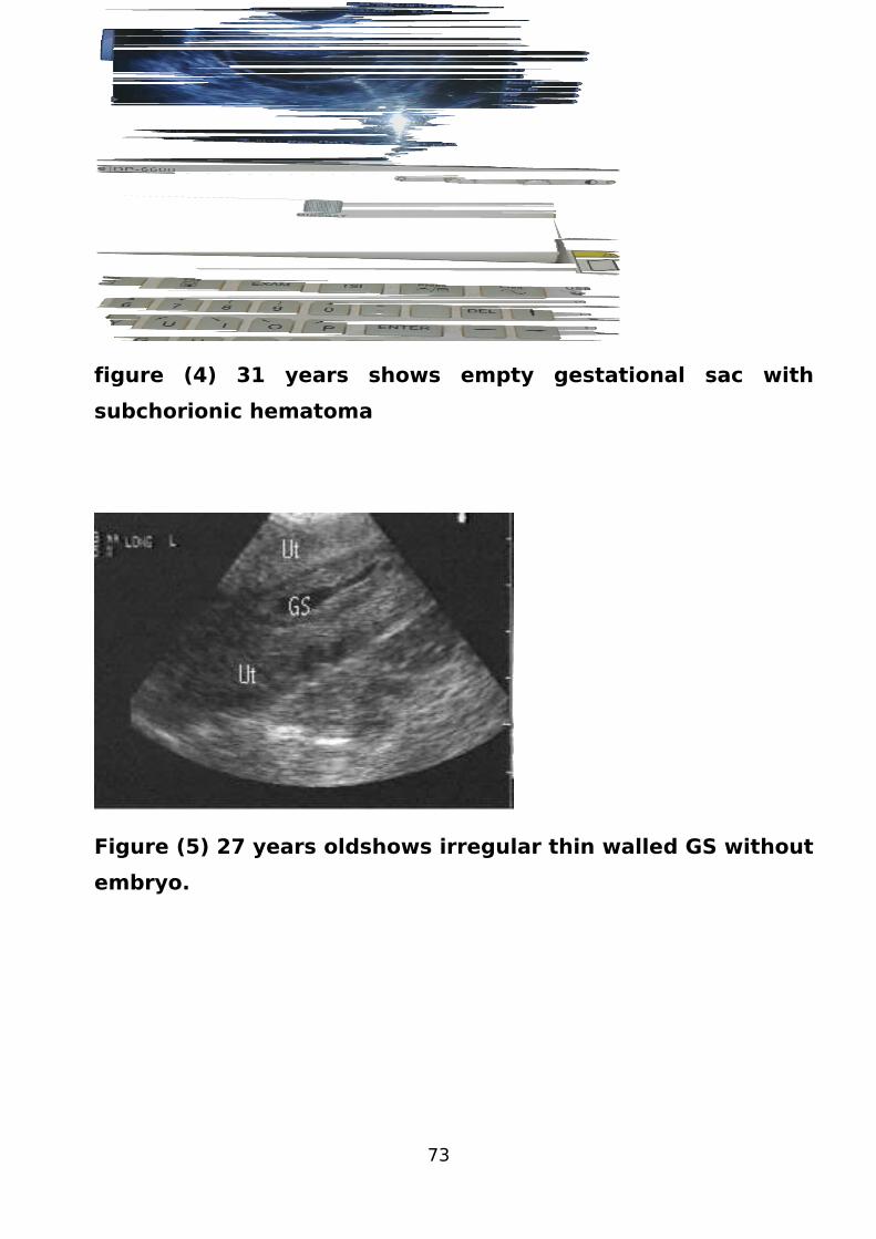
figure (4) 31 years shows empty gestational sac with
subchorionic hematoma
Figure (5) 27 years oldshows irregular thin walled GS without
embryo.
73
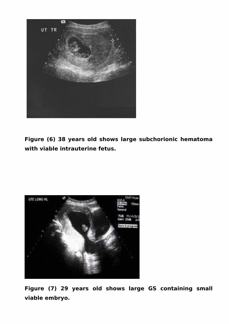
Figure (6) 38 years old shows large subchorionic hematoma
with viable intrauterine fetus.
Figure (7) 29 years old shows large GS containing small
viable embryo.
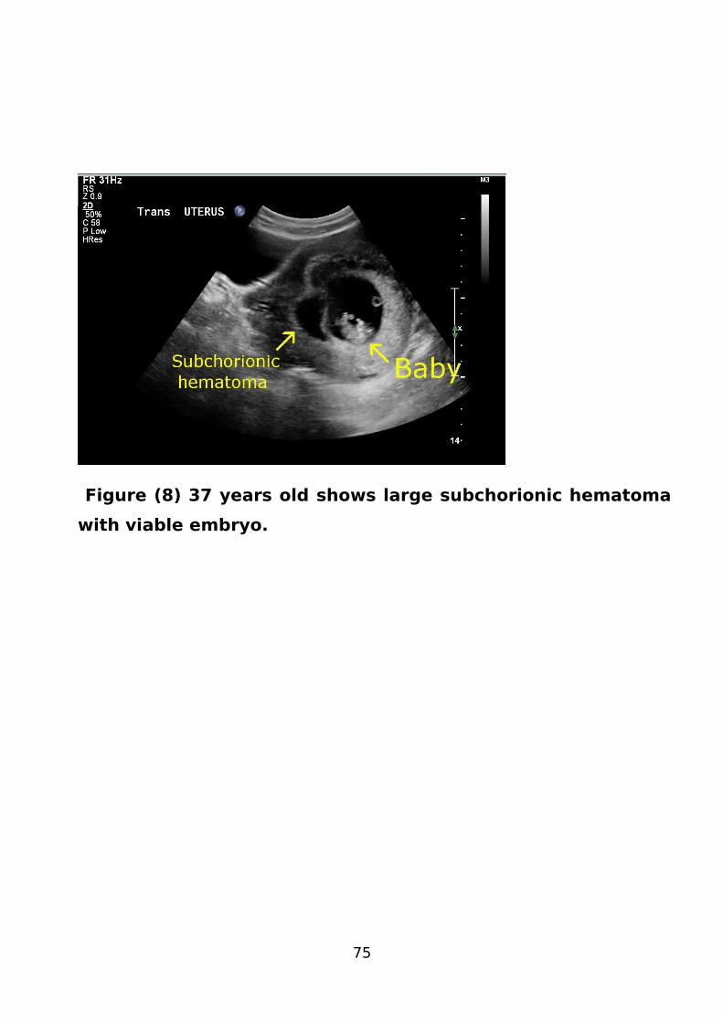
Figure (8) 37 years old shows large subchorionic hematoma
with viable embryo.
75
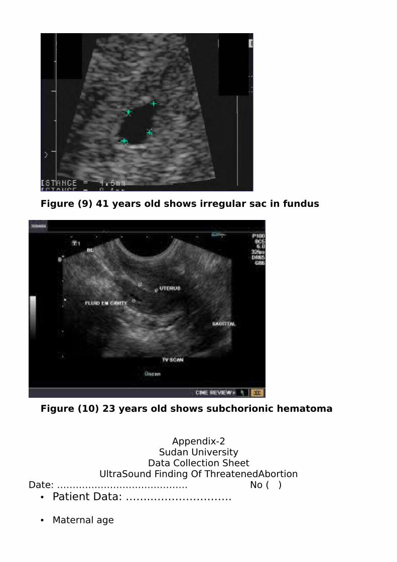
Figure (9) 41 years old shows irregular sac in fundus
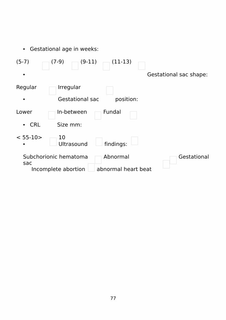
Figure (10) 23 years old shows subchorionic hematoma
Appendix-2Sudan University
Data Collection SheetUltraSound Finding Of ThreatenedAbortion
Date: …………………………………… No ( )• Patient Data: ……..………………….
• Maternal age
• Gestational age in weeks:
(5-7) (7-9) (9-11) (11-13)
• Gestational sac shape:
Regular Irregular
• Gestational sac position:
Lower In-between Fundal
• CRL Size mm:
< 55-10> 10 • Ultrasound findings:
Subchorionic hematoma Abnormal Gestational sac Incomplete abortion abnormal heart beat
77