Sbe2p and Sbe22p, Two Homologous Golgi Proteins Involved in Yeast Cell Wall Formation
18
Molecular Biology of the Cell Vol. 11, 435– 452, February 2000 Sbe2p and Sbe22p, Two Homologous Golgi Proteins Involved in Yeast Cell Wall Formation Beatriz Santos and Michael Snyder* Department of Molecular, Cellular, and Developmental Biology, Yale University, New Haven, Connecticut 06520-8103 Submitted July 26, 1999; Revised November 8, 1999; Accepted November 12, 1999 Monitoring Editor: Pam Silver The cell wall of fungal cells is important for cell integrity and cell morphogenesis and protects against harmful environmental conditions. The yeast cell wall is a complex structure consisting mainly of mannoproteins, glucan, and chitin. The molecular mechanisms by which the cell wall components are synthesized and transported to the cell surface are poorly understood. We have identified and characterized two homologous yeast proteins, Sbe2p and Sbe22p, through their suppression of a chs5 spa2 mutant strain defective in chitin synthesis and cell morphogenesis. Although sbe2 and sbe22 null mutants are viable, sbe2 sbe22 cells display several phenotypes indicative of defects in cell integrity and cell wall structure. First, sbe2 sbe22 cells display a sorbitol-remediable lysis defect at 37°C and are hypersensitive to SDS and calcofluor. Second, electron microscopic analysis reveals that sbe2 sbe22 cells have an aberrant cell wall structure with a reduced mannoprotein layer. Finally, immunofluorescence experiments reveal that in small- budded cells, sbe2 sbe22 mutants mislocalize Chs3p, a protein involved in chitin synthesis. In addition, sbe2 sbe22 diploids have a bud-site selection defect, displaying a random budding pattern. A Sbe2p–GFP fusion protein localizes to cytoplasmic patches, and Sbe2p cofractionates with Golgi proteins. Deletion of CHS5, which encodes a Golgi protein involved in the transport of Chs3p to the cell periphery, is lethal in combination with disruption of SBE2 and SBE22. Thus, we suggest a model in which Sbe2p and Sbe22p are involved in the transport of cell wall components from the Golgi apparatus to the cell surface periphery in a pathway independent of Chs5p. INTRODUCTION Yeast cell walls are essential for the maintenance of cell shape, prevention of lysis, and regulation of the uptake of substances from the environment. In spite of its apparent rigidity, the yeast cell wall is a dynamic structure that can be remodeled in response to different physiological states (e.g., budding, mating, and sporulation) or to morphological changes, such as in Candida albicans during the transition from yeast to hyphal growth (Cid et al., 1995). The Saccharo- myces cerevisiae cell wall is a complex structure composed of mannoproteins, b-1,3-glucan, and b-1,6-glucan, all cross- linked to each other and to chitin, a N-acetylglucosamine polymer (Orlean, 1997). To mediate bud formation and growth, new plasma membrane and cell wall material must be directed properly to the site of growth, presumably through the secretory pathway. How cell wall components are transported to the cell surface and whether multiple pathways are involved remain poorly understood. The iden- tification and characterization of components that partici- pate in specific steps of this transport will be crucial for the understanding of how this process occurs at the molecular level. Chitin is an essential structural component of the cell wall present at very low abundance (Shaw et al., 1991). Chitin deposition is spatially and temporally regulated throughout the yeast cell cycle and life cycle (for review, see Cid et al., 1995; Orlean, 1997). Three chitin synthase activities (CSI, CSII, and CSIII) have been described; each has a distinct function. CSIII activity is required for the formation of the chitin ring at the base of the bud and for chitin deposition in the lateral wall during vegetative growth, as well as for chitin synthesis during mating and sporulation (Shaw et al., 1991; Valdivieso et al., 1991). Several proteins, including Chs3p, Chs4p, Chs5p, Chs6p, and Chs7p, are required for the CSIII activity. Chs3p has significant homology with other chitin synthases and is the catalytic component of CSIII (Valdivieso et al., 1991). Chs3p is present at the cell periphery and in cytoplasmic patches and undergoes a dy- namic localization during the cell cycle. Chs3p localizes at the incipient bud site in unbudded cells and at the bud neck in small-budded cells and cells undergoing cytokinesis (Chuang and Schekman, 1996; Santos and Snyder, 1997). * Corresponding author. E-mail address: michael.snyder@ yale.edu. © 2000 by The American Society for Cell Biology 435
-
Upload
independent -
Category
Documents
-
view
1 -
download
0
Transcript of Sbe2p and Sbe22p, Two Homologous Golgi Proteins Involved in Yeast Cell Wall Formation

Molecular Biology of the CellVol. 11, 435–452, February 2000
Sbe2p and Sbe22p, Two Homologous Golgi ProteinsInvolved in Yeast Cell Wall FormationBeatriz Santos and Michael Snyder*
Department of Molecular, Cellular, and Developmental Biology, Yale University, New Haven,Connecticut 06520-8103
Submitted July 26, 1999; Revised November 8, 1999; Accepted November 12, 1999Monitoring Editor: Pam Silver
The cell wall of fungal cells is important for cell integrity and cell morphogenesis and protectsagainst harmful environmental conditions. The yeast cell wall is a complex structure consistingmainly of mannoproteins, glucan, and chitin. The molecular mechanisms by which the cell wallcomponents are synthesized and transported to the cell surface are poorly understood. We haveidentified and characterized two homologous yeast proteins, Sbe2p and Sbe22p, through theirsuppression of a chs5 spa2 mutant strain defective in chitin synthesis and cell morphogenesis.Although sbe2 and sbe22 null mutants are viable, sbe2 sbe22 cells display several phenotypesindicative of defects in cell integrity and cell wall structure. First, sbe2 sbe22 cells display asorbitol-remediable lysis defect at 37°C and are hypersensitive to SDS and calcofluor. Second,electron microscopic analysis reveals that sbe2 sbe22 cells have an aberrant cell wall structure witha reduced mannoprotein layer. Finally, immunofluorescence experiments reveal that in small-budded cells, sbe2 sbe22 mutants mislocalize Chs3p, a protein involved in chitin synthesis. Inaddition, sbe2 sbe22 diploids have a bud-site selection defect, displaying a random buddingpattern. A Sbe2p–GFP fusion protein localizes to cytoplasmic patches, and Sbe2p cofractionateswith Golgi proteins. Deletion of CHS5, which encodes a Golgi protein involved in the transport ofChs3p to the cell periphery, is lethal in combination with disruption of SBE2 and SBE22. Thus, wesuggest a model in which Sbe2p and Sbe22p are involved in the transport of cell wall componentsfrom the Golgi apparatus to the cell surface periphery in a pathway independent of Chs5p.
INTRODUCTION
Yeast cell walls are essential for the maintenance of cellshape, prevention of lysis, and regulation of the uptake ofsubstances from the environment. In spite of its apparentrigidity, the yeast cell wall is a dynamic structure that can beremodeled in response to different physiological states (e.g.,budding, mating, and sporulation) or to morphologicalchanges, such as in Candida albicans during the transitionfrom yeast to hyphal growth (Cid et al., 1995). The Saccharo-myces cerevisiae cell wall is a complex structure composed ofmannoproteins, b-1,3-glucan, and b-1,6-glucan, all cross-linked to each other and to chitin, a N-acetylglucosaminepolymer (Orlean, 1997). To mediate bud formation andgrowth, new plasma membrane and cell wall material mustbe directed properly to the site of growth, presumablythrough the secretory pathway. How cell wall componentsare transported to the cell surface and whether multiplepathways are involved remain poorly understood. The iden-tification and characterization of components that partici-
pate in specific steps of this transport will be crucial for theunderstanding of how this process occurs at the molecularlevel.
Chitin is an essential structural component of the cell wallpresent at very low abundance (Shaw et al., 1991). Chitindeposition is spatially and temporally regulated throughoutthe yeast cell cycle and life cycle (for review, see Cid et al.,1995; Orlean, 1997). Three chitin synthase activities (CSI,CSII, and CSIII) have been described; each has a distinctfunction. CSIII activity is required for the formation of thechitin ring at the base of the bud and for chitin deposition inthe lateral wall during vegetative growth, as well as forchitin synthesis during mating and sporulation (Shaw et al.,1991; Valdivieso et al., 1991). Several proteins, includingChs3p, Chs4p, Chs5p, Chs6p, and Chs7p, are required forthe CSIII activity. Chs3p has significant homology withother chitin synthases and is the catalytic component ofCSIII (Valdivieso et al., 1991). Chs3p is present at the cellperiphery and in cytoplasmic patches and undergoes a dy-namic localization during the cell cycle. Chs3p localizes atthe incipient bud site in unbudded cells and at the bud neckin small-budded cells and cells undergoing cytokinesis(Chuang and Schekman, 1996; Santos and Snyder, 1997).
* Corresponding author. E-mail address: [email protected].
© 2000 by The American Society for Cell Biology 435
Chs4p, Chs5p, Chs6p, Chs7p, and two members of the yeastSNAREs, Tlg1p and Tlg2p, are implicated in the highlyregulated localization of Chs3p (DeMarini et al., 1997; Santosand Snyder, 1997; Holthuis et al., 1998; Ziman et al., 1998;Trilla et al., 1999). Chs4p is involved in the proper localiza-tion of Chs3p at the bud neck through an indirect interactionwith the septins (DeMarini et al., 1997). Septins are highlyconserved cytoskeletal proteins that assemble into filamentsand are essential for cytokinesis (Longtine et al., 1996).Chs5p and Chs6p are required for the transport of Chs3pfrom an internal membrane compartment to the plasmamembrane (Santos and Snyder, 1997; Ziman et al., 1998).Chs5p colocalizes with Kex2p in the trans-Golgi network(Santos and Snyder, 1997). Chs5p also has a chitin-indepen-dent role in cell fusion during mating (Santos et al., 1997).Chs7p is an endoplasmic reticulum (ER) protein required forthe export of Chs3p from the ER (Trilla et al., 1999). Thus,these different proteins form a pathway for the transport ofChs3p, an important protein involved in cell wall synthesis.Because all of these proteins are not essential, it is possiblethat there are other as-yet-unidentified factors playing im-portant functions for cell wall construction and transport ofcell wall components.
Several proteins involved in cell wall synthesis are alsoimplicated in cell polarity and morphogenesis (Cabib et al.,1998). Polarized cell growth in yeast is a complex processthat requires the reorganization of the actin cytoskeleton,polarized secretion, and the function and regulation of sig-nal transduction cascades (reviewed by Madden and Sny-der, 1998). A number of proteins important for cell polarityin yeast have been identified. One such protein, Spa2p, islocated at polarized growth sites, including the incipientbud site of unbudded cells, the bud tips of small-buddedcells, the necks of cells undergoing cytokinesis, and theprojection tips of mating cells (Snyder, 1989). spa2 mutantsare defective in bud-site selection, apical bud growth,pseudohyphal growth, and mating projection formation(Gehrung and Snyder, 1990; Roemer et al., 1998). Spa2pinteracts physically with other cell polarity proteins, such asPea2p and Bud6p, and with components of two MAPKpathways, the mating signaling pathway and the Slt2pMAPK pathway (Sheu et al., 1998). This latter MAPK path-way functions downstream of PKC to maintain cellular in-tegrity during polarized growth (for review, see Maddenand Snyder, 1998).
chs5 and spa2 mutants share several common phenotypesaffecting polarized growth processes: they are defective inmating projection formation and cell fusion during mating,and both exhibit altered bud-site selection in diploids duringvegetative growth (Gehrung and Snyder, 1990; Santos et al.,1997; our unpublished results). Furthermore, the sequencesof these two proteins display some common structural fea-tures: both show low-level similarity with filamentous pro-teins (Chs5p with neurofilaments and Spa2p with keratinsand myosins), and both contain heptad repeats of unknownfunction (Gehrung and Snyder, 1990; Santos et al., 1997).
Here, we describe a genetic interaction between chs5 andspa2 mutants and show that the double mutant is inviable athigh temperature. To identify additional components im-portant for cell wall synthesis and cell morphogenesis, wehave isolated high-copy-number suppressors of the growthdefect of chs5 spa2 cells. We have characterized one of these
suppressors, SBE2, and its highly related gene, SBE22. sbe2sbe22 double mutants exhibit cell polarity and cell wall de-fects. We present evidence suggesting that Sbe2p andSbe22p are novel Golgi proteins required for proper cell wallformation.
MATERIALS AND METHODS
Media and Microbiological TechniquesGenetic methods and growth media were as described by Guthrieand Fink (1991). Cell lysis was visualized on YPDA (rich mediumsupplemented with adenine) containing 0.001% methylene bluedye. YPDAS is YPDA containing 1 M sorbitol. Calcofluor sensitivitywas analyzed on plates containing synthetic complete (SC) mediumsupplemented with 50 mg/ml fluorescent brightener 28 (calcofluorwhite; Sigma Chemical, St. Louis, MO). SDS sensitivity was ana-lyzed on YPDA plates containing 0.0025% SDS. Yeast transforma-tions were performed by the lithium acetate method of Ito et al.(1983).
Isolation of Multicopy Suppressors of chs5 spa2StrainA chs5D spa2D strain (Y1941) was transformed with a yeast genomicDNA library constructed in the multicopy plasmid YEp24 (Carlsonand Botstein, 1982). After transformation, cells were incubated at30°C for 24 h to allow recovery and then incubated at 37°C for 2 d.About 18,000 transformants were analyzed, and 18 transformantsthat reproducibly grew at 37°C were obtained. From each of these 18transformants, plasmids were recovered for further analysis. Eightof them failed to suppress the growth defect of chs5 spa2 at 37°Cwhen they were reintroduced into Y1941, indicating that the sup-pression event was not due to the plasmids. The genes present in theremaining 10 plasmids were identified by sequencing both ends ofthe insert.
Yeast Strains and PlasmidsYeast strains used in this study are listed in Table 1. Construction ofthe chs5::ADE2 allele is described by Santos et al. (1997),3Xmyc::CHS5 and CHS3::3XHA alleles are described by Santos andSnyder (1997), and spa2::URA3 and spa2::TRP1 alleles are describedby Gehrung and Snyder (1990).
The complete ORFs of SBE2 (from 215 base pairs upstream of theATG to the stop codon) and SBE22 (from the start codon to the stopcodon) were deleted with the use of the PCR disruption procedureof Baudin et al. (1993). A sbe2::HIS3 null allele was generated usingoligonucleotides 59-CGGGCTTCCACCTTTGCTTCATTATTTTAC-TTCAGCTCTTTAGCTTTCTGTGACGCGCGTTTCGGTGATGACGGTG-39 and 59-GAACTTAAGAAGAGATAGTCTGGTCACCAA-ACTTTTAGTACGTGCCACATACACGGGGTGATGGTTCA-CGT-AGTGGGC-39 to amplify the HIS3 gene from pRS313. Underlinedportions of primers correspond to common sequences that flankeach of the selectable markers within the pRS313–pRS316 series ofplasmids (Sikorski and Hieter, 1989). Null alleles of SBE22 werecreated similarly. A sbe22::URA3 strain was generated with the useof oligonucleotides 59-CAAATTTGTCCTTATCTTTAGTTAATAC-GGTCTAACTTGCCACGCTACTCAAGAAGCGCGTTTCGGTGATGACGGTG-39 and 59-CCAGTTTTTTTTTTCTTGTGCATGAGTGA-AATTACAGTTACAAAAAATAGGGTGATGGTTCACGTAG-TG-GGC-39 to amplify the URA3 gene from pRS316. In addition, asbe22::TRP1 null allele was generated with the use of the sameoligonucleotides and pRS314 to amplify the TRP1 gene.
Strains containing the SBE2::3XHA or SBE2::3Xmyc alleles wereconstructed with the use of the PCR epitope-tagging method ofSchneider et al. (1995). Both epitopes were integrated at the Cterminus of the coding region (before the stop codon). Primers
B. Santos and M. Snyder
Molecular Biology of the Cell436
59-CCTCAATAATATTTTCACAAAGTGGTGGTTGCACTACTAC-CGAAAATTACGTAGGGAACAAAAGCTGG-39 and 59-CTTAAGA-AGAGATAGTCTGGTCACCAAACTTTTAGTACGTGCCACATACA-CGCTACTATAGGGCGAATTGG-39 were used. Proper formation ofthe hemagglutinin (HA)-tagged and myc-tagged alleles was confirmedby PCR and immunoblot analyses.
The green fluorescent protein (GFPS65T; Heim and Tsien, 1996)was fused to the N terminus of Sbe2p. A NotI site was created byPCR after the fourth codon of SBE2, creating the plasmid pBU55(YEp24 vector). A fragment containing GFPS65T flanked by NotIsites and in frame with the SBE2 ORF was cloned in pBU55,creating pBU62. A BamHI/KpnI fragment from pBU62 containingSBE2::GFP was cloned in the same restriction sites of the centro-meric vector pRS316, creating pBU65, the plasmid used in thisstudy.
Mating Projection and Budding Pattern AnalysesFor pheromone-treatment experiments, cells were grown to early logphase, a-factor (Sigma Chemical) was added to a final concentration of5 mg/ml, and cells were incubated at 30°C with shaking for 45 min.Cultures were supplemented with a second addition of the sameamount of a-factor and incubated for another 45 min. Microscopicexamination of the cultures revealed that after 90 min most of thewild-type cells (90%) were unbudded and had formed shmoos.
Budding patterns were examined by staining cells with cal-cofluor. Mid log phase cells were fixed in 3.7% formaldehyde at30°C with rotation, washed with water, and resuspended in 50 mMTris, pH 7.5, containing 5 mg/ml calcofluor. Cells with two or morebud scars were scored and were classified into three categories: (1)bud scars only at one pole; (2) bud scars at both poles; and (3)random distribution of bud scars.
Table 1. Yeast strains used in this study
Strain Genotypes
Y603 MATa ura3-52 lys2-801 ade2-101 trp1-901 his3-D200Y601 MATa ura3-52 lys2-801 ade2-101 trp1-901 his3-D200 spa2::URA3Y1935 MATa ura3-52 lys2-801 ade2-101 trp1-901 his3-D200 chs5::ADE2Y1936 MATa ura3-52 lys2-801 ade2-101 trp1-901 his3-D200chs5::ADE2 spa2::URA3Y1937 MATa ura3-52 lys2-801 ade2-101 trp1-901 his3-D200 CDC3::3XHAY1938 MATa ura3-52 lys2-801 ade2-101 trp1-901 his3-D200 CDC3::3XHA chs5::ADE2Y1939 MATa ura3-52 lys2-801 ade2-101 trp1-901 his3-D200 CDC3::3XHA spa2::TRP1Y1940 MATa ura3-52 lys2-801 ade2-101 trp1-901 his3-D200 CDC3::3XHA chs5::ADE2 spa2::TRP1Y1941 MATa ura3-52 lys2-801 ade2-101 trp1-901 his3D-200 chs5::ADE2 spa2::TRP1Y270 MATa/a ura3-52/ura3-52 lys2-801/lys2-801 ade2-101/ade2-101 trp1-901/trp1-901 his3-D200/his3-D200Y1942 MATa/a ura3-52/ura3-52 lys2-801/lys2-801 ade2-101/ade2-101 trp1-901/trp1-901 his3-D200/his3-D200
sbe2::HIS3/sbe2::HIS3Y1943 MATa/a ura3-52/ura3-52 lys2-801/lys2-801 ade2-101/ade2-101 trp1-901/trp1-901 his3-D200/his3-D200 sbe22:
URA3/sbe22:URA3Y1944 MATa/a ura3-52/ura3-52 lys2-801/lys2-801 ade2-101/ade2-101 trp1-901/trp1-901 his3-D200/his3-D200
sbe2::HIS3/sbe2::HIS3 sbe22:URA3/sbe22::URA3Y1310 MATa ura3-52 lys2-801 ade2-101 trp1-901 his3-D200 3Xmyc::CHS5 CHS3::3XHAY1945 MATa ura3-52 lys2-801 ade2-101 trp1-901 his3-D200 3Xmyc::CHS5 CHS3::3XHA sbe2::HIS3Y1946 MATa ura3-52 lys2-801 ade2-101 trp1-901 his3-D200 3Xmyc::CHS5 CHS3::3XHA sbe2::HIS3 sbe22::TRP1Y1947 MATa ura3-52 lys2-801 ade2-101 trp1-901 his3-D200 3Xmyc::CHS5 CHS3::3XHA sbe2::HIS3 sbe22::TRP1Y1948 MATa ura3-52 lys2-801 ade2-101 trp1-901 his3-D200 3Xmyc::CHS5 SBE2::3XHANY13a MATa ura3-52NY26a MATa ura3-52 sec2-59NY405a MATa ura3-52 sec4-8NY17a MATa ura3-52 sec6-4NY759a MATa ura3-52 his4-619 sec7-1NY415a MATa ura3-52 sec16-2Y604 MATa ura3-52 lys2-801 ade2-101 trp1-901 his3-D200Y1949 MATa ura3-52 lys2-801 ade2-101 trp1-901 his3-D200 sbe2::HIS3 sbe22::TRP1Y1950 MATa ura3-52 lys2-801 ade2-101 trp1-901 his3-D200 chs5::ADE2Y1951 MATa ura3-52 lys2-801 ade2-101 trp1-901 his3-D200 sbe2::HIS3 sbe22::TRP1 chs5::ADE2Y760 MATa ura3-52 lys2-801 ade2-101 trp1-901 his3-D200 bck1:URA3Y1952 MATa ura3-52 lys2-801 ade2-101 trp1-901 his3-D200 sbe2::HIS3 sbe22::TRP1 bck1::URA3Y602 MATa ura3-52 lys2-801 ade2-101 trp1-901 his3-D200 spa2::URA3Y1953 MATa ura3-52 lys2-801 ade2-101 trp1-901 his3-D200 sbe2::HIS3 sbe22::TRP1 spa2::URA3Y1954b MATa ura3 leu2 ade2 lys2 his3 cdc24-4Y1955b MATa ura3 lys2-801 ade2-101 trp1-901 his3-D200 sbe2::HIS3 sbe22::URA3 cdc24-4Y1956b MATa cdc12-1 ura3 leu2 ade2 lys2 his3Y1957b MATa ura3-52 lys2-801 ade2-101 trp1-901 his3-D200 sbe2::HIS3 sbe22::URA3 cdc12-1Y1958 MATa ura3-52 lys2-801 ade2-101 trp1-901 his3-D200 SBE2::3XHAY1959 MATa ura3-52 lys2-801 ade2-101 trp1-901 his3-D200 SBE2::3Xmyc
a These strains are from P. Novick at Yale University.b cdc24-4 and cdc12-1 alleles originally come from J. Pringle’s strains (University of North Carolina, Chapel Hill, NC), but they have beenbackcrossed into our background.
Sbe2p Is Involved in Cell Wall Formation
Vol. 11, February 2000 437
Thin Section Electron MicroscopyCultures of wild-type (Y270), sbe2 (Y1942), sbe22 (Y1943), and sbe2sbe22 (Y1944) cells were grown overnight at 30°C in YPDAS andthen diluted into fresh YPDA medium and incubated for 6 h at 30°C(early log phase). Cells (15 ml) were fixed by adding 50% glutaral-dehyde to a final concentration of 1%, incubated for 5 min on ice,and pelleted in a tabletop centrifuge at 4°C. Cell pellets were resus-pended in 1 ml of PBS. Samples were subsequently processedaccording to the method described by Kaiser and Schekman (1990)but with 1% osmium tetroxide instead of permanganate for fixation.
Enzyme AssaysExternal invertase activity was measured as described by Goldsteinand Lampen (1975). Internal invertase was determined by assayingspheroplast lysates prepared as described by Novick and Schekman(1979). The percentage of secreted invertase represents the level ofexternal invertase divided by the total amount of the enzyme,external plus internal. For exoglucanase activity measurement, logphase cells grown in YPDA were diluted in fresh YPDA medium atOD600 5 0.25 and incubated at 25 or 37°C for 2 h. Cells werepelleted, and 200 ml of the culture supernatant was used for theassay, as described by Nebreda et al. (1986). The amount of exoglu-canase liberated in the wild-type cells at 25°C was scored as 100%secretion, and all of the other values are relative to this one. Notethat the sec6-4 strain used in these experiments has a differentbackground than Y270 and Y1944.
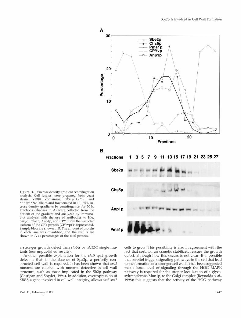
Sucrose Density Gradient CentrifugationCell lysates were prepared from strain Y1948 and analyzed asdescribed by Santos and Snyder (1997). Briefly, cells were grown inYPDA to mid log phase (2 3 107 cells/ml), and 2 g of cells (wetweight) was resuspended in 6 ml of 17% sucrose (wt/vol) in 50 mMTris-HCl, pH 7.5, 1 mM EDTA containing protease inhibitors (SigmaChemical) and 6 ml of glass beads. Cells were broken by vortexing,and the crude extract was centrifuged at 1500 3 g for 10 min. Thesupernatant was layered on top of a 33-ml linear sucrose gradient(10–65%, wt/vol) in 50 mM Tris-HCl, 1 mM EDTA, pH 7.5. Thetubes were centrifuged in a SW28 rotor at 25,000 rpm for 20 h at 4°C(Beckman Instruments, Fullerton, CA). One-milliliter fractions werecollected from the bottom of the tube with the use of a peristalticpump and analyzed by immunoblot analysis, as described by San-tos and Snyder (1997). Monoclonal anti-HA (HA.11; BABCO, Rich-mond, CA) or anti-myc (9E10; BABCO) antibodies were used fordetection of epitope-tagged Sbe2p and Chs5p, respectively. Rabbitpolyclonal antibodies against Pma1p, Anp1p, and carboxypeptidaseY (CPY) (kindly provided by C. Slayman [Yale University, NewHaven, CT], S. Munro [Laboratory of Molecular Biology, Cam-bridge, England], and P. Novick [Yale Univeristy], respectively)were used as markers of different cellular compartments. The reac-tive bands were detected with the use of anti-mouse or anti-rabbitalkaline phosphatase–conjugated antibodies (Jackson Immunore-search, West Grove, PA) and CDP-Star (Boehringer Mannheim,Indianapolis, IN) detection reagent. Protein immunoblots werescanned, and the resulting data were analyzed with the use of theMulti-Analyst software from Bio-Rad (Richmond, CA).
Indirect ImmunofluorescenceIndirect immunofluorescence was performed as outlined by Ge-hrung and Snyder (1990) and Pringle et al. (1991). Specific conditionsfor indirect immunofluorescence of epitope-tagged Chs3p andChs5p are described by Santos and Snyder (1997).
RESULTS
CHS5 and SPA2 Genetically Interactchs5 and spa2 mutants share several common phenotypes(see INTRODUCTION). In addition, we found that thesemutants display common genetic interactions with muta-tions in genes involved in morphogenesis, suggesting thatChs5p and Spa2p might act in similar processes. It has beendescribed that a spa2 disruption is lethal in combination witha septin mutant, cdc10-10 (Flescher et al., 1993), with muta-tions in genes of the Slt2p MAPK signaling cascade, such asBCK1 (SLK1) and SLT2 (Costigan et al., 1992), with mutationsin SWI4, which encodes a transcription factor functioningdownstream of the Slt2p MAPK pathway (Madden et al.,1997), and with mutations in genes required for bud emer-gence, such as BEM2 (Costigan et al., 1992). Similarly, wefound that chs5 mutants are also lethal with mutations inseptin genes, BCK1, SLT2, and BEM2 (our unpublished re-sults).
To test for a possible genetic interaction between CHS5and SPA2, we constructed and analyzed chs5 spa2 strains.The chs5 spa2 double mutant displays a stronger cell fusiondefect than either single mutant (our unpublished results).In addition, although chs5 and spa2 single mutants are in-distinguishable from the wild type in growth rate at 25, 30,or 37°C, the chs5 spa2 double mutant exhibits a severegrowth defect at 37°C that can be rescued by the addition ofthe osmotic stabilizer sorbitol (1 M) to the growth medium(Figure 1A). The double mutant also displays a growthdefect at 30°C. Microscopic analysis of chs5 spa2 cells at 30 or37°C reveals aberrant morphologies in many cells, includingelongated bud necks, multiple projections or buds, largeround cells, and a shmoo-like morphology (Figure 1B).
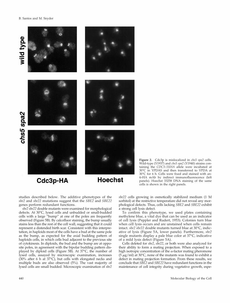
The existence of cells with elongated bud necks suggests apossible defect in neck organization in chs5 spa2 cells; there-fore, we analyzed septin organization in this mutant byexamining the localization of a functional epitope-taggedversion of the septin Cdc3p (Roemer et al., 1996). In the wildtype and in chs5 and spa2 mutants, Cdc3p shows the typicalring structure at the neck (Kim et al., 1991). In contrast,Cdc3p is dramatically mislocalized in chs5 spa2 cells at 30 or37°C (Figure 2). Abnormal septin structures are observed in75% of chs5 spa2 cells at 30°C (n 5 507); defects includefragmented rings, mislocalized rings not placed at the neck,and the presence of septin patches. This indicates that thelack of Chs5p and Spa2p directly or indirectly affects septinorganization in yeast.
Screen for High-Copy-Number Suppressors of thechs5 spa2 LethalityTo identify additional components that may function in thesame processes as CHS5 and SPA2, we searched for genesthat, when present in high copy number, suppress the chs5spa2 lethality at high temperature (see MATERIALS ANDMETHODS). Ten plasmids that reproducibly suppressed thelethality were identified. Six contained CHS5, whereas theother four plasmids each carried a different yeast genomicDNA fragment; therefore, each represents a different sup-pressor gene. These genes were designated CSR1–CSR4(chs5 spa2 rescue); CSR3 is the strongest suppressor, whereasCSR2 is the weakest (Figure 3). None of the suppressor
B. Santos and M. Snyder
Molecular Biology of the Cell438

genes complements chs5 or spa2 single mutant defects. Forinstance, chs5 spa2 mutants, like chs5, are resistant to cal-cofluor as a result of the low levels of chitin. However, noneof the plasmids suppresses this phenotype, suggesting thatoverexpression of these genes does not affect chitin levels.Likewise, overexpression of the CSR genes does not restorenormal shmoo morphology in a spa2 mutant. Therefore, theCSR genes suppress defects specific to the chs5 spa2 doublemutant.
All of the suppressor plasmids contained several ORFs.The suppressing gene was identified by testing individualsubcloned fragments for suppressing activity. CSR1 andCSR2 are novel genes (Saccharomyces Genome Database ORFdesignation YLR380w and YPR030w, respectively), CSR3corresponds to YDR351w/SBE2, and CSR4 is identical toYIL147c/SLN1. SBE2 was isolated previously as a suppres-sor of bem4 (Mack et al., 1996), but no further characteriza-tion has been reported. Sln1p is a two-component signaltransducer involved in the high-osmolarity glycerol (HOG)MAPK pathway (Ota and Varshavsky, 1993; Maeda et al.,1994). Characterization of CSR1, CSR2, and CSR4/SLN1 willbe described elsewhere. The characterization of CSR3/SBE2is described below.
Sbe2p and Sbe22p Are Highly Homologous ProteinsSBE2 encodes a hydrophilic protein of 864 amino acids. Asearch with the FASTA program revealed that S. cerevisiaepossesses another gene, YHR103w/SBE22, predicted to en-code a protein with high amino acid sequence identity toSbe2p. Sbe22p is 852 amino acids in length and contains 43%identity and 63% similarity to Sbe2p over their entire length(Figure 4). They are more divergent at the N terminus (44%similarity) and more conserved at the C terminus (71%similarity). Sbe2p and Sbe22p are novel proteins and do notexhibit high homology with other known proteins in yeastor other organisms. However, they both display low-levelsimilarity (25%) with a predicted 472-amino acid protein(C23D3.03c) of Schizosaccharomyces pombe, sharing a perfectlyconserved 8-amino acid block (RPSWLPPK) that does notexist in any other protein in the databases (Figure 4).
Sbe2p and Sbe22p proteins share some common structuralfeatures. Using the TMpred program (Hofman and Stoffel,1993), we found that Sbe2p displays a potential transmem-brane domain from amino acids 798 to 818 and, similarly,Sbe22p is predicted to have a C-terminal transmembrane do-main from amino acids 786 to 807. However, other programspredict that Sbe2p and Sbe22p are soluble proteins. Secondarystructure analysis of Sbe2p with the use of the program COILS(Lupas, 1996) identifies a potential coiled-coil region (510–560amino acids); this feature is not evident in Sbe22p. Despite thisdifference, the high level of amino acid sequence similaritybetween Sbe2p and Sbe22p suggests that they are likely redun-dant proteins with similar functions in the cell.
sbe2 sbe22 Double Mutants Display a Lysis DefectTo explore the function of SBE2 and SBE22, null mutantswere generated and isogenic single and double mutantswere analyzed. The sbe2 and sbe22 null mutants are viableand have growth rates in rich medium comparable to thoseof the isogenic wild-type strain at 25 or 30°C; however, sbe2mutants display a slow-growth phenotype at 16 and 37°C,whereas sbe22 null strains exhibit a growth rate indistin-guishable from that of wild-type cells at these temperatures(Figure 5A). Thus, SBE2 and SBE22 are not essential and aredispensable for growth.
In contrast, sbe2 sbe22 double mutants display a severegrowth defect at 37°C. This defect is completely rescued bythe addition of 1 M sorbitol (Figure 5A) and is stronger indiploids than in haploids. Therefore, unless mentioned oth-erwise, we have used homozygous diploids in all of the
Figure 1. chs5 spa2 mutant has a severe growth defect at 37°C thatcan be rescued by sorbitol. (A) Isogenic wild-type (Y603), spa2(Y601), chs5 (Y1935), and chs5 spa2 (Y1936) strains were grownovernight to stationary phase at 30°C in YPDAS. Ten-fold serialdilutions starting with the saturated culture (;5 3 107 cells/ml)were spotted in 5-ml drops onto YPDA plates (two upper panels) orYPDAS (lower panel). Plates were incubated at 30°C (upper panel)or 37°C (two lower panels). (B) Morphology of chs5 spa2 cells(Y1936) incubated at the restrictive temperature in YPDA for 6 h;elongated necks, large round cells, and shmoo-like cells are shownfrom left to right.
Sbe2p Is Involved in Cell Wall Formation
Vol. 11, February 2000 439
studies described below. The additive phenotypes of thesbe2 and sbe22 mutations suggest that the SBE2 and SBE22genes perform redundant functions.
sbe2 sbe22 double mutants were examined for morphologicaldefects. At 30°C, lysed cells and unbudded or small-buddedcells with a large “bump” at one of the poles are frequentlyobserved (Figure 5B). By calcofluor staining, the bump usuallystains less than the rest of the cell wall, suggesting that it couldrepresent a distended birth scar. Consistent with this interpre-tation, in haploids most of the cells have a bud at the same poleas the bump, as expected for the axial budding pattern ofhaploids cells, in which cells bud adjacent to the previous siteof cytokinesis. In diploids, the bud and the bump are at oppo-site poles, in agreement with the bipolar budding pattern dis-played by diploid cells (Figure 5B). At 37°C, the number oflysed cells, assayed by microscopic examination, increases(30% after 6 h at 37°C), but cells with elongated necks andmultiple buds are also observed (5%). The vast majority oflysed cells are small budded. Microscopic examination of sbe2
sbe22 cells growing in osmotically stabilized medium (1 Msorbitol) at the restrictive temperature did not reveal any mor-phological defects. Thus, cells lacking SBE2 and SBE22 exhibita strong cell lysis defect.
To confirm this phenotype, we used plates containingmethylene blue, a vital dye that can be used as an indicatorof cell lysis (Peppler and Rudert, 1953). Colonies turn bluewhen cell lysis occurs and are unstained when cells remainintact. sbe2 sbe22 double mutants turned blue at 30°C, indic-ative of lysis (Figure 5A, lower panels). Furthermore, sbe2single mutants display a pale blue color at 37°C, indicativeof a mild lysis defect (Figure 5A).
Cells deleted for sbe2, sbe22, or both were also analyzed fortheir ability to form a mating projection. When exposed to ahigh isotropic concentration of the a-factor mating pheromone(5 mg/ml) at 30°C, none of the mutants was found to exhibit adefect in mating projection formation. From these results, weconclude that SBE2 and SBE22 have redundant functions in themaintenance of cell integrity during vegetative growth, espe-
Figure 2. Cdc3p is mislocalized in chs5 spa2 cells.Wild-type (Y1937) and chs5 spa2 (Y1940) strains con-taining the CDC3::3XHA allele were incubated at30°C in YPDAS and then transferred to YPDA at30°C for 6 h. Cells were fixed and stained with an-ti-HA mAb by indirect immunofluorescence (leftpanels). Hoechst 33258 DNA staining of the samecells is shown in the right panels.
B. Santos and M. Snyder
Molecular Biology of the Cell440

cially at high temperatures, although SBE2 appears to performa more prominent role. Consistent with this interpretation,SBE22 in a multicopy plasmid does not rescue chs5 spa2 lethal-ity at high temperature (our unpublished results).
sbe2 sbe22 Mutants Are Hypersensitive toCalcofluor and SDSA sorbitol-remediable lysis defect phenotype has been de-scribed previously for mutants with a defective cell wall,such as those affected in b-1,3-glucan synthesis or in theSlt2p MAPK pathway (Cid et al., 1995). Therefore, we ana-lyzed whether sbe2 sbe22 mutants exhibit cell wall defects bytesting their sensitivity to calcofluor and SDS.
Calcofluor is toxic to yeast cells because of its interactionwith chitin and its interference with cell wall assembly (Ron-cero et al., 1988). Mutants affecting cell wall integrity aresusceptible to this agent (Lussier et al., 1997). The sensitivityof sbe2, sbe22, and sbe2 sbe22 mutants to 50 mg/ml calcofluorat 30°C was tested. Under these conditions, the sbe2 mutantis more sensitive and the sbe2 sbe22 double mutant is muchmore sensitive compared with the isogenic wild-type strain.sbe22 has no detectable defect (Figure 6A).
SDS is a toxic detergent for yeast cells because it affects cellintegrity. Mutants with a defective cell wall are usually more
sensitive to this detergent (Shimizu et al., 1994). The sbe2sbe22 double mutant is at least 10 times more sensitive to lowlevels of SDS (0.0025%) compared with wild type. The sbe2mutant is slightly more sensitive and the sbe22 mutant be-haves like the wild type (Figure 6B).
These results strongly indicate that loss of SBE2 andSBE22 function leads to a defective cell wall, suggesting theinvolvement of Sbe2p and Sbe22p in establishing or main-taining cell wall integrity.
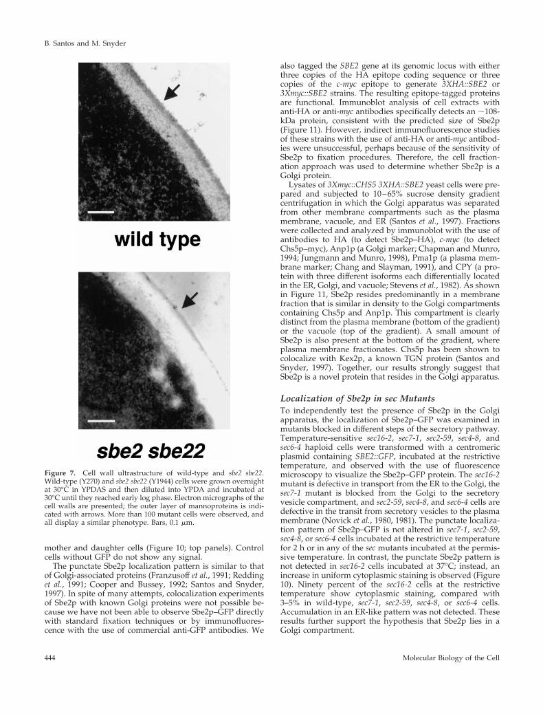
sbe2 sbe22 Mutants Exhibit an Abnormal Cell WallUltrastructureTo further explore the requirement for Sbe2p and Sbe22p forproper cell wall architecture, the morphology of the cell wallwas examined by electron microscopy. The cell wall of wild-type cells appears as a layered structure, exhibiting an elec-tron-dense fibrillar outer layer, rich in mannoproteins, andan electron-transparent amorphous inner layer (Horisbergerand Vonlanthen, 1977). The inner layer contains glucan anda small amount of chitin and may be subdivided into twodifferent zones: one closest to the plasma membrane, whichis rich in proteins, and an outer one, which contains asignificant proportion of b-1,6-glucan (Kopecka et al., 1974).
We analyzed the ultrastructure of the cell wall of sbe2,sbe22, and sbe2 sbe22 mutants growing in rich medium at30°C. Whereas sbe2 or sbe22 cells show a normal layeredstructure similar to wild-type cells, the double mutant dis-plays an aberrant cell wall architecture. The inner layer ofglucan and chitin is enlarged, and the outer layer of man-noproteins is diminished or absent (Figure 7). We also ob-served that some small-budded cells appear to lyse at the tipof their buds because their cell walls are very thin or almostabsent (data not shown). In conclusion, sbe2 sbe22 mutantsexhibit an altered cell wall, with the thickness of the man-noprotein layer being very reduced.
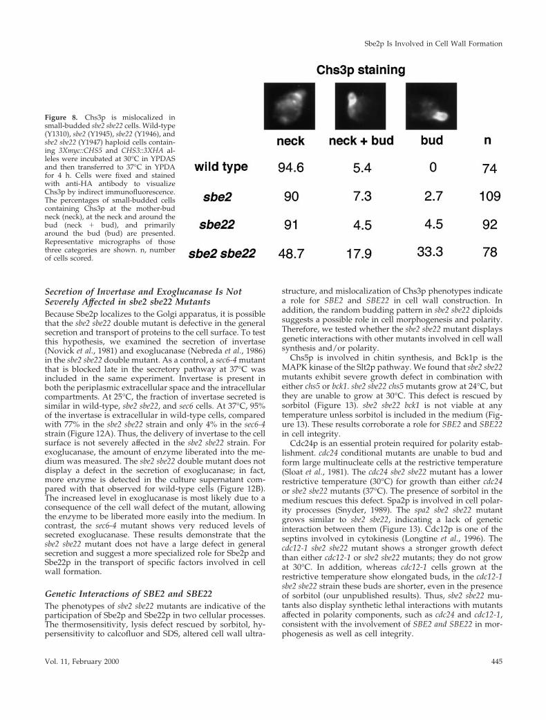
Chs3p Is Mislocalized in the sbe2 sbe22 MutantsBecause the sbe2 sbe22 mutant has a defective cell wall, weexamined the localization of Chs3p and Chs5p, two proteinsrequired for chitin synthesis, in the double mutant. In wild-type cells, Chs5p localizes in cytoplasmic patches (Santosand Snyder, 1997); this localization is not affected in sbe2,sbe22, or sbe2 sbe22 null mutants at 30 or 37°C. Localizationof Chs3p in sbe2 and sbe22 single mutants is also similar tothat in the wild type (see INTRODUCTION). In contrast, insbe2 sbe22 null mutants, Chs3p is often found at the budplasma membrane or diffuse throughout the bud in small-budded cells, instead of forming a ring at the bud neck likethose observed in wild-type cells (Figure 8). Thus, in theabsence of SBE2 and SBE22, Chs3p appears to be polarizedto the correct general location but fails to assemble properly.
sbe2 sbe22 Diploids Display a Random BuddingPatternTo further explore the role of SBE2 and SBE22, the buddingpatterns of sbe2, sbe22, and sbe2 sbe22 cells were examined.Cells grown to log phase at 30°C were stained with cal-cofluor to visualize the chitin-rich bud scars that mark pre-vious sites of budding (Hayashibe and Katohda, 1973). sbe2,sbe22, and sbe2 sbe22 haploid mutants show an axial budding
Figure 3. Multicopy suppressors of chs5 spa2 lethality at 37°C. chs5spa2 strain (Y1941) transformed with vector (YEp24) or plasmidscontaining CHS5, SPA2, or the suppressor genes CSR1, CSR2, CSR3,or CSR4 were grown overnight at 30°C in SC-Ura plus 1 M sorbitol.Ten-fold serial dilutions starting with 10% of the saturated culturewere spotted in 5-ml drops onto SC-Ura plates (top panel) or SC-Uraplus sorbitol (bottom panel) and were incubated at 37°C. The sup-pressor genes rescue the growth defect of chs5 spa2 cells to differentdegrees.
Sbe2p Is Involved in Cell Wall Formation
Vol. 11, February 2000 441
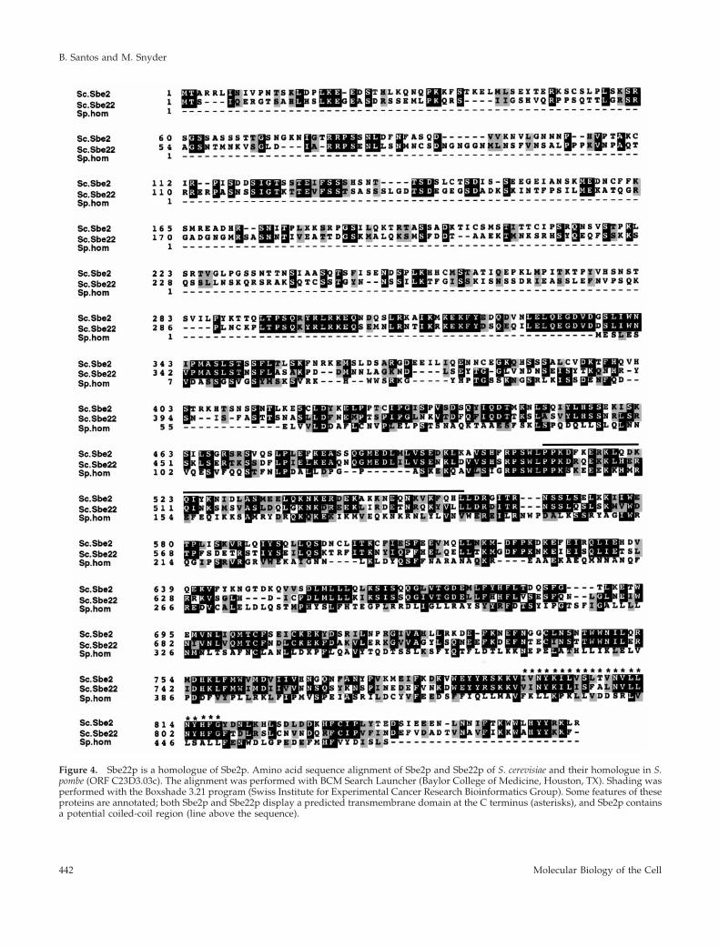
Figure 4. Sbe22p is a homologue of Sbe2p. Amino acid sequence alignment of Sbe2p and Sbe22p of S. cerevisiae and their homologue in S.pombe (ORF C23D3.03c). The alignment was performed with BCM Search Launcher (Baylor College of Medicine, Houston, TX). Shading wasperformed with the Boxshade 3.21 program (Swiss Institute for Experimental Cancer Research Bioinformatics Group). Some features of theseproteins are annotated; both Sbe2p and Sbe22p display a predicted transmembrane domain at the C terminus (asterisks), and Sbe2p containsa potential coiled-coil region (line above the sequence).
B. Santos and M. Snyder
Molecular Biology of the Cell442

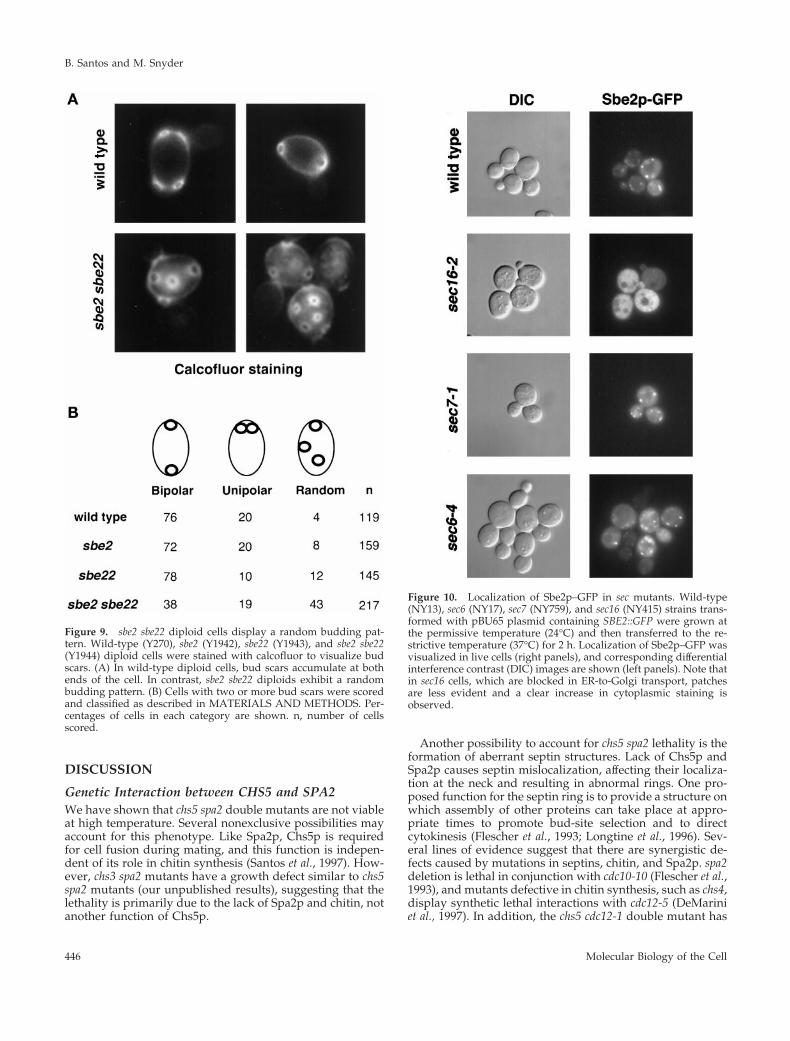
pattern similar to the wild type (Flescher et al., 1993; Chantand Pringle, 1995). In contrast to haploids, wild-type diploidcells exhibit a bipolar budding pattern, resulting in bud scarslocated at both ends of the cell. sbe2 and sbe22 diploid cellsdisplay the normal bipolar budding pattern; however, sbe2sbe22 diploids often bud randomly (Figure 9). Thus, sbe2sbe22 diploids possess a bipolar-specific budding patterndefect.
Sbe2p Localizes to Cytoplasmic Patches andCofractionates with Golgi ProteinsTo determine the subcellular localization of Sbe2p, the GFPwas fused to the N terminus of Sbe2p. This fusion protein isfully functional and rescues the thermosensitivity of sbe2sbe22 double mutants. Localization of Sbe2p was examinedin sbe2 mutant cells transformed with a centromeric plasmidcontaining SBE2::GFP and observed with the use of fluores-cence microscopy. Sbe2p–GFP localizes to cytoplasmicpatches in cells at all stages of the cell cycle. Sbe2p patchesare distributed throughout the cell and are observed in both
Figure 5. sbe2 sbe22 double mutant shows a lysis defect at 37°C. (A)Wild-type (Y270), sbe2 (Y1942), sbe22 (Y1943), and sbe2 sbe22 (Y1944)homozygous diploid strains were grown overnight at 30°C in YP-DAS. Five-fold dilutions starting with 4% of the saturated culturewere spotted in 5-ml drops onto YPDA (upper panel), YPDAS(middle panel), or YPDA plus 0.001% methylene blue (MB) dye(lower panel). Plates were incubated at 30°C (left panels) or 37°C(right panels). (B) Morphological defects of the sbe2 sbe22 mutant(Y1944) at 30°C, showing a characteristic bump at one of the poles(arrows).
Figure 6. sbe2 sbe22 mutants are sensitive to calcofluor and SDS.Wild-type (Y270), sbe2 (Y1942), sbe22 (Y1943), and sbe2 sbe22 (Y1944)homozygous diploid strains were grown overnight at 30°C in YP-DAS. (A) Five-fold serial dilutions starting with 20% of the satu-rated culture were spotted in 5-ml drops onto SC and SC containing50 mg/ml calcofluor. (B) Ten-fold dilutions starting with the satu-rated culture were spotted onto YPDA and YPDA containing0.0025% SDS. All plates were incubated at 30°C.
Sbe2p Is Involved in Cell Wall Formation
Vol. 11, February 2000 443
mother and daughter cells (Figure 10; top panels). Controlcells without GFP do not show any signal.
The punctate Sbe2p localization pattern is similar to thatof Golgi-associated proteins (Franzusoff et al., 1991; Reddinget al., 1991; Cooper and Bussey, 1992; Santos and Snyder,1997). In spite of many attempts, colocalization experimentsof Sbe2p with known Golgi proteins were not possible be-cause we have not been able to observe Sbe2p–GFP directlywith standard fixation techniques or by immunofluores-cence with the use of commercial anti-GFP antibodies. We
also tagged the SBE2 gene at its genomic locus with eitherthree copies of the HA epitope coding sequence or threecopies of the c-myc epitope to generate 3XHA::SBE2 or3Xmyc::SBE2 strains. The resulting epitope-tagged proteinsare functional. Immunoblot analysis of cell extracts withanti-HA or anti-myc antibodies specifically detects an ;108-kDa protein, consistent with the predicted size of Sbe2p(Figure 11). However, indirect immunofluorescence studiesof these strains with the use of anti-HA or anti-myc antibod-ies were unsuccessful, perhaps because of the sensitivity ofSbe2p to fixation procedures. Therefore, the cell fraction-ation approach was used to determine whether Sbe2p is aGolgi protein.
Lysates of 3Xmyc::CHS5 3XHA::SBE2 yeast cells were pre-pared and subjected to 10–65% sucrose density gradientcentrifugation in which the Golgi apparatus was separatedfrom other membrane compartments such as the plasmamembrane, vacuole, and ER (Santos et al., 1997). Fractionswere collected and analyzed by immunoblot with the use ofantibodies to HA (to detect Sbe2p–HA), c-myc (to detectChs5p–myc), Anp1p (a Golgi marker; Chapman and Munro,1994; Jungmann and Munro, 1998), Pma1p (a plasma mem-brane marker; Chang and Slayman, 1991), and CPY (a pro-tein with three different isoforms each differentially locatedin the ER, Golgi, and vacuole; Stevens et al., 1982). As shownin Figure 11, Sbe2p resides predominantly in a membranefraction that is similar in density to the Golgi compartmentscontaining Chs5p and Anp1p. This compartment is clearlydistinct from the plasma membrane (bottom of the gradient)or the vacuole (top of the gradient). A small amount ofSbe2p is also present at the bottom of the gradient, whereplasma membrane fractionates. Chs5p has been shown tocolocalize with Kex2p, a known TGN protein (Santos andSnyder, 1997). Together, our results strongly suggest thatSbe2p is a novel protein that resides in the Golgi apparatus.
Localization of Sbe2p in sec MutantsTo independently test the presence of Sbe2p in the Golgiapparatus, the localization of Sbe2p–GFP was examined inmutants blocked in different steps of the secretory pathway.Temperature-sensitive sec16-2, sec7-1, sec2-59, sec4-8, andsec6-4 haploid cells were transformed with a centromericplasmid containing SBE2::GFP, incubated at the restrictivetemperature, and observed with the use of fluorescencemicroscopy to visualize the Sbe2p–GFP protein. The sec16-2mutant is defective in transport from the ER to the Golgi, thesec7-1 mutant is blocked from the Golgi to the secretoryvesicle compartment, and sec2-59, sec4-8, and sec6-4 cells aredefective in the transit from secretory vesicles to the plasmamembrane (Novick et al., 1980, 1981). The punctate localiza-tion pattern of Sbe2p–GFP is not altered in sec7-1, sec2-59,sec4-8, or sec6-4 cells incubated at the restrictive temperaturefor 2 h or in any of the sec mutants incubated at the permis-sive temperature. In contrast, the punctate Sbe2p pattern isnot detected in sec16-2 cells incubated at 37°C; instead, anincrease in uniform cytoplasmic staining is observed (Figure10). Ninety percent of the sec16-2 cells at the restrictivetemperature show cytoplasmic staining, compared with3–5% in wild-type, sec7-1, sec2-59, sec4-8, or sec6-4 cells.Accumulation in an ER-like pattern was not detected. Theseresults further support the hypothesis that Sbe2p lies in aGolgi compartment.
Figure 7. Cell wall ultrastructure of wild-type and sbe2 sbe22.Wild-type (Y270) and sbe2 sbe22 (Y1944) cells were grown overnightat 30°C in YPDAS and then diluted into YPDA and incubated at30°C until they reached early log phase. Electron micrographs of thecell walls are presented; the outer layer of mannoproteins is indi-cated with arrows. More than 100 mutant cells were observed, andall display a similar phenotype. Bars, 0.1 mm.
B. Santos and M. Snyder
Molecular Biology of the Cell444
Secretion of Invertase and Exoglucanase Is NotSeverely Affected in sbe2 sbe22 MutantsBecause Sbe2p localizes to the Golgi apparatus, it is possiblethat the sbe2 sbe22 double mutant is defective in the generalsecretion and transport of proteins to the cell surface. To testthis hypothesis, we examined the secretion of invertase(Novick et al., 1981) and exoglucanase (Nebreda et al., 1986)in the sbe2 sbe22 double mutant. As a control, a sec6-4 mutantthat is blocked late in the secretory pathway at 37°C wasincluded in the same experiment. Invertase is present inboth the periplasmic extracellular space and the intracellularcompartments. At 25°C, the fraction of invertase secreted issimilar in wild-type, sbe2 sbe22, and sec6 cells. At 37°C, 95%of the invertase is extracellular in wild-type cells, comparedwith 77% in the sbe2 sbe22 strain and only 4% in the sec6-4strain (Figure 12A). Thus, the delivery of invertase to the cellsurface is not severely affected in the sbe2 sbe22 strain. Forexoglucanase, the amount of enzyme liberated into the me-dium was measured. The sbe2 sbe22 double mutant does notdisplay a defect in the secretion of exoglucanase; in fact,more enzyme is detected in the culture supernatant com-pared with that observed for wild-type cells (Figure 12B).The increased level in exoglucanase is most likely due to aconsequence of the cell wall defect of the mutant, allowingthe enzyme to be liberated more easily into the medium. Incontrast, the sec6-4 mutant shows very reduced levels ofsecreted exoglucanase. These results demonstrate that thesbe2 sbe22 mutant does not have a large defect in generalsecretion and suggest a more specialized role for Sbe2p andSbe22p in the transport of specific factors involved in cellwall formation.
Genetic Interactions of SBE2 and SBE22The phenotypes of sbe2 sbe22 mutants are indicative of theparticipation of Sbe2p and Sbe22p in two cellular processes.The thermosensitivity, lysis defect rescued by sorbitol, hy-persensitivity to calcofluor and SDS, altered cell wall ultra-
structure, and mislocalization of Chs3p phenotypes indicatea role for SBE2 and SBE22 in cell wall construction. Inaddition, the random budding pattern in sbe2 sbe22 diploidssuggests a possible role in cell morphogenesis and polarity.Therefore, we tested whether the sbe2 sbe22 mutant displaysgenetic interactions with other mutants involved in cell wallsynthesis and/or polarity.
Chs5p is involved in chitin synthesis, and Bck1p is theMAPK kinase of the Slt2p pathway. We found that sbe2 sbe22mutants exhibit severe growth defect in combination witheither chs5 or bck1. sbe2 sbe22 chs5 mutants grow at 24°C, butthey are unable to grow at 30°C. This defect is rescued bysorbitol (Figure 13). sbe2 sbe22 bck1 is not viable at anytemperature unless sorbitol is included in the medium (Fig-ure 13). These results corroborate a role for SBE2 and SBE22in cell integrity.
Cdc24p is an essential protein required for polarity estab-lishment. cdc24 conditional mutants are unable to bud andform large multinucleate cells at the restrictive temperature(Sloat et al., 1981). The cdc24 sbe2 sbe22 mutant has a lowerrestrictive temperature (30°C) for growth than either cdc24or sbe2 sbe22 mutants (37°C). The presence of sorbitol in themedium rescues this defect. Spa2p is involved in cell polar-ity processes (Snyder, 1989). The spa2 sbe2 sbe22 mutantgrows similar to sbe2 sbe22, indicating a lack of geneticinteraction between them (Figure 13). Cdc12p is one of theseptins involved in cytokinesis (Longtine et al., 1996). Thecdc12-1 sbe2 sbe22 mutant shows a stronger growth defectthan either cdc12-1 or sbe2 sbe22 mutants; they do not growat 30°C. In addition, whereas cdc12-1 cells grown at therestrictive temperature show elongated buds, in the cdc12-1sbe2 sbe22 strain these buds are shorter, even in the presenceof sorbitol (our unpublished results). Thus, sbe2 sbe22 mu-tants also display synthetic lethal interactions with mutantsaffected in polarity components, such as cdc24 and cdc12-1,consistent with the involvement of SBE2 and SBE22 in mor-phogenesis as well as cell integrity.
Figure 8. Chs3p is mislocalized insmall-budded sbe2 sbe22 cells. Wild-type(Y1310), sbe2 (Y1945), sbe22 (Y1946), andsbe2 sbe22 (Y1947) haploid cells contain-ing 3Xmyc::CHS5 and CHS3::3XHA al-leles were incubated at 30°C in YPDASand then transferred to 37°C in YPDAfor 4 h. Cells were fixed and stainedwith anti-HA antibody to visualizeChs3p by indirect immunofluorescence.The percentages of small-budded cellscontaining Chs3p at the mother-budneck (neck), at the neck and around thebud (neck 1 bud), and primarilyaround the bud (bud) are presented.Representative micrographs of thosethree categories are shown. n, numberof cells scored.
Sbe2p Is Involved in Cell Wall Formation
Vol. 11, February 2000 445
DISCUSSION
Genetic Interaction between CHS5 and SPA2We have shown that chs5 spa2 double mutants are not viableat high temperature. Several nonexclusive possibilities mayaccount for this phenotype. Like Spa2p, Chs5p is requiredfor cell fusion during mating, and this function is indepen-dent of its role in chitin synthesis (Santos et al., 1997). How-ever, chs3 spa2 mutants have a growth defect similar to chs5spa2 mutants (our unpublished results), suggesting that thelethality is primarily due to the lack of Spa2p and chitin, notanother function of Chs5p.
Another possibility to account for chs5 spa2 lethality is theformation of aberrant septin structures. Lack of Chs5p andSpa2p causes septin mislocalization, affecting their localiza-tion at the neck and resulting in abnormal rings. One pro-posed function for the septin ring is to provide a structure onwhich assembly of other proteins can take place at appro-priate times to promote bud-site selection and to directcytokinesis (Flescher et al., 1993; Longtine et al., 1996). Sev-eral lines of evidence suggest that there are synergistic de-fects caused by mutations in septins, chitin, and Spa2p. spa2deletion is lethal in conjunction with cdc10-10 (Flescher et al.,1993), and mutants defective in chitin synthesis, such as chs4,display synthetic lethal interactions with cdc12-5 (DeMariniet al., 1997). In addition, the chs5 cdc12-1 double mutant has
Figure 9. sbe2 sbe22 diploid cells display a random budding pat-tern. Wild-type (Y270), sbe2 (Y1942), sbe22 (Y1943), and sbe2 sbe22(Y1944) diploid cells were stained with calcofluor to visualize budscars. (A) In wild-type diploid cells, bud scars accumulate at bothends of the cell. In contrast, sbe2 sbe22 diploids exhibit a randombudding pattern. (B) Cells with two or more bud scars were scoredand classified as described in MATERIALS AND METHODS. Per-centages of cells in each category are shown. n, number of cellsscored.
Figure 10. Localization of Sbe2p–GFP in sec mutants. Wild-type(NY13), sec6 (NY17), sec7 (NY759), and sec16 (NY415) strains trans-formed with pBU65 plasmid containing SBE2::GFP were grown atthe permissive temperature (24°C) and then transferred to the re-strictive temperature (37°C) for 2 h. Localization of Sbe2p–GFP wasvisualized in live cells (right panels), and corresponding differentialinterference contrast (DIC) images are shown (left panels). Note thatin sec16 cells, which are blocked in ER-to-Golgi transport, patchesare less evident and a clear increase in cytoplasmic staining isobserved.
B. Santos and M. Snyder
Molecular Biology of the Cell446
a stronger growth defect than chs5D or cdc12-1 single mu-tants (our unpublished results).
Another possible explanation for the chs5 spa2 growthdefect is that, in the absence of Spa2p, a perfectly con-structed cell wall is required. It has been shown that spa2mutants are colethal with mutants defective in cell wallstructure, such as those implicated in the Slt2p pathway(Costigan and Snyder, 1994). In addition, overexpression ofSBE2, a gene involved in cell wall integrity, allows chs5 spa2
cells to grow. This possibility is also in agreement with thefact that sorbitol, an osmotic stabilizer, rescues the growthdefect, although how this occurs is not clear. It is possiblethat sorbitol triggers signaling pathways in the cell that leadto the formation of a stronger cell wall. It has been suggestedthat a basal level of signaling through the HOG MAPKpathway is required for the proper localization of a glyco-syltransferase, Mnn1p, to the Golgi complex (Reynolds et al.,1998); this suggests that the activity of the HOG pathway
Figure 11. Sucrose density gradient centrifugationanalysis. Cell lysates were prepared from yeaststrain Y1948 containing 3Xmyc::CHS5 andSBE2::3XHA alleles and fractionated in 10–65% su-crose density gradients by centrifugation for 20 h.Fractions (abscissa in A) were collected from thebottom of the gradient and analyzed by immuno-blot analysis with the use of antibodies to HA,c-myc, Pma1p, Anp1p, and CPY. Only the vacuolarisoform of the CPY protein (CPYvp) is represented.Sample blots are shown in B. The amount of proteinin each lane was quantified, and the results areshown in A as percentages of the total protein.
Sbe2p Is Involved in Cell Wall Formation
Vol. 11, February 2000 447

may regulate the compartmental distribution of glycosyl-transferases, which are involved in mannan synthesis,within the Golgi to provide a novel mechanism to regulatethe composition of the cell wall. Additionally, we havefound SLN1, encoding one of the receptors of the HOGpathway (Maeda et al., 1994), as a multicopy suppressor ofchs5 spa2 lethality, suggesting that signaling throughout thiscascade can help chs5 spa2 cells to survive at high tempera-ture.
Sbe2p Localizes to the Golgi ApparatusSBE2 and SBE22 genes encode highly homologous proteins.Several lines of evidence suggest that Sbe2p localizes to theGolgi apparatus. First, Sbe2p localizes to cytoplasmicpatches with a punctate pattern similar to that of Golgi-associated proteins (Franzusoff et al., 1991). Second, in su-crose gradient experiments, Sbe2p cofractionates with pro-teins demonstrated to be in the Golgi apparatus, such asChs5p and Anp1p (Santos and Snyder, 1997; Jungmann andMunro, 1998). Preliminary results indicate that Sbe22p dis-plays a profile similar to that of Sbe2p in sucrose gradients,suggesting that Sbe22p might also be a Golgi protein (ourunpublished results). Third, Sbe2p is mislocalized in a sec16mutant that blocks protein transport from the ER but not inother mutants defective in the secretory pathway but con-taining a normal Golgi apparatus, such as sec2, sec4, sec6, andsec7. This protein is not retained in the ER in the sec16mutant; the same behavior has been described for Chs5p,another Golgi protein also involved in cell wall construction.One possible explanation is that these proteins assembledirectly in the Golgi apparatus independently of the ER.
It has been reported that most proteins in the Golgi areeither integral membrane proteins (mainly glycosyltrans-ferases or proteases) or peripheral membrane proteins(Munro, 1998). Sbe2p and Sbe22p are predicted to be mem-brane proteins and localize to the Golgi apparatus but do not
Figure 12. sbe2 sbe22 mutant is not severely affected in secretion.(A) Percentage of secreted invertase relative to intracellular enzymein wild-type (Y270), sbe2 sbe22 (Y1944), and sec6-4 cells (NY17) after1 h of incubation in low-glucose medium at 25 or 37°C. (B) Percent-age of exoglucanase liberated into the medium by the same strains(see MATERIALS AND METHODS).
Figure 13. Genetic interactions of SBE2 and SBE22. Wild-type(Y604), sbe2 sbe22 (Y1949), chs5 (Y1950), sbe2 sbe22 chs5 (Y1951), bck1(Y760), sbe2 sbe22 bck1 (Y1952), cdc24 (Y1954), sbe2 sbe22 cdc24(Y1955), spa2 (Y602), sbe2 sbe22 spa2 (Y1953), cdc12 (Y1956), and sbe2sbe22 cdc12 (Y1957) strains were grown overnight at 24°C in YPDAS.Ten-fold dilutions starting with 1% of the saturated culture werespotted in 5-ml drops onto YPDA (three upper panels) or YPDAS(lower panel). Plates were incubated at different temperatures, asindicated in the figure.
B. Santos and M. Snyder
Molecular Biology of the Cell448
show similarity with glycosyltransferases or proteases, sug-gesting that they perform a different function.
Sbe2p and Sbe22p Are Involved in Cell WallIntegritySeveral lines of evidence suggest that Sbe2p and Sbe22p areinvolved in cell wall integrity. First, the sbe2 sbe22 doublemutant has a lysis defect at high temperature that is rescuedby the presence of sorbitol in the medium. This phenotypehas been described for several mutants defective in cell wallstructure, including those affecting the Slt2p pathway (Cid etal., 1995). Second, the sbe2 sbe22 double mutant displayssensitivity to compounds such as SDS and calcofluor. SDSinduces lysis of cells with fragile cell walls, and calcofluor isa dye that blocks chitin polymerization, resulting in a weak-ened cell wall. Sensitivity to these products has proved to bea powerful tool in revealing cell wall defects (Ram et al.,1994; Lussier et al., 1997). Hypersensitivity to calcofluor mayalso be an indication of an increased amount of chitin in thecell wall. Third, electron microscopic analysis of the sbe2sbe22 mutants demonstrates a strongly reduced electron-dense mannoprotein layer in the cell wall. This layer isbelieved to both contribute to the structural integrity of thecell wall and serve to exclude hydrolytic enzymes.
Several genetic screens have identified yeast mutationsthat cause defects in the synthesis of mannan, including themnn (mannan; Ballou, 1990), och (outer chain; Nagasu et al.,1992), ngd (N-glycosylation defective; Lehle et al., 1995), andldb (low dye binding; Manas et al., 1997) set of mutants. Likesbe2 sbe22 double mutants, mnn6 mutants are hypersensitiveto calcofluor (Wang et al., 1997). Lack of Cwp2, anothermannoprotein, confers an increased sensitivity to calcofluorand strongly reduces the electron-dense layer of the outsideof the cell wall (Van Der Vaart et al., 1995). Mannan is notessential for viability; however, survival without mannan isdependent on cells being able to sense a cell wall defect(Jungmann et al., 1999). Yeast cells are endowed with thecapacity to compensate for alterations in the structureand/or composition of the cell wall matrix. It has beenproposed that the cell increases the amount of chitin andchitin-bound b-1,6-glucosylated proteins as a rescue mech-anism in response to cell wall weakening (Kapteyn et al.,1997; Popolo et al., 1997). The putative compensatory mech-anism might be mediated by the PKC-dependent signal
transduction pathway (Kamada et al., 1995). Thus, mutationsthat result in defective mannans show synthetic lethalitywith components of the PKC pathway, which regulates theexpression of cell wall components (Rayner and Munro,1998). This is in agreement with the fact that sbe2 sbe22mutants, which display a reduced mannoprotein layer, aresynthetic lethal with mutants affected in chitin synthesis,such as chs5, and mutants in the Slt2p pathway, such as bck1.The lethality between sbe2 sbe22 and bck1 also suggests thatSbe2p and Sbe22p are not part of the Slt2p pathway and mayact in a parallel pathway involved in cell wall constructionand/or cell integrity. In addition, Chs3p, the catalytic com-ponent of CSIII, is mislocalized in sbe2 sbe22 mutants. Thisdefect in Chs3p polarization is similar to that of chs4 or bni4mutants that prevent the interaction between Chs3p and theseptins (DeMarini et al., 1997).
SBE2 and SBE22 Are Involved in Polarity ProcessesSeveral lines of evidence suggest that SBE2 and SBE22 genesmay play a role in yeast cell polarity. sbe2 sbe22 mutantsposses a bipolar-specific budding pattern defect. Mutationsin SPA2, RVS161, RVS167, ACT1, BNI1, and BUD6 affectbud-site selection in diploid cells but not in haploids cells(Madden and Snyder, 1998). Most of these proteins areimplicated in morphogenesis and polarized growth in yeast.Thus, Sbe2p and Sbe22p may have a role in morphogenesis.mnn10/bed1 mutants, defective in mannan deposition likesbe2 sbe22 mutants, are also defective in bud emergence;mutant cells are larger and rounder, indicative of a role inpolarized growth (Mondesert and Reed, 1996).
It is possible that the cell wall defects of sbe2 sbe22 mutantsaffect bud-site selection. Several mutants with a defectivecell wall also display defects in the budding pattern. HKR1is an essential gene that regulates b-glucan synthesis; hkr1mutants display an altered axial budding pattern in haploids(Yabe et al., 1996). In addition, rot1-1, rot2-1, and big1 strainsalso have defective cell walls and show a random buddingpattern in haploid cells (Bickle et al., 1998).
Consistent with the function of Sbe2p and Sbe22p in po-larized cell growth, sbe2 sbe22 mutants display syntheticlethality with polarity mutants such as cdc24 and cdc12. Thesbe2 sbe22 cdc12 triple mutant does not form as elongatedbuds at the restrictive temperature, as does the cdc12 mutant.Mutants defective in apical bud growth, like spa2 mutants,
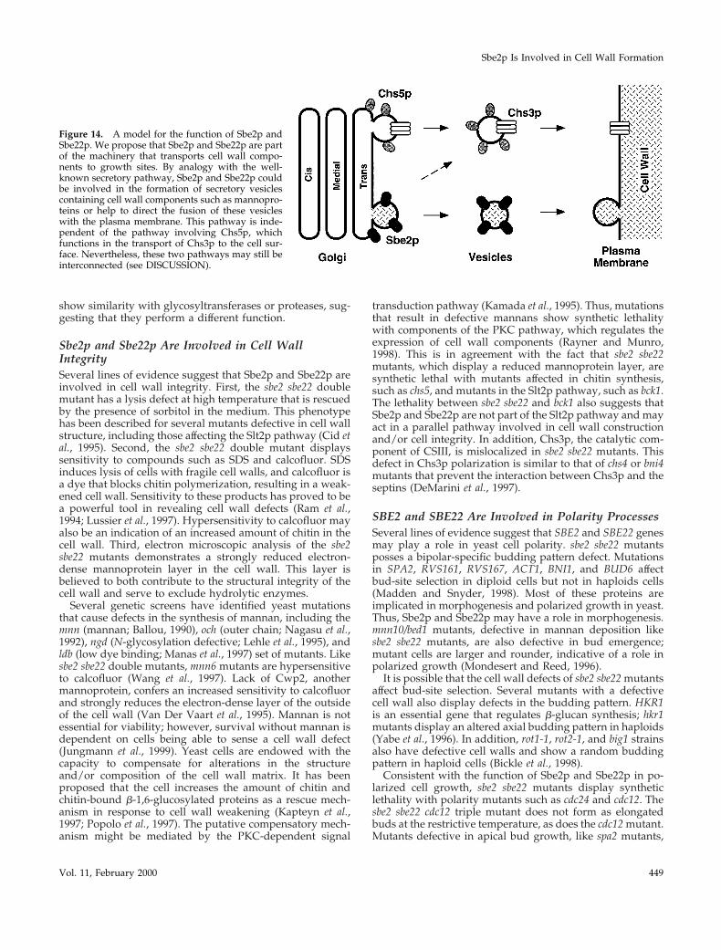
Figure 14. A model for the function of Sbe2p andSbe22p. We propose that Sbe2p and Sbe22p are partof the machinery that transports cell wall compo-nents to growth sites. By analogy with the well-known secretory pathway, Sbe2p and Sbe22p couldbe involved in the formation of secretory vesiclescontaining cell wall components such as mannopro-teins or help to direct the fusion of these vesicleswith the plasma membrane. This pathway is inde-pendent of the pathway involving Chs5p, whichfunctions in the transport of Chs3p to the cell sur-face. Nevertheless, these two pathways may still beinterconnected (see DISCUSSION).
Sbe2p Is Involved in Cell Wall Formation
Vol. 11, February 2000 449
display a random budding pattern and also fail to formelongated buds in the cdc12 background under the sameconditions (Sheu, Barral, and Snyder, unpublished data).sbe2 sbe22 mutants also show these phenotypes, suggestingthat Sbe2p and Sbe22p may also participate in apical budgrowth.
Possible Role of Sbe2p and Sbe22p ProteinsOne possible role for Sbe2p and Sbe22p proteins is that theyare part of the machinery involved in the transport of cellwall components to sites of growth. Several lines of evidencesuggest the existence of several classes of secretory vesiclescarrying different cargo. First, sec6-4 strains accumulate twotypes of 100-nm vesicles containing different proteins (Har-say and Bretscher, 1996). Second, myo2 and act1 mutantsaccumulate vesicles without affecting the secretion of well-known extracellular proteins (Johnston et al., 1991; Govin-dan et al., 1995; Mulholland et al., 1997). Third, by analogywith the well-studied secretory pathway, we have proposedpreviously that Chs5p is involved in the formation of adistinct set of vesicles required to transport Chs3p, andpossibly other polarity components, to the bud neck regionof the cell (Santos and Snyder, 1997; Madden and Snyder,1998). sbe2 sbe22 mutants are not severely defective in thesecretion of invertase or exoglucanase, suggesting a special-ized pathway for polarized secretion of cell wall compo-nents. Because sbe2 sbe22 mutants have chitin, SBE2 overex-pression is not able to rescue chs5 resistance to calcofluor,and chs5 and sbe2 sbe22 mutants are synthetic lethal, wepropose that Sbe2p and Sbe22p could help to form secretoryvesicles involved in the transport of mannoproteins to thecell wall. We suggest the existence of at least two pathwaystransporting cell wall components to the cell surface: oneinvolving Chs5p that is important for chitin synthesis, andthe other involving Sbe2p and Sbe22p that is involved in thesynthesis of the mannoprotein layer (Figure 14). BecauseChs3p is mislocalized in sbe2 sbe22 mutants, there also maybe some connection between these two pathways. Prelimi-nary evidence indicates that sbe2 sbe22 mutants are not af-fected in the incorporation of cell wall proteins such asCwp1 or Pir2p (Van Der Vaart et al., 1995; Kapteyn et al.,1999) into the cell wall. We suggest that Sbe2p and Sbe22pmay be required for either the proper localization of specificmannoproteins within the cell wall or the transport of aspecific subset of proteins required for cell wall organizationto the cell surface. Additional experiments will be requiredto elucidate more detailed roles of the Sbe2p and Sbe22pproteins.
ACKNOWLEGMENTS
We thank Gertien Smits and Frans Klis at the University of Amster-dam, who did the experiments concerning incorporation of cell wallproteins. We thank B. Manning, P. San-Segundo, Y.-J. Sheu, and tworeviewers for critical comments on the manuscript. C. Slayman andS. Munro provided antibodies. P. Novick provided strains, reagentsfor invertase assay, and anti-CPY antibodies. C. Walch-Solimenaprovided help with the invertase assay. C.R. Vazquez de Aldanaprovided advice with the exoglucanase experiment. B. Piekos pro-vided help on the electron microscopy experiments. This researchwas supported by National Institutes of Health grant GM36494 toM.S. B.S. was supported in part by a postdoctoral fellowship fromthe Ministerio de Educacion y Ciencia, Spain.
REFERENCES
Ballou, C.E. (1990). Isolation, characterization, and properties ofSaccharomyces cerevisiae mnn mutants with nonconditional proteinglycosylation defects. Methods Enzymol. 185, 440–470.
Baudin, A., Ozier-Kalogeropoulos, O., Denouel, A., Lacroute, F.,and Cullin, C. (1993). A simple and efficient method for direct genedeletion in Saccharomyces cerevisiae. Nucleic Acids Res. 21, 3329–3330.
Bickle, M., Delley, P.-A., Schmidt, A., and Hall, M.N. (1998). Cellwall integrity modulates RHO1 activity via the exchange factorROM2. EMBO J. 17, 2235–2245.
Cabib, E., Drgonova, J., and Drgon, T. (1998). Role of small Gproteins in yeast cell polarization and wall biosynthesis. Annu. Rev.Biochem. 67, 307–333.
Carlson, M., and Botstein, D. (1982). Two differentially regulatedmRNAs with different 59 ends encode secreted and intracellularforms of yeast invertase. Cell 28, 145–154.
Chang, A., and Slayman, C. (1991). Maturation of the yeast plasmamembrane [H1] ATPase involves phosphorylation during intracel-lular transport. J. Cell Biol. 115, 289–295.
Chant, J., and Pringle, J.R. (1995). Patterns of bud site selection in theyeast Saccharomyces cerevisiae. J. Cell Biol. 129, 751–765.
Chapman, R.E., and Munro, S. (1994). The functioning of the Golgiapparatus requires an ER protein encoded by ANP1, a member of anew family of genes affecting the secretory pathway. EMBO J. 13,4896–4907.
Chuang, J.S., and Schekman, R.W. (1996). Differential trafficking andtimed localization of two chitin synthase proteins, Chs2p andChs3p. J. Cell Biol. 135, 597–610.
Cid, V.J., Duran, A., del Rey, F., Snyder, M.P., Nombela, C., andSanchez, M. (1995). Molecular basis of cell integrity and morpho-genesis in Saccharomyces cerevisiae. Microbiol. Rev. 59, 345–386.
Cooper, A., and Bussey, H. (1992). Yeast Kex1p is a Golgi-associatedmembrane protein: deletions in a cytoplasmic targeting domainresult in mislocalization to the vacuolar membrane. J. Cell Biol. 119,1459–1468.
Costigan, C., Gehrung, S., and Snyder, M. (1992). A synthetic lethalscreen identifies SLK1, a novel protein kinase homolog implicatedin yeast cell morphogenesis and cell growth. Mol. Cell. Biol. 12,1162–1178.
Costigan, C., and Snyder, M. (1994). SLK1, a homolog of MAPkinase activators, mediates nutrient sensing independently of theyeast cAMP-dependent protein kinase pathway. Mol. Gen. Genet.243, 286–296.
DeMarini, D.J., Adams, A.E.M., Fares, H., De Virgilio, C., Valle, G.,Chuang, J.S., and Pringle, J.R. (1997). A septin-based hierarchy ofproteins required for localized deposition of chitin in the Saccharo-myces cerevisiae cell wall. J. Cell Biol. 139, 75–93.
Flescher, E.G., Madden, K., and Snyder, M. (1993). Componentsrequired for cytokinesis are important for bud site selection in yeast.J. Cell Biol. 122, 373–386.
Franzusoff, A., Redding, K., Crosby, J., Fuller, R.S., and Schekman,R. (1991). Localization of components involved in protein transportand processing through the yeast Golgi apparatus. J. Cell Biol. 112,27–37.
Gehrung, S., and Snyder, M. (1990). The SPA2 gene of Saccharomycescerevisiae is important for pheromone-induced morphogenesis andefficient mating. J. Cell Biol. 111, 1451–1464.
Goldstein, A., and Lampen, J.O. (1975). b-d-Fructofuranoside fruc-tohydrolase from yeast. Methods Enzymol. 42, 504–511.
B. Santos and M. Snyder
Molecular Biology of the Cell450
Govindan, B., Bowser, R., and Novick, P. (1995). The role of Myo2,a yeast class V myosin, in vesicular transport. J. Cell Biol. 128,1055–1068.
Guthrie, C., and Fink, G.R. (1991). Guide to yeast genetics andmolecular biology. In Methods in Enzymology, vol. 194, ed. J.N.Abelson and M.I. Simon, San Diego, CA: Academic Press, 1–933.
Harsay, E., and Bretscher, A. (1996). Parallel secretory pathways tothe cell surface in yeast. J. Cell Biol. 131, 297–310.
Hayashibe, M., and Katohda, S. (1973). Initiation of budding andchitin-ring. J. Gen. Appl. Microbiol. 19, 23–39.
Heim, R., and Tsien, R.Y. (1996). Engineering green fluorescentprotein for improved brightness, longer wavelengths and fluores-cent resonance energy transfer. Curr. Biol. 6, 178–182.
Hofman, K., and Stoffel, W. (1993). Tmbase: a database of membranespanning protein segments. Biol. Chem. Hoppe-Seyler 374, 166.
Holthuis, J.C.M., Nichols, B.J., and Pelham, H.R.B. (1998). The syn-taxin Tlg1p mediates trafficking of chitin synthase III to polarizedgrowth sites in yeast. Mol. Biol. Cell 9, 3383–3397.
Horisberger, M., and Vonlanthen, M. (1977). Localization of mannanand chitin on thin sections of budding yeasts with gold markers.Arch. Microbiol. 115, 1–7.
Ito, H., Fukuda, Y., Murata, K., and Kimura, A. (1983). Transforma-tion of intact yeast cells treated with alkali cations. J. Bacteriol. 153,163–168.
Johnston, G.C., Prendergast, J.A., and Singer, R.A. (1991). The Sac-charomyces cerevisiae MYO2 gene encodes an essential myosin forvectorial transport of vesicles. J. Cell Biol. 113, 539–551.
Jungmann, J., and Munro, S. (1998). Multi-protein complexes in thecis Golgi of Saccharomyces cerevisiae with alpha-1,6-mannosyltrans-ferase activity. EMBO J. 17, 423–434.
Jungmann, J., Rayner, J.C., and Munro, S. (1999). The Saccharomycescerevisiae protein Mnn10/Bed1p is a subunit of a Golgi mannosyl-transferase complex. J. Biol. Chem. 274, 6579–6585.
Kaiser, C.A., and Schekman, R. (1990). Distinct sets of SEC genesgovern transport vesicle formation and fusion early in the secretorypathway. Cell 61, 723–733.
Kamada, Y., Jung, U.S., Piotrowski, J., and Levin, D.E. (1995). Theprotein kinase C-activated MAP kinase pathway of Saccharomycescerevisiae mediates a novel aspect of the heat shock response. GenesDev. 9, 1559–1571.
Kapteyn, J.C., Ram, A.F.J., Groos, E.M., Kollar, R., Montijn, R.C.,Van Den Ende, H., Llobell, A., Cabib, E., and Klis, F.M. (1997).Altered extent of cross-linking of b1,6-glucosylated mannoproteinsto chitin in Saccharomyces cerevisiae mutants with reduced cell wallb-1,3-glucan content. J. Bacteriol. 179, 6279–6284.
Kapteyn, J.C., VanEgmond, P., Sievi, E., VanDenEnde, H.,Makarow, M., and Klis, F.M. (1999). The contribution of the O-glycosylated protein Pir2p/Hsp150 to the construction of the yeastcell wall in wild-type cells and b-1,6-glucan-deficient mutants. Mol.Microbiol. 31, 1853–1844.
Kim, H.B., Haarer, B.K., and Pringle, J.R. (1991). Cellular morpho-genesis in the Saccharomyces cerevisiae cell cycle: localization of theCDC3 gene product and the timing of events at the budding site.J. Cell Biol. 112, 535–544.
Kopecka, M., Phaff, H.J., and Fleet, G.H. (1974). Demonstration of afibrillar component in the cell wall of the yeast Saccharomyces cer-evisiae. J. Cell Biol. 62, 66–76.
Lehle, L., Eiden, A., Lehnert, K., Haselbeck, A., and Kopetzki, E.(1995). Glycoprotein biosynthesis in Saccharomyces cerevisiae: ngd29,an N-glycosylation mutant allelic to och1 having a defect in theinitiation of outer chain formation. FEBS Lett. 370, 41–45.
Longtine, M.S., DeMarini, D.J., Valencik, M.L., Al-Awar, O.S., Fares,H., De Virgilio, C., and Pringle, J.R. (1996). The septins: roles incytokinesis and other processes. Curr. Opin. Cell Biol. 8, 106–119.
Lupas, A. (1996). Prediction and analysis of coiled-coil structures.Methods Enzymol. 266, 513–525.
Lussier, M., et al. (1997). Large scale identification of genes involvedin cell surface biosynthesis and architecture in Saccharomyces cerevi-siae. Genetics 147, 435–450.
Mack, D., Nishimura, K., Dennehey, B.K., Arbogast, T., Parkinson,J., Toh-e, A., Pringle, J.R., Bender, A., and Matsui, Y. (1996). Identi-fication of the bud emergence gene BEM4 and its interactions withRho-type GTPases in Saccharomyces cerevisiae. Mol. Cell. Biol. 16,4387–4395.
Madden, K., Sheu, Y., Baetz, K., Andrews, B., and Snyder, M. (1997).SBF cell cycle regulator as a target of the yeast PKC-MAP kinasepathway. Science 275, 1781–1784.
Madden, K., and Snyder, M. (1998). Cell polarity and morphogen-esis in budding yeast. Annu. Rev. Microbiol. 52, 687–744.
Maeda, T., Wurgler-Murphy, S.M., and Saito, H. (1994). A two-component system that regulates an osmosensing MAP kinase cas-cade in yeast. Nature 369, 242–245.
Manas, P., Olivero, I., Avalos, M., and Hernandez, L.M. (1997).Isolation of new nonconditional Saccharomyces cerevisiae mutantsdefective in asparagine-linked glycosylation. Glycobiology 7, 487–497.
Mondesert, G., and Reed, S.I. (1996). BED1, a gene encoding agalactosyltransferase homologue, is required for polarized growthand efficient bud emergence in Saccharomyces cerevisiae. J. Cell Biol.132, 137–151.
Mulholland, J., Wesp, A., Riezman, H., and Botstein, D. (1997). Yeastactin cytoskeleton mutants accumulate a new class of Golgi-derivedsecretory vesicle. Mol. Biol. Cell 8, 1481–1499.
Munro, S. (1998). Localization of proteins to the Golgi apparatus.Trends Cell Biol. 8, 11–15.
Nagasu, T., Shimma, Y.I., Nakanishi, Y., Kuromitsu, J., Iwana, K.,Nakayama, K.I., Suzuki, K., and Jigami, Y. (1992). Isolation of newtemperature-sensitive mutants of Saccharomyces cerevisiae deficientin mannose outer chain elongation. Yeast 8, 535–547.
Nebreda, A.R., Villa, T.G., Villanueva, J.R., and del Rey, F. (1986).Cloning of genes related to exoglucanase production in Saccharomy-ces cerevisiae: characterization of an exo-b-glucanase structural gene.Gene 47, 245–259.
Novick, P., Ferro, S., and Schekman, R. (1981). Order of events in theyeast secretory pathway. Cell 25, 461–469.
Novick, P., Field, C., and Schekman, R. (1980). Identification of 23complementation groups required for posttranslational events inthe yeast secretory pathway. Cell 21, 205–215.
Novick, P., and Schekman, R. (1979). Secretion and cell surfacegrowth are blocked in a temperature sensitive mutant of Saccharo-myces cerevisiae. Proc. Natl. Acad. Sci. USA 76, 1858–1862.
Orlean, P. (1997). Biogenesis of yeast cell wall and surface compo-nents. In The Molecular and Cellular Biology of the Yeast Saccharo-myces cerevisiae: Cell Cycle and Cell Biology, ed. J. Pringle, J. Broach,and E. Jones, Cold Spring Harbor, NY: Cold Spring Harbor Labo-ratory, 229–362.
Ota, I.M., and Varshavsky, A. (1993). A yeast protein similar tobacterial two-component regulators. Science 262, 566–569.
Peppler, H.J., and Rudert, F.J. (1953). Comparative evaluation ofsome methods for estimation of the quality of active dry yeast.Cereal Chem. 30, 146–152.
Sbe2p Is Involved in Cell Wall Formation
Vol. 11, February 2000 451

Popolo, L., Gilardelli, D., Bonfante, P., and Vai, M. (1997). Increasein chitin as an essential response to defects in assembly of cell wallpolymers in the ggp1D mutant of Saccharomyces cerevisiae. J. Bacte-riol. 179, 463–469.
Pringle, J., Adams, A.E.M., Drubin, D.G., and Haarer, B.K. (1991).Immunofluorescence methods for yeast. Methods Enzymol. 194,565–601.
Ram, A.F.J., Wolters, A., Ten Hoopen, R., and Klis, F.M. (1994). Anew approach for isolating cell wall mutants in Saccharomyces cer-evisiae by screening for hypersensitivity to calcofluor white. Yeast10, 1019–1030.
Rayner, J.C., and Munro, S. (1998). Identification of the MNN2 andMNN5 mannosyltransferases required for forming and extendingthe mannose branches of the outer chain mannans of Saccharomycescerevisiae. J. Biol. Chem. 273, 26836–26843.
Redding, K., Holcomb, C., and Fuller, R.S. (1991). Immunolocaliza-tion of Kex2 protease identifies a putative late Golgi compartment inthe yeast Saccharomyces cerevisiae. J. Cell Biol. 113, 527–538.
Reynolds, T.B., Hopkins, B.D., Lyons, M.R., and Graham, T.R.(1998). The high osmolarity glycerol response (HOG) MAP kinasepathway controls localization of a yeast Golgi glycosyltransferase.J. Cell Biol. 143, 935–946.
Roemer, T., Vallier, L., Sheu, Y.-J., and Snyder, M. (1998). TheSpa2-related protein, Sph1p, is important for polarized growth inyeast. J. Cell Sci. 111, 479–494.
Roemer, T., Vallier, L.G., and Snyder, M. (1996). Selection of polar-ized growth sites in yeast. Trends Cell Biol. 6, 434–441.
Roncero, C., Valdivieso, M.H., Ribas, J.C., and Duran, A. (1988).Effect of calcofluor white on chitin synthases from Saccharomycescerevisiae. J. Bacteriol. 170, 1945–1949.
Santos, B., Duran, A., and Valdivieso, M.H. (1997). CHS5, a geneinvolved in chitin synthesis and mating in Saccharomyces cerevisiae.Mol. Cell. Biol. 17, 2485–2496.
Santos, B., and Snyder, M. (1997). Targeting of chitin synthase 3 topolarized growth sites in yeast requires Chs5p and Myo2p. J. CellBiol. 136, 95–110.
Schneider, B.L., Seufert, W., Steiner, B., Yang, Q.H., and Futcher,A.B. (1995). Use of PCR epitope tagging for protein tagging inSaccharomyces cerevisiae. Yeast 11, 1265–1274.
Shaw, J.A., Mol, P.C., Bowers, B., Silverman, S.J., Valdivieso, M.H.,Duran, A., and Cabib, E. (1991). The function of chitin synthases 2and 3 in the Saccharomyces cerevisiae cell cycle. J. Cell Biol. 114,111–123.
Sheu, Y.-J., Santos, B., Fortin, N., Costigan, C., and Snyder, M.(1998). Spa2p interacts with cell polarity proteins and signalingcomponents involved in yeast cell morphogenesis. Mol. Cell. Biol.18, 4053–4069.
Shimizu, J., Yoda, K., and Yamasaki, M. (1994). The hypo-osmolar-ity-sensitive phenotype of the Saccharomyces cerevisiae hpo2 mutant isdue to a mutation in PKC1, which regulates expression of b-glu-canase. Mol. Gen. Genet. 242, 641–648.
Sikorski, R., and Hieter, P. (1989). A system of shuttle vectors andyeast host strains designed for efficient manipulation of DNA inSaccharomyces cerevisiae. Genetics 122, 19–27.
Sloat, B.F., Adams, A., and Pringle, J.R. (1981). Roles of the CDC24gene product in cellular morphogenesis during the Saccharomycescerevisiae cell cycle. J. Cell Biol. 89, 395–405.
Snyder, M. (1989). The SPA2 protein of yeast localizes to sites of cellgrowth. J. Cell Biol. 108, 1419–1429.
Stevens, T., Esmon, B., and Schekman, R. (1982). Early stages in theyeast secretory pathway are required for transport of carboxypep-tidase Y to the vacuole. Cell 30, 439–448.
Trilla, J.A., Duran, A., and Roncero, C. (1999). Chs7p, a new proteininvolved in the control of protein export from the endoplasmicreticulum that is specifically engaged in the regulation of chitinsynthesis in Saccharomyces cerevisiae. J. Cell Biol. 145, 1153–1163.
Valdivieso, M.H., Mol, P.C., Shaw, J.A., Cabib, E., and Duran, A.(1991). CAL1, a gene required for activity of chitin synthase 3 inSaccharomyces cerevisiae. J. Cell Biol. 114, 101–109.
Van Der Vaart, J.M., Caro, L.H.P., Chapman, J.W., Klis, F.M., andVerrips, C.T. (1995). Identification of three mannoproteins in the cellwall of Saccharomyces cerevisiae. J. Bacteriol. 177, 3104–3110.
Wang, X., Nakayama, K., Shimma, Y., Tanaka, A., and Jigami, Y.(1997). MNN6, a member of the KRE2/MNT1 family, is the gene formannosylphosphate transfer in Saccharomyces cerevisiae. J. Biol.Chem. 272, 18117–18124.
Yabe, T., Yamada-Okabe, T., Kasahara, S., Furuichi, Y., Nakajima,T., Ichishima, E., Arisawa, M., and Yamada-Okabe, H. (1996). HKR1encodes a cell surface protein that regulates both cell wall b-glucansynthesis and budding pattern in the yeast Saccharomyces cerevisiae.J. Bacteriol. 178, 477–483.
Ziman, M., Chuang, J.S., Tsung, M., Hamamoto, S., and Schekman,R. (1998). Chs6p-dependent anterograde transport of Chs3p fromthe chitosome to the plasma membrane in Saccharomyces cerevisiae.Mol. Biol. Cell 9, 1565–1576.
B. Santos and M. Snyder
Molecular Biology of the Cell452