Drug-induced apoptosis in yeast
13
Review Drug-induced apoptosis in yeast B. Almeida, A. Silva, A. Mesquita, B. Sampaio-Marques, F. Rodrigues, P. Ludovico ⁎ Life and Health Sciences Research Institute (ICVS), School of Health Sciences, University of Minho, Campus de Gualtar, 4710-057 Braga, Portugal Received 31 October 2007; received in revised form 21 December 2007; accepted 7 January 2008 Available online 17 January 2008 Abstract In order to alter the impact of diseases on human society, drug development has been one of the most invested research fields. Nowadays, cancer and infectious diseases are leading targets for the design of effective drugs, in which the primary mechanism of action relies on the modulation of programmed cell death (PCD). Due to the high degree of conservation of basic cellular processes between yeast and higher eukaryotes, and to the existence of an ancestral PCD machinery in yeast, yeasts are an attractive tool for the study of affected pathways that give insights into the mode of action of both antitumour and antifungal drugs. Therefore, we covered some of the leading reports on drug-induced apoptosis in yeast, revealing that in common with mammalian cells, antitumour drugs induce apoptosis through reactive oxygen species (ROS) generation and altered mitochondrial functions. The evidence presented suggests that yeasts may be a powerful model for the screening/ development of PCD-directed drugs, overcoming the problem of cellular specificity in the design of antitumour drugs, but also enabling the design of efficient antifungal drugs, targeted to fungal-specific apoptotic regulators that do not have major consequences for human cells. © 2008 Elsevier B.V. All rights reserved. Keywords: Antifungal drug; Antitumour drug; Drug targets; Mitochondria; Reactive oxygen species; Yeast apoptosis 1. Introduction Throughout the history of mankind, the quest for drugs with direct or indirect impact on our well-being and longevity has been at the cutting edge of human cultural and scientific development. Medicinal consumption has increased to a new level in recent decades, fuelling a constant exploration for new agents that might cure a variety of illnesses, or at least improve life quality. Infectious diseases and cancers, due to their high mortality/morbidity rates and impact on human society, have more recently surfaced as leading target diseases for the design of effective drugs. Interestingly, most of the antitumour drugs used nowadays were first selected as antimicrobial agents; however, after the recognition of their antitumour value, their characterization substantially increased over the following years. Additionally, up to date scientific research has pointed out that the mechanism by which most, if not all, of the antitumour drugs kill tumour cells involves the induction of cell death by apoptosis [1]. In fact, the increase in knowledge on programmed cell death (PCD) itself, particularly apoptosis, as well as its deregulation in tumour cells has dramatically changed the point of view on the pharmacology of antitumour drugs. Consequently, great interest has emerged in developing new strategies that involve the modulation of key molecules that control life and death decisions, thereby offering an exciting multitude of molecular targets and therapeutic options for the future [2,3]. The budding yeast Saccharomyces cerevisiae has been successfully used as a model organism for the study of mo- lecular and cellular pathways underlying mammalian diseases. This is in part due to the high degree of conservation of basic cellular processes between yeast and higher organisms, as well as the advantages of yeast genetics [4]. Studies in yeast were the first to reveal the cellular target of rapamycin, an immunosup- pressant drug broadly used in human tissue transplants [5,6]. In the last decade compelling evidence accumulated showed that yeasts are valuable for PCD research due to their ability to undergo PCD responses which display a certain degree of conservation with apoptotic mechanisms of higher eukaryotes [7–9]. Specifically, an apoptosis-inducing factor (AIF1), cyto- chrome c (cyt c) and HtrA/Omi, that play an important role in Available online at www.sciencedirect.com Biochimica et Biophysica Acta 1783 (2008) 1436 – 1448 www.elsevier.com/locate/bbamcr ⁎ Corresponding author. Tel.: +351 253604812; fax: +351 253604809. E-mail address: [email protected] (P. Ludovico). 0167-4889/$ - see front matter © 2008 Elsevier B.V. All rights reserved. doi:10.1016/j.bbamcr.2008.01.005
-
Upload
independent -
Category
Documents
-
view
8 -
download
0
Transcript of Drug-induced apoptosis in yeast
Available online at www.sciencedirect.com
Biochimica et Biophysica Acta 1783 (2008) 1436–1448www.elsevier.com/locate/bbamcr
Review
Drug-induced apoptosis in yeast
B. Almeida, A. Silva, A. Mesquita, B. Sampaio-Marques, F. Rodrigues, P. Ludovico ⁎
Life and Health Sciences Research Institute (ICVS), School of Health Sciences, University of Minho, Campus de Gualtar, 4710-057 Braga, Portugal
Received 31 October 2007; received in revised form 21 December 2007; accepted 7 January 2008Available online 17 January 2008
Abstract
In order to alter the impact of diseases on human society, drug development has been one of the most invested research fields. Nowadays,cancer and infectious diseases are leading targets for the design of effective drugs, in which the primary mechanism of action relies on themodulation of programmed cell death (PCD). Due to the high degree of conservation of basic cellular processes between yeast and highereukaryotes, and to the existence of an ancestral PCD machinery in yeast, yeasts are an attractive tool for the study of affected pathways that giveinsights into the mode of action of both antitumour and antifungal drugs. Therefore, we covered some of the leading reports on drug-inducedapoptosis in yeast, revealing that in common with mammalian cells, antitumour drugs induce apoptosis through reactive oxygen species (ROS)generation and altered mitochondrial functions. The evidence presented suggests that yeasts may be a powerful model for the screening/development of PCD-directed drugs, overcoming the problem of cellular specificity in the design of antitumour drugs, but also enabling the designof efficient antifungal drugs, targeted to fungal-specific apoptotic regulators that do not have major consequences for human cells.© 2008 Elsevier B.V. All rights reserved.
Keywords: Antifungal drug; Antitumour drug; Drug targets; Mitochondria; Reactive oxygen species; Yeast apoptosis
1. Introduction
Throughout the history of mankind, the quest for drugs withdirect or indirect impact on our well-being and longevity hasbeen at the cutting edge of human cultural and scientificdevelopment. Medicinal consumption has increased to a newlevel in recent decades, fuelling a constant exploration for newagents that might cure a variety of illnesses, or at least improvelife quality. Infectious diseases and cancers, due to their highmortality/morbidity rates and impact on human society, havemore recently surfaced as leading target diseases for the designof effective drugs. Interestingly, most of the antitumour drugsused nowadays were first selected as antimicrobial agents;however, after the recognition of their antitumour value, theircharacterization substantially increased over the followingyears. Additionally, up to date scientific research has pointedout that the mechanism by which most, if not all, of theantitumour drugs kill tumour cells involves the induction of celldeath by apoptosis [1]. In fact, the increase in knowledge on
⁎ Corresponding author. Tel.: +351 253604812; fax: +351 253604809.E-mail address: [email protected] (P. Ludovico).
0167-4889/$ - see front matter © 2008 Elsevier B.V. All rights reserved.doi:10.1016/j.bbamcr.2008.01.005
programmed cell death (PCD) itself, particularly apoptosis, aswell as its deregulation in tumour cells has dramaticallychanged the point of view on the pharmacology of antitumourdrugs. Consequently, great interest has emerged in developingnew strategies that involve the modulation of key molecules thatcontrol life and death decisions, thereby offering an excitingmultitude of molecular targets and therapeutic options for thefuture [2,3].
The budding yeast Saccharomyces cerevisiae has beensuccessfully used as a model organism for the study of mo-lecular and cellular pathways underlying mammalian diseases.This is in part due to the high degree of conservation of basiccellular processes between yeast and higher organisms, as wellas the advantages of yeast genetics [4]. Studies in yeast were thefirst to reveal the cellular target of rapamycin, an immunosup-pressant drug broadly used in human tissue transplants [5,6].In the last decade compelling evidence accumulated showedthat yeasts are valuable for PCD research due to their ability toundergo PCD responses which display a certain degree ofconservation with apoptotic mechanisms of higher eukaryotes[7–9]. Specifically, an apoptosis-inducing factor (AIF1), cyto-chrome c (cyt c) and HtrA/Omi, that play an important role in
1437B. Almeida et al. / Biochimica et Biophysica Acta 1783 (2008) 1436–1448
the intrinsic pathway of yeast and mammalian systems havebeen identified [8,10–13]. In contrast, no components of theextrinsic apoptotic regulatory pathway (e.g., death receptorsand their ligands) have been described, showing that yeast cellsdo not completely recapitulate the mammalian apoptotic sys-tem [9]. Molecules involved in mammalian PCD but whosecounterparts are not known in yeast cells such as Bcl-2 proteinsor p53, have been expressed in yeast and studied in more detailin a genetically tractable system [14–18]. A throughout cha-racterization of the yeast conserved and non-conserved PCDregulators and processes may open up new avenues for theevaluation of drug targets and modes of action. DNA damage[19] and defects in DNA replication and cell cycle checkpointsidentified in S. cerevisiae [20] have been shown to induce celldeath resembling apoptosis in metazoans, suggesting that futurestudies in yeast may provide further valuable input regardingthe complex molecular pathways underlying these events or theeffects of some antitumour drugs directed against those targets.On the other hand, differences in the architecture of yeast PCDmay allow the targeting of non-conserved genes or geneproducts as novel and specific antifungal drug targets to combat
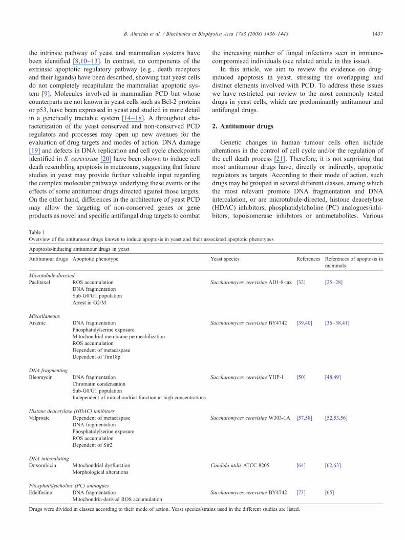
Table 1Overview of the antitumour drugs known to induce apoptosis in yeast and their ass
Apoptosis-inducing antitumour drugs in yeast
Antitumour drugs Apoptotic phenotype
Microtubule-directedPaclitaxel ROS accumulation
DNA fragmentationSub-G0/G1 populationArrest in G2/M
MiscellaneousArsenic DNA fragmentation
Phosphatidylserine exposureMitochondrial membrane permeabilizationROS accumulationDependent of metacaspaseDependent of Tim18p
DNA fragmentingBleomycin DNA fragmentation
Chromatin condensationSub-G0/G1 populationIndependent of mitochondrial function at high concentrations
Histone deacetylase (HDAC) inhibitorsValproate Dependent of metacaspase
DNA fragmentationPhosphatidylserine exposureROS accumulationDependent of Sir2
DNA intercalatingDoxorubicin Mitochondrial dysfunction
Morphological alterations
Phosphatidylcholine (PC) analoguesEdelfosine DNA fragmentation
Mitochondria-derived ROS accumulation
Drugs were divided in classes according to their mode of action. Yeast species/strai
the increasing number of fungal infections seen in immuno-compromised individuals (see related article in this issue).
In this article, we aim to review the evidence on drug-induced apoptosis in yeast, stressing the overlapping anddistinct elements involved with PCD. To address these issueswe have restricted our review to the most commonly testeddrugs in yeast cells, which are predominantly antitumour andantifungal drugs.
2. Antitumour drugs
Genetic changes in human tumour cells often includealterations in the control of cell cycle and/or the regulation ofthe cell death process [21]. Therefore, it is not surprising thatmost antitumour drugs have, directly or indirectly, apoptoticregulators as targets. According to their mode of action, suchdrugs may be grouped in several different classes, among whichthe most relevant promote DNA fragmentation and DNAintercalation, or are microtubule-directed, histone deacetylase(HDAC) inhibitors, phosphatidylcholine (PC) analogues/inhi-bitors, topoisomerase inhibitors or antimetabolites. Various
ociated apoptotic phenotypes
Yeast species References References of apoptosis inmammals
Saccharomyces cerevisiae AD1-8-tax [32] [25–28]
Saccharomyces cerevisiae BY4742 [39,40] [36–38,41]
Saccharomyces cerevisiae YHP-1 [50] [48,49]
Saccharomyces cerevisiae W303-1A [57,58] [52,53,56]
Candida utilis ATCC 8205 [64] [62,63]
Saccharomyces cerevisiae BY4742 [73] [65]
ns used in the different studies are listed.
1438 B. Almeida et al. / Biochimica et Biophysica Acta 1783 (2008) 1436–1448
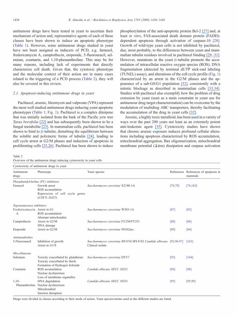
antitumour drugs have been tested in yeast to ascertain theirmechanism of action and, representative agents of each of theseclasses have been shown to induce an apoptotic phenotype(Table 1). However, some antitumour drugs studied in yeasthave not been assigned as inducers of PCD, e.g. farnesol,fredericamycin A, camptothecin, etoposide, 5-fluorouracil, sel-enium, coumarin, and 1,10-phenanthroline. This may be formany reasons, including lack of experiments that directlycharacterize cell death. Given that, the cytotoxic phenotypeand the molecular context of their action are in many casesrelated to the triggering of a PCD process (Table 2), they willalso be covered in this review.
2.1. Apoptosis-inducing antitumour drugs in yeast
Paclitaxel, arsenic, bleomycin and valproate (VPA) representthe most well studied antitumour drugs inducing yeast apoptoticphenotypes (Table 1; Fig. 1). Paclitaxel is a complex diterpenethat was initially isolated from the bark of the Pacific yew treeTaxus brevifolia [22] and has subsequently been shown to be afungal metabolite [23]. In mammalian cells, paclitaxel has beenshown to bind to β-tubulin, disturbing the equilibrium betweenthe soluble and polymeric forms of tubulin [24], leading tocell cycle arrest at G2/M phases and induction of apoptosis inproliferating cells [25,26]. Paclitaxel has been shown to induce
Table 2Overview of the antitumour drugs inducing cytotoxicity in yeast cells
Cytotoxicity of antitumour drugs in yeast
Antitumourdrugs
Phenotype Yeast species
Phosphatidylcholine (PC) inhibitorsFarnesol Growth arrest Saccharomyces cerevisiae
ROS accumulationRepression of cell cycle genes(CDC9; HAT2)
Topoisomerases inhibitorsFredericamycinA
Arrest in G1 Saccharomyces cerevisiaeROS accumulationAberrant mitochondria
Camptothecin Arrest in G2/M Saccharomyces cerevisiaeDNA damage
Etoposide Arrest in G2/M Saccharomyces cerevisiae
Antimetabolites5-Fluorouracil Inhibition of growth Saccharomyces cerevisia
Clinical isolateArrest in G1/S
MiscellaneousSelenium Toxicity exacerbated by glutathione Saccharomyces cerevisiae
Toxicity exacerbated by thiolsFormation of Hydrogen Selenide
Coumarin ROS accumulation Candida albicans ATCCNuclear dysfunctionsLoss of membrane organelles
1,10-Phenanthroline
DNA degradation Candida albicans ATCCNuclear dysfunctionsMitochondrialfunction disruption
Drugs were divided in classes according to their mode of action. Yeast species/strai
phosphorylation of the anti-apoptotic protein Bcl-2 [27] and, atleast in vitro, FAS-associated death domain protein (FADD)-dependent apoptosis through activation of caspase-10 [28].Growth of wild-type yeast cells is not inhibited by paclitaxel,due, most probably, to the differences between yeast and mam-malian tubulin residues involved in paclitaxel binding [29–31].However, mutations in the yeast β-tubulin promote the accu-mulation of intracellular reactive oxygen species (ROS), DNAfragmentation (detected by terminal dUTP nick-end labeling(TUNEL) assay), and alterations of the cell cycle profile (Fig. 1)characterized by an arrest in the G2/M phases and the ap-pearance of a sub-G0/G1 population [32], consistently with amitotic blockage as described in mammalian cells [33,34].Studies with paclitaxel also exemplify how the problem of drugextrusion by yeast (seen as a main constraint in yeast use forantitumour drug target characterization) can be overcome by themodulation of multidrug ABC transporters, thereby facilitatingthe accumulation of the drug in yeast cells [32].
Arsenic, a highly toxicmetalloid, has been used in a variety ofways over the past 200 years not least as an extremely potentanti-leukemic agent [35]. Cytotoxicity studies have shownthat chronic arsenic exposure induces profound cellular altera-tions including apoptosis characterized by ROS accumulation,mitochondrial aggregation, Bax oligomerization, mitochondrialmembrane potential (Δψm) dissipation and caspase activation
References References of apoptosis inmammals
X2180-1A [78,79] [74,162]
W303-1A [87] [82]
FY250/FY251 [88] [86]
JN362acc [89] [84]
e BY4741/BY4742 Candida albicans [92,96,97] [163]
DTY7 [93] [164]
10231 [94] [98]
10231 [95] [95,99]
ns used in the different studies are listed.
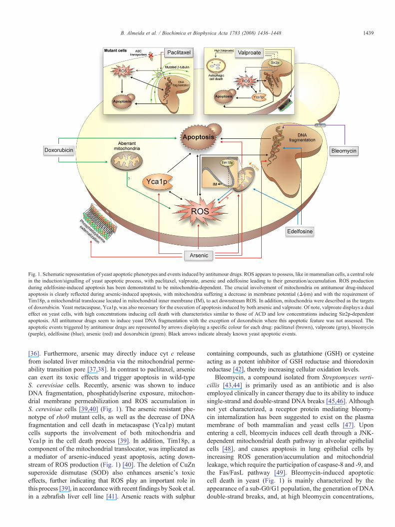
Fig. 1. Schematic representation of yeast apoptotic phenotypes and events induced by antitumour drugs. ROS appears to possess, like in mammalian cells, a central rolein the induction/signalling of yeast apoptotic process, with paclitaxel, valproate, arsenic and edelfosine leading to their generation/accumulation. ROS productionduring edelfosine-induced apoptosis has been demonstrated to be mitochondria-dependent. The crucial involvement of mitochondria on antitumour drug-inducedapoptosis is clearly reflected during arsenic-induced apoptosis, with mitochondria suffering a decrease in membrane potential (Δψm) and with the requirement ofTim18p, a mitochondrial translocase located in mitochondrial inner membrane (IM), to act downstream ROS. In addition, mitochondria were described as the targetsof doxorubicin. Yeast metacaspase, Yca1p, was also necessary for the execution of apoptosis induced by both arsenic and valproate. Of note, valproate displays a dualeffect on yeast cells, with high concentrations inducing cell death with characteristics similar to those of ACD and low concentrations inducing Sir2p-dependentapoptosis. All antitumour drugs seem to induce yeast DNA fragmentation with the exception of doxorubicin where this apoptotic feature was not assessed. Theapoptotic events triggered by antitumour drugs are represented by arrows displaying a specific colour for each drug: paclitaxel (brown), valproate (gray), bleomycin(purple), edelfosine (blue), arsenic (red) and doxorubicin (green). Black arrows indicate already known yeast apoptotic events.
1439B. Almeida et al. / Biochimica et Biophysica Acta 1783 (2008) 1436–1448
[36]. Furthermore, arsenic may directly induce cyt c releasefrom isolated liver mitochondria via the mitochondrial perme-ability transition pore [37,38]. In contrast to paclitaxel, arseniccan exert its toxic effects and trigger apoptosis in wild-typeS. cerevisiae cells. Recently, arsenic was shown to induceDNA fragmentation, phosphatidylserine exposure, mitochon-drial membrane permeabilization and ROS accumulation inS. cerevisiae cells [39,40] (Fig. 1). The arsenic resistant phe-notype of rho0 mutant cells, as well as the decrease of DNAfragmentation and cell death in metacaspase (Yca1p) mutantcells supports the involvement of both mitochondria andYca1p in the cell death process [39]. In addition, Tim18p, acomponent of the mitochondrial translocator, was implicated asa mediator of arsenic-induced yeast apoptosis, acting down-stream of ROS production (Fig. 1) [40]. The deletion of CuZnsuperoxide dismutase (SOD) also enhances arsenic's toxiceffects, further indicating that ROS play an important role inthis process [39], in accordancewith recent findings by Seok et al.in a zebrafish liver cell line [41]. Arsenic reacts with sulphur
containing compounds, such as glutathione (GSH) or cysteineacting as a potent inhibitor of GSH reductase and thioredoxinreductase [42], thereby increasing cellular oxidation levels.
Bleomycin, a compound isolated from Streptomyces verti-cillis [43,44] is primarily used as an antibiotic and is alsoemployed clinically in cancer therapy due to its ability to inducesingle-strand and double-strand DNA breaks [45,46]. Althoughnot yet characterized, a receptor protein mediating bleomy-cin internalization has been suggested to exist on the plasmamembrane of both mammalian and yeast cells [47]. Uponentering a cell, bleomycin induces cell death through a JNK-dependent mitochondrial death pathway in alveolar epithelialcells [48], and causes apoptosis in lung epithelial cells byincreasing ROS generation/accumulation and mitochondrialleakage, which require the participation of caspase-8 and -9, andthe Fas/FasL pathway [49]. Bleomycin-induced apoptoticcell death in yeast (Fig. 1) is mainly characterized by theappearance of a sub-G0/G1 population, the generation of DNAdouble-strand breaks, and, at high bleomycin concentrations,

1440 B. Almeida et al. / Biochimica et Biophysica Acta 1783 (2008) 1436–1448
the induction of a mitochondria-independent cell death process[50].
Similar to other antitumour drugs, VPA, an inhibitor of theclass I HDACs [51] can trigger apoptosis in mammalian cellsthrough caspase-dependent and -independent pathways [52,53].VPA also promotes the down-regulation of pro-survival genes,Bcl-2 and Bcl-XL, and the up-regulation of pro-apoptotic genessuch as Bax [54,55]. A recent study also demonstrates that VPAinduces caspase-dependent apoptosis in HeLa cells through theblocking of the Akt pathway [56]. Likewise, in yeast cells, VPAtriggers a cell death process that is dependent on Yca1p [57](Fig. 1). Exposure to high concentrations of VPA induces celldeath with morphological features similar to those of autopha-gic cell death (ACD), which is independent of Yca1p [57],while low VPA concentrations result in apoptotic cell deathassociated with DNA fragmentation, ROS accumulation,phosphatidylserine exposure and morphological alterationssuch as cell shrinkage [57,58]. Sun et al. showed that Sir2p orsirtuin, a class III HDAC, that is also involved in the DNAdamage response and life span extension mediated by caloricrestriction [59,60], is required for VPA-induced cell death [58].Accordingly, Δsir2 cells do not produce ROS or accumulateneutral lipids, leading to the conclusion that Sir2p has a role inlipid metabolism, which might be linked to apoptosis [58].
Other antitumour drugs presented in Table 1 and describedas inducing apoptosis in yeast cells include doxorubicin (DOX)and edelfosine. DOX is an antibiotic, originally isolated fromStreptomyces peucetius and currently used as an effective an-titumour drug [61] known to induce, among other events, thegeneration of free radicals, DNA damage and apoptosis, via aninhibition of topoisomerase II [62,63]. In yeast, DOX wasshown to induce apoptosis in Candida utilis, based merely uponmorphological observations, with reported plasma membranealterations and changes in mitochondrial shape and cristaeorganization [64] (Fig. 1). Therefore, further studies directed toknown yeast apoptotic regulators are needed in order to uncoverthe mechanism by which DOX kills yeast cells.
Edelfosine is a synthetic lipid, analogue of phosphatidylcho-line (PC), which induces apoptosis in a wide variety of tumourcells [65]. Edelfosine and its analogues contain ether linked fattyacids, as opposed to the endogenous ester linked fatty acids,rendering them more resistant to cellular phospholipases and,thus, more effective as drugs. Although not as an amplificatorymechanism, like bleomycin [49], edelfosine was found to induceFas-dependent apoptosis in leukemic cells [66]. Overexpressionof Bcl-2 or Bcl-XL was shown to be able to inhibit apoptosisinduced by this compound [65,67], which was also shown to beassociated with alterations in mitochondrial function, generationof ROS and caspase-3 activation [68,69]. Recently it wassuggested that endoplasmic reticulum may also play a majorrole in edelfosine-induced apoptosis in tumour cells [70]. Inaddition to its cytotoxic effects [71,72], edelfosine was reportedto promote apoptosis in S. cerevisiae cells characterized by aTUNEL-positive phenotype and mitochondrial dependent ROSgeneration [73], presenting similarities with edelfosine-inducedapoptosis in human tumour cells, also mediated by mitochon-dria and correlated with ROS generation [68,69].
The accumulated evidence indicates that the mechanisms ofantitumour drug-induced apoptosis in yeast share some homo-logies with the mammalian system. Particularly predominantare the involvement of mitochondria, DNA fragmentation, andespecially ROS production/accumulation. Nevertheless, not allthe studies regarding the induction of apoptosis in yeast cells byantitumour drugs explore the knowledge of yeast molecularPCD pathway(s), namely, the precise association of the apop-totic regulators and their hierarchy. Even so, the data hereinpresented point out the potential value of yeast to study PCD-based therapies and drug targets.
2.2. Cytotoxicity of antitumour drugs in yeast
As a model organism, yeast has long been used as a phar-macological tool in the identification and definition of themolecular context and of critical determinants that conferchemosensitivity to specific cytotoxic injuries induced by drugs.Several of the different antitumour drugs studied in yeast havenot been specifically assigned as inducers of PCD, although,the cytotoxic phenotype and the molecular context of theiraction are suggestive of that. Drugs such as the PC inhibitorfarnesol and some topoisomerases inhibitors are worth of furtherdiscussion (Table 2). Farnesol is known to induce apoptosis in awide variety of cell lines [74,75]. Farnesol-induced cell death isattenuated through the addition of exogenous PC or diacylgly-cerol, but not other lipids [76,77]. In yeast cells, farnesol hasbeen shown to induce growth arrest and cell death, withrepression of cell cycle genes encoding a DNA ligase (CDC9)and a histone acetyltransferase (HAT2), a process that can beinhibited by the addition of a diacylglycerol analogue [78].Although farnesol induces the generation of ROS [79,80], thefarnesol-induced cell death mechanism remains uncharacterizedin yeast cells. Nevertheless, farnesol has been described toinduce apoptosis in Aspergillus nidulans cells characterized bychromatin condensation, a TUNEL-positive phenotype, expo-sure of phosphatidylserine, and is also dependent on mitochon-drial function and ROS generation [81].
Other successful antitumour drugs take advantage of theinhibition of topoisomerases, key enzymes in DNA transcriptionand replication. Fredericamycin A (FMA), an antibiotic productof Streptomyces griseus, camptothecin, an alkaloid derived fromthe plant Camptotheca acuminate and etoposide, a derivative ofthe podophyllotoxin from Podophyllum peltatum are among theantitumour drugs known to induce apoptosis in mammalian cellsdue to the inhibition of topoisomerases [82–86]. Although theinduction of apoptosis in yeast cells by those drugs is notsupported by the available data, some lines of evidence dosupport a link. For example, FMAwas shown to induce growtharrest (G1 cell cycle phase) and the appearance of aberrantmitochondria in yeast, just as in mammalian cells, which alsoresults in the generation of high intracellular ROS levels [87].Furthermore, and even though evidence for apoptosis inducedby etoposide and camptothecin in yeast cells is scarce, thesetopoisomerase inhibitors are known to induce arrest in the G2/Mcell cycle phases and DNA damage [88,89], a phenomenon alsoobserved in other drugs inducing apoptosis in yeast [90,91].
1441B. Almeida et al. / Biochimica et Biophysica Acta 1783 (2008) 1436–1448
Other antitumour drugs, including 5-fluorouracil, selenium,coumarin and 1,10-phenanthroline, have been described as cy-totoxic agents that lead to yeast cell death [92–97]. However,the mechanism of the cell death process underlying theircytotoxicity remains unexplored. Nonetheless, treatment withcoumarin and 1,10-phenanthroline stimulates ROS genera-tion, changes in nuclear morphology and a loss of membraneorganelles [94,95], indicating that apoptotic cell death mighttake place in yeast as demonstrated in mammalian cells [98,99].
Future studies directed towards the identification of the truenature of the cell death processes that occur upon treatmentwith these drugs will bring forth important data regarding drug-induced apoptotic phenotype in yeast strengthening its claim asa useful tool for screening the cytotoxic effects of antitumourdrugs.
3. Apoptosis-inducing antifungal drugs in yeast
S. cerevisiae represents a practical and conventional systemfor studying the properties of antifungal compounds, not onlyagainst fungal human pathogens with which they are closelyrelated (e.g., Candida albicans) [100], but also with those thatare evolutionarily more distant (e.g., filamentous fungi). More-over, the majority of the currently used antifungal drugs areactive against S. cerevisiae (reviewed in [101]), thus making it asuitable model for both drug development and the elucidation ofthe mechanisms underlying drug's action.Most antifungal drugsbelong to a few structural classes that affect specific fungalcellular targets, such as ergosterol synthesis. However, many ofthese drugs are associated with a high human toxicity (e.g.amphotericin B) and/or to the selection of resistant fungal pa-thogens (e.g., azole drugs), two main constraints on the successof antifungal drug therapies. To overcome the changing tide offungal diseases, novel fungal targets for drug therapy need tobe identified. As described above, the possible architecturaldifferences between apoptotic regulators/mechanisms of yeastand mammalian cells may open the door either for the designof new antifungal drugs, or for testing the fungal-specificapoptotic-inducing abilities of the current ones. In fact, somedrugs with known antifungal capacities have already beendemonstrated to act as yeast-specific apoptotic PCD-inducers(Table 3). Among these, some are currently used in clinics whileothers are still under development. One of the best characterizedand commercially available antifungal drugs is amphotericin B(AmB), a polyene agent efficiently used for treating invasivefungal infections, but generally associated with high toxicityagainst human cells [102]. AmB binds to sterols, creating poresthat increase fungal membrane permeability to small cations,thus promoting the rapid depletion of intracellular potassium andfungal cell death [103]. Phillips et al. assessed the toxic effectsof AmB inC. albicans, revealing that AmB induces an apoptoticmechanism, with the occurrence of arrest in G2/M cell cyclephases, chromatin condensation, nuclear fragmentation, phos-phatidylserine externalization and ROS accumulation [91].
Ciclopirox olamine (CPO), a representative of a quite distinctclass of antifungal drugs, was introduced into clinical therapymore than three decades ago. CPO belongs to a group of syn-
thetic antifungal agents, hydroxypyridones that have high af-finity for trivalent metal cations [104], that are used effectivelyin clinical practice since they have a broad spectrum of actionagainst dermatophytes, yeasts, filamentous fungi and bacteria[105]. A remarkable feature of CPO is that no single case offungal resistance has been reported so far. Work performed byour group has shown that CPO leads to non-apoptotic yeast PCDcharacterized by chromatin condensation and DNA damageassociated with the appearance of a sub-G0/G1 population andarrest in G2/M cell cycle phases [90]. Notably, in contrast toAmB-induced apoptosis, CPO-induced PCD does not involveROS signalling and is associated with a TUNEL-negativephenotype; CPO-induced PCD also appears to be independent ofmetacaspase but is associated with unknown protease activities[90].
Besides the described antifungal drugs, other compoundsisolated from distinct organisms have proved to display effectiveantifungal capacities through the induction of apoptosis. Os-motin, a Tobacco pathogenesis-related protein, dermaseptins, afamily of peptides derived from the tree-frog Phyllomedusasauvagii, and pradimicin (PRM), an Actinomadura hibisca-derived antibiotic, were found to induce cell death in yeast withapoptotic features [106–109]. All of these drugs were shown toinduce nuclear fragmentation, a TUNEL-positive phenotype andthe generation of high intracellular ROS levels [106–109].However, some differences were detected among the apoptoticcell death processes triggered by these drugs. The mechanism bywhich osmotin induces apoptosis, relies on the suppression ofstress-responsive gene transcription via the RAS2/cAMP path-way, and, upstream from RAS2, on the binding of osmotin tothe plasma membrane protein Pho36, a homologue of the mam-malian receptor for the hormone adiponectin [106,110]. On theother hand, the truncated derivative of dermaseptin S3 [111],which promotes disruption of the yeast cell membrane and aderegulation in the homeostasis of intracellular pH, was shownto induce S. cerevisiae PCD associated with ROS generation andnuclear DNA fragmentation [107,108]. Interestingly, the modeof dermaseptin-induced cell death is metacaspase-independent,but dependent on Aif1p, and on the proteasomal substrate,Stm1p [108], which is also involved in yeast apoptosis [112]. Inaddition to their capacity to induce yeast apoptosis, dermaseptinshave very low human cytotoxicity. In fact, the same is true formost of the naturally occurring antimicrobial peptides fromamphibian skin, at concentrations that effectively inhibit fungalgrowth [111,113–115], making them very attractive antifungaldrugs. PRM, a mannose-binding antifungal antibiotic that cau-ses membrane permeability dysfunction, is also capable ofinducing S. cerevisiae apoptotic cell death, characterized byROS accumulation, DNA damage and nuclear fragmentation[109]. The cell death mechanism seems to be dependent on thesensor kinase, Sln1p, to which PRM can bind [116].
Another group of compounds that display antifungal capa-cities are histatins, histidine-rich cationic peptides secreted bythe parotid and the submandibular/sublingual human salivaryglands [117]. Histatin 5 has been shown to display potent fun-gicidal properties against C. albicans [117]. Although scarce,evidence for the induction of a histatin 5-mediated apoptotic
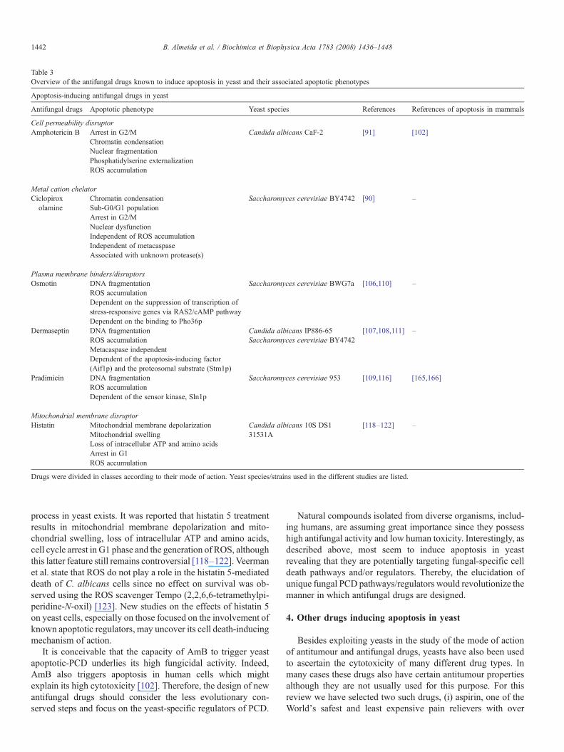
Table 3Overview of the antifungal drugs known to induce apoptosis in yeast and their associated apoptotic phenotypes
Apoptosis-inducing antifungal drugs in yeast
Antifungal drugs Apoptotic phenotype Yeast species References References of apoptosis in mammals
Cell permeability disruptorAmphotericin B Arrest in G2/M Candida albicans CaF-2 [91] [102]
Chromatin condensationNuclear fragmentationPhosphatidylserine externalizationROS accumulation
Metal cation chelatorCiclopiroxolamine
Chromatin condensation Saccharomyces cerevisiae BY4742 [90] –Sub-G0/G1 populationArrest in G2/MNuclear dysfunctionIndependent of ROS accumulationIndependent of metacaspaseAssociated with unknown protease(s)
Plasma membrane binders/disruptorsOsmotin DNA fragmentation Saccharomyces cerevisiae BWG7a [106,110] –
ROS accumulationDependent on the suppression of transcription ofstress-responsive genes via RAS2/cAMP pathwayDependent on the binding to Pho36p
Dermaseptin DNA fragmentation Candida albicans IP886-65Saccharomyces cerevisiae BY4742
[107,108,111] –ROS accumulationMetacaspase independentDependent of the apoptosis-inducing factor(Aif1p) and the proteosomal substrate (Stm1p)
Pradimicin DNA fragmentation Saccharomyces cerevisiae 953 [109,116] [165,166]ROS accumulationDependent of the sensor kinase, Sln1p
Mitochondrial membrane disruptorHistatin Mitochondrial membrane depolarization Candida albicans 10S DS1
31531A[118–122] –
Mitochondrial swellingLoss of intracellular ATP and amino acidsArrest in G1ROS accumulation
Drugs were divided in classes according to their mode of action. Yeast species/strains used in the different studies are listed.
1442 B. Almeida et al. / Biochimica et Biophysica Acta 1783 (2008) 1436–1448
process in yeast exists. It was reported that histatin 5 treatmentresults in mitochondrial membrane depolarization and mito-chondrial swelling, loss of intracellular ATP and amino acids,cell cycle arrest in G1 phase and the generation of ROS, althoughthis latter feature still remains controversial [118–122]. Veermanet al. state that ROS do not play a role in the histatin 5-mediateddeath of C. albicans cells since no effect on survival was ob-served using the ROS scavenger Tempo (2,2,6,6-tetramethylpi-peridine-N-oxil) [123]. New studies on the effects of histatin 5on yeast cells, especially on those focused on the involvement ofknown apoptotic regulators, may uncover its cell death-inducingmechanism of action.
It is conceivable that the capacity of AmB to trigger yeastapoptotic-PCD underlies its high fungicidal activity. Indeed,AmB also triggers apoptosis in human cells which mightexplain its high cytotoxicity [102]. Therefore, the design of newantifungal drugs should consider the less evolutionary con-served steps and focus on the yeast-specific regulators of PCD.
Natural compounds isolated from diverse organisms, includ-ing humans, are assuming great importance since they possesshigh antifungal activity and low human toxicity. Interestingly, asdescribed above, most seem to induce apoptosis in yeastrevealing that they are potentially targeting fungal-specific celldeath pathways and/or regulators. Thereby, the elucidation ofunique fungal PCD pathways/regulators would revolutionize themanner in which antifungal drugs are designed.
4. Other drugs inducing apoptosis in yeast
Besides exploiting yeasts in the study of the mode of actionof antitumour and antifungal drugs, yeasts have also been usedto ascertain the cytotoxicity of many different drug types. Inmany cases these drugs also have certain antitumour propertiesalthough they are not usually used for this purpose. For thisreview we have selected two such drugs, (i) aspirin, one of theWorld's safest and least expensive pain relievers with over
1443B. Almeida et al. / Biochimica et Biophysica Acta 1783 (2008) 1436–1448
100 years of proven and effective treatment against a variety ofailments, and (ii) ricin, a toxin isolated from plants that has thecapacity to inhibit protein synthesis by irreversibly inactivatingeukaryotic ribosomes.
Aspirin, or acetylsalicylic acid, is a non-steroidal anti-inflammatory drug known to induce apoptosis in mammaliancells by a variety of different mechanisms including caspaseactivation [124,125], inhibition of NF-KB activation [126], ce-ramide pathway activation [127] and p38MAP kinase activation[128]. The effects of aspirin on cell growth and its propensity toinduce apoptosis have also been studied in yeast cells. Aspi-rin was found to commit mitochondrial MnSOD-deficientS. cerevisiae cells growing in ethanol to apoptosis [129]. Inaccordance with aspirin's ROS scavenger properties, it alsoexhibits a significant antioxidant effect until the onset of overtapoptosis in yeast cells, suggesting that ROS probably do notplay a primary role in the apoptosis of cells exposed to aspirin[129]. Instead, the authors suggest that a disruption of the redoxbalance commits yeast cells to apoptosis upon aspirin treatment[130].
Ricin is naturally synthesized in the seeds of Ricinus com-munis (castor bean). This plant toxin is a type II ribosome-inactivating protein (RIP) that inhibits protein synthesis [131].It consists of a catalytic A chain (RTA) covalently joined by adisulfide bond to a cell binding B chain (RTB) and is highly toxicto eukaryotic cells [132,133]. The RTB is a lectin that bindsgalactose or N-acetylgalactosamine receptors on the surface oftarget cells and promotes subsequent endocytosis of the RTA[132,133]. Ricin induces apoptosis in a wide variety of animalcells [134] and recently the effects of ricin were studied inS. cerevisiae, using a large-scale mutagenesis screen for variantsof the precursor form of RTA (pre-RTA) that were unable to killyeast cells. Apoptotic markers, such as chromatin condensation,nuclear fragmentation and ROS accumulation were observedfor yeast cells expressing the wild-type RTA but not for cellsexpressing the nontoxic mutants, even though they still de-purinated ribosomes and inhibited translation [135]. These re-sults provide evidence showing that similar to the studies inmammalian cells, ribosome depurination and translation inhibi-tion are also not sufficient for the ricin-induced cytotoxicity inS. cerevisiae. Moreover, the mechanism of apoptotic cell deathseems to be strictly dependent on the early generation of ROS[135].
5. Conclusions and future perspectives
The field of yeast PCD, particularly apoptotic-PCD, hasgrown rapidly during the last decade [7–9]. The increasingunderstanding of yeast PCDmolecular pathways is crucial eitherfor the basic knowledge or for the application of this knowledgeto the use of yeasts as a model for cell death-based therapies.Yeasts have been intensively explored to study a wide range ofprocesses, from the basic cellular and molecular pathways tothe implications of their regulation and dysfunction in humandiseases. Previously, yeast cells containing mutations in genesassociated with a specific disease, e.g. tumour associated alte-rations in DNA repair, mitotic catastrophe, etc., have allowed the
screening of drugs that kill mutant cells more efficiently thanwild-type cells [136,137]. These strategies have been usedsuccessfully revealing several antitumour agents with a hightherapeutic advantage [138,139]. The power of yeast moleculargenetics, including the multi-faceted role of yeast in drugdiscovery is also apparent from yeast two-hybrid and three-hybrid systems that have been employed in target identificationand validation; the yeast target-based screenings such as high-throughput screening or cell based assays; phenotype-basedscreening; gene expression profiling of drug action and drug-induced haploinsufficiency (reviewed in [101,140,141]). There-fore, one may already consider “Yeast as a model in drug targetdiscovery and validation”. The question that now arises is, canwe now reasonably say that “Yeasts are also a good model inapoptosis or cell death-based therapies and drug targets”? Theexamples addressed in this review show that some therapeuticagents induce yeast apoptotic-PCD that certainly have somesimilarities with the cell death processes known in mammaliancells. For most of the cell death scenarios induced by antitumourdrugs and discussed herein, ROS and mitochondria appear ascrucial yeast and mammalian players. This evidence brings us toa relevant and recurrent theme in tumours and chemotherapy:mitochondria and ROS as therapeutic targets. Indeed, a greatvariety of drugs can directly be targeted to mitochondria toinduce apoptosis [142] or to ROS scavenging, resulting in ROSaccumulation and apoptosis (reviewed in [143,144]). BesidesROS, nitric oxide (NO), which reacts with molecular oxygen toform reactive nitrogen species (RNS) and ultimately favourscarcinogenesis [145], is also an appealing target. Somewhatparadoxically, both anti-NO and NO-based strategies have beenapplied in cancer therapy (reviewed in [145,146]) indicating aNO dichotomy and an inevitable need of modulate NO levelsaccording to the specific molecular makeup of each individualtumour cell (reviewed in [145,146]). Recently, we demonstratedthat S. cerevisiae is able to synthesize NO by an L-arginine-dependent mechanism, controlling the formation of ROS andacting as a crucial apoptotic inducer [147,148]. Following thisline of thought, yeast could be employed in the study of thesynergistic effects as well as molecular pathways that determinethe increased sensitivity of cells to antitumour drugs in thepresence of different endogenous NO levels.
Other cellular processes that have been revealed as futuretherapeutic targets include the proteasome, Heat Shock Protein90 (HSP90), and non-apoptotic PCD pathways including ACD,all of which could be explored using yeast. Yeast proteasomefunction has already been linked to apoptotic cell death [112]. Asthe proteasome is a critical enzymatic complex for fundamentalpathways in cell survival and proliferation, its inhibition couldbe a potential antitumour therapy [149,150]. The establishedlink between proteasome and yeast apoptosis suggests that aproteasome inhibition-based therapy could also be investigatedin yeast.
The molecular chaperone HSP90, required to ensure the cor-rect conformation, activity, intracellular localization, and proteo-lytic turnover of a range of proteins that are involved in cellgrowth, differentiation, and survival [151,152], is also an at-tractive target for tumour therapy. It is already known that

1444 B. Almeida et al. / Biochimica et Biophysica Acta 1783 (2008) 1436–1448
inhibition of HSP90's function causes degradation of the socalled “client proteins”, which are reported to be involved intumourigenesis [151], via the ubiquitin-proteasome pathway[153,154]. Interestingly, our recent observations point to aprotective role of HSP90 members in yeast apoptosis (Almeida,B. et al., unpublished data). Using yeast to assess for HSP90“client proteins”, upon treatment with HSP90 inhibitors, couldeasily contribute to the understanding of its mode of actionand role in tumourigenesis. Regarding non-apoptotic PCDprocess such as ACD, accumulated evidence has shown thatthis phenomenon also occurs in yeast cells [155–157]. Sincemany reports show that antitumour drug-induced cell deathmay involve non-apoptotic PCD through caspase-independentpathways [158,159] or even through the induction of ACD [160],the yeast system seems promising for revealing clues on thefoundation of new opportunities to design targeting therapy topromote non-apoptotic cell death of tumour cells.
An interesting link between HSPs, the proteasome andautophagy relates to the fact that they all act as cellular defensesin neurodegenerative disorders, especially those that involveprotein misfolding. Given the fact that yeast is being used as amodel to study several neurodegenerative disorders involvingprotein misfolding and aggregation [161], it seems feasible toalso use yeast for screening of drugs that are able to increasesurvival by acting on these targets.
Although yeast cells are useful for the study of the cytotoxiceffects of a panoply of drugs, their primary relevance might bedirected to the design of new antifungal drugs. A newgeneration of antifungal drugs is urgently needed given theproblems associated with ones currently in use and to theincreasing number of invasive fungal infections in immuno-compromised patients. In this sense, the exploration of yeastPCD processes to identify molecules that allow the specificmanipulation of yeast cell death without causing serious sideeffects on human cells is appealing. Until recently thecumulative knowledge on yeast PCD shows a high conservationof cell death processes and regulators, however substantialdifferences will necessarily be detected among molecules and/or pathways as the field develops. One good example is thefungal metacaspases which seem to be the main executors of awide range of apoptotic stimuli. Even though metacaspases areorthologs of caspases, they display enough structural dissim-ilarity to allow the design/screening of compounds or moleculesthat selectively activate metacaspases and not caspases. For thisto be possible more effort needs to be applied to the study ofyeast PCD.
On the other hand, we must not disregard the existence ofdistinct cellular machineries linked to yeast PCD induction thatmay be relevant as future therapeutic targets. In fact, one of themain problems regarding the design of antitumour drugs is thecellular specificity; ergo, some drugs are effective only against aparticular kind of tumour cells while ineffective for others. Assimple eukaryotic microorganisms with less complex PCD re-gulation without the idiosyncrasies of different cell types, yeastsare undoubtedly important models for the design of therapiesdirected to basic molecular pathways, thus overcoming theproblem of cellular specificity.
The examples presented throughout this review show invery distinct ways the real utility of yeasts in drug-induced celldeath discovery. In addition, the plethora of tools available,along with our knowledge of PCD also makes yeast a highlyvaluable model organism for drug target identification andvalidation. Future studies are required in order to fully cha-racterize the “ups and downs” of yeast PCD and definitivelyexpose the extent of potential benefits that yeast may present tostudy these issues.
Acknowledgments
The authors apologize to the researchers whose work was notcited or discussed in this review. The authors would also like tothank Agostinho Almeida for all the helpful suggestions and forthe critical reading of the manuscript. This work was supportedby a grant from FCT— Fundação para a Ciência e a Tecnologia,Portugal (POCI/BIA-BCM/57364/2004). B.A., A.S., A.M. andB.S.M. have fellowships from FCT (SFRH/BD/15317/2005,SFRH/BD/33125/2007, SFRH/BD/32464/2006 and SFRH/BI/15406/2005, respectively).
References
[1] C.B. Thompson, Apoptosis in the pathogenesis and treatment of disease,Science 267 (1995) 1456–1462.
[2] U. Fischer, K. Schulze-Osthoff, New approaches and therapeuticstargeting apoptosis in disease, Pharmacol. Rev. 57 (2005) 187–215.
[3] U. Fischer, K. Schulze-Osthoff, Apoptosis-based therapies and drugtargets, Cell Death Differ 12 (Suppl 1) (2005) 942–961.
[4] L.H. Hartwell, Nobel Lecture. Yeast and cancer, Biosci. Rep. 22 (2002)373–394.
[5] J. Heitman, N.R. Movva, M.N. Hall, Targets for cell cycle arrest by theimmunosuppressant rapamycin in yeast, Science 253 (1991) 905–909.
[6] S.L. Schreiber, G.R. Crabtree, The mechanism of action of cyclosporin Aand FK506, Immunol. Today 13 (1992) 136–142.
[7] F. Madeo, E. Herker, S. Wissing, H. Jungwirth, T. Eisenberg, K.U.Frohlich, Apoptosis in yeast, Curr. Opin. Microbiol. 7 (2004) 655–660.
[8] P. Ludovico, F. Madeo, M. Silva, Yeast programmed cell death: anintricate puzzle, IUBMB Life 57 (2005) 129–135.
[9] K.U. Frohlich, H. Fussi, C. Ruckenstuhl, Yeast apoptosis—from genes topathways, Semin. Cancer Biol. 17 (2007) 112–121.
[10] S. Wissing, P. Ludovico, E. Herker, S. Buttner, S.M. Engelhardt, T.Decker, A. Link, A. Proksch, F. Rodrigues, M. Corte-Real, K.U.Frohlich, J. Manns, C. Cande, S.J. Sigrist, G. Kroemer, F. Madeo, AnAIF orthologue regulates apoptosis in yeast, J. Cell Biol. 166 (2004)969–974.
[11] P. Ludovico, F. Rodrigues, A. Almeida, M.T. Silva, A. Barrientos, M.Corte-Real, Cytochrome c release and mitochondria involvement inprogrammed cell death induced by acetic acid in Saccharomycescerevisiae, Mol. Biol. Cell 13 (2002) 2598–2606.
[12] B. Fahrenkrog, U. Sauder, U. Aebi, The S. cerevisiae HtrA-like proteinNma111p is a nuclear serine protease that mediates yeast apoptosis,J. Cell Sci. 117 (2004) 115–126.
[13] C.W. Gourlay, W. Du, K.R. Ayscough, Apoptosis in yeast—mechanismsand benefits to a unicellular organism, Mol. Microbiol. 62 (2006)1515–1521.
[14] W.Greenhalf, C. Stephan, B.Chaudhuri, Role ofmitochondria andC-terminalmembrane anchor of Bcl-2 in Bax induced growth arrest and mortality inSaccharomyces cerevisiae, FEBS Lett. 380 (1996) 169–175.
[15] H. Zha, H.A. Fisk, M.P. Yaffe, N. Mahajan, B. Herman, J.C. Reed,Structure–function comparisons of the proapoptotic protein Bax in yeastand mammalian cells, Mol. Cell Biol. 16 (1996) 6494–6508.
1445B. Almeida et al. / Biochimica et Biophysica Acta 1783 (2008) 1436–1448
[16] B. Ink, M. Zornig, B. Baum, N. Hajibagheri, C. James, T. Chittenden, G.Evan, Human Bak induces cell death in Schizosaccharomyces pombewith morphological changes similar to those with apoptosis inmammalian cells, Mol. Cell Biol. 17 (1997) 2468–2474.
[17] J. Smardova, J. Smarda, J. Koptikova, Functional analysis of p53 tumorsuppressor in yeast, Differentiation 73 (2005) 261–277.
[18] D. Grochova, J. Vankova, J. Damborsky, B. Ravcukova, J. Smarda, B.Vojtesek, J. Smardova, Analysis of transactivation capability andconformation of p53 temperature-dependent mutants and their reactiva-tion by amifostine in yeast, Oncogene (2007).
[19] W.C. Burhans, M. Weinberger, M.A. Marchetti, L. Ramachandran, G.D'Urso, J.A. Huberman, Apoptosis-like yeast cell death in response toDNA damage and replication defects, Mutat. Res. 532 (2003) 227–243.
[20] M. Weinberger, L. Ramachandran, L. Feng, K. Sharma, X. Sun, M.Marchetti, J.A. Huberman, W.C. Burhans, Apoptosis in budding yeastcaused by defects in initiation of DNA replication, J. Cell Sci. 118 (2005)3543–3553.
[21] P. Perego, L. Gatti, N. Carenini, L. Dal Bo, F. Zunino, Apoptosis inducedby extracellular glutathione is mediated by H(2)O(2) production andDNA damage, Int. J. Cancer 87 (2000) 343–348.
[22] M.C. Wani, H.L. Taylor, M.E. Wall, P. Coggon, A.T. McPhail, Plantantitumor agents. VI. The isolation and structure of taxol, a novelantileukemic and antitumor agent from Taxus brevifolia, J. Am. Chem.Soc. 93 (1971) 2325–2327.
[23] A. Stierle, G. Strobel, D. Stierle, Taxol and taxane production by Taxo-myces andreanae, an endophytic fungus of Pacific yew, Science 260(1993) 214–216.
[24] W.B. Derry, L. Wilson, M.A. Jordan, Substoichiometric binding of taxolsuppresses microtubule dynamics, Biochemistry 34 (1995) 2203–2211.
[25] C.M. Woods, J. Zhu, P.A. McQueney, D. Bollag, E. Lazarides, Taxol-induced mitotic block triggers rapid onset of a p53-independent apoptoticpathway, Mol. Med. 1 (1995) 506–526.
[26] K. Torres, S.B. Horwitz, Mechanisms of Taxol-induced cell death areconcentration dependent, Cancer Res. 58 (1998) 3620–3626.
[27] V. Ganansia-Leymarie, P. Bischoff, J.P. Bergerat, V. Holl, Signaltransduction pathways of taxanes-induced apoptosis, Curr. Med. Chem.Anticancer Agents 3 (2003) 291–306.
[28] S.J. Park, C.H. Wu, J.D. Gordon, X. Zhong, A. Emami, A.R. Safa, Taxolinduces caspase-10-dependent apoptosis, J. Biol. Chem. 279 (2004)51057–51067.
[29] J.V. Kilmartin, Purification of yeast tubulin by self-assembly in vitro,Biochemistry 20 (1981) 3629–3633.
[30] G. Barnes, K.A. Louie, D. Botstein, Yeast proteins associated withmicrotubules in vitro and in vivo, Mol Biol Cell 3 (1992) 29–47.
[31] C.J. Bode, M.L. Gupta Jr., E.A. Reiff, K.A. Suprenant, G.I. Georg, R.H.Himes, Epothilone and paclitaxel: unexpected differences in promotingthe assembly and stabilization of yeast microtubules, Biochemistry 41(2002) 3870–3874.
[32] T.B. Foland, W.L. Dentler, K.A. Suprenant, M.L. Gupta, R.H. Himes,Paclitaxel-induced microtubule stabilization causes mitotic block andapoptotic-like cell death in a paclitaxel-sensitive strain of Saccharomycescerevisiae, Yeast 22 (2005) 971–978.
[33] P.B. Schiff, J. Fant, S.B. Horwitz, Promotion of microtubule assembly invitro by taxol, Nature 277 (1979) 665–667.
[34] M.A. Jordan, Mechanism of action of antitumor drugs that interact withmicrotubules and tubulin, Curr. Med. Chem. Anticancer Agents 2 (2002)1–17.
[35] K. Alimoghaddam, A. Shariftabrizi, S.M. Tavangar, Z. Sanaat, S.Rostami, M. Jahani, A. Ghavamzadeh, Anti-leukemic and anti-angiogen-esis efficacy of arsenic trioxide in new cases of acute promyelocyticleukemia, Leuk. Lymphoma. 47 (2006) 81–88.
[36] N. Haga, N. Fujita, T. Tsuruo, Involvement of mitochondrial aggregationin arsenic trioxide (As2O3)-induced apoptosis in human glioblastomacells, Cancer Sci. 96 (2005) 825–833.
[37] J. Bustamante, L. Nutt, S. Orrenius, V. Gogvadze, Arsenic stimulatesrelease of cytochrome c from isolated mitochondria via induction ofmitochondrial permeability transition, Toxicol. Appl. Pharmacol. 207(2005) 110–116.
[38] Y. Zheng, Y. Shi, C. Tian, C. Jiang, H. Jin, J. Chen, A. Almasan, H. Tang,Q. Chen, Essential role of the voltage-dependent anion channel (VDAC)in mitochondrial permeability transition pore opening and cytochrome crelease induced by arsenic trioxide, Oncogene 23 (2004) 1239–1247.
[39] L. Du, Y. Yu, J. Chen, Y. Liu, Y. Xia, Q. Chen, X. Liu, Arsenic inducescaspase- and mitochondria-mediated apoptosis in Saccharomycescerevisiae, FEMS Yeast Res. 7 (2007) 860–865.
[40] L. Du, Y. Yu, Z. Li, J. Chen, Y. Liu, Y. Xia, X. Liu, Tim18, a componentof the mitochondrial translocator, mediates yeast cell death induced byarsenic, Biochemistry (Mosc) 72 (2007) 843–847.
[41] S.H. Seok, M.W. Baek, H.Y. Lee, D.J. Kim, Y.R. Na, K.J. Noh, S.H. Park,H.K. Lee, B.H. Lee, D.Y. Ryu, J.H. Park, Arsenite-induced apoptosis isprevented by antioxidants in zebrafish liver cell line, Toxicol. In Vitro 21(2007) 870–877.
[42] M.F. Hughes, Arsenic toxicity and potential mechanisms of action,Toxicol. Lett. 133 (2002) 1–16.
[43] H. Umezawa, Bleomycin and other antitumor antibiotics of highmolecular weight, Antimicrobial Agents Chemother (Bethesda) 5(1965) 1079–1085.
[44] H. Umezawa, K. Maeda, T. Takeuchi, Y. Okami, New antibiotics,bleomycin A and B, J. Antibiot. (Tokyo) 19 (1966) 200–209.
[45] O. Tounekti, G. Pron, J. Belehradek Jr., L.M. Mir, Bleomycin, anapoptosis-mimetic drug that induces two types of cell death depending onthe number of molecules internalized, Cancer Res. 53 (1993) 5462–5469.
[46] O. Tounekti, J. Belehradek, Jr., L.M. Mir, Relationships between DNAfragmentation, chromatin condensation, and changes in flow cytometryprofiles detected during apoptosis, Exp. Cell. Res. 217 (1995) 506–516.
[47] M. Aouida, O. Tounekti, O. Belhadj, L.M. Mir, Comparative roles of thecell wall and cell membrane in limiting uptake of xenobiotic molecules bySaccharomyces cerevisiae, Antimicrob. Agents Chemother. 47 (2003)2012–2014.
[48] V.Y. Lee, C. Schroedl, J.K. Brunelle, L.J. Buccellato, O.I. Akinci, H.Kaneto, C. Snyder, J. Eisenbart, G.R. Budinger, N.S. Chandel,Bleomycin induces alveolar epithelial cell death through JNK-dependentactivation of the mitochondrial death pathway, Am. J. Physiol. Lung CellMol. Physiol. 289 (2005) L521–L528.
[49] S.B. Wallach-Dayan, G. Izbicki, P.Y. Cohen, R. Gerstl-Golan, A. Fine, R.Breuer, Bleomycin initiates apoptosis of lung epithelial cells by ROS butnot by Fas/FasL pathway, Am. J. Physiol. Lung Cell Mol. Physiol. 290(2006) L790–L796.
[50] M. Aouida, H. Mekid, O. Belhadj, L.M. Mir, O. Tounekti, Mitochondria-independent morphological and biochemical apoptotic alterationspromoted by the anti-tumor agent bleomycin in Saccharomycescerevisiae, Biochem. Cell Biol. 85 (2007) 49–55.
[51] M. Gottlicher, S. Minucci, P. Zhu, O.H. Kramer, A. Schimpf, S. Giavara,J.P. Sleeman, F. Lo Coco, C. Nervi, P.G. Pelicci, T. Heinzel, Valproic aciddefines a novel class of HDAC inhibitors inducing differentiation oftransformed cells, EMBO J. 20 (2001) 6969–6978.
[52] R. Kawagoe, H. Kawagoe, K. Sano, Valproic acid induces apoptosis inhuman leukemia cells by stimulating both caspase-dependent and-independent apoptotic signaling pathways, Leuk. Res. 26 (2002)495–502.
[53] A. Angelucci, A. Valentini, D. Millimaggi, G.L. Gravina, R. Miano, V.Dolo, C. Vicentini, M. Bologna, G. Federici, S. Bernardini, Valproic acidinduces apoptosis in prostate carcinoma cell lines by activation ofmultiple death pathways, Anticancer Drugs 17 (2006) 1141–1150.
[54] W.T. Shen, T.S. Wong, W.Y. Chung, M.G. Wong, E. Kebebew, Q.Y. Duh,O.H. Clark, Valproic acid inhibits growth, induces apoptosis, andmodulates apoptosis-regulatory and differentiation gene expression inhuman thyroid cancer cells, Surgery 138 (2005) 979–984 discussion984–975.
[55] S. Armeanu, A. Pathil, S. Venturelli, P. Mascagni, T.S. Weiss, M.Gottlicher, M. Gregor, U.M. Lauer, M. Bitzer, Apoptosis on hepatomacells but not on primary hepatocytes by histone deacetylase inhibitorsvalproate and ITF2357, J. Hepatol. 42 (2005) 210–217.
[56] J. Chen, F.M. Ghazawi, W. Bakkar, Q. Li, Valproic acid and butyrateinduce apoptosis in human cancer cells through inhibition of geneexpression of Akt/protein kinase B, Mol. Cancer 5 (2006) 71.

1446 B. Almeida et al. / Biochimica et Biophysica Acta 1783 (2008) 1436–1448
[57] K.Mitsui, D. Nakagawa,M. Nakamura, T. Okamoto, K. Tsurugi, Valproicacid induces apoptosis dependent of Yca1p at concentrations that mildlyaffect the proliferation of yeast, FEBS Lett. 579 (2005) 723–727.
[58] Q. Sun, L. Bi, X. Su, K. Tsurugi, K. Mitsui, Valproate induces apoptosisby inducing accumulation of neutral lipids which was prevented bydisruption of the SIR2 gene in Saccharomyces cerevisiae, FEBS Lett. 581(2007) 3991–3995.
[59] L. Guarente, Sir2 links chromatin silencing, metabolism, and aging,Genes Dev. 14 (2000) 1021–1026.
[60] S.J. Lin, M. Kaeberlein, A.A. Andalis, L.A. Sturtz, P.A. Defossez, V.C.Culotta, G.R. Fink, L. Guarente, Calorie restriction extends Saccharomycescerevisiae lifespan by increasing respiration, Nature 418 (2002) 344–348.
[61] R.B. Weiss, The anthracyclines: will we ever find a better doxorubicin?Semin. Oncol. 19 (1992) 670–686.
[62] D.A. Gewirtz, A critical evaluation of the mechanisms of action proposedfor the antitumor effects of the anthracycline antibiotics adriamycin anddaunorubicin, Biochem. Pharmacol. 57 (1999) 727–741.
[63] G. Minotti, P. Menna, E. Salvatorelli, G. Cairo, L. Gianni, Anthracy-clines: molecular advances and pharmacologic developments in anti-tumor activity and cardiotoxicity, Pharmacol. Rev. 56 (2004) 185–229.
[64] E. Keyhani, J. Keyhani, Plasma membrane alteration is an early signalingevent in doxorubicin-induced apoptosis in the yeast Candida utilis, AnnN YAcad Sci 1030 (2004) 369–376.
[65] F. Mollinedo, J.L. Fernandez-Luna, C. Gajate, B. Martin-Martin, A.Benito, R. Martinez-Dalmau, M. Modolell, Selective induction ofapoptosis in cancer cells by the ether lipid ET-18-OCH3 (Edelfosine):molecular structure requirements, cellular uptake, and protection by Bcl-2and Bcl-X(L), Cancer Res 57 (1997) 1320–1328.
[66] C. Gajate, F. Mollinedo, The antitumor ether lipid ET-18-OCH(3) inducesapoptosis through translocation and capping of Fas/CD95 into membranerafts in human leukemic cells, Blood 98 (2001) 3860–3863.
[67] O. Cuvillier, E. Mayhew, A.S. Janoff, S. Spiegel, Liposomal ET-18-OCH(3) induces cytochrome c-mediated apoptosis independently of CD95(APO-1/Fas) signaling, Blood 94 (1999) 3583–3592.
[68] A.S. Vrablic, C.D. Albright, C.N. Craciunescu, R.I. Salganik, S.H. Zeisel,Altered mitochondrial function and overgeneration of reactive oxygenspecies precede the induction of apoptosis by 1-O-octadecyl-2-methyl-rac-glycero-3-phosphocholine in p53-defective hepatocytes, FASEB J. 15(2001) 1739–1744.
[69] C. Gajate, A.M. Santos-Beneit, A. Macho, M. Lazaro, A. Hernandez-DeRojas, M. Modolell, E. Munoz, F. Mollinedo, Involvement ofmitochondria and caspase-3 in ET-18-OCH(3)-induced apoptosis ofhuman leukemic cells, Int. J. Cancer 86 (2000) 208–218.
[70] T. Nieto-Miguel, C. Gajate, F. Mollinedo, Differential targets andsubcellular localization of antitumor alkyl-lysophospholipid in leukemicversus solid tumor cells, J. Biol. Chem. 281 (2006) 14833–14840.
[71] P.K. Hanson, L. Malone, J.L. Birchmore, J.W. Nichols, Lem3p isessential for the uptake and potency of alkylphosphocholine drugs,edelfosine and miltefosine, J. Biol. Chem. 278 (2003) 36041–36050.
[72] V. Zaremberg, C. Gajate, L.M. Cacharro, F. Mollinedo, C.R. McMaster,Cytotoxicity of an anti-cancer lysophospholipid through selectivemodification of lipid raft composition, J. Biol. Chem. 280 (2005)38047–38058.
[73] H. Zhang, C. Gajate, L.P. Yu, Y.X. Fang, F. Mollinedo, Mitochondrial-derived ROS in edelfosine-induced apoptosis in yeasts and tumor cells,Acta Pharmacol. Sin 28 (2007) 888–894.
[74] M.L. Anthony, M. Zhao, K.M. Brindle, Inhibition of phosphatidylcholinebiosynthesis following induction of apoptosis in HL-60 cells, J. Biol.Chem. 274 (1999) 19686–19692.
[75] J.H. Joo, G. Liao, J.B. Collins, S.F. Grissom, A.M. Jetten, Farnesol-inducedapoptosis in human lung carcinoma cells is coupled to the endoplasmicreticulum stress response, Cancer Res. 67 (2007) 7929–7936.
[76] M.M. Wright, A.L. Henneberry, T.A. Lagace, N.D. Ridgway, C.R.McMaster, Uncoupling farnesol-induced apoptosis from its inhibition ofphosphatidylcholine synthesis, J. Biol. Chem. 276 (2001) 25254–25261.
[77] M.M. Taylor, K. Macdonald, A.J. Morris, C.R. McMaster, Enhancedapoptosis through farnesol inhibition of phospholipase D signaltransduction, FEBS J. 272 (2005) 5056–5063.
[78] K. Machida, T. Tanaka, Y. Yano, S. Otani, M. Taniguchi, Farnesol-induced growth inhibition in Saccharomyces cerevisiae by a cell cyclemechanism, Microbiology 145 (Pt 2) (1999) 293–299.
[79] K. Machida, T. Tanaka, K. Fujita, M. Taniguchi, Farnesol-inducedgeneration of reactive oxygen species via indirect inhibition of themitochondrial electron transport chain in the yeast Saccharomycescerevisiae, J. Bacteriol. 180 (1998) 4460–4465.
[80] K. Machida, T. Tanaka, Farnesol-induced generation of reactive oxygenspecies dependent on mitochondrial transmembrane potential hyperpo-larization mediated by F(0)F(1)-ATPase in yeast, FEBS Lett. 462 (1999)108–112.
[81] C.P. Semighini, J.M. Hornby, R. Dumitru, K.W. Nickerson, S.D. Harris,Farnesol-induced apoptosis in Aspergillus nidulans reveals a possiblemechanism for antagonistic interactions between fungi, Mol. Microbiol.59 (2006) 753–764.
[82] M.D. Latham, C.K. King, P. Gorycki, T.L. Macdonald, W.E. Ross,Inhibition of topoisomerases by fredericamycin A, Cancer Chemother.Pharmacol. 24 (1989) 167–171.
[83] J.T. Hartmann, H.P. Lipp, Camptothecin and podophyllotoxin deriva-tives: inhibitors of topoisomerase I and II — mechanisms of action,pharmacokinetics and toxicity profile, Drug Saf. 29 (2006) 209–230.
[84] S.H. Kaufmann, Cell death induced by topoisomerase-targeted drugs:more questions than answers, Biochim. Biophys. Acta 1400 (1998)195–211.
[85] A. Montecucco, G. Biamonti, Cellular response to etoposide treatment,Cancer Lett. 252 (2007) 9–18.
[86] A. Albihn, H. Mo, Y. Yang, M. Henriksson, Camptothecin-inducedapoptosis is enhanced by Myc and involves PKCdelta signaling, Int.J. Cancer 121 (2007) 1821–1829.
[87] Y. Imamura, M. Yukawa, K. Kimura, H. Takahashi, Y. Suzuki, M. Ojika,Y. Sakagami, E. Tsuchiya, Fredericamycin A affects mitochondrialinheritance and morphology in Saccharomyces cerevisiae, Biosci.Biotechnol. Biochem. 69 (2005) 2213–2218.
[88] E.A. Kauh, M.A. Bjornsti, SCT1 mutants suppress the camptothecinsensitivity of yeast cells expressing wild-type DNA topoisomerase I,Proc. Natl. Acad. Sci. U. S. A. 92 (1995) 6299–6303.
[89] M. Sabourin, J.L. Nitiss, K.C. Nitiss, K. Tatebayashi, H. Ikeda, N.Osheroff, Yeast recombination pathways triggered by topoisomerase II-mediated DNA breaks, Nucleic Acids Res. 31 (2003) 4373–4384.
[90] B. Almeida, B. Sampaio-Marques, J. Carvalho, M.T. Silva, C. Leao, F.Rodrigues, P. Ludovico, An atypical active cell death process underliesthe fungicidal activity of ciclopirox olamine against the yeast Sacchar-omyces cerevisiae, FEMS Yeast Res. 7 (2007) 404–412.
[91] A.J. Phillips, I. Sudbery, M. Ramsdale, Apoptosis induced by environ-mental stresses and amphotericin B in Candida albicans, Proc Natl AcadSci U S A 100 (2003) 14327–14332.
[92] J. Hoskins, J. Scott Butler, Evidence for distinct DNA- and RNA-basedmechanisms of 5-fluorouracil cytotoxicity in Saccharomyces cerevisiae,Yeast 24 (2007) 861–870.
[93] A. Tarze, M. Dauplais, I. Grigoras, M. Lazard, N.T. Ha-Duong, F.Barbier, S. Blanquet, P. Plateau, Extracellular production of hydrogenselenide accounts for thiol-assisted toxicity of selenite against Sacchar-omyces cerevisiae, J. Biol. Chem. 282 (2007) 8759–8767.
[94] B. Thati, A. Noble, R. Rowan, B.S. Creaven, M. Walsh, M. McCann, D.Egan, K. Kavanagh, Mechanism of action of coumarin and silver(I)–coumarin complexes against the pathogenic yeast Candida albicans,Toxicol. In Vitro 21 (2007) 801–808.
[95] B. Coyle, P. Kinsella, M. McCann, M. Devereux, R. O'Connor, M.Clynes, K. Kavanagh, Induction of apoptosis in yeast and mammaliancells by exposure to 1,10-phenanthroline metal complexes, Toxicol. InVitro 18 (2004) 63–70.
[96] C. Kesavan, A.G. Joyee, 5-fluorouracil altered morphology and inhibitedgrowth of Candida albicans, J Clin Microbiol 43 (2005) 6215–6216.
[97] L. Seiple, P. Jaruga, M. Dizdaroglu, J.T. Stivers, Linking uracil baseexcision repair and 5-fluorouracil toxicity in yeast, Nucleic Acids Res. 34(2006) 140–151.
[98] B. Thati, A. Noble, B.S. Creaven, M. Walsh, M. McCann, K. Kavanagh,M. Devereux, D.A. Egan, A study of the role of apoptotic cell death and
1447B. Almeida et al. / Biochimica et Biophysica Acta 1783 (2008) 1436–1448
cell cycle events mediating the mechanism of action of 6-hydroxycou-marin-3-carboxylatosilver in human malignant hepatic cells, Cancer Lett.250 (2007) 128–139.
[99] X. Cai, N. Pan, G. Zou, Copper-1,10-phenanthroline-induced apoptosisin liver carcinoma Bel-7402 cells associates with copper overload,reactive oxygen species production, glutathione depletion and oxidativeDNA damage, Biometals 20 (2007) 1–11.
[100] S.M. Barns, D.J. Lane, M.L. Sogin, C. Bibeau, W.G. Weisburg, Evolutionaryrelationships among pathogenic Candida species and relatives, J. Bacteriol.173 (1991) 2250–2255.
[101] T.R. Hughes, Yeast and drug discovery, Funct Integr Genomics 2 (2002)199–211.
[102] D.E. Varlam, M.M. Siddiq, L.A. Parton, H. Russmann, Apoptosiscontributes to amphotericin B-induced nephrotoxicity, Antimicrob.Agents Chemother. 45 (2001) 679–685.
[103] M. Kleinberg, What is the current and future status of conventionalamphotericin B? Int. J. Antimicrob. Agents 27 (Suppl 1) (2006) 12–16.
[104] S.H. Leem, J.E. Park, I.S. Kim, J.Y. Chae, A. Sugino, Y. Sunwoo, Thepossible mechanism of action of ciclopirox olamine in the yeast Sac-charomyces cerevisiae, Mol. Cells 15 (2003) 55–61.
[105] K. Kokjohn, M. Bradley, B. Griffiths, M. Ghannoum, Evaluation of invitro activity of ciclopirox olamine, butenafine HCl and econazole nitrateagainst dermatophytes, yeasts and bacteria, Int. J. Dermatol. 42 (Suppl 1)(2003) 11–17.
[106] M.L. Narasimhan, B. Damsz, M.A. Coca, J.I. Ibeas, D.J. Yun, J.M. Pardo,P.M. Hasegawa, R.A. Bressan, A plant defense response effector inducesmicrobial apoptosis, Mol. Cell 8 (2001) 921–930.
[107] C.O. Morton, A. Hayes, M. Wilson, B.M. Rash, S.G. Oliver, P. Coote,Global phenotype screening and transcript analysis outlines the inhibitorymode(s) of action of two amphibian-derived, {alpha}-helical, cationicpeptides on Saccharomyces cerevisiae, Antimicrob. Agents Chemother.(2007).
[108] C.O. Morton, S.C. Dos Santos, P. Coote, An amphibian-derived, cationic,alpha-helical antimicrobial peptide kills yeast by caspase-independent butAIF-dependent programmed cell death, Mol. Microbiol. 65 (2007)494–507.
[109] F. Hiramoto, N. Nomura, T. Furumai, T. Oki, Y. Igarashi, Apoptosis-likecell death of Saccharomyces cerevisiae induced by a mannose-bindingantifungal antibiotic, pradimicin, J. Antibiot. (Tokyo) 56 (2003) 768–772.
[110] M.L. Narasimhan, M.A. Coca, J. Jin, T. Yamauchi, Y. Ito, T. Kadowaki,K.K. Kim, J.M. Pardo, B. Damsz, P.M. Hasegawa, D.J. Yun, R.A.Bressan, Osmotin is a homolog of mammalian adiponectin and controlsapoptosis in yeast through a homolog of mammalian adiponectinreceptor, Mol. Cell 17 (2005) 171–180.
[111] A. Mor, P. Nicolas, The NH2-terminal alpha-helical domain 1–18 ofdermaseptin is responsible for antimicrobial activity, J. Biol. Chem. 269(1994) 1934–1939.
[112] M. Ligr, I. Velten, E. Frohlich, F. Madeo, M. Ledig, K.U. Frohlich, D.H.Wolf, W. Hilt, The proteasomal substrate Stm1 participates in apoptosis-like cell death in yeast, Mol. Biol. Cell 12 (2001) 2422–2432.
[113] A. Mor, M. Amiche, P. Nicolas, Structure, synthesis, and activity ofdermaseptin b, a novel vertebrate defensive peptide from frog skin:relationship with adenoregulin, Biochemistry 33 (1994) 6642–6650.
[114] A. Mor, K. Hani, P. Nicolas, The vertebrate peptide antibioticsdermaseptins have overlapping structural features but target specificmicroorganisms, J. Biol. Chem. 269 (1994) 31635–31641.
[115] M. Zasloff,Magainins, a class of antimicrobial peptides fromXenopus skin:isolation, characterization of two active forms, and partial cDNA sequenceof a precursor, Proc. Natl. Acad. Sci. U. S. A. 84 (1987) 5449–5453.
[116] F. Hiramoto, N. Nomura, T. Furumai, Y. Igarashi, T. Oki, Pradimicinresistance of yeast is caused by a mutation of the putative N-glycosylationsites of osmosensor protein Sln1, Biosci. Biotechnol. Biochem. 69 (2005)238–241.
[117] F.G. Oppenheim, T. Xu, F.M. McMillian, S.M. Levitz, R.D. Diamond, G.D.Offner, R.F. Troxler, Histatins, a novel family of histidine-rich proteins inhuman parotid secretion. Isolation, characterization, primary structure, andfungistatic effects on Candida albicans, J. Biol. Chem. 263 (1988)7472–7477.
[118] R. Isola, M. Isola, G. Conti, M.S. Lantini, A. Riva, Histatin-inducedalterations in Candida albicans: a microscopic and submicroscopiccomparison, Microsc. Res. Tech. 70 (2007) 607–616.
[119] S.E. Koshlukova, T.L. Lloyd, M.W. Araujo, M. Edgerton, Salivaryhistatin 5 induces non-lytic release of ATP from Candida albicansleading to cell death, J Biol Chem 274 (1999) 18872–18879.
[120] D. Baev, X.S. Li, J. Dong, P. Keng, M. Edgerton, Human salivary histatin5 causes disordered volume regulation and cell cycle arrest in Candidaalbicans, Infect Immun 70 (2002) 4777–4784.
[121] E.J. Helmerhorst, W. van't Hof, P. Breeuwer, E.C. Veerman, T. Abee, R.F.Troxler, A.V. Amerongen, F.G. Oppenheim, Characterization of histatin 5with respect to amphipathicity, hydrophobicity, and effects on cell andmitochondrial membrane integrity excludes a candidacidal mechanism ofpore formation, J. Biol. Chem. 276 (2001) 5643–5649.
[122] E.J. Helmerhorst, R.F. Troxler, F.G. Oppenheim, The human salivarypeptide histatin 5 exerts its antifungal activity through the formation ofreactive oxygen species, Proc. Natl. Acad. Sci. U. S. A. 98 (2001)14637–14642.
[123] E.C. Veerman, K. Nazmi, W. Van't Hof, J.G. Bolscher, A.L. Den Hertog,A.V. Nieuw Amerongen, Reactive oxygen species play no role in thecandidacidal activity of the salivary antimicrobial peptide histatin 5,Biochem. J. 381 (2004) 447–452.
[124] B. Bellosillo, M. Pique, M. Barragan, E. Castano, N. Villamor, D.Colomer, E. Montserrat, G. Pons, J. Gil, Aspirin and salicylate induceapoptosis and activation of caspases in B-cell chronic lymphocyticleukemia cells, Blood 92 (1998) 1406–1414.
[125] L. Klampfer, J. Cammenga, H.G. Wisniewski, S.D. Nimer, Sodiumsalicylate activates caspases and induces apoptosis of myeloid leukemiacell lines, Blood 93 (1999) 2386–2394.
[126] E. Kopp, S. Ghosh, Inhibition of NF-kappa B by sodium salicylate andaspirin, Science 265 (1994) 956–959.
[127] T.A. Chan, P.J. Morin, B. Vogelstein, K.W. Kinzler, Mechanismsunderlying nonsteroidal antiinflammatory drug-mediated apoptosis,Proc. Natl. Acad. Sci. U. S. A. 95 (1998) 681–686.
[128] P. Schwenger, P. Bellosta, I. Vietor, C. Basilico, E.Y. Skolnik, J. Vilcek,Sodium salicylate induces apoptosis via p38 mitogen-activated proteinkinase but inhibits tumor necrosis factor-induced c-Jun N-terminal kinase/stress-activated protein kinase activation, Proc. Natl. Acad. Sci. U. S. A. 94(1997) 2869–2873.
[129] R. Balzan, K. Sapienza, D.R. Galea, N. Vassallo, H. Frey, W.H. Bannister,Aspirin commits yeast cells to apoptosis depending on carbon source,Microbiology 150 (2004) 109–115.
[130] K. Sapienza, R. Balzan, Metabolic aspects of aspirin-induced apoptosis inyeast, FEMS Yeast Res. 5 (2005) 1207–1213.
[131] Y. Endo, K. Mitsui, M. Motizuki, K. Tsurugi, The mechanism of action ofricin and related toxic lectins on eukaryotic ribosomes. The site andthe characteristics of the modification in 28 S ribosomal RNA caused bythe toxins, J. Biol. Chem. 262 (1987) 5908–5912.
[132] M.R. Hartley, J.M. Lord, Cytotoxic ribosome-inactivating lectins fromplants, Biochim. Biophys. Acta 1701 (2004) 1–14.
[133] S. Olsnes, J.V. Kozlov, Ricin, Toxicon. 39 (2001) 1723–1728.[134] S. Narayanan, K. Surendranath, N. Bora, A. Surolia, A.A. Karande,
Ribosome inactivating proteins and apoptosis, FEBS Lett. 579 (2005)1324–1331.
[135] X.P. Li, M. Baricevic, H. Saidasan, N.E. Tumer, Ribosome depurinationis not sufficient for ricin-mediated cell death in Saccharomycescerevisiae, Infect. Immun. 75 (2007) 417–428.
[136] L.H. Hartwell, Yeast and cancer, Biosci. Rep. 24 (2004) 523–544.[137] P. Perego, G.S. Jimenez, L. Gatti, S.B. Howell, F. Zunino, Yeast mutants
as a model system for identification of determinants of chemosensitivity,Pharmacol. Rev. 52 (2000) 477–492.
[138] L.H. Hartwell, P. Szankasi, C.J. Roberts, A.W. Murray, S.H. Friend,Integrating genetic approaches into the discovery of anticancer drugs,Science 278 (1997) 1064–1068.
[139] J.A. Simon, P. Szankasi, D.K. Nguyen, C. Ludlow, H.M. Dunstan, C.J.Roberts, E.L. Jensen, L.H. Hartwell, S.H. Friend, Differential toxicities ofanticancer agents among DNA repair and checkpoint mutants of Sac-charomyces cerevisiae, Cancer Res. 60 (2000) 328–333.
1448 B. Almeida et al. / Biochimica et Biophysica Acta 1783 (2008) 1436–1448
[140] C.D. Armour, P.Y. Lum, From drug to protein: using yeast genetics forhigh-throughput target discovery, Curr. Opin. Chem. Biol. 9 (2005)20–24.
[141] M. Menacho-Marquez, J.R. Murguia, Yeast on drugs: Saccharomycescerevisiae as a tool for anticancer drug research, Clin. Transl. Oncol. 9(2007) 221–228.
[142] L. Galluzzi, N. Larochette, N. Zamzami, G. Kroemer, Mitochondria astherapeutic targets for cancer chemotherapy, Oncogene 25 (2006)4812–4830.
[143] J.P. Fruehauf, F.L. Meyskens, Jr., Reactive oxygen species: a breath of lifeor death? Clin. Cancer Res. 13 (2007) 789–794.
[144] R.H. Engel, A.M. Evens, Oxidative stress and apoptosis: a new treatmentparadigm in cancer, Front Biosci. 11 (2006) 300–312.
[145] S. Mocellin, V. Bronte, D. Nitti, Nitric oxide, a double edged sword incancer biology: searching for therapeutic opportunities, Med. Res. Rev.27 (2007) 317–352.
[146] D.G. Hirst, T. Robson, Nitrosative stress in cancer therapy, Front Biosci.12 (2007) 3406–3418.
[147] B. Almeida, S. Buttner, S. Ohlmeier, A. Silva, A. Mesquita, B. Sampaio-Marques, N.S. Osorio, A. Kollau, B. Mayer, C. Leao, J. Laranjinha, F.Rodrigues, F. Madeo, P. Ludovico, NO-mediated apoptosis in yeast,J. Cell Sci. 120 (2007) 3279–3288.
[148] N.S. Osorio, A. Carvalho, A.J. Almeida, S. Padilla-Lopez, C. Leao, J.Laranjinha, P. Ludovico, D.A. Pearce, F. Rodrigues, Nitric oxidesignaling is disrupted in the yeast model for Batten disease, Mol. Biol.Cell 18 (2007) 2755–2767.
[149] C. Montagut, A. Rovira, J. Albanell, The proteasome: a novel target foranticancer therapy, Clin. Transl. Oncol. 8 (2006) 313–317.
[150] J.P. Spano, J.O. Bay, J.Y. Blay, O. Rixe, Proteasome inhibition: a newapproach for the treatment of malignancies, Bull Cancer 92 (2005)E61–E66 945–952.
[151] A. Maloney, P. Workman, HSP90 as a new therapeutic target for cancertherapy: the story unfolds, Expert Opin. Biol. Ther. 2 (2002) 3–24.
[152] L. Whitesell, S.L. Lindquist, HSP90 and the chaperoning of cancer, Nat.Rev. Cancer 5 (2005) 761–772.
[153] P. Connell, C.A. Ballinger, J. Jiang, Y. Wu, L.J. Thompson, J.Hohfeld, C. Patterson, The co-chaperone CHIP regulates proteintriage decisions mediated by heat-shock proteins, Nat. Cell Biol. 3(2001) 93–96.
[154] J. Demand, S. Alberti, C. Patterson, J. Hohfeld, Cooperation of aubiquitin domain protein and an E3 ubiquitin ligase during chaperone/proteasome coupling, Curr. Biol. 11 (2001) 1569–1577.
[155] E. Ogier-Denis, P. Codogno, Autophagy: a barrier or an adaptive responseto cancer, Biochim. Biophys. Acta 1603 (2003) 113–128.
[156] M. Motizuki, S. Yokota, K. Tsurugi, Autophagic death after cell cyclearrest at the restrictive temperature in temperature-sensitive cell divisioncycle and secretory mutants of the yeast Saccharomyces cerevisiae, Eur.J. Cell Biol. 68 (1995) 275–287.
[157] H. Abeliovich, Mitophagy: the life-or-death dichotomy includes yeast,Autophagy 3 (2007) 275–277.
[158] R. Kim, M. Emi, K. Tanabe, Y. Uchida, K. Arihiro, The role of apoptoticor nonapoptotic cell death in determining cellular response to anticancertreatment, Eur. J. Surg. Oncol. 32 (2006) 269–277.
[159] R. Kim, M. Emi, K. Tanabe, S. Murakami, Y. Uchida, K. Arihiro,Regulation and interplay of apoptotic and non-apoptotic cell death,J. Pathol. 208 (2006) 319–326.
[160] Y. Kondo, S. Kondo, Autophagy and cancer therapy, Autophagy 2 (2006)85–90.
[161] M.Y. Sherman, P.J. Muchowski, Making yeast tremble: yeast models astools to study neurodegenerative disorders, Neuromolecular Med. 4(2003) 133–146.
[162] G.Melnykovych, J.S. Haug, C.M.Goldner, Growth inhibition of leukemiacell line CEM-C1 by farnesol: effects of phosphatidylcholine anddiacylglycerol, Biochem. Biophys Res Commun 186 (1992) 543–548.
[163] A. Morio, H. Miyamoto, H. Izumi, T. Futagawa, T. Oh, A. Yamazaki, H.Konno, Enhanced induction of apoptosis in lung adenocarcinoma afterpreoperative chemotherapy with tegafur and uracil (UFT), Surg. Today34 (2004) 822–827.
[164] L. Zuo, J. Li, Y. Yang, X. Wang, T. Shen, C.M. Xu, Z.N. Zhang, Sodiumselenite induces apoptosis in acute promyelocytic leukemia-derived NB4cells by a caspase-3-dependent mechanism and a redox pathway differentfrom that of arsenic trioxide, Ann. Hematol. 83 (2004) 751–758.
[165] T. Oki, Y. Yamazaki, T. Furumai, Y. Igarashi, Pradimicin, a mannose-binding antibiotic, induced carbohydrate-mediated apoptosis in U937cells, Biosci. Biotechnol. Biochem. 61 (1997) 1408–1410.
[166] T. Oki, Y. Yamazaki, N. Nomura, T. Furumai, Y. Igarashi, Involvement ofCa2+ ion and reactive oxygen species as a mediator in pradimicin-induced apoptosis, J. Antibiot. (Tokyo) 52 (1999) 455–459.