Puma Is a Dominant Regulator of Oxidative Stress Induced Bax Activation and Neuronal Apoptosis
Upload
universitasnegeripadangCategory
view
3download
0
doi:10.1182/blood-2001-12-0327Prepublished online May 13, 2002;
Campo, Emili Montserrat and Dolors ColomerBeatriz Bellosillo, Neus Villamor, Armando Lopez-Guillermo, Silvia Marce, Francesc Bosch, Elias leukemiaconformational changes of Bax and Bak in B-cell chronic lymphocytic Spontaneous and drug-induced apoptosis is mediated by
(4212 articles)Neoplasia � (746 articles)Apoptosis �
Articles on similar topics can be found in the following Blood collections
http://bloodjournal.hematologylibrary.org/site/misc/rights.xhtml#repub_requestsInformation about reproducing this article in parts or in its entirety may be found online at:
http://bloodjournal.hematologylibrary.org/site/misc/rights.xhtml#reprintsInformation about ordering reprints may be found online at:
http://bloodjournal.hematologylibrary.org/site/subscriptions/index.xhtmlInformation about subscriptions and ASH membership may be found online at:
articles must include the digital object identifier (DOIs) and date of initial publication. priority; they are indexed by PubMed from initial publication. Citations to Advance online prior to final publication). Advance online articles are citable and establish publicationyet appeared in the paper journal (edited, typeset versions may be posted when available Advance online articles have been peer reviewed and accepted for publication but have not
Copyright 2011 by The American Society of Hematology; all rights reserved.Washington DC 20036.by the American Society of Hematology, 2021 L St, NW, Suite 900, Blood (print ISSN 0006-4971, online ISSN 1528-0020), is published weekly
For personal use only. by guest on September 25, 2013. bloodjournal.hematologylibrary.orgFrom For personal use only. by guest on September 25, 2013. bloodjournal.hematologylibrary.orgFrom For personal use only. by guest on September 25, 2013. bloodjournal.hematologylibrary.orgFrom For personal use only. by guest on September 25, 2013. bloodjournal.hematologylibrary.orgFrom For personal use only. by guest on September 25, 2013. bloodjournal.hematologylibrary.orgFrom For personal use only. by guest on September 25, 2013. bloodjournal.hematologylibrary.orgFrom For personal use only. by guest on September 25, 2013. bloodjournal.hematologylibrary.orgFrom For personal use only. by guest on September 25, 2013. bloodjournal.hematologylibrary.orgFrom For personal use only. by guest on September 25, 2013. bloodjournal.hematologylibrary.orgFrom For personal use only. by guest on September 25, 2013. bloodjournal.hematologylibrary.orgFrom For personal use only. by guest on September 25, 2013. bloodjournal.hematologylibrary.orgFrom For personal use only. by guest on September 25, 2013. bloodjournal.hematologylibrary.orgFrom For personal use only. by guest on September 25, 2013. bloodjournal.hematologylibrary.orgFrom For personal use only. by guest on September 25, 2013. bloodjournal.hematologylibrary.orgFrom For personal use only. by guest on September 25, 2013. bloodjournal.hematologylibrary.orgFrom For personal use only. by guest on September 25, 2013. bloodjournal.hematologylibrary.orgFrom For personal use only. by guest on September 25, 2013. bloodjournal.hematologylibrary.orgFrom For personal use only. by guest on September 25, 2013. bloodjournal.hematologylibrary.orgFrom For personal use only. by guest on September 25, 2013. bloodjournal.hematologylibrary.orgFrom For personal use only. by guest on September 25, 2013. bloodjournal.hematologylibrary.orgFrom For personal use only. by guest on September 25, 2013. bloodjournal.hematologylibrary.orgFrom For personal use only. by guest on September 25, 2013. bloodjournal.hematologylibrary.orgFrom For personal use only. by guest on September 25, 2013. bloodjournal.hematologylibrary.orgFrom For personal use only. by guest on September 25, 2013. bloodjournal.hematologylibrary.orgFrom For personal use only. by guest on September 25, 2013. bloodjournal.hematologylibrary.orgFrom For personal use only. by guest on September 25, 2013. bloodjournal.hematologylibrary.orgFrom For personal use only. by guest on September 25, 2013. bloodjournal.hematologylibrary.orgFrom For personal use only. by guest on September 25, 2013. bloodjournal.hematologylibrary.orgFrom For personal use only. by guest on September 25, 2013. bloodjournal.hematologylibrary.orgFrom

1
Spontaneous and drug-induced apoptosis is mediated by
conformational changes of Bax and Bak in B-cell chronic
lymphocytic leukemia.
Beatriz Bellosillo, Neus Villamor, Armando López-Guillermo, Silvia Marcé,
Francesc Bosch, Elias Campo, Emili Montserrat, Dolors Colomer
Hematopathology Unit, Departments of Hematology and Pathology, Institute of Hematology and Oncology, Postgraduate School of Hematology Farreras-Valentí, Institut d’Investigacions
Biomèdiques August Pi i Sunyer (IDIBAPS), Hospital Clínic, University of Barcelona, Barcelona, Spain.
Running title: Bax and Bak changes precede apoptosis in CLL.
Scientific Section Heading: Neoplasia
B. Bellosillo and S. Marcé are recipients of a research fellowship from the Instituto de Salud Carlos III and Fondo de Investigaciones Sanitarias (FIS), respectively. This work was supported in part by FIS grant numbers 99/0189 and 00/0946, José Carreras International Foundation Against Leukemia (EM/P-01), and by the Asociación Española Contra el Cáncer.
Corresponding author: Emili Montserrat, MD, Department of HematologyHospital Clínic, Villarroel 170, 08036 Barcelona, SpainTel/fax: +34-93-2275475 / Fax: +34-93-2275572e-mail:[email protected]
Word counts: (text: 3531); (abstract: 227)
Copyright 2002 American Society of Hematology
Blood First Edition Paper, prepublished online May 13, 2002; DOI 10.1182/blood-2001-12-0327

2
ABSTRACT
The role of Bax and Bak, two proapoptotic proteins of the Bcl-2 family, was analyzed in
primary B-cell chronic lymphocytic leukemia (CLL) cells following in vitro treatment with
fludarabine, dexamethasone or the combination of fludarabine with cyclophosphamide
and mitoxantrone (FCM). A strong correlation was found between the number of
apoptotic cells and the percentage of cells stained with antibodies recognizing
conformational changes of Bax (n=33, r=0.836, p<0.001) or Bak (n=10, r=0.948,
p<0.001). Preincubation of CLL cells with Z-VAD.fmk, a broad caspase inhibitor,
abolished caspase-3 activation, exposure of phosphatidylserine residues, and reactive
oxygen species (ROS) generation, partially reverted the loss of transmembrane
mitochondrial potential (∆Ψm), but did not affect Bax or Bak conformational changes.
These results indicate that the conformational changes of Bax and Bak occur
upstream of caspase activation or are caspase-independent. Following drug-induced
apoptosis, Bax integrates into mitochondria, as demonstrated by fluorescence
microscopy and Western blot, without changes in the total amount of Bax or Bak
protein. Fludarabine and FCM induce p53 stabilization, but it does not seem to be
essential in inducing Bax and Bak conformational changes, as they are also observed
in dexamethasone-treated CLL cells. These results demonstrate that, in CLL cells, the
change in the intracellular localization of Bax from cytosol to mitochondria, and the
conformational changes of Bax and Bak are one of the early steps in the induction of
cell death.
3
INTRODUCTION
B-cell chronic lymphocytic leukemia (CLL), the most common adult leukemia in
Western countries, is characterized by the accumulation of long-lived, functionally
inactive, mature appearing neoplastic B-lymphocytes.1 The clonal excess of B-cells is
mainly caused by a decrease in cell death rather than increased cell proliferation.2
Although no curative treatment is currently available for CLL patients, several drugs
have shown high activity against the disease, including purine analogues,
glucocorticoids, alkylating agents or combinations of these drugs.3 Experimental
evidence indicates that in CLL cells these drugs mainly exert their cytotoxic effects by
inducing apoptosis.4-7
Signal transduction pathways involved in drug-induced apoptosis converge on a
common pathway that consists of effector molecules (caspases), adaptor molecules
(Apaf-1), and regulatory molecules [Bcl-2 family members, inhibitors of apoptosis
(IAPs), and Smac/DIABLO]. The integration of this cell death machinery takes place in
the mitochondrion. Thus, in response to an apoptotic signal, the outer mitochondrial
membrane is permeabilized, with this resulting in the release of cytochrome c, among
others. Cytochrome c binds to Apaf-1 and results in the recruitment and activation of
caspase-9, which subsequently activates downstream effectors, such as caspase-3.8
Bcl-2 family proteins are major regulators of mitochondria-dependent apoptosis. The
members of this family contain up to four highly conserved sequence regions and can
be divided into three subgroups: antiapoptotic members, such as Bcl-2 and Mcl-1, with
Bcl-2 sequence homology (BH) at BH1, BH2, BH3 and BH4 domains; proapoptotic
proteins, such as Bax and Bak, with sequence homology at BH1, BH2 and BH3
domains; and, finally, proapoptotic proteins that share only the BH3 domain, such as
Bid, Bik, Noxa and Bim. 9

4
Certain pro- and antiapoptotic members, such as Bcl-2, Bcl-XL and Bak, reside
predominantly in the mitochondria, whereas other members such as Bax, Bid and Bad
reside in the cytosol of healthy cells.9 Recently, it has been proposed that translocation
of Bax into the outer mitochondrial membrane plays a key role in the induction of cell
death machinery.10 Bax translocation involves a conformational change that exposes
the NH2-terminus and the hydrophobic COOH-terminus that targets mitochondria.11,12
Bak is another proapoptotic member that is implicated in cell death induction. Similarly
to Bax, upregulation or conformational changes of Bak seem to be necessary to
induce apoptosis. 13
CLL cells contain high levels of the antiapoptotic Bcl-2 protein.14 Increased ratios of
Bcl-2 relative to its proapoptotic antagonist Bax have been correlated with refractory
disease, progression of the disease, and shorter survival.15-17 Higher levels of the
antiapoptotic protein Mcl-1 have also been correlated with the failure to achieve
complete remission in patients treated with alkylating agents or purine analogues.18
Moreover, a decrease in Bcl-2 and Mcl-1 proteins and an increase in Bax and p53
proteins were observed following in vitro incubation of CLL cells with fludarabine.
The aim of this study was to analyze the role of Bax and Bak in response to drug-
induced cytotoxicity in CLL. For this purpose, conformational changes of Bax and Bak,
its cellular redistribution, and modifications of other apoptosis-related proteins were
analyzed in primary CLL cells following in vitro incubation with several drugs.

5
METHODS
Patients. Thirty- three patients (15 men and 18 women) with a median age of 65 years
diagnosed with CLL were included in the study. The diagnosis was established
according to the World Health Organization classification.20 All patients were informed
of the investigational nature of this study and informed consent was obtained from
each patient in accordance with Hospital Clinic Ethical Committee.
Reagents. Fludarabine monophosphate was obtained from Schering AG (Berlin,
Germany), mafosfamide from ASTAMedica AG (Frankfurt, Germany), mitoxantrone
from Lederle Laboratories (Gosport, Hampshire, UK), dexamethasone from Merck
KGaA (Darmstadt, Germany), and N-benzyloxycarbonyl-Val-Ala-Asp-fluoromethyl
ketone (Z-VAD.fmk) from Bachem (Bubendorf, Switzerland).
Antibodies. The following antibodies were used: mouse monoclonal anti-cytochrome c
(clones 7H8.2C12 and 6H2.B4, BD-Pharmingen, San Diego, CA), rabbit polyclonal
anti-caspase-3, active form (BD-Pharmingen), rabbit polyclonal anti-Bax antibody
directed against aminoacids 43-61 (BD-Pharmingen), rabbit polyclonal anti-Bax
antibodies (N20 and ∆21, Santa Cruz Biotechnology, Inc., Santa Cruz, CA), rabbit
polyclonal anti-Bax against aminoacids 1-20 (Upstate Biotechnology, Lake Placid, NY),
mouse monoclonal anti-Bax antibody (clone YTH-6A7, Trevigen, Inc., Gaithersburg,
MD), mouse monoclonal anti-Bak antibodies (clone G317-1, BD-Pharmingen; Ab-1,
Oncogene Research Products, Boston, MA), polyclonal rabbit antibody generated
against residues 2-14 of human Bak (Calbiochem-Novabiochem Corporation, San
Diego, CA), rabbit polyclonal anti-Mcl-1 antibody (S-19, Santa Cruz Biotechnology,
Inc), mouse monoclonal anti-Bcl-2 antibody (DAKO, Glostrup, Denmark), mouse
monoclonal anti-p53 antibodies (clone DO7, DAKO; Ab-2, Oncogene Research
Products), and rabbit polyclonal anti-poly-ADP ribose polymerase (PARP) antibody
(Roche Diagnostics GmbH, Mannheim, Germany).
6
Isolation and culture of cells. Mononuclear cells were isolated from peripheral blood
samples by centrifugation on a Ficoll/Hypaque (Seromed, Berlin, Germany) gradient
and either used directly or cryopreserved in liquid nitrogen in the presence of 10%
DMSO. Manipulation due to freezing/thawing did not influence the cell response.
Lymphocytes were cultured at a concentration of 5x10⁶ cells/mL in RPMI 1640 culture
medium supplemented with 10% heat inactivated fetal calf serum (Gibco BRL, Paisley,
Scotland), 2 mM glutamine and 0.04 mg/mL gentamicin, at 37°C in a humidified
atmosphere containing 5% carbon dioxide. Cells were incubated for 24 hours with
fludarabine (5 µg/mL), dexamethasone (10 µM) or the combination of fludarabine (1
µg/mL) with mafosfamide (1 µg/mL), the active form of cyclophosphamide in vitro, and
mitoxantrone (0.5 µg/mL) (FCM).
Analysis of cell viability by annexin V binding. Exposure of phosphatidylserine
residues was quantified by surface annexin V staining as previously described.7 Briefly,
cells were washed in binding buffer (10 mM HEPES, pH 7.4, 2.5 mM CaCl2, 140 mM
NaCl), resuspended in 200 µL and incubated with 0.5 µg/mL of annexin V fluorescein
isothiocyanate (FITC) (Bender Medsystems, Vienna, Austria) for 15 minutes in the
dark. Cells were washed again and resuspended in binding buffer. Five µL (20 µg/mL)
of propidium iodide (Sigma Chemicals Co., St. Louis, MO) was added to each sample
prior to flow cytometric analysis (FACScan, Becton Dickinson, Palo Alto, CA). Ten
thousand cells were acquired per sample using CELLquest software and data were
analyzed with the Paint-a-gate Pro software (Becton Dickinson). All experiments were
performed in duplicate.
Assessment of mitochondrial transmembrane potential (∆Ψ∆Ψ∆Ψ∆Ψm) and reactive
oxygen species (ROS) production. As previously described, 21 changes in ∆Ψ∆Ψ∆Ψ∆Ψm were
evaluated by staining with 1 nM 3,3’-dihexyloxacarbocyanine iodide (DiOC6[3];
Molecular Probes, Eugene, OR). ROS production was determined by staining with 2

7
µM dihydroethidine (DHE; Molecular Probes). Cells were incubated with the dyes for
15 minutes at 37°C, washed, resuspended in PBS and analyzed by flow cytometry.
Ten thousand cells were acquired in a FACScan flow cytometer. All experiments were
performed in duplicate.
Flow cytometric detection of intracellular proteins. Cells were fixed and
permeabilized using the Cytofix/CytopermTM kit (BD-Pharmingen) for 20 minutes at
4°C, pelleted and washed with Perm/WashTM buffer (BD-Pharmingen). Cells were then
stained with the antibodies against the active form of caspase-3, Bax, Bak, cytochrome
c, or Bcl-2 (0.25 µg/1x106 cells) for 20 minutes at room temperature, washed in
Perm/WashTM buffer, stained with goat anti-rabbit- FITC (SuperTechs, Bethesda, MD),
goat anti-mouse-FITC (DAKO) or goat anti-mouse-PE (DAKO), and analyzed in a
FACScan. For p53 (clone DO7) detection, cellular fixation was performed in 0.5%
paraformaldehid and 80% ethanol at 4°C for at least 1 hour.
Western blot. Cells were lysed in 80 mM Tris HCl pH 6.8, 2% SDS, 10% glycerol, 0.1
M DTT and equal amounts of protein were separated by electrophoresis on 12%
polyacrylamide gel and transferred to Immobilon-P (Millipore, Bedford, MA)
membranes. The membranes were incubated with the indicated antibodies, and
antibody binding was detected using secondary antibodies conjugated to horseradish
peroxidase and an enhanced chemiluminiscence (ECL) detection kit (Amersham,
Buckinghamshire, UK). Mitochondrial and cytosolic protein extracts were obtained from
50x106 cells/condition using the ApoAlert cell fractionation kit (CLONTECH
Laboratories, Inc., Palo Alto, CA). In some cases, pelleted mitochondria were
incubated in 0.1 M Na2CO3, pH 11.5, for 20 min on ice and centrifuged to separate
supernatants and mitoplasts.22
Statistical analysis. Correlations between Bax and Bak positive staining and other
parameters of cell death, as well as, correlation between Bax and Bak conformational

8
changes were analyzed by means of the Pearson correlation test, or the
nonparametric Spearman test when appropriate, using SPSS 10.0 software package
(SPSS, Inc, IL). Differences between apoptosis induced by drugs were analyzed by the
Wilcoxon nonparametric test.

9
RESULTS
Drug induced apoptosis is associated with conformational changes of Bax
Lymphocytes from 33 CLL patients were incubated with fludarabine, dexamethasone
or the FCM combination. As previously demonstrated,7,23 these drugs decreased cell
viability and induced the characteristic features of apoptosis. The number of apoptotic
cells, assessed by annexin V binding, was higher in FCM-treated cells (61% ± 22.5)
than in cells treated with fludarabine (35.1 % ± 17.4) (p<0.001) or dexamethasone
(49.8% ± 19.1) (p=0.02) alone. As shown below, drug-induced apoptosis involved
mitochondrial alterations including a loss of ∆Ψm, generation of ROS, and cytochrome
c release. Moreover, the caspase cascade was activated, as determined by the
detection of the active form of caspase-3, and the proteolytic cleavage of PARP, an
endogenous substrate of caspases.
Involvement of Bax on drug-induced apoptosis was analyzed in cells from all patients
using an antibody directed against the NH2-terminal region of Bax (clone YTH-6A7).
This region is occluded in unstressed intact cells and hence is not available for binding
by Bax NH2-terminal epitope-specific antibodies.24,25 Few untreated cells resulted
stained with this antibody (17.6% ± 10.9). An increase in the number of Bax positive
cells was observed following incubation with fludarabine (26.1% ± 14.4),
dexamethasone (37.7% ± 21.7) or FCM (51.5% ± 25.1) (p<0.001 in all cases). Figure
1A shows annexin V binding and Bax staining from one representative case. In drug-
treated cells a cluster of cells with Bax-associated fluorescence was observed. The
number of both spontaneous and drug-induced apoptotic cells directly correlated with
the number of Bax positive cells (r=0.836, p<0.001) (Figure 1B).
Similar results were obtained using other antibodies directed against the NH2-terminal
epitopes of Bax (∆21 and N20 from Santa Cruz, and polyclonal anti-Bax from Upstate

10
Biotechnology). On the contrary, when antibodies directed against aminoacids 43-61
were used, no differences in the fluorescence pattern between control and drug-
treated CLL cells were observed (data not shown).
Bax conformational changes precede caspase activation in drug-induced apoptosis
To establish whether the changes in Bax conformation preceded or followed caspase
activation, cells from eight patients were pre-incubated in the presence or absence of
200 µM Z-VAD.fmk prior to the addition of FCM. As seen in figure 2, the inhibition of
the caspase pathway reverted FCM-induced phosphatidylserine exposure, ROS
generation, caspase-3 activation, and PARP proteolysis (data not shown). Loss of ∆Ψm
was partially reverted by inhibition of caspases. In contrast, Bax conformational
changes were observed despite inhibition of the caspase cascade. Similar results were
obtained with fludarabine or dexamethasone alone (data not shown). These results
place the conformational changes of Bax upstream of the caspase activation or in a
caspase-independent manner.
Analysis of apoptosis-related proteins during drug-induced apoptosis in CLL
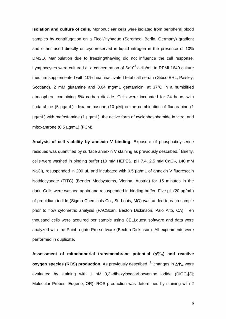
After FCM incubation no changes in the overall protein levels of Bax, Bak and Bcl-2
were detected, whereas downregulation of Mcl-1, upregulation of p53, and proteolysis
of PARP were observed (figure 3A). The cellular localization of Bcl-2 family proteins
(Bax, Bcl-2, Bak, Mcl-1) was analyzed by Western Blot in mitochondrial and cytosolic
protein fractions. As shown in figure 3B, in untreated CLL cells, Bcl-2, Mcl-1 and Bak
were present only in the mitochondrial fraction, whereas Bax was found predominantly
in this fraction, but it was also detected in the cytosolic fraction. Incubation with FCM
induced a shift in the cellular localization of Bax from the cytosolic to the mitochondrial
fraction, as well as, a decrease in the levels of Mcl-1.
To assess if these proteins were integrated or only attached to mitochondria,
mitochondrial fractions from untreated and FCM-treated cells were incubated with

11
Na2CO3 to remove proteins attached to mitochondria (Figure 3B). In untreated CLL
cells, Bak and Bcl-2 were only detected in the pellet of alkali-treated mitochondria,
corresponding to proteins integrated into the mitochondria, whereas Bax and Mcl-1
were also detected in the alkali supernatant. Interestingly, in FCM-treated cells, Bax
was only found in the pellet of alkali-treated mitochondria, with this suggesting that Bax
had been integrated into mitochondria.
These results were corroborated by visualization of immunostaining with anti-Bax
(clone YHT-6A7) (Figure 4). In untreated CLL cells no staining with this monoclonal
antibody was observed, whereas upon FCM treatment a clear punctuate staining was
detected, indicating that a conformational change of Bax had been induced. This
pattern indicated that Bax had a mitochondrial localization in drug-treated cells, which
was confirmed by simultaneous staining with Bcl-2 (data not shown). In addition, a
double staining with Bax and cytochrome c was performed. As seen in the right panels
of figure 4, an intense and punctuated staining for cytochrome c was observed in
untreated cells, where cytochrome c remained intact in the mitochondria. In contrast, in
FCM-treated cells, a weak and diffuse cytochrome c staining was observed,
suggesting that this protein had been released from mitochondria. Interestingly, in both
control and FCM-treated cells, double staining confirmed that cells having lost
cytochrome c staining corresponded to those in which Bax had undergone
conformational changes. Loss of cytochrome c was also observed by flow cytometry
and confirmed by Western Blot of cytosolic and mitochondrial fractions (figure 5).
Bak underwent conformational changes during drug-induced apoptosis and correlated
with Bax
Cells from ten CLL patients were incubated with fludarabine, dexamethasone or FCM
for 24 hours and labeled with antibodies directed against the NH2-terminal region of
Bak (Ab-1 and 2-14 antibodies). As seen in figure 6A, cells undergoing apoptosis

12
showed a positive staining, which directly correlated with the degree of apoptosis
(r=0.948, p<0.001) (figure 6B), as well as, with the amount of Bax positive cells
(r=0.905, p<0.001) (figure 6C).
As observed with Bax protein, pre-incubation of cells from four CLL patients with Z-
VAD.fmk, did not prevent Bak conformational change, whereas all the other cellular
features of apoptosis were completely reverted, except for loss of ∆Ψm that was only
partially reverted (data not shown).
Conformational changes of Bax and Bak are independent of p53 activation
It has been reported that Bax and Bak are regulated by p53 protein.26,27 For this
reason, p53 stabilization during drug-induced apoptosis was analyzed in cells from 13
CLL patients, either by flow cytometry or by Western Blot (figure 7A and 7B).
Stabilization of p53 was detected following in vitro treatment with fludarabine or FCM in
all cases analyzed. However, no activation of p53 protein was observed in any case
after treatment with dexamethasone, although conformational changes of Bax and Bak
were observed. Preincubation of cells with Z-VAD.fmk did not prevent stabilization of
p53 protein (figure 7B). These results suggest that p53 stabilization is not essential for
conformational changes of Bax and Bak, and precedes caspase activation.
Localization of p53 was analyzed on cytosolic and mitochondrial fractions. No protein
was observed in cytosolic fractions of either untreated or treated cells, or in
mitochondrial fractions of untreated cells. Interestingly, p53 was detected in
mitochondrial fractions of FCM-treated cells and remained in this fraction following
treatment with alkali (figure 7C).

13
DISCUSSION
In the present study we have investigated the subcellular localization and the
translocation of Bax and Bak, two proapoptotic members of the Bcl-2 family, in CLL
cells after drug-induced apoptosis. This study shows, for the first time, that treatment
of primary CLL cells with either dexamethasone, fludarabine or FCM induce
conformational changes of Bax and Bak that precede cell death.
Bax is a member of the Bcl-2 family of proteins that participates in the induction of
apoptosis in response to a variety of apoptotic stimuli.28 We and others have previously
described that the overall Bax protein levels are not altered during fludarabine- or
FCM-induced apoptosis in CLL cells,7,18 in contrast to other cell types.29,30 In this study,
we demonstrate that Bax conformational change is one of the first steps in drug-
induced apoptosis in CLL cells, and that it is accompanied by the characteristic
features of the mitochondrial-dependent apoptosis pathway. Conformational changes
of Bax were not blocked by the presence of the broad caspase inhibitor Z-VAD.fmk,
indicating that Bax translocation precedes caspase activation or is caspase-
independent, as previously observed in some experimental models using cell lines.25,31
The loss of ∆Ψm is only partially reverted by Z-VAD.fmk, with this suggesting that an
initial loss of ∆Ψm is independent of caspases and may be due to integration of Bax
into the outer mitochondrial membrane. In fact, recombinant Bax is able to form ion
channels in artificial membranes.32
Previous studies showed that Bax is a cytosolic protein in healthy cells, unless it is
loosely attached to the mitochondria. Upon induction of apoptosis, Bax translocates to
mitochondria 11,33 and undergoes a conformational change that unmasks both the
amino and the hydrophobic portion of the carboxi-terminus; the latter being important
for its proapoptotic function.11,34 This translocation is followed by the insertion of Bax

14
into the outer mitochondrial membrane, becoming an integral membrane protein. Our
results show that albeit Bax is detected in the mitochondrial fraction of both treated and
untreated CLL cells, Bax is only inserted into the mitochondrial membranes and
becomes an integral mitochondrial protein after FCM-treatment. This is in agreement
with previous reports which showed that the association of Bax with mitochondrial
membranes changes from a weak to a strong insertion following apoptosis triggering.35
Bak is another proapoptotic member of the Bcl-2 family and shows high homology to
Bax in size, sequence and biological activity.9 Recently, a role for Bak in the
mechanism of releasing mitochondrial intermembrane proteins in human leukemic cell
lines in response to cytotoxic drugs has been described.22 Indeed, it has been reported
that Bak-deficient Jurkat cells are resistant to apoptosis.36 Our results demonstrate that
in primary CLL cells a conformational change of Bak is observed after genotoxic and
non-genotoxic (dexamethasone) drug-induced apoptosis. In contrast to Bax, Bak is an
integral mitochondrial protein in healthy cells and modifications in this protein following
apoptosis triggering can only be detected, as in the present study, by exposure of its
amino-terminal region. The conformational changes of Bak also occurred before
caspase activation, and directly correlated with the number of apoptotic cells and the
number of Bax positive cells. The conformational changes of these proteins are
supposed to modify the protein-protein interactions that seem to be important for the
integration of damage signals and the commitment of the cell to apoptotic death.13
Mitochondrial apoptosis signaling requires either Bak, which usually resides in
mitochondria, or Bax, which translocates to mitochondria after apoptosis triggering.
Furthermore, the coexistence of Bax and Bak in cells provides a redundancy in the
apoptotic signaling pathway. In CLL cells, in contrast to other models using cell
lines,37,38 activation of apoptosis by different stimuli simultaneously activates
conformational changes of both Bax and Bak proteins. Recently, a coalescence of both
Bax and Bak in clusters adjacent to the mitochondrial membrane has been observed,39

15
and its formation seems essential for cell death. Also, it has been described that cells
lacking both Bax and Bak, but not cells lacking one of these proteins, are resistant to
multiple apoptotic stimuli, as well as, to tBid induced cytochrome c release and
apoptosis.36
The apoptotic signals upstream of Bax and Bak are not completely known. Although it
has been suggested that an increase in intracellular pH, occurring in the cytosol of
cells undergoing apoptosis, induces a structural modification of cytosolic Bax followed
by its translocation to the mitochondria 40, the nuclear magnetic resonance
spectroscopy of Bax protein demonstrates the absence of conformational changes of
Bax at pH ranging from 6 to 8.34 Moreover, albeit it has been described that the
Na+/H+ exchange activity is drastically decreased in CLL cells in comparison with
normal B cells 41,42, conformational changes of Bax are also observed in normal B cells
(data not shown).
The binding of cell death receptors by their ligands activates procaspase 8, which, in
turn, truncates Bid (tBid). This truncated protein translocates to mitochondria where it
directly binds to Bax or Bak leading to a conformational change of these proteins.10,36
Nevertheless, conformational changes of Bax and Bak are also observed in absence
of tBid.36 Other proteins such as p53, MEKK1 and c-myc have also been described as
upstream regulators of Bax and Bak conformation.27,37,43,44, We have previously
described that fludarabine and FCM combination induce the stabilization of p53
protein,7 whereas this phenomenon is not observed in dexamethasone-treated CLL
cells. In our model, Bax and Bak conformational changes occurred in response to both
p53-dependent and independent cytotoxic drugs, indicating that p53 is not required to
execute the apoptotic program. Furthermore, our results also suggest that p53 is
translocated and integrated into mitochondria following in vitro treatment with FCM.
These results are concordant with previous studies describing this shift in the
localization of p53 after drug-treatment.45

16
Bcl-2 family members are the cellular guardkeepers of cell survival or death. A
complex regulation of its members, not completely understood, has been proposed.
Although Bax and Bak act as redundant proteins, specific regulation for each protein,
as well as, a differential response to apoptotic stimuli with a predominant use of Bax or
Bak in the cell death process have been described.37,46 The regulatory mechanisms
proposed include post-transcriptional regulation, phosphorylation, dimerization and
protein displacement.10 Therefore, the effect of these mechanisms should be taken
into account when analyzing the role of these proteins as prognostic factors, in addition
to overall protein levels and ratios between pro- and antiapoptotic members.
In summary, this study demonstrates that Bax and Bak, two proapoptotic members of
the Bcl-2 family, undergo conformational changes in CLL cells in response to drug-
induced apoptosis. These conformational changes precede mitochondrial dysfunction
and caspase activation and are independent of p53 activation. The conformational
changes of Bax and Bak directly correlate with cell death, suggesting an implication of
both proteins in the apoptotic pathway of CLL cells.

17
References
1. Rozman C, Montserrat E. Chronic lymphocytic leukemia. N Engl J Med.
1995;333:1052-1057.
2. Reed JC. Molecular biology of chronic lymphocytic leukemia. Semin Oncol.
1998;25:11-18.
3. Byrd JC, Waselenko JK, Keating M, Rai K, Grever MR. Novel therapies for
chronic lymphocytic leukemia in the 21st century. Semin Oncol. 2000;27:587-
597.
4. Robertson LE, Chubb S, Meyn RE, et al. Induction of apoptotic cell death in
chronic lymphocytic leukemia by 2-chloro-2'-deoxyadenosine and 9-beta-D-
arabinosyl-2-fluoroadenine. Blood. 1993;81:143-150.
5. Begleiter A, Lee K, Israels LG, Mowat MR, Johnston JB. Chlorambucil induced
apoptosis in chronic lymphocytic leukemia (CLL) and its relationship to clinical
efficacy. Leukemia. 1994;8 Suppl 1:S103-S106.
6. McConkey DJ, Aguilar-Santelises M, Hartzell P, et al. Induction of DNA
fragmentation in chronic B-lymphocytic leukemia cells. J Immunol.
1991;146:1072-1076.
7. Bellosillo B, Villamor N, Colomer D, Pons G, Montserrat E, Gil J. In vitro
evaluation of fludarabine in combination with cyclophosphamide and/or
mitoxantrone in B-cell chronic lymphocytic leukemia. Blood. 1999;94:2836-2843.
8. Herr I, Debatin KM. Cellular stress response and apoptosis in cancer therapy.
Blood. 2001;98:2603-2614.
9. Adams JM, Cory S. Life-or-death decisions by the Bcl-2 protein family. Trends
Biochem Sci. 2001;26:61-66.
10. Gross A, McDonnell JM, Korsmeyer SJ. BCL-2 family members and the
mitochondria in apoptosis. Genes Dev. 1999;13:1899-1911.
11. Gross A, Jockel J, Wei MC, Korsmeyer SJ. Enforced dimerization of BAX results
in its translocation, mitochondrial dysfunction and apoptosis. EMBO J.
1998;17:3878-3885.

18
12. Nechushtan A, Smith CL, Hsu YT, Youle RJ. Conformation of the Bax C-terminus
regulates subcellular location and cell death. EMBO J. 1999;18:2330-2341.
13. Griffiths GJ, Dubrez L, Morgan CP, et al. Cell damage-induced conformational
changes of the pro-apoptotic protein Bak in vivo precede the onset of apoptosis.
J Cell Biol. 1999;144:903-914.
14. Hanada M, Delia D, Aiello A, Stadtmauer E, Reed JC. bcl-2 gene
hypomethylation and high-level expression in B-cell chronic lymphocytic
leukemia. Blood. 1993;82:1820-1828.
15. Robertson LE, Plunkett W, McConnell K, Keating MJ, McDonnell TJ. Bcl-2
expression in chronic lymphocytic leukemia and its correlation with the induction
of apoptosis and clinical outcome. Leukemia. 1996;10:456-459.
16. Pepper C, Bentley P, Hoy T. Regulation of clinical chemoresistance by bcl-2 and
bax oncoproteins in B-cell chronic lymphocytic leukaemia. Br J Haematol.
1996;95:513-517.
17. Pepper C, Hoy T, Bentley DP. Bcl-2/Bax ratios in chronic lymphocytic leukaemia
and their correlation with in vitro apoptosis and clinical resistance. Br J Cancer.
1997;76:935-938.
18. Kitada S, Andersen J, Akar S, et al. Expression of apoptosis-regulating proteins
in chronic lymphocytic leukemia: correlations with In vitro and In vivo
chemoresponses. Blood. 1998;91:3379-3389.
19. Pepper C, Thomas A, Hoy T, Fegan C, Bentley P. Flavopiridol circumvents Bcl-2
family mediated inhibition of apoptosis and drug resistance in B-cell chronic
lymphocytic leukaemia. Br J Haematol. 2001;114:70-77.
20. Jaffe ES, Harris NL, Stein H, Vardiman JWE. World Health organization
Classification of Tumours. Pathology and Genetics of Tumours of
Haematopoietic and Lymphoid Tissues. 2001.
21. Bellosillo B, Villamor N, Lopez-Guillermo A, et al. Complement-mediated cell
death induced by rituximab in B-cell lymphoproliferative disorders is mediated in
vitro by a caspase-independent mechanism involving the generation of reactive
oxygen species. Blood. 2001;98:2771-2777.

19
22. Wang GQ, Gastman BR, Wieckowski E, et al. A role for mitochondrial Bak in
apoptotic response to anticancer drugs. J Biol Chem. 2001;276:34307-34317.
23. Bellosillo B, Dalmau M, Colomer D, Gil J. Involvement of CED-3/ICE proteases in
the apoptosis of B-chronic lymphocytic leukemia cells. Blood. 1997;89:3378-
3384.
24. Hsu YT, Youle RJ. Bax in murine thymus is a soluble monomeric protein that
displays differential detergent-induced conformations. J Biol Chem.
1998;273:10777-10783.
25. Desagher S, Osen-Sand A, Nichols A, et al. Bid-induced conformational change
of Bax is responsible for mitochondrial cytochrome c release during apoptosis. J
Cell Biol. 1999;144:891-901.
26. Pearson AS, Spitz FR, Swisher SG, et al. Up-regulation of the proapoptotic
mediators Bax and Bak after adenovirus-mediated p53 gene transfer in lung
cancer cells. Clin Cancer Res. 2000;6:887-890.
27. Miyashita T, Reed JC. Tumor suppressor p53 is a direct transcriptional activator
of the human bax gene. Cell. 1995;80:293-299.
28. Knudson CM, Korsmeyer SJ. Bcl-2 and Bax function independently to regulate
cell death. Nat Genet. 1997;16:358-363.
29. Schuler M, Bossy-Wetzel E, Goldstein JC, Fitzgerald P, Green DR. p53 induces
apoptosis by caspase activation through mitochondrial cytochrome c release. J
Biol Chem. 2000;275:7337-7342.
30. Zhou XM, Wong BC, Fan XM, et al. Non-steroidal anti-inflammatory drugs induce
apoptosis in gastric cancer cells through up-regulation of bax and bak.
Carcinogenesis. 2001;22:1393-1397.
31. Gilmore AP, Metcalfe AD, Romer LH, Streuli CH. Integrin-mediated survival
signals regulate the apoptotic function of Bax through its conformation and
subcellular localization. J Cell Biol. 2000;149:431-446.
32. Bernardi P, Broekemeier KM, Pfeiffer DR. Recent progress on regulation of the
mitochondrial permeability transition pore; a cyclosporin-sensitive pore in the
inner mitochondrial membrane. J Bioenerg Biomembr. 1994;26:509-517.

20
33. Hsu YT, Wolter KG, Youle RJ. Cytosol-to-membrane redistribution of Bax and
Bcl-X(L) during apoptosis. Proc Natl Acad Sci U S A. 1997;94:3668-3672.
34. Suzuki M, Youle RJ, Tjandra N. Structure of Bax: coregulation of dimer formation
and intracellular localization. Cell. 2000;103:645-654.
35. Goping IS, Gross A, Lavoie JN, et al. Regulated targeting of BAX to
mitochondria. J Cell Biol. 1998;143:207-215.
36. Wei MC, Zong WX, Cheng EH, et al. Proapoptotic BAX and BAK: a requisite
gateway to mitochondrial dysfunction and death. Science. 2001;292:727-730.
37. Mandic A, Viktorsson K, Molin M, et al. Cisplatin induces the proapoptotic
conformation of Bak in a deltaMEKK1-dependent manner. Mol Cell Biol.
2001;21:3684-3691.
38. Wang GQ, Wieckowski E, Goldstein LA, et al. Resistance to granzyme B-
mediated cytochrome c release in Bak-deficient cells. J Exp Med.
2001;194:1325-1337.
39. Nechushtan A, Smith CL, Lamensdorf I, Yoon SH, Youle RJ. Bax and Bak
coalesce into novel mitochondria-associated clusters during apoptosis. J Cell
Biol. 2001;153:1265-1276.
40. Khaled AR, Kim K, Hofmeister R, Muegge K, Durum SK. Withdrawal of IL-7
induces Bax translocation from cytosol to mitochondria through a rise in
intracellular pH. Proc Natl Acad Sci U S A. 1999;96:14476-14481.
41. Ghigo D, Gaidano G, Treves S, et al. Na+/H+ antiporter has different properties
in human B lymphocytes according to CD5 expression and malignant phenotype.
Eur J Immunol. 1991;21:583-588.
42. Gaidano G, Ghigo D, Schena M, et al. Na+/H+ exchange activation mediates the
lipopolysaccharide-induced proliferation of human B lymphocytes and is impaired
in malignant B-chronic lymphocytic leukemia lymphocytes. J Immunol.
1989;142:913-918.
43. Mitchell KO, Ricci MS, Miyashita T, et al. Bax is a transcriptional target and
mediator of c-myc-induced apoptosis. Cancer Res. 2000;60:6318-6325.
44. Kannan K, Amariglio N, Rechavi G, et al. DNA microarrays identification of

21
primary and secondary target genes regulated by p53. Oncogene. 2001;20:2225-
2234.
45. Marchenko ND, Zaika A, Moll UM. Death signal-induced localization of p53
protein to mitochondria. A potential role in apoptotic signaling. J Biol Chem.
2000;275:16202-16212.
46. Ke N, Godzik A, Reed JC. Bcl-B, a novel Bcl-2 family member that differentially
binds and regulates Bax and Bak. J Biol Chem. 2001;276:12481-12484.

22
FIGURE LEGENDS
Figure 1. Drug-induced apoptosis is accompanied by conformational changes in
Bax. A. Cells from a representative CLL patient were incubated in the presence or
absence of 5 µg/mL fludarabine, 10 µM dexamethasone or the FCM combination for
24 hours. Bax conformational changes were determined by staining with anti-Bax
(clone YHT-6A7) and flow cytometric analysis. Cell viability was quantified by Annexin
V binding. In FCM-treated cells a shift in the signal of fluorescence 2 (585 nm) and 3
(630 nm) was observed due to the incorporation of mitoxantrone. B. Correlation
between cell viability and Bax-positive cells following incubation with medium alone,
fludarabine, dexamethasone or the FCM combination in cells from 33 CLL patients.
Bax positive cells (%)
Ap
op
toti
c ce
lls (
%)
100806040200
100
80
60
40
20
0
FCM
29%
60%
87%
Dexamethasone
4%
17%
13%
Annexin V
Bax
Control
Pro
pid
ium
iod
ide
2%
7%
Flu
ore
scen
ce 2
4%
Fludarabine
3%
29%
18%
B
A
FCM
Fludarabine
Dexamethasone
Control
r=0.836p<0.001

23
Figure 2. Conformational changes of Bax during drug-induced apoptosis
precede caspase activation. Cells from a representative CLL patient were incubated
with medium alone or the FCM combination in the presence or absence of 200 µM
ZVAD.fmk for 24 hours. Z-VAD.fmk was preincubated for 1 hour prior to the addition of
FCM. In FCM-treated cells a shift in the signal of fluorescence 2 (585 nm) and 3 (630
nm) was observed due to the incorporation of mitoxantrone. Flow cytometric dot plots
of: A. Cell viability as determined by annexin V binding. B. Loss of ∆Ψm and ROS
generation by dual staining with DIOC6 and DHE. C. Quantification of the active form
of caspase-3. D. Conformational changes of Bax as determined by staining with anti-
Bax (clone YHT-6A7).
Annexin V
DiOC6
Control FCM Z-VAD.fmk FCM+ Z-VAD.fmk
7%
21%
9%
62%
3%
21%
Pro
pid
ium
Iod
ide
4%
8%
Dih
ydro
eth
idin
e
24%
67%
75%
22%
17%
69%
28%
44%
Bax
Flu
ore
scen
ce 2
20% 70% 25% 69%
Active caspase-3
Flu
ore
scen
ce 2
20% 72% 4% 4%
A
B
C
D
24
Figure 3. Apoptosis related proteins during drug-induced cell death. A. Whole
cell lysates were obtained from 2x10⁶ cells from a representative CLL patient
incubated in the absence (C) or presence of the FCM combination for 24 hours, and
analyzed by Western Blot. B. Cytosolic and mitochondrial fractions were obtained from
50x106 cells incubated in the absence (C) or presence of the FCM combination for 24
hours. Mitochondrial fractions were either untreated or treated with alkali. All fractions
were analyzed by Western Blot. The position of the proteolytic fragment (85 kD) of
PARP is indicated by an arrow.
A B
PARP
Mcl-1
p53
Bax
Bak
Bcl-2
C FCM
Mitochondria
C FCM
Cytosol
Bax
Mcl-1
FCM FCMC C
Alkali-treated Mitochondria
Pellets Supernatants
Bak
C FCM
w
Bcl-2

25
Figure 4. Cellular localization of Bax and cytochrome c during drug-induced
apoptosis. Cells from a representative patient were incubated in the absence or
presence of the FCM combination for 24 hours. Cells were immunostained with anti-
Bax (clone YHT-6A7, green fluorescence), and anti-cytochrome c (red fluorescence).
Photographs of the same fields were obtained with optical filters specific for green and
red light. Arrows indicate the same apoptotic cell showing a punctuated Bax staining
and having lost cytochrome c. Asterisks on the lower panel indicate viable cells
(negative staining for Bax and bright punctuated signal for cytochrome c).

26
Figure 5. Analysis of cytochrome c in drug-induced apoptosis. A. Flow cytometric
analysis of cytochrome c staining. Cells from a representative CLL patient were
incubated in the presence or absence of 5 µg/mL fludarabine, 10 µM dexamethasone
or the FCM combination for 24 hours, stained with anti-cytochrome c and analyzed by
flow cytometry. B. Western blot of cytochrome c in mitochondrial and cytosolic
fractions obtained from 50x106 cells of a representative CLL patient incubated in the
presence or absence of FCM for 24 hours.
Cytochrome c
FludarabineControl
Nu
mb
ero
f E
ven
ts
100 101 102 103 104
2%
100 101 102 103 104
2%
FCM
100 101 102 103 104
33%
Dexamethasone
103100 101 102 104
8%
B
A
Cytochrome c
C FCM
Mitochondria Cytosol
C FCM

27
Figure 6. Bak conformational changes in drug-induced apoptosis of CLL cells. A.
Dot plots of Bak conformational changes in cells from one representative patient
following incubation with medium alone, 5 µg/mL fludarabine, 10 µM dexamethasone or
the FCM combination for 24 hours. B. Correlation between cell viability and Bak-
positive cells following incubation of cells from 10 patients with medium alone,
fludarabine, dexamethasone or the FCM combination. C. Correlation of Bax and Bax
positive cells following incubation of cells from 10 patients with medium alone,
fludarabine, dexamethasone or the FCM combination for 24 hours.
Drug
Fludarabine
Dexamethasone
FCM
Control
Bak positive cells (%)
Ap
op
toti
c ce
lls (
%)
Bak
Flu
ore
scen
ce 2
45%
FCM
61%
Dexamethasone
10%
Control
16%
Fludarabine
Bak positive cells (%)
Bax
po
siti
ve c
ells
(%
)
100806040200
100
80
60
40
20
0
A
CB
r=0.905p<0.001
100806040200
100
80
60
40
20
0
r=0.948p<0.001
19 % 28 % 73 % 54 %Annexin V+ cells

28
Figure 7. p53 activation is independent of Bax conformational changes.
A. Flow cytometric analysis of conformational changes of Bax and p53 activation in
cells from a representative CLL patient incubated with medium alone or in the
presence of 5 µg/mL fludarabine, 10 µM dexamethasone or the FCM combination. B.
Western Blot analysis of p53 protein. Whole cell lysates were obtained from 2x10⁶
cells from a representative CLL patient incubated with medium alone (C), fludarabine
(F), FCM, or dexamethasone (D), in the presence (+) or absence (-) of 200 µM of
ZVAD.fmk for 24 hours, and were analyzed by Western Blot. C. Cytosolic and
mitochondrial fractions were obtained from 50x106 cells incubated in the absence (C)
or presence of the FCM combination for 24 hours. Mitochondrial fractions were either
left untreated or treated with alkali. All fractions were analyzed by Western Blot.
C F FCM D C F FCM DZ-VAD.fmk - - - - + + + +
B
A
C
p53
100 101 102 103 104
2%
100 101 102 103 104
65%
100 101 102 103 104
5%
100 101 102 103 104
89%
Nu
mb
ero
f E
ven
ts
Control Fludarabine Dexamethasone FCM
Bax
Flu
ore
scen
ce 2
21% 32% 55%69%
C FCM FCM FCMC C
Alkali-treated Mitochondria
Pellets Supernatants
C FCM
Cytosol Mitochondria
Copyright © 2022 FDOKUMEN