HIV1 Vpr-Induced Apoptosis Is Cell Cycle Dependent and Requires Bax but Not ANT
14
HIV-1 Vpr-Induced Apoptosis Is Cell Cycle Dependent and Requires Bax but Not ANT Joshua L. Andersen 1 , Jason L. DeHart 1 , Erik S. Zimmerman 1 , Orly Ardon 1 , Baek Kim 2 , Guillaume Jacquot 3 , Serge Benichou 3 , Vicente Planelles 1* 1 Department of Pathology, University of Utah School of Medicine, Salt Lake City, Utah, United States of America, 2 Department of Microbiology and Immunology, University of Rochester Medical Center, Rochester, New York, United States of America, 3 Departement de Maladies Infectieuses, Institut Cochin, Institut National de la Sante ´ et de la Recherche Me ´ dicale, Paris, France The HIV-1 accessory protein viral protein R (Vpr) causes G 2 arrest and apoptosis in infected cells. We previously identified the DNA damage–signaling protein ATR as the cellular factor that mediates Vpr-induced G 2 arrest and apoptosis. Here, we examine the mechanism of induction of apoptosis by Vpr and how it relates to induction of G 2 arrest. We find that entry into G 2 is a requirement for Vpr to induce apoptosis. We investigated the role of the mitochondrial permeability transition pore by knockdown of its essential component, the adenine nucleotide translocator. We found that Vpr-induced apoptosis was unaffected by knockdown of ANT. Instead, apoptosis is triggered through a different mitochondrial pore protein, Bax. In support of the idea that checkpoint activation and apoptosis induction are functionally linked, we show that Bax activation by Vpr was ablated when ATR or GADD45a was knocked down. Certain mutants of Vpr, such as R77Q and I74A, identified in long-term nonprogressors, have been proposed to inefficiently induce apoptosis while activating the G 2 checkpoint in a normal manner. We tested the in vitro phenotypes of these mutants and found that their abilities to induce apoptosis and G 2 arrest are indistinguishable from those of HIV-1 NL4–3 vpr, providing additional support to the idea that G 2 arrest and apoptosis induction are mechanistically linked. Citation: Andersen JL, DeHart JL, Zimmerman ES, Ardon O, Kim B, et al. (2006) HIV-1 Vpr-induced apoptosis is cell cycle dependent and requires Bax but not ANT. PLoS Pathog 2(12): e127. doi:10.1371/journal.ppat.0020127 Introduction Loss of CD4 þ lymphocytes is a hallmark of progression to acquired immune deficiency syndrome (AIDS). The mecha- nisms proposed to explain the loss and dysfunction of CD4 þ T cells are multiple and include indirect effects of viral infection, such as generalized activation of the immune system, CD8 þ cytotoxic T lymphocyte–mediated killing of infected cells, as well as direct effects of infection such as virus budding, and expression of certain viral genes (reviewed in [1–3]). Analysis of virus dynamics in vivo has revealed that a significant portion of CD4 þ T-cell death is due to virus- induced cytotoxicity in the infected cells [4–6], and the estimated half-life of infected lymphocytes is on the order of 2 d (reviewed in [7]). Therefore, studies that seek to under- stand the molecular and cellular basis of HIV-1–induced death in T cells are critical toward explaining how immune deterioration results from HIV-1 infection. The HIV-1 viral protein R (Vpr) has emerged as a major proapoptotic gene product (reviewed in [8,9]). In an effort to characterize the apoptotic signaling cascade induced by Vpr downstream of the mitochondria, Muthumani et al. [10,11] demonstrated that Vpr induces apoptosis via the intrinsic pathway. This pathway is characterized by cytochrome c release and caspase 9 activation and is triggered in the absence of death receptor ligation. Vpr induces a second type of cytopathic effect by blocking the cell cycle in the G 2 phase. We, and others, have previously shown that Vpr causes activation of the G 2 checkpoint protein, ATR (ataxia and telangiectasia mutated and Rad3 related), a serine/threonine kinase responsive to DNA damage and replication stress [12–14]. Furthermore, activation of ATR by Vpr is required for Vpr-induced G 2 arrest and involves the ATR-associated molecules Rad17 and the Rad9- Rad1-Hus1 trimer [14–16]. A cause–effect relationship between the two deleterious actions of Vpr (G 2 arrest and apoptosis) has not been established. Indirect evidence supports the notion that induction of G 2 arrest and apoptosis are linked. For example, treatment of cells with either caffeine, an inhibitor of ATR/ ATM checkpoint function, or small interfering RNA (siRNA) specific to ATR, relieves both Vpr-induced G 2 arrest and apoptosis [17,18]. In addition, Yuan et al. [19,20] demon- strated that siRNA knockdown of Wee1, a Cdk1 inhibitor that is activated by DNA damage, abrogated both Vpr-induced G 2 arrest and apoptosis. Taken together, these data suggest a model in which checkpoint activation and apoptosis signaling Editor: Michael Malim, King’s College London, United Kingdom Received May 11, 2006; Accepted October 25, 2006; Published December 1, 2006 Copyright: Ó 2006 Andersen et al. This is an open-access article distributed under the terms of the Creative Commons Attribution License, which permits unrestricted use, distribution, and reproduction in any medium, provided the original author and source are credited. Abbreviations: ANT, adenine nucleotide transporter; ATM, ataxia telangiectasia mutated; ATR, ataxia and telangiectasia mutated and Rad3 related; BRCA1, breast cancer–associated protein 1; DAPI, 49,6-diamidino-2-phenylindole dihydrochloride; DSB, double-strand break; GADD45a, growth arrest and DNA damage–responsive protein a; GFP, green fluorescent protein; HIV-1, human immunodeficiency virus type 1; MNNG, N-methyl-N9-nitro-N-nitrosoguanidine; mRFP, monomeric red fluorescent protein; PARP, poly-ADP-ribose polymerase; PTPC, permeability transition pore complex; siRNA, small interfering RNA; Smac, second mitochon- dria-derived activator of caspase; VDAC, voltage-dependent anion channel; Vpr, viral protein R * To whom correspondence should be addressed. E-mail: Vicente.planelles@path. utah.edu PLoS Pathogens | www.plospathogens.org December 2006 | Volume 2 | Issue 12 | e127 1106
-
Upload
independent -
Category
Documents
-
view
0 -
download
0
Transcript of HIV1 Vpr-Induced Apoptosis Is Cell Cycle Dependent and Requires Bax but Not ANT

HIV-1 Vpr-Induced Apoptosis Is Cell CycleDependent and Requires Bax but Not ANTJoshua L. Andersen
1, Jason L. DeHart
1, Erik S. Zimmerman
1, Orly Ardon
1, Baek Kim
2, Guillaume Jacquot
3,
Serge Benichou3
, Vicente Planelles1*
1 Department of Pathology, University of Utah School of Medicine, Salt Lake City, Utah, United States of America, 2 Department of Microbiology and Immunology, University
of Rochester Medical Center, Rochester, New York, United States of America, 3 Departement de Maladies Infectieuses, Institut Cochin, Institut National de la Sante et de la
Recherche Medicale, Paris, France
The HIV-1 accessory protein viral protein R (Vpr) causes G2 arrest and apoptosis in infected cells. We previouslyidentified the DNA damage–signaling protein ATR as the cellular factor that mediates Vpr-induced G2 arrest andapoptosis. Here, we examine the mechanism of induction of apoptosis by Vpr and how it relates to induction of G2
arrest. We find that entry into G2 is a requirement for Vpr to induce apoptosis. We investigated the role of themitochondrial permeability transition pore by knockdown of its essential component, the adenine nucleotidetranslocator. We found that Vpr-induced apoptosis was unaffected by knockdown of ANT. Instead, apoptosis istriggered through a different mitochondrial pore protein, Bax. In support of the idea that checkpoint activation andapoptosis induction are functionally linked, we show that Bax activation by Vpr was ablated when ATR or GADD45awas knocked down. Certain mutants of Vpr, such as R77Q and I74A, identified in long-term nonprogressors, have beenproposed to inefficiently induce apoptosis while activating the G2 checkpoint in a normal manner. We tested the invitro phenotypes of these mutants and found that their abilities to induce apoptosis and G2 arrest areindistinguishable from those of HIV-1NL4–3 vpr, providing additional support to the idea that G2 arrest and apoptosisinduction are mechanistically linked.
Citation: Andersen JL, DeHart JL, Zimmerman ES, Ardon O, Kim B, et al. (2006) HIV-1 Vpr-induced apoptosis is cell cycle dependent and requires Bax but not ANT. PLoSPathog 2(12): e127. doi:10.1371/journal.ppat.0020127
Introduction
Loss of CD4þ lymphocytes is a hallmark of progression toacquired immune deficiency syndrome (AIDS). The mecha-nisms proposed to explain the loss and dysfunction of CD4þTcells are multiple and include indirect effects of viralinfection, such as generalized activation of the immunesystem, CD8þ cytotoxic T lymphocyte–mediated killing ofinfected cells, as well as direct effects of infection such asvirus budding, and expression of certain viral genes (reviewedin [1–3]). Analysis of virus dynamics in vivo has revealed that asignificant portion of CD4þ T-cell death is due to virus-induced cytotoxicity in the infected cells [4–6], and theestimated half-life of infected lymphocytes is on the order of2 d (reviewed in [7]). Therefore, studies that seek to under-stand the molecular and cellular basis of HIV-1–induceddeath in T cells are critical toward explaining how immunedeterioration results from HIV-1 infection.
The HIV-1 viral protein R (Vpr) has emerged as a majorproapoptotic gene product (reviewed in [8,9]). In an effort tocharacterize the apoptotic signaling cascade induced by Vprdownstream of the mitochondria, Muthumani et al. [10,11]demonstrated that Vpr induces apoptosis via the intrinsicpathway. This pathway is characterized by cytochrome crelease and caspase 9 activation and is triggered in theabsence of death receptor ligation.
Vpr induces a second type of cytopathic effect by blockingthe cell cycle in the G2 phase. We, and others, have previouslyshown that Vpr causes activation of the G2 checkpointprotein, ATR (ataxia and telangiectasia mutated and Rad3related), a serine/threonine kinase responsive to DNA damageand replication stress [12–14]. Furthermore, activation of
ATR by Vpr is required for Vpr-induced G2 arrest andinvolves the ATR-associated molecules Rad17 and the Rad9-Rad1-Hus1 trimer [14–16].A cause–effect relationship between the two deleterious
actions of Vpr (G2 arrest and apoptosis) has not beenestablished. Indirect evidence supports the notion thatinduction of G2 arrest and apoptosis are linked. For example,treatment of cells with either caffeine, an inhibitor of ATR/ATM checkpoint function, or small interfering RNA (siRNA)specific to ATR, relieves both Vpr-induced G2 arrest andapoptosis [17,18]. In addition, Yuan et al. [19,20] demon-strated that siRNA knockdown of Wee1, a Cdk1 inhibitor thatis activated by DNA damage, abrogated both Vpr-induced G2
arrest and apoptosis. Taken together, these data suggest amodel in which checkpoint activation and apoptosis signaling
Editor: Michael Malim, King’s College London, United Kingdom
Received May 11, 2006; Accepted October 25, 2006; Published December 1, 2006
Copyright: � 2006 Andersen et al. This is an open-access article distributed underthe terms of the Creative Commons Attribution License, which permits unrestricteduse, distribution, and reproduction in any medium, provided the original authorand source are credited.
Abbreviations: ANT, adenine nucleotide transporter; ATM, ataxia telangiectasiamutated; ATR, ataxia and telangiectasia mutated and Rad3 related; BRCA1, breastcancer–associated protein 1; DAPI, 49,6-diamidino-2-phenylindole dihydrochloride;DSB, double-strand break; GADD45a, growth arrest and DNA damage–responsiveprotein a; GFP, green fluorescent protein; HIV-1, human immunodeficiency virustype 1; MNNG, N-methyl-N9-nitro-N-nitrosoguanidine; mRFP, monomeric redfluorescent protein; PARP, poly-ADP-ribose polymerase; PTPC, permeabilitytransition pore complex; siRNA, small interfering RNA; Smac, second mitochon-dria-derived activator of caspase; VDAC, voltage-dependent anion channel; Vpr,viral protein R
* To whom correspondence should be addressed. E-mail: [email protected]
PLoS Pathogens | www.plospathogens.org December 2006 | Volume 2 | Issue 12 | e1271106
by Vpr are functionally associated and that such anassociation stems from the ability of Vpr to activate ATR[12–14,17].
On the other hand, evidence from mutagenesis studiessuggests that checkpoint activation and apoptosis may beseparable effects of Vpr. Thus, mutants of Vpr have beendescribed that induce normal levels of G2 arrest but arepartially impaired for induction of apoptosis [21,22]. Onemodel to explain the proapoptotic activity of Vpr wasproposed by Jacotot et al. [23] and Vieira et al. [24], whoobserved that recombinant Vpr associates with the adeninenucleotide transporter (ANT) on purified mitochondria, todirectly promote release of mitochondrion-associated cyto-chrome c and apoptosis. This finding suggested that Vprinitiated the commitment to apoptosis at the mitochondrialmembrane by binding to ANT, rather than by activatingupstream stress signaling pathways derived from checkpointactivation.
In the present study, we examine the relationship betweeninduction of G2 arrest and onset of apoptosis by Vpr. We findthat entry into G2 is a requirement for Vpr to induceapoptosis. Since the requirement for entry into G2 seemedinconsistent with the reported ability of Vpr to bind to ANTand promote apoptosis in a cell cycle–independent manner,we then examined the requirement for ANT. We find thatVpr-induced apoptosis is unaffected by knockdown of ANT.Instead, Vpr-induced apoptosis is dependent on the presenceof Bax and is concomitant with Bax activation. Furthermore,Bax activation is a result of proapoptotic signals transducedby Vpr through the upstream stress proteins ATR andGADD45a (growth arrest and DNA damage–responsiveprotein a) because knockdown of ATR or GADD45aprevented activation of Bax. We also demonstrate that,despite the striking similarities between the signaling eventsinduced by Vpr and those induced by genotoxic stress,
important differences can be found. Specifically, Vpr-induced apoptosis is abrogated by checkpoint inhibition,while apoptosis induced by genotoxic agents is exacerbatedby checkpoint inhibition. Taken together, these resultsdemonstrate that Vpr activates the ATR-initiated DNAdamage–signaling pathway to link checkpoint activation andcommitment to apoptosis.
Results
G2 Arrest Precedes the Release of Mitochondrial Smac andCaspase ActivationTreatment with caffeine, an inhibitor of ATR and ATM, or
siRNA targeted to ATR, abrogates Vpr-induced G2 arrest andapoptosis [14,17,18]. One possible explanation for theseobservations would be that Vpr induces apoptosis as adownstream consequence of sustained G2 checkpoint activa-tion. This idea, however, is inconsistent with a previouslypublished model in which Vpr was proposed to directly bindto the mitochondrial ANT to cause release of proapoptoticfactors in a cell cycle–independent manner [23,24].To begin to differentiate between these models, we
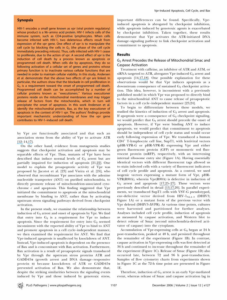
studied the kinetics of induction of G2 arrest and apoptosis.If apoptosis were a consequence of G2 checkpoint signaling,we would predict that G2 arrest should precede the onset ofapoptosis. However, if Vpr were binding ANT to induceapoptosis, we would predict that commitment to apoptosisshould be independent of cell cycle status and would occurearly following expression of Vpr. We transduced a humanCD4þ T-lymphocyte cell line, SupT1, with lentiviral vectors(pHR-VPR-G or pHR-VPR-R) expressing Vpr and eithergreen fluorescent protein (GFP) or monomeric red fluo-rescent protein (mRFP), respectively, with an interveninginternal ribosome entry site (Figure 1A). Having essentiallyidentical vectors with different fluorescent tags allowed usto stain infected cells with a variety of fluorescent indicatorsof cell cycle profile and apoptosis. As a control, we usedisogenic vectors expressing a mutant form of Vpr, pHR-VPR(R80A), wherein Vpr(R80A) is defective in induction ofG2 arrest and apoptosis [17,25–27]. These vectors werepreviously described in detail [13,17,26]. In parallel experi-ments, we transduced SupT1 cells with VSV-G pseudotyped,env-defective vector derived from HIV-1NL4–3 (DHIV3;Figure 1A) or a mutant form of the previous vector withVpr deleted (DHIV3-DVPR). At various time points, cultureswere harvested and partitioned for further analyses.Analyses included cell cycle profile, induction of apoptosisas measured by caspase activation, and Western blot todetect release of Smac (second mitochondria-derived acti-vator of caspase) into the cytoplasm.Accumulation of Vpr-expressing cells at G2 began at 24 h
post-transduction, peaked at 48 h, and persisted throughoutthe remainder of the experiment (Figure 1B). In contrast,caspase activation in Vpr-expressing cells was first detected at36 h and continued to increase throughout the remainder ofthe experiment (Figure 1C). Release of Smac (Figure 1D) alsooccurred late, between 72 and 96 h post-transduction.Samples of flow cytometry charts from experiments shownin Figure 1C at the 72-h time point are presented in Figure1E.Therefore, induction of G2 arrest is an early Vpr-mediated
event, whereas release of Smac and caspase activation lag in
PLoS Pathogens | www.plospathogens.org December 2006 | Volume 2 | Issue 12 | e1271107
Vpr-Induced Apoptosis, Cell Cycle, and Bax
Synopsis
HIV-1 encodes a small gene known as vpr (viral protein regulatory)whose product is a 96–amino acid protein. HIV-1 infects cells of theimmune system, such as CD4-positive lymphocytes. When cellsbecome infected with HIV-1, two deleterious effects result fromexpression of the vpr gene. One effect of vpr is to manipulate thecell cycle by blocking the cells in G2 (the phase of the cell cycleimmediately preceding mitosis). Thus, cells infected with HIV-1 ceaseto proliferate, due to the action of vpr. A second effect of vpr is theinduction of cell death by a process known as apoptosis orprogrammed cell death. When cells die by apoptosis, they do sofollowing activation of a cellular set of genes and proteins whoseprimary function is to inactivate various cellular functions that areneeded in order to maintain cellular viability. In this study, Andersenet al. demonstrate that the above two effects of vpr are linked. Inparticular, the authors show that the blockade in cell proliferation inG2 is a requirement toward the onset of programmed cell death.Programmed cell death can be accomplished by a number ofcellular proteins known as ‘‘executioners.’’ Various executionerproteins reside on the mitochondrial membranes and may triggerrelease of factors from the mitochondria, which in turn willprecipitate the onset of apoptosis. In this work Anderson et al.identify the mitochondrial protein, Bax, as the key executioner ofapoptosis in the context of HIV-1 vpr. The authors’ findings provideimportant mechanistic understanding of how the vpr genecontributes to HIV-1–induced cell death.
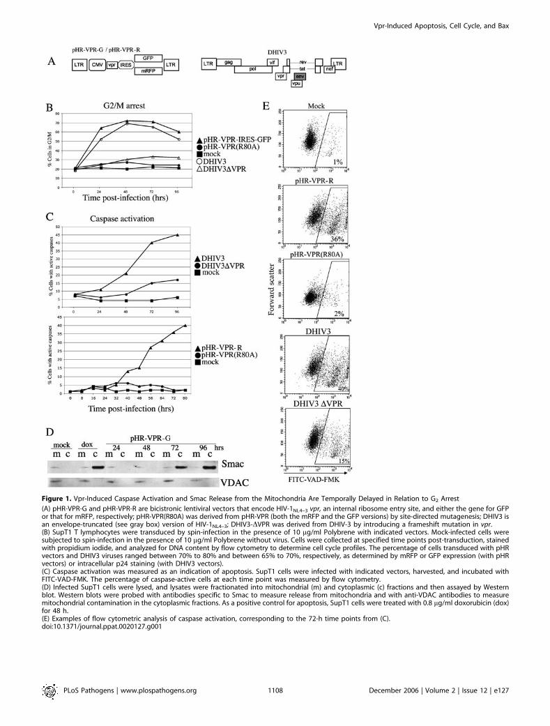
Figure 1. Vpr-Induced Caspase Activation and Smac Release from the Mitochondria Are Temporally Delayed in Relation to G2 Arrest
(A) pHR-VPR-G and pHR-VPR-R are bicistronic lentiviral vectors that encode HIV-1NL4–3 vpr, an internal ribosome entry site, and either the gene for GFPor that for mRFP, respectively; pHR-VPR(R80A) was derived from pHR-VPR (both the mRFP and the GFP versions) by site-directed mutagenesis; DHIV3 isan envelope-truncated (see gray box) version of HIV-1NL4–3; DHIV3-DVPR was derived from DHIV-3 by introducing a frameshift mutation in vpr.(B) SupT1 T lymphocytes were transduced by spin-infection in the presence of 10 lg/ml Polybrene with indicated vectors. Mock-infected cells weresubjected to spin-infection in the presence of 10 lg/ml Polybrene without virus. Cells were collected at specified time points post-transduction, stainedwith propidium iodide, and analyzed for DNA content by flow cytometry to determine cell cycle profiles. The percentage of cells transduced with pHRvectors and DHIV3 viruses ranged between 70% to 80% and between 65% to 70%, respectively, as determined by mRFP or GFP expression (with pHRvectors) or intracellular p24 staining (with DHIV3 vectors).(C) Caspase activation was measured as an indication of apoptosis. SupT1 cells were infected with indicated vectors, harvested, and incubated withFITC-VAD-FMK. The percentage of caspase-active cells at each time point was measured by flow cytometry.(D) Infected SupT1 cells were lysed, and lysates were fractionated into mitochondrial (m) and cytoplasmic (c) fractions and then assayed by Westernblot. Western blots were probed with antibodies specific to Smac to measure release from mitochondria and with anti-VDAC antibodies to measuremitochondrial contamination in the cytoplasmic fractions. As a positive control for apoptosis, SupT1 cells were treated with 0.8 lg/ml doxorubicin (dox)for 48 h.(E) Examples of flow cytometric analysis of caspase activation, corresponding to the 72-h time points from (C).doi:10.1371/journal.ppat.0020127.g001
PLoS Pathogens | www.plospathogens.org December 2006 | Volume 2 | Issue 12 | e1271108
Vpr-Induced Apoptosis, Cell Cycle, and Bax
Figure 2. The Apoptotic Effect of Vpr Is Lost in Cells Synchronized in G1/S
(A) HeLa cells were transduced with pHR-VPR-G or mock-transduced and then incubated with 2 mM thymidine. After 24 h of incubation, cells wereharvested, stained with PI, and analyzed for DNA content by flow cytometry to determine the percentage of cells in G1/S and G2/M. Transduction efficiencyof pHR-VPR in both thymidine-treated and cycling cells was 70% to 75% as determined by analysis of GFP expression by flow cytometry (unpublished data).(B) Cells from experiments shown in (A) were stained for DAPI at 72 h postinfection, in order to evaluate apoptosis via chromatin morphology(C) Quantitation of apoptosis in DAPI-stained samples shown in (B); incubation with 25 lM etoposide for 48 h or 1 lM staurosporine for 8 h wasincluded in both cycling and thymidine-treated cells, for comparative purposes.(D) Cycling or thymidine-synchronized HeLa cells were mock-transduced or transduced with either pHR-VPR-R or pHR-VPR(R80A) for apoptosis analysis,using FITC-VAD-FMK. As positive controls for apoptosis, cycling and synchronized cells were treated with etoposide or staurosporine as shown in (C).(E) Cycling or thymidine-synchronized HeLa cells expressing Vpr or treated with 25 lM etoposide for 48 h were lysed and analyzed by Western blot forPARP cleavage as a marker of apoptosis/caspase activity. The caspase-cleaved PARP band is observed at 89 kDa.(F) Cell lysates from pHR-VPR–transduced cells, with or without thymidine block, were harvested at 12 and 24 h post-transduction and analyzed byWestern blot for Vpr expression with antibodies specific to the amino-terminal hemagglutinin tag.doi:10.1371/journal.ppat.0020127.g002
PLoS Pathogens | www.plospathogens.org December 2006 | Volume 2 | Issue 12 | e1271109
Vpr-Induced Apoptosis, Cell Cycle, and Bax
relation to cell cycle arrest by approximately 24 h. Since G2
arrest clearly precedes apoptosis, we found it compelling toask whether transition into G2 was required for the commit-ment to apoptosis in Vpr-expressing cells.
G1/S Block Abolishes the Induction of Apoptosis by VprWe designed an experiment to prevent Vpr-expressing
cells from entering G2 and then asked whether these cellswould still be susceptible to Vpr-induced apoptosis. Wechose to perform the experiment in HeLa cells since thesecells can be synchronized by incubation with 2 mMthymidine with minimal toxicity [28]. HeLa cells representan appropriate cell type to study Vpr-induced apoptosis,because ATR activation, breast cancer–associated protein 1(BRCA1) phosphorylation, and GADD45a upregulation areconserved from HeLa cells to primary human CD4þ
lymphocytes [14,17,26,29]. HeLa cells were transduced withpHR-VPR-G and, 4 h later, treated with 2 mM thymidine,which arrests cells at the G1/S boundary [28]. DNA contentanalysis showed that thymidine treatment effectivelysynchronized uninfected cells in G1/S (Figure 2A). Thymidinesynchronization of pHR-VPR-G–infected cells resulted in82.9% cells in G1/S and 17.0% cells in G2/M. Thecomparatively high number of cells in G2/M in thesynchronized, pHR-VPR–infected culture (17%) reflects cellsthat reached G2/M prior to synchronization and arrested dueto Vpr expression. To evaluate the frequency of apoptoticcells in these cultures, cells were stained with the nuclear dye49,6-diamidino-2-phenylindole dihydrochloride (DAPI). DA-PI binds to chromatin and reveals the presence of aconspicuous pyknotic nuclear morphology that is associatedwith late stages of apoptosis. We have previously describedthe use of DAPI for analysis of Vpr-induced apoptosis[17,30]. Figure 2B shows fluorescence microscopy photo-graphs from this analysis, and quantitations are shown inFigure 2C. DAPI staining revealed that at 48 h post-transduction, 23% of cells transduced with Vpr wereapoptotic if allowed to cycle, whereas only 7% wereapoptotic if treated with thymidine (Figure 2C). The back-ground level of apoptosis in uninfected cells treated withthymidine was 4%.
To address whether the decreased sensitivity of G1/S-arrested cells to Vpr-induced apoptosis could be due tothymidine treatment itself, cells were treated with a pharma-cologic apoptosis inducer (etoposide or staurosporine) andthen allowed to cycle, or not (thymidine incubation).Staurosporine is a kinase inhibitor and potent inducer ofapoptosis with no known cell cycle specificity. Etoposide is atopoisomerase II inhibitor and an inducer of double-strandbreaks (DSBs) and, like Vpr, causes G2 arrest [31,32]. Treat-ment with staurosporine or etoposide effectively inducedapoptosis in both G1/S-arrested and cycling cells (Figure 2C).
To confirm the previous apoptosis results, we performedparallel experiments using pHR-VPR-R and staining forcaspase activity with FITC-VAD-FMK (Figure 2D) and bymeasuring poly-ADP-ribose polymerase (PARP) cleavage(Figure 2E). Analysis of Vpr expression by Western blotrevealed that thymidine treatment did not affect expressionof Vpr (Figure 2F). These results are in agreement with thoseobtained by DAPI staining and confirm that synchronizationof cells in G1/S alleviates Vpr-induced apoptosis.
Release of Vpr-Expressing Cells from G1/S Block Leads toG2 Arrest-Dependent ApoptosisThe previous results indicate that the proapoptotic effect
of Vpr is lost when cells are artificially maintained in G1/S. Todetermine whether induction of apoptosis by Vpr specificallyrequires transition into G2, cells were synchronized as in theprevious experiment and then were released from thymidineblock and allowed to reenter the cell cycle. Released cellswere harvested at specified intervals to examine cell cycleprogression (Figure 3A) and apoptosis (Figure 3B). Bothmock-treated and Vpr-expressing cells entered G2 at 12 hpostrelease. Mock-infected cells completed the first divisioncycle at 18 h postrelease and continued to cycle (Figure 3A,upper panels), while cells expressing Vpr persisted in G2 forthe remainder of the experiment (Figure 3A, lower panels). Asmall amount of cells (17.4%) in the Vpr-transduced culturereached G1 at 18 h postrelease. The reason for this is that theefficiency of transduction with pHR-VPR was about 80% andtherefore about 20% are untransduced.We then asked when, after release from the thymidine
block, Vpr-expressing cells would enter apoptosis. Cells whichwere synchronized, maintained in G1 for 48 h, and thenreleased (Figure 3B, lanes 2–6) began to display detectablePARP cleavage by 12 h postrelease (Figure 3B, lane 3).Therefore, the onset of apoptosis in Vpr-expressing cells isconcomitant with entry into the G2 phase. In the absence ofsynchronization in G1 (Figure 3B, lanes 7–11), Vpr-expressingcells begin to display PARP cleavage by 24 h post-trans-duction. Synchronized cells, in the absence of Vpr expression,did not display PARP cleavage after release (unpublisheddata).Parallel samples from the previous synchronization experi-
ment were also analyzed by DAPI staining (Figure 3C). Theseexperiments revealed chromatin fragmentation/condensationin Vpr-expressing cells at 24 and 48 h postrelease (Figure 3C).Taken together, these data indicate that Vpr-induced
apoptosis requires entry into the G2 phase and artificiallymaintaining cells in G1 effectively prevents the onset ofapoptosis.
Inhibition of ATR Prevents Vpr-Induced Apoptosis butExacerbates Genotoxin-Induced ApoptosisDue to the phenotypic similarities between Vpr and
genotoxic stress, many studies on Vpr have been modeledbased on the current understanding of DNA damage signal-ing [12,14,33]. Several reports have shown that suppression ofG2 checkpoint-activating kinases after exposure to genotoxicagents overrides cell cycle arrest but exacerbates apoptosis[34–37]. The reason for this increase in cell death is thoughtto be the inability of cells to successfully duplicate DNA or toalign and segregate damaged chromosomes during mitosis(reviewed in [35]). Thus, we set out to compare in parallel theeffect of ATR suppression on Vpr- versus genotoxin-inducedapoptosis. We reasoned that if Vpr causes irreparable DNAdamage, then the effect of checkpoint overriding on Vpr-induced apoptosis should mirror that of cells treated withgenotoxic agents. To test this, we treated cells with siRNAsspecific to ATR, ATM, or nonspecific siRNA. At 48 hfollowing siRNA transfection, we treated cells with 25 lMN-methyl-N9-nitro-N-nitrosoguanidine (MNNG), an SN1-typemethylating agent and inducer of G2 arrest, or 25 lMetoposide. At 24- and 48-h time points following the addition
PLoS Pathogens | www.plospathogens.org December 2006 | Volume 2 | Issue 12 | e1271110
Vpr-Induced Apoptosis, Cell Cycle, and Bax
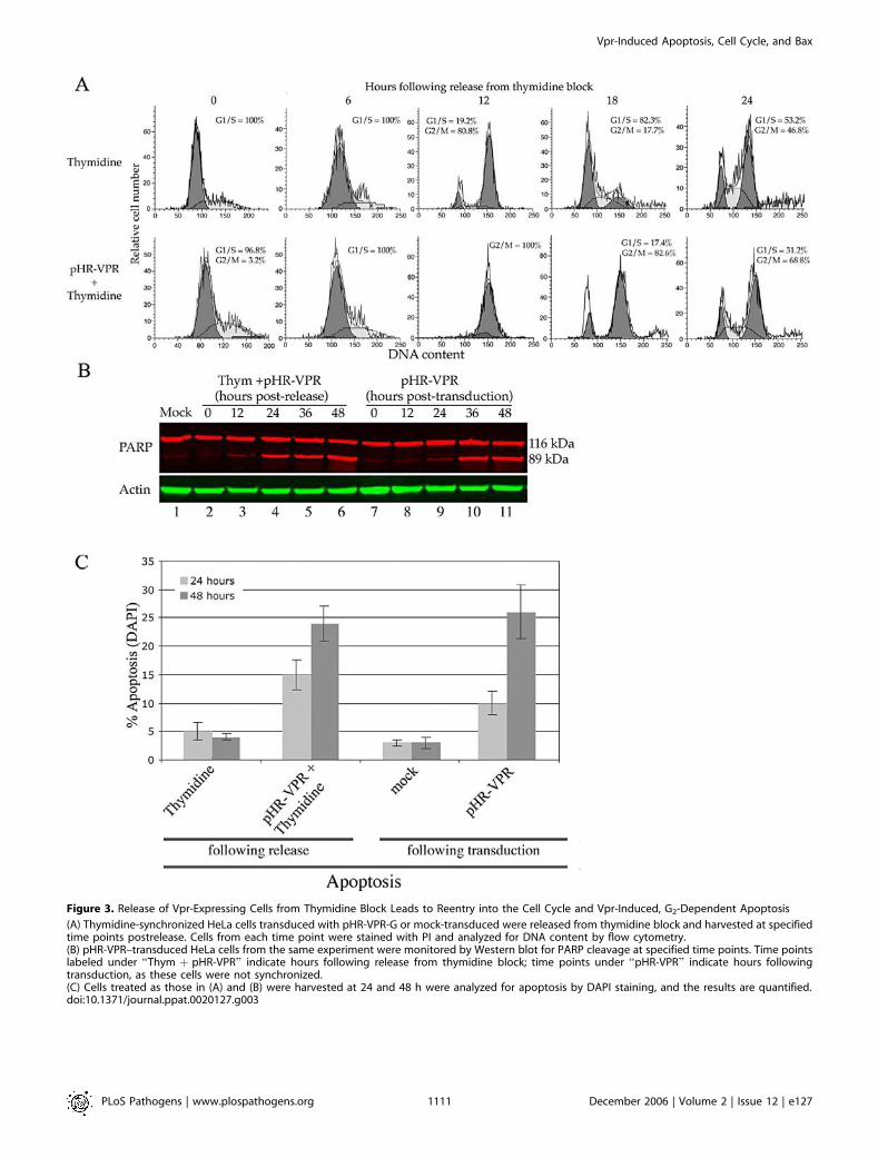
Figure 3. Release of Vpr-Expressing Cells from Thymidine Block Leads to Reentry into the Cell Cycle and Vpr-Induced, G2-Dependent Apoptosis
(A) Thymidine-synchronized HeLa cells transduced with pHR-VPR-G or mock-transduced were released from thymidine block and harvested at specifiedtime points postrelease. Cells from each time point were stained with PI and analyzed for DNA content by flow cytometry.(B) pHR-VPR–transduced HeLa cells from the same experiment were monitored by Western blot for PARP cleavage at specified time points. Time pointslabeled under ‘‘Thym þ pHR-VPR’’ indicate hours following release from thymidine block; time points under ‘‘pHR-VPR’’ indicate hours followingtransduction, as these cells were not synchronized.(C) Cells treated as those in (A) and (B) were harvested at 24 and 48 h were analyzed for apoptosis by DAPI staining, and the results are quantified.doi:10.1371/journal.ppat.0020127.g003
PLoS Pathogens | www.plospathogens.org December 2006 | Volume 2 | Issue 12 | e1271111
Vpr-Induced Apoptosis, Cell Cycle, and Bax
of etoposide or MNNG, we measured caspase activation(Figure 4A and 4B; for simplicity, only one time point isshown in 4A). Knockdown of ATR dramatically exacerbatedetoposide- and MNNG-induced apoptosis (14.5% to 60.7%and 6.2% to 35.4%, respectively, at 48 h). Knockdown of ATMled to a modest increase in genotoxin-induced apoptosis(14.5% to 21.3% for etoposide and 6.2% to 10% for MNNG,at 48 h). In contrast, Vpr-induced caspase activation wasdramatically inhibited by ATR knockdown (from 41.2% to14.2%) but was unaffected by ATM knockdown (41.2% to39.4%). DAPI staining showed a similar ATR-dependentdecrease in Vpr-induced apoptosis (Figure 4C). The level ofknockdown achieved by each siRNA was determined byWestern blot with antibodies specific for the endogenousproteins (Figure 4D).
In conclusion, we demonstrated the requirement for ATRcheckpoint activation through two different methodologies:(a) manipulation by G1/S synchronization and (b) ATRknockdown. We also conclude that this aspect of Vprfunction represents a departure from the manner in whichcheckpoint activation relates to apoptosis in the context ofgenotoxic agents.
Vpr-Induced Apoptosis Does Not Require ANT but IsMediated via Bax, Downstream of ATR Activation
Early reports suggested that the permeability transitionpore complex (PTPC), which consists of VDAC, ANT, andcyclophilin D, was involved in release of apoptotic factorsfrom the mitochondria in response to various apoptoticstimuli (reviewed in [38]) and, in particular, for Vpr [23].However, later reports have put in question the role of thePTPC in DNA damage–induced apoptosis [39–42]. Forexample, mouse cells deficient in ANT are fully capable ofundergoing apoptosis in response to DNA damage and,instead, show increased resistance to necrosis in response tohigh intracellular Ca2þ [39]. Mouse cells deficient in cyclo-philin D, which is required for VDAC function, are fullycapable of undergoing apoptosis in response to genotoxins[40,42]. Large oligomeric complexes containing Bax andpossessing apoptotic pore function do not contain ANT orVDAC [43]. Bax is required for activation of the mitochon-drial pore-forming complex that responds to DNA damage(reviewed in [44,45]) and the pore-forming function of Bax isindependent of ANT or VDAC [43,46–48].
Thus, we asked whether Vpr-induced apoptosis is mediatedthrough ANT or, alternatively, through Bax. We addressedthis question using siRNAs that would specifically down-regulate Bax or ANT, in the context of pHR-VPR-R trans-duction. Bax knockdown led to a dramatic decrease in Vpr-induced apoptosis (from 41.2% in the presence of nonspecificsiRNA to 12.6% in the presence of Bax siRNA; Figure 4A),whereas ANT knockdown had no appreciable effect onapoptosis (41.2% versus 39.5%; Figure 4A). These resultswere also confirmed by analysis of DAPI-stained nuclei(Figure 4C). The levels of knockdown achieved by each siRNAare shown in Figure 4D.
Activation of Bax is associated with a conformationalchange that exposes an N- terminal epitope detected bymonoclonal antibody Bax6A7 [49]. Thus, reactivity withBax6A7 provides a direct measurement of Bax activation.To probe for Bax activation in the presence of Vpr,peripheral blood CD4þ lymphocytes were infected with either
DHIV3 or DHIV3-DVPR, cells were lysed, and immunoreac-tivity with Bax6A7 antibody was tested by immunoprecipita-tion followed by Western blot (Figure 4E). Reactivity withBax6A7 was increased following infection with DHIV3, whencompared with that of mock- or DHIV3-DVPR–infected cells.Our previous studies established that Vpr-induced ATR
activation leads to BRCA1 phosphorylation and GADD45aupregulation and that both ATR and GADD45a are requiredfor Vpr-induced apoptosis [14,17]. Based on previous findingsand data reported here, we propose that ATR activation is anupstream signaling event in the pathway leading to Vpr-induced cell cycle arrest and apoptosis. If this were correct,then it would follow that Bax activation by Vpr is also ATRdependent. In order to examine the requirement of ATR inVpr-induced Bax activation, we treated cells with siRNAsspecific to either ATR or GADD45a prior to transductionwith pHR-VPR-R. We observed that the increase in Bax6A7reactivity was reduced to basal levels in the presence of eitherATR or GADD45a knockdown but not when using anonspecific siRNA (Figure 4F). Therefore, these resultsprovide additional support for a model in which Baxactivation is a principal effector of Vpr-induced apoptosisdownstream of G2 checkpoint activation by the ATR kinase.
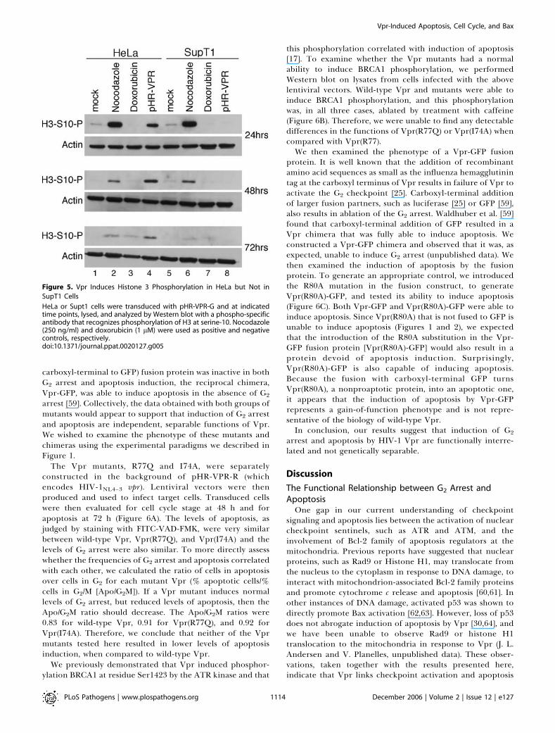
Effect of Vpr on Mitotic EntryIt is unclear whether cells arrested in G2 by Vpr transition
into mitosis. Early measurements of the mitotic indexsuggested that Vpr induced G2 arrest and concomitantlyinhibited entry into mitosis [50–53]. Later studies, however,showed that cells expressing Vpr develop mitotic abnormal-ities such as multipolar spindles, mislocalization of certainspindle pole body proteins, and defects in cytokinesis [54,55],collectively indicating mitotic entry. To reexamine the issueof mitotic entry in the context of Vpr, we measuredphosphorylation of histone 3 (H3) at serine-10 [56,57] in thepresence of Vpr (Figure 5). As positive and negative controls,we incubated cells with nocodazole (lanes 2 and 6) anddoxorubicin (lanes 3 and 7), respectively. Doxorubicin is agenotoxic agent that intercalates into DNA, inhibits top-oisomerase 2 and induces G2 arrest via Cdc2 Tyr15phosphorylation, without allowing mitotic entry. Nocodazolearrests cells in mitosis and exerts its effect by depolymerizingmicrotubules. HeLa cells transduced with pHR-VPR dis-played a high level of H3 phosphorylation (lane 4), whereastransduced SupT1 cells displayed no detectable H3 phos-phorylation (lane 8). Therefore, Vpr expression leads tomitotic entry in HeLa cells following G2 arrest, whereas Vprcauses sustained G2 arrest in SupT1 cells without subsequentmitotic entry.
Analysis of G2 Arrest and Apoptosis Induction by ClinicallyRelevant Vpr Alleles and by a Vpr-GFP Fusion ChimeraSeveral natural or laboratory-constructed mutants of HIV-
1 Vpr have been reported that selectively ablate either G2
arrest or apoptosis. These mutants can be categorized intotwo groups. The first group includes vpr alleles with aminoacid substitutions that have been observed in long-termnonprogressors, with a reduced ability to cause apoptosis butwith normal induction of G2 arrest. These substitutionsinclude Q3R [22], R77Q [21], and I47A [58]. The second groupincludes Vpr-GFP and GFP-Vpr fusion proteins. In partic-ular, it was reported that while a GFP-Vpr (whereby Vpr is
PLoS Pathogens | www.plospathogens.org December 2006 | Volume 2 | Issue 12 | e1271112
Vpr-Induced Apoptosis, Cell Cycle, and Bax
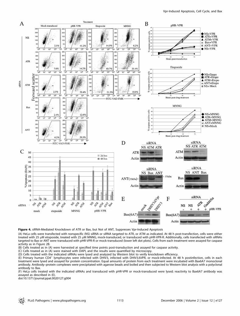
Figure 4. siRNA-Mediated Knockdown of ATR or Bax, but Not of ANT, Suppresses Vpr-Induced Apoptosis
(A) HeLa cells were transfected with nonspecific (NS) siRNA or siRNA targeted to ATR, or ATM as indicated. At 48 h post-transfection, cells were eithertreated with 25 lM etoposide, treated with 25 lM MNNG, mock-transduced, or transduced with pHR-VPR-R. Additionally, cells transfected with siRNAstargeted to Bax or ANT were transduced with pHR-VPR-R or mock-transduced (lower left dot plots). Cells from each treatment were assayed for caspaseactivity as in Figure 2B.(B) Cells treated as in (A) were harvested at specified time points post-transduction and assayed for caspase activity.(C) Cells treated as in (A) were stained with DAPI, and the results were quantified by microscopy.(D) Cells treated with the indicated siRNAs were lysed and analyzed by Western blot to verify knockdown efficiency.(E) Primary human CD4þ lymphocytes were infected with DHIV3, infected with DHIV3DVPR, or mock-infected. At 48 h postinfection, cells in eachtreatment were lysed and assayed for protein concentration. Equal amounts of protein from each treatment were incubated with Bax6A7 monoclonalantibody. Antibody–protein complexes were precipitated with agarose beads and boiled and then subjected to Western blot analysis with a polyclonalantibody to Bax.(F) HeLa cells treated with the indicated siRNAs and transduced with pHR-VPR or mock-transduced were lysed; reactivity to Bax6A7 antibody wasassayed as described in (E).doi:10.1371/journal.ppat.0020127.g004
PLoS Pathogens | www.plospathogens.org December 2006 | Volume 2 | Issue 12 | e1271113
Vpr-Induced Apoptosis, Cell Cycle, and Bax
carboxyl-terminal to GFP) fusion protein was inactive in bothG2 arrest and apoptosis induction, the reciprocal chimera,Vpr-GFP, was able to induce apoptosis in the absence of G2
arrest [59]. Collectively, the data obtained with both groups ofmutants would appear to support that induction of G2 arrestand apoptosis are independent, separable functions of Vpr.We wished to examine the phenotype of these mutants andchimeras using the experimental paradigms we described inFigure 1.
The Vpr mutants, R77Q and I74A, were separatelyconstructed in the background of pHR-VPR-R (whichencodes HIV-1NL4–3 vpr). Lentiviral vectors were thenproduced and used to infect target cells. Transduced cellswere then evaluated for cell cycle stage at 48 h and forapoptosis at 72 h (Figure 6A). The levels of apoptosis, asjudged by staining with FITC-VAD-FMK, were very similarbetween wild-type Vpr, Vpr(R77Q), and Vpr(I74A) and thelevels of G2 arrest were also similar. To more directly assesswhether the frequencies of G2 arrest and apoptosis correlatedwith each other, we calculated the ratio of cells in apoptosisover cells in G2 for each mutant Vpr (% apoptotic cells/%cells in G2/M [Apo/G2M]). If a Vpr mutant induces normallevels of G2 arrest, but reduced levels of apoptosis, then theApo/G2M ratio should decrease. The Apo/G2M ratios were0.83 for wild-type Vpr, 0.91 for Vpr(R77Q), and 0.92 forVpr(I74A). Therefore, we conclude that neither of the Vprmutants tested here resulted in lower levels of apoptosisinduction, when compared to wild-type Vpr.
We previously demonstrated that Vpr induced phosphor-ylation BRCA1 at residue Ser1423 by the ATR kinase and that
this phosphorylation correlated with induction of apoptosis[17]. To examine whether the Vpr mutants had a normalability to induce BRCA1 phosphorylation, we performedWestern blot on lysates from cells infected with the abovelentiviral vectors. Wild-type Vpr and mutants were able toinduce BRCA1 phosphorylation, and this phosphorylationwas, in all three cases, ablated by treatment with caffeine(Figure 6B). Therefore, we were unable to find any detectabledifferences in the functions of Vpr(R77Q) or Vpr(I74A) whencompared with Vpr(R77).We then examined the phenotype of a Vpr-GFP fusion
protein. It is well known that the addition of recombinantamino acid sequences as small as the influenza hemagglutinintag at the carboxyl terminus of Vpr results in failure of Vpr toactivate the G2 checkpoint [25]. Carboxyl-terminal additionof larger fusion partners, such as luciferase [25] or GFP [59],also results in ablation of the G2 arrest. Waldhuber et al. [59]found that carboxyl-terminal addition of GFP resulted in aVpr chimera that was fully able to induce apoptosis. Weconstructed a Vpr-GFP chimera and observed that it was, asexpected, unable to induce G2 arrest (unpublished data). Wethen examined the induction of apoptosis by the fusionprotein. To generate an appropriate control, we introducedthe R80A mutation in the fusion construct, to generateVpr(R80A)-GFP, and tested its ability to induce apoptosis(Figure 6C). Both Vpr-GFP and Vpr(R80A)-GFP were able toinduce apoptosis. Since Vpr(R80A) that is not fused to GFP isunable to induce apoptosis (Figures 1 and 2), we expectedthat the introduction of the R80A substitution in the Vpr-GFP fusion protein [Vpr(R80A)-GFP] would also result in aprotein devoid of apoptosis induction. Surprisingly,Vpr(R80A)-GFP is also capable of inducing apoptosis.Because the fusion with carboxyl-terminal GFP turnsVpr(R80A), a nonproaptotic protein, into an apoptotic one,it appears that the induction of apoptosis by Vpr-GFPrepresents a gain-of-function phenotype and is not repre-sentative of the biology of wild-type Vpr.In conclusion, our results suggest that induction of G2
arrest and apoptosis by HIV-1 Vpr are functionally interre-lated and not genetically separable.
Discussion
The Functional Relationship between G2 Arrest andApoptosisOne gap in our current understanding of checkpoint
signaling and apoptosis lies between the activation of nuclearcheckpoint sentinels, such as ATR and ATM, and theinvolvement of Bcl-2 family of apoptosis regulators at themitochondria. Previous reports have suggested that nuclearproteins, such as Rad9 or Histone H1, may translocate fromthe nucleus to the cytoplasm in response to DNA damage, tointeract with mitochondrion-associated Bcl-2 family proteinsand promote cytochrome c release and apoptosis [60,61]. Inother instances of DNA damage, activated p53 was shown todirectly promote Bax activation [62,63]. However, loss of p53does not abrogate induction of apoptosis by Vpr [30,64], andwe have been unable to observe Rad9 or histone H1translocation to the mitochondria in response to Vpr (J. L.Andersen and V. Planelles, unpublished data). These obser-vations, taken together with the results presented here,indicate that Vpr links checkpoint activation and apoptosis
Figure 5. Vpr Induces Histone 3 Phosphorylation in HeLa but Not in
SupT1 Cells
HeLa or Supt1 cells were transduced with pHR-VPR-G and at indicatedtime points, lysed, and analyzed by Western blot with a phospho-specificantibody that recognizes phosphorylation of H3 at serine-10. Nocodazole(250 ng/ml) and doxorubicin (1 lM) were used as positive and negativecontrols, respectively.doi:10.1371/journal.ppat.0020127.g005
PLoS Pathogens | www.plospathogens.org December 2006 | Volume 2 | Issue 12 | e1271114
Vpr-Induced Apoptosis, Cell Cycle, and Bax

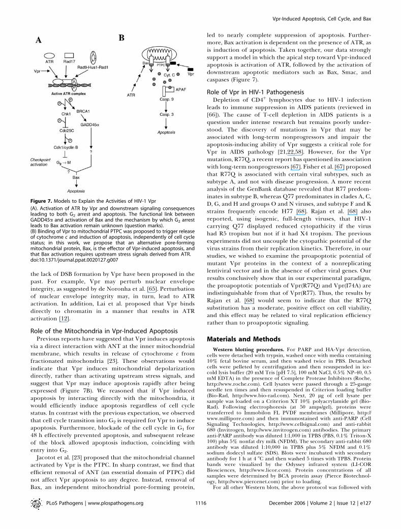
in a novel manner and that a key element is the requirementfor entry into G2 (Figure 7A).
In support of a cause–effect relationship between Vpr-induced G2 arrest and apoptosis, several reports demonstratethat the apoptotic effect of Vpr can be overridden bysuppressing G2-specific cell cycle–regulating kinases [17–20].Yuan et al. [19,20] found that Wee1, a kinase which negativelyregulates Cdk1 activity and thus regulates the G2-to-Mtransition, is activated by Vpr. Furthermore, Yuan et al.[19,20] showed that suppression of Wee1 in the presence ofVpr abrogated both Vpr-induced G2 arrest and apoptosis.
The Role of ATR in Vpr-Induced Apoptosis Differs from Its
Role in Genotoxic Stress-Induced ApoptosisWe also demonstrate that ATR knockdown exacerbates
genotoxin-induced apoptosis but, surprisingly, relieves Vpr-
induced apoptosis [17]. The different effects of ATR knock-down on Vpr- versus genotoxin-induced apoptosis suggestthat Vpr activity fundamentally differs from genotoxic stress.More specifically, it is tempting to speculate that such adifference resides in the fact that Vpr does not cause physicalDNA damage (such as DSBs) whereas genotoxic agents do. Infact, three lines of evidence indicate that Vpr activates ATRin a manner that does not involve generation of DSBs: (1)Pulse-field gel electrophoresis revealed no DSBs in vpr-expressing cells [12]. (2) Vpr fails to induce phosphorylationof ATM at serine-1981, a residue linked to DSB signaling andpotently phosphorylated in response to ionizing radiation[12]. (3) And, as we have shown here, in contrast withetoposide-induced apoptosis, Vpr-induced apoptosis is re-lieved under conditions of checkpoint suppression.Models for Vpr activity that would be in agreement with
Figure 6. Functional Analysis of Vpr Mutants
(A) SupT1 cells were infected with pHR-VPR-R or indicated mutants, at an MOI of 0.5. At 48 h postinfection, cells were stained with hypotonic PI todetermine the cell cycle profiles. At 72 h postinfection, cells were incubated with FITC-VAD-FMK and analyzed by flow cytometry to determine thepercentage of cells with active caspases.(B) Cells from above treatments were lysed at 48 h postinfection, and Western blot was performed to assay for phosphorylation of BRCA1 at Ser1423 bythe ATR kinase. To establish the role of ATR in BRCA1 phosphorylation, parallel infections were treated with caffeine (2 mM).(C) Induction of apoptosis by Vpr-GFP and Vpr(R80A)-GFP fusion proteins. HPB-ALL cells were transfected with indicated constructs or mock-transfectedand 48 after transfection, phosphatidylserine exposure was analyzed by flow cytometry using phycoerythrin-conjugated annexin V.doi:10.1371/journal.ppat.0020127.g006
PLoS Pathogens | www.plospathogens.org December 2006 | Volume 2 | Issue 12 | e1271115
Vpr-Induced Apoptosis, Cell Cycle, and Bax
the lack of DSB formation by Vpr have been proposed in thepast. For example, Vpr may perturb nuclear envelopeintegrity, as suggested by de Noronha et al. [65]. Perturbationof nuclear envelope integrity may, in turn, lead to ATRactivation. In addition, Lai et al. proposed that Vpr bindsdirectly to chromatin in a manner that results in ATRactivation [12].
Role of the Mitochondria in Vpr-Induced ApoptosisPrevious reports have suggested that Vpr induces apoptosis
via a direct interaction with ANT at the inner mitochondrialmembrane, which results in release of cytochrome c fromfractionated mitochondria [23]. These observations wouldindicate that Vpr induces mitochondrial depolarizationdirectly, rather than activating upstream stress signals, andsuggest that Vpr may induce apoptosis rapidly after beingexpressed (Figure 7B). We reasoned that if Vpr inducedapoptosis by interacting directly with the mitochondria, itwould efficiently induce apoptosis regardless of cell cyclestatus. In contrast with the previous expectation, we observedthat cell cycle transition into G2 is required for Vpr to induceapoptosis. Furthermore, blockade of the cell cycle in G1 for48 h effectively prevented apoptosis, and subsequent releaseof the block allowed apoptosis induction, coinciding withentry into G2.
Jacotot et al. [23] proposed that the mitochondrial channelactivated by Vpr is the PTPC. In sharp contrast, we find thatefficient removal of ANT (an essential domain of PTPC) didnot affect Vpr apoptosis to any degree. Instead, removal ofBax, an independent mitochondrial pore-forming protein,
led to nearly complete suppression of apoptosis. Further-more, Bax activation is dependent on the presence of ATR, asis induction of apoptosis. Taken together, our data stronglysupport a model in which the apical step toward Vpr-inducedapoptosis is activation of ATR, followed by the activation ofdownstream apoptotic mediators such as Bax, Smac, andcaspases (Figure 7).
Role of Vpr in HIV-1 PathogenesisDepletion of CD4þ lymphocytes due to HIV-1 infection
leads to immune suppression in AIDS patients (reviewed in[66]). The cause of T-cell depletion in AIDS patients is aquestion under intense research but remains poorly under-stood. The discovery of mutations in Vpr that may beassociated with long-term nonprogressors and impair theapoptosis-inducing ability of Vpr suggests a critical role forVpr in AIDS pathology [21,22,58]. However, for the Vprmutation, R77Q, a recent report has questioned its associationwith long-term nonprogressors [67]. Fisher et al. [67] proposedthat R77Q is associated with certain viral subtypes, such assubtype A, and not with disease progression. A more recentanalysis of the GenBank database revealed that R77 predom-inates in subtype B, whereas Q77 predominates in clades A, C,D, G, and H and groups O and N viruses, and subtype F and Kstrains frequently encode H77 [68]. Rajan et al. [68] alsoreported, using isogenic, full-length viruses, that HIV-1carrying Q77 displayed reduced cytopathicity if the virushad R5 tropism but not if it had X4 tropism. The previousexperiments did not uncouple the cytopathic potential of thevirus strains from their replication kinetics. Therefore, in ourstudies, we wished to examine the proapoptotic potential ofmutant Vpr proteins in the context of a nonreplicatinglentiviral vector and in the absence of other viral genes. Ourresults conclusively show that in our experimental paradigm,the proapoptotic potentials of Vpr(R77Q) and Vpr(I74A) areindistinguishable from that of Vpr(R77). Thus, the results byRajan et al. [68] would seem to indicate that the R77Qsubstitution has a moderate, positive effect on cell viability,and this effect may be related to viral replication efficiencyrather than to proapoptotic signaling.
Materials and Methods
Western blotting procedures. For PARP and HA-Vpr detection,cells were detached with trypsin, washed once with media containing10% fetal bovine serum, and then washed twice in PBS. Detachedcells were pelleted by centrifugation and then resuspended in ice-cold lysis buffer (20 mM Tris [pH 7.5], 100 mM NaCl, 0.5% NP-40, 0.5mM EDTA) in the presence of Complete Protease Inhibitors (Roche,http://www.roche.com). Cell lysates were passed through a 25-gaugeneedle ten times and then resuspended in Criterion loading buffer(Bio-Rad, http://www.bio-rad.com). Next, 20 lg of cell lysate persample was loaded on a Criterion XT 10% polyacrylamide gel (Bio-Rad). Following electrophoresis (at 50 amps/gel), proteins weretransferred to Immobilon FL PVDF membranes (Millipore, http://www.millipore.com) and then immunostained with anti-PARP (CellSignaling Technologies, http://www.cellsignal.com) and anti-rabbit680 (Invitrogen, http://www.invitrogen.com) antibodies. The primaryanti-PARP antibody was diluted 1:1,000 in TPBS (PBS, 0.1% Triton-X100) plus 5% nonfat dry milk (NFDM). The secondary anti-rabbit 680antibody was diluted 1:10,000 in TPBS plus 5% NFDM and 0.1%sodium dodecyl sulfate (SDS). Blots were incubated with secondaryantibody for 1 h at 4 8C and then washed 5 times with TPBS. Proteinbands were visualized by the Odyssey infrared system (LI-CORBiosciences, http://www.licor.com). Protein concentrations of allsamples were determined by BCA protein assay (Pierce Biotechnol-ogy, http://www.piercenet.com) prior to loading.
For all other Western blots, the above protocol was followed with
Figure 7. Models to Explain the Activities of HIV-1 Vpr
(A). Activation of ATR by Vpr and downstream signaling consequencesleading to both G2 arrest and apoptosis. The functional link betweenGADD45a and activation of Bax and the mechanism by which G2 arrestleads to Bax activation remain unknown (question marks).(B) Binding of Vpr to mitochondrial PTPC was proposed to trigger releaseof cytochrome c and induction of apoptosis, independently of cell cyclestatus; in this work, we propose that an alternative pore-formingmitochondrial protein, Bax, is the effector of Vpr-induced apoptosis, andthat Bax activation requires upstream stress signals derived from ATR.doi:10.1371/journal.ppat.0020127.g007
PLoS Pathogens | www.plospathogens.org December 2006 | Volume 2 | Issue 12 | e1271116
Vpr-Induced Apoptosis, Cell Cycle, and Bax

the exception that secondary HRP-conjugated antibodies werediluted in TPBS plus 5% NFDM and bands were visualized by ECLdetection, using ECL plus (Amersham Biosciences, http://www.amersham.com). For Bax 6A7 immunoprecipitation, cells were lysedin CHAPS buffer (10 mM HEPES, 150 mM NaCl, 1% CHAPS,protease inhibitors) on ice for 10 min, followed by ten passes througha 25-gauge needle. Then, 2 lg of Bax 6A7 monoclonal antibody(Abcam, http://www.abcam.com) was added to 1 mg of cellular proteinand incubated 8 h at 4 8C under constant mixing followed byincubation with protein A/G beads (Santa Cruz Biotechnology, http://www.scbt.com) according to the manufacturer’s protocol. Protein–bead complexes were then washed with lysis buffer 3 times, boiled for3 min, and subject to Western blot with anti-Bax antibody (CellSignaling Technologies) and ECL detection according to methodsdescribed above. Antibodies used were anti-ATR (obtained from Dr.Paul Nghiem, Harvard University, Boston, Massachusetts, UnitedStates), anti-GADD45 (Santa Cruz Biotechnology), anti-Bax (CellSignaling Technologies), anti–cytochrome c (Santa Cruz Biotechnol-ogy), anti-Smac/Diablo (Imgenex Corporation, http://www.imgenex.-com), and Bax 6A7 (Abcam).
Cell cycle analysis. Cells were harvested and counted; 1 3 106 cellswere aliquoted into tubes and washed once with cold PBS. Washedcells were resuspended in 0.5 ml of cold PBS, following which 2 ml ofice-cold ethanol was slowly added with gently vortexing. Ethanol-fixed cells were left overnight at�20 8C. The following day, cells werecentrifuged at 828 3 g, and ethanol was washed from the cells. Cellswere then resuspended in propidium iodide staining buffer (10 lg/mlpropidium iodide, 2% FBS, 11.25 kU RNase A/ml, 0.02% sodiumazide) and allowed to incubate for 10 min on ice prior to analysis ofDNA content by flow cytometry. Cell cycle profiles were furtheranalyzed using Modfit software (Verity Software, http://www.vsh.com)to derive percentages of cells in different phases of cell cycle.
Viral vectors and virus. DHIV3 and pHR lentiviral vectors wereproduced by transient transfection of HEK293T cells. For produc-tion of pHR vectors, pHR-VPR or pHR-VPR(R80A) was cotrans-fected with pCMVDR8.2DVPR [69], and pHCMV-VSVG [70] bycalcium phosphate–mediated transfection [18]. Virus-containingsupernatants were harvested at 24, 48, and 72 h post-transfection.Supernatants were cleared of cellular debris by centrifugation at828 3 g for 10 min, and virus in cleared supernatants wasconcentrated by centrifugation at 115,889 3 g for 2 h at 4 8C.Virus was titered by infection of HeLa cells and subsequent flowcytometric analysis of the reporter molecule GFP. Vector titers werecalculated with the equation [(F 3 C0)/V] 3 D, where F is thefrequency of GFP-positive cells found by flow cytometry, C0 is thetotal number of target cells at the time of infection, V is the volumeof inoculum, and D is the virus dilution factor. The virus dilutionfactor used for titrations was 10. The total number of target cells atthe time of infection for titer was 0.5 3 106. Infections wereperformed at a multiplicity of infection (MOI) of 2.5 with 10 lg ofPolybrene/ml for 8 h. Infections of siRNA-treated cells wereperformed 48 h after siRNA transfection. T cells and HeLa cellswere transduced with virus diluted in cell culture media with 8 lg/ml and 10 lg/ml Polybrene, respectively. For HeLa cell transduction,virus-containing media was washed after 8 h and replaced with freshculture media. T cells were transduced by spinoculation aspreviously described [71].
The HIV-1 molecular clone HIV-1NL4–3 was transfected into 2 3107 HEK293FT cells by calcium phosphate transfection. At 48 h aftertransfection, HEK293FT cells were co-cultured with 107 MT-2 cellsfor 5 h. MT-2 cells were then cultured alone until approximately 75%of cell clumps showed syncitia. Virus-containing supernatants werethen cleared of cells and debris by centrifugation at 2,000 rpm for 10min. Viral stocks were then frozen at �80 8C. Spin infections wereperformed as described above.
Apoptosis assays. For analysis of apoptotic nuclear morphology,cells were fixed in the well with 2% paraformaldehyde (in PBS) for 15min at room temperature (RT). Fixed cells were then permeabilizedin 0.1% Triton X-100 (in PBS) for 15 min at RT, gently washed 2 timesin PBS, and then incubated in 0.5 lg/ml DAPI (Invitrogen) for 45 minat 37 8C. Apoptotic nuclei were identified by fluorescence micro-scopy, counted, and divided by total cells in the field to determinepercent apoptosis. For cell counting, fields were chosen at random,and a minimum of 1,000 cells were counted per treatment in eachexperiment. Standard deviations were derived from three separateexperiments. PARP cleavage was assayed by Western blot as described
above. For analysis of caspase activity, cells were stained with FITC-VAD-FMK (Promega, http://www.promega.com) or RED-VAD-FMK(Calbiochem/EMD Biosciences, http://www.emdbiosciences.com) ac-cording to manufacturer’s protocols. Caspase-stained cells wereanalyzed by flow cytometry.
Phosphatidylserine exposure at the cell surface was analyzed aspreviously described [72] by flow cytometry using phycoerythrin-conjugated annexin V (Annexin V-PE, Bender MedSystems, http://www.bendermedsystems.com).
siRNA treatments. All siRNA treatments were performed withDharmacon anti-human smart pool siRNA duplexes (Dharmacon,http://www.dharmacon.com). Smart pool siRNAs were transfected at afinal concentration of 100 nM into exponentially growing HeLa cellswith Oligofectamine (Invitrogen) according to the manufacturer’sprotocol. Cells were split 1:3 at 24 h post-transfection. At 48 h post-transfection, cells were harvested to verify knockdown by Westernblot or subject to experimental treatments.
Cell culture. For experiments in which cell synchronization or theuse of siRNAs was to be employed, we used the human cervical cancercell line HeLa, which was maintained in Dulbecco’s modified Eagle’smedium (Cambrex BioScience (formerly BioWhittaker), http://www.cambrex.com), supplemented with 10% FCS and 2 mM L-glutamine. For experiments in which viral transduction and measure-ment of cell cycle/apoptosis were the aims, we used the human T-cellline SupT1, which was maintained in RPMI 1640 (CambrexBioScience), supplemented with 10% FCS and L-glutamine.
Peripheral blood mononuclear cells were obtained with Leukopaksfrom unidentified, healthy donors (American Red Cross, http://www.redcross.org), and CD4þ lymphocytes were purified using anti-CD4 magnetic beads (Dynal/Invitrogen) according to the manufac-turer’s instructions. CD4þ T cells were activated by culture in RPMIplus 10% FBS plus 1 mM L-glutamine with 3 anti-CD3/anti-CD28beads per cell (Invitrogen) for 2 d, changing medium daily. After 2 d,recombinant interleukin 2 was added to the culture medium at aconcentration of 100 units/ml.
Drug treatments. Thymidine was diluted to a concentration of 2mM in culture media and then added to exponentially growing HeLacells. For experiments in which thymidine treatment was combinedwith viral gene expression, thymidine-containing media was addedimmediately following transduction. For experiments in whichthymidine treatment was combined with genotoxic drugs, cells wereincubated in thymidine-containing media for 4 h prior to theaddition of genotoxins. Etoposide (Sigma Aldrich, http://www.signaaldrich.com) and MNNG (Sigma Aldrich) were diluted in culturemedia to a concentration of 25 lM and incubated with cells for 3 h.Following incubation, genotoxin-containing media was removed andreplaced with fresh media with or without thymidine.
Supporting InformationAccession Numbers
The GenBank (http://www.ncbi.nlm.nih.gov/Genbank) accession num-bers for the proteins discussed in this paper are ANT (P12235[GenPept]), ATM (NM_000051), ATR (NM_001184), BRCA1(NP_009233), GADD45a (NM_001924), HIV-1NL4–3 VPR(AAB60574 [GenPept]), and VDAC (P21796 [GenPept]).
Acknowledgments
We thank Dr. Wayne Green, Dr. Douglas Grossman, Namson Hawk,and Michael Blackwell for technical assistance and Dr. Christopher D.Freel for insight into potential Vpr interactions. We are grateful toDr. Paul Nghiem for providing antibodies to ATR.
Author contributions. JLA, GJ, SB, and VP conceived and designedthe experiments. JLA, JLD, GJ, and SB performed the experiments.JLA, JLD, GJ, SB, and VP analyzed the data. JLA, JLD, ESZ, OA, BK,and VP contributed reagents/materials/analysis tools. JLA and VPwrote the paper.
Funding. This research was supported by National Institutes ofHealth (NIH) research grant AI49057 to VP. JLA is supported by NIHTraining Grant in Microbial Pathogenesis T32 AI055434. ESZ issupported by NIH Genetics Training Grant T32 GM07464.
Competing interests. The authors have declared that no competinginterests exist.
PLoS Pathogens | www.plospathogens.org December 2006 | Volume 2 | Issue 12 | e1271117
Vpr-Induced Apoptosis, Cell Cycle, and Bax
References1. Derdeyn CA, Silvestri G (2005) Viral and host factors in the pathogenesis of
HIV infection. Curr Opin Immunol 17: 366–373.2. Gougeon ML (2003) Apoptosis as an HIV strategy to escape immune attack.
Nat Rev Immunol 3: 392–404.3. Roshal M, Zhu Y, Planelles V (2001) Apoptosis in AIDS. Apoptosis 6: 103–
116.4. Haase AT (1999) Population biology of HIV-1 infection: Viral and CD4þ T
cell demographics and dynamics in lymphatic tissues. Annu Rev Immunol17: 625–656.
5. Perelson AS, Neumann AU, Markowitz M, Leonard JM, Ho DD (1996) HIV-1 dynamics in vivo: Virion clearance rate, infected cell life-span, and viralgeneration time. Science 271: 1582–1586.
6. Wei X, Ghosh SK, Taylor ME, Johnson VA, Emini EA, et al. (1995) Viraldynamics in human immunodeficiency virus type 1 infection. Nature 373:117–122.
7. Simon V, Ho DD (2003) HIV-1 dynamics in vivo: Implications for therapy.Nat Rev Microbiol 1: 181–190.
8. Andersen JL, Planelles V (2005) The role of Vpr in HIV-1 pathogenesis.Curr HIV Res 3: 43–51.
9. Le Rouzic E, Benichou S (2005) The Vpr protein from HIV-1: Distinct rolesalong the viral life cycle. Retrovirology 2: 11.
10. Muthumani K, Zhang D, Hwang DS, Kudchodkar S, Dayes NS, et al. (2002)Adenovirus encoding HIV-1 Vpr activates caspase 9 and induces apoptoticcell death in both p53 positive and negative human tumor cell lines.Oncogene 21: 4613–4625.
11. Muthumani K, Hwang DS, Desai BM, Zhang D, Dayes N, et al. (2002) HIV-1Vpr induces apoptosis through caspase 9 in T cells and peripheral bloodmononuclear cells. J Biol Chem 277: 37820–37831.
12. Lai M, Zimmerman ES, Planelles V, Chen J (2005) Activation of the ATRpathway by human immunodeficiency virus type 1 Vpr involves its directbinding to chromatin in vivo. J Virol 79: 15443–15451.
13. Roshal M, Kim B, Zhu Y, Nghiem P, Planelles V (2003) Activation of theATR-mediated DNA damage response by the HIV-1 viral protein R. J BiolChem 278: 25879–25886.
14. Zimmerman ES, Chen J, Andersen JL, Ardon O, Dehart JL, et al. (2004)Human immunodeficiency virus type 1 Vpr-mediated G2 arrest requiresRad17 and Hus1 and induces nuclear BRCA1 and c-H2AX focus formation.Mol Cell Biol 24: 9286–9294.
15. Bao S, Tibbetts RS, Brumbaugh KM, Fang Y, Richardson DA, et al. (2001)ATR/ATM-mediated phosphorylation of human Rad17 is required forgenotoxic stress responses. Nature 411: 969–974.
16. Zou L, Cortez D, Elledge SJ (2002) Regulation of ATR substrate selection byRad17-dependent loading of Rad9 complexes onto chromatin. Genes Dev16: 198–208.
17. Andersen JL, Zimmerman ES, Dehart JL, Murala S, Ardon O, et al. (2005)ATR and GADD45alpha mediate HIV-1 Vpr-induced apoptosis. Cell DeathDiffer 12: 326–334.
18. Zhu Y, Gelbard HA, Roshal M, Pursell S, Jamieson BD, et al. (2001)Comparison of cell cycle arrest, transactivation, and apoptosis induced bythe simian immunodeficiency virus SIVagm and human immunodeficiencyvirus type 1 vpr genes. J Virol 75: 3791–3801.
19. Yuan H, Kamata M, Xie YM, Chen IS (2004) Increased levels of Wee-1kinase in G(2) are necessary for Vpr- and gamma irradiation-induced G(2)arrest. J Virol 78: 8183–8190.
20. Yuan H, Xie YM, Chen IS (2003) Depletion of Wee-1 kinase is necessary forboth human immunodeficiency virus type 1 Vpr- and gamma irradiation-induced apoptosis. J Virol 77: 2063–2070.
21. Lum JJ, Cohen OJ, Nie Z, Weaver JG, Gomez TS, et al. (2003) Vpr R77Q isassociated with long-term nonprogressive HIV infection and impairedinduction of apoptosis. J Clin Invest 111: 1547–1554.
22. Somasundaran M, Sharkey M, Brichacek B, Luzuriaga K, Emerman M, et al.(2002) Evidence for a cytopathogenicity determinant in HIV-1 Vpr. ProcNatl Acad Sci U S A 99: 9503–9508.
23. Jacotot E, Ravagnan L, Loeffler M, Ferri KF, Vieira HL, et al. (2000) TheHIV-1 viral protein R induces apoptosis via a direct effect on themitochondrial permeability transition pore. J Exp Med 191: 33–46.
24. Vieira HL, Haouzi D, El Hamel C, Jacotot E, Belzacq AS, et al. (2000)Permeabilization of the mitochondrial inner membrane during apoptosis:Impact of the adenine nucleotide translocator. Cell Death Differ 7: 1146–1154.
25. Di Marzio P, Choe S, Ebright M, Knoblauch R, Landau NR (1995)Mutational analysis of cell cycle arrest, nuclear localization and virionpackaging of human immunodeficiency virus type 1 Vpr. J Virol 69: 7909–7916.
26. Zimmerman ES, Sherman MP, Blackett JL, Neidleman JA, Kreis C, et al.(2006) HIV-1 Vpr induces DNA replication stress in vitro and in vivo. JVirol 80: 10407–10418.
27. Gaynor EM, Chen IS (2001) Analysis of apoptosis induced by HIV-1 Vprand examination of the possible role of the hHR23A protein. Exp Cell Res267: 243–257.
28. Firket H, Mahieu P (1967) Synchronization of division induced in HeLacells with an excess of thymidine. Studies on blockage of various stages ofthe cell cycle. Exp Cell Res 45: 11–22.
29. Ardon O, Zimmerman ES, Andersen JL, DeHart JL, Blackett J, et al. (2006)Induction of G2 arrest and binding to cyclophilin A are independentphenotypes of human immunodeficiency virus type 1 Vpr. J Virol 80: 3694–3700.
30. Shostak LD, Ludlow J, Fisk J, Pursell S, Rimel BJ, et al. (1999) Roles of p53and caspases in the induction of cell cycle arrest and apoptosis by HIV-1vpr. Exp Cell Res 251: 156–165.
31. Stojic L, Mojas N, Cejka P, Di Pietro M, Ferrari S, et al. (2004) Mismatchrepair-dependent G2 checkpoint induced by low doses of SN1 typemethylating agents requires the ATR kinase. Genes Dev 18: 1331–1344.
32. Clifford B, Beljin M, Stark GR, Taylor WR (2003) G2 arrest in response totopoisomerase II inhibitors: The role of p53. Cancer Res 63: 4074–4081.
33. Poon B, Jowett JB, Stewart SA, Armstrong RW, Rishton GM, et al. (1997)Human immunodeficiency virus type 1 vpr gene induces phenotypic effectssimilar to those of the DNA alkylating agent, nitrogen mustard. J Virol 71:3961–3971.
34. Hammond EM, Dorie MJ, Giaccia AJ (2004) Inhibition of ATR leads toincreased sensitivity to hypoxia/reoxygenation. Cancer Res 64: 6556–6562.
35. Castedo M, Perfettini JL, Roumier T, Andreau K, Medema R, et al. (2004)Cell death by mitotic catastrophe: A molecular definition. Oncogene 23:2825–2837.
36. Yu Q, La Rose J, Zhang H, Takemura H, Kohn KW, et al. (2002) UCN-01inhibits p53 up-regulation and abrogates gamma-radiation-induced G(2)-Mcheckpoint independently of p53 by targeting both of the checkpointkinases, Chk2 and Chk1. Cancer Res 62: 5743–5748.
37. Truman JP, Gueven N, Lavin M, Leibel S, Kolesnick R, et al. (2005) Down-regulation of ATM protein sensitizes human prostate cancer cells toradiation-induced apoptosis. J Biol Chem 280: 23262–23272.
38. Verrier F, Mignotte B, Jan G, Brenner C (2003) Study of PTPC compositionduring apoptosis for identification of viral protein target. Ann N Y AcadSci 1010: 126–142.
39. Kokoszka JE, Waymire KG, Levy SE, Sligh JE, Cai J, et al. (2004) The ADP/ATP translocator is not essential for the mitochondrial permeabilitytransition pore. Nature 427: 461–465.
40. Baines CP, Kaiser RA, Purcell NH, Blair NS, Osinska H, et al. (2005) Loss ofcyclophilin D reveals a critical role for mitochondrial permeabilitytransition in cell death. Nature 434: 658–662.
41. Li Y, Johnson N, Capano M, Edwards M, Crompton M (2004) Cyclophilin-Dpromotes the mitochondrial permeability transition but has oppositeeffects on apoptosis and necrosis. Biochem J 383: 101–109.
42. Nakagawa T, Shimizu S, Watanabe T, Yamaguchi O, Otsu K, et al. (2005)Cyclophilin D-dependent mitochondrial permeability transition regulatessome necrotic but not apoptotic cell death. Nature 434: 652–658.
43. Antonsson B, Montessuit S, Sanchez B, Martinou JC (2001) Bax is present asa high molecular weight oligomer/complex in the mitochondrial membraneof apoptotic cells. J Biol Chem 276: 11615–11623.
44. Norbury CJ, Zhivotovsky B (2004) DNA damage-induced apoptosis.Oncogene 23: 2797–2808.
45. Cory S, Huang DC, Adams JM (2003) The Bcl-2 family: Roles in cell survivaland oncogenesis. Oncogene 22: 8590–8607.
46. Kuwana T, Mackey MR, Perkins G, Ellisman MH, Latterich M, et al. (2002)Bid, Bax, and lipids cooperate to form supramolecular openings in theouter mitochondrial membrane. Cell 111: 331–342.
47. Rostovtseva TK, Antonsson B, Suzuki M, Youle RJ, Colombini M, et al.(2004) Bid, but not Bax, regulates VDAC channels. J Biol Chem 279: 13575–13583.
48. Annis MG, Soucie EL, Dlugosz PJ, Cruz-Aguado JA, Penn LZ, et al. (2005)Bax forms multispanning monomers that oligomerize to permeabilizemembranes during apoptosis. EMBO J 24: 2096–2103.
49. Rathmell JC, Fox CJ, Plas DR, Hammerman PS, Cinalli RM, et al. (2003) Akt-directed glucose metabolism can prevent Bax conformation change andpromote growth factor-independent survival. Mol Cell Biol 23: 7315–7328.
50. He J, Choe S, Walker R, Di Marzio P, Morgan DO, et al. (1995) Humanimmunodeficiency virus type 1 viral protein R (Vpr) arrests cells in the G2phase of the cell cycle by inhibiting p34cdc2 activity. J Virol 69: 6705–6711.
51. Jowett JB, Planelles V, Poon B, Shah NP, Chen ML, et al. (1995) The humanimmunodeficiency virus type 1 vpr gene arrests infected T cells in the G2þM phase of the cell cycle. J Virol 69: 6304–6313.
52. Re F, Braaten D, Franke EK, Luban J (1995) Human immunodeficiencyvirus type 1 Vpr arrests the cell cycle in G2 by inhibiting the activation ofp34cdc2-cyclin B. J Virol 69: 6859–6864.
53. Bartz SR, Rogel ME, Emerman M (1996) Human immunodeficiency virustype 1 cell cycle control: Vpr is cytostatic and mediates G2 accumulation bya mechanism which differs from DNA damage checkpoint control. J Virol70: 2324–2331.
54. Chang F, Re F, Sebastian S, Sazer S, Luban J (2004) HIV-1 Vpr inducesdefects in mitosis, cytokinesis, nuclear structure, and centrosomes. Mol BiolCell 15: 1793–1801.
55. Watanabe N, Yamaguchi T, Akimoto Y, Rattner JB, Hirano H, et al. (2000)Induction of M-phase arrest and apoptosis after HIV-1 Vpr expressionthrough uncoupling of nuclear and centrosomal cycle in HeLa cells. ExpCell Res 258: 261–269.
56. Hendzel MJ, Wei Y, Mancini MA, Van Hooser A, Ranalli T, et al. (1997)Mitosis-specific phosphorylation of histone H3 initiates primarily withinpericentromeric heterochromatin during G2 and spreads in an ordered
PLoS Pathogens | www.plospathogens.org December 2006 | Volume 2 | Issue 12 | e1271118
Vpr-Induced Apoptosis, Cell Cycle, and Bax

fashion coincident with mitotic chromosome condensation. Chromosoma106: 348–360.
57. Juan G, Traganos F, James WM, Ray JM, Roberge M, et al. (1998) Histone H3phosphorylation and expression of cyclins A and B1 measured in individualcells during their progression through G2 and mitosis. Cytometry 32: 71–77.
58. Zhao Y, Chen M, Wang B, Yang J, Elder RT, et al. (2002) Functionalconservation of HIV-1 Vpr and variability in a mother-child pair of long-term non-progressors. Virus Res 89: 103–121.
59. Waldhuber MG, Bateson M, Tan J, Greenway AL, McPhee DA (2003) Studieswith GFP-Vpr fusion proteins: Induction of apoptosis but ablation of cell-cycle arrest despite nuclear membrane or nuclear localization. Virology313: 91–104.
60. Konishi A, Shimizu S, Hirota J, Takao T, Fan Y, et al. (2003) Involvement ofhistone H1.2 in apoptosis induced by DNA double-strand breaks. Cell 114:673–688.
61. Ishii H, Inageta T, Mimori K, Saito T, Sasaki H, et al. (2005) Frag1, ahomolog of alternative replication factor C subunits, links replicationstress surveillance with apoptosis. Proc Natl Acad Sci U S A 102: 9655–9660.
62. Chipuk JE, Bouchier-Hayes L, Kuwana T, Newmeyer DD, Green DR (2005)PUMA couples the nuclear and cytoplasmic proapoptotic function of p53.Science 309: 1732–1735.
63. Chipuk JE, Kuwana T, Bouchier-Hayes L, Droin NM, Newmeyer DD, et al.(2004) Direct activation of Bax by p53 mediates mitochondrial membranepermeabilization and apoptosis. Science 303: 1010–1014.
64. Stewart SA, Poon B, Jowett JB, Xie Y, Chen IS (1999) Lentiviral delivery ofHIV-1 Vpr protein induces apoptosis in transformed cells. Proc Natl AcadSci U S A 96: 12039–12043.
65. de Noronha CM, Sherman MP, Lin HW, Cavrois MV, Moir RD, et al. (2001)Dynamic disruptions in nuclear envelope architecture and integrityinduced by HIV-1 Vpr. Science 294: 1105–1108.
66. Hazenberg MD, Hamann D, Schuitemaker H, Miedema F (2000) T celldepletion in HIV-1 infection: how CD4þ T cells go out of stock. NatImmunol 1: 285–289.
67. Fischer A, Lejczak C, Lambert C, Roman F, Servais J, et al. (2004) Is the VprR77Q mutation associated with long-term non-progression of HIVinfection? AIDS 18: 1346–1347.
68. Rajan D, Wildum S, Rucker E, Schindler M, Kirchhoff F (2006) Effect ofR77Q, R77A and R80A changes in Vpr on HIV-1 replication and CD4 T celldepletion in human lymphoid tissue ex vivo. AIDS 20: 831–836.
69. An DS, Morizono K, Li QX, Mao SH, Lu S, et al. (1999) An inducible humanimmunodeficiency virus type 1 (HIV-1) vector which effectively suppressesHIV-1 replication. J Virol 73: 7671–7677.
70. Akkina RK, Walton RM, Chen ML, Li QX, Planelles V, et al. (1996) High-efficiency gene transfer into CD34þ cells with a human immunodeficiencyvirus type 1-based retroviral vector pseudotyped with vesicular stomatitisvirus envelope glycoprotein G. J Virol 70: 2581–2585.
71. O’Doherty U, Swiggard WJ, Malim MH (2000) Human immunodeficiencyvirus type 1 spinoculation enhances infection through virus binding. J Virol74: 10074–10080.
72. Py B, Slomianny C, Auberger P, Petit PX, Benichou S (2004) Siva-1 and analternative splice form lacking the death domain, Siva-2, similarly induceapoptosis in T lymphocytes via a caspase-dependent mitochondrialpathway. J Immunol 172: 4008–4017.
PLoS Pathogens | www.plospathogens.org December 2006 | Volume 2 | Issue 12 | e1271119
Vpr-Induced Apoptosis, Cell Cycle, and Bax