Human brain microvascular endothelial cells resist elongation due to shear stress
Upload
independentCategory
view
4download
0
APOBEC3G Inhibits Elongation of HIV-1 ReverseTranscriptsKate N. Bishop1¤, Mohit Verma1, Eun-Young Kim2, Steven M. Wolinsky2, Michael H. Malim1*
1 Department of Infectious Diseases, King’s College London School of Medicine, Guy’s Hospital, London, United Kingdom, 2 Division of Infectious Diseases, Northwestern
University Feinberg School of Medicine, Chicago, Illinois, United States of America
Abstract
APOBEC3G (A3G) is a host cytidine deaminase that, in the absence of Vif, restricts HIV-1 replication and reduces the amountof viral DNA that accumulates in cells. Initial studies determined that A3G induces extensive mutation of nascent HIV-1cDNA during reverse transcription. It has been proposed that this triggers the degradation of the viral DNA, but there is nowmounting evidence that this mechanism may not be correct. Here, we use a natural endogenous reverse transcriptase assayto show that, in cell-free virus particles, A3G is able to inhibit HIV-1 cDNA accumulation not only in the absence ofhypermutation but also without the apparent need for any target cell factors. We find that although reverse transcriptioninitiates in the presence of A3G, elongation of the cDNA product is impeded. These data support the model that A3Greduces HIV-1 cDNA levels by inhibiting synthesis rather than by inducing degradation.
Citation: Bishop KN, Verma M, Kim E-Y, Wolinsky SM, Malim MH (2008) APOBEC3G Inhibits Elongation of HIV-1 Reverse Transcripts. PLoS Pathog 4(12): e1000231.doi:10.1371/journal.ppat.1000231
Editor: Thomas J. Hope, Northwestern University, United States of America
Received September 15, 2008; Accepted November 5, 2008; Published December 5, 2008
Copyright: � 2008 Bishop et al. This is an open-access article distributed under the terms of the Creative Commons Attribution License, which permitsunrestricted use, distribution, and reproduction in any medium, provided the original author and source are credited.
Funding: This study was supported by the Royal Society, the United Kingdom Medical Research Council, and the National Institutes of Health (AI070072). KNB isa Royal Society Dorothy Hodgkin Research Fellow. MHM is an Elizabeth Glaser Scientist.
Competing Interests: The authors have declared that no competing interests exist.
* E-mail: [email protected]
¤ Current address: Division of Virology, National Institute for Medical Research, London, United Kingdom
Introduction
APOBEC3G (A3G) is a potent anti-viral polynucleotide cytidine
deaminase, initially identified as the cellular target of the HIV-1
viral infectivity factor (Vif) protein [1]. Since this discovery, many
APOBEC proteins from various species have been shown to
inhibit the replication of a diverse range of viruses and
retrotransposons (see [2–4] for reviews). However, the exact
mechanism(s) by which APOBEC proteins elicit these effects is
unresolved, enduringly controversial, and may differ among
APOBEC family members.
Studies on HIV-1 have revealed that in the absence of the Vif
protein, human A3G from virus-producing cells is packaged into
HIV-1 particles [5–9]. Two phenotypes are then observed when
these particles infect new target cells: first, nascent viral reverse
transcripts are extensively mutated [10–12]. Most of the mutations
detected are G-to-A changes in the positive sense, coding strand,
implying that deamination of cytidines must occur predominantly
on the negative sense strand of cDNA. This, in turn, locates A3G
to the site and time of reverse transcription. The fixation of such
hypermutation in proviral sequences would presumably lead to the
expression of inactive or truncated viral proteins, producing non-
infectious virions. The second observed phenotype is a reduction
in the accumulation of viral cDNA in target cells [11,13–20]. This
could occur either by triggering degradation of reverse transcripts
or by inhibiting DNA synthesis.
We, and others, initially proposed a mechanism that linked
hypermutation to DNA degradation [10,11]. Specifically, the
hypothesis was that cellular enzymes known as uracil DNA
glycosylases would recognise and remove uracil in the viral cDNA
leaving abasic sites. These sites would then be cleaved by cellular
apurinic/apyrimidinic endonucleases resulting in the degradation
of single-stranded reverse transcripts. However, recent studies
have challenged this theory. Several groups have now shown that
knocking out or inhibiting the cellular uracil DNA glycosylases
UNG2 and SMUG1 does not rescue the defect in viral cDNA
accumulation [16,17,21]. Furthermore, there is mounting evi-
dence to suggest that hypermutation can be dispensable for
APOBEC-mediated antiviral activity (reviewed in [3]). For
example, various APOBEC proteins are able to inhibit MMTV,
HBV, AAV or retrotransposons with little or no discernible editing
activity [22–24]; A3G expressed in unstimulated CD4+ T-cells can
inhibit incoming HIV-1 without inducing widespread hypermuta-
tion [25]; levels of mutations detected in HIV-1 do not correlate
with the degree of viral inhibition or cDNA levels [13,21]; and
finally, engineered catalytically inactive APOBEC mutants can still
inhibit HIV-1, MMTV, HBV or retrotransposons in cultured cell
experiments [14,23,24,26,27]. In the absence of editing, therefore,
what would be the trigger for viral cDNA degradation?
Alternatively, it is plausible that APOBEC proteins, which are
known to bind both RNA and single stranded DNA [28–30], may
be able to prevent the synthesis of cDNA by interfering with the
process of reverse transcription. In this regard, various conflicting
reports have suggested that A3G is able to inhibit numerous steps
during HIV-1 replication, including primer tRNA annealing,
minus and plus strand transfer, primer tRNA processing and
removal, DNA elongation, and proviral integration [11,13–
20,31,32]. It is possible that minor blocks at any (or all) of these
steps could, together, accumulate to produce the potent overall
inhibition of HIV-1 infectivity. In addition, it has recently been
PLoS Pathogens | www.plospathogens.org 1 December 2008 | Volume 4 | Issue 12 | e1000231

reported that the observed block to HBV replication is not due to
A3G-mediated DNA degradation, but instead due to an inhibition
of HBV early minus strand DNA synthesis [33,34].
We have previously shown that A3G and A3F can elicit a
pronounced block to early viral DNA accumulation and that this
decrease correlates well with the block to viral infectivity although
these proteins induce very different levels of hypermutation during
HIV-1 infection [13,14,35]. We therefore decided to employ an
alternative methodology to examine the effects of APOBEC
proteins on the early steps of reverse transcription more closely,
namely using using viral particles isolated from cell supernatants to
study natural endogenous reverse transcription (nERT). We show
here that A3G-mediated inhibition of cDNA accumulation is
independent of both the target cell and hypermutation. Using this
assay system, we also show there is no defect in tRNAlys3 priming
of reverse transcription in virions. However, a small decrease in
the amount of cDNA can already be observed 16 bases after the
start of reverse transcription, and this reduction amplifies with
distance from the tRNAlys3 primer. The block to cDNA titrates
with A3G concentration and can be induced by endogenous levels
of A3G. Together with existing data, our results support the
proposal that A3G inhibits the elongation of HIV-1 DNA by
reverse transcriptase, probably by steric hindrance, rather than
promoting the degradation of viral cDNA.
Results
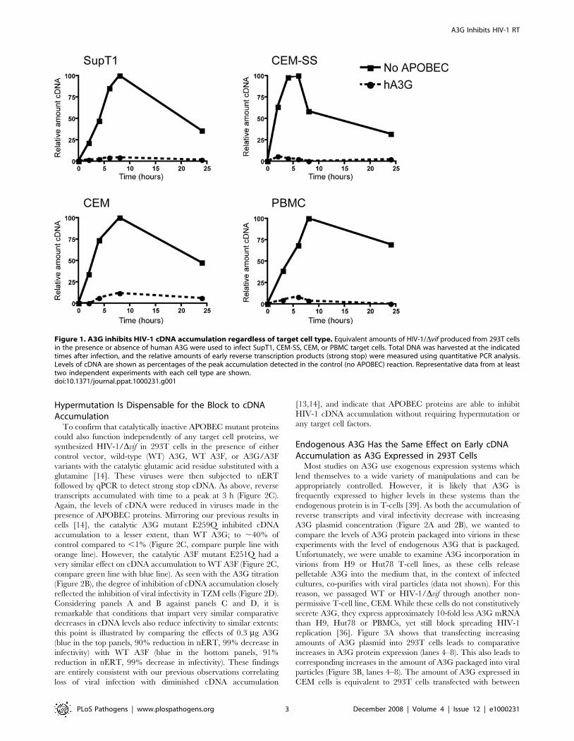
Inhibition of cDNA Accumulation Is Independent ofTarget Cell Type
We have previously shown that human A3G and A3F inhibit
the accumulation of early (strong stop) cDNA products during
HIV-1 infection of SupT1 cells (a T-cell line) [13,14]. It has been
proposed that this is due to specific degradation of viral cDNA by
target cell enzymes [10,11]. We therefore wanted to test the effects
of A3G on cDNA accumulation in different target cells. We
synthesized vif-deficient (Dvif) HIV-1 in the presence or absence of
A3G in 293T cells and used this virus to infect three different
human T-cell lines or peripheral blood mononuclear cells
(PBMCs). In case the expression of endogenous A3G affected
the accumulation of viral cDNA, we chose two so-called
permissive T-cell lines that express little or no endogenous A3G,
SupT1 and CEM-SS, and one cell line, CEM, known to express
A3G and be non-permissive for HIV-1/Dvif replication [1,35,36].
PBMCs include CD4+ T-cells, the natural targets of HIV-1
infection, which also express A3G. Cells were harvested at
different times after infection and the relative amounts of HIV-1
strong stop cDNA were measured by quantitative (q)PCR. As seen
previously, in the absence of A3G, the level of reverse transcription
products increased with time to a peak around 8 h and then
declined (Figure 1, filled lines). The levels of transcripts detected in
cells infected with virus produced in the presence of A3G were
very low at all time points and only just detectable, regardless of
which target cell culture had been infected (Figure 1, dotted lines).
At the peak of accumulation, the amount of cDNA from these
viruses ranged from ,2–10% of the level of cDNA from control
viruses made in the absence of A3G. There was no significant
difference between PBMCs and T-cell lines, or between permissive
and non-permissive cell types, indicating the inhibition of cDNA
accumulation under these experimental conditions is independent
of endogenous A3G in the target cell, and can occur in a range of
different T-cells.
Target Cell Proteins Are Dispensable for the Block tocDNA Accumulation
As the A3G-mediated inhibition of viral cDNA accumulation
seemed to be independent of the type of target cell infected, we
wondered whether any target cell factors were required for this
effect at all. Previous studies have tested the contribution of
particular cellular proteins to the antiviral activity of A3G by
knocking down individual proteins, specifically uracil DNA
glycosylases [16,17,21]. By employing a nERT assay where cell-
free viral particles are incubated in the presence of dNTPs and a
membrane pore-forming substance called melittin in vitro [37,38],
we could test the involvement of all target cell proteins on A3G’s
influence on viral cDNA accumulation at once. Under these
conditions, reverse transcription of the viral RNA proceeds using
the natural tRNAlys3 primer and virion reverse transcriptase
enzyme. We carried out nERT reactions on HIV-1/Dvif produced
in 293T cells in the presence of increasing concentrations of A3G,
and measured the amount of strong stop cDNA present at various
times after the start of the reaction by qPCR (Figure 2A). As seen
with infections of target cells, the amount of viral cDNA increased
with time in all reactions, peaking around 3 h after the addition of
nucleotides. Strikingly, the amount of strong stop DNA that
accumulated decreased with increasing A3G concentration.
Virions made in the presence of the highest dose of A3G (1 mg
transfected plasmid; Figure 2A, purple line) only accumulated
,5% of the amount detected in virions made in the absence of
A3G (Figure 2A, red line). This was very similar to the decreases in
strong stop cDNA seen in T-cell cultures when viruses were made
in the presence of 1 mg of A3G plasmid (Figure 1), suggesting that
additional factors present in target cells are not required for this
defect. The decrease in cDNA accumulation also notably
correlated with decreasing viral infectivity as measured in a single
cycle infection of TZM b-gal reporter cells (Figure 2B). Similar
results were also obtained using an HIV-1 vector system (data not
shown), confirming that this phenotype is not specific for the strain
of HIV-1 used. Accordingly, because target cell proteins are
dispensable for the A3G-mediated suppression of reverse tran-
script accumulation, we considered the nERT assay to be a
simpler, more defined system with which to study this aspect of
APOBEC protein function.
Author Summary
APOBEC proteins are cell-encoded factors that inhibit thereplication of numerous retroviruses, such as HIV-1, andretrotransposons. In many cases, inhibition is clearlyassociated with cytidine-to-uridine editing of viral ortransposon DNA. On the other hand, a number of studieswith particular APOBEC protein/substrate combinations, orengineered proteins that are editing-deficient, have indi-cated that inhibitory mechanism(s) distinct from editing arealso operative. Here, we have analyzed the effects ofAPOBEC3G, a potent HIV-1 inhibitor, on viral reversetranscription using cell-free viruses (natural endogenousreverse transcriptase assays). We report that APOBEC3Ginhibits viral DNA synthesis in a dose-dependent fashion,and does not require editing capabilities to do so. Becausethe addition of the first nucleotide to the tRNA primer isunaffected by A3G and the magnitude of inhibitionincreases as later reverse transcription intermediates aremeasured, we suggest that APOBEC3G acts by impedingthe translocation of the reverse transcriptase enzyme alongits RNA template, perhaps by binding directly to the RNA.These results provide novel insight into the biologicalactivities of this class of host anti-viral proteins.
A3G Inhibits HIV-1 RT
PLoS Pathogens | www.plospathogens.org 2 December 2008 | Volume 4 | Issue 12 | e1000231
Hypermutation Is Dispensable for the Block to cDNAAccumulation
To confirm that catalytically inactive APOBEC mutant proteins
could also function independently of any target cell proteins, we
synthesized HIV-1/Dvif in 293T cells in the presence of either
control vector, wild-type (WT) A3G, WT A3F, or A3G/A3F
variants with the catalytic glutamic acid residue substituted with a
glutamine [14]. These viruses were then subjected to nERT
followed by qPCR to detect strong stop cDNA. As above, reverse
transcripts accumulated with time to a peak at 3 h (Figure 2C).
Again, the levels of cDNA were reduced in viruses made in the
presence of APOBEC proteins. Mirroring our previous results in
cells [14], the catalytic A3G mutant E259Q inhibited cDNA
accumulation to a lesser extent, than WT A3G; to ,40% of
control compared to ,1% (Figure 2C, compare purple line with
orange line). However, the catalytic A3F mutant E251Q had a
very similar effect on cDNA accumulation to WT A3F (Figure 2C,
compare green line with blue line). As seen with the A3G titration
(Figure 2B), the degree of inhibition of cDNA accumulation closely
reflected the inhibition of viral infectivity in TZM cells (Figure 2D).
Considering panels A and B against panels C and D, it is
remarkable that conditions that impart very similar comparative
decreases in cDNA levels also reduce infectivity to similar extents:
this point is illustrated by comparing the effects of 0.3 mg A3G
(blue in the top panels, 90% reduction in nERT, 99% decrease in
infectivity) with WT A3F (blue in the bottom panels, 91%
reduction in nERT, 99% decrease in infectivity). These findings
are entirely consistent with our previous observations correlating
loss of viral infection with diminished cDNA accumulation
[13,14], and indicate that APOBEC proteins are able to inhibit
HIV-1 cDNA accumulation without requiring hypermutation or
any target cell factors.
Endogenous A3G Has the Same Effect on Early cDNAAccumulation as A3G Expressed in 293T Cells
Most studies on A3G use exogenous expression systems which
lend themselves to a wide variety of manipulations and can be
appropriately controlled. However, it is likely that A3G is
frequently expressed to higher levels in these systems than the
endogenous protein is in T-cells [39]. As both the accumulation of
reverse transcripts and viral infectivity decrease with increasing
A3G plasmid concentration (Figure 2A and 2B), we wanted to
compare the levels of A3G protein packaged into virions in these
experiments with the level of endogenous A3G that is packaged.
Unfortunately, we were unable to examine A3G incorporation in
virions from H9 or Hut78 T-cell lines, as these cells release
pelletable A3G into the medium that, in the context of infected
cultures, co-purifies with viral particles (data not shown). For this
reason, we passaged WT or HIV-1/Dvif through another non-
permissive T-cell line, CEM. While these cells do not constitutively
secrete A3G, they express approximately 10-fold less A3G mRNA
than H9, Hut78 or PBMCs, yet still block spreading HIV-1
replication [36]. Figure 3A shows that transfecting increasing
amounts of A3G plasmid into 293T cells leads to comparative
increases in A3G protein expression (lanes 4–8). This also leads to
corresponding increases in the amount of A3G packaged into viral
particles (Figure 3B, lanes 4–8). The amount of A3G expressed in
CEM cells is equivalent to 293T cells transfected with between
Figure 1. A3G inhibits HIV-1 cDNA accumulation regardless of target cell type. Equivalent amounts of HIV-1/Dvif produced from 293T cellsin the presence or absence of human A3G were used to infect SupT1, CEM-SS, CEM, or PBMC target cells. Total DNA was harvested at the indicatedtimes after infection, and the relative amounts of early reverse transcription products (strong stop) were measured using quantitative PCR analysis.Levels of cDNA are shown as percentages of the peak accumulation detected in the control (no APOBEC) reaction. Representative data from at leasttwo independent experiments with each cell type are shown.doi:10.1371/journal.ppat.1000231.g001
A3G Inhibits HIV-1 RT
PLoS Pathogens | www.plospathogens.org 3 December 2008 | Volume 4 | Issue 12 | e1000231

0.03 and 0.1 mg A3G plasmid (Figure 3A, compare lane 1 with
lanes 5 and 6). The level, as expected, is reduced in CEM cells
infected with WT HIV-1 (lane 2) due to Vif expression inducing
the degradation of A3G. Expression is not completely abolished as
a proportion of these cells are presumably not infected at the time
of sampling and therefore still express normal levels of A3G. As
previously reported [5,7,9], WT HIV-1 particles from CEM cells
do not package detectable A3G (Figure 3B, lane 2). As with the
cellular expression, the amount of endogenous A3G packaged into
HIV-1/Dvif virions corresponds to that detected in viruses from
293T cells transfected with between 0.03 and 0.1 mg A3G plasmid
(Figure 3B, compare lane 3 with lanes 5 and 6). This compares
well with a previous report where HIV-1/Dvif virions from
PBMCs packaged the equivalent amount of A3G as viruses made
in 293T cells transfected with a 1:5 molar ratio of A3G: proviral
DNA [39]. In our experiment, transfecting 0.3 mg A3G plasmid
also gives approximately a 1:5 molar ratio, and CEM cells express
,10-fold less A3G than PBMCs, corresponding to 0.03 mg A3G
plasmid.
When a nERT reaction was carried out on viruses from CEM
cells, followed by qPCR for strong stop cDNA, the relative amount
of viral cDNA detected in HIV-1/Dvif virions also corresponded to
that in viruses from 293T cells transfected with between 0.03 and
0.1 mg A3G plasmid (compare Figure 3C with Figure 2A). The
reduction in cDNA levels is similar to that detected previously in
CEM cells by non-PCR based methods [40]. This implies that
endogenous A3G has a similar effect on cDNA accumulation as
exogenously expressed A3G in 293T cells. In other words, the
specific activity of the protein with respect to this attribute is
similar irrespective of the cell type used for expression.
Based on the 293T titration data from Figure 2B, this modest
reduction in strong stop cDNA levels in Dvif viruses from CEM
cells would be expected to lead to between an ,55–90% reduction
in viral infectivity. However, Dvif viruses from CEM cells were
Figure 2. A3G inhibits HIV-1 cDNA accumulation in the absence of target cells and hypermutation. (A) HIV-1/Dvif viruses were generatedfrom 293T transfected with a fixed amount of proviral plasmid (3 mg) and increasing amounts of A3G plasmid (from 0 to 1 mg). Viruses weresubjected to natural endogenous reverse transcription reactions, and DNA was harvested at the indicated times after the addition of dNTPs. Therelative amounts of early reverse transcription products (strong stop) were measured using quantitative PCR analysis and are represented as inFigure 1. (B) The virus preparations described in (A) were used in parallel to challenge TZM b-gal indicator cells, and productive infection wasmeasured after 24 h as the induction of b-galactosidase activity, monitored using a chemiluminescent substrate. Infectivity is reported as counts persec. (C) HIV-1/Dvif viruses were produced from 293T cells in the presence of either control vector, wild-type (WT) A3G, WT A3F, or A3G/A3F variantswith the catalytic glutamic acid residue altered to a glutamine. Viruses were subjected to natural endogenous reverse transcription reactions, andDNA was harvested at the indicated times after the addition of dNTPs. The relative amounts of early reverse transcription products (strong stop) weremeasured using quantitative PCR analysis. (D) The virus preparations described in (C) were used in parallel to challenge TZM b-gal indicator cells, andviral infectivity was measured as in (B). All experiments were performed with at least two independent sets of viruses, representative results areshown.doi:10.1371/journal.ppat.1000231.g002
A3G Inhibits HIV-1 RT
PLoS Pathogens | www.plospathogens.org 4 December 2008 | Volume 4 | Issue 12 | e1000231

slightly less infectious than this, showing ,95% reduction in
infectivity compared to WT HIV-1 (Figure 3D). This suggests that
the antiviral properties of A3G may be accentuated when
expressed in T-cells either through the action of cofactors provided
by virus producing cells [41] or through direct effects on the
protein itself [42,43]. Regarding the former possibility, a recent
report showing that endogenous A3G has significantly lower
cytidine deaminase activity than A3G produced in 293T cells
would indicate that any additional activities are independent of
hypermutation [36].
A3G Does Not Affect Initiation of Reverse TranscriptionSeveral possible mechanisms could be responsible for the
APOBEC-mediated decrease in the accumulation of HIV-1 strong
stop cDNA. Whilst rapid degradation is still a formal possibility,
we have shown that it would have to occur in the absence of
Figure 3. Endogenous A3G has the same effect on early cDNA accumulation as equivalent amounts of A3G expressed in 293T cells.(A) Immunoblot analysis of A3G expression in uninfected CEM cells, CEM cells infected with either WT HIV-1 or HIV-1/Dvif, or 293T cells transfectedwith a fixed amount of HIV-1/Dvif proviral plasmid (3 mg) and increasing amounts of A3G plasmid (0, 0.03, 0.1, 0.3 or 1 mg). Below the blot, the graphshows the quantity of A3G expressed relative to HSP90. (B) Viral incorporation of A3G was measured by immunoblot analysis of purified virionsharvested from the cells in (A). Below the blot, the graph shows the quantity of A3G incorporated relative to HIV-1 CA-p24. (C) WT HIV-1 or HIV-1/Dvifwere passaged through CEM cells and used in natural endogenous reverse transcription reactions. DNA was harvested at the indicated times after theaddition of dNTPs, and the relative amounts of early reverse transcription products (strong stop) were measured using quantitative PCR analysis. (D)The virus preparations from CEM cells described in (C) were used in parallel to challenge TZM b-gal indicator cells, and productive infection wasmeasured as the induction of b-galactosidase activity, monitored using a chemiluminescent substrate. Infectivity is reported as counts per sec.doi:10.1371/journal.ppat.1000231.g003
A3G Inhibits HIV-1 RT
PLoS Pathogens | www.plospathogens.org 5 December 2008 | Volume 4 | Issue 12 | e1000231
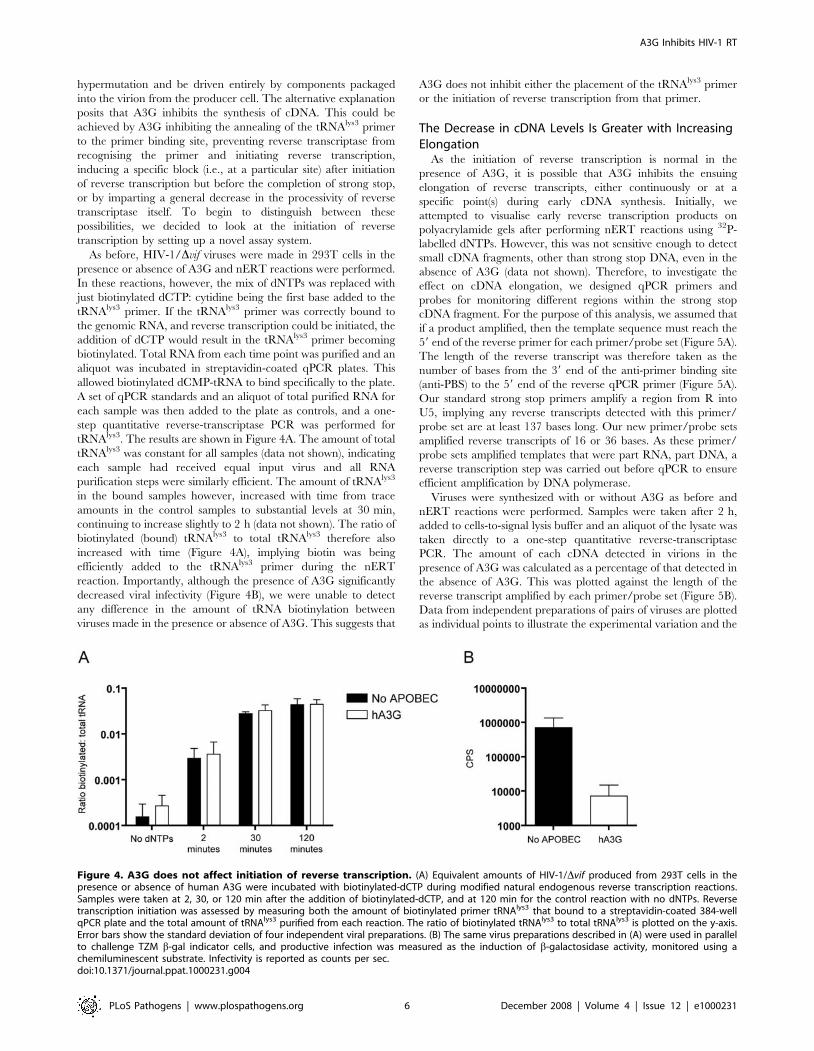
hypermutation and be driven entirely by components packaged
into the virion from the producer cell. The alternative explanation
posits that A3G inhibits the synthesis of cDNA. This could be
achieved by A3G inhibiting the annealing of the tRNAlys3 primer
to the primer binding site, preventing reverse transcriptase from
recognising the primer and initiating reverse transcription,
inducing a specific block (i.e., at a particular site) after initiation
of reverse transcription but before the completion of strong stop,
or by imparting a general decrease in the processivity of reverse
transcriptase itself. To begin to distinguish between these
possibilities, we decided to look at the initiation of reverse
transcription by setting up a novel assay system.
As before, HIV-1/Dvif viruses were made in 293T cells in the
presence or absence of A3G and nERT reactions were performed.
In these reactions, however, the mix of dNTPs was replaced with
just biotinylated dCTP: cytidine being the first base added to the
tRNAlys3 primer. If the tRNAlys3 primer was correctly bound to
the genomic RNA, and reverse transcription could be initiated, the
addition of dCTP would result in the tRNAlys3 primer becoming
biotinylated. Total RNA from each time point was purified and an
aliquot was incubated in streptavidin-coated qPCR plates. This
allowed biotinylated dCMP-tRNA to bind specifically to the plate.
A set of qPCR standards and an aliquot of total purified RNA for
each sample was then added to the plate as controls, and a one-
step quantitative reverse-transcriptase PCR was performed for
tRNAlys3. The results are shown in Figure 4A. The amount of total
tRNAlys3 was constant for all samples (data not shown), indicating
each sample had received equal input virus and all RNA
purification steps were similarly efficient. The amount of tRNAlys3
in the bound samples however, increased with time from trace
amounts in the control samples to substantial levels at 30 min,
continuing to increase slightly to 2 h (data not shown). The ratio of
biotinylated (bound) tRNAlys3 to total tRNAlys3 therefore also
increased with time (Figure 4A), implying biotin was being
efficiently added to the tRNAlys3 primer during the nERT
reaction. Importantly, although the presence of A3G significantly
decreased viral infectivity (Figure 4B), we were unable to detect
any difference in the amount of tRNA biotinylation between
viruses made in the presence or absence of A3G. This suggests that
A3G does not inhibit either the placement of the tRNAlys3 primer
or the initiation of reverse transcription from that primer.
The Decrease in cDNA Levels Is Greater with IncreasingElongation
As the initiation of reverse transcription is normal in the
presence of A3G, it is possible that A3G inhibits the ensuing
elongation of reverse transcripts, either continuously or at a
specific point(s) during early cDNA synthesis. Initially, we
attempted to visualise early reverse transcription products on
polyacrylamide gels after performing nERT reactions using 32P-
labelled dNTPs. However, this was not sensitive enough to detect
small cDNA fragments, other than strong stop DNA, even in the
absence of A3G (data not shown). Therefore, to investigate the
effect on cDNA elongation, we designed qPCR primers and
probes for monitoring different regions within the strong stop
cDNA fragment. For the purpose of this analysis, we assumed that
if a product amplified, then the template sequence must reach the
59 end of the reverse primer for each primer/probe set (Figure 5A).
The length of the reverse transcript was therefore taken as the
number of bases from the 39 end of the anti-primer binding site
(anti-PBS) to the 59 end of the reverse qPCR primer (Figure 5A).
Our standard strong stop primers amplify a region from R into
U5, implying any reverse transcripts detected with this primer/
probe set are at least 137 bases long. Our new primer/probe sets
amplified reverse transcripts of 16 or 36 bases. As these primer/
probe sets amplified templates that were part RNA, part DNA, a
reverse transcription step was carried out before qPCR to ensure
efficient amplification by DNA polymerase.
Viruses were synthesized with or without A3G as before and
nERT reactions were performed. Samples were taken after 2 h,
added to cells-to-signal lysis buffer and an aliquot of the lysate was
taken directly to a one-step quantitative reverse-transcriptase
PCR. The amount of each cDNA detected in virions in the
presence of A3G was calculated as a percentage of that detected in
the absence of A3G. This was plotted against the length of the
reverse transcript amplified by each primer/probe set (Figure 5B).
Data from independent preparations of pairs of viruses are plotted
as individual points to illustrate the experimental variation and the
Figure 4. A3G does not affect initiation of reverse transcription. (A) Equivalent amounts of HIV-1/Dvif produced from 293T cells in thepresence or absence of human A3G were incubated with biotinylated-dCTP during modified natural endogenous reverse transcription reactions.Samples were taken at 2, 30, or 120 min after the addition of biotinylated-dCTP, and at 120 min for the control reaction with no dNTPs. Reversetranscription initiation was assessed by measuring both the amount of biotinylated primer tRNAlys3 that bound to a streptavidin-coated 384-wellqPCR plate and the total amount of tRNAlys3 purified from each reaction. The ratio of biotinylated tRNAlys3 to total tRNAlys3 is plotted on the y-axis.Error bars show the standard deviation of four independent viral preparations. (B) The same virus preparations described in (A) were used in parallelto challenge TZM b-gal indicator cells, and productive infection was measured as the induction of b-galactosidase activity, monitored using achemiluminescent substrate. Infectivity is reported as counts per sec.doi:10.1371/journal.ppat.1000231.g004
A3G Inhibits HIV-1 RT
PLoS Pathogens | www.plospathogens.org 6 December 2008 | Volume 4 | Issue 12 | e1000231
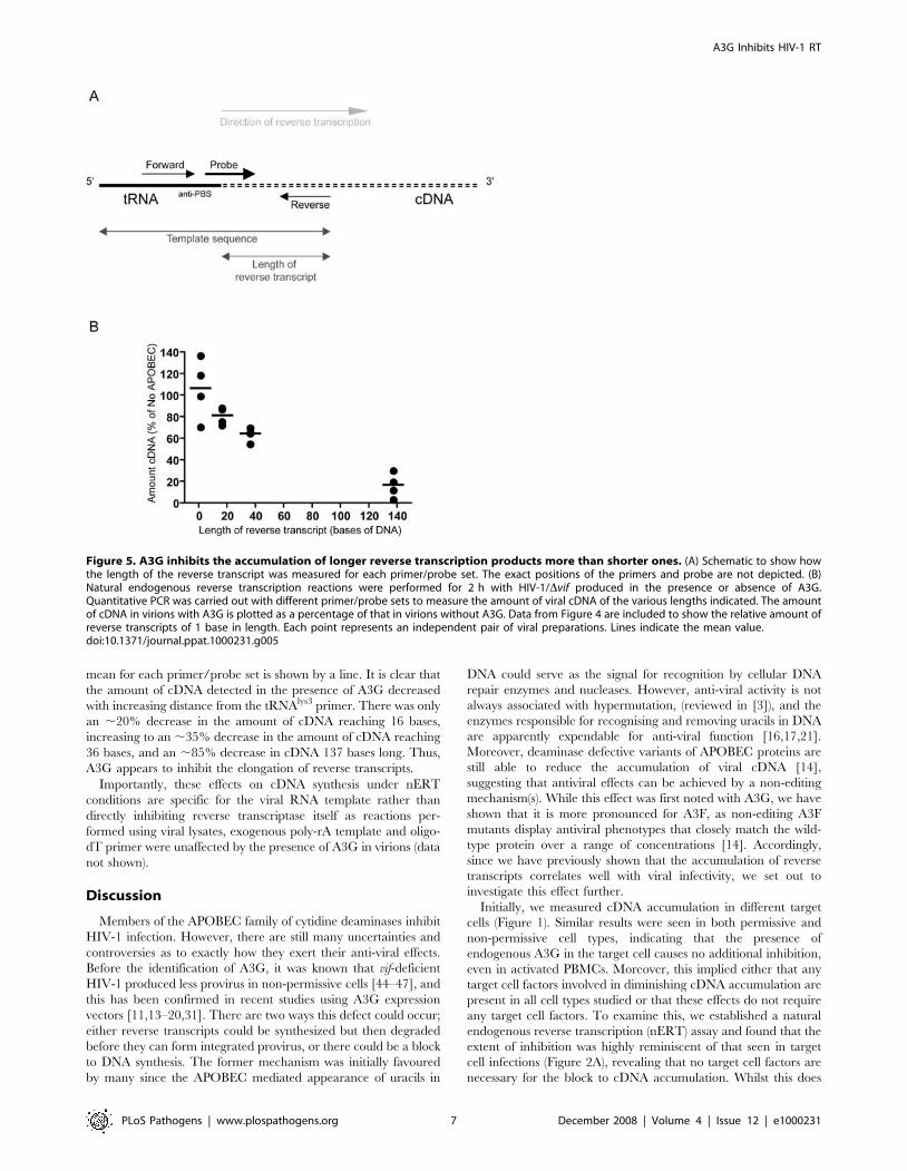
mean for each primer/probe set is shown by a line. It is clear that
the amount of cDNA detected in the presence of A3G decreased
with increasing distance from the tRNAlys3 primer. There was only
an ,20% decrease in the amount of cDNA reaching 16 bases,
increasing to an ,35% decrease in the amount of cDNA reaching
36 bases, and an ,85% decrease in cDNA 137 bases long. Thus,
A3G appears to inhibit the elongation of reverse transcripts.
Importantly, these effects on cDNA synthesis under nERT
conditions are specific for the viral RNA template rather than
directly inhibiting reverse transcriptase itself as reactions per-
formed using viral lysates, exogenous poly-rA template and oligo-
dT primer were unaffected by the presence of A3G in virions (data
not shown).
Discussion
Members of the APOBEC family of cytidine deaminases inhibit
HIV-1 infection. However, there are still many uncertainties and
controversies as to exactly how they exert their anti-viral effects.
Before the identification of A3G, it was known that vif-deficient
HIV-1 produced less provirus in non-permissive cells [44–47], and
this has been confirmed in recent studies using A3G expression
vectors [11,13–20,31]. There are two ways this defect could occur;
either reverse transcripts could be synthesized but then degraded
before they can form integrated provirus, or there could be a block
to DNA synthesis. The former mechanism was initially favoured
by many since the APOBEC mediated appearance of uracils in
DNA could serve as the signal for recognition by cellular DNA
repair enzymes and nucleases. However, anti-viral activity is not
always associated with hypermutation, (reviewed in [3]), and the
enzymes responsible for recognising and removing uracils in DNA
are apparently expendable for anti-viral function [16,17,21].
Moreover, deaminase defective variants of APOBEC proteins are
still able to reduce the accumulation of viral cDNA [14],
suggesting that antiviral effects can be achieved by a non-editing
mechanism(s). While this effect was first noted with A3G, we have
shown that it is more pronounced for A3F, as non-editing A3F
mutants display antiviral phenotypes that closely match the wild-
type protein over a range of concentrations [14]. Accordingly,
since we have previously shown that the accumulation of reverse
transcripts correlates well with viral infectivity, we set out to
investigate this effect further.
Initially, we measured cDNA accumulation in different target
cells (Figure 1). Similar results were seen in both permissive and
non-permissive cell types, indicating that the presence of
endogenous A3G in the target cell causes no additional inhibition,
even in activated PBMCs. Moreover, this implied either that any
target cell factors involved in diminishing cDNA accumulation are
present in all cell types studied or that these effects do not require
any target cell factors. To examine this, we established a natural
endogenous reverse transcription (nERT) assay and found that the
extent of inhibition was highly reminiscent of that seen in target
cell infections (Figure 2A), revealing that no target cell factors are
necessary for the block to cDNA accumulation. Whilst this does
Figure 5. A3G inhibits the accumulation of longer reverse transcription products more than shorter ones. (A) Schematic to show howthe length of the reverse transcript was measured for each primer/probe set. The exact positions of the primers and probe are not depicted. (B)Natural endogenous reverse transcription reactions were performed for 2 h with HIV-1/Dvif produced in the presence or absence of A3G.Quantitative PCR was carried out with different primer/probe sets to measure the amount of viral cDNA of the various lengths indicated. The amountof cDNA in virions with A3G is plotted as a percentage of that in virions without A3G. Data from Figure 4 are included to show the relative amount ofreverse transcripts of 1 base in length. Each point represents an independent pair of viral preparations. Lines indicate the mean value.doi:10.1371/journal.ppat.1000231.g005
A3G Inhibits HIV-1 RT
PLoS Pathogens | www.plospathogens.org 7 December 2008 | Volume 4 | Issue 12 | e1000231
not refute a degradative mechanism for cDNA turnover, it does
eliminate proteasomal degradation and implies that any endo- or
exonucleases involved must be copurified or packaged into viral
particles. As cDNA levels were also reduced by deaminase
deficient APOBEC variants (Figure 2C), an alternative trigger
for degradation other than uracils in DNA would be necessary.
Theoretically, A3G could cause the specific packaging of an as yet
unidentified nuclease responsible for degrading nascent reverse
transcripts. However, there is no direct evidence in the literature
for A3G-induced degradation and, as we expand upon below, this
is not our preferred explanation.
The fact that the block to cDNA accumulation can be detected
in nERT reactions allowed us to investigate this process in ways
that are not possible in cells, but resemble HIV-1 infections more
closely than reconstitution studies using purified components. For
instance, we were able to monitor the initiation of reverse
transcription in virions (Figure 4). Recently, the Kleiman group
have reported that both A3G and A3F prevent tRNAlys3 primer
annealing in vitro via an interaction with nucleocapsid [19,31,48].
However, annealing was not measured directly, and in a very
thorough recent study, Iwatani et al., found no effect on primer
placement or initiation of the +1 cDNA, again in vitro using
purified components [15]. By using a novel variation of the nERT
assay to study reverse transcription initiation in viral particles, we
have shown that A3G does not affect the addition of a biotinylated
dCTP first base to the tRNAlys3 primer. We acknowledge that
some reverse transcription may have initiated prior to incubation
with biotinylated dCTP, but as we detect more than a two log
increase in biotinylation after 30 min, we assert that this represents
significant initiation in our experiment.
Although there was no block to the initiation of reverse
transcription, we could detect a significant reduction in the level of
strong stop cDNA in cells (Figure 1) and nERT reactions (Figures 2
and 5) in the presence of A3G. This indicates either that there is an
early block to reverse transcription or that cDNA is degraded very
rapidly. To address whether reverse transcription was inhibited at
a particular position or whether elongation was progressively
impeded, we designed new qPCR primer/probe sets to measure
reverse transcripts shorter than the 137 nucleotide standard strong
stop product. The amounts of cDNA detected in the presence of
A3G decreased with increasing distance from the tRNAlys3 primer
(Figure 5B), from an ,20% reduction in cDNA reaching 16 bases
to an ,35% decrease at 36 bases and an ,85% decrease at 137
bases. Some variation was seen between independent virus
preparations (indicated by individual points) most likely reflecting
minor differences in A3G expression and packaging. From these
data, it therefore appears that there is no single abrupt point of
termination to reverse transcription.
The results of our study are in complete agreement with a
recent report from Iwatani et al. who show that purified
recombinant A3G is able to inhibit the elongation of exogenous
cDNA by HIV-1 reverse transcriptase in vitro [15]. They
hypothesise that A3G binding to HIV-1 RNA or single stranded
DNA physically blocks RT movement along the template. If A3G
bound genomic RNA at random points, this would imply that the
likelihood of synthesizing a given product would decrease with
increasing product length, as borne out here in Figure 5. The fact
that A3G has been shown to bind several different RNA molecules
implies that binding is not particularly sequence specific [6,28,49–
52]. Work on HBV has recently revealed that A3G is also able to
inhibit the early steps in minus-strand DNA synthesis in this virus
via a block to DNA strand elongation [33].
Other groups have reported a less dramatic decrease in early
products than we see here, with escalating reductions at
progressively later stages of replication [17,18,20]. Indeed, we
have also published a greater decrease in the levels of later
products compared to early transcripts [14]. The difference in the
magnitude of the early effect between different groups has been
attributed to differences in A3G expression levels, and we concur
with this view since we show clearly that both the levels of early
cDNA accumulation and infectivity titrate down with ascending
A3G concentration (Figure 2A and 2B).
Whilst the levels of endogenous A3G expression and viral
incorporation were low in CEM cells, the effect on early cDNA
accumulation was consistent with that seen with exogenous A3G
(Figures 2 and 3). We note, however, that the relative deficit in
infectivity imparted by APOBEC proteins in CEM cells is greater
than anticipated on the bases of the nERT assay. Unfortunately,
we found the efficiency of strand transfer and second strand
synthesis to be very low in nERT reactions, and therefore it was
not possible to use this system to investigate the effect on later steps
in reverse transcription with any degree of quantification or
certainty (data not shown). Possible underlying reasons are
discussed above, and are a subject for future investigations.
Importantly, a mode of action that involves direct binding of A3G
to viral RNA would be expected to be sensitive to A3G
concentration: if A3G can bind to multiple regions of the RNA
genome with equal preference, then the probability of binding to a
region within R or U5, and therefore blocking strong stop cDNA
synthesis, would increase with A3G concentration. Theoretically, a
single molecule of A3G may be all that would be required to
inhibit the synthesis of a full length reverse transcript, such that
even low levels of endogenous A3G would be sufficient to prevent
viral infection.
Based on recent findings, we suggest that the relative contribution
of editing and non-editing effects to the antiviral phenotype of A3G
may depend upon its circumstances. If the inhibition of cDNA
synthesis is inefficient, the resulting nascent transcripts may still be
inactivated via hypermutation and loss of genetic integrity, and the
production of such mutated viral sequences has been well
documented in both cultured cells and in vivo [7,10–12,35,53–56].
Interestingly, the editing capability of endogenous A3G in cultured
T-cells appears to be reduced compared to A3G produced in 293T
cells [36]. In addition, viral cDNAs recovered from PBMCs infected
with HIV-1/Dvif harbour lower levels of G-to-A mutations
compared to cDNAs from H9 cells infected with HIV-1/Dvif, both
in terms of the number of clones carrying mutations and the number
of mutations per clone [57]. This might suggest a lesser role for
hypermutation in vivo, although this issue requires further
investigation. Nevertheless, the capacity to suppress HIV-1 infection
via dual mechanisms presumably serves to enhance the potency of
this class of antiviral proteins.
Recently, two groups have shown that HIV-1/Dvif can replicate
in stable cell lines expressing deaminase-defective A3G mutants
but not wild-type A3G [58,59]. They have interpreted these results
as proving that deamination is essential for A3G activity. We
suggest that such observations should be interpreted with caution
as we have shown that the A3G E259Q mutant also has a
substantially reduced ability to inhibit cDNA accumulation
(Figure 2 and [14]). An alternative explanation for these findings
is that disruption of the deaminase motif of A3G also affects an
attribute necessary for inhibiting reverse transcription; since the C-
terminal deaminase domain of A3G must engage nucleic acids for
the purpose of editing, one possibility is that this defect could be in
RNA binding. At present, it is unclear what effects such mutations
have on the protein structure and function.
In this study, we have shown that A3G-mediated inhibition of
cDNA accumulation occurs independent of both the target cell
A3G Inhibits HIV-1 RT
PLoS Pathogens | www.plospathogens.org 8 December 2008 | Volume 4 | Issue 12 | e1000231

and hypermutation. Although there is no defect in tRNAlys3
priming of reverse transcription, a small decrease in the amount of
cDNA can already be observed at 16 bases, and this reduction
increases with distance from the tRNAlys3 primer. These data
bolster the argument against a degradative mechanism being
responsible for the decreases in viral cDNA that are observed in
the presence of A3G, and support the proposal that A3G inhibits
the elongation of nascent HIV-1 cDNA by steric hindrance of
reverse transcriptase through direct binding to viral genomic
RNA.
Materials and Methods
Plasmid ConstructsAll APOBEC proteins were expressed from pcDNA3.1 as
previously described; for WT human A3G [1], A3G E259Q [27],
WT A3F and A3F E251Q [14]. Expression vectors for wild type
and Dvif HIV-1 (pIIIB and pIIIB/Dvif, respectively), have been
described before [35], and contain a mutation at nucleotide 567 of
the provirus to create a G to A substitution in the U5 region of the
59-LTR, that would copy to the 39-LTR during reverse
transcription.
Cells and Virus ProductionAll cell lines were maintained under standard conditions. Dvif
HIV-1 stocks were typically prepared by co-transfection of 293T
cells with proviral plasmid and A3G expression plasmid (or empty
pcDNA3.1 vector) at a ratio of 3:1. For the A3G titration
experiment shown in Figure 2, 3 mg of Dvif provirus was co-
transfected with varying amounts of A3G plasmid as indicated.
For the experiment that included A3F and A3G/A3F mutants
(Figure 2) a ratio of 1:1 provirus to APOBEC expression vector
was used. The media was changed ,18 h after transfection and
replaced with DMEM containing 20 ml/ml RQ1-DNase (Pro-
mega). After 8 h, the cells were washed and fresh media was added
for a further 17 h, before virus containing supernatants were
harvested. To study endogenous A3G, wild-type or Dvif HIV-1
pseudotyped with the G-protein from vesicular stomatitis virus was
made in 293T cells and used to infect CEM cells by spin infection
at 12006g for 2.5 h at 20uC. The cells were extensively washed
and allowed to grow for 24 h before the media was changed again,
and virus containing supernatants were harvested 16 h following
this. All viral titres were quantitated by enzyme-linked immuno-
sorbent assay (ELISA) for p24CA content.
Viral InfectionsViral infectivities were determined in single-cycle assays by
challenging 105 TZM-b-gal indicator cells [60] with viruses
corresponding to 5 ng p24CA, and measuring the induced
expression of b-galactosidase activity in cell lysates after ,28 h,
using the Galacto-Star system from Applied Biosystems.
For quantitative PCR analysis of cDNA in target cells, viral
preparations containing 30 ng of p24CA were added to 106 SupT1,
CEM-SS or CEM cells or PBMCs rotating at 4uC for 2 h. Cells
were then washed, resuspended in fresh RPMI media, and
incubated at 37uC for 0–24 h. Total DNA was purified using the
DNeasy kit from Qiagen, and eluted in a total volume of 200 ml.
After treatment with Dpn1 for 2 h at 37uC, 2 ml of DNA was
analysed by real-time quantitative PCR.
Natural Endogenous Reverse Transcription Assays (nERT)Viral preparations containing 10–25 ng of p24CA were pelleted
by centrifugation for 1 h at 20,0006g at 4uC. Virions were
resuspended in PBS, 2.5 mM MgCl2, 15 mg/ml melittin and
1 mM dNTPs, and incubated at 37uC. Control reactions were
incubated without any dNTPs for the same length of time as the
longest time point. At the specified time points, aliquots were
removed and equal volumes of PBS containing 40 mg/ml
‘‘carrier’’ salmon sperm DNA added. DNA was then purified
using using the DNeasy kit from Qiagen, and eluted in a total
volume of 200 ml. After treatment with Dpn1 for 2 h at 37uC, 2 ml
of DNA was analysed by real-time quantitative PCR. For later
experiments, an equal volume of 26 Cells-to-signal lysis buffer
(Ambion) was added instead of PBS with carrier DNA, and
samples were diluted 1:10 in water before analysing 2 ml directly in
real-time quantitative PCR.
Reverse Transcription Initiation nERT AssayFor this assay, a nERT reaction was performed essentially as
above, with the exception of the dNTP addition: pelletted virions
were resuspended in PBS, 2.5 mM MgCl2, 15 mg/ml melittin and
0.5mM biotin-11-dCTP (Jena Bioscience), and incubated at 37uC.
Control reactions were incubated without any dNTPs for 2 h. At
the specified time points, aliquots were removed, added to QIAzol
lysis buffer (Qiagen) and frozen at 280uC. RNA was purified using
the Qiagen miRNeasy kit and eluted in 30 ml of water. 2 ml 106PBS was added to 18 ml RNA to make the final samples 16PBS
and the samples were heated at 95–100uC for 3 min and cooled
instantly on ice to separate tRNA from genomic RNA. Samples
were then added to streptavidin coated, 384-well, Strep Thermo-
Fast plates (Abgene) and incubated at 37uC for 30 min. Plates
were extensively washed and all liquid aspirated. Standards and
aliquots of total purified RNA were added to the plates before the
addition of Ag-path-ID One-Step RT-PCR Kit reagents (Ambion)
and tRNAlys3 levels were measured by quantitative PCR analysis.
Quantitative PCRStrong stop reverse transcription products were detected using
primers that amplify the region between nucleotides 500 and 635
of the provirus: oHC64 (59-TAACTAGGGAACCCACTGC) and
oHC65 (59-GCTAGAGATTTTCCACACTG) with probe
oHC66 (59-FAM-ACACAACAGACGGGCACACACTA-
TAMRA). Reactions were performed in triplicate, in TaqMan
Universal PCR master mix (UNG-less) using 900 nM of each
primer, and 250 nM probe. After 10 min at 95uC, reactions were
cycled through 15 sec at 95uC followed by 1 min at 60uC for 40
repeats, carried out on an ABI Prism model 7900HT (Applied
Biosystems). The Dvif-HIV-1 expression vector, pIIIB/Dvif, was
diluted into purified SupT1 cellular DNA to create a series of
control samples that were used to calculate relative cDNA copy
numbers and confirm the linearity of the assay.
tRNAlys3 was detected using primers tRNA-15for (59-
GTCGGTAGAGCATCAGACTTTTAATCT) and Long PBS-G-
(C)7rev (59-CCCCCCCGTGGCGCCCGAACAGGGACTT-
GAAC) with MGB probe Probe-tRNA43for (59-FAM-AGGGTC-
CAGGGTTC-MGB). An oligo with sequence: 59-CCCCCCCG-
TGGCGCCCGAACAGGGACTTGAACCCTGGACCCTCA-
GATTAAAAGTCTGATGCTCTACCGAC was serially diluted
in water to create a standard curve. Note that this primer/probe set
was originally designed to detect tRNAlys3 with a 39 extension. One-
step quantitative reverse transcription PCR was performed using the
Ag-path-ID One-Step RT-PCR Kit from Ambion using 20 ml
reaction volumes and final primer and probe concentrations of 450
nM and 125 nM respectively. Cycling conditions were 45uC for
15 min then 95uC for 10 min, followed by 40 cycles of 15 sec at 95uCand 45 sec at 60uC.
Earlier products of reverse transcription either 16 or 36 bases
from the 39 end of the tRNAlys3 primer were detected with set 2 or
A3G Inhibits HIV-1 RT
PLoS Pathogens | www.plospathogens.org 9 December 2008 | Volume 4 | Issue 12 | e1000231

set 3 primer/probe sets respectively. Set 2 primer sequences were:
tRNA-15for and PBSU5-rev, (59-GGAAAATCTCTAG-
CAGTGGCG) with MGB probe Probe-tRNA43for. Set 3 primer
sequences were tRNA-41for (59-TGAGGGTCCAGGGTT-
CAAGT) and U5-rev-3 (59-CAGACCCTTTTAGTCAGTGTG-
GAA) with probe Probe-PBSU5f-3 (59-FAM-CCTGTTCG-
GGCGCCACTGCT-BHQ1). An oligo with sequence: 59GTC-
GGTAGAGCATCAGACTTTTAATCTGAGGGTCCAGGGT-
TCAAGTCCCTGTTCGGGCGCCACTGCTAGAGATTTTC-
CACACTGACTAAAAGGGTCTGAGGGATCTCTA was seri-
ally diluted in water to create a standard curve. One-step
quantitative reverse transcription PCR was performed using the
Ag-path-ID One-Step RT-PCR Kit using 20 ml reaction volumes
and final primer and probe concentrations of 400 nM and 120 nM
respectively. Cycling conditions were as above for tRNAlys3.
All primer and probe sequences, as well as their respective
amplicons, are also provided in Table S1.
Immunoblot AnalysisVirions were concentrated by centrifugation through a 20% (w/
v) sucrose cushion before immunoblot analysis. Human A3G
protein, heat shock protein (HSP) 90 or viral p24CA proteins were
detected in whole cell lysates of uninfected or HIV-1 infected
CEM cells or transfected 293T cells, or in virion lysates, using a
polyclonal rabbit serum to A3G, HSP 90ab (H-114, Santa Cruz
Biotechnology), or 24-2, a monoclonal anti-CA antibody,
respectively, secondary antibodies IRDye800CW Goat Anti-rabbit
and IRDye800CW Anti-mouse (LI-COR Biosciences UK Ltd)
and Li-cor Odyssey infrared imaging and quantitation.
Supporting Information
Table S1 Names and sequences of qPCR primers and probes
used.
Found at: doi:10.1371/journal.ppat.1000231.s001 (0.04 MB PDF)
Author Contributions
Conceived and designed the experiments: KNB EYK SMW MHM.
Performed the experiments: KNB MV. Analyzed the data: KNB MV
MHM. Contributed reagents/materials/analysis tools: KNB. Wrote the
paper: KNB MHM.
References
1. Sheehy AM, Gaddis NC, Choi JD, Malim MH (2002) Isolation of a human gene
that inhibits HIV-1 infection and is suppressed by the viral Vif protein. Nature
418: 646–650.
2. Chiu YL, Greene WC (2008) The APOBEC3 cytidine deaminases: an innatedefensive network opposing exogenous retroviruses and endogenous retro-
elements. Annu Rev Immunol 26: 317–353.
3. Holmes RK, Malim MH, Bishop KN (2007) APOBEC-mediated viralrestriction: not simply editing? Trends Biochem Sci 32: 118–128.
4. Malim MH, Emerman M (2008) HIV-1 accessory proteins—Ensuring viral
survival in a hostile environment. Cell Host Microbe 3: 388–398.
5. Sheehy AM, Gaddis NC, Malim MH (2003) The antiretroviral enzymeAPOBEC3G is degraded by the proteasome in response to HIV-1 Vif. Nat Med
9: 1404–1407.
6. Bogerd HP, Cullen BR (2008) Single-stranded RNA facilitates nucelocapsid:A-POBEC3G complex formation. RNA 14: 1228–1236.
7. Mariani R, Chen D, Schrofelbauer B, Navarro F, Konig R, et al. (2003) Species-
specific exclusion of APOBEC3G from HIV-1 virions by Vif. Cell 114: 21–31.
8. Soros VB, Yonemoto W, Greene WC (2007) Newly synthesized APOBEC3G is
incorporated into HIV virions, inhibited by HIV RNA, and subsequentlyactivated by RNase H. PLoS Pathog 3: e15. doi:10.1371/journal.ppat.0030015.
9. Yu X, Yu Y, Liu B, Luo K, Kong W, et al. (2003) Induction of APOBEC3G
ubiquitination and degradation by an HIV-1 Vif-Cul5-SCF complex. Science302: 1056–1060.
10. Harris RS, Bishop KN, Sheehy AM, Craig HM, Petersen-Mahrt SK, et al.
(2003) DNA deamination mediates innate immunity to retroviral infection. Cell113: 803–809.
11. Mangeat B, Turelli P, Caron G, Friedli M, Perrin L, et al. (2003) Broad
antiretroviral defence by human APOBEC3G through lethal editing of nascent
reverse transcripts. Nature 424: 99–103.
12. Zhang H, Yang B, Pomerantz RJ, Zhang C, Arunachalam SC, et al. (2003) The
cytidine deaminase CEM15 induces hypermutation in newly synthesized HIV-1
DNA. Nature 424: 94–98.
13. Bishop KN, Holmes RK, Malim MH (2006) Antiviral potency of APOBECproteins does not correlate with cytidine deamination. J Virol 80: 8450–8458.
14. Holmes RK, Koning FA, Bishop KN, Malim MH (2007) APOBEC3F can
inhibit the accumulation of HIV-1 reverse transcription products in the absenceof hypermutation. Comparisons with APOBEC3G. J Biol Chem 282:
2587–2595.
15. Iwatani Y, Chan DS, Wang F, Maynard KS, Sugiura W, et al. (2007)Deaminase-independent inhibition of HIV-1 reverse transcription by APO-
BEC3G. Nucleic Acids Res 35: 7096–7108.
16. Kaiser SM, Emerman M (2006) Uracil DNA glycosylase is dispensable for
human immunodeficiency virus type 1 replication and does not contribute to theantiviral effects of the cytidine deaminase Apobec3G. J Virol 80: 875–882.
17. Mbisa JL, Barr R, Thomas JA, Vandegraaff N, Dorweiler IJ, et al. (2007)
Human immunodeficiency virus type 1 cDNAs produced in the presence ofAPOBEC3G exhibit defects in plus-strand DNA transfer and integration. J Virol
81: 7099–7110.
18. Anderson JL, Hope TJ (2008) APOBEC3G restricts early HIV-1 replication inthe cytoplasm of target cells. Virology 375: 1–12.
19. Guo F, Cen S, Niu M, Saadatmand J, Kleiman L (2006) Inhibition of tRNAlys-
primed reverse transcription by human APOBEC3G during human immuno-
deficiency virus type 1 replication. J Virol 80: 11710–11722.
20. Luo K, Wang T, Liu B, Tian C, Xiao Z, et al. (2007) Cytidine deaminases
APOBEC3G and APOBEC3F interact with human immunodeficiency virus
type 1 integrase and inhibit proviral DNA formation. J Virol 81: 7238–7248.
21. Langlois MA, Neuberger MS (2008) Human APOBEC3G can restrict retroviral
infection in avian cells and acts independently of both UNG and SMUG1.
J Virol 82: 4660–4664.
22. Chen H, Lilley CE, Yu Q, Lee DV, Chou J, et al. (2006) APOBEC3A is a potent
inhibitor of adeno-associated virus and retrotransposons. Curr Biol 16: 480–485.
23. Okeoma CM, Lovsin N, Peterlin BM, Ross SR (2007) APOBEC3 inhibits mouse
mammary tumour virus replication in vivo. Nature 445: 927–930.
24. Turelli P, Mangeat B, Jost S, Vianin S, Trono D (2004) Inhibition of hepatitis B
virus replication by APOBEC3G. Science 303: 1829.
25. Chiu YL, Soros VB, Kreisberg JF, Stopak K, Yonemoto W, et al. (2005) Cellular
APOBEC3G restricts HIV-1 infection in resting CD4+ T cells. Nature 435:
108–114.
26. Bogerd HP, Wiegand HL, Hulme AE, Garcia-Perez JL, O’Shea KS, et al. (2006)
Cellular inhibitors of long interspersed element 1 and Alu retrotransposition.
Proc Natl Acad Sci U S A 103: 8780–8785.
27. Newman EN, Holmes RK, Craig HM, Klein KC, Lingappa JR, et al. (2005)
Antiviral function of APOBEC3G can be dissociated from cytidine deaminase
activity. Curr Biol 15: 166–170.
28. Iwatani Y, Takeuchi H, Strebel K, Levin JG (2006) Biochemical activities of
highly purified, catalytically active human APOBEC3G: correlation with
antiviral effect. J Virol 80: 5992–6002.
29. Yu Q, Konig R, Pillai S, Chiles K, Kearney M, et al. (2004) Single-strand
specificity of APOBEC3G accounts for minus-strand deamination of the HIV
genome. Nat Struct Mol Biol 11: 435–442.
30. Chelico L, Pham P, Calabrese P, Goodman MF (2006) APOBEC3G DNA
deaminase acts processively 39R59 on single-stranded DNA. Nat Struct Mol Biol
13: 392–399.
31. Guo F, Cen S, Niu M, Yang Y, Gorelick RJ, et al. (2007) The interaction of
APOBEC3G with human immunodeficiency virus type 1 nucleocapsid inhibits
tRNA3Lys annealing to viral RNA. J Virol 81: 11322–11331.
32. Li XY, Guo F, Zhang L, Kleiman L, Cen S (2007) APOBEC3G inhibits DNA
strand transfer during HIV-1 reverse transcription. J Biol Chem 282:
32065–32074.
33. Nguyen DH, Gummuluru S, Hu J (2007) Deamination-independent inhibition
of hepatitis B virus reverse transcription by APOBEC3G. J Virol 81: 4465–4472.
34. Rosler C, Kock J, Kann M, Malim MH, Blum HE, et al. (2005) APOBEC-
mediated interference with hepadnavirus production. Hepatology 42: 301–309.
35. Bishop KN, Holmes RK, Sheehy AM, Davidson NO, Cho SJ, et al. (2004)
Cytidine deamination of retroviral DNA by diverse APOBEC proteins. Curr
Biol 14: 1392–1396.
36. Thielen BK, Klein KC, Walker LW, Rieck M, Buckner JH, et al. (2007) T cells
contain an RNase-insensitive inhibitor of APOBEC3G deaminase activity. PLoS
Pathog 3: 1320–1334. doi:10.1371/journal.ppat.0030135.
37. Boone LR, Skalka A (1980) Two species of full-length cDNA are synthesized in
high yield by melittin-treated avian retrovirus particles. Proc Natl Acad Sci U S A
77: 847–851.
38. Zhang H, Dornadula G, Pomerantz RJ (1996) Endogenous reverse transcription
of human immunodeficiency virus type 1 in physiological microenviroments: An
important stage for viral infection of nondividing cells. J Virol 70: 2809–2824.
A3G Inhibits HIV-1 RT
PLoS Pathogens | www.plospathogens.org 10 December 2008 | Volume 4 | Issue 12 | e1000231
39. Xu H, Chertova E, Chen J, Ott DE, Roser JD, et al. (2007) Stoichiometry of the
antiviral protein APOBEC3G in HIV-1 virions. Virology 360: 247–256.40. Ohagen A, Gabuzda D (2000) Role of Vif in stability of the human
immunodeficiency virus type 1 core. J Virol 74: 11055–11066.
41. Han Y, Wang X, Dang Y, Zheng YH (2008) APOBEC3G and APOBEC3Frequire an endogenous cofactor to block HIV-1 replication. PLoS Pathog 4:
e1000095. doi:10.1371/journal.ppat.1000095.42. Shirakawa K, Takaori-Kondo A, Yokoyama M, Izumi T, Matsui M, et al.
(2008) Phosphorylation of APOBEC3G by protein kinase A regulates its
interaction with HIV-1 Vif. Nat Struct Mol Biol 15: 1184–1191. doi:10.1038/nsmb.1497.
43. Watashi K, Khan M, Yedavalli VR, Yeung ML, Strebel K, et al. (2008) Humanimmunodeficiency virus type 1 replication and regulation of APOBEC3G by
peptidyl prolyl isomerase Pin1. J Virol 82: 9928–9936.44. Courcoul M, Patience C, Rey F, Blanc D, Harmache A, et al. (1995) Peripheral
blood mononuclear cells produce normal amounts of defective Vif- human
immunodeficiency virus type 1 particles which are restricted for thepreretrotranscription steps. J Virol 69: 2068–2074.
45. Simon JH, Malim MH (1996) The human immunodeficiency virus type 1 Vifprotein modulates the postpenetration stability of viral nucleoprotein complexes.
J Virol 70: 5297–5305.
46. Sova P, Volsky DJ (1993) Efficiency of viral DNA synthesis during infection ofpermissive and nonpermissive cells with vif-negative human immunodeficiency
virus type 1. J Virol 67: 6322–6326.47. von Schwedler U, Song J, Aiken C, Trono D (1993) Vif is crucial for human
immunodeficiency virus type 1 proviral DNA synthesis in infected cells. J Virol67: 4945–4955.
48. Yang Y, Guo F, Cen S, Kleiman L (2007) Inhibition of initiation of reverse
transcription in HIV-1 by human APOBEC3F. Virology 365: 92–100.49. Gallois-Montbrun S, Holmes RK, Swanson CM, Fernandez-Ocana M,
Byers HL, et al. (2008) Comparison of cellular ribonucleoprotein complexesassociated with the APOBEC3F and APOBEC3G antiviral proteins. J Virol 82:
5636–5642.
50. Khan MA, Goila-Gaur R, Opi S, Miyagi E, Takeuchi H, et al. (2007) Analysis ofthe contribution of cellular and viral RNA to the packaging of APOBEC3G into
HIV-1 virions. Retrovirology 4: 48.
51. Navarro F, Bollman B, Chen H, Konig R, Yu Q, et al. (2005) Complementary
function of the two catalytic domains of APOBEC3G. Virology 333: 374–386.
52. Wang T, Tian C, Zhang W, Luo K, Sarkis PT, et al. (2007) 7SL RNA mediates
virion packaging of the antiviral cytidine deaminase APOBEC3G. J Virol 81:
13112–13124.
53. Lecossier D, Bouchonnet F, Clavel F, Hance AJ (2003) Hypermutation of HIV-1
DNA in the absence of the Vif protein. Science 300: 1112.
54. Janini M, Rogers M, Birx DR, McCutchan FE (2001) Human immunodefi-
ciency virus type 1 DNA sequences genetically damaged by hypermutation are
often abundant in patient peripheral blood mononuclear cells and may be
generated during near-simultaneous infection and activation of CD4(+) T cells.
J Virol 75: 7973–7986.
55. Kieffer TL, Kwon P, Nettles RE, Han Y, Ray SC, et al. (2005) GRA
hypermutation in protease and reverse transcriptase regions of human
immunodeficiency virus type 1 residing in resting CD4+ T cells in vivo. J Virol
79: 1975–1980.
56. Vartanian JP, Meyerhans A, Sala M, Wain-Hobson S (1994) GRA
hypermutation of the human immunodeficiency virus type 1 genome: Evidence
for dCTP pool imbalance during reverse transcription. Proc Natl Acad Sci U S A
91: 3092–3096.
57. Knoepfel SA, Salisch NC, Huelsmann PM, Rauch P, Walter H, et al. (2008)
Comparison of G-to-A mutation frequencies induced by APOBEC3 proteins in
H9 cells and peripheral blood mononuclear cells in the context of impaired
processivities of drug-resistant human immunodeficiency virus type 1 reverse
transcriptase variants. J Virol 82: 6536–6545.
58. Miyagi E, Opi S, Takeuchi H, Khan M, Goila-Gaur R, et al. (2007)
Enzymatically active APOBEC3G is required for efficient inhibition of human
immunodeficiency virus type 1. J Virol 81: 13346–13353.
59. Schumacher AJ, Hache G, Macduff DA, Brown WL, Harris RS (2008) The
DNA deaminase activity of human APOBEC3G is required for Ty1, MusD, and
human immunodeficiency virus type 1 restriction. J Virol 82: 2652–2660.
60. Wei X, Decker JM, Liu H, Zhang Z, Arani RB, et al. (2002) Emergence of
resistant human immunodeficiency virus type 1 in patients receiving fusion
inhibitor (T-20) monotherapy. Antimicrob Agents Chemother 46: 1896–1905.
A3G Inhibits HIV-1 RT
PLoS Pathogens | www.plospathogens.org 11 December 2008 | Volume 4 | Issue 12 | e1000231
Copyright © 2022 FDOKUMEN