Biochemical and Structural Analysis of the Interaction between the UBA(2) Domain of the DNA Repair...
10
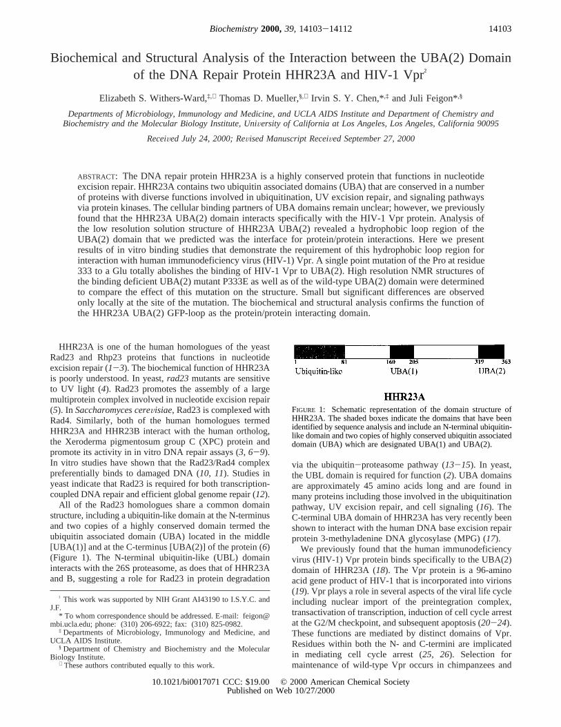
Biochemical and Structural Analysis of the Interaction between the UBA(2) Domain of the DNA Repair Protein HHR23A and HIV-1 Vpr ² Elizabeth S. Withers-Ward, ‡,⊥ Thomas D. Mueller, §,⊥ Irvin S. Y. Chen,* ,‡ and Juli Feigon* ,§ Departments of Microbiology, Immunology and Medicine, and UCLA AIDS Institute and Department of Chemistry and Biochemistry and the Molecular Biology Institute, UniVersity of California at Los Angeles, Los Angeles, California 90095 ReceiVed July 24, 2000; ReVised Manuscript ReceiVed September 27, 2000 ABSTRACT: The DNA repair protein HHR23A is a highly conserved protein that functions in nucleotide excision repair. HHR23A contains two ubiquitin associated domains (UBA) that are conserved in a number of proteins with diverse functions involved in ubiquitination, UV excision repair, and signaling pathways via protein kinases. The cellular binding partners of UBA domains remain unclear; however, we previously found that the HHR23A UBA(2) domain interacts specifically with the HIV-1 Vpr protein. Analysis of the low resolution solution structure of HHR23A UBA(2) revealed a hydrophobic loop region of the UBA(2) domain that we predicted was the interface for protein/protein interactions. Here we present results of in vitro binding studies that demonstrate the requirement of this hydrophobic loop region for interaction with human immunodeficiency virus (HIV-1) Vpr. A single point mutation of the Pro at residue 333 to a Glu totally abolishes the binding of HIV-1 Vpr to UBA(2). High resolution NMR structures of the binding deficient UBA(2) mutant P333E as well as of the wild-type UBA(2) domain were determined to compare the effect of this mutation on the structure. Small but significant differences are observed only locally at the site of the mutation. The biochemical and structural analysis confirms the function of the HHR23A UBA(2) GFP-loop as the protein/protein interacting domain. HHR23A is one of the human homologues of the yeast Rad23 and Rhp23 proteins that functions in nucleotide excision repair (1-3). The biochemical function of HHR23A is poorly understood. In yeast, rad23 mutants are sensitive to UV light (4). Rad23 promotes the assembly of a large multiprotein complex involved in nucleotide excision repair (5). In Saccharomyces cereVisiae, Rad23 is complexed with Rad4. Similarly, both of the human homologues termed HHR23A and HHR23B interact with the human ortholog, the Xeroderma pigmentosum group C (XPC) protein and promote its activity in in vitro DNA repair assays (3, 6-9). In vitro studies have shown that the Rad23/Rad4 complex preferentially binds to damaged DNA (10, 11). Studies in yeast indicate that Rad23 is required for both transcription- coupled DNA repair and efficient global genome repair (12). All of the Rad23 homologues share a common domain structure, including a ubiquitin-like domain at the N-terminus and two copies of a highly conserved domain termed the ubiquitin associated domain (UBA) located in the middle [UBA(1)] and at the C-terminus [UBA(2)] of the protein (6) (Figure 1). The N-terminal ubiquitin-like (UBL) domain interacts with the 26S proteasome, as does that of HHR23A and B, suggesting a role for Rad23 in protein degradation via the ubiquitin-proteasome pathway (13-15). In yeast, the UBL domain is required for function (2). UBA domains are approximately 45 amino acids long and are found in many proteins including those involved in the ubiquitination pathway, UV excision repair, and cell signaling (16). The C-terminal UBA domain of HHR23A has very recently been shown to interact with the human DNA base excision repair protein 3-methyladenine DNA glycosylase (MPG) (17). We previously found that the human immunodeficiency virus (HIV-1) Vpr protein binds specifically to the UBA(2) domain of HHR23A (18). The Vpr protein is a 96-amino acid gene product of HIV-1 that is incorporated into virions (19). Vpr plays a role in several aspects of the viral life cycle including nuclear import of the preintegration complex, transactivation of transcription, induction of cell cycle arrest at the G2/M checkpoint, and subsequent apoptosis (20-24). These functions are mediated by distinct domains of Vpr. Residues within both the N- and C-termini are implicated in mediating cell cycle arrest (25, 26). Selection for maintenance of wild-type Vpr occurs in chimpanzees and ² This work was supported by NIH Grant AI43190 to I.S.Y.C. and J.F. * To whom correspondence should be addressed. E-mail: feigon@ mbi.ucla.edu; phone: (310) 206-6922; fax: (310) 825-0982. ‡ Departments of Microbiology, Immunology and Medicine, and UCLA AIDS Institute. § Department of Chemistry and Biochemistry and the Molecular Biology Institute. ⊥ These authors contributed equally to this work. FIGURE 1: Schematic representation of the domain structure of HHR23A. The shaded boxes indicate the domains that have been identified by sequence analysis and include an N-terminal ubiquitin- like domain and two copies of highly conserved ubiquitin associated domain (UBA) which are designated UBA(1) and UBA(2). 14103 Biochemistry 2000, 39, 14103-14112 10.1021/bi0017071 CCC: $19.00 © 2000 American Chemical Society Published on Web 10/27/2000
-
Upload
independent -
Category
Documents
-
view
0 -
download
0
Transcript of Biochemical and Structural Analysis of the Interaction between the UBA(2) Domain of the DNA Repair...
Biochemical and Structural Analysis of the Interaction between the UBA(2) Domainof the DNA Repair Protein HHR23A and HIV-1 Vpr†
Elizabeth S. Withers-Ward,‡,⊥ Thomas D. Mueller,§,⊥ Irvin S. Y. Chen,*,‡ and Juli Feigon*,§
Departments of Microbiology, Immunology and Medicine, and UCLA AIDS Institute and Department of Chemistry andBiochemistry and the Molecular Biology Institute, UniVersity of California at Los Angeles, Los Angeles, California 90095
ReceiVed July 24, 2000; ReVised Manuscript ReceiVed September 27, 2000
ABSTRACT: The DNA repair protein HHR23A is a highly conserved protein that functions in nucleotideexcision repair. HHR23A contains two ubiquitin associated domains (UBA) that are conserved in a numberof proteins with diverse functions involved in ubiquitination, UV excision repair, and signaling pathwaysvia protein kinases. The cellular binding partners of UBA domains remain unclear; however, we previouslyfound that the HHR23A UBA(2) domain interacts specifically with the HIV-1 Vpr protein. Analysis ofthe low resolution solution structure of HHR23A UBA(2) revealed a hydrophobic loop region of theUBA(2) domain that we predicted was the interface for protein/protein interactions. Here we presentresults of in vitro binding studies that demonstrate the requirement of this hydrophobic loop region forinteraction with human immunodeficiency virus (HIV-1) Vpr. A single point mutation of the Pro at residue333 to a Glu totally abolishes the binding of HIV-1 Vpr to UBA(2). High resolution NMR structures ofthe binding deficient UBA(2) mutant P333E as well as of the wild-type UBA(2) domain were determinedto compare the effect of this mutation on the structure. Small but significant differences are observedonly locally at the site of the mutation. The biochemical and structural analysis confirms the function ofthe HHR23A UBA(2) GFP-loop as the protein/protein interacting domain.
HHR23A is one of the human homologues of the yeastRad23 and Rhp23 proteins that functions in nucleotideexcision repair (1-3). The biochemical function of HHR23Ais poorly understood. In yeast,rad23 mutants are sensitiveto UV light (4). Rad23 promotes the assembly of a largemultiprotein complex involved in nucleotide excision repair(5). In Saccharomyces cereVisiae, Rad23 is complexed withRad4. Similarly, both of the human homologues termedHHR23A and HHR23B interact with the human ortholog,the Xeroderma pigmentosum group C (XPC) protein andpromote its activity in in vitro DNA repair assays (3, 6-9).In vitro studies have shown that the Rad23/Rad4 complexpreferentially binds to damaged DNA (10, 11). Studies inyeast indicate that Rad23 is required for both transcription-coupled DNA repair and efficient global genome repair (12).
All of the Rad23 homologues share a common domainstructure, including a ubiquitin-like domain at the N-terminusand two copies of a highly conserved domain termed theubiquitin associated domain (UBA) located in the middle[UBA(1)] and at the C-terminus [UBA(2)] of the protein (6)(Figure 1). The N-terminal ubiquitin-like (UBL) domaininteracts with the 26S proteasome, as does that of HHR23Aand B, suggesting a role for Rad23 in protein degradation
via the ubiquitin-proteasome pathway (13-15). In yeast,the UBL domain is required for function (2). UBA domainsare approximately 45 amino acids long and are found inmany proteins including those involved in the ubiquitinationpathway, UV excision repair, and cell signaling (16). TheC-terminal UBA domain of HHR23A has very recently beenshown to interact with the human DNA base excision repairprotein 3-methyladenine DNA glycosylase (MPG) (17).
We previously found that the human immunodeficiencyvirus (HIV-1) Vpr protein binds specifically to the UBA(2)domain of HHR23A (18). The Vpr protein is a 96-aminoacid gene product of HIV-1 that is incorporated into virions(19). Vpr plays a role in several aspects of the viral life cycleincluding nuclear import of the preintegration complex,transactivation of transcription, induction of cell cycle arrestat the G2/M checkpoint, and subsequent apoptosis (20-24).These functions are mediated by distinct domains of Vpr.Residues within both the N- and C-termini are implicatedin mediating cell cycle arrest (25, 26). Selection formaintenance of wild-type Vpr occurs in chimpanzees and
† This work was supported by NIH Grant AI43190 to I.S.Y.C. andJ.F.
* To whom correspondence should be addressed. E-mail: [email protected]; phone: (310) 206-6922; fax: (310) 825-0982.
‡ Departments of Microbiology, Immunology and Medicine, andUCLA AIDS Institute.
§ Department of Chemistry and Biochemistry and the MolecularBiology Institute.
⊥ These authors contributed equally to this work.
FIGURE 1: Schematic representation of the domain structure ofHHR23A. The shaded boxes indicate the domains that have beenidentified by sequence analysis and include an N-terminal ubiquitin-like domain and two copies of highly conserved ubiquitin associateddomain (UBA) which are designated UBA(1) and UBA(2).
14103Biochemistry2000,39, 14103-14112
10.1021/bi0017071 CCC: $19.00 © 2000 American Chemical SocietyPublished on Web 10/27/2000
humans infected with HIV-1, supporting the idea that Vprconfers an important selective advantage to the virus (27).
Vpr-induced cell-cycle arrest and apoptosis of HIV-1infected cells would have a profound effect on T-cells, theprimary target of HIV-1, contributing to the immunederegulation associated with AIDS (28). HIV-1 infectedCD4+ T-cells would fail to undergo activation and clonalexpansion. Vpr-mediated apoptosis would contribute todepletion of the CD4+ cells that is seen in HIV-infectedindividuals. Therefore, Vpr may be an important viral proteinto target in the development of therapeutic reagents fortreatment of HIV-1-infected individuals. In addition tofinding that Vpr interacts specifically with the UBA(2)domain of HHR23A, we previously demonstrated thatoverexpression of HHR23A or a fragment encompassing theUBA(2) domain would partially alleviate cell cycle arrestmediated by Vpr (18). This suggests a significant functionalrole for the interactions between Vpr and HHR23A. Wehypothesized that Vpr may mediate cell cycle arrest and/orapoptosis by binding to the UBA(2) domain of HHR23A,thereby leading to deregulation of normal cellular processesinvolved in the control and signaling of DNA repair. Thus,an understanding of the structural requirements for interactionbetween Vpr and UBA(2) may not only provide insight intothe role of HHR23A in Vpr function but may also lead tostrategies for the development of therapeutic reagents thatwould interfere with Vpr function.
We have previously reported a low resolution structureof UBA(2) of HHR23A determined by homonuclear NMRspectroscopy (29). The structural motif of the UBA(2)domain is composed of threeR-helices folded around ahydrophobic core. Analysis of the NMR structure revealedan unusually large hydrophobic area of the protein that wepredicted to be an interface for protein/protein interactionsand might be the Vpr-interacting region of the HHR23AUBA(2) domain. This hydrophobic area of the UBA(2)domain is in the vicinity of the GFP-loop (331GFP333) whichforms a sharp turn betweenR-helices 1 and 2. To furthercharacterize the amino acids that are critical for interactionwith HHR23A UBA(2), synthetic peptides derived from theUBA domains ofS. cereVisiae (2) and an unrelated proteinc-cbl (30) that is involved in signal transduction in hemato-poietic cells were assayed for binding to Vpr in vitro. Theamino acid sequence of these UBA domains differs at andaround the GFP-loop. The results indicate that the Pro withinthe GFP-loop of the HHR23A UBA(2) domain is requiredfor interaction with Vpr. Further studies with mutant peptidesdemonstrate that the Pro within the GFP-loop is necessarybut not sufficient for binding to Vpr. Furthermore, theHHR23A UBA(1) domain and the UBA domain from c-cbldo not interact with Vpr, suggesting that the interactionbetween the HHR23A UBA(2) domain and Vpr is a highlyspecific one. High-resolution structures of both the wild typeand the P333E mutant of UBA(2) were determined by NMRto address the question of how this mutation affects the struc-ture and/or hydrophobic surface of the protein. Comparisonof wild-type and P333E mutant structures reveals that substi-tution of the Pro residue 333 with a Glu results in changesin the shape and charge of the hydrophobic patch predictedto be the Vpr-interacting domain. These changes can accountfor the loss of the ability to bind Vpr. The structural analysisof a Vpr-interacting protein and identification of a Vpr-inter-
acting interface provide a rationale for the future developmentof therapeutic reagents that interfere with Vpr function.
MATERIALS AND METHODSExpression of GST Fusion Proteins in Bacteria.The HIV-1
Vpr GST fusion protein was expressed and purified asdescribed with some modifications (18). Briefly, the HIV-1NL4-3 coding sequence, inserted into the pGEX-KG expres-sion plasmid, was expressed inEscherichia coliBL21 (DE3).Following disruption of the cells by sonication and centrifu-gation, the cells were dialyzed overnight at 4°C against PBS.The dialysate was then applied directly to a prepackedglutathione sepharose column (Pharmacia Biotech) andpurified according to the manufacturer’s instructions. Frac-tions were collected and analyzed by SDS-polyacrylamidegel electrophoresis (PAGE). Proteins were visualized bystaining the polyacrylamide gels with Coomassie Blue orby transferring the proteins to nitrocellulose and staining withPonceau S. These assays confirmed that all fractions werefree of contaminating bacterial proteins. Those fractionscontaining the bulk of the GSTVpr fusion protein werepooled and dialyzed overnight at 4°C against PBS. Proteinconcentration was determined by the Bradford method.Purified proteins were stored at-70 °C.
Synthesis and Preparation of Peptides.Peptides corre-sponding to UBA domains were biochemically synthesizedby Fmoc (fluorenylmethoxycarbonyl)/tert-butyl based solid-phase peptide chemistry method on an ABI 431A (Perkin-Elmer) peptide synthesizer utilizing a single couplingprogram to carry out the chain assembly. The crude peptidewas purified to 95% homogeneity by preparative reverse-phase HPLC on a Vydac C18 column. The composition andpurity of each peptide was assessed using electro-sprayionization (ESI) mass spectrometry and amino acid analysis.For detection purposes, all peptides were synthesized witha biotin molecule at their N-termini. The sequences of thepeptides used in these studies are listed below with mutationsindicated in boldface type.
HHR23A UBA(2): QEKEAIERLKALGFPESLVIQAYF-ACEKNENLAANFLLSQNFDDE; HHR23A UBA(2)P333E:QEKEAIERLKALGFEESLVIQAYFACEKNENLAANF-LLSQNFDDE; HHR23A UBA(2)333GFP/ERD335: QEKEAI-ERLKALGFERDLVIQAYFACEKNENLAANFLLSQNF-DDE; HHR23A UBA(1): SEYETMLTEIMSMGFERERVV-AALRASYNNPHRAVEYLLTGIPGS; S. cereVisiaeUBA-(2): EDDQAISRLCELGFERDLVIQVYFACDKNEEAA-ANILFSDHAD; S. cereVisiae UBA(2)E370P: EDDQAIS-RLCELGFPRDLVIQVYFACDKNEEAAANILFSDHAD; S.cereVisiaeUBA(2)370PES372: EDDQAISRLCELGFPESLVIQ-VYFACDKNEEAAANILFSDHAD; UBA c-cbl: SPQLSS-EIENLMSQGYSYQDIQKALVIAQNNIEMAKNILRE.
Samples for binding assays were prepared by dissolvingthe purified lyophilized peptide in 10 mM Hepes, pH 6.8 (1mg/mL). In some cases, the pH was adjusted to 7.0 by theaddition of 1 N NaOH. Aliquots were stored at-70 °C andthawed just prior to use.
In Vitro Binding Assay.This assay is a slightly modifiedversion of a binding assay that we have previously developedand tested with all of the relevant controls (18). GlutathioneSepharose 4B (Pharmacia Biotech) was prepared by washing0.5 mL of the suspension obtained from the manufacturerthree times in prebinding buffer (100 mM NaCl, 25 mM Tris-
14104 Biochemistry, Vol. 39, No. 46, 2000 Withers-Ward et al.
HCl [pH 7.5], 0.1% Triton X-100) (1 mL/wash) and thenresuspending it in prebinding buffer in a total volume of 2mL. GSTVpr or GST (10µg/reaction) was added to 100µLof the glutathione Sepharose suspension. After samples wereincubated at room temperature (30-60 min), preboundcomplexes were washed once in prebinding buffer (1 mL/reaction) and then twice with buffer A (20 mM Tris-HCl,pH 7.5, 100 mM NaCl, 5 mM DTT). Peptides were diluted4-fold in buffer A, and 50µg was added to each reaction.After a 30-min incubation at room temperature, samples werewashed three times in buffer B (20 mM Tris-HCl, pH 7.5,300 mM NaCl, 1 mM DTT, 0.5% NP40) (1 mL/wash).Samples were then incubated in blocking buffer [1× TNT(20 mM Tris, 500 mM NaCl, pH 7.5, 0.3% Tween 20 (v/v),4.0% nonfat dry milk (w/v), 1.0% BSA (w/v)] (0.2 mL/sample) for 30 min at room temperature, and then washedthree times in buffer B. After a 30-min incubation in astreptavidin horseradish peroxidase conjugate (AmershamLife Science Inc.) [diluted 1/5000 in TNT that was 0.2% inBSA (w/v)], samples were washed three times in buffer B.1-Step Turbo TMB-ELISA solution (Pierce Chemical Co.)was then added to each sample (0.25 mL/sample). After a10-min incubation at room temperature, 150µL of sulfuricacid (1.2 N) was added to each sample, and the absorbancewas read at 450 nm in a spectrophotometer.
Preparation of the UBA(2) Domain.A DNA fragmentencoding the UBA(2) domain (residues 319-363) wasamplified by PCR from full-length HHR23A gene (18) andinserted into the pGEX-2T vector (Pharmacia) between theBamHI andEcoRI restriction sites.E. coli strain BL21(DE3)-pLysS was then transformed with the plasmid, and thetransformed bacteria were cultured in either LB or M9 media.To prepare15N- or 15N- and13C-labeled protein,15NH4Cl or[13C6]glucose was used as sole nitrogen or carbon source inthe growth medium. Expression of GST-UBA(2) fusionprotein was induced with 1 mM isopropyl-â-D-thiogalacto-pyranoside when the absorbance of the culture reachedapproximately 0.7 optical density units at 550 nm. Thetemperature was lowered from 37 to 30°C during theexpression to increase the yield of soluble fusion protein.The cells were harvested 3 h (4.5 h for the preparation oflabeled protein in M9 medium) after induction and resus-pended in 10 mM Tris-HCl, pH 8.0, 1 mM EDTA, 0.375 Msucrose (15 mL/L of culture). Cells were lysed by adding 1vol of BugBuster (Novagen), 1 mg/mL lysozyme (Sigma),and 0.5 U of Benzonase (Novagen). The GST fusion proteinwas cleaved using biotinylated thrombin (Novagen). Theprotease was then removed with streptavidin agarose, andthe UBA(2) protein was finally purified by an anion-exchange chromatography step (high performance Q sepharose,Pharmacia). The final purified UBA(2) protein consisted ofresidues 319-363 of the HHR23A protein and an N-terminalGly-Ser peptide derived from the expression vector. Typi-cally, between 5 (15N- and 13C-labeled) and 12.5 mg (15N-labeled) of purified UBA(2) protein was obtained from 1 Lof M9 medium. Initial NMR studies for the wild-type proteinas well as the NMR analysis of the UBA(2) mutant P333Ewere performed using peptides derived from chemicalsynthesis as previously described (29).
NMR Analysis and Structure Calculation of the Wild-TypeUBA(2) Domain and the Mutant P333E.NMR samples ofwild-type UBA(2) contained between 1.5 and 3 mM protein
in 50 mM sodium phosphate, pH 6.5, 100 mM sodiumchloride, 2 mM dithiothreitol-d8 (DTT), 0.02% sodium azide,and 5%2H2O. All spectra were acquired at 300 K with aBruker DRX 500 or DRX 600 spectrometer. A series ofthree-dimensional experiments (15N-HSQC-NOE-HSQC,15N-HSQC TOCSY, CBCA(CO)NH, CBCANH, HBHA(CO)-NH, HBHANH, HCCH-COSY, and HCCH-TOCSY) wereacquired for the assignments of the1H, 15N, and 13Cresonances (31). All NMR data was processed using XWIN-NMR (Bruker) and analyzed with the software XEASY (32).Distance information was obtained from15N- or 13C-edited3D NOESY experiments as well as a series of 2D NOESYexperiments with mixing times ranging from 80 to 300 ms.NOE connectivities obtained from 3D NOESY experimentswere categorized into strong, medium, weak, and very weak,corresponding to upper limits for proton-proton distancesof 3.0, 4.0, 4.5, and 5.0 Å, respectively. 2D NOESY spectrawere analyzed and integrated using the software packagesXEASY (32) and SPSCAN (33). No stereospecific assign-ments of methylene protons or methyl groups were used.Upper limit distance restraints were then generated from theNOE volumes using DYANA (34). In total, 779 NOE-derived distance restraints (173 intraresidue, 196 sequential,219 medium range, and 191 long range) were used for thewild-type UBA(2). In the final stages of refinement, 17hydrogen bonds were introduced based on a regular patternof NOEs of the type HRiHâi+3 and HRiNHi+3, as well as carbonand proton chemical shift information suggesting a regularR-helix.
The proton resonances of the UBA(2)P333E were assignedby analysis of a series of 2D1H NMR experiments composedof TOCSY, DQF-COSY, and NOESY. NOE connectivitieswere derived from a series of 2D NOESY experimentsacquired with mixing times ranging from 100 to 350 ms.Distance restraints were generated as described above. A totalof 826 NOE-derived distance restraints (204 intraresidue, 182sequential, 226 medium range, and 214 long range) wereused for the structure calculation of UBA(2)P333E. Althoughalmost all NOE-derived distance restraints were derived fromthe 2D NOESY data acquired on the mutant sample, anadditional 148 distance restraints were taken from the wild-type UBA(2) 3D data set due to spectral overlap in the 2DNOESY spectra of the mutant. All of these were locatedwithin the hydrophobic core and were far from the site ofmutation. Identical proton chemical shifts between the wild-type and the mutant protein indicate that the structuralenvironment for those protons is the same, and use of thoserestraints does not bias the structure of the mutant protein.
The structures of wild-type UBA(2) and its mutant P333Ewere calculated from an extended peptide conformation witha simulated annealing protocol using XPLOR (35). Thesimulated annealing protocol consisted of 35 ps dynamicsat 2000 K (integration time steps of 5 fs) and 35 ps slowcooling (10 K/cycle). Final minimization was performedusing 50 steps of Adopted Raphson Newton algorithm(Quanta98, MSI). For each protein, 100 structures werecalculated, of which 18 (wild-type) and 21 (mutant P333E)were selected based on lowest total and NOE energy.
Coordinates for the 18 lowest energy structures of UBA-(2) (accession code 1DV0) and UBA(2)P333E (accessioncode 1F4I) have been deposited in the RCSB protein databank.
Structure/Function Analysis of HHR23A UBA(2) Biochemistry, Vol. 39, No. 46, 200014105
RESULTS AND DISCUSSION
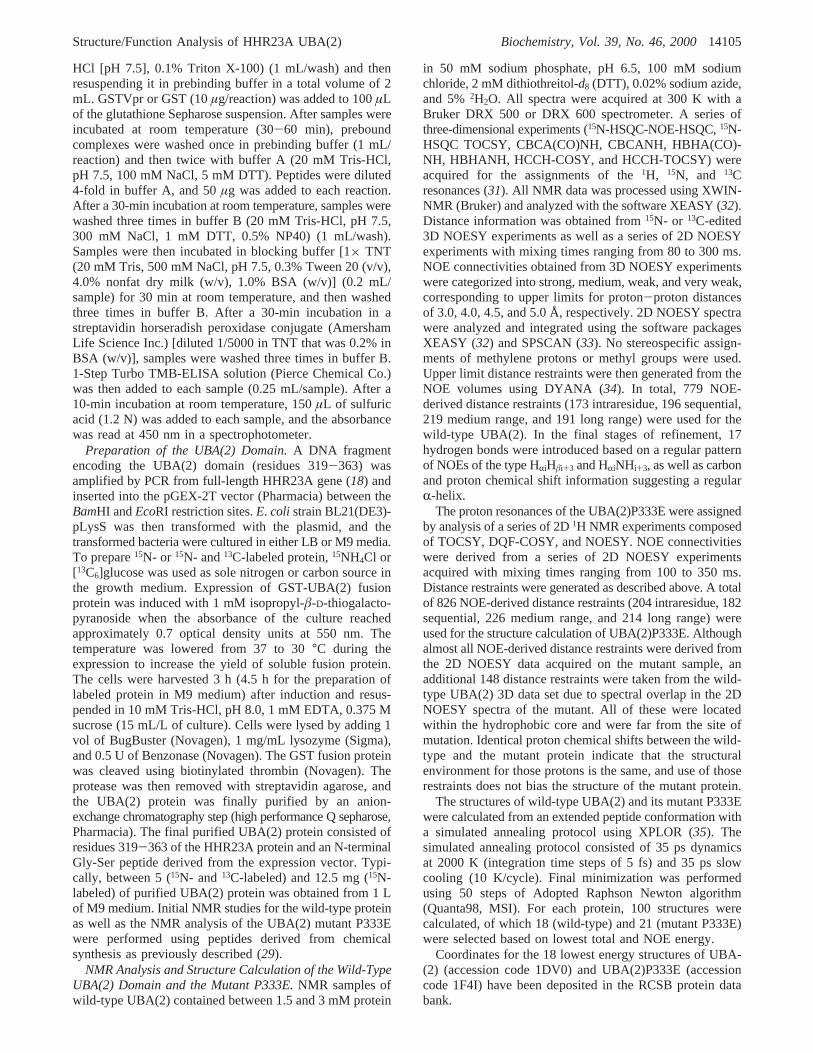
Binding of Vpr to UBA Domains Is Specific to the UBA-(2) of HHR23A.Our previous studies showed that Vpr bindsspecifically to UBA(2) of HHR23A and HHR23B. Since theUBA domain is a common structural motif, the binding toVpr of a variety of other UBA domains was tested using abinding assay based on one that we have previously described(18) (Figure 2). Binding of GSTVpr-associated proteins wasdetected by ELISA. The amino acid sequences and alignmentof the peptides that were tested are shown in Figure 3, panelA. The results of the in vitro binding assays using peptidesderived from the internal UBA domain of HHR23A [UBA-(1)], S. cereVisiae Rad23A UBA(2), and the UBA domain
of an unrelated protein, c-cbl (30), which is involved in signaltransduction in hematopoeitic cells, are shown in Figure 3,panel B. None of these peptides show binding to GSTVprin this assay. TheS. cereVisiae Rad23 UBA(2) domaincontains several hydrophilic residues in the vicinity of theGFP-loop. Western blot analysis (29) also did not show anydetectable binding ofS. cereVisiae Rad23 UBA(2) toGSTVpr (unpublished results). The absence of binding tothe UBA domains of c-cbl,S. cereVisiae Rad23, as well asthe UBA(1) of HHR23A indicates that the interactionbetween the HHR23A UBA(2) domain and Vpr is a highlyspecific one and not simply the result of the promiscuousinteraction of Vpr with a domain found within many proteinswith diverse cellular functions. Furthermore, these resultssupport the prediction that the presence of hydrophilicresidues at and around the region of the GFP-loop mightpreclude Vpr binding and lend support to the proposal thatthe GFP-loop is the interface for Vpr interaction.
Identification of Critical Residues on HHR23A for Bindingto Vpr.As a next step in characterizing the amino acids thatare critical for interaction with HHR23A UBA(2), weperformed in vitro binding studies with synthetic mutantpeptides derived from the UBA(2) domains ofS. cereVisiaeand HHR23A. A comparison of the amino acid sequence ofthe UBA domains in the vicinity of the GFP-loop indicatedthe presence of several charged residues in theS. cereVisiaeUBA(2) domain at and around the GFP-loop that mightcontribute to the loss of binding to GSTVpr. In particular,
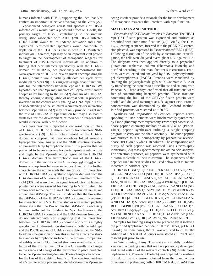
FIGURE 2: Schematic of the in vitro binding assay (18). Bindingreactions were performed in the following manner. Ten microgramsof GSTVpr or GST bound to glutathione sepharose was mixed with50 µg of biotinylated UBA(2) peptide and incubated at roomtemperature for 1 h. GST-containing complexes were selectivelyrecovered and incubated with streptavidin conjugated to horseradishperoxidase. The presence of bound biotinylated peptide wasdetermined by reading the absorbance at 450 nm after the additionof tetramethyl benzidine substrate solution (1-Step Turbo TMB-ELISA, Pierce Chemical Co.) and sulfuric acid.
FIGURE 3: (A) Amino acid sequences and alignment of UBA domains: HHR23A UBA(2),S. cereVisiaeRad23A UBA(2), c-cbl UBA, andHHR23A UBA(1). The amino acids that compriseR-helices 1, 2, and 3 are indicated above the sequence. The sequence of amino acids arecolored green (hydrophobic), red (negatively charged), orange (polar uncharged), and blue (positively charged). (B) Results of bindingassays with HHR23A UBA(2), HHR23A UBA(2) P333E,S. cereVisiaeRad23A UBA(2), c-cbl UBA, and HHR23A UBA(1). (C) Resultsof binding assays with HHR23A andS. cereVisiaeRad23 UBA(2) mutant peptides. Binding reactions were performed as described in theMaterials and Methods section. Data shown are values obtained from duplicate samples from a typical experiment.
14106 Biochemistry, Vol. 39, No. 46, 2000 Withers-Ward et al.
we predicted that the Pro residue at position 333 of HHR23AUBA(2) might be critical for interaction with Vpr. To assessthe contribution of this residue to the HHR23A UBA(2)/Vpr interaction, a mutant HHR23A UBA(2) peptide contain-ing a glutamic acid residue at position 333 [HHR23AUBA(2)P333E)] was chemically synthesized and assayed forbinding. This P333E substitution completely abolishedbinding of the peptide to GSTVpr (Figure 3, panel C). TheHHR23A UBA(1) domain contains a glutamic acid residueat the position corresponding to position 333 in the HHR23AUBA(2) domain, and the c-cbl UBA domain contains a serineat that position. These results indicate that the proline atposition 333 of HHR23A UBA(2) is necessary for interactionwith GSTVpr.
Further studies with mutant synthetic UBA(2) peptidesdemonstrate that the Pro at position 333 is critical for binding
but not sufficient on its own for the interaction of UBA(2)with GSTVpr. Neither an E370P substitution nor a370PES372
substitution made within theS. cereVisiae UBA(2) confersupon those peptides the ability to bind GSTVpr (Figure 3,panel C). These in vitro binding results demonstrate theimportance of the hydrophobic GFP-loop region for interac-tion of UBA(2) with Vpr.
Refined High-Resolution Structures of Wild-Type UBA-(2) of HHR23A.To further explore the role of the criticalP333 residue on binding of UBA(2) to Vpr, we undertookto determine the structure of UBA(2) P333E. During theassignment of this mutant protein using 2D proton NMR datarecorded from a synthetic peptide, we discovered a mismatchbetween the assignments for the wild-type UBA(2) (29) andthe mutant. To resolve the conflict, the structure of HHR23AUBA(2) was redetermined using heteronuclear NMR spec-
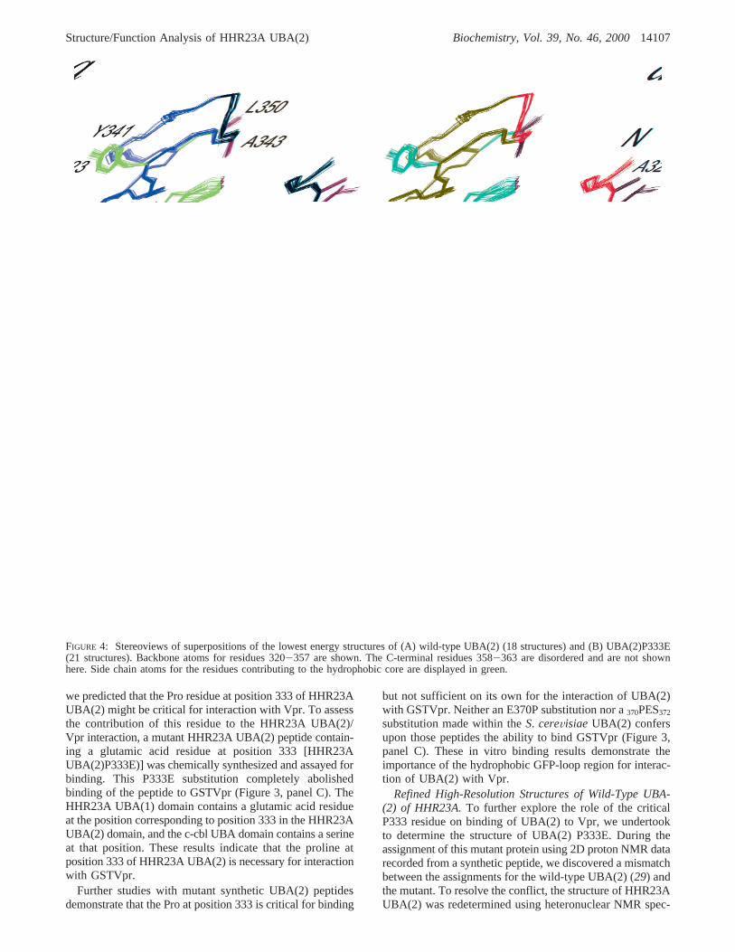
FIGURE 4: Stereoviews of superpositions of the lowest energy structures of (A) wild-type UBA(2) (18 structures) and (B) UBA(2)P333E(21 structures). Backbone atoms for residues 320-357 are shown. The C-terminal residues 358-363 are disordered and are not shownhere. Side chain atoms for the residues contributing to the hydrophobic core are displayed in green.
Structure/Function Analysis of HHR23A UBA(2) Biochemistry, Vol. 39, No. 46, 200014107
troscopy on a13C-,15N-labeled protein. The high resolutionstructure was necessary for the structure comparison, toanalyze the small structural changes that might be causedby the point mutation of Pro333 to Glu. For the preparationof labeled protein, the cDNA of UBA(2) encoding theresidues 319-363 was cloned into the expression vectorpGEX-2T. Analysis of the isotope-edited 3D data revealeda misassignment in the original data due to spectral overlap.Specifically, NOEs were incorrectly assigned between theside chain of Leu330 and the aromatic ring protons ofTyr341. The correct NOE connectivities are found to bebetween the side chains of Ile324 and Tyr341 as well asGlu348 and Tyr341. For the highly refined structure of thewild-type UBA(2) domain 779 NOE-derived distance re-straints (vs 380 in the previously reported structure) wereobtained. Simulated annealing calculations were employedto generate an ensemble of 18 structures (Figure 4, panelA) consistent with the NMR data. The structures exhibit goodcovalent geometry and have no NOE violations greater than0.5 Å. Figure 4, panel A, shows the quality of the calculatedstructures with a root-mean-square (rms) deviation betweenthe atomic coordinates of the backbone and all heavy atomswith respect to the average coordinates for the residues withinthe threeR-helices (residues 320-330, 337-344 and 350-357) of only 0.178( 0.037 Å and 0.686( 0.064 Å,respectively. Complete restraint and structural statistics arepresented in Table 1.
In the refined structure, the three helix bundle is morecompact and the orientation of the interhelical angles changedslightly (Figure 4, panel A). Interhelical angles weredetermined using the program RIBBONS2.6 (36). Helices1 and 2 are arranged at an angle of 120° (equivalent to-60°)and helices 2 and 3 exhibit an angle of 126° (or -54°). Thesevalues are close to the optimum of 52° reported for a “knobin hole” packing of the side chains. In contrast, theinterhelical angle between helices 1 and 3 is only about 30°.A tight “knob in hole” packing for the side chains of thesetwo helices is not required due to the larger distance betweenthem. The resulting change in the structure of the UBA(2)domain is mainly a shift of the firstR-helix of the three-helical bundle along its axis of about 3 Å. The orientationof the first helix with respect to the other two helices is onlyslightly affected (about 15°).
The structure of the UBA(2) domain is well-ordered fromresidues Glu320 to Ser357. In contrast to the low resolutionstructure of UBA(2) (29), a full set of NOEs of the typeHRiHâi+3 and HRiNHi+3, were found for residues Glu320 toLeu330, showing that the first helix is extended to theN-terminal end of the UBA(2) domain. The C-terminalresidues Asn359 to Asp363 show no medium or long rangeNOE connectivities and are disordered. Carbon and protonchemical shifts for these residues are similar to those foundfor random coil conformations. The first loop connectinghelices 1 and 2 is well-ordered, and the residues Gly331 toLeu336 show many NOE connectivities to residues locatedin the hydrophobic core. NOE connectivities for P333 clearlyindicate that the Pro residue adopts a trans conformation.The second loop, consisting of the residues Glu345 toAsn349, is far more exposed on the surface. Line broadeningfor several residues in this loop, namely Lys346 and Glu348,indicates that this loop is dynamic, likely due to slowchemical exchange processes.
One interesting feature of the UBA(2) domain is thepresence of large hydrophobic patches at the protein surface(Figure 5). Several hydrophobic residues that show contactswith the hydrophobic core are partially exposed on thesurface. The largest area is made up of residues Leu327,Ala329, and Leu330 in helix 1, residues Gly331, Phe332,Pro333, and Leu336 in the loop 1, and residues Ala351 andLeu355 in helix 3, covering a total of about 596 Å2 (epitope1). This corresponds to approximately 18% of the totalsolvent accessible surface area of 3288 Å2. A secondhydrophobic region (epitope 2) located on the opposite sideof the UBA(2) molecule consists of the residues Ala323,Ile324 (helix 1), Ile338, Phe341, Ala342 (helix2), Leu349(helix3), and Phe359 (C-terminus). The total surface area is555 Å2, but in contrast to the hydrophobic epitope 1, thisarea is not as compact and the residues do not form aconsecutive epitope. Such large hydrophobic surface patchesare rather unusual and as previously noted are likely bindingsites for other proteins. The site of interaction of HIV-1 Vprwith the UBA(2) domain was therefore suggested to occur
Table 1: Input Data for Structure Calculation and StructuralStatistics UBA(2) [18 Structures] and UBA(2)P333E [21 Structures]
Distance Restraints UBA(2) UBA(2)P333E
total 796 843NOE-derived
intraresidue 173 204sequential (|i - j| ) 1) 196 182medium-range (|i - j| e 4) 219 226long-range (|i - j| > 4) 191 214
hydrogen bondsmedium-range(within-helices)
17 17
Structure Statistics
RMSD from experimentaldistance restraints (Å)
0.047( 0.001 0.052( 0.0003
RMSD from idealized geometrybonds (Å) 0.005( 0.00004 0.005( 0.00005angles (deg) 0.616( 0.007 0.617( 0.004
energies (kcal mol-1)ENOE 11.7( 0.4 9.8( 0.5Ebond 17.1( 0.3 18.3( 0.4Eangles 74.9( 1.6 74.8( 1.0EL.-J.
a -262( 9 -270( 6RMSD from avg structure (Å)
backbone atoms insecondary structureb
0.18( 0.04 0.12( 0.03
backbone atoms forresidues 3-9
0.21( 0.06 0.16( 0.07
all heavy atoms insecondary structureb
0.69( 0.06 0.66( 0.08
all heavy atoms forresidues 3-39
0.72( 0.07 0.71( 0.07
Procheck analysisresidues in most favoredregion (%)
79.7 76.1
residues in additionalallowed region (%)
15.9 20.9
residues in generouslyallowed regions (%)
3.7 2.6
residues in disallowedregions (%)
0.8 0.4
a Lennart-Jones energy was calculated after the accepted structureswere subjected to a final minimization (50 steps Adopted RaphsonNewton algorithm, QUANTA98) using the CHARMM force field andthe NOE-derived distance restraints.b Secondary structure is composedof residues 3-12, 19-25, and 32-39. These residues are numbered321-330, 337-343, and 350-357 in HHR23A.
14108 Biochemistry, Vol. 39, No. 46, 2000 Withers-Ward et al.
between the hydrophobic region of Vpr and epitope 1.Epitope 2 is a potential site for other protein-proteininteractions in the XPC complex.
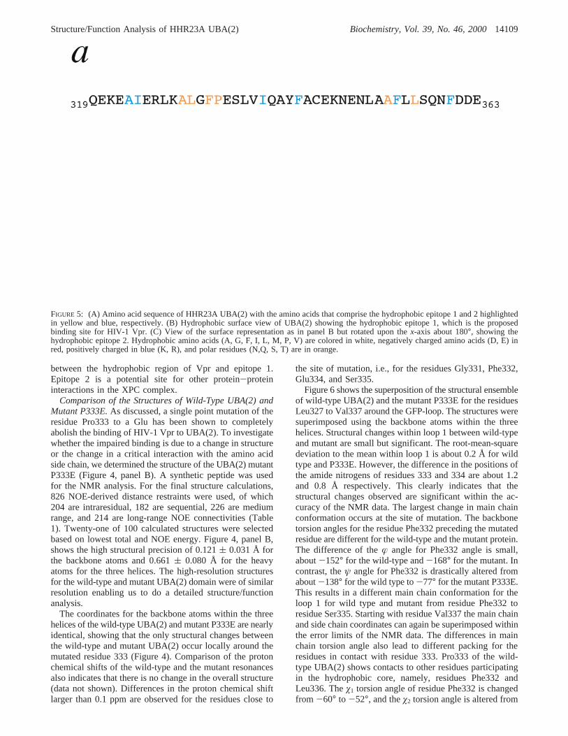
Comparison of the Structures of Wild-Type UBA(2) andMutant P333E.As discussed, a single point mutation of theresidue Pro333 to a Glu has been shown to completelyabolish the binding of HIV-1 Vpr to UBA(2). To investigatewhether the impaired binding is due to a change in structureor the change in a critical interaction with the amino acidside chain, we determined the structure of the UBA(2) mutantP333E (Figure 4, panel B). A synthetic peptide was usedfor the NMR analysis. For the final structure calculations,826 NOE-derived distance restraints were used, of which204 are intraresidual, 182 are sequential, 226 are mediumrange, and 214 are long-range NOE connectivities (Table1). Twenty-one of 100 calculated structures were selectedbased on lowest total and NOE energy. Figure 4, panel B,shows the high structural precision of 0.121( 0.031 Å forthe backbone atoms and 0.661( 0.080 Å for the heavyatoms for the three helices. The high-resolution structuresfor the wild-type and mutant UBA(2) domain were of similarresolution enabling us to do a detailed structure/functionanalysis.
The coordinates for the backbone atoms within the threehelices of the wild-type UBA(2) and mutant P333E are nearlyidentical, showing that the only structural changes betweenthe wild-type and mutant UBA(2) occur locally around themutated residue 333 (Figure 4). Comparison of the protonchemical shifts of the wild-type and the mutant resonancesalso indicates that there is no change in the overall structure(data not shown). Differences in the proton chemical shiftlarger than 0.1 ppm are observed for the residues close to
the site of mutation, i.e., for the residues Gly331, Phe332,Glu334, and Ser335.
Figure 6 shows the superposition of the structural ensembleof wild-type UBA(2) and the mutant P333E for the residuesLeu327 to Val337 around the GFP-loop. The structures weresuperimposed using the backbone atoms within the threehelices. Structural changes within loop 1 between wild-typeand mutant are small but significant. The root-mean-squaredeviation to the mean within loop 1 is about 0.2 Å for wildtype and P333E. However, the difference in the positions ofthe amide nitrogens of residues 333 and 334 are about 1.2and 0.8 Å respectively. This clearly indicates that thestructural changes observed are significant within the ac-curacy of the NMR data. The largest change in main chainconformation occurs at the site of mutation. The backbonetorsion angles for the residue Phe332 preceding the mutatedresidue are different for the wild-type and the mutant protein.The difference of theæ angle for Phe332 angle is small,about-152° for the wild-type and-168° for the mutant. Incontrast, theψ angle for Phe332 is drastically altered fromabout-138° for the wild type to-77° for the mutant P333E.This results in a different main chain conformation for theloop 1 for wild type and mutant from residue Phe332 toresidue Ser335. Starting with residue Val337 the main chainand side chain coordinates can again be superimposed withinthe error limits of the NMR data. The differences in mainchain torsion angle also lead to different packing for theresidues in contact with residue 333. Pro333 of the wild-type UBA(2) shows contacts to other residues participatingin the hydrophobic core, namely, residues Phe332 andLeu336. Theø1 torsion angle of residue Phe332 is changedfrom -60° to -52°, and theø2 torsion angle is altered from
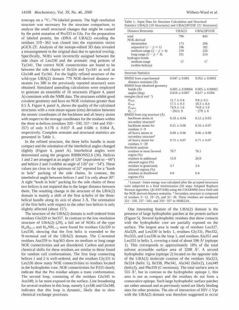
FIGURE 5: (A) Amino acid sequence of HHR23A UBA(2) with the amino acids that comprise the hydrophobic epitope 1 and 2 highlightedin yellow and blue, respectively. (B) Hydrophobic surface view of UBA(2) showing the hydrophobic epitope 1, which is the proposedbinding site for HIV-1 Vpr. (C) View of the surface representation as in panel B but rotated upon thex-axis about 180°, showing thehydrophobic epitope 2. Hydrophobic amino acids (A, G, F, I, L, M, P, V) are colored in white, negatively charged amino acids (D, E) inred, positively charged in blue (K, R), and polar residues (N,Q, S, T) are in orange.
Structure/Function Analysis of HHR23A UBA(2) Biochemistry, Vol. 39, No. 46, 200014109
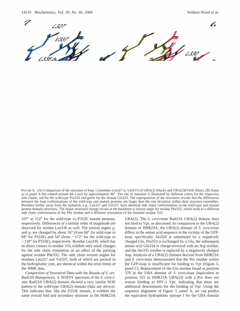
107° to 152° for the wild-type vs P333E mutant proteins,respectively. Differences of a similar order of magnitude areobserved for residue Leu336 as well. The torsion anglesø1
andø2 are changed by about 16° (from 68° for wild-type to84° for P333E) and 54° (from -172° for the wild-type to-118° for P333E), respectively. Residue Leu330, which hasno direct contact to residue 333, exhibits only small changesfor the side chain orientation as an effect of the packingagainst residue Phe332. The side chain torsion angles forresidues Leu327 and Val337, both of which are packed inthe hydrophobic core, are identical within the error limits ofthe NMR data.
Comparison of Structural Data with the Results of S. cer.Rad23A Mutagenesis.A NOESY spectrum of theS. cereVi-siaeRad23A UBA(2) domain showed a very similar NOEpattern to the wild-type UBA(2) domain (data not shown).This indicates that, like the P333E mutant, it exhibits thesame overall fold and secondary structure as the HHR23A
UBA(2). The S. cereVisiae Rad23A UBA(2) domain doesnot bind to Vpr, as discussed. In comparison to the UBA(2)domain of HHR23A, the UBA(2) domain ofS. cereVisiaediffers in the amino acid sequence in the vicinity of the GFP-loop, specifically Ala329 is substituted by a negativelycharged Glu, Pro333 is exchanged by a Glu, the subsequentamino acid Glu334 is charge-reversed with an Arg residue,and the Ser335 residue is replaced by a negatively chargedAsp. Analysis of a UBA(2) chimera derived from HHR23AandS. cereVisiae demonstrated that the Pro residue withinthe GFP-loop is insufficient for binding to Vpr (Figure 3,panel C). Replacement of the Glu residue found at position370 in the UBA domain ofS. cereVisiae [equivalent toposition 333 in HHR23A UBA(2)] with a Pro does notrestore binding to HIV-1 Vpr, indicating that there areadditional determinants for the binding of Vpr. Using thesequence alignment of Figure 3, panel A, we can predictthe equivalent hydrophobic epitope 1 for the UBA domain
FIGURE 6: (A) Comparison of the structure of loop 1 (residues Leu327 to Val337) of UBA(2) (black) and UBA(2)P333E (blue). (B) Sameas in panel A but rotated around thex-axis by approximately 90°. The site of mutation is illustrated by different colors for the respectiveside chains, red for the wild-type Pro333 and green for the mutant Glu333. The superposition of the structures reveals that the differencesbetween the loop conformations of the wild-type and mutant proteins are larger than the rms deviation within their structure ensembles.Residues farther away from the mutation, e.g., Leu327 and Val337, have identical side chain conformations in the wild-type and mutantprotein domain structures. The major structural change occurs at the backboneψ torsion angle for residue Phe332, which leads to a differentside chain conformation of the Phe residue and a different orientation of the mutated residue 333.
14110 Biochemistry, Vol. 39, No. 46, 2000 Withers-Ward et al.
of S. cereVisiae. This analysis shows that in addition to thechange to Glu at position 333, two other residues that arepart of the hydrophobic patch are different between the twoUBA domains, i.e., Ala329 and Leu356, which are replacedby Glu and Phe, respectively. This change of two smallhydrophobic residues for a charged and much larger aminoacid, respectively, is likely to have a significant effect onthe structure of the proposed interaction surface betweenUBA(2) and Vpr.
SUMMARY AND CONCLUSIONS
The UBA domains are found in a number of proteinsbesides HHR23A with diverse functions involved in ubiq-uitination, DNA repair, and signaling pathways via proteinkinases (16). One of the unusual features of the UBA(2)domain of HHR23A is the presence of large hydrophobicpatches at the protein surface. The existence of these largehydrophobic patches strongly suggests that these may serveas binding sites for other proteins. For HIV-1 Vpr, weconfirmed by site-directed mutagenesis that one of thehydrophobic patches encompassing Pro333 is critical for theinteraction between Vpr and UBA(2) (18). To understandthe structural basis of the loss in binding upon mutation ofPro 333 to Glu, we determined the structures of the wild-type UBA(2) domain and the mutant P333E to high resolu-tion. A comparison shows that while the overall fold of theUBA domain is not affected by the substitution, there aresignificant changes that occur locally around the site ofmutation. The high accuracy of the structures revealed thatsubstitution of a Pro residue 333 with a Glu resulted inchanges of the shape and charge of the hydrophobic patchpredicted to be the Vpr-interacting domain. These changesaccount for the loss of the ability to bind Vpr.
The structural and genetic information developed in thestudy should facilitate the identification and characterizationof cellular proteins that bind through UBA. One hypothesisthat can be addressed with the structural knowledge obtainedhere is that Vpr may compete or alter the interaction betweenUBA(2) and a normal cellular protein involved in DNArepair signaling processes. To date, despite their ubiquitousoccurrence, only two cellular binding partners for UBAdomains have been identified. Ubiquitin has been shown tobind to the UBA domain of p62, a ligand of the SH2 domainof p56lck (37). However, the very low sequence homologybetween the UBA domain of p62 and UBA(2) of HHR23Adoes not suggest a similar interaction between UBA(2) andubiquitin. Significantly for this work, HHR23A and B havebeen shown to interact with the 3-methyladenine DNAglycosylase (MPG) protein and have been proposed to serveas universal DNA damage recognition accessory proteins.Furthermore, MPG binds specifically to the UBA(2) ofHHR23A. It is therefore possible that HIV-1 Vpr maycompete with MPG for binding to HHR23A, thus affectingDNA nucleotide excision repair. This provides support forour hypothesis that the interaction of HIV-1 Vpr with thisdomain may be important to its functional role in mediatingcell cycle arrest.
ACKNOWLEDGMENT
We thank Dr. Thorsten Dieckmann for advice in the earlystages of this work. We thank Dr. Mark A. Jarosinski and
the Peptide Synthesis group at AmGen (Boulder, Co) forsynthesis of the peptides used in the binding assays and theUBA(2)P333E peptide used in the structural studies and Mr.Evan Feinstein for help in manuscript preparation.
REFERENCES
1. Lombaerts, M., Goeloe, J. I., den Dulk, H., Brandsma, J. A.,and Brouwer, J. (2000)Biochem. Biophys. Res. Commun. 268,210-215.
2. Watkins, J. F., Sung, P., Prakash, L., and Prakash, S. (1993)Mol. Cell. Biol. 13, 7757-7765.
3. Masutani, C., Sugasawa, K., Yanagisawa, J., Sonoyama, T.,Ui, M., Enomoto, T., Takio, K., Tanaka, K., Vanderspek, P.J., Bootsma, D., Hoeijmakers, J. H. J., and Hanaoka, F. (1994)EMBO J. 13, 1831-1843.
4. Madura, K., and Prakash, S. (1990)Nucleic Acids Res. 18,4737-4742.
5. Guzder, S. N., Habraken, Y., Sung, P., Prakash, L., andPrakash, S. (1995)J. Biol. Chem. 270, 12973-12976.
6. Sugasawa, K., Masutani, C., Uchida, A., Maekawa, T.,Vanderspek, P. J., Bootsma, D., Hoeijmakers, J. H. J., andHanaoka, F. (1996)Mol. Cell. Biol. 16, 4852-4861.
7. Masutani, C., Araki, M., Sugasawa, K., van der Spek, P. J.,Yamada, A., Uchida, A., Maekawa, T., Bootsma, D., Hoeij-makers, J. H., and Hanaoka, F. (1997)Mol. Cell Biol. 17,6915-6923.
8. Li, L., Lu, X. Y., Peterson, C., and Legerski, R. (1997)Mutat.Res. 383, 197-203.
9. Wang, Z. G., Wei, S. G., Reed, S. H., Wu, X. H., Svejstrup,J. Q., Feaver, W. J., Kornberg, R. D., and Friedberg, E. C.(1997)Mol. Cell. Biol. 17, 635-643.
10. Jansen, L. E. T., Verhage, R. A., and Brouwer, J. A. (1998)J. Biol. Chem. 273, 33111-33114.
11. Guzder, S. N., Sung, P., Prakash, L., and Prakash, S. (1998)J. Biol. Chem. 273, 31541-31546.
12. Mueller, J. P., and Smerdon, M. J. (1996)Mol. Cell. Biol. 16,2361-2368.
13. Schauber, C., Chen, L., Tongaonkar, P., Vega, I., Lambertson,D., Potts, W., and Madura, K. (1998)Nature 391, 715-718.
14. Lambertson, D., Chen, L., and Madura, K. (1999)Genetics153, 69-79.
15. Hiyama, H., Yokoi, M., Masutani, C., Sugasawa, K., Maekawa,T., Tanaka, K., Hoeijmakers, J. H. J., and Hanaoka, F. (1999)J. Biol. Chem. 274, 28019-28025.
16. Hofmann, K., and Bucher, P. (1996)Trends Biochem. Sci. 21,172-173.
17. Miao, F., Bouziane, M., Dammann, R., Masutani, C., Hanaoka,F., Pfeifer, G. P., and O’Connor, T. R. (2000)J. Biol. Chem.275, 28433-28438.
18. Withers-Ward, E. S., Jowett, J. B. M., Stewart, S. A., Xie, Y.M., Garfinkel, A., Shibagaki, Y., Chow, S. A., Shah, N.,Hanaoka, F., Sawitz, D. G., Armstrong, R. W., Souza, L. M.,and Chen, I. S. Y. (1997)J. Virol. 71, 9732-9742.
19. Emerman, M. (1996)Curr. Biol. 6, 1096-1103.20. Jowett, J. B. M., Planelles, V., Poon, B., Shah, N. P., Chen,
M. L., and Chen, I. S. Y. (1995)J. Virol. 69, 6304-6313.21. He, J. L., Choe, S., Walker, R., Dimarzio, P., Morgan, D. O.,
and Landau, N. R. (1995)J. Virol. 69, 6705-6711.22. Re, F., Braaten, D., Franke, E. K., and Luban, J. (1995)J.
Virol. 69, 6859-6864.23. Rogel, M. E., Wu, L. I., and Emerman, M. (1995)J. Virol.
69, 882-888.24. Stewart, S. A., Poon, B., Jowett, J. B. M., and Chen, I. S. Y.
(1997)J. Virol. 71, 5579-5592.25. Mahalingam, S., Ayyavoo, V., Patel, M., KieberEmmons, T.,
and Weiner, D. B. (1997)J. Virol. 71, 6339-6347.26. Di Marzio, P., Choe, S., Ebright, M., Knoblauch, R., and
Landau, N. R. (1995)J. Virol. 69, 7909-7916.27. Goh, W. C., Rogel, M. E., Kinsey, C. M., Michael, S. F., Fultz,
P. N., Nowak, M. A., Hahn, B. H., and Emerman, M. (1998)Nat. Med. 4, 65-71.
Structure/Function Analysis of HHR23A UBA(2) Biochemistry, Vol. 39, No. 46, 200014111
28. Poon, B., Grovit-Ferbas, K., Stewart, S. A., and Chen, I. S.Y. (1998)Science 281, 266-269.
29. Dieckmann, T., Withers-Ward, E. S., Jarosinski, M. A., Liu,C. F., Chen, I. S. Y., and Feigon, J. (1998)Nat. Struct. Biol.5, 1042-1047.
30. Blake, T. J., Shapiro, M., Morse, H. C., and Langdon, W. Y.(1991)Oncogene 6, 653-657.
31. Cavanagh, J., Fairbrother, W. J., Palmer, A. G., III, andSkelton, N. J. (1996)Protein NMR Spectroscopy: Principlesand Practice, Academic Press, Inc., San Diego.
32. Bartels, C., Xia, T. H., Billeter, M., Guntert, P., and Wuthrich,K. (1995)J. Biomol. NMR 6, 1-10.
33. Glaser, R. W., and Wu¨thrich, K. (1997) http://www.mol.bio-l.ethz.ch/wuthrich/software/spscan/.
34. Guntert, P., Mumenthaler, C., and Wuthrich, K. (1997)J. Mol.Biol. 273, 283-298.
35. Brunger, A. T. (1992)X-PLOR (Version 3.1) Manual, YaleUniversity Press, New Haven and London.
36. Carson, M. (1991)J. App. Crystal. 24, 958-961.37. Vadlamudi, R. K., Joung, I., Strominger, J. L., and Shin, J.
(1996)J. Biol. Chem. 271, 20235-20237.
BI0017071
14112 Biochemistry, Vol. 39, No. 46, 2000 Withers-Ward et al.