Point Mutations in the HIV1 Matrix Protein Turn Off the Myristyl Switch
21
Point mutations in the HIV-1 matrix protein turn off the myristyl switch Jamil S. Saad 1 , Erin Loeliger 1 , Paz Luncsford 1 , Mellisa Liriano 1 , Janet Tai 1 , Andrew Kim 1 , Jaime Miller 1 , Anjali Joshi 2 , Eric O. Freed 2 , and Michael F. Summers 1,* 1 Howard Hughes Medical Institute and Department of Chemistry and Biochemistry, University of Maryland Baltimore County, 1000 Hilltop Circle, Baltimore, Maryland 21250 USA 2 Virus-Cell Interaction Section, HIV Drug Resistance Program, National Cancer Institute, Frederick, MD 21702-1201 Abstract During the late phase of HIV-1 replication, newly synthesized retroviral Gag proteins are targeted to lipid raft regions of specific cellular membranes, where they assemble and bud to form new virus particles. Gag binds preferentially to the plasma membrane (PM) of most hematopoietic cell types, a process mediated by interactions between the cellular PM marker phosphatidylinositol-(4,5)- bisphosphate (PI(4,5)P 2 ) and Gag’s N-terminally-myristoylated matrix (MA) domain. We recently demonstrated that PI(4,5)P 2 binds to a conserved cleft on MA and promotes myristate exposure, suggesting a role as both a direct membrane anchor and myristyl switch trigger. Here we show that PI(4,5)P 2 is also capable of binding to MA proteins containing point mutations that inhibit membrane binding in vitro, and in vivo, including V7R, L8A and L8I. However, these mutants do not exhibit PI(4,5)P 2 - or concentration-dependent myristate exposure. NMR studies of V7R and L8A MA reveal minor structural changes that appear to be responsible for stabilizing the myristate-sequestered (myr (s)) species and inhibiting exposure. Unexpectedly, the myristyl group of a revertant mutant with normal PM targeting properties (V7R,L21K) is also tightly sequestered and insensitive to PI(4,5) P 2 binding. This mutant binds PI(4,5)P 2 with two-fold higher affinity compared with the native protein, suggesting a potential compensatory mechanism for membrane binding. Keywords Human immunodeficiency virus type-1 (HIV-1); Gag; myristyl (myr); matrix (MA); phosphatidylinositol-4,5-bisphosphate (PI(4,5)P 2 ); analytical ultracentrifugation (AU); nuclear magnetic resonance (NMR) Introduction The assembly of retroviruses in infected cells proceeds by complex, multi-stepped mechanisms that are only beginning to become understood. The major structural constituent of retroviruses is called Gag, a multi-domain protein that is post-translationally acylated (acetylated in the alpharetroviruses and myristoylated in most other retroviruses). Several thousand copies of Gag assemble to form a single virus particle, which is enveloped by a lipid bilayer derived * Corresponding author: Phone: (410)-455-2527; FAX: (410)-455-1174; Email: [email protected]. Publisher's Disclaimer: This is a PDF file of an unedited manuscript that has been accepted for publication. As a service to our customers we are providing this early version of the manuscript. The manuscript will undergo copyediting, typesetting, and review of the resulting proof before it is published in its final citable form. Please note that during the production process errors may be discovered which could affect the content, and all legal disclaimers that apply to the journal pertain. NIH Public Access Author Manuscript J Mol Biol. Author manuscript; available in PMC 2008 February 16. Published in final edited form as: J Mol Biol. 2007 February 16; 366(2): 574–585. NIH-PA Author Manuscript NIH-PA Author Manuscript NIH-PA Author Manuscript
-
Upload
independent -
Category
Documents
-
view
0 -
download
0
Transcript of Point Mutations in the HIV1 Matrix Protein Turn Off the Myristyl Switch

Point mutations in the HIV-1 matrix protein turn off the myristylswitch
Jamil S. Saad1, Erin Loeliger1, Paz Luncsford1, Mellisa Liriano1, Janet Tai1, Andrew Kim1,Jaime Miller1, Anjali Joshi2, Eric O. Freed2, and Michael F. Summers1,*
1 Howard Hughes Medical Institute and Department of Chemistry and Biochemistry, University of MarylandBaltimore County, 1000 Hilltop Circle, Baltimore, Maryland 21250 USA
2 Virus-Cell Interaction Section, HIV Drug Resistance Program, National Cancer Institute, Frederick, MD21702-1201
AbstractDuring the late phase of HIV-1 replication, newly synthesized retroviral Gag proteins are targetedto lipid raft regions of specific cellular membranes, where they assemble and bud to form new virusparticles. Gag binds preferentially to the plasma membrane (PM) of most hematopoietic cell types,a process mediated by interactions between the cellular PM marker phosphatidylinositol-(4,5)-bisphosphate (PI(4,5)P2) and Gag’s N-terminally-myristoylated matrix (MA) domain. We recentlydemonstrated that PI(4,5)P2 binds to a conserved cleft on MA and promotes myristate exposure,suggesting a role as both a direct membrane anchor and myristyl switch trigger. Here we show thatPI(4,5)P2 is also capable of binding to MA proteins containing point mutations that inhibit membranebinding in vitro, and in vivo, including V7R, L8A and L8I. However, these mutants do not exhibitPI(4,5)P2- or concentration-dependent myristate exposure. NMR studies of V7R and L8A MA revealminor structural changes that appear to be responsible for stabilizing the myristate-sequestered (myr(s)) species and inhibiting exposure. Unexpectedly, the myristyl group of a revertant mutant withnormal PM targeting properties (V7R,L21K) is also tightly sequestered and insensitive to PI(4,5)P2 binding. This mutant binds PI(4,5)P2 with two-fold higher affinity compared with the nativeprotein, suggesting a potential compensatory mechanism for membrane binding.
KeywordsHuman immunodeficiency virus type-1 (HIV-1); Gag; myristyl (myr); matrix (MA);phosphatidylinositol-4,5-bisphosphate (PI(4,5)P2); analytical ultracentrifugation (AU); nuclearmagnetic resonance (NMR)
IntroductionThe assembly of retroviruses in infected cells proceeds by complex, multi-stepped mechanismsthat are only beginning to become understood. The major structural constituent of retrovirusesis called Gag, a multi-domain protein that is post-translationally acylated (acetylated in thealpharetroviruses and myristoylated in most other retroviruses). Several thousand copies ofGag assemble to form a single virus particle, which is enveloped by a lipid bilayer derived
* Corresponding author: Phone: (410)-455-2527; FAX: (410)-455-1174; Email: [email protected]'s Disclaimer: This is a PDF file of an unedited manuscript that has been accepted for publication. As a service to our customerswe are providing this early version of the manuscript. The manuscript will undergo copyediting, typesetting, and review of the resultingproof before it is published in its final citable form. Please note that during the production process errors may be discovered which couldaffect the content, and all legal disclaimers that apply to the journal pertain.
NIH Public AccessAuthor ManuscriptJ Mol Biol. Author manuscript; available in PMC 2008 February 16.
Published in final edited form as:J Mol Biol. 2007 February 16; 366(2): 574–585.
NIH
-PA Author Manuscript
NIH
-PA Author Manuscript
NIH
-PA Author Manuscript
from the host cell. Although the Gag proteins of the betaretroviruses assemble in the cytoplasmprior to budding from the plasma membrane1, the Gag proteins of most other retroviruses(including the human immunodeficiency virus type-1, HIV-1) assemble at cellular membranesand require the assistance of host factors associated with the endosomal protein sortingmachinery2,3. In most hematopoetic cell types, the HIV-1 Gag proteins assemble and bud fromthe plasma membrane (PM)4,5. Some studies suggest that assembly in macrophages occursmainly at the membranes of late endosomes and/or multivesicular bodies (MVBs)6–8, althoughthe significance of endosomal assembly has recently been questioned9. The Gag proteins co-localize at punctate sites10,11, and analysis of the lipid and lipid-bound protein componentsof isolated virions indicates that the viral membranes are derived from lipid raft microdomains.Lipid rafts are enriched with cholesterol and sphingolipids with saturated fatty acid chains, andform liquid-ordered membrane structures that serve as preferential binding sites for specificcellular proteins. Several animal viruses are known to associate with rafts upon cellular entryand/or egress11.
The trafficking of Gag to membrane assembly sites is mediated by the protein’s N-terminalmatrix (MA) domain. Tight binding of the HIV-1 Gag protein to the PM is driven by a bipartitesignal that constitutes a myristyl group (myr) and a cluster of basic residues on the surface ofthe MA protein 12,13. Mutations and/or deletions within the first 6 residues (Gly-2 to Ser-6;myristate group is residue number 1) of MA that affect Gag myristylation 14–18, or within thepolybasic region of MA 5,12,19,20, impair membrane binding in vitro and can retarget Gag tothe cytoplasm and/or intracellular compartments in vivo. In addition, there is considerableevidence that the affinity of MA binding for membranes is significantly lower than that of theintact Gag molecules, which led to the suggestion that Gag binding may be mediated by amyristyl switch mechanism.21,22 In addition to promoting membrane binding, myristateexposure appears to be required for targeting Gag to lipid rafts. Resh and co-workers showedthat substitution of the saturated myristyl group of HIV-1 Gag by unsaturated lipids reducesthe affinity of Gag for rafts, but not for membranes in general, leading to particle assemblydefect.23
Mutagenesis studies have identified residues in the N-terminus of HIV-1 MA that are criticalfor proper membrane selection, including point mutations such as V7R, L8A and L8I. 19,24–26 These substitutions give rise to a phenotype similar to that observed for the unmyristoylatedprotein, although they do not block myristoylation. They severely inhibit virus assembly andrelease, cause significant reductions in PM binding, and lead to significant increases incytosolic localization of Gag. 19,24–26 Interestingly, the assembly-defective phenotypeimposed by the V7R mutation is reversed by second-site compensatory mutations elsewherein the MA domain. Biochemical studies have shown that L21K and K98E mutations reversedthe membrane-binding defect caused by V7R and significantly increased Gag binding to thePM. 24 These findings suggested that mutations at positions 7 and 8 may block myristateexposure, and that compensatory substitutions at positions 21 and 98 might restore exposure.
NMR studies confirmed that the HIV-1 MA myristyl group is capable of adopting sequestered(myr(s)) and exposed (myr(e)) conformations, and demonstrated that myristate exposure canbe promoted by factors that increase protein self-association27. More recently, the myristylswitch equilibrium was shown to be sensitive to binding by phosphatidyl-4,5-bisphosphate (PI(4,5)P2), a cellular factor that serves as a membrane marker for proteins that need to associatespecifically with the plasma membrane28–31. PI(4,5)P2 plays a major role in directing HIV-1Gag to the PM32, and appears to function by acting as both a trigger for myristate exposureand as a direct membrane anchor31. To determine the mechanism(s) by which the position-7and –8 mutations affect in vivo Gag localization and virus assembly, we conducted NMR andthermodynamic studies on the HIV-1 MA mutants V7R, L8A, L8I, L21K, and V7R/L21K(Figure 1), and studied interactions between these mutants and PI(4,5)P2. Our findings reveal
Saad et al. Page 2
J Mol Biol. Author manuscript; available in PMC 2008 February 16.
NIH
-PA Author Manuscript
NIH
-PA Author Manuscript
NIH
-PA Author Manuscript
that the single-residue mutations do not inhibit PI(4,5)P2 binding, but do inhibit PI(4,5)P2- andconcentration-dependent myristate exposure and suggest a novel potential compensatorymechanism for membrane selection and binding by the V7R/L21K revertant mutant.
ResultsV7R, L8A and L8I point mutations inhibit myristate exposure and protein self-association
Single point mutations in the HIV-1 MA protein (V7R, L8A and L8I) were made by site-directed mutagenesis of a co-expression vector harboring the yeast N-terminal myristyltransferase (yNMT) and HIV-1 MA gene. 27 These substitutions did not adversely affectmyristylation efficiencies (~100%, compared to ~90/10% myr/myr(-) for the wild-type MAprotein; Mmeas:Mcalc = 15801.4:15801.7, 15701.9:15702.5, and 15744.9:15744.6 for V7R,L8A and L8I MA, respectively). 1H and 15N NMR signals observed in two dimensional (2D)[1H–15N] HSQC spectra obtained for V7R, L8A and L8I were insensitive to concentrationover the range of 50 μM to 0.9 mM (Figure 1). In contrast, for the wild type (WT) myrMAprotein, a subset of signals shift progressively toward the frequencies observed for myr(-)MAupon increasing the protein concentration due to a concomitant shift in the monomer-trimerequilibrium toward the trimeric species and exposure of the myristyl group.27 The absence ofconcentration-dependent shifts for V7R, L8A, and L8I indicates that these mutants exist in aunique conformation over the range of concentrations tested. Sedimentation equilibrium (SE)data obtained for V7R, L8A and L8I are similar in appearance to SE data obtained for myr(-)MA, and confirm that the myrMA mutants remain monomeric at concentrations as high as 1mM.
NMR analysis of V7R-, L8A- and L8I-myrMATo elucidate the conformational changes that occur upon substituting a single amino acid inthe N-terminus of myrMA, a combination of 15N-, 13C-, 15N/13C-, 13C/13C- and 13C-edited/12C-double-half-filtered NOE experiments were collected for V7R, L8A, and L8Iproteins. In all spectra obtained for the three mutants, minor changes were observed in thepattern of NMR shifts and intensities for residues located in close proximity to the mutationsite (Ser-6 – Gly-10). For residues 11–132, no new intraprotein NOEs were detected that wouldbe indicative of an altered protein conformation. Representative portions of the 4D 15N,13C-edietd NOE data obtained for V7R are presented in Figure 2. NOE cross-peaks between proteinresidues and myristate group are not observed in this dataset because the myristate group isnot 13C-labeled. However, 2D NOESY, 3D 13C-edited NOE, and 3D 13C-edited/12C-double-half-filtered NOE data obtained for V7R, L8A, and L8I show numerous unambiguous NOEsbetween the myristate group and the side chains of Val-/Arg-7, Leu-/Ala-/Ile-8, Trp-16, ILe-34,Val-35, Ser-38, Gly-49, Leu-51, Glu-52, and Leu-85 (Figure 2 and 3). For V7R, NOEs betweenthe myristate group and both Arg-7 (Hβ2/3, Hγ2/3, and Hδ2/3) and Leu-8 (Hβ2/3, Hγ, andHδ1/2) side chains are clearly observed in the 3D 13C-edited/12C-double-half-filtered NOEdata (Figure 2), indicating that the myristate group is sequestered and that the side chains ofArg-7 and Leu-8 are packed against the myristate group. Similar NOEs between the myristategroup and side chains of Val-7 and Ala/Ile-8 were also observed for L8A and L8I (Figure 3).NOEs between the myristate group and the side chains of Val-7 and Leu-8 were also observedfor the WT myrMA protein. 27 These NMR observations indicate that mutation of Arg-7 andLeu-8 does not greatly alter the conformation of the loop and that residues 7 and 8 play animportant role into the stabilization of the myr(s) form.
For V7R and L8I, the terminal methyl group (myr-C14H3) exhibited an upfield chemical shiftcomparable to that observed for the WT myrMA protein (~0.5 ppm). 27 Structural analysis ofthe WT myrMA protein revealed that the terminal myr-C14H3 group packs against the side
Saad et al. Page 3
J Mol Biol. Author manuscript; available in PMC 2008 February 16.
NIH
-PA Author Manuscript
NIH
-PA Author Manuscript
NIH
-PA Author Manuscript
chain of Trp-16. For L8A, the myr-C14H3 group exhibited a larger upfield shift (~0.3 ppm),indicating that the methyl group is packed even closer to the aromatic ring of Trp-16.
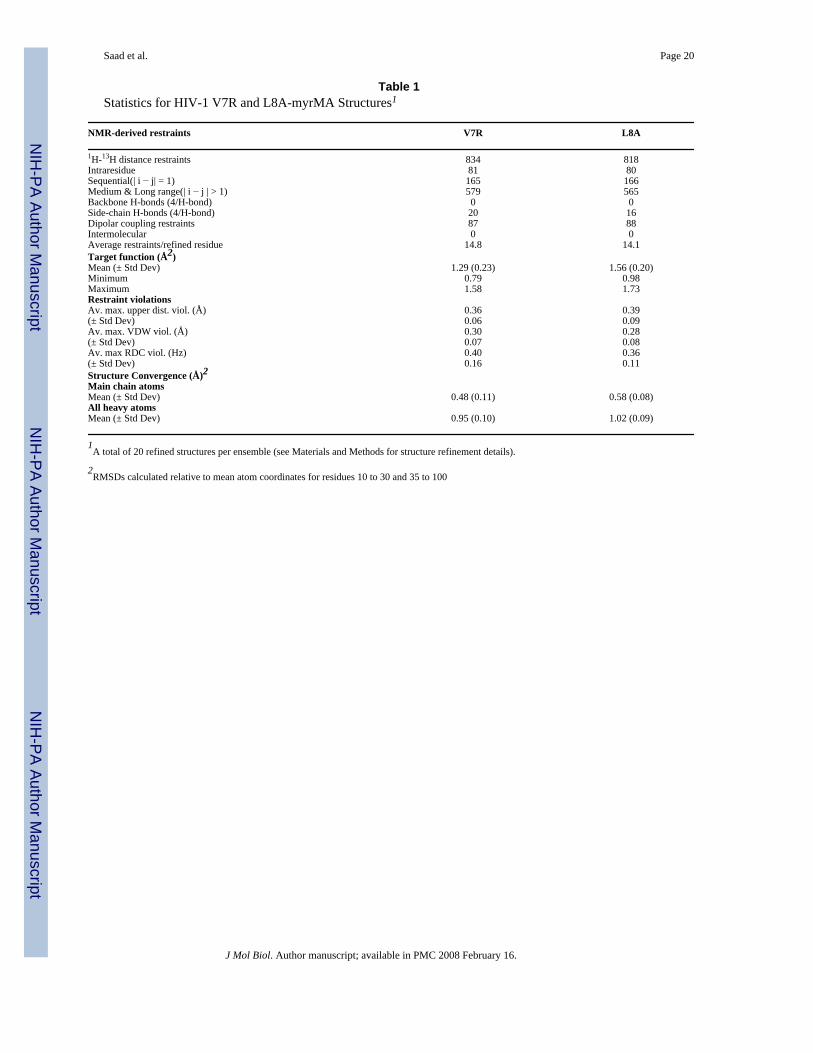
Structures of V7R- and L8A-myrMAWe chose V7R and L8A proteins as representative models to study the structural changes thatled to the stabilization of myristyl-sequestered form. Superpositions of 20 structures for V7Rand L8A generated from NMR data (500–800 μM and 35°C) are shown in Figure 4. Thebackbone atoms of Val-7–Ile-104 are well defined and are very similar to the coordinates ofthe WT myrMA protein (Figure 5 and Table 1),27,31 indicating that single mutations in theN-terminal loop had no effect on the globular fold of the protein. The myristyl group adoptsan extended conformation with the myr-C14H3 group packing in close proximity to the sidechains of core residues Trp-16, Ile-34, and Leu-85, and other myristate methylene groupsinteracting with the side chains of Ala-5, Ser-6, Val-7, Ile-34, Ser-38, Pro-48, Val-35, Leu-51,and Glu-52. In all spectra obtained, NOEs to the myristate were observed, but no newintraprotein NOEs were detected that would be indicative of an altered protein conformation.In addition, the relative NOE intensities and cross-peak patterns matched the data obtained forthe WT myr(s)MA protein. 27,31
The main differences between the structures are localized within the disordered loop formedby residues Gly-2 – Ser-9. For V7R, the side chain of Arg-7 is poised to make a salt bridgewith the side chain of Glu-52, and the mythelene groups of Arg-7 contribute to the hydrophobicinteractions with the methylene groups of the myristate (Figure 6). For L8A, the smaller sizeof Ala-8 creates a better cavity for the myristate group, which packs closer to Trp-16 asevidenced by the upfield shift of the terminal myr-C14H3 (Figure 6). Thus, structural changesimplicated by the chemical shift data involve only residues located near the mutation site, whichmay have only average intrinsic conformational mobility.
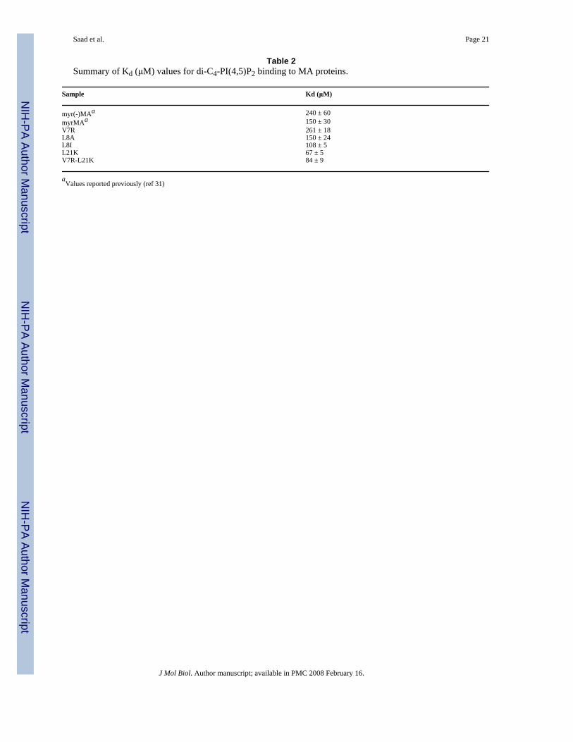
PI(4,5)P2 binds to myrMA mutants but does not trigger myristate exposureBinding studies were conducted with di-C4-phosphatidylinositol phosphate (di-C4-PI(4,5)P2), a soluble form of PI(4,5)P2 containing truncated lipids. Representative 1H-15N HSQCNMR data obtained upon titration of V7R with di-C4-PI(4,5)P2 are shown in Figure 7. Titrationof di-C4-PI(4,5)P2 led to significant changes in the backbone 1H and 15N NMR chemical shiftsof residues Arg-22, Lys-26, Lys-27, His-33, Glu-73, Glu-74, Leu-75 and Ser-77, (Group 1;ΔδHN ((Δδ1H)2+(Δδ15N)2)1/2 = 0.1–0.8 ppm) (Figure 7). These residues reside on the β-II-V cleft, and were previously shown to contribute to the PI(4,5)P2 binding site. Non-linear leastsquares fits of the titration data afforded a dissociation constant (Kd) value (261 ± 18 μM),similar to those observed for WT myr(-)MA and myrMA (Figure 7 and Table 2). Titration ofdi-C4-PI(4,5)P2 into L8A and L8I mutants also led to signal shifts and Kd values that are verysimilar to those obtained for V7R (Table 2). NMR data obtained for V7R (Figure 7), L8A, andL8I proteins confirmed that di-C4-PI(4,5)P2 binds to these mutants in a manner identical tothat observed for the WT myr(-) and myrMA proteins 31.
Interestingly, 1H and 15N NMR signal corresponding to residues Gly-2-Ser-9, Glu-12-Asp-15,Arg-39, and His 89 “group 2” did not exhibit detectable chemical shift changes upon titrationof V7R, L8A, and L8I with di-C4-PI(4,5)P2 (Figure 7). Chemical shift changes of “group 2”were observed for WT myrMA upon addition of di-C4-PI(4,5)P2 and NMR-based structuralstudies confirmed that they reflect di-C4-PI(4,5)P2-dependent exposure of the myristyl group.31 In addition, intermolecular NOEs between protein residues and myristate group are clearlyobserved in the 3D 13C-edited/12C-double-half-filtered NOE experiment obtained for theV7R:di-C4-PI(4,5)P2 complex (Figure 7), indicating that di-C4-PI(4,5)P2 binding does nottrigger myristyl exposure.
Saad et al. Page 4
J Mol Biol. Author manuscript; available in PMC 2008 February 16.
NIH
-PA Author Manuscript
NIH
-PA Author Manuscript
NIH
-PA Author Manuscript
Compensatory mutant V7R/L21K-myrMA binds PI(4,5)P2 with enhanced affinityTo understand how a compensatory mutation (Leu-21) reversed the membrane-binding defectimposed by V7R, we collected NMR and thermodynamic data for V7R/L21K and L21K proteinsamples. 2D [1H–15N] HSQC spectra obtained for L21K at concentrations of 50 – 900 μMshowed that as protein concentration is increased, several signals (e.g., those corresponding toSer 6, Gly 10, Gly 11) shifted progressively towards the corresponding frequencies of myr(-)MA. These signal shifts are similar to those observed for WT myrMA, and are indicative of ashift in equilibrium from myr(s) to myr(e) form. SE data obtained for L21K protein confirmthat the protein exists in a monomer-trimer equilibrium with Kassoc = 3.7 ± 0.4 × 10⁸ M−2 (datanot shown). Interestingly, [1H–15N] HSQC spectra for V7R/L21K show that signals wereinsensitive to protein concentration and SE data confirm that V7R/L21K is a monomer,indicating that a second mutation (L21K) does not trigger myristyl exposure.
Structural studies on the WT myrMA protein bound to PI(4,5)P2 revealed that the side chainsof Leu-21 play a major role in stabilizing the hydrophobic interactions with the 2’-acyl chainof PI(4,5)P2. Thus, we hypothesized that substitution of Leu-21 with lysine may affect thebinding affinity of PI(4,5)P2 to the MA protein. For L21K and V7R/L21K, 1H-15N HSQCNMR data obtained upon titration with di-C4-PI(4,5)P2 showed that “group 1” peaks exhibitedsimilar shifts to those observed for V7R, L8A and L8I as well as WT myr(-)MA and myrMA.Non-linear least squares fits of the titration data for L21K and V7R/L21K afforded bindingaffinity values (Figure 7 and Table 2), which are ~2-fold greater than WT myrMA. For L21K,group 2 signals also exhibited di-C4-PI(4,5)P2-dependent 1H-15N HSQC signals that shiftprogressively toward values observed for myr(-)MA. Analysis of a combinationof 15N-, 15N/13C- and 13C-edited/12C-double-half-filtered NOE experiments confirm that di-C4-PI(4,5)P2 binds to the II-V-β cleft on L21K and V7R/L21K in a manner very similar to theWT myrMA protein (Figure 7). Interestingly, for V7R/L21K the 1H-15N HSQC signalscorresponding to “group 2” residues did not exhibit chemical shift changes upon titration withdi-C4-PI(4,5)P2, indicating that although di-C4-PI(4,5)P2 binds to V7R/L21K with affinitysimilar to that observed for L21K, that binding does not trigger myristate exposure.
Point mutations near the amino-terminus of myrMA inhibit PM binding in vivoPrevious studies demonstrated that point mutations near the amino terminus of myrMA canlead to defects in membrane association. 19,24–26 We have now determined the in vivolocalization patterns of HIV-1 Gag proteins containing the following point mutations: L8E,L8V, S9A, S9E, V7R, and V7R/L21K (Figure 1). As expected from our membrane bindingstudies, all of the single mutants show an increase in hazy cytosolic staining relative to WT(Figure 8), consistent with their membrane-binding defect. This hazy, non-punctate stainingpattern is typical of HIV-1 Gag mutants that are defective in membrane binding33,34. ThisGag localization phenotype is distinct from that induced by mutations in the MA "80's domain”or the MA basic domain, which show retargeting to the MVB 5,12,19,35. Thus, consistent withearlier biochemical data24, these mutations appear to induce defects in membrane bindingrather than targeting. Interestingly, V7R/L21K displays a highly punctate, non-diffuse cell-surface Gag localization pattern, consistent with the ability of the L21K mutation to correctthe membrane-binding defect imposed by V7R24.
DiscussionPrevious in vivo mutagenesis studies have provided important insights into the mechanism ofretrovirus assembly. Mutations of the first five residues of the HIV-1 MA domain that inhibitmyristoylation lead to the accumulation of Gag in the cytosol, indicating that myristoylationis required for efficient membrane binding. Substitution of basic residues between Lys-18 andLys-32, which form a basic patch on the surface of MA, retargets Gag to a cytoplasmic
Saad et al. Page 5
J Mol Biol. Author manuscript; available in PMC 2008 February 16.
NIH
-PA Author Manuscript
NIH
-PA Author Manuscript
NIH
-PA Author Manuscript
compartment 5,12,19,35, and in vitro membrane binding studies support the hypothesis thatthe myristyl group and basic patch cooperatively facilitate membrane binding 5,12,19,35.Mutations at positions 7 and 8 of the MA domain (V7R, L8A, and L8I), which do not inhibitmyristoylation, have also been shown to inhibit membrane binding in cells, suggesting that themutations might inhibit myristate exposure. 19,24–26 The findings presented here demonstratethat these mutations inhibit PM localization in vivo and turn off the myristyl switch. Themyristyl switch equilibrium is sensitive to subtle molecular changes, and even the conservativesubstitution of Leu-8 by Ile is sufficient to block myristate exposure under conditions in whichexposure is normally highly favored. These studies further demonstrate that the mutations leadto only minor structural changes that are proximal to the mutation sites.
Recent studies indicate that PI(4,5)P2, a member of a family of differentially phosphorylatedphosphatidylinositides that serve as membrane markers for specific cellular proteins 28–30,plays a critical role in the ultimate localization of Gag at the PM.32 PI(4,5)P2 is normallyassociated with the inner leaflet of the plasma membrane 29, and depletion of PI(4,5)P2 leadsto accumulation of Gag at the membranes of late endosomes and MVBs 32. In addition,induction of PI(4,5)P2-enriched endosomes retargets Gag to endosomes and induces intra-vesicle budding 32. In both cases, virus production is severely attenuated32. PI(4,5)P2 bindsMA in an “extended lipid” conformation in which the inositol head group and the 2’-fatty acidchain bind to a hydrophobic cleft formed by helices II and V, and a meandering β-hairpin (β-II–V), and the remaining 1’-fatty acid and exposed myristyl group bracket a conserved basicsurface patch previously implicated in membrane binding36. These findings suggested to usthat the position-7 and –8 mutations, and the revertant mutants, might affect PI(4,5)P2 bindingor the sensitivity of PI(4,5)P2 binding on the myristyl switch. The present studies demonstratethat PI(4,5)P2 binds to V7R, L8A, and L8I with affinities similar to that observed for the nativeprotein. Furthermore, the NMR chemical shift and NOE data indicate these mutants bind PI(4,5)P2 in a manner that is essentially identical to that observed for the wild-type protein.However, PI(4,5)P2 binding does not trigger myristate exposure in the V7R, L8A and L8Imutants, indicating that the coupling between the PI(4,5)P2 binding site and the pocket thatsequesters the myristyl group is weak. The fact that PI(4,5)P2 binding is not attenuated by thesemutations suggests that membrane binding in vivo may be mediated by a tripartite mechanism,in which the exposed myristyl group, the extended PI(4,5)P2 bridge, and the basic patchcollectively contribute to binding.
The defects in virus assembly and membrane binding imposed by amino acid substitutions atpositions 7 and 8 can be reversed by intragenic second-site changes in the core of theprotein24–26. In particular, L21K and K98E reversed the phenotype imposed by V7R, whichled to the suggestion that these second mutations likely trigger myristate exposure to facilitatemembrane binding. The present studies unexpectedly reveal that substitution of Leu-21 by Lysdoes not promote myristate exposure in either the L21K or L21K/V7R MA proteins. Instead,these mutants bind PI(4,5)P2 with ~2-fold greater affinity than either the WT or V7R, L8A,and L8I mutants of the MA protein. It thus appears that this rather modest increase in bindingaffinity is sufficient to compensate for the inability of the protein to expose its myristyl group.
In related studies, the substitution of Val-7 and Leu-8 by glutamine (V7Q and L8Q) in thesimian immunodeficiency virus (SIV) MA protein was shown to dramatically inhibit virusproduction and membrane association without affecting myristoylation 37. HIV-1 and SIVMA proteins are highly homologous (~50% sequence identity and ~80% homologous), andthe three-dimensional structures of the unmyristoylated MA proteins solved by NMR and X-ray techniques are very similar 36,38,39. It is thus likely that the mechanism used by SIV toregulate membrane targeting and binding is similar to the mechanism employed by HIV-1.
Saad et al. Page 6
J Mol Biol. Author manuscript; available in PMC 2008 February 16.
NIH
-PA Author Manuscript
NIH
-PA Author Manuscript
NIH
-PA Author Manuscript
In summary, we have demonstrated that mutations near the amino-terminus of the HIV-1 MAprotein that inhibit membrane binding in vitro also lead to cytosolic localization of Gag invivo. This loss of membrane association is due to the inability of the mutant MA proteins toexpose their myristyl group. The myristyl switch equilibrium is highly sensitive to mutationsnear the N-terminus of the protein, and even the conservative substitution of Leu 8 by Ile issufficient to “lock off” the myristyl switch. The myristyl switch unexpectedly remains lockedoff in the revertant mutant V7R/L21K, which assembles at the PM and exhibits a wild-typephenotype. This behavior appears to result from enhanced interactions with PI(4,5)P2, whichbinds L21K- and V7R/L21K-MA with affinities that are about two-fold greater than that ofthe native MA protein. However, we cannot rule out the possibility that the increased chargeand location of the L21K substitution might also directly influence membrane binding, andfuture studies of the interactions between these proteins with membranes, including thosecontaining PI(4,5)P2, are warranted.
Materials and MethodsSample preparation
HIV-1 myrMA constructs containing amino acid changes V7R, L8I, L8A, L21K and V7R/L21K were made by using QuickChange XL site-directed mutagenesis kit (Stratagene, La Jolla,CA) and sequenced at the DNA sequencing facility, UMBC. Protein samples were preparedas described,27,38 except that a modified protocol was applied to prepare 15N-, and 13C-,15N-labeled samples.40 Cells were first grown in 4L LB media at 37°C until O.D.600 reached ~0.6– 0.7. Next, cells were spun down and washed with 1X M9 salt before transferring them to 1LM9 minimal media containing 15NH4Cl as the sole nitrogen source to produce 15N-labeledprotein. At this stage, cells were supplemented with myristic acid (10 mg/liter; Sigma) thenwere grown for 1h before induction with isopropyl -D-thiogalactoside (1 mM). Cells weregrown at 37°C for ~12 – 14h, lysed using microfluidizer, and the proteins purified by cobaltaffinity chromatography (Clontech, Mountain View, CA) and ion exchange columnchromatography (SP column). Traces of unmyristoylated species (for L21K) were separatedfrom myristoylated sample by butyl-Sepharose hydrophobicity chromatography (GEHealthcare, Pittsburg, PA). We obtained a high yield (~25 mg/L) of 93 – 97%isotopically 15N- and 15N/13C-enriched proteins. Molecular weights and efficiency ofmyristylation were confirmed by electrospray ionization mass spectrometry. Phosphoinositidedi-C4-PI(4,5)P2 (Echelon, Salt Lake City, UT) was obtained commercially and used withoutfurther purification. Samples for all NMR experiments were prepared in 50 mM sodiumphosphate at pH 5.5, 100 mM NaCl and 5 mM DTT. NaCl was excluded from the NMR bufferfor di-C4-PI(4,5)P2 titration experiments.
NMR spectroscopyNMR data were collected with Bruker DRX (800 MHz 1H) and DMX (600 MHz 1H)spectrometers equipped with cryoprobes, processed with NMRPIPE41 and analyzed withNMRVIEW42. A combination of two-, three- and four-dimensional NOESY data wereobtained for combinations of natural abundance, 15N- and 15N-,13C-labeled protein samples(35 °C). Protein backbone signals were assigned using standard triple resonance methods(HNCA and HNCOCA), and side chain signals were assigned from 3D and 4D 15N-, 13C-,and 15N/13C-edited NOESY data. Intermolecular 1H-1H NOEs between 15N-,13C-labeledprotein and unlabeled myristate group were assigned from 2D (1H-1H) and 3D (13C- and 13C-edited/12C-double-half-filtered) NOESY data (see references 43–45 and citations there-in). 1H-15N residual dipolar couplings were measured using a modified in-phase/anti-phase(IPAP) HSQC experiment 46 for V7R and L8A samples aligned in polyacrylamide gel 47.Binding isotherms from 1H-15N NMR HSQC titration experiments were calculated withORIGIN 7.0 software (MicoCal, Northampton, MA).
Saad et al. Page 7
J Mol Biol. Author manuscript; available in PMC 2008 February 16.
NIH
-PA Author Manuscript
NIH
-PA Author Manuscript
NIH
-PA Author Manuscript

Analytical ultracentrifugationSedimentation equilibrium measurements were collected on a Beckman XL-I Optima systemequipped with a four-hole An-60 rotor (Beckman Coulter) and cells were equipped withdouble-sectored centerpieces of path length 12 or 3 mm and quartz windows. Protein sampleswere prepared in 50 mM sodium phosphate buffer at pH 5.5 containing 100 mM NaCl and 2mM Tris (2-carboxy-ethyl)-phosphine-HCl. Loading concentrations varied from 50 to 150μM for all samples. Rotor speeds were at 22,000 and 26,000 rpm, and temperature was 20 °C.Scans were obtained by using the absorption optics system with 0.002 cm step size and fouraverages per point, and acquired at wavelengths of 280 and 295 nm. Partial specific volumes(v-bar) and molar extinction coefficients were calculated by using the program SEDNTERP(www.jphilo.mail-way.com) and buffer densities were measured pycnometrically. Dataanalysis was performed by using NONLIN.48 Equilibrium association constants weredetermined by global analysis of data acquired from samples prepared at two loadingconcentrations and centrifuged at two rotor speeds.
Structure calculationsStructures were calculated in torsion angle space with CYANA(http://www.las.jp/index_eg.html) starting from random initial angles. Upper interprotondistance bounds of 2.7, 3.3 and 5.0 Å (with appropriate corrections for pseudoatoms) wereemployed for NOE cross peaks of strong, medium, and weak intensity respectively, whichwere qualitatively determined following intensity normalization of the different NOE data sets.No backbone hydrogen bond or chemical shift-based torsion angle restraints were employed.Statistical information is provided in Table 1. Structure figures were generated with PyMOL(http://pymol.sourceforge.net).
Fluorescence microscopyConstruction of derivatives of the HIV-1 pNL4-3 clone containing MA mutations V7R, L8V,L8E, S9A, S9E and V7R/L21K has been described elsewhere.19,24,25 The numbering schemeof these mutations has been changed to be consistent with the scheme we used in earlierstructural studies. HeLa cells cultured in chamber slides (Nunc) were transfected with eitherwild type pNL4-3 or the indicated MA mutants in the pNL4-3 backbone using theLipofectamine 2000 reagent (Invitrogen, Carlsbad, CA). Cells were fixed 24–48 hrposttransfection with 3.7% formaldehyde in 100 mM sodium phosphate buffer (pH 7.2) for 20min, and permeabilized with phosphate-buffered saline (PBS) containing 0.1% Triton X-100.Gag protein in transfected cells was visualized by incubating with anti-p17 monoclonalantibody (Advanced Biotechnologioes Inc, Columbia, MD) diluted in PBS containing 3% BSAfollowed by Texas red-conjugated anti-mouse IgG. Cells were mounted with Aqua Poly Mount(Polysciences Inc, Warrington, PA) and examined using a Delta Vision RT microscope.
Atomic coordinatesThe atomic coordinates have been deposited in the Protein Data Bank, www.pdb.org, and areavailable under the accession codes 2JMG and 2NV3 for V7R and L8A, respectively.
Supplementary MaterialRefer to Web version on PubMed Central for supplementary material.
Acknowledgements
This work was supported by NIH grant AI30917. We thank the HHMI staff and colleagues at UMBC, Dr. DorothyBeckett (University of Maryland, College Park), and David King (HHMI, U.C. Berkeley) for technical support. Thisresearch was supported in part by the Intramural Research Program of the NIH, National Cancer Institute, Center for
Saad et al. Page 8
J Mol Biol. Author manuscript; available in PMC 2008 February 16.
NIH
-PA Author Manuscript
NIH
-PA Author Manuscript
NIH
-PA Author Manuscript
Cancer Research (EOF). P.J. and M.L. are Meyerhoff undergraduate scholars, and E.L. and J.T. are UMBC Presidentialand MARC undergraduate scholars, respectively.
References1. Coffin, JM.; Hughes, SH.; Varmus, HE. Retroviruses. Cold Spring Harbor Laboratory Press; Plainview,
N.Y.: 1997.2. Morita E, Sundquist WI. Retrovirus Budding. Annual Review of Cell and Develpomental Biology
2004;20:395–425.3. von Schwedler UK, Stuchell M, Muller B, Ward DM, Chung HY, Morita E, Wang HE, Davis T, He
GP, Cimbora DM, Scott A, Krausslich HG, Kaplan J, Morham SG, Sundquist WI. The protein networkof HIV budding. Cell 2003;114:701–713. [PubMed: 14505570]
4. Dong X, Li H, Derdowski A, Ding L, Burnett A, Chen X, Peters TR, Dermody TS, Woodruff E, WangJJ, Spearman P. AP-3 directs the intracellular trafficking of HIV-1 Gag and plays a key role in particleassembly. Cell 2005;120:663–674. [PubMed: 15766529]
5. Hermida-Matsumoto L, Resh MD. Localization of Human Immunodeficiency virus Type 1 Gag andenv at the Plasma Membrane by Confocal Imagine. Journal of Virology 2000;74:8670–8679.[PubMed: 10954568]
6. Nguyen DG, Booth A, Gould SJ, Hildreth JE. Evidence that HIV budding in primary macrophagesoccurs through the exosome release pathway. J Biol Chem 2003;278:52347–52354. [PubMed:14561735]
7. Ono A, Freed EO. Cell-Type-Dependent Tageting of Human Immunodeficiency Virus Type 1Assembly to the Plasma Membrane and the Multivesicular body. Journal of Virology 2004;78:1552–1563. [PubMed: 14722309]
8. Raposo G, Moore M, Innes D, Leijendekker R, Leigh-Brown A, Benaroch P, Geuze H. Humanmacrophages accumulate HIV-1 particles in MHC II compartments. Traffic 2002;3:718–729.[PubMed: 12230470]
9. Jouvenet, N.; Neil, SJD.; Bess, C.; Johnson, MC.; Virgen, CA.; Simon, SM.; Bieniasz, PD. PLoSBiology. 2006. Plasma membrane is the site of productive HIV-1 particle assembly. in press
10. Ono A, Freed EO. Plasma membrane rafts play a critical role in HIV-1 assembly and release. ProcNatl Acad Sci USA 2001;98:13925–13930. [PubMed: 11717449]
11. Ono A, Freed EO. Role of lipid rafts in virus replication. Adv Virus Res 2005;64:311–358. [PubMed:16139599]
12. Yuan X, Yu X, Lee TH, Essex M. Mutations in the N-Terminal Region of Human ImmunodeficiencyVirus Type I Matrix Protein Block Intracellular Transport of the Gag Precursor. Journal of Virology1993;67:6387–6394. [PubMed: 8411340]
13. Zhou W, Parent LJ, Wills JW, Resh MD. Identification of a Membrane-Binding Domain Within theAmino-Terminal Region of Human Immunodeficiency Virus Type 1 Gag Protein Which Interactswith Acidic Phospholipids. Journal of Virology 1994;68:2556–2569. [PubMed: 8139035]
14. Bryant M, Ratner L. Myristoylation-Dependent Replication and Assembly of HumanImmunodeficiency Virus 1. Proceedings of the National Academy of Sciences USA 1990;87:523–527.
15. Copeland NG, Jenkins NA, Nexo B, Schultz AM, Rein A, Mikkelsen T, Jorgensen P. Poorly expressedendogenous ecotropic provirus of DBA/2 mice encodes a mutant Pr65gag protein that is notmyristylated. J Virol 1988;62:479–487. [PubMed: 2826810]
16. Göttlinger HG, Sodroski JG, Haseltine WA. Role of capsid precursor processing and myristoylationin morphogenesis and infectivity of human immunodeficiency virus type 1. Proceedings of theNational Academy of Sciences USA 1989;86 :5781–5785.
17. Spearman P, Horton R, Ratner L, Kuli-Zade I. Membrane binding of human immunodeficiency virustype 1 matrix protein in vivo supports a conformational myristyl switch mechanism. J Virol1997;71:6582–6592. [PubMed: 9261380]
18. Spearman P, Wang JJ, Vander Heyden N, Ratner L. Identification of Human Immunodeficiency VirusType 1 Gag Protein Domains Essential to Membrane Binding and Particle Assembly. Journal ofVirology 1994;68:3232–3242. [PubMed: 8151785]
Saad et al. Page 9
J Mol Biol. Author manuscript; available in PMC 2008 February 16.
NIH
-PA Author Manuscript
NIH
-PA Author Manuscript
NIH
-PA Author Manuscript
19. Freed EO, Orenstein JM, Buckler-White AJ, Martin MA. Single Amino Acid Changes in the HumanImmunodeficiency Virus Type 1 Matrix Protein Block Virus Particle Production. Journal of Virology1994;68:5311–5320. [PubMed: 8035531]
20. Ono A, Orenstein JM, Freed EO. Role of the Gag Matrix Domain in Targeting HumanImmunodeficiency Virus Type 1 assembly. Journal of Virology 2000;74:2855–2866. [PubMed:10684302]
21. Bouamr F, Scarlata S, Carter CA. Role of myristylation in HIV-1 Gag assembly. Biochemistry2003;42:6408–6417. [PubMed: 12767222]
22. Zhou W, Resh MD. Differential membrane binding of the human immunodeficiency virus type 1matrix protein. J Virol 1996;70:8540–8548. [PubMed: 8970978]
23. Lindwasser OW, Resh MD. Myristoylation as a target for inhibiting HIV assembly: Unsaturated fattyacids block viral budding. Proc Natl Acad Sci USA 2002;99:13037–13042. [PubMed: 12244217]
24. Ono A, Freed EO. Binding of Human Immunodeficiency Virus Type 1 gag to membrane: Role of thematrix amino terminus. J Virol 1999;73:4136–4144. [PubMed: 10196310]
25. Ono A, Huang M, Freed EO. Characterization of human immunodeficiency virus type 1 matrixrevertants: effects on virus assembly, Gag processing, and Env incorporation into virions. J Virol1997;71:4409–4418. [PubMed: 9151831]
26. Paillart JC, Gottlinger HG. Opposing effects of human immunodeficiency virus type 1 matrixmutations support a myristyl switch model of Gag membrane targeting. J Virol 1999;73:2604–2612.[PubMed: 10074105]
27. Tang C, Loeliger E, Luncsford P, Kinde I, Beckett D, Summers MF. Entropic switch regulatesmyristate exposure in the HIV-1 matrix protein. Proc Natl Acad Sci USA 2004;101:517–522.[PubMed: 14699046]
28. Martin TFJ. PI(4,5)P2 regulation of surface membrane traffic. Curr Opin Cell Biol 2001;13:493–499.[PubMed: 11454457]
29. Behnia R, Munro S. Organelle identity and the signposts for membrane traffic. Nature 2005;438:597–604. [PubMed: 16319879]
30. McLaughlin S, Murray D. Plasma membrane phosphoinositide organization by protein electrostatics.Nature 2005;438:605–611. [PubMed: 16319880]
31. Saad JS, Miller J, Tai J, Kim A, Ghanam RH, Summers MF. Structural basis for targeting HIV-1 Gagproteins to the plasma membrane for virus assembly. Proc Natl Acad Sci USA 2006;103:11364–11369. [PubMed: 16840558]
32. Ono A, Ablan SD, Lockett SJ, Nagashima K, Freed EO. Phosphatidylinositol (4,5) bisphosphateregulates HIV-1 Gag targeting to the plasma membrane. Proceedings of the National Academy ofSciences 2004;101:14889–14894.
33. Joshi A, Nagashima K, Freed EO. Mutation of dileucine-like motifs in the human immunodeficiencyvirus type 1 capsid disrupts virus assembly, gag-gag interactions, gag-membrane binding, and virionmaturation. Journal of Virology 2006;80:7939–7951. [PubMed: 16873251]
34. Ono A, Demirov D, Freed EO. Relationship between human immunodeficiency virus Type-1 Gagmultimerization and membrane binding. J Virol 2000;74:5142–5150. [PubMed: 10799589]
35. Freed EO, Englund G, Martin AM. Role of the Basic Domain of Human Immondeficiency VirusType 1 Matrix in Macrophage Infection. Journal of Virology 1995;69:3949–3954. [PubMed:7745752]
36. Hill CP, Worthylake D, Bancroft DP, Christensen AM, Sundquist WI. Crystal Structures of theTrimeric HIV-1 Matrix Protein: Implications for Membrane Association. Proceedings of the NationalAcademy of Sciences USA 1996;93:3099–3104.
37. Gonzalez SA, Affranchino JL. Substitution of Leucine 8 in Simian Immunodeficiency Virus MatrixProtein Impairs Particle Formation Without Affecting N-Myristylation of the Gag Precursor.Virology 1998;240:27–35. [PubMed: 9448686]
38. Massiah MA, Starich MR, Paschall C, Summers MF, Christensen AM, Sundquist WI. Threedimensional structure of the human immunodeficiency virus type 1 matrix protein. J Mol Biol1994;244:198–223. [PubMed: 7966331]
39. Rao Z, Belyaev AS, Fry E, Roy P, Jones IM, Stuart DI. Crystal structure of SIV matrix antigen andimplications for virus assembly. Nature 1995;378:743–747. [PubMed: 7501025]
Saad et al. Page 10
J Mol Biol. Author manuscript; available in PMC 2008 February 16.
NIH
-PA Author Manuscript
NIH
-PA Author Manuscript
NIH
-PA Author Manuscript
40. Marley J, Lu M, Bracken C. A method for efficient isotopic labeling of recombinant proteins. Journalof Biomolecular NMR 2001;20:71–75. [PubMed: 11430757]
41. Delaglio F, Grzesiek S, Vuister GW, Zhu G, Pfeifer J, Bax A. NMRPipe: A multidimensional spectralprocessing system based on UNIX pipes. J Biomol NMR 1995;6:277–293. [PubMed: 8520220]
42. Johnson BA, Blevins RA. NMRview: a Computer Program for the Visualization and Analysis ofNMR Data. J Biomol NMR 1994;4:603–614.
43. Wüthrich, K. NMR of Proteins and Nucleic Acids. John Wiley & Sons; New York: 1986.44. Kay LE, Clore GM, Bax A, Gronenborn AM. Four-dimensional heteronuclear triple-resonance NMR
spectroscopy of interleukin-1β in solution. Science 1990;249:411–414. [PubMed: 2377896]45. Folkers PJM, Folmer RHA, Konings RNH, Hilbers CW. Overcoming the ambiguity problem
encountered in the analysis of nuclear Overhauser magnetic resonance spectra of symmetric dimerproteins. J Am Chem Soc 1993;115:3798–3799.
46. Ishii Y, Markus MA, Tycko R. Controlling residual dipolar couplings in high-resolution NMR ofproteins by strain induced alignment in a gel. J Biomol NMR 2001;21:141–151. [PubMed: 11727977]
47. Chou JJ, Gaemers S, Howder B, Louis JM, Bax A. A simple apparatus for generating strechedpolyacrylamide gels, yeilding uniform alignment of proteins and detergent micelles. J Biomol NMR2001;21:377–382. [PubMed: 11824758]
48. Johnson ML, Faunt LM. Parameter estimation by least-squares methods. Methods Enzymol1992;210:1–37. [PubMed: 1584035]
Abbreviations usedMA
myristoylated HIV-1 matrix protein
Gag myristoylated HIV-1 Gag polyprotein
myr(-) unmyristoylated
myr(s) myristate-sequestered state
myr(e) myristate-exposed state
PI(4,5)P2 phosphatidylinositol-(4,5)-bisphosphate
NMR nuclear magnetic resonance
HSQC heteronuclear single quantum coherence
AU analytical ultracentrifugation
RDC residual dipolar coupling
Saad et al. Page 11
J Mol Biol. Author manuscript; available in PMC 2008 February 16.
NIH
-PA Author Manuscript
NIH
-PA Author Manuscript
NIH
-PA Author Manuscript
Figure 1.(a) A representation of the MA sequence showing positions of the mutations in red. (b) Overlayof 2D 1H-15N HSQC spectra collected at different concentrations [600–900 μM (black), 300μM red, 150 μM (blue), 50 μM (magenta)] for V7R, L8A, and L8I samples. (c) Representativesedimentation profiles obtained for myr(-)MA, WT and mutant myrMA proteins [(26,000 rpm,20°C, 100 μM)]. For WT myrMA, best fits were obtained for a monomer-trimer equilibriumaffording association constant (Kassoc) of 2.5 ± 0.6 × 10⁸ M−2.27 Sedimentation equilibriumcurves for myr(-)MA, V7R, L8A, and L8I fit best to monomeric species.
Saad et al. Page 12
J Mol Biol. Author manuscript; available in PMC 2008 February 16.
NIH
-PA Author Manuscript
NIH
-PA Author Manuscript
NIH
-PA Author Manuscript
Figure 2.Representative NMR data obtained for V7R. (a) 4D 15N,13C-edited NOE data showingintramolecular and intermolecular NOEs for residues Arg-7 (left) and Leu-8 (right). NOEcross-peaks to the myristate group are not observed because the myristate group is not 13C-labeled. (b) 13C-edited/12C-double-half-filtered NOE data showing unambiguously assignedintermolecular NOEs between unlabeled myristyl group and 13C-labeled protein. Solid linesdenote 1H-12C breakthrough doublets NOE peaks.
Saad et al. Page 13
J Mol Biol. Author manuscript; available in PMC 2008 February 16.
NIH
-PA Author Manuscript
NIH
-PA Author Manuscript
NIH
-PA Author Manuscript
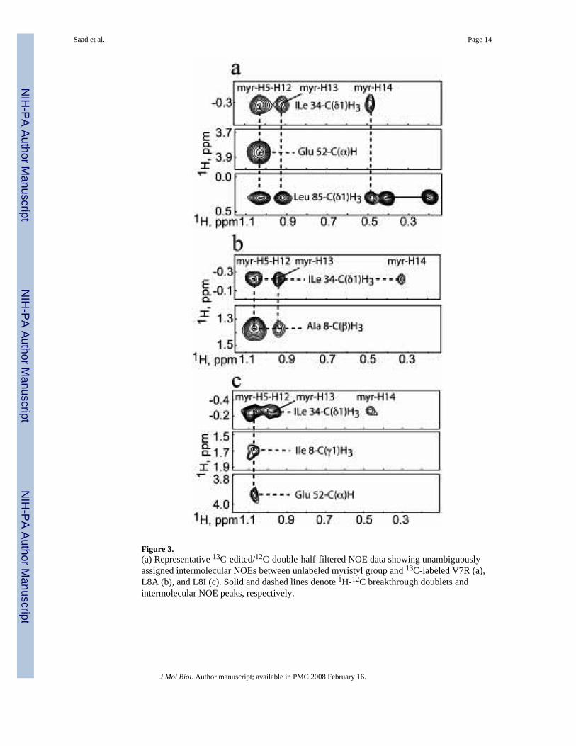
Figure 3.(a) Representative 13C-edited/12C-double-half-filtered NOE data showing unambiguouslyassigned intermolecular NOEs between unlabeled myristyl group and 13C-labeled V7R (a),L8A (b), and L8I (c). Solid and dashed lines denote 1H-12C breakthrough doublets andintermolecular NOE peaks, respectively.
Saad et al. Page 14
J Mol Biol. Author manuscript; available in PMC 2008 February 16.
NIH
-PA Author Manuscript
NIH
-PA Author Manuscript
NIH
-PA Author Manuscript
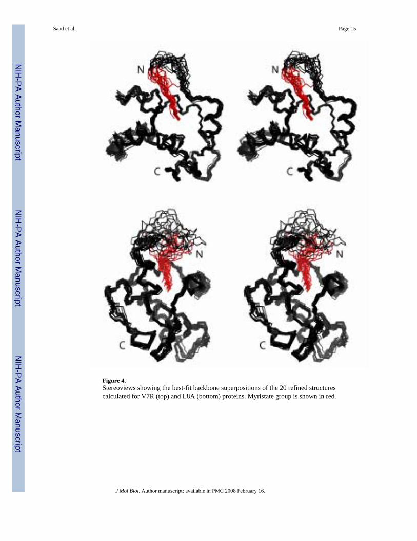
Figure 4.Stereoviews showing the best-fit backbone superpositions of the 20 refined structurescalculated for V7R (top) and L8A (bottom) proteins. Myristate group is shown in red.
Saad et al. Page 15
J Mol Biol. Author manuscript; available in PMC 2008 February 16.
NIH
-PA Author Manuscript
NIH
-PA Author Manuscript
NIH
-PA Author Manuscript
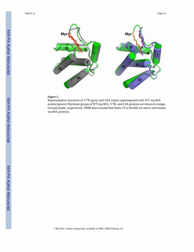
Figure 5.Representative structures of V7R (grey) and L8A (slate) superimposed with WT myrMAprotein (green). Myristate groups of WT myrMA, V7R- and L8A proteins are shown in orange,red and purple, respectively. NMR data revealed that Helix VI is flexible for native and mutantmyrMA proteins.
Saad et al. Page 16
J Mol Biol. Author manuscript; available in PMC 2008 February 16.
NIH
-PA Author Manuscript
NIH
-PA Author Manuscript
NIH
-PA Author Manuscript
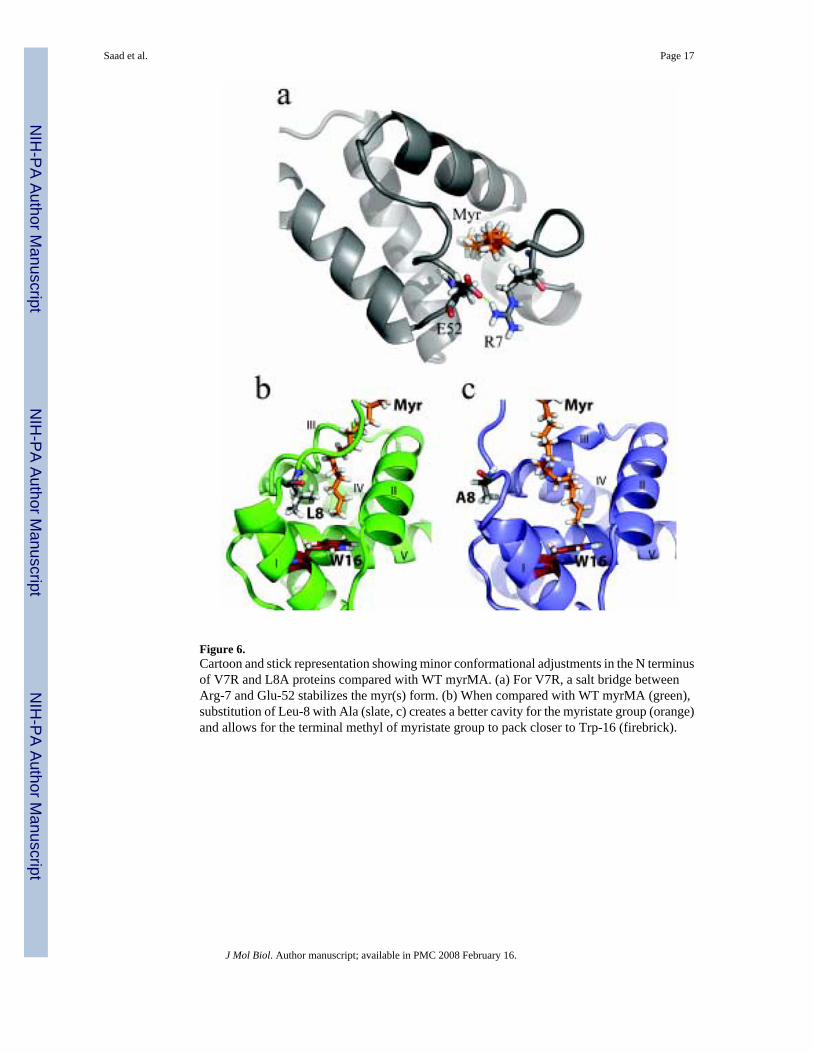
Figure 6.Cartoon and stick representation showing minor conformational adjustments in the N terminusof V7R and L8A proteins compared with WT myrMA. (a) For V7R, a salt bridge betweenArg-7 and Glu-52 stabilizes the myr(s) form. (b) When compared with WT myrMA (green),substitution of Leu-8 with Ala (slate, c) creates a better cavity for the myristate group (orange)and allows for the terminal methyl of myristate group to pack closer to Trp-16 (firebrick).
Saad et al. Page 17
J Mol Biol. Author manuscript; available in PMC 2008 February 16.
NIH
-PA Author Manuscript
NIH
-PA Author Manuscript
NIH
-PA Author Manuscript
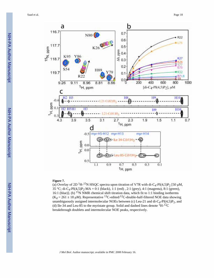
Figure 7.(a) Overlay of 2D 1H-15N HSQC spectra upon titration of V7R with di-C4-PI(4,5)P2 [50 μM,35 °C; di-C4-PI(4,5)P2:MA = 0:1 (black), 1:1 (red) , 2:1 (grey), 4:1 (magenta), 8:1 (green),16:1 (blue)]. (b) 15N NMR chemical shift titration data, which fit to 1:1 binding isotherms(Kd = 261 ± 18 μM). Representative 13C-edited/12C-double-half-filtered NOE data showingunambiguously assigned intermolecular NOEs between (c) Leu-21 and di-C4-PI(4,5)P2, and(d) Ile-34 and Leu-85 to the myristate group. Solid and dashed lines denote 1H-12Cbreakthrough doublets and intermolecular NOE peaks, respectively.
Saad et al. Page 18
J Mol Biol. Author manuscript; available in PMC 2008 February 16.
NIH
-PA Author Manuscript
NIH
-PA Author Manuscript
NIH
-PA Author Manuscript
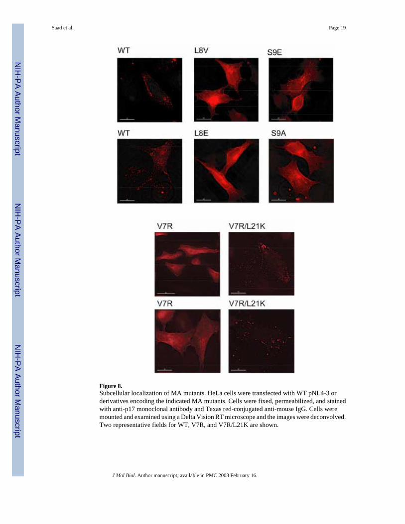
Figure 8.Subcellular localization of MA mutants. HeLa cells were transfected with WT pNL4-3 orderivatives encoding the indicated MA mutants. Cells were fixed, permeabilized, and stainedwith anti-p17 monoclonal antibody and Texas red-conjugated anti-mouse IgG. Cells weremounted and examined using a Delta Vision RT microscope and the images were deconvolved.Two representative fields for WT, V7R, and V7R/L21K are shown.
Saad et al. Page 19
J Mol Biol. Author manuscript; available in PMC 2008 February 16.
NIH
-PA Author Manuscript
NIH
-PA Author Manuscript
NIH
-PA Author Manuscript
NIH
-PA Author Manuscript
NIH
-PA Author Manuscript
NIH
-PA Author Manuscript
Saad et al. Page 20
Table 1Statistics for HIV-1 V7R and L8A-myrMA Structures1
NMR-derived restraints V7R L8A
1H-13H distance restraints 834 818Intraresidue 81 80Sequential(| i − j| = 1) 165 166Medium & Long range(| i − j | > 1) 579 565Backbone H-bonds (4/H-bond) 0 0Side-chain H-bonds (4/H-bond) 20 16Dipolar coupling restraints 87 88Intermolecular 0 0Average restraints/refined residue 14.8 14.1Target function (Å2)Mean (± Std Dev) 1.29 (0.23) 1.56 (0.20)Minimum 0.79 0.98Maximum 1.58 1.73Restraint violationsAv. max. upper dist. viol. (Å) 0.36 0.39(± Std Dev) 0.06 0.09Av. max. VDW viol. (Å) 0.30 0.28(± Std Dev) 0.07 0.08Av. max RDC viol. (Hz) 0.40 0.36(± Std Dev) 0.16 0.11Structure Convergence (Å)2Main chain atomsMean (± Std Dev) 0.48 (0.11) 0.58 (0.08)All heavy atomsMean (± Std Dev) 0.95 (0.10) 1.02 (0.09)
1A total of 20 refined structures per ensemble (see Materials and Methods for structure refinement details).
2RMSDs calculated relative to mean atom coordinates for residues 10 to 30 and 35 to 100
J Mol Biol. Author manuscript; available in PMC 2008 February 16.
NIH
-PA Author Manuscript
NIH
-PA Author Manuscript
NIH
-PA Author Manuscript
Saad et al. Page 21
Table 2Summary of Kd (μM) values for di-C4-PI(4,5)P2 binding to MA proteins.
Sample Kd (μM)
myr(-)MAa 240 ± 60myrMAa 150 ± 30V7R 261 ± 18L8A 150 ± 24L8I 108 ± 5L21K 67 ± 5V7R-L21K 84 ± 9
aValues reported previously (ref 31)
J Mol Biol. Author manuscript; available in PMC 2008 February 16.