Bax-dependent caspase-3 activation is a key determinant in p53-induced apoptosis in neurons
10
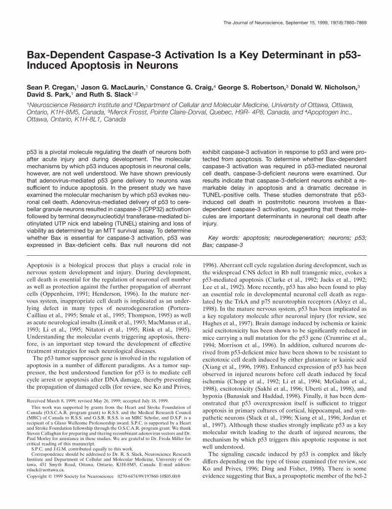
Bax-Dependent Caspase-3 Activation Is a Key Determinant in p53- Induced Apoptosis in Neurons Sean P. Cregan, 1 Jason G. MacLaurin, 1 Constance G. Craig, 4 George S. Robertson, 3 Donald W. Nicholson, 3 David S. Park, 1 and Ruth S. Slack 1,2 1 Neuroscience Research Institute and 2 Department of Cellular and Molecular Medicine, University of Ottawa, Ottawa, Ontario, K1H-8M5, Canada, 3 Merck Frosst, Pointe Claire-Dorval, Quebec, H9R- 4P8, Canada, and 4 Apoptogen Inc., Ottawa, Ontario, K1H-8L1, Canada p53 is a pivotal molecule regulating the death of neurons both after acute injury and during development. The molecular mechanisms by which p53 induces apoptosis in neuronal cells, however, are not well understood. We have shown previously that adenovirus-mediated p53 gene delivery to neurons was sufficient to induce apoptosis. In the present study we have examined the molecular mechanism by which p53 evokes neu- ronal cell death. Adenovirus-mediated delivery of p53 to cere- bellar granule neurons resulted in caspase-3 (CPP32) activation followed by terminal deoxynucleotidyl transferase-mediated bi- otinylated UTP nick end labeling (TUNEL) staining and loss of viability as determined by an MTT survival assay. To determine whether Bax is essential for caspase-3 activation, p53 was expressed in Bax-deficient cells. Bax null neurons did not exhibit caspase-3 activation in response to p53 and were pro- tected from apoptosis. To determine whether Bax-dependent caspase-3 activation was required in p53-mediated neuronal cell death, caspase-3-deficient neurons were examined. Our results indicate that caspase-3-deficient neurons exhibit a re- markable delay in apoptosis and a dramatic decrease in TUNEL-positive cells. These studies demonstrate that p53- induced cell death in postmitotic neurons involves a Bax- dependent caspase-3 activation, suggesting that these mole- cules are important determinants in neuronal cell death after injury. Key words: apoptosis; neurodegeneration; neurons; p53; Bax; caspase-3 Apoptosis is a biological process that plays a crucial role in nervous system development and injury. During development, cell death is essential for the regulation of neuronal cell number as well as protection against the further propagation of aberrant cells (Oppenheim, 1991; Henderson, 1996). In the mature ner- vous system, inappropriate cell death is implicated as an under- lying defect in many types of neurodegeneration (Portera- Cailliau et al., 1995; Smale et al., 1995; Thompson, 1995) as well as acute neurological insults (Linnik et al., 1993; MacManus et al., 1993; Li et al., 1995; Nitatori et al., 1995; Rink et al., 1995). Understanding the molecular events triggering apoptosis, there- fore, is an important step toward the development of effective treatment strategies for such neurological diseases. The p53 tumor suppressor gene is involved in the regulation of apoptosis in a number of different paradigms. As a tumor sup- pressor, the best understood function for p53 is to mediate cell cycle arrest or apoptosis after DNA damage, thereby preventing the propagation of damaged cells (for review, see Ko and Prives, 1996). Aberrant cell cycle regulation during development, such as the widespread CNS defect in Rb null transgenic mice, evokes a p53-mediated apoptosis (Clarke et al., 1992; Jacks et al., 1992; Lee et al., 1992). More recently, p53 has also been found to play an essential role in developmental neuronal cell death as regu- lated by the TrkA and p75 neurotrophin receptors (Aloyz et al., 1998). In the mature nervous system, p53 has been implicated as a key regulatory molecule after neuronal injury (for review, see Hughes et al., 1997). Brain damage induced by ischemia or kainic acid excitotoxicity has been shown to be significantly reduced in mice carrying a null mutation for the p53 gene (Crumrine et al., 1994; Morrison et al., 1996). In addition, cultured neurons de- rived from p53-deficient mice have been shown to be resistant to excitotoxic cell death induced by either glutamate or kainic acid (Xiang et al., 1996, 1998). Enhanced expression of p53 has been observed in injured neurons before cell death induced by focal ischemia (Chopp et al., 1992; Li et al., 1994; McGahan et al., 1998), excitotoxicity (Sakhi et al., 1996; Uberti et al., 1998), and hypoxia (Banasiak and Haddad, 1998). Finally, it has been dem- onstrated that p53 overexpression itself is sufficient to trigger apoptosis in primary cultures of cortical, hippocampal, and sym- pathetic neurons (Slack et al., 1996; Xiang et al., 1996; Jordan et al., 1997). Although these studies strongly implicate p53 as a key molecular switch leading to the death of injured neurons, the mechanism by which p53 triggers this apoptotic response is not well understood. The signaling cascade induced by p53 is complex and likely differs depending on the type of tissue examined (for review, see Ko and Prives, 1996; Ding and Fisher, 1998). There is some evidence suggesting that Bax, a proapoptotic member of the bcl-2 Received March 8, 1999; revised May 26, 1999; accepted July 18, 1999. This work was supported by grants from the Heart and Stroke Foundation of Canada (O.S.C.A.R. program grant) to R.S.S. and the Medical Research Council (MRC) of Canada to R.S.S. and G.S.R. R.S.S. is an MRC Scholar, and D.S.P. is a recipient of a Glaxo Wellcome Professorship award. S.P.C. is supported by a Heart and Stroke Foundation fellowship through the O.S.C.A.R. program grant. We thank Steven C allaghan for preparing and titering recombinant adenovirus vectors and Dr. Paul Morley for assistance in these studies. We are grateful to Dr. Freda Miller for critical reading of this manuscript. S.P.C. and J.G.M. contributed equally to this work. Correspondence should be addressed to Dr. R. S. Slack, Neuroscience Research Institute and Department of Cellular and Molecular Medicine, University of Ot- tawa, 451 Smyth Road, Ottawa, Ontario, K1H-8M5, Canada. E-mail address: [email protected]. Copyright © 1999 Society for Neuroscience 0270-6474/99/197860-10$05.00/0 The Journal of Neuroscience, September 15, 1999, 19(18):7860–7869
-
Upload
independent -
Category
Documents
-
view
0 -
download
0
Transcript of Bax-dependent caspase-3 activation is a key determinant in p53-induced apoptosis in neurons
Bax-Dependent Caspase-3 Activation Is a Key Determinant in p53-Induced Apoptosis in Neurons
Sean P. Cregan,1 Jason G. MacLaurin,1 Constance G. Craig,4 George S. Robertson,3 Donald W. Nicholson,3David S. Park,1 and Ruth S. Slack1,2
1Neuroscience Research Institute and 2Department of Cellular and Molecular Medicine, University of Ottawa, Ottawa,Ontario, K1H-8M5, Canada, 3Merck Frosst, Pointe Claire-Dorval, Quebec, H9R- 4P8, Canada, and 4Apoptogen Inc.,Ottawa, Ontario, K1H-8L1, Canada
p53 is a pivotal molecule regulating the death of neurons bothafter acute injury and during development. The molecularmechanisms by which p53 induces apoptosis in neuronal cells,however, are not well understood. We have shown previouslythat adenovirus-mediated p53 gene delivery to neurons wassufficient to induce apoptosis. In the present study we haveexamined the molecular mechanism by which p53 evokes neu-ronal cell death. Adenovirus-mediated delivery of p53 to cere-bellar granule neurons resulted in caspase-3 (CPP32) activationfollowed by terminal deoxynucleotidyl transferase-mediated bi-otinylated UTP nick end labeling (TUNEL) staining and loss ofviability as determined by an MTT survival assay. To determinewhether Bax is essential for caspase-3 activation, p53 wasexpressed in Bax-deficient cells. Bax null neurons did not
exhibit caspase-3 activation in response to p53 and were pro-tected from apoptosis. To determine whether Bax-dependentcaspase-3 activation was required in p53-mediated neuronalcell death, caspase-3-deficient neurons were examined. Ourresults indicate that caspase-3-deficient neurons exhibit a re-markable delay in apoptosis and a dramatic decrease inTUNEL-positive cells. These studies demonstrate that p53-induced cell death in postmitotic neurons involves a Bax-dependent caspase-3 activation, suggesting that these mole-cules are important determinants in neuronal cell death afterinjury.
Key words: apoptosis; neurodegeneration; neurons; p53;Bax; caspase-3
Apoptosis is a biological process that plays a crucial role innervous system development and injury. During development,cell death is essential for the regulation of neuronal cell numberas well as protection against the further propagation of aberrantcells (Oppenheim, 1991; Henderson, 1996). In the mature ner-vous system, inappropriate cell death is implicated as an under-lying defect in many types of neurodegeneration (Portera-Cailliau et al., 1995; Smale et al., 1995; Thompson, 1995) as wellas acute neurological insults (Linnik et al., 1993; MacManus et al.,1993; Li et al., 1995; Nitatori et al., 1995; Rink et al., 1995).Understanding the molecular events triggering apoptosis, there-fore, is an important step toward the development of effectivetreatment strategies for such neurological diseases.
The p53 tumor suppressor gene is involved in the regulation ofapoptosis in a number of different paradigms. As a tumor sup-pressor, the best understood function for p53 is to mediate cellcycle arrest or apoptosis after DNA damage, thereby preventingthe propagation of damaged cells (for review, see Ko and Prives,
1996). Aberrant cell cycle regulation during development, such asthe widespread CNS defect in Rb null transgenic mice, evokes ap53-mediated apoptosis (Clarke et al., 1992; Jacks et al., 1992;Lee et al., 1992). More recently, p53 has also been found to playan essential role in developmental neuronal cell death as regu-lated by the TrkA and p75 neurotrophin receptors (Aloyz et al.,1998). In the mature nervous system, p53 has been implicated asa key regulatory molecule after neuronal injury (for review, seeHughes et al., 1997). Brain damage induced by ischemia or kainicacid excitotoxicity has been shown to be significantly reduced inmice carrying a null mutation for the p53 gene (Crumrine et al.,1994; Morrison et al., 1996). In addition, cultured neurons de-rived from p53-deficient mice have been shown to be resistant toexcitotoxic cell death induced by either glutamate or kainic acid(Xiang et al., 1996, 1998). Enhanced expression of p53 has beenobserved in injured neurons before cell death induced by focalischemia (Chopp et al., 1992; Li et al., 1994; McGahan et al.,1998), excitotoxicity (Sakhi et al., 1996; Uberti et al., 1998), andhypoxia (Banasiak and Haddad, 1998). Finally, it has been dem-onstrated that p53 overexpression itself is sufficient to triggerapoptosis in primary cultures of cortical, hippocampal, and sym-pathetic neurons (Slack et al., 1996; Xiang et al., 1996; Jordan etal., 1997). Although these studies strongly implicate p53 as a keymolecular switch leading to the death of injured neurons, themechanism by which p53 triggers this apoptotic response is notwell understood.
The signaling cascade induced by p53 is complex and likelydiffers depending on the type of tissue examined (for review, seeKo and Prives, 1996; Ding and Fisher, 1998). There is someevidence suggesting that Bax, a proapoptotic member of the bcl-2
Received March 8, 1999; revised May 26, 1999; accepted July 18, 1999.This work was supported by grants from the Heart and Stroke Foundation of
Canada (O.S.C.A.R. program grant) to R.S.S. and the Medical Research Council(MRC) of Canada to R.S.S. and G.S.R. R.S.S. is an MRC Scholar, and D.S.P. is arecipient of a Glaxo Wellcome Professorship award. S.P.C. is supported by a Heartand Stroke Foundation fellowship through the O.S.C.A.R. program grant. We thankSteven Callaghan for preparing and titering recombinant adenovirus vectors and Dr.Paul Morley for assistance in these studies. We are grateful to Dr. Freda Miller forcritical reading of this manuscript.
S.P.C. and J.G.M. contributed equally to this work.Correspondence should be addressed to Dr. R. S. Slack, Neuroscience Research
Institute and Department of Cellular and Molecular Medicine, University of Ot-tawa, 451 Smyth Road, Ottawa, Ontario, K1H-8M5, Canada. E-mail address:[email protected] © 1999 Society for Neuroscience 0270-6474/99/197860-10$05.00/0
The Journal of Neuroscience, September 15, 1999, 19(18):7860–7869
family of cell death-regulating genes, may be involved in p53-induced apoptosis. The Bax gene contains p53 consensus se-quences within its promoter and has been shown to be transcrip-tionally regulated by p53 (Miyashita et al., 1994; Miyashita andReed, 1995). Furthermore, Bax induction has been observedduring p53-mediated cell death in a number of non-neuronal cellsystems (Selvakumaran et al., 1994; Zhan et al., 1994; Brady et al.,1996). In neuronal cells the role of Bax in p53-induced apoptosisremains unclear; however, it appears to be important in a numberof different neuronal death paradigms (Deckwerth et al., 1996;Miller et al., 1997; Johnson et al., 1998; Xiang et al., 1998).
One set of molecules that appear to be modulated by thepresence of Bax are the caspases (Miller et al., 1997; Martinou etal., 1998). Caspases, a family of cysteine proteases implicated inthe commitment and execution of apoptotic cell death, exist asproenzymes until cleaved in response to apoptotic stimuli (forreview, see Thornberry and Lazebnik, 1998). Recent reportsindicate that caspases may play a role in neuronal cell deathduring development (Kuida et al., 1996, 1998; Woo et al., 1998) aswell as after neuronal injury (Loddick et al., 1996; Gillardon etal., 1997; Gottron et al., 1997; Hara et al., 1997; Cheng et al., 1998;Endres et al., 1998; Ni et al., 1998; Park et al., 1998).
To examine the molecular cascade by which p53 induces neu-ronal cell death, we have used primary cultures of cerebellargranule neurons (CGNs) derived from transgenic mice carryingnull mutations for either the Bax or the caspase-3 gene (CPP32).By using adenovirus-mediated p53 gene delivery to primary neu-ronal cells, we demonstrate that neuronal apoptosis induced byp53 is mediated by Bax and caspase-3.
MATERIALS AND METHODSTransgenic mice. Bax-deficient transgenic mice were generously providedby Dr. Stanley Korsmeyer (Knudson et al., 1995) and are now availablefrom Jackson Laboratories (Bar Harbor, ME). Bax mice were originallyon a mixed 129/C57BL6 strain (Knudson et al., 1995) but have since beenbackcrossed to C57BL6 (12–14 times) and can therefore be considered a
C57BL6 genetic background. Transgenic mice carrying a caspase-3 nullmutation were obtained from Dr. Don Nicholson (Merck Frosst, Can-ada) (E. Keramis and D. S. Park, unpublished observations). The phe-notype of the caspase-3-deficient mice used in these experiments wassimilar to those described previously (Kuida et al., 1996; Woo et al.,1998). All caspase-3-deficient mice were maintained on a C57BL6 back-ground to maintain genetic uniformity. Bax null mice were genotyped asdescribed previously (Knudson et al., 1995). Caspase-3 null mice weregenotyped by PCR according to the following protocol. The usual PCRreaction buffer contained 2.25 mM MgCl2 and 5% DMSO. The primersfor the wild-type caspase-3 alleles were CTAAGTTAACCAAAGT-GAGCACCGA (sense) and ATGAATCAAGGCAGCATAGTACTCC(antisense). For detection of the targeted allele, the same sense primerand the following antisense primer GTCGATCCACTAGTTCTA-GAGCGGC (LZ1) were used. Conditions were set as follows: 94°C, 2min (1 cycle); 94°C, 30 sec, 60°C, 1 min, 72°C, 1 min (30 cycles); 72°C, 5min (1 cycle).Cell culture. Transgenic pups were genotyped at postnatal day 4–6, afterwhich the appropriate animals were selected for experimentation, andneurons were cultured from brains individually. Primary cultures ofcerebellar granule neurons were obtained from dissociated cerebella ofpostnatal day 8 or 9 mice, as described previously (Levi et al., 1984;Miller and Johnson, 1996) with some modifications. Brains were removedand placed into separate dishes containing solution A (124 mM NaCl,
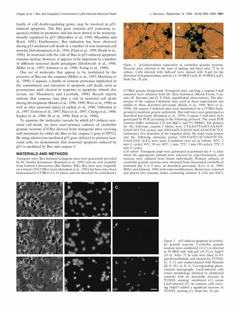
Figure 1. b-Galactosidase expression in cerebellar granule neurons.Neurons were infected at the time of plating and fixed after 72 hr inculture. Cells infected with AdLacZ were stained with X-gal for thedetection of b-galactosidase activity (A, 10 MOI LacZ; B, 50 MOI LacZ).Scale bar, 20 mm.
Figure 2. p53 induces apoptosis in cerebel-lar granule neurons. Cerebellar granuleneurons were uninfected (A–C) or infectedat 50 MOI with AdLacZ (D–F) or Adp53(G–I). After 72 hr cells were fixed in 4%paraformaldehyde and stained for TUNEL(C, F, I ) and counterstained with Hoechst(B, E, H ). A, D, G, Corresponding phase-contrast micrographs. LacZ-infected cellsretain morphology identical to uninfectedcontrols, with no detectable increase inTUNEL staining: uninfected (C) versusLacZ-infected (F). In contrast, cells carry-ing Adp53 exhibit a significant increase inTUNEL staining ( I ). Scale bar, 20 mm.
Cregan et al. • Bax and Caspase-3 Involvement in p53-Induced Neuronal Death J. Neurosci., September 15, 1999, 19(18):7860–7869 7861

5.37 mM KCl, 1 mM NaH2PO4, 1.2 mM MgSO4, 14.5 mM D-(1)-glucose,25 mM HEPES, 3 mg/ml BSA, pH 7.4) in which the cerebella weredissected, meninges removed, and tissue sliced into small pieces. Tissuewas briefly centrifuged and transferred to solution A containing 0.25mg/ml trypsin, then incubated at 37°C for 18 min. After the addition of0.082 mg/ml trypsin inhibitor (Boehringer Mannheim, Indianapolis, IN)and 0.25 mg/ml DNase I (Boehringer Mannheim), tissue was incubatedat 25°C for 2 min. After a brief centrifugation, the resulting pellet wasgently triturated in solution A yielding suspension that was furtherincubated for 10 min at 25°C in solution A containing 2.7 mM MgSO4 and0.03 mM CaCl2. After a final centrifugation the pellet was resuspended inEMEM media (Sigma, St. Louis, MO) containing 10% dialyzed FBS(Sigma), 25 mM KCl, 2 mM glutamine (Life Technologies BRL, Gaith-ersburg, MD), 25 mM glucose, and 0.1 mg/ml gentamycin (Sigma) andfiltered through a cell strainer (size 70 mm; Falcon). Cells were plated ata density of 1.5 3 10 6 cells per milliliter of medium on either Nuncfour-well or 35 3 10 mm dishes (Life Technologies BRL) coated withpoly-D-lysine (Sigma). Cytosine-b-arabinoside (10 mM; Sigma) was added24 hr after plating.Recombinant adenovirus infection. Recombinant adenovirus vectors car-rying the human p53 or LacZ expression cassettes were kindly providedby Dr. Frank Graham (McMaster University, Hamilton, Ontario) (Bac-chetti and Graham, 1993). In preliminary studies it was determined thattreatment of cells with recombinant adenovirus vectors at a multiplicityof infection (MOI) of 50 pfu/cell resulted in a high degree of transgeneexpression (as shown by X-gal staining) with minimal toxicity; thus allfurther experiments were performed at this titer. Recombinant adeno-virus vectors were added to cell suspensions immediately before plating.Cell survival assay. Two different assays were used to measure cellsurvival: TUNEL labeling and a quantitative MTT assay. The colori-metric MTT survival assay (Cell Titer Kit, Promega, Madison, WI) thatmeasures the mitochondrial conversion of the tetrazolium salt to a blueformizan salt was used as described previously (Slack et al., 1996). Toassay apoptosis, TUNEL labeling was used to visualize cells with frag-mented DNA. Cells were harvested 72 and 96 hr after p53 or LacZ genedelivery and fixed in 4% paraformaldehyde for 10 min followed byacetone/methanol (1:1) for 1 min. Cells were then washed in threechanges of PBS and incubated for 1 hr at 37°C with 75 ml of a mixture(Boehringer Mannheim) consisting of 0.5 ml terminal transferase, 0.95 mlbiotin-16-dUTP, 6.0 ml CoCl2, 15.0 ml 5 3 TdT buffer, and 52.55 mldistilled water. Cells were then washed three times in PBS and incubatedwith a streptavidin Cy2 secondary antibody (Jackson ImmunoresearchLaboratories, West Grove, PA). Cells were counterstained with Hoechst33258, and the fraction of TUNEL-positive cells was determined. Aminimum of 500 cells was scored for each treatment, and the datarepresent the mean of three independent experiments.
Caspase protease activity assays. Cells were harvested and extracted for15 min on ice in a lysis buffer consisting of 1 mM KCl, 10 mM HEPES,pH 7.4, 1.5 mM MgCl2, 1 mM DTT, 1 mM PMSF, 5 mg /ml leupeptin, 2mg /ml aprotinin, and 10% glycerol. Lysates were centrifuged for 10min at 15,000 rpm, and supernatants were removed and assayed forprotein content. To measure caspase activity, aliquots of cell extractscontaining 10 mg of protein were added to a reaction buffer (25 mMHEPES at pH 7.4, 10 mM DTT, 10% sucrose, 0.1% CHAPS) (Stefaniset al., 1996) containing 15 mM N-acetyl-Asp-Glu-Val-Asp-(7-amino-4trifluoromethyl-coumarin (DEVD-AFC), 40 mM N-acetyl-Asp-Glu-Val-Asp-p-nitroanilide (DEVD-pNA) for caspase-3-like protease activ-ity, or 40 mM N-acetyl-Tyr-Val-Ala-Asp-p-nitroanilide (YVAD-pNA)(Biomol Research Laboratories, Plymouth Meeting, PA) for caspase-1-like protease activity and incubated at 37°C for 2 hr. DEVD-AFCcleavage was measured using an SLM 8000 fluorometer (excitation 400nm and emission 505 nm), and DEVD-pNA and YVAD-pNA cleavagewere measured at 405 nm in a spectrophotometer.Western blot analysis. Cells were extracted as above, and aliquots con-taining 30 mg of protein were separated on a 10% acrylamide gel andtransferred to a nitrocellulose membrane. After they were blocked for 2hr with 5% skim milk, membranes were incubated for 1 hr with either arabbit polyclonal antibody directed against p53 (CM-1; Novacastra Lab-oratories, Newcastle upon Tyne, UK), a mouse monoclonal antibody(mAb) specific for caspase-3 (Merck Frosst Research, Pointe Claire-Dorval, PQ), LacZ (Cappell, Durham, NC), or a goat polyclonal anti-body directed against actin (Santa Cruz Biotechnologies, Santa Cruz,CA). After three washes with TPBS (25 mM Na2HPO4, 5 mM NaH2PO4,0.9% NaCl, 0.1% Tween-20), membranes were incubated for 1 hr at 25°Cwith the appropriate secondary antibody, washed five times for 5 mineach in TPBS, and then developed by an enhanced chemiluminescencesystem according to the manufacturer’s instructions (Amersham, Arling-ton Heights, IL).Immunofluorescence. For immunofluorescence detection of human p53delivered by adenovirus vectors, neuronal cultures were fixed for 10 minin 4% paraformaldehyde followed by acetone/methanol (1:1) for 1 min.Cells were then washed three times with PBS and incubated at 25°C for1 hr with a mouse mAb specific for an amino-terminal epitope of humanp53 (DO-1; Santa Cruz). After three washes in PBS, cells were incubatedfor 1 hr at 25°C with a Cy3-conjugated goat anti-mouse secondaryantibody (Jackson Immunoresearch Laboratories). Cells were thenwashed three times in PBS, counterstained with Hoechst 33258, andvisualized by fluorescent microscopy.
RESULTSp53 induces apoptosis in cultured cerebellargranule neuronsRecombinant adenovirus-mediated delivery of p53 has previouslybeen shown to induce cell death in various post-mitotic neurons(Slack et al., 1996; Xiang et al., 1996; Jordan et al., 1997). Todetermine whether p53 overexpression could induce cell deathin cultured cerebellar granule neurons, CGNs were infected at50 MOI with recombinant adenoviruses carrying an expressioncassette for either the human p53 gene (Adp53) or the
Figure 3. p53 expression in cerebellar granule neurons. Neurons infectedat 50 MOI with either AdLacZ (A, B), as controls, or Adp53 (C, D) werefixed in 4% paraformaldehyde after 72 hr and stained with an antibodydirected against human p53 (B, D) and counterstained with Hoechst (A,C). Scale bar, 20 mm.
Figure 4. p53 and LacZ protein levels in cerebellar granule neurons.Neurons were either uninfected or infected at 50 MOI at the time ofplating with either AdLacZ or Adp53. After 48 and 72 hr, protein wasextracted and Western blot analysis was performed to visualize transgeneexpression.
7862 J. Neurosci., September 15, 1999, 19(18):7860–7869 Cregan et al. • Bax and Caspase-3 Involvement in p53-Induced Neuronal Death
b-galactosidase reporter gene (AdLacZ). The AdLacZ vector wasused to determine the efficiency of transduction as well as toassess the effect of viral infection on neuronal viability as de-scribed previously (Slack et al., 1996). At a virus titer of 50 MOI,.90% of neurons infected with AdLacZ were found to expresshigh levels of b-galactosidase as determined by X-gal staining(Fig. 1). After 72 hr in culture, uninfected neurons and neuronsinfected with the control virus AdLacZ displayed healthy cellbodies and an extensive neuritic network (Fig. 2A,D). In contrast,neurons infected with Adp53 exhibited obvious signs of cellulardegradation, including phase-bright, pyknotic cell bodies as wellas blebbing and dissolution of neuritic processes (Fig. 2G). Thiscell death was accompanied by a significant increase in the frac-tion of cells exhibiting chromatin condensation (Fig. 2B,E, H)and positive TUNEL labeling (Fig. 2C,F, I), indicative of anapoptotic mode of cell death.
To confirm that this cell death was associated with increasedexpression of p53, neurons infected at 50 MOI with either Adp53or AdLacZ were immunostained with an antibody specific for
human p53 to distinguish from endogenous p53. As expected,neurons infected with AdLacZ were not immunoreactive forhuman p53 (Fig. 3A,B), whereas .80% of the neurons infectedwith Adp53 exhibited intense immunopositive staining (Fig.3C, D). To determine more precisely the extent of transgeneexpression, cell extracts were prepared from CGN cultures 48 and72 hr after infection with AdLacZ or Adp53, and the level of p53protein was assessed by Western blot analysis, this time using anantibody that recognizes both rodent and human p53. Theseexperiments demonstrate that infection with Adp53 resulted in asubstantial increase in p53 expression within 48 hr of infection(Fig. 4). The lack of detectable p53 in extracts from uninfectedcells or cells infected with AdLacZ suggests that the endogenouslevels of p53 were comparatively small and that the infectionprocess itself did not significantly alter p53 expression in thesecells. Taken together, these results indicate that expression of p53can directly induce apoptotic cell death in cultured cerebellargranule neurons.
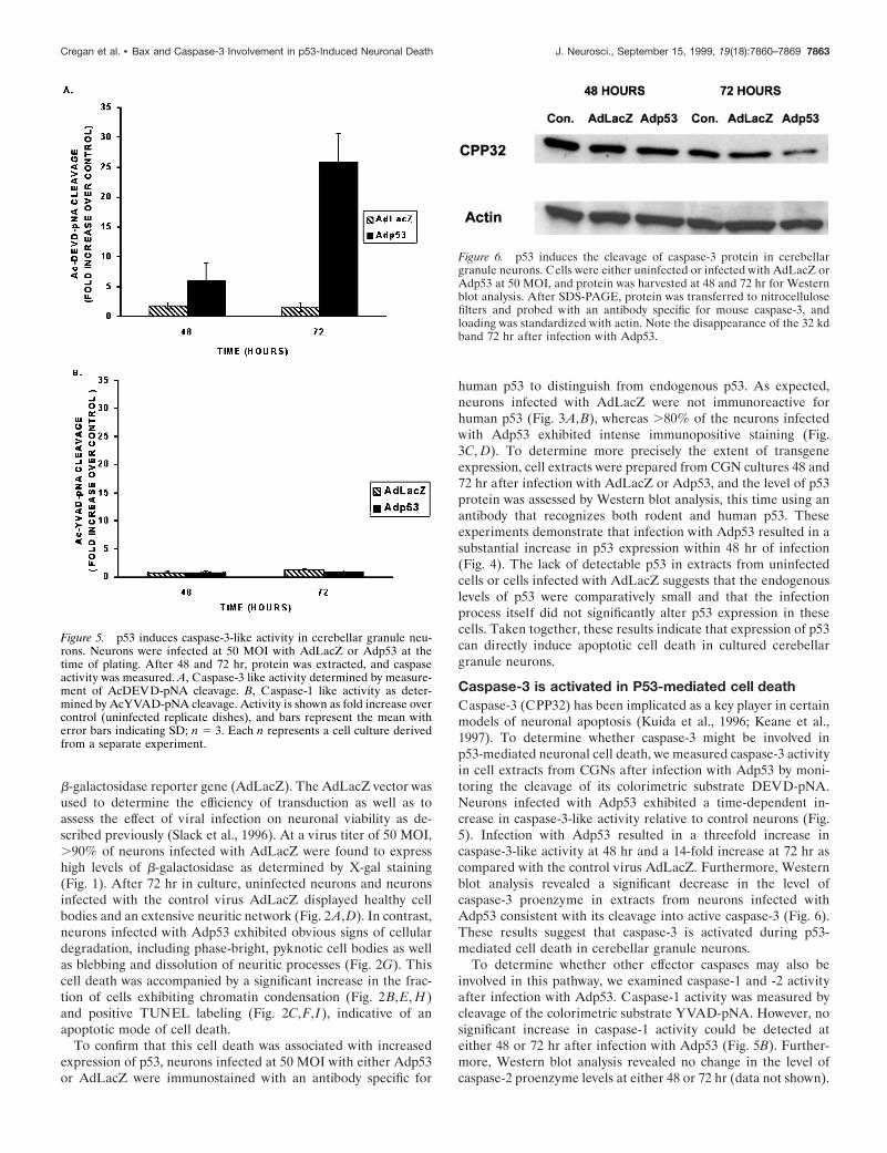
Caspase-3 is activated in P53-mediated cell deathCaspase-3 (CPP32) has been implicated as a key player in certainmodels of neuronal apoptosis (Kuida et al., 1996; Keane et al.,1997). To determine whether caspase-3 might be involved inp53-mediated neuronal cell death, we measured caspase-3 activityin cell extracts from CGNs after infection with Adp53 by moni-toring the cleavage of its colorimetric substrate DEVD-pNA.Neurons infected with Adp53 exhibited a time-dependent in-crease in caspase-3-like activity relative to control neurons (Fig.5). Infection with Adp53 resulted in a threefold increase incaspase-3-like activity at 48 hr and a 14-fold increase at 72 hr ascompared with the control virus AdLacZ. Furthermore, Westernblot analysis revealed a significant decrease in the level ofcaspase-3 proenzyme in extracts from neurons infected withAdp53 consistent with its cleavage into active caspase-3 (Fig. 6).These results suggest that caspase-3 is activated during p53-mediated cell death in cerebellar granule neurons.
To determine whether other effector caspases may also beinvolved in this pathway, we examined caspase-1 and -2 activityafter infection with Adp53. Caspase-1 activity was measured bycleavage of the colorimetric substrate YVAD-pNA. However, nosignificant increase in caspase-1 activity could be detected ateither 48 or 72 hr after infection with Adp53 (Fig. 5B). Further-more, Western blot analysis revealed no change in the level ofcaspase-2 proenzyme levels at either 48 or 72 hr (data not shown).
Figure 5. p53 induces caspase-3-like activity in cerebellar granule neu-rons. Neurons were infected at 50 MOI with AdLacZ or Adp53 at thetime of plating. After 48 and 72 hr, protein was extracted, and caspaseactivity was measured. A, Caspase-3 like activity determined by measure-ment of AcDEVD-pNA cleavage. B, Caspase-1 like activity as deter-mined by AcYVAD-pNA cleavage. Activity is shown as fold increase overcontrol (uninfected replicate dishes), and bars represent the mean witherror bars indicating SD; n 5 3. Each n represents a cell culture derivedfrom a separate experiment.
Figure 6. p53 induces the cleavage of caspase-3 protein in cerebellargranule neurons. Cells were either uninfected or infected with AdLacZ orAdp53 at 50 MOI, and protein was harvested at 48 and 72 hr for Westernblot analysis. After SDS-PAGE, protein was transferred to nitrocellulosefilters and probed with an antibody specific for mouse caspase-3, andloading was standardized with actin. Note the disappearance of the 32 kdband 72 hr after infection with Adp53.
Cregan et al. • Bax and Caspase-3 Involvement in p53-Induced Neuronal Death J. Neurosci., September 15, 1999, 19(18):7860–7869 7863

These results suggest that caspase-1 and -2 are not involved in thiscell death pathway.
Bax-deficient neurons are resistant to p53-inducedcell deathTo determine whether Bax was required in this p53-mediated celldeath pathway, cell survival was examined in CGNs derived fromBax wild-type and Bax-deficient mice after infection with eitherAdp53 or AdLacZ. In Bax 1/1 neurons, a significant decrease incell survival could be detected as early as 48 hr after infectionwith Adp53 (Fig. 7A). Neuronal cell death continued to increasegradually with time, and by 96 hr there was an approximate 80%loss in neuronal survival. In contrast, Bax 2/2 neurons infectedwith Adp53 exhibited only a marginal decrease in cell survivalrelative to AdLacZ controls that remained constant for at least120 hr after infection. Consistent with this increased resistance,Bax-deficient neurons infected with Adp53 appeared remarkablyhealthy when examined by light microscopy (Fig. 7B).
To determine whether this enhanced survival of Bax-deficientneurons was associated with a decrease in apoptotic cell death,Bax 1/1 and Bax 2/2 neurons were infected with Adp53 orAdLacZ, and the frequency of TUNEL-positive cells was deter-mined after 72 hr. In Bax 1/1 neurons, infection with Adp53resulted in a dramatic increase in the frequency of TUNEL-positive cells (;70%) in comparison with neurons infected with
the control virus AdLacZ (Fig. 8). In contrast, Bax2/2 neuronsinfected with Adp53 exhibited only a small increase in TUNEL-positive cells (;10%) relative to control. The fact that p53-mediated cell death is dramatically reduced in the absence of Baxsuggests that Bax must play a prominent role in this cell deathpathway.
Bax has been shown to be transcriptionally regulated by p53(Miyashita et al., 1994; Miyashita and Reed, 1995),and Bax in-duction has been observed in certain p53-mdiated cell deathparadigms (Selvakumaran et al., 1994; Zhan et al., 1994; Brady etal., 1996). Therefore, we examined Bax protein expression inwild-type cerebellar granule neurons after infection with Adp53.Interestingly, p53 overexpression did not cause a significant in-crease in Bax protein levels (data not shown). Therefore, al-though Bax appears to play a critical role in p53-mediated celldeath, this did not appear to involve induction of Bax expression.
Bax is required for p53-induced caspase-3 activationWe next examined caspase-3 activity in Bax 1/1 and Bax 2/2neurons after infection with Adp53 to determine whether Bax wasrequired for p53-mediated caspase-3 activation. In Bax 1/1 neu-rons, infection with Adp53 resulted in an approximately 13-foldincrease in caspase-3 like activity as compared with the controlvirus AdLacZ (Fig. 9). This increase in caspase-3 like activity,
Figure 7. Bax deficiency renders cerebellar granule neuronsresistant to p53-induced cell death. A, Cells were eitheruninfected or infected with AdLacZ or Adp53 at 50 MOI,and an MTT survival assay was conducted at 48, 72, 96, and120 hr. Control, uninfected cells were considered 100%, andresults are reported as the percentage of control. Points arethe average of cultures derived from three separate brainswith matching wild-type littermates (n 5 3), with error barsshowing SD. B, Phase-contrast photomicrographs of Bax 1/1and 2/2 neurons 72 hr after infection at 50 MOI with eitherAdLacZ or Adp53. Scale bar, 60 mm.
7864 J. Neurosci., September 15, 1999, 19(18):7860–7869 Cregan et al. • Bax and Caspase-3 Involvement in p53-Induced Neuronal Death

however, was absent in Bax-deficient neurons (Fig. 9), indicatingthat Bax is required for p53-induced caspase-3 activation.
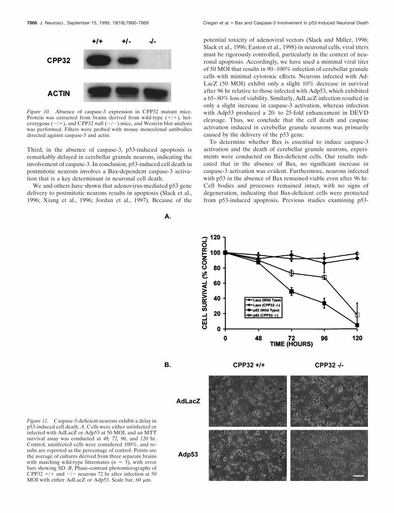
Caspase-3-deficient neurons exhibit increasedresistance to P53-induced cell deathBecause caspase-3 appears to be the predominant caspase in-volved in p53-induced cell death, we next asked whethercaspase-3 was required in this p53-mediated cell death pathway.Survival was examined in CGNs derived from CPP32 1/1 orCPP32 2/2 mice after infection with Adp53. The genotype ofthese mice was determined by PCR, and the absence of caspase-3protein was confirmed by Western blot analysis (Fig. 10). Similarto Bax-deficient neurons, neurons deficient in caspase-3 werefound to be significantly more resistant to p53-induced cell deaththan their wild-type counterparts (Fig. 11). However, survival inCPP32 2/2 neurons infected with Adp53 did eventually declinerelative to control cultures infected with AdLacZ (Fig. 11A). Thisindicates that the absence of caspase-3 delays cell death withinthe first 96 hr, but death will ensue independent of caspase-3.Consistent with the observed decrease in survival, after 72 hrCPP322/2 neurons infected with Adp53 exhibited some signs ofcell death when examined by light microscopy; however, theyappeared significantly more intact than similarly treatedCPP321/1 neurons (Fig. 11B). These results suggest that al-though caspase-3 appears to be involved in p53-induced celldeath, there may be additional death pathways through which p53and Bax can mediate their effects.
It has been suggested that caspase-3 is responsible for theactivation of endonucleases involved in the internucleosomalDNA fragmentation process that occurs during apoptosis (Enariet al., 1998; Sakahira et al., 1998). Therefore, TUNEL labelingwas examined in caspase-3-deficient neurons to determinewhether apoptotic DNA fragmentation was being blocked inthese cells. Consistent with this hypothesis, the fraction ofTUNEL-positive cells was significantly decreased in CPP322/2neurons after infection with Adp53 as compared with controlneurons infected with AdLacZ (Fig. 12). Furthermore, mostTUNEL-positive neurons in the CPP322/2 cultures did notexhibit the characteristic pyknotic nucleus and highly condensedchromatin structure typically observed in wild-type neurons (datanot shown).
DISCUSSIONRecent evidence indicates that p53 is a pivotal molecule regulat-ing the death of many cell types, including postmitotic neurons(White, 1996; Hughes et al., 1997). We therefore examined themolecular mechanisms by which p53 induces apoptosis in neuro-nal cells. The results of these experiments support a number ofconclusions. First, we demonstrate that p53-induced apoptosis incerebellar granule neurons results in a strong activation ofcaspase-3. Second, caspase-3 activity and the subsequent celldeath induced by p53 is dependent on the presence of Bax. Cellsdeficient in Bax exhibit normal morphology and little change insurvival relative to LacZ controls, despite the presence of p53.
Figure 8. Bax-deficient neurons are resistant to p53-mediated apoptosis. Cerebellar granule neurons were infected at 50 MOI with AdLacZ or Adp53at the time of plating. A, After 72 hr in culture, Bax 1/1 (a–d) or Bax 2/2 (e–h) neurons were fixed and stained with TUNEL (b, d, f, h) andcounterstained with Hoechst (a, c, e, g). Scale bar, 100 mm. B, Quantitation of TUNEL-positive cells. Data represent the mean and SD from threeindependent experiments.
Figure 9. p53-induced caspase-3-like activity is dependent on Bax.Neurons were infected at 50 MOI with AdLacZ or Adp53 at the timeof plating. After 72 hr in culture, protein was extracted, and AcDEVD-AFC cleavage activity was measured in Bax-deficient and wild-type cells.Bars represent the average of three separate experiments, and error barsshow SD.
Cregan et al. • Bax and Caspase-3 Involvement in p53-Induced Neuronal Death J. Neurosci., September 15, 1999, 19(18):7860–7869 7865
Third, in the absence of caspase-3, p53-induced apoptosis isremarkably delayed in cerebellar granule neurons, indicating theinvolvement of caspase-3. In conclusion, p53-induced cell death inpostmitotic neurons involves a Bax-dependent caspase-3 activa-tion that is a key determinant in neuronal cell death.
We and others have shown that adenovirus-mediated p53 genedelivery to postmitotic neurons results in apoptosis (Slack et al.,1996; Xiang et al., 1996; Jordan et al., 1997). Because of the
potential toxicity of adenoviral vectors (Slack and Miller, 1996;Slack et al., 1996; Easton et al., 1998) in neuronal cells, viral titersmust be rigorously controlled, particularly in the context of neu-ronal apoptosis. Accordingly, we have used a minimal viral titerof 50 MOI that results in 90–100% infection of cerebellar granulecells with minimal cytotoxic effects. Neurons infected with Ad-LacZ (50 MOI) exhibit only a slight 10% decrease in survivalafter 96 hr relative to those infected with Adp53, which exhibiteda 65–80% loss of viability. Similarly, AdLacZ infection resulted inonly a slight increase in caspase-3 activation, whereas infectionwith Adp53 produced a 20- to 25-fold enhancement in DEVDcleavage. Thus, we conclude that the cell death and caspaseactivation induced in cerebellar granule neurons was primarilycaused by the delivery of the p53 gene.
To determine whether Bax is essential to induce caspase-3activation and the death of cerebellar granule neurons, experi-ments were conducted on Bax-deficient cells. Our results indi-cated that in the absence of Bax, no significant increase incaspase-3 activation was evident. Furthermore, neurons infectedwith p53 in the absence of Bax remained viable even after 96 hr.Cell bodies and processes remained intact, with no signs ofdegeneration, indicating that Bax-deficient cells were protectedfrom p53-induced apoptosis. Previous studies examining p53-
Figure 10. Absence of caspase-3 expression in CPP32 mutant mice.Protein was extracted from brains derived from wild-type (1/1), het-erozygous (2/1), and CPP32 null (2/2) mice, and Western blot analysiswas performed. Filters were probed with mouse monoclonal antibodiesdirected against caspase-3 and actin.
Figure 11. Caspase-3-deficient neurons exhibit a delay inp53-induced cell death. A, Cells were either uninfected orinfected with AdLacZ or Adp53 at 50 MOI, and an MTTsurvival assay was conducted at 48, 72, 96, and 120 hr.Control, uninfected cells were considered 100%, and re-sults are reported as the percentage of control. Points arethe average of cultures derived from three separate brainswith matching wild-type littermates (n 5 3), with errorbars showing SD. B, Phase-contrast photomicrographs ofCPP32 1/1 and 2/2 neurons 72 hr after infection at 50MOI with either AdLacZ or Adp53. Scale bar, 60 mm.
7866 J. Neurosci., September 15, 1999, 19(18):7860–7869 Cregan et al. • Bax and Caspase-3 Involvement in p53-Induced Neuronal Death

induced cell death in Bax-deficient cortical neurons demonstratedsignificant protection at viral titers up to 250 MOI; however, cellsinfected at higher titers (500 MOI) exhibited significant cell deathdespite the absence of Bax (Xiang et al., 1998). Thus, the involve-ment of Bax in p53-induced apoptosis in cortical neurons remainsunclear. It is possible that the function of Bax in the cell deathprocess could vary depending on the cell type being examined.One advantage of cerebellar granule neurons is that relatively lowviral titers (50 MOI) are sufficient to transduce 90–100% of thecells. Under these conditions, Bax-deficient neurons are clearlyprotected against p53-induced apoptosis.
It has been proposed that p53-mediates cell death throughtranscriptional induction of the Bax gene. Consistent with thisproposal, the Bax gene has been reported to contain a p53-responsive element within its promoter region (Miyashita et al.,1994; Miyashita and Reed, 1995), and Bax induction has beenobserved during p53-mediated cell death in certain cell systems(Selvakumaran et al., 1994; Zhan et al., 1994; Brady et al., 1996).Although this study demonstrates that adenovirus-mediated p53transduction can induce Bax-dependent cell death in cerebellargranule neurons, this does not appear to require upregulation ofBax expression (results not shown). Likewise, it has been re-ported that Bax protein levels are not elevated duringcamptothecin- or radiation-induced cell death of cortical andhippocampal neurons, respectively, although cell death was dem-onstrated to be Bax-dependent (Johnson et al., 1998; Xiang et al.,1998).
The mechanism by which Bax becomes activated in the absenceof increased protein levels is not clear. Recent reports, however,have indicated that after exposure to certain apoptotic stimuliBax is translocated from the cytoplasm to the mitochondrialmembrane and that this process is essential for cell death to occur(Wolter et al., 1997; Goping et al., 1998; Zhang et al., 1998). Onehypothesis is that Bax forms pores in the mitochondrial mem-brane, resulting in depolarization and the consequent release ofcytochrome c. Cytoplasmic cytochrome c has been reported tofacilitate the interaction between APAF-1 and caspase-9, result-ing in the activation of the caspase cascade (Li et al., 1997; Zouet al., 1997). Bax activity could also be affected by alterations inthe activity of Bax binding partners such as Bcl-XL (Oltvai et al.,1993; Merry and Korsmeyer, 1997). It is believed that Bax pro-motes cell death when in its homodimeric form but is inactive
when heterodimerized with anti-apoptotic family members(Oltvai and Korsmeyer, 1994; for review, see Adams and Cory,1998). Thus, Bax may be activated indirectly by the downregula-tion of its anti-apoptotic antagonists.
The results of the present study indicate that p53-mediatedactivation of caspase-3 is dependent on Bax. Previous studieshave shown that Bax is required for caspase activation afterpotassium withdrawal-induced cell death of cerebellar granuleneurons (Miller et al., 1997). Furthermore, it has been demon-strated that Bax overexpression itself can induce caspase activa-tion in neuronal cells (Vekrellis et al., 1997; Martinou et al.,1998). Thus, one possible mechanism by which Bax may functionin the p53-mediated cell death pathway is through the activationof caspases. Our finding that caspase-3-like activity was essen-tially blocked in Bax-deficient neurons is consistent with thisinterpretation. It should be noted, however, that the Bax mutationresults in significantly greater protection against cell death thanthe caspase-3 null mutation, clearly indicating that Bax has func-tions beyond the activation of caspase-3-like activity.
The question as to why the Bax mutation is protective againstp53-induced apoptosis whereas caspase-3 deficiency results in adelay of apoptosis may be explained by the involvement of mul-tiple caspases. Previous studies have shown that Bax is a keyregulator of caspase activation in neuronal cell types (Miller etal., 1997; Vekrellis et al., 1997). Thus, it is possible that othercaspases may be activated in response to p53-mediated Bax acti-vation, thereby facilitating cell death in the absence of caspase-3.Alternatively, Bax may initiate a caspase-independent death pro-gram in caspase-3-deficient cells. Previous studies using pan-caspase inhibitors have suggested that Bax can evoke a caspase-independent program to execute cell death (Miller et al., 1997).The fact that caspase-3-deficient neurons eventually die but donot exhibit the typical pyknotic morphology supports the possi-bility that a caspase-independent death program may be involved.
Neuronal cell death induced by adenovirus-mediated p53 de-livery was significantly delayed in caspase-3-deficient neurons.However, caspase-3 does not appear to play an essential role inthis death pathway because caspase-3-deficient neurons express-ing p53 did eventually die. Unlike CPP32 1/1 neurons, CPP32-deficient neurons did not exhibit typical chromatin condensation,suggesting that caspase-3 is involved in the breakdown of thenucleus and DNA fragmentation in neuronal cells. Indeed, one
Figure 12. Caspase-3-deficient neurons expressing p53 exhibit decreased TUNEL staining. Cerebellar granule neurons were infected at 50 MOI withAdLacZ or Adp53 at the time of plating. A, After 72 hr in culture, CPP321/1 (a–d) or CPP322/2 (e–h) neurons were fixed and stained with TUNEL(b, d, f, h) and counterstained with Hoechst (a, c, e, g). Scale bar, 100 mm. B, Quantitation of TUNEL-positive cells. Data represent the mean and SDfrom three independent experiments.
Cregan et al. • Bax and Caspase-3 Involvement in p53-Induced Neuronal Death J. Neurosci., September 15, 1999, 19(18):7860–7869 7867

prominent function of caspase-3 appears to be in the activation ofcaspase-activated deoxyribonuclease (CAD). CAD has been im-plicated as one of the endonucleases responsible for cleavage ofDNA into internucleosomal fragments characteristic of apoptosis(Enari et al., 1998; Sakahira et al., 1998).
In summary, p53-induced cell death has been shown to occur inresponse to a wide range of cellular perturbations, includingDNA damage, cell cycle deregulation, and more recently, isch-emic insult in neurons (for review, see Ko and Prives, 1996;Hughes et al., 1997). Results from our studies as well as those ofother laboratories have demonstrated that p53 expression alone iscapable of inducing apoptosis in postmitotic neurons (Slack et al.,1996; Xiang et al., 1996; Jordan et al., 1997). The mechanisms bywhich p53 can induce cell death are complex and vary dependingon the cell type examined (for review, see Ding and Fisher, 1998).To determine the precise mechanism by which p53 functions inneuronal cells, we questioned the involvement of Bax, one of theputative regulatory targets for p53 (Miyashita et al., 1994), andcaspase-3, a key regulator of neuronal cell death during develop-ment (Kuida et al., 1996). The results of our experiments dem-onstrate that p53 mediates a Bax-dependent caspase-3 activationin neurons, and that in the absence of caspase-3 a significant delayin neuronal cell death occurs. We therefore show that Bax andcaspase-3 are key determinants in the p53 apoptotic signalingcascade in neurons.
REFERENCESAdams JM, Cory S (1998) The Bcl-2 protein family: arbiters of cell
survival. Science 281:1322–1325.Aloyz RS, Bamji SX, Pozniak CD, Toma JG, Atwal J, Kaplan DR, Miller
FD (1998) P53 is essential for developmental neuron death as regu-lated by the TrkA and p75 neurotrophin receptors. J Cell Biol143:1691–1703.
Bacchetti S, Graham FL (1993) Inhibition of cell proliferation by anadenovirus vector expressing the human wild-type p53 protein. Int JOncol 3:781–788.
Banasiak KJ, Haddad GG (1998) Hypoxia-induced apoptosis: effect ofhypoxic severity and role of p53 in neuronal cell death. Brain Res29:295–304.
Brady HJ, Solomons GS, Bobel dijk RC, Berns AJ (1996) T-cells frombax-alpha transgenic mice show accelerated apoptosis in response tostimuli but do not show restored DNA damage induced cell death in theabsence of p53. EMBO J 15:1221–1230.
Cheng Y, Deshmukh M, D’Costa A, Demaro JA, Gidday JM, Shah A,Sun Y, Jacquin MF, Johnson EM, Holtzman DM (1998) Caspaseinhibitor affords neuroprotection with delayed administration in a ratmodel of neonatal hypoxic-ischemic brain injury. J Clin Invest101:1992–1999.
Chopp M, Li Y, Zhang ZG, Freytag SO (1992) P53 expression in brainafter middle cerebral artery occlusion in the rat. Biochem Biophys ResCommun 182:1201–1207.
Clarke AR, Maandag ER, Van Roon M, Van der Lugt NMT, Van derValk M, Hooper MI, Berns A, Te Riele H (1992) Requirement for afunctional Rb-1 gene in murine development. Nature 359:328–330.
Crumrine RC, Thomas AL, Morgan PF (1994) Attenuation of p53 ex-pression protects against focal ischemic damage in transgenic mice.J Cereb Blood Flow Metab 14:887–891.
Deckwerth TL, Elliott JL, Knudson CM, Johnson EM, Snider WD,Korsmeyer SJ (1996) Bax is required for neuronal death after trophicfactor deprivation and during development. Neuron 17:401–411.
Ding HF, Fisher DE (1998) Mechanisms of p53-mediated apoptosis.Crit Rev Oncol 9:83–98.
Easton RM, Johnson EM, Creedon DJ (1998) Analysis of events leadingto neuronal death after infection with E1-deficient adenoviral vectors.Mol Cell Neurosci 11:334–347.
Enari M, Sakahira H, Yokoyama H, Okawa K, Iwamatsu A, Nagata S(1998) A caspase-activated DNase that degrades DNA during apopto-sis, and its inhibitor ICAD. Nature 391:43–50.
Endres M, Namura S, Shimizu-Sasamata M, Waeber C, Zhang L, Gomez-
Isla T, Hyman BT, Moskowitz MA (1998) Attenuation of delayedneuronal death after mild focal ischemia in mice by inhibition of thecaspase family. J Cereb Blood Flow Metab 18:238–247.
Gillardon F, Bottiger B, Schmitz B, Zimmermann M, Hossmann KA(1997) Activation of CPP-32 protease in hippocampal neurons follow-ing ischemia and epilepsy. Brain Res Mol Brain Res 50:16–22.
Goping IS, Gross A, Lavoie JN, Nguyen M, Jemmerson R, Roth K,Korsmeyer SJ, Shore GC (1998) Regulated targeting of Bax to mito-chondria. J Cell Biol 143:207–215.
Gottron FJ, Ying HS, Choi DW (1997) Caspase inhibition selectivelyreduces the apoptotic component of oxygen-glucose deprivation-induced cortical neuronal cell death. Mol Cell Neurosci 9:159–169.
Hara H, Friedlander RM, Gagliardini V, Ayata C, Fink K, Huang Z,Shimizu-Sasamata M, Yuan J, Moskowitz MA (1997) Inhibition ofinterleukin 1beta converting enzyme family proteases reduces ischemicand excitotoxic neuronal damage. Proc Natl Acad Sci USA94:2007–2012.
Henderson CE (1996) Programmed cell death in the developing nervoussystem. Neuron 17:579–585.
Hughes PE, Alexi T, Schreiber SS (1997) A role for the tumour suppres-sor gene p53 in regulating neuronal apoptosis. NeuroReport 8:5–12.
Jacks T, Fazeli A, Schmitt EM, Bronson RT, Goodell MA, Weinberg RA(1992) Effects of an Rb mutation in the mouse. Nature 359:295–300.
Johnson, MD, Xiang H, London S, Kinoshita Y, Knudson M, MaybergM, Korsmeyer SJ, Morrison RS (1998) Evidence for involvement ofbax and p53, but not caspases, in radiation-induced cell death ofcultured postnatal hippocampal neurons. J Neurosci Res 54:721–733.
Jordan J, Galindo RS, Prehn JHM, Weichselbaum RR, Beckett M,Ghadge GD, Roos RP, Leiden JM, Miller RJ (1997) p53 expressioninduces apoptosis in hippocampal pyramidal neuron cultures. J Neuro-sci 17:1397–1405.
Keane RW, Srinivasan A, Foster LM, Testa MP, Ord T, Nonner D, WangHG, Reed JC Bredesen DE, Kayalar C (1997) Activation of CPP32during apoptosis of neurons and astrocytes. J Neurosci Res 48:168–180.
Knudson CM, Tung KSK, Tourtelotte WG, Brown GAJ, Korsmeyer SJ(1995) Bax-deficient mice with lymphoid hyperplasia and male germcell death. Science 270:96–99.
Ko LJ, Prives C (1996) p53: puzzle and paradigm. Genes Dev10:1054–1072.
Kuida K, Zheng TS, Na S, Kuan C-Y, Yang D, Karasuyama, Rakic HP,Flavell RA (1996) Decreased apoptosis in the brain and prematurelethality in CPP32-deficient mice. Nature 384:368–372.
Kuida K, Haydar T, Kuan C-Y, Gu Y, Taya C, Karasuyama H, Su MS-S,Rakic P, Flavell RA (1998) Reduced apoptosis and cytochromec-mediated caspase activation in mice lacking caspase 9. Cell94:325–337.
Lee EHP, Chang CY, Hu N, Wang YCJ, Lai CC, Herrup K, Lee WH,Bradley A (1992) Mice deficient for Rb are nonviable and show de-fects in neurogenesis and haematopoiesis. Nature 359:288–294.
Levi GF, Aloisi MT, Ciotti A, Gallo V (1984) Autographic localizationand depolarization-induced release of acidic amino acids in differenti-ating cerebellar granule cell cultures. Brain Res. 290:77–86.
Li P, Nijhawan D, Budihardjo I, Srinivasula SM, Ahmad M, Alnemri ES,Wang X (1997) Cytochrome c and dATP- dependent formation ofAPAF-1/caspase-9 complex initiates an apoptotic protease cascade.Cell 91:479–489.
Li Y, Chopp M, Zhang ZG, Zaloga C, Niewenhuis L, Gautam S (1994)P53-immunoreactive protein and p53 mRNA expression after transientmiddle cerebral artery occlusion in rats. Stroke 25:849–855.
Li Y, Sharov VG, Jiang N, Zaloga C, Sabbah HN, Chopp M (1995)Ultrastructural and light microscopic evidence of apoptosis after mid-dle cerebral artery occlusion in rat. Am J Pathol 146:1045–1051.
Linnik MD, Zobrist RH, Hatfield MD (1993) Evidence supporting arole for programmed cell death in focal cerebral ischemia in rats.Stroke 24:2002–2009.
Loddick SA, MacKenzie A, Rothwell NJ (1996) An ICE inhibitor,z-VAD-DCB attenuates ischemic brain damage in the rat. NeuroRe-port 7:1465–1468.
MacManus JP, Buchan AM, Hill IE, Rasquinha I, Preston E (1993)Global ischemia can cause DNA fragmentation indicative of apoptosisin rat brain. Neurosci Lett 164:89–92.
Martinou I, Missotten M, Fernandez PA, Sadoul R, Martinou JC (1998)Bax and Bak proteins require caspase activity to trigger apoptosis insympathetic neurons. NeuroReport 9:15–19.
McGahan L, Hakim AM, Robertson GS (1998) Hippocampal myc and
7868 J. Neurosci., September 15, 1999, 19(18):7860–7869 Cregan et al. • Bax and Caspase-3 Involvement in p53-Induced Neuronal Death
p53 expression following transient global ischemia. Mol Brain Res56:133–145.
Merry DE, Korsmeyer SJ (1997) Bcl-2 gene family in the nervous sys-tem. Annu Rev Neurosci 20:245–267.
Miller TM, Johnson Jr EM (1996) Metabolic and genetic analysis ofapoptosis in rat cerebellar granule cells. J Neurosci 16:7487–7495.
Miller TM, Moulder KL, Knudson CM, Creedon DJ, Deshmukh M,Korsmeyer SJ, Johnson EM (1997) Bax deletion further orders thecell death pathway in cerebellar granule cells and suggests a caspase-independent pathway to cell death. J Cell Biol 139:205–217.
Miyashita T, Reed JC (1995) Tumor suppressor p53 is a direct transcrip-tional activator of the human Bax gene. Cell 80:293–299.
Miyashita TS, Krajewski S, Krajewska M, Wang HG, Lin HK, Lieber-mann DK, Hoffman B, Reed JC (1994) Tumour suppressor p53 is aregulator of bcl-2 and Bax gene expression in vitro and in vivo. Onco-gene 9:1799–1805.
Morrison RS, Wenzel HJ, Kinoshita Y, Robbins CA, Donehower LA,Schwartzkroin PA (1996) Loss of the p53 tumor suppressor geneprotects neurons from kainate-induced cell death. J Neurosci16:1337–1345.
Ni B, Wu X, Su Y, Stephenson D, Smalstig EB, Clemens J, Paul SM(1998) Transient global forebrain ischemia induces a prolonged ex-pression of the caspase-3 mRNA in rat hippocampal CA1 pyramidalneurons. J Cereb Blood Flow Metab 18:248–256.
Nitatori T, Sato N, Waguri S, Karasawa Y, Araki H, Shibanai K, Komi-nami E, Uchiyama Y (1995) Delayed neuronal death in the CA1pyramidal cell layer of the gerbil hippocampus following transientischemia is apoptosis. J Neurosci 15:1001–1011.
Oltvai ZN, Korsmeyer SJ (1994) Checkpoints of dueling dimers foildeath wishes. Cell 79:189–192.
Oltvai ZN, Milliman CL, Korsmeyer SJ (1993) Bcl-2 heterodimerizes invivo with a conserved homolog, Bax, that accelerates programmed celldeath. Cell 74:609–619.
Oppenheim RW (1991) Cell death during development of the nervoussystem. Annu Rev Neurosci 14:453–501.
Park DS, Morris EJ, Stefanis L, Troy CM, Shelanski ML, Geller HM,Greene LA (1998) Multiple pathways of neuronal death induced byDNA-damaging agents, NGF deprivation, and oxidative stress. J Neu-rosci 18:830–840.
Portera-Cailliau C, Hedreen JC, Price DL, Koliatsos VE (1995) Evi-dence for apoptotic cell death in Huntington disease and excitotoxicanimal models. J Neurosci 15:3775–3787.
Rink A, Fung KM, Trojanowski JQ, Lee VM, Neugebauer E, McIntoshTK (1995) Evidence of apoptotic cell death after experimental trau-matic brain injury in the rat. Am J Pathol 147:1575–1583.
Sakahira H, Enari M, Nagata S (1998) Cleavage of CAD inhibitor inCAD activation and DNA degradation during apoptosis. Nature391:96–99.
Sakhi S, Sun N, Wing LL, Mehta P, Schreiber SS (1996) Nuclear accu-mulation of p53 protein following kainic acid-induced seizures. Neu-roReport 7:493–496.
Selvakumaran M, Lin HK, Miyashita T, Wang HG, Krajewski S, Reed
JC, Hoffman B, Liebermann D (1994) Immediately early upregulationof bax expression by p53 but not TGFb1: a paradigm for distinctapoptotic pathways. Oncogene 9:1791–1798.
Slack RS, Miller FD (1996) Viral vectors for modulating gene expressionin neurons. Curr Opin Neurobiol 6:576–583.
Slack RS, Belliveau DJ, Rosenberg M, Atwal J, Lochmuller H, Aloyz R,Haghighi A, Lach B, Seth P, Cooper E, Miller FD (1996) Adenovirus-mediated gene transfer of the tumor suppressor, p53, induces apoptosisin postmitotic neurons. J Cell Biol 135:1085–1096.
Smale G, Nichols NR, Brady DR, Finch CE, Horton WE (1995) Evi-dence for apoptotic cell death in Alzheimer’s disease. Exp Neurol133:225–230.
Stefanis L, Park DS, Yan CYI, Farinelli SE, Troy CM, Shelanski ML,Green LA (1996) Induction of CPP32-like activity in PC12 cells bywithdrawal of trophic support. J Biol Chem 48:30663–30671.
Thompson CB (1995) Apoptosis in the pathogenesis and treatment ofdisease. Science 267:1456–1462.
Thornberry NA, Lazebnik Y (1998) Caspases: enemies within. Science281:1312–1316.
Uberti D, Belloni M, Grilli M, Spano P, Memo M (1998) Induction oftumour-suppressor phosphoprotein p53 in the apoptosis of cultured ratcerebellar neurons triggered by excitatory amino acids. Eur J Neurosci10:246–254.
Vekrellis K, McCarthy MJ, Watson A, Whitfield J, Rubin LL, Ham J(1997) Bax promotes neuronal cell death and is downregulated duringthe development of the nervous system. Development 124:1239–1249.
White E (1996) Life, death, and the pursuit of apoptosis. Genes Dev10:1–15.
Wolter KG, Hsu Y-T, Smith CL, Nechushtan A, Xi XG, Youle RJ (1997)Movement of Bax from the cytosol to mitochondria during apoptosis.J Cell Biol 139:1281–1292.
Woo M, Hakem R, Soengas MS, Duncan GS, Shahinian A, Kagi D,Hakem A, McCurrach M, Khoo W, Kaufman SA, Senaldi G, HowardT, Lowe SW, Mak TW (1998) Essential contribution of caspase-3/CPP32 to apoptosis and its associated nuclear changes. Genes Dev12:806–819.
Xiang H, Hochman DW, Saya H, Fujiwara T, Schwartzkroin PA, Mor-rison RS (1996) Evidence for p53-mediated modulation of neuronalviability. J Neurosci 16:6753–6765.
Xiang H, Kinoshita Y, Knudson CM, Korsmeyer SJ, Schwartzkroin PA,Morrison RS (1998) Bax involvement in p53-mediated neuronal celldeath. J Neurosci 18:1363–1373.
Zhan Q, Fan S, Bae I, Guillof C, Liebermann DA, O’Conner PM,Fornace AJ (1994) Induction of bax by genotoxic stress in human cellscorrelates with normal p53 status and apoptosis. Oncogene9:3743–3751.
Zhang H, Heim J, Meyhack B (1998) Redistribution of Bax from cytosolto membranes is induced by apoptotic stimuli and is an early step in theapoptotic pathway. Biochem Biophys Res Commun 251:454–459.
Zou H, Henzel WJ, Liu X, Lutschg A, Wang X (1997) Apaf-1, a humanprotein homologous to C. elegans CED-4 participates in cytochromec-dependent activation of caspase-3. Cell 90:405–413.
Cregan et al. • Bax and Caspase-3 Involvement in p53-Induced Neuronal Death J. Neurosci., September 15, 1999, 19(18):7860–7869 7869