Human colon cancer cells lacking Bax resist curcumin-induced apoptosis and Bax requirement is...
11
Human colon cancer cells lacking Bax resist curcumin-induced apoptosis and Bax requirement is dispensable with ectopic expression of Smac or downregulation of Bcl-XL Ramachandran Rashmi, Santhosh Kumar and Devarajan Karunagaran Cancer Biology Laboratory, Rajiv Gandhi Centre for Biotechnology, Thiruvananthapuram, Kerala 695 014, India To whom correspondence should be addressed. Tel: þ91 471 2347975; Fax: þ91 471 2348096; E-mail: [email protected] Multiple apoptotic stimuli induce conformational changes in Bax, a proapoptotic protein from the Bcl-2 family and its deficiency is a frequent cause of chemoresistance in colon adenocarcinomas. Curcumin, a dietary compound from turmeric, is known to induce apoptosis in a variety of cancer cells. To understand the role of Bax in curcumin- induced apoptosis we used HCT116 human colon cancer cells with one allele of Bax gene (Bax þ/ ) and Bax knockout HCT116 (Bax / ) cells in which Bax gene is inactivated by homologous recombination. Cell viability decreased in a concentration-dependent manner in Bax þ/ cells treated with curcumin (0--50 mM) whereas only minimal changes in viability were observed in Bax / cells upon curcumin treatment. In Bax / cells curcumin-induced activation of caspases 9 and 3 was blocked and that of caspase 8 remained unaltered. Curcumin-induced release of cyto- chrome c, Second mitochondria derived activator of cas- pase (Smac) and apoptosis inducing factor (AIF) was also blocked in Bax / cells and reintroduction of Bax, down- regulation of the antiapoptotic protein Bcl-XL by antisense DNA as well as the overexpression of Smac, highly sensit- ized the Bax / cells toward curcumin-induced apoptosis. There was no considerable difference in the percentage of apoptotic cells in Bak RNAi transfected Bax þ/ or Bax / cells treated with curcumin when compared with their corresponding vector transfected cells treated with curcu- min. The present study demonstrates the role of Bax but not Bak as a critical regulator of curcumin-induced apop- tosis and implies the potential of targeting antiapoptotic proteins like Bcl-XL or overexpression of proapoptotic proteins like Smac as interventional approaches to deal with Bax-deficient chemoresistant cancers for curcumin- based therapy. Introduction Bcl-2 family members are important regulators of apoptosis that include antiapoptotic (Bcl-2, Bcl-XL and Mcl-1), proapoptotic (Bax and Bak) and the BH3-domain-only (Bim, Bid and Bik) proteins (1). Anticancer drugs or radiation induce the release of cytochrome c from mitochondria which together with apoptosis protease activating factor-1 (Apaf-1) and pro- caspase 9 forms the apoptosome complex (2). Caspase 9 sub- sequently activates caspase 3, the cysteine protease that can cleave the majority of caspase substrates and other caspases ensuring peaceful elimination of the cell (3--5). Apart from cytochrome c, the release of second mitochondria derived activator of caspase (Smac) from mitochondria also ensures continued caspase activation needed for ultimate cell death by suppressing the caspase inhibitory function of inhibitors of apoptosis protein (IAPs) (6--11). Independent of caspases, apoptosis inducing factor (AIF) and endonuclease G can induce DNA fragmentation once they are released from the mitochondria (12,13). Caspase 8 cleaves Bid to form truncated Bid that translocates to mitochondria and oligomerizes Bak or Bax into pores so as to allow the release of cytochrome c and amplify the signals downstream of caspase activation (14,15). Bcl-2 and Bcl-XL perform their antiapoptotic function by inhibiting the release of cytochrome c from the mitochondria either by preventing the translocation or activation the Bax or Bak proteins (15,16). Abrogation of the release of proapoptotic proteins from the mitochondria often leads to impaired apoptosis with sub- sequent chemoresistance. Experimental evidence involving various human tumors, animal models and in vitro reconstitu- tion assays suggest the essential role of Bax, Bak or both for triggering apoptotic cell death (17--20). In non-apoptotic cells, Bax is a soluble monomeric protein, diffusely distributed in the cytoplasm (21). Diverse apoptotic stimuli induce a conforma- tional change in Bax unmasking specific epitopes needed for membrane insertion, thereby facilitating its translocation to the outer mitochondrial membrane and oligomerization (22,23). Studies using mouse embryonic fibroblasts deficient in both Bax and Bak substantiated the essential role of these proteins in apoptosis (20). Recently, it has been shown that Bax defi- ciency renders cancer cells resistant to several anticancer drugs acting through the mitochondria or endoplasmic reticulum stress (18,24). A subset of human colon cancers with micro- satellite mutator phenotype often shows chemoresistance to conventional therapy subsequent to loss in the function of Bax or Bak (25,26). A proper understanding of how Bax deficiency results in resistance against conventional drugs could offer promising opportunities for circumventing the chemoresistance with con- ventional chemotherapeutics. Several studies have shown that curcumin, the yellow pigment isolated from Curcuma longa, is a potent inhibitor of the proliferation of several cancer cells (27--29). Recently, we have shown that heat shock provides resistance against curcumin-induced apoptosis of human colon cancer cells suggesting that interventional approaches to mod- ify the expression of antiapoptotic proteins such as the heat shock proteins have the potential to make curcumin-based Abbreviations: Apaf-1, apoptosis protease activating factor-1; AIF, apoptosis inducing factor; AFC, 7-amino-4-trifluoromethyl coumarin; DAPI, 4,6-diamidino-2-phenylindole; GFP, green fluorescent protein; IAP, inhibitor of apoptosis proteins; MTT, 3-(4,5-dimethylthiazol-2-yl)-2,5- diphenyltetrazolium bromide; PARP, poly (ADP) ribose polymerase; PBS, phosphate buffered saline; Smac, second mitochondria derived activator of caspase; TRAIL, tumor necrosis factor-related apoptosis-inducing ligand. Carcinogenesis vol.26 no.4 # Oxford University Press 2005; all rights reserved. 713 Carcinogenesis vol.26 no.4 pp.713--723, 2005 doi:10.1093/carcin/bgi025 by guest on September 2, 2014 http://carcin.oxfordjournals.org/ Downloaded from
Transcript of Human colon cancer cells lacking Bax resist curcumin-induced apoptosis and Bax requirement is...

Human colon cancer cells lacking Bax resist curcumin-induced apoptosisand Bax requirement is dispensable with ectopic expression of Smacor downregulation of Bcl-XL
Ramachandran Rashmi, Santhosh Kumarand Devarajan Karunagaran�
Cancer Biology Laboratory, Rajiv Gandhi Centre for Biotechnology,Thiruvananthapuram, Kerala 695 014, India
�To whom correspondence should be addressed. Tel: þ91 471 2347975;Fax: þ91 471 2348096;E-mail: [email protected]
Multiple apoptotic stimuli induce conformational changesin Bax, a proapoptotic protein from the Bcl-2 family andits deficiency is a frequent cause of chemoresistance incolon adenocarcinomas. Curcumin, a dietary compoundfrom turmeric, is known to induce apoptosis in a varietyof cancer cells. To understand the role of Bax in curcumin-induced apoptosis we used HCT116 human colon cancercells with one allele of Bax gene (Baxþ/�) and Bax knockoutHCT116 (Bax�/�) cells in which Bax gene is inactivated byhomologous recombination. Cell viability decreased in aconcentration-dependent manner in Baxþ/� cells treatedwith curcumin (0--50 mM) whereas only minimal changesin viability were observed in Bax�/� cells upon curcumintreatment. In Bax
�/�cells curcumin-induced activation
of caspases 9 and 3 was blocked and that of caspase 8remained unaltered. Curcumin-induced release of cyto-chrome c, Second mitochondria derived activator of cas-pase (Smac) and apoptosis inducing factor (AIF) was alsoblocked in Bax�/� cells and reintroduction of Bax, down-regulation of the antiapoptotic protein Bcl-XL by antisenseDNA as well as the overexpression of Smac, highly sensit-ized the Bax�/� cells toward curcumin-induced apoptosis.There was no considerable difference in the percentage ofapoptotic cells in Bak RNAi transfected Baxþ/� or Bax�/�
cells treated with curcumin when compared with theircorresponding vector transfected cells treated with curcu-min. The present study demonstrates the role of Bax butnot Bak as a critical regulator of curcumin-induced apop-tosis and implies the potential of targeting antiapoptoticproteins like Bcl-XL or overexpression of proapoptoticproteins like Smac as interventional approaches to dealwith Bax-deficient chemoresistant cancers for curcumin-based therapy.
Introduction
Bcl-2 family members are important regulators of apoptosisthat include antiapoptotic (Bcl-2, Bcl-XL and Mcl-1),
proapoptotic (Bax and Bak) and the BH3-domain-only (Bim,Bid and Bik) proteins (1). Anticancer drugs or radiation inducethe release of cytochrome c from mitochondria which togetherwith apoptosis protease activating factor-1 (Apaf-1) and pro-caspase 9 forms the apoptosome complex (2). Caspase 9 sub-sequently activates caspase 3, the cysteine protease that cancleave the majority of caspase substrates and other caspasesensuring peaceful elimination of the cell (3--5). Apart fromcytochrome c, the release of second mitochondria derivedactivator of caspase (Smac) from mitochondria also ensurescontinued caspase activation needed for ultimate cell death bysuppressing the caspase inhibitory function of inhibitors ofapoptosis protein (IAPs) (6--11). Independent of caspases,apoptosis inducing factor (AIF) and endonuclease G caninduce DNA fragmentation once they are released from themitochondria (12,13). Caspase 8 cleaves Bid to form truncatedBid that translocates to mitochondria and oligomerizes Bak orBax into pores so as to allow the release of cytochrome c andamplify the signals downstream of caspase activation (14,15).Bcl-2 and Bcl-XL perform their antiapoptotic function byinhibiting the release of cytochrome c from the mitochondriaeither by preventing the translocation or activation the Bax orBak proteins (15,16).Abrogation of the release of proapoptotic proteins from
the mitochondria often leads to impaired apoptosis with sub-sequent chemoresistance. Experimental evidence involvingvarious human tumors, animal models and in vitro reconstitu-tion assays suggest the essential role of Bax, Bak or both fortriggering apoptotic cell death (17--20). In non-apoptotic cells,Bax is a soluble monomeric protein, diffusely distributed in thecytoplasm (21). Diverse apoptotic stimuli induce a conforma-tional change in Bax unmasking specific epitopes needed formembrane insertion, thereby facilitating its translocation to theouter mitochondrial membrane and oligomerization (22,23).Studies using mouse embryonic fibroblasts deficient in bothBax and Bak substantiated the essential role of these proteinsin apoptosis (20). Recently, it has been shown that Bax defi-ciency renders cancer cells resistant to several anticancer drugsacting through the mitochondria or endoplasmic reticulumstress (18,24). A subset of human colon cancers with micro-satellite mutator phenotype often shows chemoresistance toconventional therapy subsequent to loss in the function ofBax or Bak (25,26).A proper understanding of how Bax deficiency results in
resistance against conventional drugs could offer promisingopportunities for circumventing the chemoresistance with con-ventional chemotherapeutics. Several studies have shown thatcurcumin, the yellow pigment isolated from Curcuma longa, isa potent inhibitor of the proliferation of several cancer cells(27--29). Recently, we have shown that heat shock providesresistance against curcumin-induced apoptosis of human coloncancer cells suggesting that interventional approaches to mod-ify the expression of antiapoptotic proteins such as the heatshock proteins have the potential to make curcumin-based
Abbreviations: Apaf-1, apoptosis protease activating factor-1; AIF, apoptosisinducing factor; AFC, 7-amino-4-trifluoromethyl coumarin; DAPI,4,6-diamidino-2-phenylindole; GFP, green fluorescent protein; IAP,inhibitor of apoptosis proteins; MTT, 3-(4,5-dimethylthiazol-2-yl)-2,5-diphenyltetrazolium bromide; PARP, poly (ADP) ribose polymerase; PBS,phosphate buffered saline; Smac, second mitochondria derived activator ofcaspase; TRAIL, tumor necrosis factor-related apoptosis-inducing ligand.
Carcinogenesis vol.26 no.4 # Oxford University Press 2005; all rights reserved. 713
Carcinogenesis vol.26 no.4 pp.713--723, 2005doi:10.1093/carcin/bgi025
by guest on September 2, 2014
http://carcin.oxfordjournals.org/D
ownloaded from

therapy more effective (30). In a subsequent study wehave shown that antisense inhibition of hsp 70 restores thesensitivity of human colon cancer cells to curcumin-inducedapoptosis (31).In the present study we have attempted to understand the role
of Bax in curcumin-induced apoptosis using isogenic humancolon cancer cells that differ only in the presence or absence ofBax gene. Further, we have explored ways for using curcuminas a potential apoptosis-inducing compound for Bax-deficientcolon cancers. The results from this study suggest the essentialrole of Bax in curcumin-induced apoptosis since Bax defi-ciency almost completely prevented the release of cytochromec, AIF and Smac with the subsequent inhibition of caspases 3,9 and 8. The reintroduction of Bax, the overexpression ofSmac or AIF, or the downregulation of Bcl-XL are shown assome of the novel strategies to sensitize the human coloncancer cells to curcumin-induced cell death.
Materials and methods
Cell culture and vectors
The HCT116 human colon adenocarcinoma cell line having one intact Baxallele (Baxþ/�) and its Bax-deficient derivative (Bax�/�) generated by genetargeting have been described previously (26) and provided by Dr BertVogelstein (Johns Hopkins University School of Medicine, Baltimore, MD).The cells were maintained on Dulbecco’s Modified Eagle Medium (LifeTechnologies, Inc.) supplemented with 10% (v/v) heat inactivated FetalBovine Serum (Sigma) in an atmosphere of 95% air and 5% CO2. Bax--GFPexpression vector was a gift from Dr Clark W.Distelhorst (Case WesternReserve University Medical School, Cleveland, Ohio). The full-length Smaccloned in pcDNA3 vector was obtained from Dr X.Wang (University of TexasSouthWestern Medical Centre, Howard Hughes Medical Institute, Dallas, TX)and the antisense construct for full-length Bcl-XL cloned in pcDNA3 wasobtained from Dr G.Filmus (University of Toronto, Canada). pBS/U6 BakRNAi was a gift from Dr G.Chinnadurai (St Louis University, Institute forMolecular Virology, MO).
Reagents and antibodies
Curcumin, MTT [3-(4,5-dimethylthiazol-2-yl)-2,5-diphenyltetrazoliumbromide] and DAPI (4,6-diamidino-2-phenylindole) were procured fromSigma (St Louis, MO). Rabbit polyclonal antibodies AIF (sc-5586), poly(ADP) ribose polymerase PARP (sc-7150), Bcl-XL (sc-7195), b-actin(sc-7120), goat polyclonal antibody to Smac (sc-12683) and mouse mono-clonal antibody to Bax (sc-7480) were purchased from Santa Cruz Biotechno-logy and all the secondary antibodies were obtained from Sigma (St Louis,MO). Mouse monoclonal antibody to cytochrome c (clone 6H2.B4) wasobtained from Imgenex. Rabbit polyclonal antibodies to caspase 3 (#9662),cleaved caspase 3 (#9661) and cleaved PARP (#9541) and caspase 9 (#9502)and a mouse monoclonal antibody to caspase 8 (#9746) were obtained fromCell Signaling Technology (Beverly, MA). Mouse monoclonal Bak antibodyspecific for conformationally active Bak was purchased from Oncogene(# AM03).
Transient and stable transfections
All transient transfections were performed with Lipofectamine 2000 as per themanufacturer’s instructions (Life Technologies, Inc.) to achieve high transfec-tion efficiency. The transfection reagent was diluted in OptiMeM medium,mixed with DNA and diluted again using the serum-free medium. The complexwas incubated with the cell at 60--80% confluency for 12 h after which acompletely fresh medium was added. For stable transfection of Bax--GFPLipofectamine-mediated gene transfer was used as per manufacturer’s protocol(Life Technologies, Inc.). After 24 h of transfection the cells were selectedwith 800 mg/ml of G418 for 45 days and stable clones were maintained in400 mg/ml of G418 and analyzed for protein expression.
MTT assay
MTT was used to measure the viability (32). Baxþ/� and Bax�/� cells seededat a density of 5� 103 cells per well in 96-well plates were allowed to grow for24 h and incubated with or without curcumin (25 mM) for 24, 48 or 72 h.Aliquot of 2 mg/ml MTT in serum-free medium was then added to each welland incubated for 2 h. The formazan crystals formed were dissolved in iso-propanol and spectrophotometric absorbance was measured using a 96-well
plate reader (Bio-Rad) at 540 nm and the results were expressed as a percent-age over the untreated control.
Western blotting
The cells were scraped, washed three times in PBS, lysed in a buffer [50 mMTris--Cl (pH 7.4), 1% NP-40, 40 mM NaF, 10 mM NaCl, 10 mM Na3VO4,1 mM phenylmethylsulfonyl fluoride, 10 mM DTT and 1 mg/ml each ofleupeptin and aprotinin], centrifuged and protein concentration was determ-ined by Bradford’s method as per standard protocol. The samples were boiledin SDS sample buffer for 7 min and loaded onto SDS--PAGE; the separatedproteins were transferred onto nitrocellulose membrane by wet transfer methodusing Bio-Rad electrotransfer apparatus. After blocking with 10% non-fat milkin TBS containing 0.2% Tween-20, the membrane was incubated with theprimary antibody followed by an HRP conjugated secondary antibody andthe protein bands were visualized by 3,30-diaminobenzidine/H2O2 substratemixture (Sigma).
Isolation of cytosolic fraction by digitonin lysis method
The cells (untreated or treated with curcumin) were harvested, washed twotimes with PBS and the pellet was resuspended in digitonin lysis buffer (75 mMNaCl, 1 mM NaH2PO4, 8 mM Na2HPO4, 250 mM sucrose and 190 mg/ml ofdigitonin) containing protease inhibitors and incubated on ice for 5 min. Thereleasate was centrifuged at 15 000 r.p.m. at 4�C for 30 min and used forwestern blotting as described above using antibodies to AIF, Smac or cyto-chrome c and appropriate secondary antibodies.
Assessment of chromatin condensation
The cells were grown on 12 mm cover slips and exposed to 25 mM of curcuminin a subconfluent stage for 24 h. The monolayer of cells were washed in PBSand fixed with 3% paraformaldehyde for 10 min at room temperature. Thefixed cells were permeabilized with 0.2% Triton X-100 in PBS for 10 min atroom temperature and incubated with 0.5 mg/ml of DAPI for 5 min. Theapoptotic nuclei (intensely stained, fragmented nuclei and condensed chro-matin) were scored in percentage from 200--300 cells/sample with at least twoinvestigators using a fluorescent microscope (Nikon TE 300).
Immunofluorescent staining
The cells grown on glass cover slips, after appropriate treatments, were fixed,permeabilized as before and incubated with the respective primary antibodyfor 2 h at 37�C. After extensive washing with TBS containing 0.2% Tween-20,the cells were incubated with rhodamine conjugated secondary antibody at adilution of 1:50 for 45 min in the dark. For immunofluorescent staining of Baxprotein, the cells were permeabilized with 0.0125% zwitterionic detergent,3-[(3-cholamidopropyl) dimethylammonio]-1-propanesulfonic acid, in PBSto avoid artifactual activation of Bax (21). The cover slips were mountedwith 50% glycerol--PBS, viewed under Nikon epifluorescent microscope andphotographed.
Determination of caspase activities
The subconfluent cells growing on 100-mm dishes treated with or withoutcurcumin (25 mM) for 8, 16 or 24 h were assayed spectrofluorimetrically forthe enzymatic activities of caspases 3, 9 and 8. Briefly, the whole cell lysatewas incubated with 50 mM of synthetic tetrapeptide substrates linked to afluophor, 7-amino-4-trifluoromethyl coumarin (AFC), specific for caspase 3(Ac-DEVD-AFC), caspase 9 (Ac-LEHD-AFC) or caspase 8 (Z-IETD-AFC) ina total volume of 500 ml of reaction buffer [50 mMHEPES--KOH, pH 7.0, 10%glycerol, 0.1% 3-(cholamidopropyl)-dimethylammonio-1-propane sulfonate, 2mM EDTA and 2 mMDTT] at 37�C for 1 h. The released AFC was quantitatedusing a spectrofluorimeter (Perkin Elmer, LS-50B) with the excitation andemission wavelengths of 405 and 500 nm, respectively. Values of relativefluorescence units released per milligram of protein were calculated. Further,the cleaved fragments of caspases 3, 9 and 8 were detected by western blottingusing specific antibodies that detect the intact mother band as well as thecorresponding cleaved fragments described earlier.
Results
Expression of Bax in HCT116 Baxþ/� and HCT116 Bax�/�
cells and changes in cell viability and immunolocalization ofBax induced by curcumin
Multidomain proapoptotic proteins like Bax and Bak playpivotal roles in the release of apoptogenic proteins from themitochondria into the cytosol in response to apoptotic stimuli.To study the involvement of Bax in curcumin-induced apop-tosis, we used HCT116 human colon cancer cells with one
R.Rashmi, S.Kumar and D.Karunagaran
714
by guest on September 2, 2014
http://carcin.oxfordjournals.org/D
ownloaded from

allele of Bax gene (Baxþ/�) and Bax knockout HCT116(Bax�/�) cells in which Bax gene is inactivated by homolog-ous recombination (26). Both the cell lines were analyzed forthe presence of Bax by western blotting of the totalcell extracts and the results confirm the presence of Bax inBaxþ/� cells and its absence in Bax�/� cells whereas b-actinwas present in both the cell lines (Figure 1A). To study therelative cytotoxic effects of curcumin, Baxþ/� or Bax�/� cellswere incubated with or without different concentrations ofcurcumin for different time intervals and the cytotoxicityassay was done using MTT. In Baxþ/� cells treated with 10,25 or 50 mM curcumin, cell viability over the untreated controlwas 84, 40 and 18% at 24 h; 74, 24 and 10% at 48 h and 70, 16and 4% at 72 h, respectively (Figure 1B). However, Bax�/�
cells showed marked resistance to curcumin treatment at allthe tested concentrations even after 72 h (Figure 1B) and thusthe cell viability decreased in a concentration-dependent man-ner in Baxþ/� cells treated with curcumin, whereas only min-imal changes in viability were observed in Bax�/� cells uponcurcumin treatment. We looked at the migration of native Baxprotein using a monoclonal antibody and tracking it withrhodamine conjugated secondary antibody in the presence orabsence of curcumin (25 mM) for 24 h. Baxþ/� cells showeda uniform diffuse staining throughout the cytoplasm (control)and migrated to the mitochondrial outer membrane (intensegranular staining pattern) upon treatment with curcuminwhereas the Bax�/� cells showed no specific staining in thepresence or absence of curcumin (Figure 1C). These resultssuggest that Bax is required for curcumin-induced cell deathof human colon cancer cells, and curcumin stimulates themigration of Bax from cytoplasm to the outer membrane ofmitochondria.
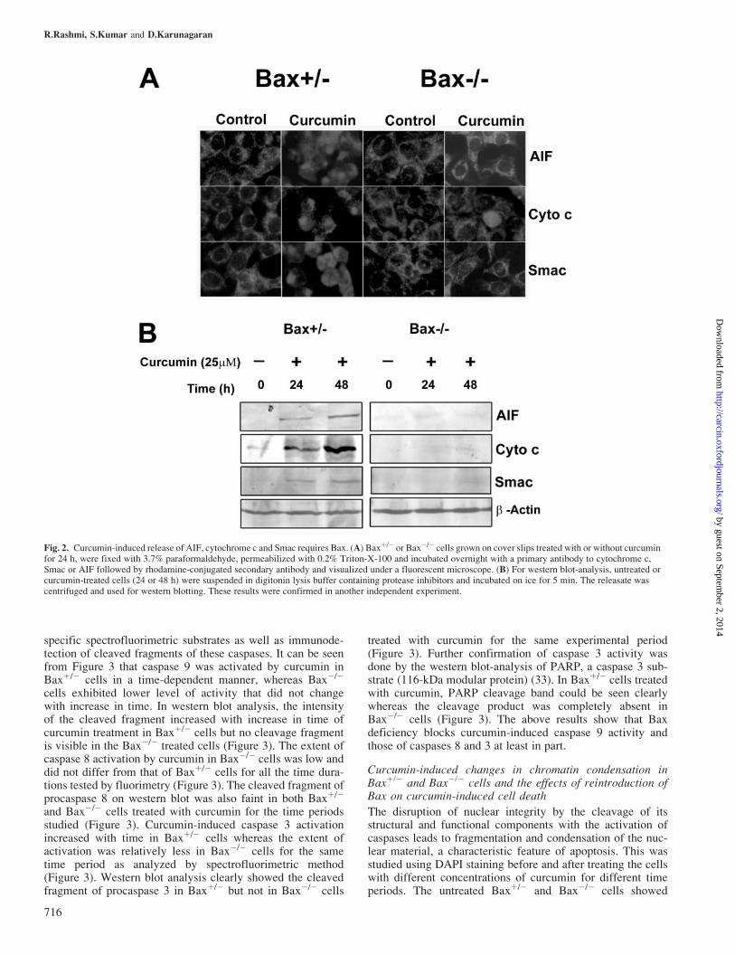
Release of cytochrome c, Smac or AIF from mitochondriaassessed by immunofluorescence or western blotting in thepresence or absence of curcumin
Early during apoptosis, cytochrome c, Smac and AIF, retainedwithin mitochondria, are released from the intermembranespace into the cytosol. We performed immunofluorescenceanalysis with specific antibodies for cytochrome c, Smac andAIF in Baxþ/� or Bax�/� cells treated with or without curcu-min (25 mM) for 24 h. As shown in Figure 2A, cytochrome c,Smac and AIF showed a granular pattern of staining (mito-chondrial localization) in untreated Baxþ/� or Bax�/� cellsand upon curcumin treatment a diffuse cytosolic distributionof cytochrome c, Smac and AIF was observed in Baxþ/� butnot Bax�/� cells. The patterns of release of cytochrome c,Smac and AIF into the cytosol were characterized further bywestern blotting of digitonin-permeabilized samples. Figure 2Bshows that upon curcumin treatment, all three moleculeswere released from Baxþ/� cells whereas their release wasalmost completely blocked in Bax�/� cells and b-actin levelsof all the fractions were similar. These data indicate thatcurcumin-induced release of the mitochondrial apoptogenicmolecules such as cytochrome c, Smac and AIF requires thepresence of Bax.
Effect of curcumin on the activities of caspases 9, 3 and 8assessed by spectrofluorometry and western blotting andPARP cleavage in Baxþ/� or Bax�/� cells
In many systems, caspases 8 and 9 act as initiators and caspase3, the effector, signals for the final execution of the cells.The processing of procaspases 9, 3 and 8 was assessed using
Fig. 1. Bax is required for curcumin-induced cell death. (A) Whole cellextracts (50 mg) prepared from HCT116 Baxþ/� or HCT116 Bax�/� cellswere separated on 12% SDS--PAGE and analyzed for Bax or b-actin (loadingcontrol) expression by western blotting. The experiment was repeated manytimes with similar results. (B) Baxþ/� or Bax�/� cells grown on 96-wellplates were exposed to different concentrations (0, 10, 25 or 50 mM) ofcurcumin for 24, 48 or 72 h and cell viability (expressed as a percentage overuntreated control) was determined by MTT assay. The mean values oftriplicate samples are shown and error bars indicate SDs and the experimentswere repeated three times with similar results. (C) Baxþ/� or Bax�/� cellsgrown on cover slips treated with or without curcumin for 24 h, werefixed with 3.7% paraformaldehyde, permeabilized with 0.0125% of3-[(3-cholamidopropyl) dimethylammonio]-1-propanesulfonic acid in PBSand incubated overnight with a primary antibody to Bax and then incubatedagain with rhodamine-conjugated secondary antibody and visualized undera fluorescent microscope. The experimental results were confirmed inanother independent experiment.
Requirement of Bax for curcumin-induced apoptosis
715
by guest on September 2, 2014
http://carcin.oxfordjournals.org/D
ownloaded from
specific spectrofluorimetric substrates as well as immunode-tection of cleaved fragments of these caspases. It can be seenfrom Figure 3 that caspase 9 was activated by curcumin inBaxþ/� cells in a time-dependent manner, whereas Bax�/�
cells exhibited lower level of activity that did not changewith increase in time. In western blot analysis, the intensityof the cleaved fragment increased with increase in time ofcurcumin treatment in Baxþ/� cells but no cleavage fragmentis visible in the Bax�/� treated cells (Figure 3). The extent ofcaspase 8 activation by curcumin in Bax�/� cells was low anddid not differ from that of Baxþ/� cells for all the time dura-tions tested by fluorimetry (Figure 3). The cleaved fragment ofprocaspase 8 on western blot was also faint in both Baxþ/�
and Bax�/� cells treated with curcumin for the time periodsstudied (Figure 3). Curcumin-induced caspase 3 activationincreased with time in Baxþ/� cells whereas the extent ofactivation was relatively less in Bax�/� cells for the sametime period as analyzed by spectrofluorimetric method(Figure 3). Western blot analysis clearly showed the cleavedfragment of procaspase 3 in Baxþ/� but not in Bax�/� cells
treated with curcumin for the same experimental period(Figure 3). Further confirmation of caspase 3 activity wasdone by the western blot-analysis of PARP, a caspase 3 sub-strate (116-kDa modular protein) (33). In Baxþ/� cells treatedwith curcumin, PARP cleavage band could be seen clearlywhereas the cleavage product was completely absent inBax�/� cells (Figure 3). The above results show that Baxdeficiency blocks curcumin-induced caspase 9 activity andthose of caspases 8 and 3 at least in part.
Curcumin-induced changes in chromatin condensation inBaxþ/� and Bax�/� cells and the effects of reintroduction ofBax on curcumin-induced cell death
The disruption of nuclear integrity by the cleavage of itsstructural and functional components with the activation ofcaspases leads to fragmentation and condensation of the nuc-lear material, a characteristic feature of apoptosis. This wasstudied using DAPI staining before and after treating the cellswith different concentrations of curcumin for different timeperiods. The untreated Baxþ/� and Bax�/� cells showed
Fig. 2. Curcumin-induced release of AIF, cytochrome c and Smac requires Bax. (A) Baxþ/� or Bax�/� cells grown on cover slips treated with or without curcuminfor 24 h, were fixed with 3.7% paraformaldehyde, permeabilized with 0.2% Triton-X-100 and incubated overnight with a primary antibody to cytochrome c,Smac or AIF followed by rhodamine-conjugated secondary antibody and visualized under a fluorescent microscope. (B) For western blot-analysis, untreated orcurcumin-treated cells (24 or 48 h) were suspended in digitonin lysis buffer containing protease inhibitors and incubated on ice for 5 min. The releasate wascentrifuged and used for western blotting. These results were confirmed in another independent experiment.
R.Rashmi, S.Kumar and D.Karunagaran
716
by guest on September 2, 2014
http://carcin.oxfordjournals.org/D
ownloaded from

Fig. 3. Bax deficiency blocks curcumin-induced activation of caspases (9, 8 and 3) and PARP cleavage. Whole cell extracts (50 mg) prepared from cellstreated with or without curcumin (25mM) for 0, 24 or 48 h were assessed for the activation of caspase 9 using a fluorimetric substrate (Ac-LEHD-AFC) (left panel)in a reaction buffer at 37�C for 1 h. Caspase 9 activation was also assessed by western blotting (right panel). Activation of caspase 3 by curcumin at the indicatedperiods of time was determined by using a fluorimetric substrate of caspase 3 (Ac-DEVD-AFC) (left panel) and by western blot-analysis (right panel).Caspase 8 activation was assessed using a spectrofluorimetric substrate (Z-IETD-AFC) (left panel) and western blot (right panel). All these experiments wererepeated at least two times with similar results and the error bars denote standard deviation. Lysates (60 mg of protein) prepared after treating the Baxþ/�
or Bax�/� cells with or without curcumin (25 mM) for the indicated time periods were analyzed for PARP with a specific polyclonal antibody.The experiment was repeated twice with similar results.
Requirement of Bax for curcumin-induced apoptosis
717
by guest on September 2, 2014
http://carcin.oxfordjournals.org/D
ownloaded from

uniform diffuse staining with DAPI (data not shown)whereas upon 10, 25 and 50 mM curcumin treatment, nuclearcondensation and fragmentation of Baxþ/� cells were observedin 20, 45 and 65% at 24 h, 25, 60 and 78% at 48 h and 30, 70
and 85% at 72 h, respectively (Figure 4A). Bax�/� cells treatedwith 10, 25 and 50 mM curcumin were found to exhibit apop-totic nuclei about 3, 7 and 12% at 24 h, 4, 10 and 18% at 48 hand 6, 14 and 10% at 72 h, respectively (Figure 4A). These
Fig. 4. Curcumin-induced condensation of chromatin and nuclei is prevented in Bax knockout cells and Bax transfection sensitizes them to curcumin-inducedapoptosis. (A) Cells were seeded on to cover slips, treated with or without curcumin (25 mM) for 24 h, fixed using 3% paraformaldehyde and stained withDAPI. Cells with condensed and fragmented chromatin were counted in five different fields and the mean values of triplicate samples expressed in percentage areshown and these results were confirmed in another independent experiment. (B) Baxþ/� and Bax�/� cells were pretreated with 50 mM of IETD-fmkor Z-VAD-fmk followed by curcumin for 24 h and then processed as described before for DAPI staining. Cells with condensed and fragmented chromatin werecounted in five different fields and the mean values of triplicate samples expressed in percentage are shown and another independent experiment confirmedthese results. (C) Baxþ/� or Bax�/� cells were stably transfected with vector alone or Bax--GFP construct using Lipofectamine as per manufacturer’s instructions(Life Technologies, Inc.) and whole cell extracts prepared from G418-resistant clones were analyzed for GFP expression by western blot (12% gel) and b-actinwas used as a loading control. The experiment was repeated at least two times with similar results. (D) Cells after transfection with control vector orBax--GFP were treated with or without curcumin and the migration of GFP-tagged Bax protein was monitored under fluorescent microscope and representativemicrographs are shown. (E) Cells with condensed and fragmented chromatin from the experiment described above were counted in five different fields andthe mean values of triplicate samples expressed in percentage are shown and another independent experiment confirmed these results.
R.Rashmi, S.Kumar and D.Karunagaran
718
by guest on September 2, 2014
http://carcin.oxfordjournals.org/D
ownloaded from
results clearly show that Bax is required for curcumin-inducednuclear condensation and fragmentation in human colon can-cer cells. To determine whether the rate of cell death in Baxþ/�
and Bax�/� cells is sensitive to caspase inhibition, we usedcaspase 8 inhibitor and a pan-caspase inhibitor with curcumintreatment. The cells were then stained with DAPI to observethe difference in the rate of cell death. It was found thatcurcumin treatment together with IETD-fmk (caspase 8 inhib-itor) showed no significant difference in cell death betweenBaxþ/� and Bax�/� cells compared to those treated with cur-cumin alone. However, in Baxþ/� cells treated with Z-VAD-fmk (pan-caspase inhibitor) and curcumin ~28% of cells wereapoptotic as compared with 10% in Bax�/� cells (Figure 4B).These results suggest that curcumin-induced apoptosis ismediated at least in part through caspases other than caspase 8.The above data suggest that the cells deficient in Bax resist
curcumin-induced apoptosis and to see how the addition ofBax into Baxþ/� or its reintroduction into Bax�/�cells, wouldaffect curcumin-induced translocation of Bax from the cytosolinto mitochondria, we have established stable clones express-ing Bax--GFP and used one of them for further experiments.Western blotting with an antibody to GFP confirmed the pres-ence of GFP in the stable clone (Bax--GFP) and its absencein the vector-transfected cells, while the b-actin levels wereunaltered in both the cells (Figure 4C). Upon curcumin treat-ment granular mitochondrial GFP staining was observedwhereas the untreated cells showed cytosolic pattern of stain-ing indicating the migration of overexpresed Bax--GFP fusionprotein to the mitochondria in Baxþ/� or Bax�/�cells(Figure 4D). Baxþ/� cells (containing one allele of Bax) trans-fected either with the control vector or with Bax--GFP respon-ded to curcumin whereas the Bax�/� cells responded well tocurcumin only when transfected with Bax--GFP but not to thecontrol vector (Figure 4E). These experiments confirm theimportant role of Bax in the induction of apoptosis in humancolon cancer cells by curcumin.
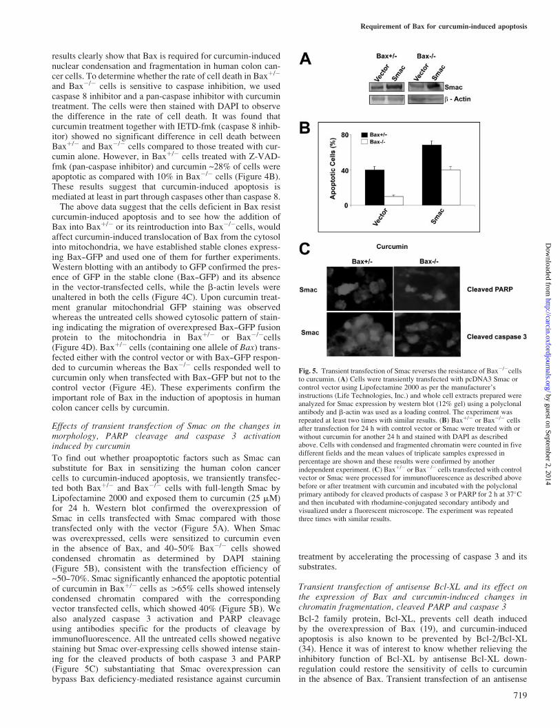
Effects of transient transfection of Smac on the changes inmorphology, PARP cleavage and caspase 3 activationinduced by curcumin
To find out whether proapoptotic factors such as Smac cansubstitute for Bax in sensitizing the human colon cancercells to curcumin-induced apoptosis, we transiently transfec-ted both Baxþ/� and Bax�/� cells with full-length Smac byLipofectamine 2000 and exposed them to curcumin (25 mM)for 24 h. Western blot confirmed the overexpression ofSmac in cells transfected with Smac compared with thosetransfected only with the vector (Figure 5A). When Smacwas overexpressed, cells were sensitized to curcumin evenin the absence of Bax, and 40--50% Bax�/� cells showedcondensed chromatin as determined by DAPI staining(Figure 5B), consistent with the transfection efficiency of~50--70%. Smac significantly enhanced the apoptotic potentialof curcumin in Baxþ/� cells as 465% cells showed intenselycondensed chromatin compared with the correspondingvector transfected cells, which showed 40% (Figure 5B). Wealso analyzed caspase 3 activation and PARP cleavageusing antibodies specific for the products of cleavage byimmunofluorescence. All the untreated cells showed negativestaining but Smac over-expressing cells showed intense stain-ing for the cleaved products of both caspase 3 and PARP(Figure 5C) substantiating that Smac overexpression canbypass Bax deficiency-mediated resistance against curcumin
treatment by accelerating the processing of caspase 3 and itssubstrates.
Transient transfection of antisense Bcl-XL and its effect onthe expression of Bax and curcumin-induced changes inchromatin fragmentation, cleaved PARP and caspase 3
Bcl-2 family protein, Bcl-XL, prevents cell death inducedby the overexpression of Bax (19), and curcumin-inducedapoptosis is also known to be prevented by Bcl-2/Bcl-XL(34). Hence it was of interest to know whether relieving theinhibitory function of Bcl-XL by antisense Bcl-XL down-regulation could restore the sensitivity of cells to curcuminin the absence of Bax. Transient transfection of an antisense
Fig. 5. Transient transfection of Smac reverses the resistance of Bax�/�cellsto curcumin. (A) Cells were transiently transfected with pcDNA3 Smac orcontrol vector using Lipofectamine 2000 as per the manufacturer’sinstructions (Life Technologies, Inc.) and whole cell extracts prepared wereanalyzed for Smac expression by western blot (12% gel) using a polyclonalantibody and b-actin was used as a loading control. The experiment wasrepeated at least two times with similar results. (B) Baxþ/� or Bax�/� cellsafter transfection for 24 h with control vector or Smac were treated with orwithout curcumin for another 24 h and stained with DAPI as describedabove. Cells with condensed and fragmented chromatin were counted in fivedifferent fields and the mean values of triplicate samples expressed inpercentage are shown and these results were confirmed by anotherindependent experiment. (C) Baxþ/� or Bax�/� cells transfected with controlvector or Smac were processed for immunofluorescence as described abovebefore or after treatment with curcumin and incubated with the polyclonalprimary antibody for cleaved products of caspase 3 or PARP for 2 h at 37�Cand then incubated with rhodamine-conjugated secondary antibody andvisualized under a fluorescent microscope. The experiment was repeatedthree times with similar results.
Requirement of Bax for curcumin-induced apoptosis
719
by guest on September 2, 2014
http://carcin.oxfordjournals.org/D
ownloaded from

construct of Bcl-XL (pcDNA3 AsBcl-XL) significantlydownregulated the level of expression of Bcl-XL in Baxþ/�
or Bax�/� cells while it had no effect on b-actin levelsas confirmed by western blot analysis (Figure 6A). DAPIstaining indicated that ~60% of Bax�/� cells showed con-densed nuclear morphology when Bcl-XL is downregulatedcompared with 12% of vector-transfected cells, and 85%of antisense Bcl-XL transfected Baxþ/� cells were highlyapoptotic compared with 45% in the corresponding vector-transfected cells (Figure 6B). Immunofluorescent detectionof active caspase 3 and cleaved PARP also supported thefinding that antisense inhibition of Bcl-XL restored theresponsiveness to curcumin in Bax�/� cells while enhancing
it in Baxþ/� cells (Figure 6C). These results suggest thatdownregulation of Bcl-XL is an effective option to sensitizehuman colon cancer cells deficient in Bax.
Effect of Bak RNAi on curcumin-induced apoptosis of Baxþ/�
and Bax�/� cells
To understand the role of Bak in curcumin-induced apoptosis,Baxþ/� or Bax�/� cells were treated with or without curcumin
Fig. 6. Downregulation of Bcl-XL is sufficient to overcome the resistanceof Bax�/� cells to curcumin. (A) Cells were transiently transfected withpcDNA3 AsBcl-XL or control vector with Lipofectamine 2000 as per themanufacturer’s instructions (Life Technologies, Inc.) and whole cell extractsprepared were analyzed for Bcl-XL expression by western blot (12% gel)using a polyclonal antibody and b-actin was used as a loading control and theexperiment repeated at least two times with similar results. (B) Baxþ/� orBax�/� cells (24 h after transfection with pcDNA3 AsBcl-XL or controlvector) were treated with or without curcumin for 24 h and stained withDAPI as described above. Cells with condensed and fragmented chromatinfrom the experiment described were counted in five different fields and themean values of triplicate samples expressed in percentage are shown andthese results were confirmed by another independent experiment. (C) Baxþ/�
or Bax�/� cells (24 h after transfection with pcDNA3 AsBcl-XL or controlvector) were treated with or without curcumin for 24 h, processed asdescribed previously and incubated with a polyclonal primary antibody forcleaved caspase 3 or PARP for 2 h at 37�C and then incubated withrhodamine-conjugated secondary antibody and visualized underfluorescent microscope. The experiment was repeated three times withsimilar results.
Fig. 7. Bax is more important than Bak in curcumin-induced apoptosis.(A) Baxþ/� or Bax�/� cells were treated with or without curcumin for 24 h,processed for activated Bak as previously described. The cells wereincubated with a monoclonal primary antibody for conformational specificBak for 2 h at 37�C and then incubated with rhodamine-conjugatedsecondary antibody and visualized under fluorescent microscope. Theexperiment was repeated three times with similar results. (B) Cells weretransiently transfected with pBS/U6 Bak RNAi or control vector withLipofectamine 2000 as per the manufacturer’s instructions (LifeTechnologies, Inc.) and whole cell extracts prepared were analyzed forBcl-XL expression by western blot (15% gel) using a monoclonal antibodyand b-actin was used as a loading control and the experiment repeated atleast two times with similar results. (C) Baxþ/� or Bax�/� cells (24 h aftertransfection with pBS/U6 Bak RNAi or control vector) were treated with orwithout curcumin for 24 h, processed for DAPI as described earlier.Apoptotic cells with condensed and fragmented chromatin were counted infive different fields and the mean values of triplicate samples expressed inpercentage are shown and these results were confirmed by anotherindependent experiment.
R.Rashmi, S.Kumar and D.Karunagaran
720
by guest on September 2, 2014
http://carcin.oxfordjournals.org/D
ownloaded from

for 24 h and immunostained with conformational specificantibody to detect activated Bak. Both Baxþ/� and Bax�/�
cells were positive for activated Bak upon curcumin treatmentbut not the untreated control cells (Figure 7A). To understandthe relative importance of Bax and Bak in curcumin-inducedapoptosis Baxþ/� or Bax�/� cells were transiently transfectedwith Bak RNAi vector by Lipofectamine 2000 to downregu-late Bak. Furthermore, western blotting confirmed the down-regulation of Bak in RNAi transfected cells (Figure 7B). After24 h of transfection with Bak RNAi vector, the cells weretreated with curcumin (25 mM) for 24 h, and there was noconsiderable difference in the percentage of apoptotic cells inBak RNAi transfected Baxþ/� or Bax�/� cells compared withtheir corresponding vector-transfected cells treated with cur-cumin (Figure 7C). In Bcl-XL downregulated or Smac over-expressed Baxþ/� and Bax�/� cells, there was no difference inthe staining for activated Bak (data not shown).The above results when considered together, demonstrate
the requirement of Bax but not Bak in curcumin-inducedapoptosis, but this requirement is dispensable if the cells canexpress additional amounts of alternative proapoptoticmolecules such as Smac or downregulate antiapoptoticmolecules like Bcl-XL under Bax-deficient conditions.
Discussion
Deficiency of Bax is a frequent cause for resistance againsttherapy in at least 15% of human colon, gastric and endo-metrial cancers (35--37). In this study we demonstrate for thefirst time that Bax is required for curcumin-induced apoptosis,particularly for the release of apoptogenic molecules, cyto-chrome c, Smac and AIF from the mitochondria. However,the requirement for Bax can be bypassed with ectopic expres-sion of Smac or downregulation of Bcl-XL to sensitize thehuman colon adenocarcinoma cells to curcumin-induced apop-tosis. The Bax-deficient derivative of HCT116 human coloncancer cells used in our study is partially resistant to theapoptotic effects of the chemotherapeutic agent 5-fluorouraciland totally resistant to the chemopreventive agent sulindac andother non-steroidal antiinflammatory drugs (26). In addition,this cell line is resistant to apoptosis induced by a variety ofagents including UV, bortezomib, staurosporine, tumor nec-rosis factor-related apoptosis-inducing ligand (TRAIL) andthapsigargin (18,24,38,39). Proapoptotic drugs such asstaurosporine and actinomycin D require the presence of Baxto induce cytochrome c release and cell death (40). Curcuminacts through both mitochondrial- and receptor-mediated apop-totic pathways (34) and we have shown earlier that it inducesthe release of cytochrome c, Smac and AIF in human coloncancer cells (30,31). Partial but not complete activation ofcaspase 8 by curcumin observed in Bax�/� cells is consistentwith the notion that although caspase 8 activation is upstreamof Bax the complete activation of caspase 8 requires the inter-action of Bax with Bid, a known substrate of caspase 8 (14,15).Curcumin-induced release of Smac into the cytosol from
Baxþ/� and Bax�/� cells transfected with Smac is likely tohave contributed to the sequestering of IAPs and the con-sequent activation of caspases 9 and 3, and PARP cleavage(41). Under the conditions of Smac transfection, sufficientamount of Smac may be available in the cytosol to inhibitthe IAPs thereby enhancing caspase activity which in turnacts on the mitochondria to amplify the release of cytochrome
c and such a feedback amplification loop has been reportedearlier (42). Smac enhances the sensitivity of Jurkat cells toTRAIL and Epothilone B derivative-induced apoptosis (43)and stable expression of Smac in neuroblastoma cellssensitized them to apoptosis induced by TRAIL, cisplatin,doxorubicin and etoposide by the activation of caspases andcleavage of caspase substrates (44). Overexpression of Smacrestores the apoptotic sensitivity of Bax knockout cells tothapsigargin-induced apoptosis (24). Infection of ovariancarcinoma cells with a recombinant adenovirus encodingSmac leads to cell death at multiplicities of infection in aconcentration-dependent manner independent of cytochromec release (45).Bcl-XL and Bak predominantly reside on the mitochondrial
outer membrane surface (46,47) and accumulation of Baxmultimers in mitochondria is essential to neutralize the func-tion of Bcl-XL and to activate Bak-induced release of cyto-chrome c (19). From the present study it is apparent that Bakconformational specific change in response to curcumin treat-ment is independent of Bax, Smac overexpression or Bcl-XLdownregulation. The active Bak alone cannot induce cyto-chrome c release or other downstream events leading to celldeath in the absence of Bax. Our findings suggest that Bakconformational change may be an early step in curcumin-induced apoptosis. In the absence of Bax the effect of Bak isneutralized by Bcl-XL thus preventing Bak-mediated cyto-chrome c release. It has been reported that the truncated Bidcan serve as a death ligand which moves from the cytosol tothe mitochondria to activate the Bak- or Bax-induced cyto-chrome c release (40,48). Once Bcl-XL is downregulated, Bidcan probably induce the release of cytochrome c by oligomer-izing Bak, independent of Bax. Even in the absence of Bax,under the condition of downregulated levels of Bcl-XL, cur-cumin may have induced conformational changes in Bakthereby inducing the release of cytochrome c and cell death.It is possible that mitochondrial Bak is always kept in aninactive conformation by its close association with the antiap-optotic protein Bcl-XL which is relatively more abundantthan Bcl-2 in Bax�/� cells (R.Rashmi, S.Kumar andD.Karunagaran, unpublished data). Mouse embryonic fibro-blasts defective in both Bax and Bak were resistant to apop-tosis induced by overexpression of various BH3 proteins suchas Bid, Bim and Noxa (20). However, cells deficient in eitherBax or Bak were not significantly defective when apoptosiswas induced by various agents, suggesting the importance ofrelative expression of proapoptotic and antiapoptotic pro-teins in chemoresistance. Presumably the proteins of Bcl-2family interact with each other to set a survival thresholdfor the cell.Although many studies suggest that AIF is caspase-
independent (12,49) some reports suggest that caspase inhib-itors prevent the effects of AIF favoring the idea that at least,part of its action is caspase-dependent (50,51). Further studiesare needed to clarify the mechanisms by which AIF is releasedfrom mitochondria in response to curcumin. The present studyhas highlighted Bax and not Bak as a critical regulator ofcurcumin-induced apoptosis and the results implicate Baxreintroduction, Smac transfection and Bcl-XL downregulationas novel and potential strategies to sensitize Bax-deficienttumors resistant to curcumin therapy. Our findings supportthe notion that the relative ratio of proapoptotic and anti-apoptotic proteins in a cell determines their sensitivity todrug-induced apoptosis highlighting the contribution of
Requirement of Bax for curcumin-induced apoptosis
721
by guest on September 2, 2014
http://carcin.oxfordjournals.org/D
ownloaded from
relative expression of proapoptotic and antiapoptotic proteinsto chemoresistance.
Acknowledgements
This work was supported by funding from the Kerala State Council forScience, Technology and the Environment (D.K.), a grant from the LifeSciences Research Board, Defence Research Development Organization,Government of India (D.K. and S.K.) and a Senior Research Fellowship (R.R.)of the Council of Scientific and Industrial Research, Government of India.
References
1.Martinou,J.C. and Green,D.R. (2001) Breaking the mitochondrial barrier.Nature Rev. Mol. Cell. Biol., 2, 63--67.
2.Liu,X., Kim,C.N., Yang,J., Jemmerson,R. and Wang,X. (1996) Inductionof apoptotic program in cell-free extracts: requirement for dATP andcytochrome c. Cell, 86, 147--157.
3.Li,P., Nijhawan,D., Budihardjo,I., Srinivasula,S.M., Ahmad,M.,Alnemri,E.S. and Wang,X. (1997) Cytochrome c and dATP-dependentformation of Apaf-1/caspase-9 complex initiates an apoptotic proteasecascade. Cell, 91, 479--489.
4.Zou,H., Li,Y., Liu,X. and Wang,X. (1999) An APAF-1 � cytochromec multimeric complex is a functional apoptosome that activatesprocaspase-9. J. Biol. Chem., 274, 11549--11556.
5.Slee,E.A., Adrain,C. and Martin,S.J. (1999) Serial killers: ordering caspaseactivation events in apoptosis. Cell Death Differ., 6, 1067--1074.
6.Du,C., Fang,M., Li,Y., Li,L. and Wang,X. (2000) Smac, a mitochondrialprotein that promotes cytochrome c-dependent caspase activation byeliminating IAP inhibition. Cell, 102, 33--42.
7.Verhagen,A.M., Ekert,P.G., Pakusch,M., Silke,J., Connolly,L.M.,Reid,G.E., Moritz,R.L., Simpson,R.J. and Vaux,D.L. (2000)Identification of DIABLO, a mammalian protein that promotes apoptosisby binding to and antagonizing IAP proteins. Cell, 102, 43--53.
8.Suzuki,Y., Imai,Y., Nakayama,H., Takahashi,K., Takio,K. andTakahashi,R. (2001) A serine protease, HtrA2, is released from themitochondria and interacts with XIAP, inducing cell death. Mol. Cell, 8,613--621.
9.Hegde,R., Srinivasula,S.M., Zhang,Z., Wassell,R., Mukattash,R.,Cilenti,L., DuBois,G., Lazebnik,Y., Zervos,A.S., Fernandes-Alnemri,T.and Alnemri,E.S. (2002) Identification of Omi/HtrA2 as a mitochondrialapoptotic serine protease that disrupts inhibitor of apoptosis protein--caspaseinteraction. J. Biol. Chem., 277, 432--438.
10.Martins,L.M., Iaccarino,I., Tenev,T., Gschmeissner,S., Totty,N.F.,Lemoine,N.R., Savopoulos,J., Gray,C.W., Creasy,C.L., Dingwall,C. andDownward,J. (2002) The serine protease Omi/HtrA2 regulatesapoptosis by binding XIAP through a reaper-like motif. J. Biol. Chem.,277, 439--444.
11.Verhagen,A.M., Silke,J., Ekert,P.G., Pakusch,M., Kaufmann,H.,Connolly,L.M., Day,C.L., Tikoo,A., Burke,R., Wrobel,C., Moritz,R.L.,Simpson,R.J. and Vaux,D.L. (2002) HtrA2 promotes cell death through itsserine protease activity and its ability to antagonize inhibitor of apoptosisproteins. J. Biol. Chem., 277, 445--454.
12.Susin,S.A., Lorenzo,H.K., Zamzami,N., Marzo,I., Snow,B.E.,Brothers,G.M., Mangion,J., Jacotot,E., Costantini,P., Loeffler,M.,Larochette,N., Goodlett,D.R., Aebersold,R., Siderovski,D.P.,Penninger,J.M. and Kroemer,G. (1999) Molecular characterization ofmitochondrial apoptosis-inducing factor. Nature, 397, 441--446.
13.Li,L.Y., Luo,X. and Wang,X. (2001) Endonuclease G is an apoptoticDNase when released from mitochondria. Nature, 412, 95--99.
14.Li,H., Zhu,H., Xu,C.J. and Yuan,J. (1998) Cleavage of BID by caspase 8mediates the mitochondrial damage in the Fas pathway of apoptosis. Cell,94, 491--501.
15.Korsmeyer,S.J., Wei,M.C., Saito,M., Weiler,S., Oh,K.J. andSchlesinger,P.H. (2000) Pro-apoptotic cascade activates BID, whicholigomerizes BAK or BAX into pores that result in the release ofcytochrome c. Cell Death Differ., 7, 1166--1173.
16.Chao,D.T. and Korsmeyer,S.J. (1998) BCL-2 family: regulators of celldeath. Annu. Rev. Immunol., 16, 395--419.
17.Nechushtan,A., Smith,C.L., Lamensdorf,I., Yoon,S.H. and Youle,R.J.(2001) Bax and Bak coalesce into novel mitochondria-associated clustersduring apoptosis. J. Cell Biol., 153, 1265--1276.
18.Theodorakis,P., Lomonosova,E. and Chinnadurai,G. (2002) Criticalrequirement of BAX for manifestation of apoptosis induced by multiplestimuli in human epithelial cancer cells. Cancer Res., 62, 3373--3376.
19.Griffiths,G.J., Dubrez,L., Morgan,C.P., Jones,N.A., Whitehouse,J.,Corfe,B.M., Dive,C. and Hickman,J.A. (1999) Cell damage-inducedconformational changes of the pro-apoptotic protein Bak in vivo precedethe onset of apoptosis. J. Cell Biol., 144, 903--914.
20.Kandasamy,K., Srinivasula,S.M., Alnemri,E.S., Thompson,C.B.,Korsmeyer,S.J., Bryant,J.L. and Srivastava,R.K. (2003) Involvement ofproapoptotic molecules Bax and Bak in tumor necrosis factor-relatedapoptosis-inducing ligand (TRAIL)-induced mitochondrial disruption andapoptosis: differential regulation of cytochrome c and Smac/DIABLOrelease. Cancer Res., 63, 1712--1721.
21.Hsu,Y.T. and Youle,R.J. (1998) Bax in murine thymus is a solublemonomeric protein that displays differential detergent-induced conforma-tions. J. Biol. Chem., 273, 10777--10783.
22.Makin,G.W., Corfe,B.M., Griffiths,G.J., Thistlethwaite,A., Hickman,J.A.and Dive,C. (2001) Damage-induced Bax N-terminal change, transloca-tion to mitochondria and formation of Bax dimers/complexes occurregardless of cell fate. EMBO J., 20, 6306--6315.
23.Suzuki,M., Youle,R.J. and Tjandra,N. (2000) Structure of Bax: coregu-lation of dimer formation and intracellular localization. Cell, 103,645--654.
24.Yamaguchi,H., Bhalla,K. and Wang,H.G. (2003) Bax plays a pivotal rolein thapsigargin-induced apoptosis of human colon cancer HCT116 cellsby controlling Smac/Diablo and Omi/HtrA2 release from mitochondria.Cancer Res., 63, 1483--1489.
25.Rampino,N., Yamamoto,H., Ionov,Y., Li,Y., Sawai,H., Reed,J.C. andPerucho,M. (1997) Somatic frameshift mutations in the BAX gene incolon cancers of the microsatellite mutator phenotype. Science, 275,967--969.
26.Zhang,L., Yu,J., Park,B.H., Kinzler,K.W. and Vogelstein,B. (2000) Roleof BAX in the apoptotic response to anticancer agents. Science, 290,989--992.
27.Kuo,M.L., Huang,T.S. and Lin,J.K. (1996) Curcumin, an antioxidant andanti-tumor promoter, induces apoptosis in human leukemia cells. Biochim.Biophys. Acta, 1317, 95--100.
28.Piwocka,K., Zablocki,K., Wieckowski,M.R., Skierski,J., Feiga,I., Szopa,J.,Drela,N., Wojtczak,L. and Sikora,E. (1999) A novel apoptosis-likepathway, independent of mitochondria and caspases, induced by curcuminin human lymphoblastoid T (Jurkat) cells. Exp. Cell Res., 249, 299--307.
29.Chen,H., Zhang,Z.S., Zhang,Y.L. and Zhou,D.Y. (1999) Curcumininhibits cell proliferation by interfering with the cell cycle and inducingapoptosis in colon carcinoma cells. Anticancer Res., 19, 3675--3680.
30.Rashmi,R., Santhosh Kumar,T.R. and Karunagaran,D. (2003) Humancolon cancer cells differ in their sensitivity to curcumin-induced apoptosisand heat shock protects them by inhibiting the release of apoptosis-inducing factor and caspases. FEBS Lett., 538, 19--24.
31.Rashmi,R., Kumar,S. and Karunagaran,D. (2004) Ectopic expression ofHsp70 confers resistance and silencing its expression sensitizes humancolon cancer cells to curcumin-induced apoptosis. Carcinogenesis, 25,179--187.
32.Alley,M.C., Scudiero,D.A., Monks,A., Hursey,M.L., Czerwinski,M.J.,Fine,D.L., Abbott,B.J., Mayo,J.G., Shoemaker,R.H. and Boyd,M.R.(1988) Feasibility of drug screening with panels of human tumor celllines using a microculture tetrazolium assay. Cancer Res., 48, 589--601.
33.Tewari,M., Quan,L.T., O’Rourke,K., Desnoyers,S., Zeng,Z., Beidler,D.R.,Poirier,G.G., Salvesen,G.S. and Dixit,V.M. (1995) Yama/CPP32 beta,a mammalian homolog of CED-3, is a CrmA-inhibitable proteasethat cleaves the death substrate poly(ADP-ribose) polymerase. Cell, 81,801--809.
34.Anto,R.J., Mukhopadhyay,A., Denning,K. and Aggarwal,B.B. (2002)Curcumin (diferuloylmethane) induces apoptosis through activation ofcaspase-8, BID cleavage and cytochrome c release: its suppression byectopic expression of Bcl-2 and Bcl-xl. Carcinogenesis, 23, 143--150.
35. Ionov,Y., Yamamoto,H., Krajewski,S., Reed,J.C. and Perucho,M. (2000)Mutational inactivation of the proapoptotic gene BAX confers selectiveadvantage during tumor clonal evolution. Proc. Natl Acad. Sci. USA, 97,10872--10877.
36.Kitada,S., Krajewska,M., Zhang,X., Scudiero,D., Zapata,J.M., Wang,H.G.,Shabaik,A., Tudor,G., Krajewski,S., Myers,T.G., Johnson,G.S.,Sausville,E.A. and Reed,J.C. (1998) Expression and location of pro-apoptotic Bcl-2 family protein BAD in normal human tissues and tumorcell lines. Am. J. Pathol., 152, 51--61.
37.Perucho,M. (1996) Cancer of the microsatellite mutator phenotype. Biol.Chem., 377, 675--684.
R.Rashmi, S.Kumar and D.Karunagaran
722
by guest on September 2, 2014
http://carcin.oxfordjournals.org/D
ownloaded from

38.Ravi,R. and Bedi,A. (2002) Requirement of BAX for TRAIL/Apo2L-induced apoptosis of colorectal cancers: synergism with sulindac-mediatedinhibition of Bcl-xL. Cancer Res., 62, 1583--1587.
39.Yu,J., Tiwari,S., Steiner,P. and Zhang,L. (2003) Differential apoptoticresponse to the proteasome inhibitor bortezomib [VELCADE(TM),PS-341] in Bax-deficient and p21-deficient colon cancer cells. CancerBiol. Ther., 2, 694--699.
40.Wei,M.C., Zong,W.X., Cheng,E.H., Lindsten,T., Panoutsakopoulou,V.,Ross,A.J., Roth,K.A., MacGregor,G.R., Thompson,C.B. andKorsmeyer,S.J. (2001) Proapoptotic BAX and BAK: a requisite gatewayto mitochondrial dysfunction and death. Science, 292, 727--730.
41.Srinivasula,S.M., Hegde,R., Saleh,A., Datta,P., Shiozaki,E., Chai,J.,Lee,R.A., Robbins,P.D., Fernandes-Alnemri,T., Shi,Y. and Alnemri,E.S.(2001) A conserved XIAP-interaction motif in caspase-9 and Smac/DIABLO regulates caspase activity and apoptosis. Nature, 410, 112--116.
42.Marzo,I., Susin,S.A., Petit,P.X., Ravagnan,L., Brenner,C., Larochette,N.,Zamzami,N. and Kroemer,G. (1998) Caspases disrupt mitochondrialmembrane barrier function. FEBS Lett., 427, 198--202.
43.Guo,F., Nimmanapalli,R., Paranawithana,S., Wittman,S., Griffin,D.,Bali,P., O’Bryan,E., Fumero,C., Wang,H.G. and Bhalla,K. (2002)Ectopic overexpression of second mitochondria-derived activator ofcaspases (Smac/DIABLO) or cotreatment with N-terminus of Smac/DIABLO peptide potentiates epothilone B derivative-(BMS 247550) andApo-2L/TRAIL-induced apoptosis. Blood, 99, 3419--3426.
44.Fulda,S., Wick,W., Weller,M. and Debatin,K.M. (2002) Smac agonistssensitize for Apo2L/TRAIL- or anticancer drug-induced apoptosis andinduce regression of malignant glioma in vivo. Nature Med., 8,808--815.
45.McNeish,I.A., Bell,S., McKay,T., Tenev,T., Marani,M. and Lemoine,N.R.(2003) Expression of Smac/DIABLO in ovarian carcinoma cells inducesapoptosis via a caspase-9-mediated pathway. Exp. Cell Res., 286,186--198.
46.Wang,K., Yin,X.M., Chao,D.T., Milliman,C.L. and Korsmeyer,S.J. (1996)BID: a novel BH3 domain-only death agonist. Genes Dev., 10, 2859--2869.
47.Rao,L., Modha,D. and White,E. (1997) The E1B 19K protein associateswith lamins in vivo and its proper localization is required for inhibition ofapoptosis. Oncogene, 15, 1587--1597.
48.Wei,M.C., Lindsten,T., Mootha,V.K., Weiler,S., Gross,A., Ashiya,M.,Thompson,C.B. and Korsmeyer,S.J. (2000) tBID, a membrane-targeteddeath ligand, oligomerizes BAK to release cytochrome c. Genes Dev., 14,2060--2071.
49.Loeffler,M., Daugas,E., Susin,S.A., Zamzami,N., Metivier,D.,Nieminen,A.L., Brothers,G., Penninger,J.M. and Kroemer,G. (2001)Dominant cell death induction by extramitochondrially targetedapoptosis-inducing factor. FASEB J., 15, 758--767.
50.Arnoult,D., Gaume,B., Karbowski,M., Sharpe,J.C., Cecconi,F. andYoule,R.J. (2003) Mitochondrial release of AIF and EndoG requirescaspase activation downstream of Bax/Bak-mediated permeabilization.EMBO J., 22, 4385--4399.
51.Arnoult,D., Karbowski,M. and Youle,R.J. (2003) Caspase inhibitionprevents the mitochondrial release of apoptosis-inducing factor. CellDeath Differ., 10, 845--849.
Received May 17, 2004; revised December 23, 2004;accepted January 1, 2005
Requirement of Bax for curcumin-induced apoptosis
723
by guest on September 2, 2014
http://carcin.oxfordjournals.org/D
ownloaded from