Distinct Functions for Arf Guanine Nucleotide Exchange Factors at the Golgi Complex: GBF1 and BIGs...
13
Molecular Biology of the Cell Vol. 19, 523–535, February 2008 Distinct Functions for Arf Guanine Nucleotide Exchange Factors at the Golgi Complex: GBF1 and BIGs Are Required for Assembly and Maintenance of the Golgi Stack and trans-Golgi Network, Respectively Florin Manolea,* Alejandro Claude, † Justin Chun,* Javier Rosas, † and Paul Melanc ¸on* *Department of Cell Biology, University of Alberta, Edmonton, AB, T6G 2H7 Canada; and † Instituto de Bioquı ´mica, Universidad Austral de Chile, Campus Isla Teja, Casilla 567, Valdivia, Chile Submitted May 1, 2007; Revised October 31, 2007; Accepted November 6, 2007 Monitoring Editor: Benjamin Glick We examined the relative function of the two classes of guanine nucleotide exchange factors (GEFs) for ADP-ribosylation factors that regulate recruitment of coat proteins on the Golgi complex. Complementary overexpression and RNA-based knockdown approaches established that GBF1 regulates COPI recruitment on cis-Golgi compartments, whereas BIGs appear specialized for adaptor proteins on the trans-Golgi. Knockdown of GBF1 and/or COPI did not prevent export of VSVGtsO45 from the endoplasmic reticulum (ER), but caused its accumulation into peripheral vesiculotubular clusters. In contrast, knockdown of BIG1 and BIG2 caused loss of clathrin adaptor proteins and redistribution of several TGN markers, but had no impact on COPI and several Golgi markers. Surprisingly, brefeldin A–inhibited guanine nucleotide exchange factors (BIGs) knockdown prevented neither traffic of VSVGtsO45 to the plasma membrane nor assembly of a polarized Golgi stack. Our observations indicate that COPII is the only coat required for sorting and export from the ER exit sites, whereas GBF1 but not BIGs, is required for COPI recruitment, Golgi subcompartmentalization, and cargo progression to the cell surface. INTRODUCTION The Golgi complex, the central sorting station of the secre- tory pathway, exhibits a very intricate organization that likely reflects the complexity of trafficking and sorting events that take place within this organelle (Rambourg and Clermont, 1990; Mogelsvang et al., 2004). It comprises three main structural elements: two extensive tubular-reticular networks flanking the cis- and trans-sides of a central stack of flat disk-shaped cisternae. The flat cisternae that form the core of the Golgi complex (compact zones) appear intercon- nected by lateral tubular networks (noncompact zones) to form a continuous membrane ribbon (Rambourg and Cler- mont, 1990; Mogelsvang et al., 2004). Subcellular fraction- ation and immunocytochemical analysis further established that cis-, medial, and trans-Golgi elements contain different sets of resident enzymes and that the Golgi complex is therefore functionally compartmentalized (Farquhar and Palade, 1998; Polishchuk and Mironov, 2004). For example, the early acting enzyme mannosidase I localizes to cis-Golgi membranes, whereas later acting ones such mannosidase II (ManII) and sialyltransferase concentrate in medial and trans compartments, respectively. Imaging of live cells revealed that the Golgi complex is not static as initially assumed from its intricate structure, but rather surprisingly dynamic and linked to several other organelles by active bidirectional transport routes (Boni- facino and Glick, 2004). In animal cells, cargo initially trans- located into the endoplasmic reticulum (ER) is selected for transport from specialized ER exit sites (ERES) to vesiculo- tubular clusters (VTCs; Fromme and Schekman, 2005; Tang et al., 2005). Shortly after their formation, these pleiomorphic carriers are transported on microtubules toward the Golgi complex (Presley et al., 1997; Scales et al., 1997) where they collect, fuse into a network, and subsequently become a flattened cis-Golgi cisterna (Bonifacino and Glick, 2004); pe- ripheral VTCs and those accumulated near the Golgi are collectively called ER-Golgi intermediate compartment or (ERGIC; Appenzeller-Herzog and Hauri, 2006). Current ev- idence suggests that cargo molecules then progress through the Golgi stack by a process termed cisternal maturation whereby newly formed cis-cisternae containing cargo pro- gressively move toward the trans side as they lose early acting Golgi enzymes and acquire late acting ones (Puthen- veedu and Linstedt, 2005; Losev et al., 2006; Matsuura-Tokita et al., 2006). At the trans-Golgi network (TGN), cargo is then This article was published online ahead of print in MBC in Press (http://www.molbiolcell.org/cgi/doi/10.1091/mbc.E07– 04 – 0394) on November 14, 2007. Address correspondence to: Paul Melanc ¸on (Paul.Melancon@ UAlberta.ca). Abbreviations used: AP, adaptor protein; Arf, ADP-ribosylation factor; BFA, brefeldin A; BIG, brefeldin A–inhibited guanine nucle- otide exchange factor; COP, coat protein; ERES, ER exit sites; ERGIC, ER-Golgi intermediate compartment; GBF, Golgi-specific brefeldin A resistance factor; GEF, guanine nucleotide exchange factor; GGA, gamma ear Golgi-localized Arf-binding protein; IF, immunofluorescence; ManII, mannosidase II; KD, knockdown; PM, plasma membrane; shRNA, short hairpin RNA; siRNA, small in- hibitory RNA; TGN, trans-Golgi Network; VSVG, vesicular stoma- titis virus glycoprotein; VTC, vesiculotubular cluster. © 2008 by The American Society for Cell Biology 523 http://www.molbiolcell.org/content/suppl/2007/11/14/E07-04-0394.DC1.html Supplemental Material can be found at:
-
Upload
independent -
Category
Documents
-
view
0 -
download
0
Transcript of Distinct Functions for Arf Guanine Nucleotide Exchange Factors at the Golgi Complex: GBF1 and BIGs...
Molecular Biology of the CellVol. 19, 523–535, February 2008
Distinct Functions for Arf Guanine Nucleotide ExchangeFactors at the Golgi Complex: GBF1 and BIGs AreRequired for Assembly and Maintenance of the GolgiStack and trans-Golgi Network, RespectivelyFlorin Manolea,* Alejandro Claude,† Justin Chun,* Javier Rosas,†and Paul Melancon*
*Department of Cell Biology, University of Alberta, Edmonton, AB, T6G 2H7 Canada; and †Instituto deBioquımica, Universidad Austral de Chile, Campus Isla Teja, Casilla 567, Valdivia, Chile
Submitted May 1, 2007; Revised October 31, 2007; Accepted November 6, 2007Monitoring Editor: Benjamin Glick
We examined the relative function of the two classes of guanine nucleotide exchange factors (GEFs) for ADP-ribosylationfactors that regulate recruitment of coat proteins on the Golgi complex. Complementary overexpression and RNA-basedknockdown approaches established that GBF1 regulates COPI recruitment on cis-Golgi compartments, whereas BIGsappear specialized for adaptor proteins on the trans-Golgi. Knockdown of GBF1 and/or COPI did not prevent export ofVSVGtsO45 from the endoplasmic reticulum (ER), but caused its accumulation into peripheral vesiculotubular clusters.In contrast, knockdown of BIG1 and BIG2 caused loss of clathrin adaptor proteins and redistribution of several TGNmarkers, but had no impact on COPI and several Golgi markers. Surprisingly, brefeldin A–inhibited guanine nucleotideexchange factors (BIGs) knockdown prevented neither traffic of VSVGtsO45 to the plasma membrane nor assembly of apolarized Golgi stack. Our observations indicate that COPII is the only coat required for sorting and export from the ERexit sites, whereas GBF1 but not BIGs, is required for COPI recruitment, Golgi subcompartmentalization, and cargoprogression to the cell surface.
INTRODUCTION
The Golgi complex, the central sorting station of the secre-tory pathway, exhibits a very intricate organization thatlikely reflects the complexity of trafficking and sortingevents that take place within this organelle (Rambourg andClermont, 1990; Mogelsvang et al., 2004). It comprises threemain structural elements: two extensive tubular-reticularnetworks flanking the cis- and trans-sides of a central stackof flat disk-shaped cisternae. The flat cisternae that form thecore of the Golgi complex (compact zones) appear intercon-nected by lateral tubular networks (noncompact zones) toform a continuous membrane ribbon (Rambourg and Cler-mont, 1990; Mogelsvang et al., 2004). Subcellular fraction-ation and immunocytochemical analysis further established
that cis-, medial, and trans-Golgi elements contain differentsets of resident enzymes and that the Golgi complex istherefore functionally compartmentalized (Farquhar andPalade, 1998; Polishchuk and Mironov, 2004). For example,the early acting enzyme mannosidase I localizes to cis-Golgimembranes, whereas later acting ones such mannosidase II(ManII) and sialyltransferase concentrate in medial and transcompartments, respectively.
Imaging of live cells revealed that the Golgi complex is notstatic as initially assumed from its intricate structure, butrather surprisingly dynamic and linked to several otherorganelles by active bidirectional transport routes (Boni-facino and Glick, 2004). In animal cells, cargo initially trans-located into the endoplasmic reticulum (ER) is selected fortransport from specialized ER exit sites (ERES) to vesiculo-tubular clusters (VTCs; Fromme and Schekman, 2005; Tanget al., 2005). Shortly after their formation, these pleiomorphiccarriers are transported on microtubules toward the Golgicomplex (Presley et al., 1997; Scales et al., 1997) where theycollect, fuse into a network, and subsequently become aflattened cis-Golgi cisterna (Bonifacino and Glick, 2004); pe-ripheral VTCs and those accumulated near the Golgi arecollectively called ER-Golgi intermediate compartment or(ERGIC; Appenzeller-Herzog and Hauri, 2006). Current ev-idence suggests that cargo molecules then progress throughthe Golgi stack by a process termed cisternal maturationwhereby newly formed cis-cisternae containing cargo pro-gressively move toward the trans side as they lose earlyacting Golgi enzymes and acquire late acting ones (Puthen-veedu and Linstedt, 2005; Losev et al., 2006; Matsuura-Tokitaet al., 2006). At the trans-Golgi network (TGN), cargo is then
This article was published online ahead of print in MBC in Press(http://www.molbiolcell.org/cgi/doi/10.1091/mbc.E07–04–0394)on November 14, 2007.
Address correspondence to: Paul Melancon ([email protected]).
Abbreviations used: AP, adaptor protein; Arf, ADP-ribosylationfactor; BFA, brefeldin A; BIG, brefeldin A–inhibited guanine nucle-otide exchange factor; COP, coat protein; ERES, ER exit sites;ERGIC, ER-Golgi intermediate compartment; GBF, Golgi-specificbrefeldin A resistance factor; GEF, guanine nucleotide exchangefactor; GGA, gamma ear Golgi-localized Arf-binding protein; IF,immunofluorescence; ManII, mannosidase II; KD, knockdown; PM,plasma membrane; shRNA, short hairpin RNA; siRNA, small in-hibitory RNA; TGN, trans-Golgi Network; VSVG, vesicular stoma-titis virus glycoprotein; VTC, vesiculotubular cluster.
© 2008 by The American Society for Cell Biology 523 http://www.molbiolcell.org/content/suppl/2007/11/14/E07-04-0394.DC1.htmlSupplemental Material can be found at:
sorted to destinations that include the endosome, plasmamembrane (PM), lysosomes, or secretory granules (Rodri-guez-Boulan and Musch, 2005).
Formation of cargo carriers depends on the spatially andtemporally regulated membrane recruitment of specific coatproteins (COPs) from the cytoplasm. At the membrane,COPs select cargo and serve as a scaffold for membranedeformation and vesicle budding (Bonifacino and Glick,2004; Rabouille and Klumperman, 2005). Transport betweenthe ER and the Golgi complex involves two types of COPs.The COPI coat, first identified in situ at the periphery of theGolgi (Orci et al., 1986), has been implicated in both antero-grade and retrograde traffic between the Golgi and VTCs(Duden, 2003). COPII-coated structures on the other hand,form at ERES and mediate export of cargo from the ER(Barlowe, 2003; Tang et al., 2005). Packaging of endosome-targeted cargo at the TGN involves clathrin and severaladaptor proteins (AP), including the multimeric AP-1, AP-3,and AP-4 and the monomeric gamma ear Golgi-localizedArf-binding protein (GGAs; Bonifacino, 2004; Robinson,2004). Current evidence suggests a multistep process inwhich all three GGAs act in concert at the TGN to concen-trate their ligands in coated regions for eventual transfer toAP-1 (Ghosh and Kornfeld, 2004). The recently identifiedexomer may play similar function for specialized endosomalcargo (Wang et al., 2006).
The recruitment of COPs and their adaptors is controlledby small GTPases, which in turn are regulated by guaninenucleotide exchange factors (GEFs) and GTPase-activatingproteins (D’Souza-Schorey and Chavrier, 2006; Nie andRandazzo, 2006). Sar1 initiates recruitment of COPII (Satoand Nakano, 2007) whereas ADP-ribosylation factors (Arfs)regulate assembly of not only COPI, but also AP-1, -3, and -4and the GGAs (D’Souza-Schorey and Chavrier, 2006). Arfshave been divided into three classes based on sequencesimilarity. Arf1 and Arf3, members of the class I (Arf1-3), aswell as the class II Arf5, have been localized to the Golgicomplex; in contrast, Arf6, the only class III Arf, associatesprimarily with the plasma membrane and endosomes(Donaldson and Honda, 2005). Several Arf-GEFs have beencharacterized, all of which contain a conserved Sec7 domainresponsible for Arf activation (Cox et al., 2004; Mouratou etal., 2005). Two Arf-GEF subfamilies called GBF1 and BIGsappear to regulate Arf activation for coat recruitment on theGolgi complex. Both of these GEFs are inhibited by thefungal metabolite brefeldin A (BFA; Mansour et al., 1999;Togawa et al., 1999; Niu et al., 2005; Zhao et al., 2006). GBF1and BIGs localize to cis- and trans-compartments of the Golgicomplex, respectively, where they have been proposed tofacilitate recruitment of the COPI coat and clathrin adapters(Claude et al., 1999; Kawamoto et al., 2002; Shinotsuka et al.,2002a; Zhao et al., 2002, 2006; Garcia-Mata et al., 2003).
Cell-free assays with Saccharomyces cerevisiae extracts un-ambiguously established that Sar1-dependent recruitmentof COPII drives cargo sorting, as well as budding and re-lease of carriers targeted to the Golgi complex (Barlowe,2003; Fromme and Schekman, 2005). No such general agree-ment over the mechanism of cargo export from the ER existsfor animal cells, however. In these cells, treatment with BFAor expression of a GDP-arrested Arf mutant blocks export ofanterograde cargo from the ER and interferes with its con-centration at ERES (Ward et al., 2001; Barzilay et al., 2005).Such observations suggest that formation and release ofGolgi carriers in animal cells is more complex and likelyinvolves a two-step process that depends on sequential ac-tion of both Sar1 and Arfs (Garcia-Mata et al., 2003; Altan-Bonnet et al., 2004). In this two-step model, Sar1 initially
recruits COPII, concentrates cargo, and organizes ER exportdomains by recruiting additional peripheral proteins suchSNAREs and rab1 and its effector p115 (Moyer et al., 2001;Weide et al., 2001). Subsequently, recruitment of GBF1, pos-sibly through its interaction with p115 (Garcia-Mata andSztul, 2003) leads to Arf activation and the recruitment ofnumerous effectors that will mature the ER export domainsinto ERGIC membranes before their release as separate car-riers bound for the Golgi complex (Altan-Bonnet et al., 2004).This model is consistent with the observation that expres-sion of a dominant negative mutant of rab1b causes dis-persal of the Golgi as observed with BFA, possibly by pre-venting sequential recruitment of p115 and GBF1 (Alvarez etal., 2003). The two-step model is further supported by thefact that several enteroviral 3A proteins that target GBF1 andblock Arf activation, also prevent export from the ER andtrap cargo in ERES rather than peripheral VTCs (Wessels etal., 2006a,b).
Here, we report complementary overexpression and (KD)knockdown studies that probe in more detail the role ofGBF1 during cargo export from the ER and examine therelative functions of the two Golgi-localized Arf-GEFs in themaintenance and function of the Golgi complex.
MATERIALS AND METHODS
Tissue Culture and ReagentsMedia and culture reagents were purchased from Invitrogen (Carlsbad, CA).Disposable plastic ware and culture six-well plates were purchased fromFalcon Plastics (Oxnard, CA). Monolayers of HeLa, A-549, and BHK-21 cellswere maintained in DMEM supplemented with 10% fetal bovine serum(Sigma-Aldrich, St. Louis, MO), 100 �g/ml penicillin G, 100 �g/ml strepto-mycin, and 2 mM glutamine. For incubations at 32°C, cells were shifted intoCO2-independent media (Invitrogen, Great Island, NY). For the experimentdescribed in Figures 6 and 8, we switched to HeLa cells obtained fromECACC (Sigma-Aldrich, 93021013) were used because they displayed bettermorphology and clear separation of cis- and trans-Golgi compartments. BFAand monensin were purchased from Sigma-Aldrich (St. Louis, MO). FuGENE6 and complete protease inhibitor cocktail tablets were purchased from RocheDiagnostics (Indianapolis, IN). All other chemicals were reagent grade andunless otherwise indicated were purchased from Sigma-Aldrich.
Antibodies9D4 serum was raised against Sec7d-encompassing recombinant proteinscontaining residues 513–873 of human GBF1. Sera raised against GBF1 (9D2;1:200) and BIG1 (9D3; 1:500) were described previously (Zhao et al., 2002;Claude et al., 2003). The following additional antibodies were used: anti-BIG2(a kind gift from Dr. K. Nakayama, Kyoto University, Kyoto, Japan) at 1:100;anti-GFP (Dr. G. Eitzen, University of Alberta, Edmonton, AB, Canada) at1:2000; anti-AP-1 (clone 88, BD Biosciences Pharmigen) at 1:600; anti-TGN46(AbD Serotec, Kidlington, Oxford, United Kingdom) at 1:500. Only for im-munoblot analysis we used anti-BIG2 (Bethyl Laboratories, Montgomery, TX)and anti-calnexin (Stressgen Biotechnologies, Victoria, BC, Canada). Otherantibodies raised against Sec31, giantin, p115 (7D1), ManII, ERGIC-53 (G1/95), and �COP (M3A5) were used as previously described (Claude et al., 1999;Zhao et al., 2006). Secondary antibodies for this study were as follows:ALEXA488-, ALEXA594-, or ALEXA660-conjugated goat or donkey anti-rabbit and anti-mouse antibodies, as well as Alexa555-conjugated donkeyanti-sheep antibody (Molecular Probes, Eugene, OR) at 1:600.
Preparation of Cell Extracts and Analysis by RT-PCR andImmunoblotsFor RT-PCR analysis, 1–2 � 106 cells grown in each well of a six-well platewere trypsinized, washed, and processed using the RNAeasy kit according tomanufacturer’s instructions (Qiagen, Chatsworth, CA). Recovered RNA wasanalyzed using Qiagen one-step RT-PCR kit according to manufacturer’sinstructions using gene-specific primers. PCR conditions and cycle numberswere optimized for each primer pair to yield single products of expected sizewhose level varied in proportion to the amount of RNA added.
Immunoblot analysis of detergent extracts prepared from control and KDcells were carried out essentially as described previously (Zhao et al., 2006).For determination of GBF1 and BIGs KD efficiency and specificity, HeLa cellstransfected as described with the appropriate amounts of small interferingRNAs (siRNAs) were lysed after 72 h. Seventy-five-micrograms of eachsample were transferred to a nitrocellulose membrane and probed with
F. Manolea et al.
Molecular Biology of the Cell524
antisera for GBF1 (9D4, 1:2500), BIG1 (9D3, 1:1000), BIG2 (1:1000), TGN46(1:2000), AP-1 (1:5000), calnexin (1:20000), �-COP (M3A5, 1:3000), and GGA3(1:5000) and detected with HRP-conjugated goat anti-rabbit (Bio-Rad Labo-ratories, Hercules, CA).
Overexpression of VSVG-tsO45 and Other cDNAsThe vesicular stomatitis virus glycoprotein (VSVG)-green fluorescent protein(GFP)-encoding plasmid was a kind gift from Dr. John F. Presley (McGillUniversity, Montreal, QC, Canada). A VSVG-tsO45 virus stock was obtainedfrom Dr. William Balch (Scripps Institute, La Jolla, CA) and grown into aworking stock by infection of BHK cells at low multiplicity of infection.Measurement of VSVG traffic in siRNA-treated cells involved separate trans-fection steps and various combinations of temperature shifts, as illustrated inFigure 3. Briefly, HeLa cells plated at �15% confluency were transfected withthe appropriate siRNA duplexes 24 h later. For VSVG-tsO45-GFP expression,KD cells were washed 50 h after RNA transfection, transfected again with 1�g of VSVG-encoding plasmid, and returned to a 37°C incubator for a further18 h. Transfected cells were then transferred to a 40°C water-jacketed CO2incubator for 4 h, followed by shift to the permissive temperature 32°C forvarious lengths of time. This shortened incubation at 40oC minimized cellularstress and proved sufficient to accumulate newly synthesized VSVG-proteinin the ER. The 32°C incubation was performed in a water bath and requiredthe use of CO2 independent media. Cells were fixed at different time pointsafter shift to 32°C, as specified. For experiments shown in Figure 3B, cellswere transferred directly from 37 to 32°C to bypass the temperature shift to40°C. For experiments involving live virus, cells were infected with VSV-tsO45 1 h before being shifted to 40°C as described (Zhao et al., 2006).VSVG-GFP was detected either directly using intrinsic GFP fluorescence or byimmunofluorescence (IF) using a combination of antibody raised against GFPand ALEXA488-conjugated secondary antibody. The latter method yieldedstronger and more stable signal that permitted analysis of cells with low tomoderate levels of VSVG in order to avoid artifacts due to overloading the ERwith unfolded proteins. Both methods of detection gave similar results.Experiments involving live virus or plasmid-driven expression of VSVG-GFPyielded identical results.
For the monensin treatment experiments, HeLa cells grown to �60% den-sity were transfected with 2 �g plasmid encoding GalT-GFP per 60-mm plate.After 24 h, cells were replated on glass coverslips at �15% confluency andtransfected with the appropriate siRNA duplexes 24 h later. Seventy-twohours after siRNA transfection, cells were treated with either 4 �M monensinor equivalent volume of methanol for the periods of time specified and thenfixed and processed for IF.
For Arf-GEF overexpression studies (Supplementary Figure 1), BHK cellsgrown on glass coverslips to �50% density were transfected with 1 �g ofpurified pCEP4 vector plasmid encoding either GBF1 (Claude et al., 1999) orBIG1 (Mansour et al., 1999), using FuGENE 6 according to the manufacturer’sinstructions. For exogenous expression of Golgi markers, HeLa cells werecotransfected with 1 �g of plasmid encoding HA-furin that was obtained Dr.J. Bonifacino (Cell Biology and Metabolism Branch, NIH, Bethesda, MD).Twenty-four hours after transfection, cells were treated with 10 �M BFA or anequivalent volume of DMSO, fixed, and processed for IF using the indicatedantibodies.
Construction of the Anti-GBF1, -BIG1, and -BIG2 shorthairpin RNA–expressing pSUPER-tet PlasmidsTo create an inducible form of pSUPER (OligoEngine, Seattle, WA, USA)under the control of the tet repressor, we replaced the promoter sequence ofthis plasmid with the promoter of the pTER plasmid (Clontech, MountainView, CA). Briefly, a 2498-base pair fragment between the HindIII andBamH1 sites from pTER was first amplified by PCR and cloned into thepGEM-T Easy vector (Promega, Madison, WI) using overhanging 3� deoxya-denine residues. A 1537-base pair fragment containing the promoter with thetet repressor sequence was then liberated with HindIII and AflII and used toreplace the pSUPER promoter using the same restriction sites. The resultingpSUPER-tet plasmid was verified by sequencing.
To generate Arf-GEF-targeting plasmids, pSUPER-tet was linearized bydouble digestion with HindIII and BglII and synthetic 60-base pair oligomersencoding the desired short hairpin RNA (shRNA) sequences were ultimatelyinserted using these restriction sites. The double-stranded oligo sequenceswere designed with cohesive BglII and HindIII sites: (5�) GATCCCCTTCAA-GAGAYYYYYYYYYYYYYYYYYYYTTTTTA (3�) where the 19-nucleotide Xand Y residues corresponded to the target sense and antisense sequencesidentified by Dharmacon (Lafayette, CO) as likely candidates for siRNA-mediated silencing of GBF1, BIG1, and BIG2 expression. All constructionswere verified by sequencing. Plasmids were transfected into HeLa cells using1 �g of plasmid per well of a six-well dish, and cells were fixed after 96 h toallow an extra 24 h for expression and processing of shRNAs.
siRNA MethodsPools and individual siRNAs targeting different regions of human (h) GBF1(MU-019783), hBIG1 (MU-012207), hBIG2 (MQ-012208), and h�-COP (MQ-
017940) were purchased from Dharmacon. We followed the Oligofectamine(Invitrogen) transfection protocol for HeLa cells as described (Harborth et al.,2001). Different combinations of targeting duplexes, time points, and siRNAconcentrations were assessed to optimize conditions for most effective KD.RT-PCR of cell extracts established that Arf-GEF mRNA levels dropped below10% by 30 h and remained knocked down for as long as 72 h. Immunoblotanalysis of cell extracts was consistent with effective and specific KD (Sup-plementary Figure 2). Interestingly, IF analysis showed that GBF1 was effec-tively knocked down in 50–60% of the cells after 72 h after siRNA transfec-tion; in contrast, BIG1 appeared less stable and was undetectable by IF ingreater than 70% of the cells as early as 48 h after transfection.
For the experiments presented here, HeLa cells were incubated with a poolof siRNAs targeting sequences 2 and 3 for GBF1, each at a concentration of 100nM. For BIGs KD, we used a pool of siRNAs targeting sequences 2 and 3 forBIG1 (75 nM each) and 1–4 for BIG2 (50 nM each). For �-COP KD we usedsequence 2 at 200 nM. Note that lower siRNA concentrations (50 nM insteadof 75 nM for BIG1 and 25 nM instead of 50 nM for BIG2) were sufficient foreffective BIGs KD (loss of BIG1 and redistribution of AP-1). As control, cellswere exposed to matching concentrations (200–300 nM) of a nonspecific GL2luciferase siRNA designed as described (Elbashir et al., 2002).
IF Microscopy and Image AnalysisCells grown on glass coverslips were washed in phosphate-buffered saline(PBS) and fixed with 3% paraformaldehyde in PBS at room temperature for 20min. Labeling of cells with different antibodies was carried out as describedpreviously (Zhao et al., 2002). Unless otherwise indicated, images were ob-tained by standard IF with an Axioskop II microscope (Carl Zeiss, Thorn-wood, NY) using a 63� objective (Plan-APOCHROMAT; NA 1.4) andequipped with a CoolSNAP HQ Photometrics camera (Tucson, AZ). Confocalimages (see Figures 4, 6, and 8) were acquired using an LSM 510 (Carl Zeiss)equipped with a similar 63� objective. When two markers were imaged in thesame cells, each fluorophore was excited and detected sequentially (multi-track mode) to avoid channel bleed-through. Laser intensity and filters wereadjusted to give maximum signal and avoid saturation. Unless otherwiseindicated, a single focal plane (0.8–1 �m) was analyzed.
For Figure 8, the line profile analysis was performed using Image-ProPlusSoftware (Media Cybernetics, Silver Spring, MD). To examine signal distri-bution within a single stack, we selected red and green structures of similarintensity that clearly appeared contiguous and in close proximity (less then0.8 �m). Quantification of signal overlap was performed using MetaMorph(Universal Imaging, West Chester, PA).
RESULTS
GBF1 and BIG1 Regulate the Recruitment of DifferentCoat Proteins on the Golgi ComplexSeveral overexpression studies have largely supported func-tional and selective links between the cis-Golgi localizedGBF1 and the COPI coat and on the other hand the trans-Golgi localized BIGs and the clathrin adapter AP-1. Forexample, Nakayama and colleagues established that overex-pression of GBF1 protects Arfs and COPI against BFA-in-duced redistribution (Kawamoto et al., 2002). This protectiveeffect appears specific because gross overexpression of GBF1did not prevent release of the clathrin adapter AP-1 (Sup-plementary Figure 1B), whereas even moderate GBF1 over-expression was sufficient to abrogate the effects of BFA onCOPI and the medial Golgi marker Man II (SupplementaryFigure 1, A and C). As expected from this specificity, over-expression of GBF1, although it protects the Golgi stack(Claude et al., 1999; Kawamoto et al., 2002; Niu et al., 2005;Zhao et al., 2006; see also Supplementary Figure 1C), did notprevent redistribution of the TGN detected with exogenoushemagglutinin (HA)-furin (Supplementary Figure 1D).
Overexpression of BIGs has effects opposite to GBF1 onstabilization of COPI and clathrin adaptors. Previous workdemonstrated that BIG2 overexpression prevented release ofthe clathrin adaptor AP-1 but not that of COPI (Shinotsukaet al., 2002b). Similarly, BIG1 overexpression had no protec-tive effect on COPI (Supplementary Figure 1A) and the Golgistack (Supplementary Figure 1C), but did prevent the effectsof BFA on the membrane recruitment of the clathrin adaptorAP-1 (Supplementary Figure 1B). Altogether, these results
Distinct Functions for Arf-GEFs at Golgi
Vol. 19, February 2008 525
confirm that the two Arf-GEF subfamilies regulate the re-cruitment of distinct coat proteins on the Golgi complex.
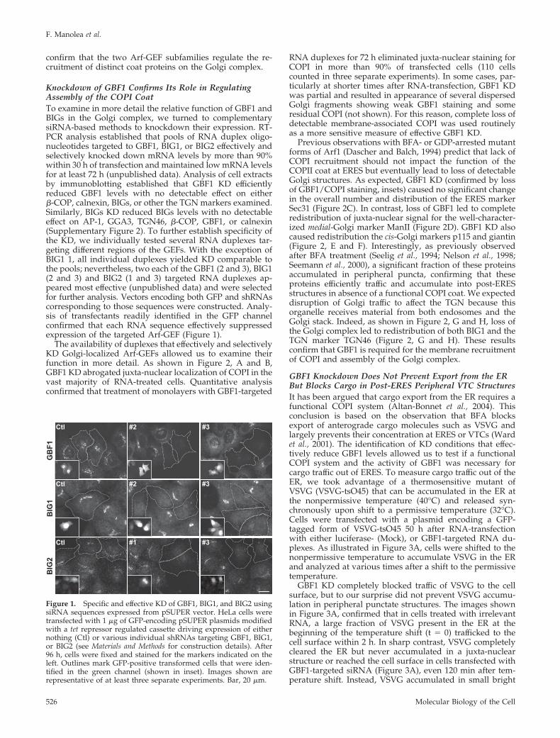
Knockdown of GBF1 Confirms Its Role in RegulatingAssembly of the COPI CoatTo examine in more detail the relative function of GBF1 andBIGs in the Golgi complex, we turned to complementarysiRNA-based methods to knockdown their expression. RT-PCR analysis established that pools of RNA duplex oligo-nucleotides targeted to GBF1, BIG1, or BIG2 effectively andselectively knocked down mRNA levels by more than 90%within 30 h of transfection and maintained low mRNA levelsfor at least 72 h (unpublished data). Analysis of cell extractsby immunoblotting established that GBF1 KD efficientlyreduced GBF1 levels with no detectable effect on either�-COP, calnexin, BIGs, or other the TGN markers examined.Similarly, BIGs KD reduced BIGs levels with no detectableeffect on AP-1, GGA3, TGN46, �-COP, GBF1, or calnexin(Supplementary Figure 2). To further establish specificity ofthe KD, we individually tested several RNA duplexes tar-geting different regions of the GEFs. With the exception ofBIG1 1, all individual duplexes yielded KD comparable tothe pools; nevertheless, two each of the GBF1 (2 and 3), BIG1(2 and 3) and BIG2 (1 and 3) targeted RNA duplexes ap-peared most effective (unpublished data) and were selectedfor further analysis. Vectors encoding both GFP and shRNAscorresponding to those sequences were constructed. Analy-sis of transfectants readily identified in the GFP channelconfirmed that each RNA sequence effectively suppressedexpression of the targeted Arf-GEF (Figure 1).
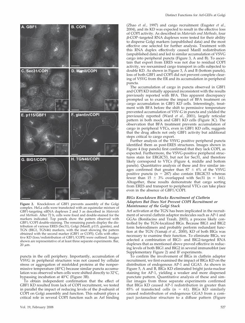
The availability of duplexes that effectively and selectivelyKD Golgi-localized Arf-GEFs allowed us to examine theirfunction in more detail. As shown in Figure 2, A and B,GBF1 KD abrogated juxta-nuclear localization of COPI in thevast majority of RNA-treated cells. Quantitative analysisconfirmed that treatment of monolayers with GBF1-targeted
RNA duplexes for 72 h eliminated juxta-nuclear staining forCOPI in more than 90% of transfected cells (110 cellscounted in three separate experiments). In some cases, par-ticularly at shorter times after RNA-transfection, GBF1 KDwas partial and resulted in appearance of several dispersedGolgi fragments showing weak GBF1 staining and someresidual COPI (not shown). For this reason, complete loss ofdetectable membrane-associated COPI was used routinelyas a more sensitive measure of effective GBF1 KD.
Previous observations with BFA- or GDP-arrested mutantforms of Arf1 (Dascher and Balch, 1994) predict that lack ofCOPI recruitment should not impact the function of theCOPII coat at ERES but eventually lead to loss of detectableGolgi structures. As expected, GBF1 KD (confirmed by lossof GBF1/COPI staining, insets) caused no significant changein the overall number and distribution of the ERES markerSec31 (Figure 2C). In contrast, loss of GBF1 led to completeredistribution of juxta-nuclear signal for the well-character-ized medial-Golgi marker ManII (Figure 2D). GBF1 KD alsocaused redistribution the cis-Golgi markers p115 and giantin(Figure 2, E and F). Interestingly, as previously observedafter BFA treatment (Seelig et al., 1994; Nelson et al., 1998;Seemann et al., 2000), a significant fraction of these proteinsaccumulated in peripheral puncta, confirming that theseproteins efficiently traffic and accumulate into post-ERESstructures in absence of a functional COPI coat. We expecteddisruption of Golgi traffic to affect the TGN because thisorganelle receives material from both endosomes and theGolgi stack. Indeed, as shown in Figure 2, G and H, loss ofthe Golgi complex led to redistribution of both BIG1 and theTGN marker TGN46 (Figure 2, G and H). These resultsconfirm that GBF1 is required for the membrane recruitmentof COPI and assembly of the Golgi complex.
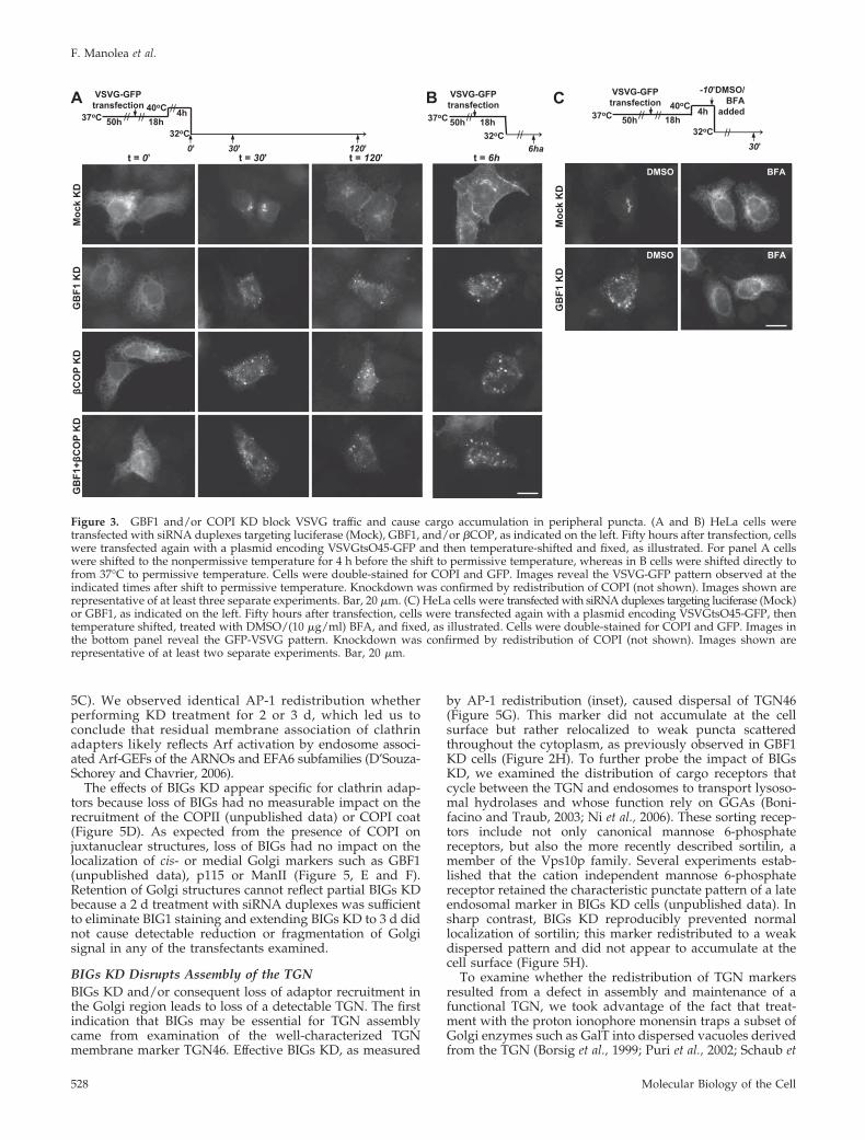
GBF1 Knockdown Does Not Prevent Export from the ERBut Blocks Cargo in Post-ERES Peripheral VTC StructuresIt has been argued that cargo export from the ER requires afunctional COPI system (Altan-Bonnet et al., 2004). Thisconclusion is based on the observation that BFA blocksexport of anterograde cargo molecules such as VSVG andlargely prevents their concentration at ERES or VTCs (Wardet al., 2001). The identification of KD conditions that effec-tively reduce GBF1 levels allowed us to test if a functionalCOPI system and the activity of GBF1 was necessary forcargo traffic out of ERES. To measure cargo traffic out of theER, we took advantage of a thermosensitive mutant ofVSVG (VSVG-tsO45) that can be accumulated in the ER atthe nonpermissive temperature (40°C) and released syn-chronously upon shift to a permissive temperature (32°C).Cells were transfected with a plasmid encoding a GFP-tagged form of VSVG-tsO45 50 h after RNA-transfectionwith either luciferase- (Mock), or GBF1-targeted RNA du-plexes. As illustrated in Figure 3A, cells were shifted to thenonpermissive temperature to accumulate VSVG in the ERand analyzed at various times after a shift to the permissivetemperature.
GBF1 KD completely blocked traffic of VSVG to the cellsurface, but to our surprise did not prevent VSVG accumu-lation in peripheral punctate structures. The images shownin Figure 3A, confirmed that in cells treated with irrelevantRNA, a large fraction of VSVG present in the ER at thebeginning of the temperature shift (t � 0) trafficked to thecell surface within 2 h. In sharp contrast, VSVG completelycleared the ER but never accumulated in a juxta-nuclearstructure or reached the cell surface in cells transfected withGBF1-targeted siRNA (Figure 3A), even 120 min after tem-perature shift. Instead, VSVG accumulated in small bright
BIG
1G
BF1
Ctl #2 #3
#2 #3Ctl
BIG
2
#1 #3Ctl
Figure 1. Specific and effective KD of GBF1, BIG1, and BIG2 usingsiRNA sequences expressed from pSUPER vector. HeLa cells weretransfected with 1 �g of GFP-encoding pSUPER plasmids modifiedwith a tet repressor regulated cassette driving expression of eithernothing (Ctl) or various individual shRNAs targeting GBF1, BIG1,or BIG2 (see Materials and Methods for construction details). After96 h, cells were fixed and stained for the markers indicated on theleft. Outlines mark GFP-positive transformed cells that were iden-tified in the green channel (shown in inset). Images shown arerepresentative of at least three separate experiments. Bar, 20 �m.
F. Manolea et al.
Molecular Biology of the Cell526
puncta in the cell periphery. Importantly, accumulation ofVSVG in peripheral structures was not caused by cellularstress or aggregation of misfolded proteins at the nonper-missive temperature (40°C) because similar puncta accumu-lation was observed when cells were shifted directly to 32°C,bypassing incubation at 40°C (Figure 3B).
To obtain independent confirmation that the effect ofGBF1 KD resulted from lack of COPI recruitment, we testedin parallel the impact of reducing levels of the �-subunit ofCOPI on Golgi assembly and function. This subunit plays acritical role in several COPI function such as Arf binding
(Zhao et al., 1997) and cargo recruitment (Eugster et al.,2004), and its KD was expected to result in the effective lossof COPI activity. As described in Materials and Methods, four�-COP–targeted RNA duplexes were tested for their abilityto disperse Golgi markers (unpublished data) and the mosteffective one selected for further analysis. Treatment withthis RNA duplex effectively caused ManII redistribution(unpublished data) and led to similar accumulation of VSVGcargo into peripheral puncta (Figure 3, A and B). To ascer-tain that export from ERES was not due to residual COPIactivity, we reexamined cargo transport in cells subjected todouble KD. As shown in Figure 3, A and B (bottom panels),loss of both GBF1 and COPI did not prevent complete clear-ing of VSVG from the ER and its accumulation in peripheralpuncta.
The accumulation of cargo in puncta observed in GBF1and COPI KD initially appeared inconsistent with the resultspreviously reported with BFA. This apparent discrepancyprompted us to examine the impact of BFA treatment oncargo accumulation in GBF1 KD cells. Interestingly, treat-ment with BFA before the shift to permissive temperatureprevented accumulation of VSV-G in puncta and yielded thepreviously reported (Ward et al., 2001), largely reticularpattern in both mock and GBF1 KD cells (Figure 3C). Theobservation that BFA treatment prevents accumulation ofcargo in peripheral VTCs, even in GBF1 KD cells, suggeststhat the drug affects not only GBF1 activity but additionalsteps critical to cargo export.
Further analysis of the VSVG positive peripheral punctaidentified them as post-ERES structures. Images shown inFigure 4 (top panels) first confirmed that they lack COPI, asexpected. Furthermore, the VSVG-positive peripheral struc-tures stain for ERGIC53, but not for Sec31, and thereforelikely correspond to VTCs (Figure 4, middle and bottompanels). Quantitative analysis of these and five similar im-ages confirmed that greater than 87 � 6% of the VSVGpositive puncta (n � 287) also contain ERGIC53 whereasfewer than 15 � 3% overlapped with Sec31 (n � 161).Altogether, these results demonstrate that cargo sortingfrom ERES and transport to peripheral VTCs can take placeeven in the absence of GBF1/COPI.
BIGs Knockdown Blocks Recruitment of ClathrinAdaptors But Does Not Prevent COPI Recruitment orMaintenance of the Golgi StackArf activation at the TGN has been implicated in the recruit-ment of several clathrin adaptor molecules such as AP-1 andGGAs (Bonifacino and Traub, 2003), a process likely con-trolled by the TGN-localized BIGs. Because BIG1 and BIG2form heterodimers and probably perform redundant func-tion at the TGN (Yamaji et al., 2000), KD of both BIGs wasnecessary to examine their function. To eliminate BIGs, weselected a combination of BIG1- and BIG2-targeted RNAduplexes that as mentioned above proved effective in reduc-ing levels of both BIG1 and BIG2 in several immunoblot (seeSupplementary Figure 2) and IF experiments.
To confirm the involvement of BIGs in clathrin adaptorrecruitment, we first examined the impact of BIGs KD on thedistribution of endogenous AP-1 and GGA3. As shown inFigure 5, A and B, BIGs KD eliminated bright juxta-nuclearstaining for AP-1, yielding a weaker and more dispersedpunctate pattern. Quantitative analysis of these and sim-ilar images from three separate experiments confirmedthat BIGs KD caused AP-1 redistribution in greater than85% of transfected cells (n � 61). BIGs KD similarlycaused redistribution of endogenous GGA3 from a com-pact juxtanuclear structure to a diffuse pattern (Figure
A. GBF1 B. COPI
H. TGN46/COPI
E. p115/GBF1
D. ManII/COPI
F. giantin/COPI
G. BIG1/COPI
C. Sec31/COPI
Figure 2. Knockdown of GBF1 prevents assembly of the Golgicomplex. HeLa cells were transfected with an equimolar mixture ofGBF1-targeting siRNA duplexes 2 and 3 as described in Materialsand Methods. After 72 h, cells were fixed and double-stained for themarkers indicated. Top panels show the pattern observed withGBF1/COPI double-staining. The remaining panels display the dis-tribution of various ERES (Sec31), Golgi (ManII, p115, giantin), andTGN (BIG1, TGN46) markers, with the inset showing the patternobtained with the second marker (GBF1 or COPI). Cells with effec-tive KD (loss/redistribution of GBF1/COPI) were outlined. Imagesshown are representative of at least three separate experiments. Bar,20 �m.
Distinct Functions for Arf-GEFs at Golgi
Vol. 19, February 2008 527
5C). We observed identical AP-1 redistribution whetherperforming KD treatment for 2 or 3 d, which led us toconclude that residual membrane association of clathrinadapters likely reflects Arf activation by endosome associ-ated Arf-GEFs of the ARNOs and EFA6 subfamilies (D’Souza-Schorey and Chavrier, 2006).
The effects of BIGs KD appear specific for clathrin adap-tors because loss of BIGs had no measurable impact on therecruitment of the COPII (unpublished data) or COPI coat(Figure 5D). As expected from the presence of COPI onjuxtanuclear structures, loss of BIGs had no impact on thelocalization of cis- or medial Golgi markers such as GBF1(unpublished data), p115 or ManII (Figure 5, E and F).Retention of Golgi structures cannot reflect partial BIGs KDbecause a 2 d treatment with siRNA duplexes was sufficientto eliminate BIG1 staining and extending BIGs KD to 3 d didnot cause detectable reduction or fragmentation of Golgisignal in any of the transfectants examined.
BIGs KD Disrupts Assembly of the TGNBIGs KD and/or consequent loss of adaptor recruitment inthe Golgi region leads to loss of a detectable TGN. The firstindication that BIGs may be essential for TGN assemblycame from examination of the well-characterized TGNmembrane marker TGN46. Effective BIGs KD, as measured
by AP-1 redistribution (inset), caused dispersal of TGN46(Figure 5G). This marker did not accumulate at the cellsurface but rather relocalized to weak puncta scatteredthroughout the cytoplasm, as previously observed in GBF1KD cells (Figure 2H). To further probe the impact of BIGsKD, we examined the distribution of cargo receptors thatcycle between the TGN and endosomes to transport lysoso-mal hydrolases and whose function rely on GGAs (Boni-facino and Traub, 2003; Ni et al., 2006). These sorting recep-tors include not only canonical mannose 6-phosphatereceptors, but also the more recently described sortilin, amember of the Vps10p family. Several experiments estab-lished that the cation independent mannose 6-phosphatereceptor retained the characteristic punctate pattern of a lateendosomal marker in BIGs KD cells (unpublished data). Insharp contrast, BIGs KD reproducibly prevented normallocalization of sortilin; this marker redistributed to a weakdispersed pattern and did not appear to accumulate at thecell surface (Figure 5H).
To examine whether the redistribution of TGN markersresulted from a defect in assembly and maintenance of afunctional TGN, we took advantage of the fact that treat-ment with the proton ionophore monensin traps a subset ofGolgi enzymes such as GalT into dispersed vacuoles derivedfrom the TGN (Borsig et al., 1999; Puri et al., 2002; Schaub et
A C B 37 o C
32 o C 6h a
VSVG-GFP transfectio n
18 h //
// 50 h 37 o C 40 o C
32 o C 0 ’ 30 ’ 12 0 ’
4h
VSVG-GF P transfection
18 h // 50 h // //
37 o C 40 o C
32 o C 30 ’
4h
VSVG-GFP transfectio n
18 h
- 10 ’DMSO/ BF A
adde d // 50 h //
//
Moc
k K
D
t = 0 ’
GB
F1 K
D
t = 30 ’ t = 12 0 ’
βCO
P K
D
GB
F1+ β
CO
PK
D
t = 6h
Moc
k K
D
GB
F1 K
D
BF A DMS O
BF A DMSO
Figure 3. GBF1 and/or COPI KD block VSVG traffic and cause cargo accumulation in peripheral puncta. (A and B) HeLa cells weretransfected with siRNA duplexes targeting luciferase (Mock), GBF1, and/or �COP, as indicated on the left. Fifty hours after transfection, cellswere transfected again with a plasmid encoding VSVGtsO45-GFP and then temperature-shifted and fixed, as illustrated. For panel A cellswere shifted to the nonpermissive temperature for 4 h before the shift to permissive temperature, whereas in B cells were shifted directly tofrom 37°C to permissive temperature. Cells were double-stained for COPI and GFP. Images reveal the VSVG-GFP pattern observed at theindicated times after shift to permissive temperature. Knockdown was confirmed by redistribution of COPI (not shown). Images shown arerepresentative of at least three separate experiments. Bar, 20 �m. (C) HeLa cells were transfected with siRNA duplexes targeting luciferase (Mock)or GBF1, as indicated on the left. Fifty hours after transfection, cells were transfected again with a plasmid encoding VSVGtsO45-GFP, thentemperature shifted, treated with DMSO/(10 �g/ml) BFA, and fixed, as illustrated. Cells were double-stained for COPI and GFP. Images inthe bottom panel reveal the GFP-VSVG pattern. Knockdown was confirmed by redistribution of COPI (not shown). Images shown arerepresentative of at least two separate experiments. Bar, 20 �m.
F. Manolea et al.
Molecular Biology of the Cell528
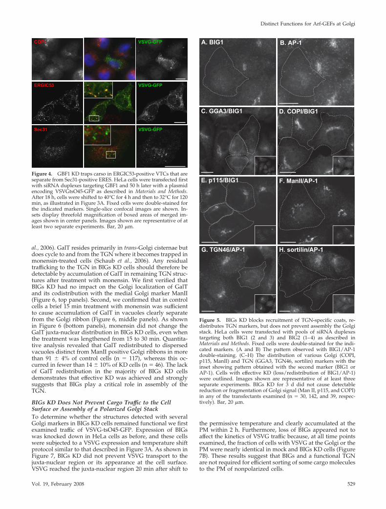
al., 2006). GalT resides primarily in trans-Golgi cisternae butdoes cycle to and from the TGN where it becomes trapped inmonensin-treated cells (Schaub et al., 2006). Any residualtrafficking to the TGN in BIGs KD cells should therefore bedetectable by accumulation of GalT in remaining TGN struc-tures after treatment with monensin. We first verified thatBIGs KD had no impact on the Golgi localization of GalTand its codistribution with the medial Golgi marker ManII(Figure 6, top panels). Second, we confirmed that in controlcells a brief 15 min treatment with monensin was sufficientto cause accumulation of GalT in vacuoles clearly separatefrom the Golgi ribbon (Figure 6, middle panels). As shownin Figure 6 (bottom panels), monensin did not change theGalT juxta-nuclear distribution in BIGs KD cells, even whenthe treatment was lengthened from 15 to 30 min. Quantita-tive analysis revealed that GalT redistributed to dispersedvacuoles distinct from ManII positive Golgi ribbons in morethan 91 � 4% of control cells (n � 117), whereas this oc-curred in fewer than 14 � 10% of KD cells (n � 46). The lackof GalT redistribution in the majority of BIGs KD cellsdemonstrates that effective KD was achieved and stronglysuggests that BIGs play a critical role in assembly of theTGN.
BIGs KD Does Not Prevent Cargo Traffic to the CellSurface or Assembly of a Polarized Golgi StackTo determine whether the structures detected with severalGolgi markers in BIGs KD cells remained functional we firstexamined traffic of VSVG-tsO45-GFP. Expression of BIGswas knocked down in HeLa cells as before, and these cellswere subjected to a VSVG expression and temperature shiftprotocol similar to that described in Figure 3A. As shown inFigure 7, BIGs KD did not prevent VSVG transport to thejuxta-nuclear region or its appearance at the cell surface.VSVG reached the juxta-nuclear region 20 min after shift to
the permissive temperature and clearly accumulated at thePM within 2 h. Furthermore, loss of BIGs appeared not toaffect the kinetics of VSVG traffic because, at all time pointsexamined, the fraction of cells with VSVG at the Golgi or thePM were nearly identical in mock and BIGs KD cells (Figure7B). These results suggest that BIGs and a functional TGNare not required for efficient sorting of some cargo moleculesto the PM of nonpolarized cells.
COPI VSVG-GFP
ERGIC53 VSVG-GFP
Sec31 VSVG-GFP
Figure 4. GBF1 KD traps carso in ERGIC53-positive VTCs that areseparate from Sec31-positive ERES. HeLa cells were transfected firstwith siRNA duplexes targeting GBF1 and 50 h later with a plasmidencoding VSVGtsO45-GFP as described in Materials and Methods.After 18 h, cells were shifted to 40°C for 4 h and then to 32°C for 120min, as illustrated in Figure 3A. Fixed cells were double-stained forthe indicated markers. Single-slice confocal images are shown. In-sets display threefold magnification of boxed areas of merged im-ages shown in center panels. Images shown are representative of atleast two separate experiments. Bar, 20 �m.
F. ManII/AP-1
G. TGN46/AP-1
A. BIG1
D. COPI/BIG1
B. AP-1
E. p115/BIG1
H. sortilin/AP-1
C. GGA3/BIG1
Figure 5. BIGs KD blocks recruitment of TGN-specific coats, re-distributes TGN markers, but does not prevent assembly the Golgistack. HeLa cells were transfected with pools of siRNA duplexestargeting both BIG1 (2 and 3) and BIG2 (1–4) as described inMaterials and Methods. Fixed cells were double-stained for the indi-cated markers. (A and B) The pattern observed with BIG1/AP-1double-staining. (C–H) The distribution of various Golgi (COPI,p115, ManII) and TGN (GGA3, TGN46, sortilin) markers with theinset showing pattern obtained with the second marker (BIG1 orAP-1). Cells with effective KD (loss/redistribution of BIG1/AP-1)were outlined. Images shown are representative of at least threeseparate experiments. BIGs KD for 3 d did not cause detectablereduction or fragmentation of Golgi signal (Man II, p115, and COPI)in any of the transfectants examined (n � 30, 142, and 39, respec-tively). Bar, 20 �m.
Distinct Functions for Arf-GEFs at Golgi
Vol. 19, February 2008 529

The observation that BIGs KD does not prevent cargotraffic to the cell surface suggests that the GBF1/COPI coatmachinery may be sufficient to drive assembly a Golgi stackthat cannot only produce carriers but may have retained itscharacteristic polarized organization. We tested this possi-bility by examining the impact of BIGs KD on the relative
distribution of several markers with limited distributionwithin the Golgi stack. HeLa cells were subjected to BIGs KDand examined by IF using a triple labeling protocol. Asshown in Figure 8, BIGs KD did not prevent assembly of apolarized Golgi stack in which cis-Golgi markers such asp115 and GBF1 remain well resolved from a trans-cisterna
Moc
k K
D /
Mon
15’
BIG
s K
D /
Mon
30’
BIG
s K
D /
Met
OH
30’
GalT-GFPManII
GalT-GFPManII
GalT-GFPManII
Figure 6. BIGs KD prevents monensin-in-duced redistribution of GalT-GFP. HeLa cellswere transfected with a plasmid encodingGalT-GFP and a pool of siRNA duplexes tar-geting either luciferase (Mock), or BIG1 andBIG2 (BIGs) as described in Materials andMethods. Seventy-two hours after siRNAtransfection, cells were treated with eithermethanol or 4 �M monensin for the indicatedperiods of time. Cells were then fixed andstained for ManII and AP-1. Cells with effec-tive BIGs KD were identified by loss of AP-1juxtanuclear staining. Single-slice confocalimages of the ManII (red) and GFP signal areshown. Merged images shown on the right.Images shown are representative of at leastthree separate experiments. Bar, 20 �m.
% c
ells
with
VSV
G in
str
uctu
re
Moc
k K
D
t = 0’ t = 20 = t’ 60’t = 40 = t’ 120’
BIG
s K
D *
* *
*
**
*
* *
**
0
20
40
60
80
100
0 20 40 60 80 100 120
Time (min)
Golgi
Golgi + PM
A B
Figure 7. BIGs KD does not block traffic of VSVG to the cell surface. HeLa cells were transfected with siRNA duplexes targeting eitherluciferase (Mock) or BIG1 and BIG2 (BIGs), as indicated on the left. Fifty hours after transfection, cells were transfected again with a plasmidencoding VSVGtsO45-GFP, then temperature shifted, and fixed, as illustrated in Figure 3A. (A) Cells fixed at the indicated times aftershift-down to 32°C were double-stained for BIG1 and GFP. Knockdown was confirmed by redistribution of BIG1 (not shown). Images revealthe GFP-VSVG pattern observed at the times shown above the panels. Arrowheads mark plasma membrane (PM) where VSVG accumulates.Cells with effective KD (loss of BIG1) were marked by asterisks. Images shown are representative of at least six separate experiments. Bar,20 �m. (B) At each time point after release for both mock (F and Œ, solid line) and BIGs KD (E and ‚, dashed line) cells, a minimum of 25cells were scored for presence of VSVG at Golgi only (E, F), or at Golgi and PM (‚, Œ). The fraction of cells with the indicated pattern wasexpressed as percentage and is shown as a function of time after shift-down to 32°C.
F. Manolea et al.
Molecular Biology of the Cell530
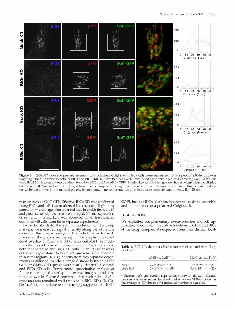
marker such as GalT-GFP. Effective BIGs KD was confirmedusing BIG1 and AP-1 as markers (blue channel). Rightmostpanels show an image of an enlarged area in which the red (cis)and green (trans) signals have been merged. Normal separationof cis- and trans-markers was observed in all transfectantsexamined (68 cells from three separate experiments).
To better illustrate the spatial resolution of the Golgimarkers, we measured signal intensity along the white lineshown in the merged image and reported values for eachmarker in the graphs on the right. The graphs confirmedgood overlap of BIG1 and AP-1 with GalT-GFP in mock-treated cells and clear separation of cis- and trans-markers inboth mock-treated and BIGs KD cells. Quantitative analysisof the average distance between cis- and trans-Golgi markersin several regions (n � 6) of cells from two separate exper-iments established that the average distance between p115/GalT or GBF1/GalT peaks were nearly identical in controland BIGs KD cells. Furthermore, quantitative analysis offluorescence signal overlap in several images similar tothose shown in Figure 8 confirmed that both pairs of cis/trans markers remained well resolved in BIGs KD cells (Ta-ble 1). Altogether, these results strongly suggest that GBF1/
COPI, but not BIGs/clathrin, is essential to drive assemblyand maintenance of a polarized Golgi stack.
DISCUSSION
We exploited complementary overexpression and KD ap-proaches to examine the relative functions of GBF1 and BIGsat the Golgi complex. As expected from their distinct local-
D ista nc e (P ixe l)0 10 20 30 40 50
0
10 0
20 0AP-1
BIG1
BIG1
AP-1
GalT-GFP
GalT-GFP
GalT-GFP
GalT-GFP
GBF1
p115
p115
GBF1
Moc
k K
DM
ock
KD
DK s
GIB
DK s
GIB
D ista nc e (P ixe l)0 10 20 30 40 50
0
10 0
20 0
D ista nc e (P ixe l)0 10 20 30 40 50
0
10 0
20 0
D ista nc e (P ixe l)0 10 20 30 40 50
0
10 0
20 0
Figure 8. BIGs KD does not prevent assembly of a polarized Golgi stack. HeLa cells were transfected with a pool of siRNA duplexestargeting either luciferase (Mock), or BIG1 and BIG2 (BIGs). After 48 h, cells were transfected again with a plasmid encoding GalT-GFP. Cellswere fixed 24 h later and double-stained for either BIG1/p115 or AP-1/GBF1. Single-slice confocal images are shown. Merged images displaythe red and GFP signal from the enlarged boxed areas. Graphs in the right column report pixel intensity profiles in all three channels alongthe white bar shown in the merged panels. Images shown are representative of at least three separate experiments. Bar, 20 �m.
Table 1. BIGs KD does not affect separation of cis- and trans-Golgimarkersa
p115 vs. GalT (%) GBF1 vs. GalT (%)
Mock 29 � 5% (n � 6) 36 � 9% (n � 8)BIGs KD 27 � 9% (n � 19) 30 � 10% (n � 23)
a The extent of signal overlap in percentages between the two indicatedmarkers was measured as described in Materials and Methods. Shown isthe average � SD obtained for indicated number of samples.
Distinct Functions for Arf-GEFs at Golgi
Vol. 19, February 2008 531

ization, overexpression of GBF1 and BIG1 had oppositeeffects on the sensitivity of the COPI and AP-1 coats to BFA.GBF1 stabilized COPI and protected cis-compartments butnot AP-1 or HA-furin positive trans-Golgi membranes; incontrast BIG1 protected AP-1 but not COPI. Knockdownwith several specific RNA duplexes or shRNAs confirmedthat GBF1 and BIGs perform distinct functions in the Golgicomplex. GBF1 KD caused redistribution of COPI and mostGolgi markers to a diffuse pattern, whereas tethering factorssuch as p115 and giantin redistributed into puncta andreticular structures. Contrary to expectation, KD of GBF1and COPI, singly or in combination, did not prevent exportof VSVGtsO45 from the ER, but caused its accumulation intoERGIC53-positive puncta separate from Sec31-positiveERES. On the other hand, BIGs KD had no impact on theCOPI machinery or several Golgi markers but caused loss ofclathrin adaptor proteins as well as redistribution of TGN46and the sorting receptor sortilin. Further tests with monen-sin confirmed loss of detectable sorting of GalT to the TGN.Despite these clear effects on the TGN, BIGs KD preventedneither traffic of VSVGtsO45 to the PM, nor assembly of apolarized Golgi stack. Our observations indicate that COPIIis the only coat required for sorting and export from ERES.Furthermore, GBF1 but not BIGs appears necessary for COPIrecruitment and cargo progression from VTCs to cell sur-face. The maintenance of a polarized Golgi stack in absenceof BIGs suggests that the GBF1-COPI machinery is sufficientto drive the maturation process responsible for Golgi orga-nization.
Effectiveness and Selectivity of Golgi ArfGEFs KDSeveral experiments established that the KD methodsused in our study led to selective and effective loss of thetargeted proteins. Multiple observations confirmed theselectivity of the KD effects. These include the fact thatsimilar KD effects were observed using any one of severalsequences targeting different regions of the mRNA, andwhether sequences were delivered by direct transfectionof duplexes or by plasmid-driven synthesis of short hair-pins RNA. The observation that targeting either �-COP orGBF1 had the same impact on ER export and the Golgicomplex further supports our conclusion that KDs wereselective.
The extent of KD varied within any given cell population,and depended on the nature of the target and length oftreatment. These variations allowed us to identify with con-fidence cells displaying effective KD of targeted proteins. Forexample, we could readily recognize cells with partial KDfor GBF1 at shorter treatment times (48 h); these cells lackedor showed little detectable GBF1 but still displayed a frag-mented Golgi complex stained weakly with COPI. Bylengthening treatment time to 72 h and selecting cells withno remaining COPI staining, we could ensure that all cellsanalyzed had effective GBF1 KD. In the case of BIGs, shorttreatment (48 h) proved effective at eliminating BIG anddispersing AP-1 IF signal. The fact that lengthening treat-ment to 72 h had no further impact on the distribution ofTGN and Golgi markers established that effective KD hadbeen achieved.
GBF1 Is Essential for COPI Recruitment and Assembly ofthe Golgi Complex, But Is Not Required for CargoConcentration and Export from ERESResults obtained through our combined use of overexpres-sion and RNA-dependent silencing established that GBF1,but not BIGs, is required to activate Arfs for recruitment ofCOPI. The localization of GBF1 to early compartments of the
Golgi complex first suggested a functional link betweenGBF1 and COPI (Kawamoto et al., 2002; Zhao et al., 2002).Subsequent studies revealed that overexpression of thecharge-reversal dominant negative mutant GBF1[E794K](Garcia-Mata et al., 2003) or microinjection of neutralizingGBF1 antibodies (Zhao et al., 2006) caused loss of COPIrecruitment. The loss of COPI recruitment and Golgi struc-ture after GBF1 KD (Figure 2), as well as the coat andcompartment-specific protection conferred by GBF1 overex-pression (Supplementary Figure 1), extend these observa-tions and confirm this functional link.
The availability of tools for the effective KD of GBF1 andCOPI allowed us to test the two-step model which proposesthat Arfs and COPI are required for traffic out of ERES.Contrary to predictions from this model, we observed thatthe GBF1/COPI machinery was not required for the concen-tration and export of VSVG cargo from ERES. These resultsare consistent with a wealth of information derived fromyeast cell-free assays (Barlowe, 2003; Sato and Nakano, 2007)and the recent identification of COPII carriers in animal cells(Zeuschner et al., 2006). These results are also consistent withour previous demonstration that GBF1 does not associatewith ERES, but rather with VTCs that are close but physi-cally separate from ERES (Zhao et al., 2006).
To explain the apparent block in cargo export by BFA orArf mutants (Ward et al., 2001), we propose that these treat-ments do not block ER export, but rather prevent theirretention in VTCs by promoting retrograde traffic fromVTCs to ER. This model is based on our recent demonstra-tion that BFA causes VTCs to lose their cargo to the ERthrough a microtubule-dependent mechanism (Zhao et al.,2006). This could occur if the inactive GBF1 trapped onmembranes by BFA (Niu et al., 2005; Szul et al., 2005; Zhao etal., 2006) somehow interfered with protein sorting at ERESand/or VTCs. Alternatively, BFA could affect additionaltargets such as BARS, which has been implicated in mem-brane scission of COPI vesicles (Yang et al., 2005) and whoseactivity is inhibited by BFA-induced ADP-ribosylation(Weigert et al., 1999).
As initially suggested by the impact of the GBF1[E794K]mutant (Garcia-Mata et al., 2003), GBF1 KD prevented theformation of motile transport competent carriers necessaryfor assembly and maintenance of the Golgi complex. InGBF1 KD cells, VSVG cargo accumulated in VTCs that con-tained several tethering factors such as p115 and giantin, butfailed to either mature into Golgi resident enzyme-contain-ing structures or to associate with microtubules and migrateto the cell center. The fact that VSVG but not Golgi residentenzymes accumulated in VTCs likely reflects the presence inVSVG of a di-acidic sorting signal efficiently recognized byCOPII (Nishimura et al., 1999; Sato and Nakano, 2007). Thereason for the lack of movement to the cell center remainsunknown but, as proposed by Sztul and colleagues (Garcia-Mata et al., 2003), it may be related to the absence of COPI-driven active protein sorting that normally drives formationof membrane domains critical for recruitment of other pro-teins such as rabs and motors/accessory proteins (Short etal., 2005).
BIGs Are Required for Recruitment of Clathrin Adaptorand Maintenance of the TGNPrevious work established that BIGs localize at the TGN andoverlap with clathrin, suggesting that BIGs regulate Arfactivation for recruitment of GGAs and other adaptor pro-teins such as AP-1 (Yamaji et al., 2000; Zhao et al., 2002). Weconfirmed here the functional link between BIG1 and AP-1by showing that BIG1 overexpression stabilizes AP-1 but not
F. Manolea et al.
Molecular Biology of the Cell532
COPI against dispersal after short BFA treatment. Na-kayama and colleagues confirmed a similar link betweenBIG2 and AP-1 using related approaches (Shinotsuka et al.,2002b). As predicted, BIGs KD caused loss of both AP-1 andGGA3 from the juxta-nuclear region. This redistribution ofAP-1 is similar to that reported after BFA treatment ofseveral cell lines in which the Golgi complex is either natu-rally resistant to BFA or acquired resistance after mutagen-esis. For example, the Golgi stack of MDCK and PtK1 cells(Robinson and Kreis, 1992) or mutagenized CHO-K1 cells(Torii et al., 1995) remains unperturbed after BFA treatment,whereas some AP-1 localizes to disperse puncta. In all cases,residual membrane association of clathrin adapters likely re-sults from Arf activation by endosome-associated BFA resis-tant Arf-GEFs of the ARNO and EFA6 subfamilies (D’Souza-Schorey and Chavrier, 2006).
Previous attempts to test the model that GGAs and APsrecognize sorting signals in endosomal-targeted cargo anddrive maturation of the TGN, focused on blocking the func-tion of either AP1, -3, and -4 or GGAs (Bonifacino and Traub,2003; Gleeson et al., 2004). However, expression of dominantnegative mutants or silencing of either types of adaptors ledto variable outcomes ranging from tubulation of the Golgi,accumulation of endosomal cargo in the TGN or its dispersalto peripheral endosome structures (Puertollano et al.,2001a,b; Ghosh et al., 2003). These apparent discrepanciesshould not be surprising, however, because current evidencesuggests that adaptors function in both anterograde andretrograde traffic and at the TGN participate in a multistepprocess involving both GGAs and APs (Ghosh and Korn-feld, 2004); loss of only a subset of the adaptors wouldimbalance this process, with complex consequences. Effec-tive KD of BIGs circumvented this problem by preventingrecruitment of both APs and GGAs to the Golgi complex.Under these conditions, we could readily detect loss of arecognizable juxta-nuclear TGN structure stained by TGN46or sortilin. Disruption of sorting to the TGN was confirmedby clear loss of GalT accumulation in dispersed vacuolesafter monensin treatment. We predict that simultaneous si-lencing of all GGAs and several APs will be required toobserve effects similar to those we report for BIGs KD.
GBF1, But Not BIGs, Is Required for Assembly of aPolarized Golgi StackOne of the more unexpected results of our study was theobservation that BIGs KD did not prevent assembly of apolarized Golgi stack in mammalian cells; these stacks notonly retained a degree of polarization similar to that ofcontrol cells, but also efficiently trafficked VSVG to the cellsurface (Figures 7 and 8). These observations were surpris-ing because previous work in S. cerevisiae had establishedthat loss of Sec7p, the single orthologue of BIGs, completelyalters Golgi morphology and blocks protein secretion. Forexample, Sec7 temperature-sensitive mutants accumulatelarge numbers of stacked Golgi membranes (Berkeley bod-ies) with concomitant block in traffic to the vacuole and cellsurface (Novick et al., 1980; Esmon et al., 1981; Franzusoffand Schekman, 1989; Rambourg et al., 1993; Deitz et al.,2000). Although undetectable levels of remaining BIGs couldaccount for those observations, we consider this possibilityextremely unlikely as argued above. We propose insteadthat the apparent discrepancy between the impact of Sec7inactivation and BIGs KD may reflect differences in theorganization of the secretory pathways of S. cerevisiae andanimals.
Yeast Golgi elements appear as fine or coarse nodularnetworks that are neither cisternal nor arranged in stacks
(Morin-Ganet et al., 2000; Rambourg et al., 2001; Kepes et al.,2005). These morphogenetic studies revealed a gradualtransformation of Golgi elements after their initial assemblyfrom ER-derived vesicles: from tubular clusters, they be-come a network of fine tubules linked by nodes that trans-form into a thicker nodular network that eventually releasessecretory granules by rupture of tubular areas. The presenceof mixed forms containing two networks of different calibers(Morin-Ganet et al., 2000) actually suggests that the gradualtransformation from early to late elements involves con-certed action of both the COPI and clathrin coats within acontinuous network. This view is consistent with recentdescription of Golgi maturation in live yeast (Losev et al.,2006; Matsuura-Tokita et al., 2006) and may explain why lossof Sec7 would prevent maturation of the nodular networkand cause accumulation of membranes unable to releasecargo carriers.
In contrast, the Golgi complex of animal cells occurs as astructured stack of discrete flattened cisternae with varyingextent of fenestration that is flanked by extensive tubular-reticular networks (Rambourg and Clermont, 1990; Thorne-Tjomsland et al., 1998; Mogelsvang et al., 2004). Our resultssuggest that the GBF1-dependent recruitment of the COPIcoat is sufficient to promote the formation of specializedmembrane domains and cargo carriers that can move cargofrom VTCs, assemble onto the Golgi matrix and subse-quently drive the maturation process to yield a polarizedstack. The fact that one can eliminate BIGs and maintainmuch of this organization suggests that these mechanismsare robust and that stacking of separate cisternae may per-mit the observed uncoupling of cis- and trans-acting coats.
The observation that VSVG trafficked normally to the cellsurface in BIGs KD cells remains consistent with the well-established role of the TGN in sorting of cargo to variousdestinations in both nonpolarized and polarized cells (Boni-facino and Traub, 2003; Rodriguez-Boulan et al., 2005; Ro-driguez-Boulan and Musch, 2005; Bard and Malhotra, 2006).Maturation by the COPI coat in animal cells may allow thecreation of membrane domains on the trans-side that becomeenriched in anterograde cargo proteins and eventually peeloff the stack to carry some or all of its content to the cellsurface. Under normal conditions such intermediates wouldlikely be absorbed in the TGN from which cargo would bethen sorted to its various destinations. Our results establishthat whatever mechanisms normally drive traffic of VSVGfrom the TGN remain operational in the absence of BIGs/clathrin-dependent sorting. The absence of a detectable TGNin BIGs KD cells raises one additional issue: why so few, ifany, of the membranes released from the Golgi stack remainin this area. Further work will clearly be required to estab-lish when and how the BIGs machinery is recruited to latecisternae to facilitate membrane retention, formation of theTGN, and sorting of endosomal cargo.
ACKNOWLEDGMENTS
We thank L. Channon for maintenance of cultured cells and RT-PCR analysis,Z. Shapovalova for construction of pSUPER vectors targeting BIG2, and F.Mast for advice on image quantitation as well as H. Chan for technical helpwith confocal microscopy. We thank Drs. K. Nakayama and H.-W. Shin(Kyoto University) for the BIG2 antibody and Dr. J. Rohrer (University ofZurich) for advice on the monensin experiments. Finally, we thank Drs. T.Hobman and Z. Wang (University of Alberta) for helpful discussions. Thisstudy was initiated with funds from the Alberta Heritage Foundation forMedical Research and supported by grants (P.M.) from the Canadian Insti-tutes of Health Research and the Human Frontier Science Program. A.C. andJ.R. were supported by the Fondecyt Grant 1030346.
Distinct Functions for Arf-GEFs at Golgi
Vol. 19, February 2008 533
REFERENCES
Altan-Bonnet, N., Sougrat, R., and Lippincott-Schwartz, J. (2004). Molecularbasis for Golgi maintenance and biogenesis. Curr. Opin. Cell Biol. 16, 364–372.
Alvarez, C., Garcia-Mata, R., Brandon, E., and Sztul, E. (2003). COPI recruit-ment is modulated by a Rab1b-dependent mechanism. Mol. Biol. Cell 14,2116–2127.
Appenzeller-Herzog, C., and Hauri, H. P. (2006). The ER-Golgi intermediatecompartment (ERGIC): in search of its identity and function. J. Cell Sci. 119,2173–2183.
Bard, F., and Malhotra, V. (2006). The formation of TGN-to-plasma-mem-brane transport carriers. Annu. Rev. Cell Dev. Biol. 22, 439–455.
Barlowe, C. (2003). Signals for COPII-dependent export from the ER: what’sthe ticket out? Trends Cell Biol. 13, 295–300.
Barzilay, E., Ben-Califa, N., Hirschberg, K., and Neumann, D. (2005). Uncou-pling of brefeldin a-mediated coatomer protein complex-I dissociation fromGolgi redistribution. Traffic 6, 794–802.
Bonifacino, J. S. (2004). The GGA proteins: adaptors on the move. Nat. Rev.Mol. Cell Biol. 5, 23–32.
Bonifacino, J. S., and Glick, B. S. (2004). The mechanisms of vesicle buddingand fusion. Cell 116, 153–166.
Bonifacino, J. S., and Traub, L. M. (2003). Signals for sorting of transmembraneproteins to endosomes and lysosomes. Annu. Rev. Biochem. 72, 395–447.
Borsig, L., Imbach, T., Hochli, M., and Berger, E. G. (1999).alpha1,3Fucosyltransferase VI is expressed in HepG2 cells and codistributedwith beta1,4galactosyltransferase I in the golgi apparatus and monensin-induced swollen vesicles. Glycobiology 9, 1273–1280.
Claude, A., Zhao, B. P., Kuziemsky, C. E., Dahan, S., Berger, S. J., Yan, J. P.,Armold, A. D., Sullivan, E. M., and Melancon, P. (1999). GBF1, a novelGolgi-associated BFA-resistant guanine nucleotide exchange factor that dis-plays specificity for ADP-ribosylation factor 5. J. Cell Biol. 146, 71–84.
Claude, A., Zhao, B. P., and Melancon, P. (2003). Characterization of alterna-tively spliced and truncated forms of the Arf guanine nucleotide exchangefactor GBF1 defines regions important for activity. Biochem. Biophys. Res.Commun. 303, 160–169.
Cox, R., Mason-Gamer, R. J., Jackson, C. L., and Segev, N. (2004). Phylogeneticanalysis of Sec7-domain-containing Arf nucleotide exchangers. Mol. Biol. Cell15, 1487–1505.
D’Souza-Schorey, C., and Chavrier, P. (2006). ARF proteins: roles in mem-brane traffic and beyond. Nat. Rev. Mol. Cell Biol. 7, 347–358.
Dascher, C., and Balch, W. E. (1994). Dominant inhibitory mutants of ARF1block endoplasmic reticulum to Golgi transport and trigger disassembly ofthe Golgi apparatus. J. Biol. Chem. 269, 1437–1448.
Deitz, S. B., Rambourg, A., Kepes, F., and Franzusoff, A. (2000). Sec7p directsthe transitions required for yeast Golgi biogenesis. Traffic 1, 172–183.
Donaldson, J. G., and Honda, A. (2005). Localization and function of Arffamily GTPases. Biochem. Soc. Trans. 33, 639–642.
Duden, R. (2003). ER-to-Golgi transport: COP I and COP II function (Review).Mol. Membr. Biol. 20, 197–207.
Elbashir, S. M., Harborth, J., Weber, K., and Tuschl, T. (2002). Analysis of genefunction in somatic mammalian cells using small interfering RNAs. Methods26, 199–213.
Esmon, B., Novick, P., and Schekman, R. (1981). Compartmentalized assem-bly of oligosaccharides on exported glycoproteins in yeast. Cell 25, 451–460.
Eugster, A., Frigerio, G., Dale, M., and Duden, R. (2004). The alpha- andbeta�-COP WD40 domains mediate cargo-selective interactions with distinctdi-lysine motifs. Mol. Biol. Cell 15, 1011–1023.
Farquhar, M. G., and Palade, G. E. (1998). The Golgi apparatus: 100 years ofprogress and controversy. Trends Cell Biol. 8, 2–10.
Franzusoff, A., and Schekman, R. (1989). Functional compartments of theyeast Golgi apparatus are defined by the sec7 mutation. EMBO J. 8, 2695–2702.
Fromme, J. C., and Schekman, R. (2005). COPII-coated vesicles: flexibleenough for large cargo? Curr. Opin. Cell Biol. 17, 345–352.
Garcia-Mata, R., and Sztul, E. (2003). The membrane-tethering protein p115interacts with GBF1, an ARF guanine-nucleotide-exchange factor. EMBO Rep.4, 320–325.
Garcia-Mata, R., Szul, T., Alvarez, C., and Sztul, E. (2003). ADP-ribosylationfactor/COPI-dependent events at the endoplasmic reticulum-Golgi interfaceare regulated by the guanine nucleotide exchange factor GBF1. Mol. Biol. Cell14, 2250–2261.
Ghosh, P., Griffith, J., Geuze, H. J., and Kornfeld, S. (2003). Mammalian GGAsact together to sort mannose 6-phosphate receptors. J. Cell Biol. 163, 755–766.
Ghosh, P., and Kornfeld, S. (2004). The GGA proteins: key players in proteinsorting at the trans-Golgi network. Eur. J. Cell Biol. 83, 257–262.
Gleeson, P. A., Lock, J. G., Luke, M. R., and Stow, J. L. (2004). Domains of theTGN: coats, tethers and G proteins. Traffic 5, 315–326.
Harborth, J., Elbashir, S. M., Bechert, K., Tuschl, T. C., and Weber, K. (2001).Identification of essential genes in cultured mammalian cells using smallinterfering RNAs. J. Cell Science 114, 4557–4565.
Kawamoto, K., Yoshida, Y., Tamaki, H., Torii, S., Shinotsuka, C., Yamashina,S., and Nakayama, K. (2002). GBF1, a guanine nucleotide exchange factor forADP-ribosylation factors, is localized to the cis-Golgi and involved in mem-brane association of the COPI coat. Traffic 3, 483–495.
Kepes, F., Rambourg, A., and Satiat-Jeunemaitre, B. (2005). Morphodynamicsof the secretory pathway. Int. Rev. Cytol. 242, 55–120.
Losev, E., Reinke, C. A., Jellen, J., Strongin, D. E., Bevis, B. J., and Glick, B. S.(2006). Golgi maturation visualized in living yeast. Nature 441, 1002–1006.
Mansour, S. J., Skaug, J., Zhao, X. H., Giordano, J., Scherer, S. W., andMelancon, P. (1999). p200 ARF-GEP1, a Golgi-localized guanine nucleotideexchange protein whose Sec7 domain is targeted by the drug brefeldin A.Proc. Natl. Acad. Sci. USA 96, 7968–7973.
Matsuura-Tokita, K., Takeuchi, M., Ichihara, A., Mikuriya, K., and Nakano, A.(2006). Live imaging of yeast Golgi cisternal maturation. Nature 441, 1007–1010.
Mogelsvang, S., Marsh, B. J., Ladinsky, M. S., and Howell, K. E. (2004).Predicting function from structure: 3D structure studies of the mammalianGolgi complex. Traffic 5, 338–345.
Morin-Ganet, M. N., Rambourg, A., Deitz, S. B., Franzusoff, A., and Kepes, F.(2000). Morphogenesis and dynamics of the yeast Golgi apparatus. Traffic 1,56–68.
Mouratou, B., Biou, V., Joubert, A., Cohen, J., Shields, D. J., Geldner, N.,Jurgens, G., Melancon, P., and Cherfils, J. (2005). The domain architecture oflarge guanine nucleotide exchange factors for the small GTP-binding proteinArf. BMC Genomics 6, 20
Moyer, B. D., Allan, B. B., and Balch, W. E. (2001). Rab1 interaction with aGM130 effector complex regulates COPII vesicle cis-Golgi tethering. Traffic 2,268–276.
Nelson, D. S., Alvarez, C., Gao, Y. S., Garcia-Mata, R., Fialkowski, E., andSztul, E. (1998). The membrane transport factor TAP/p115 cycles between theGolgi and earlier secretory compartments and contains distinct domainsrequired for its localization and function. J. Cell Biol. 143, 319–331.
Ni, X., Canuel, M., and Morales, C. R. (2006). The sorting and trafficking oflysosomal proteins. Histol. Histopathol. 21, 899–913.
Nie, Z., and Randazzo, P. A. (2006). Arf GAPs and membrane traffic. J. CellSci. 119, 1203–1211.
Nishimura, N., Bannykh, S., Slabough, S., Matteson, J., Altschuler, Y., Hahn,K., and Balch, W. E. (1999). A di-acidic (DXE) code directs concentration ofcargo during export from the endoplasmic reticulum. J. Biol. Chem. 274,15937–15946.
Niu, T. K., Pfeifer, A. C., Lippincott-Schwartz, J., and Jackson, C. L. (2005).Dynamics of GBF1, a Brefeldin A-sensitive Arf1 exchange factor at the Golgi.Mol. Biol. Cell 16, 1213–1222.
Novick, P., Field, C., and Schekman, R. (1980). Identification of 23 comple-mentation groups required for post-translational events in the yeast secretorypathway. Cell 21, 205–215.
Orci, L., Glick, B. S., and Rothman, J. E. (1986). A new type of coated vesicularcarrier that appears not to contain clathrin: its possible role in protein trans-port within the Golgi stack. Cell 46, 171–184.
Polishchuk, R. S., and Mironov, A. A. (2004). Structural aspects of Golgifunction. Cell Mol. Life Sci. 61, 146–158.
Presley, J. F., Cole, N. B., Schroer, T. A., Hirschberg, K., Zaal, K. J., andLippincott-Schwartz, J. (1997). ER-to-Golgi transport visualized in living cells[see comments]. Nature 389, 81–85.
Puertollano, R., Aguilar, R. C., Gorshkova, I., Crouch, R. J., and Bonifacino,J. S. (2001a). Sorting of mannose 6-phosphate receptors mediated by theGGAs. Science 292, 1712–1716.
Puertollano, R., Randazzo, P. A., Presley, J. F., Hartnell, L. M., and Bonifacino,J. S. (2001b). The GGAs promote ARF-dependent recruitment of clathrin to theTGN. Cell 105, 93–102.
F. Manolea et al.
Molecular Biology of the Cell534
Puri, S., Bachert, C., Fimmel, C. J., and Linstedt, A. D. (2002). Cycling of earlyGolgi proteins via the cell surface and endosomes upon lumenal pH disrup-tion. Traffic 3, 641–653.
Puthenveedu, M. A., and Linstedt, A. D. (2005). Subcompartmentalizing theGolgi apparatus. Curr. Opin. Cell Biol. 17, 369–375.
Rabouille, C., and Klumperman, J. (2005). Opinion: The maturing role of COPIvesicles in intra-Golgi transport. Nat. Rev. Mol. Cell Biol. 6, 812–817.
Rambourg, A., and Clermont, Y. (1990). Three-dimensional electron micros-copy: structure of the Golgi apparatus. Eur. J. Cell Biol. 51, 189–200.
Rambourg, A., Clermont, Y., and Kepes, F. (1993). Modulation of the Golgiapparatus in Saccharomyces cerevisiae sec7 mutants as seen by three-dimen-sional electron microscopy. Anat. Rec. 237, 441–452.
Rambourg, A., Jackson, C. L., and Clermont, Y. (2001). Three dimensionalconfiguration of the secretory pathway and segregation of secretion granulesin the yeast Saccharomyces cerevisiae. J. Cell Sci. 114, 2231–2239.
Robinson, M. S. (2004). Adaptable adaptors for coated vesicles. Trends CellBiol. 14, 167–174.
Robinson, M. S., and Kreis, T. E. (1992). Recruitment of coat proteins ontoGolgi membranes in intact and permeabilized cells: effects of brefeldin A andG protein activators. Cell 69, 129–138.
Rodriguez-Boulan, E., Kreitzer, G., and Musch, A. (2005). Organization ofvesicular trafficking in epithelia. Nat. Rev. Mol. Cell Biol. 6, 233–247.
Rodriguez-Boulan, E., and Musch, A. (2005). Protein sorting in the Golgicomplex: shifting paradigms. Biochim. Biophys. Acta 1744, 455–464.
Sato, K., and Nakano, A. (2007). Mechanisms of COPII vesicle formation andprotein sorting. FEBS Lett. 581, 2076–2082.
Scales, S. J., Pepperkok, R., and Kreis, T. E. (1997). Visualization of ER-to-Golgi transport in living cells reveals a sequential mode of action for COPIIand COPI. Cell 90, 1137–1148.
Schaub, B. E., Berger, B., Berger, E. G., and Rohrer, J. (2006). Transition ofgalactosyltransferase 1 from trans-Golgi cisterna to the trans-Golgi network issignal mediated. Mol. Biol. Cell 17, 5153–5162.
Seelig, H. P., Schranz, P., Schroter, H., Wiemann, C., and Renz, M. (1994).Macrogolgin—a new 376 kD Golgi complex outer membrane protein as targetof antibodies in patients with rheumatic diseases and HIV infections. J.Autoimmun. 7, 67–91.
Seemann, J., Jokitalo, E., Pypaert, M., and Warren, G. (2000). Matrix proteinscan generate the higher order architecture of the Golgi apparatus. Nature 407,1022–1026.
Shinotsuka, C., Waguri, S., Wakasugi, M., Uchiyama, Y., and Nakayama, K.(2002a). Dominant-negative mutant of BIG2, an ARF-guanine nucleotide ex-change factor, specifically affects membrane trafficking from the trans-Golginetwork through inhibiting membrane association of AP-1 and GGA coatproteins. Biochem. Biophys. Res. Commun. 294, 254–260.
Shinotsuka, C., Yoshida, Y., Kawamoto, K., Takatsu, H., and Nakayama, K.(2002b). Overexpression of an ADP-ribosylation factor-guanine nucleotideexchange factor, BIG2, uncouples brefeldin A-induced adaptor protein-1 coatdissociation and membrane tubulation. J. Biol. Chem. 277, 9468–9473.
Short, B., Haas, A., and Barr, F. A. (2005). Golgins and GTPases, givingidentity and structure to the Golgi apparatus. Biochim. Biophys. Acta 1744,383–395.
Szul, T., Garcia-Mata, R., Brandon, E., Shestopal, S., Alvarez, C., and Sztul, E.(2005). Dissection of membrane dynamics of the ARF-guanine nucleotideexchange factor GBF1. Traffic 6, 374–385.
Tang, B. L., Wang, Y., Ong, Y. S., and Hong, W. (2005). COPII and exit fromthe endoplasmic reticulum. Biochim. Biophys. Acta 1744, 293–303.
Thorne-Tjomsland, G., Dumontier, M., and Jamieson, J. C. (1998). 3D topog-raphy of noncompact zone Golgi tubules in rat spermatids: a computer-assisted serial section reconstruction study. Anat. Rec. 250, 381–396.
Togawa, A., Morinaga, N., Ogasawara, M., Moss, J., and Vaughan, M. (1999).Purification and cloning of a brefeldin A-inhibited guanine nucleotide- ex-change protein for ADP-ribosylation factors. J. Biol. Chem. 274, 12308–12315.
Torii, S., Banno, T., Watanabe, T., Ikehara, Y., Murakami, K., and Nakayama,K. (1995). Cytotoxicity of brefeldin A correlates with its inhibitory effect onmembrane binding of COP coat proteins. J. Biol. Chem. 270, 11574–11580.
Wang, C. W., Hamamoto, S., Orci, L., and Schekman, R. (2006). Exomer: a coatcomplex for transport of select membrane proteins from the trans-Golginetwork to the plasma membrane in yeast. J. Cell Biol. 174, 973–983.
Ward, T. H., Polishchuk, R. S., Caplan, S., Hirschberg, K., and Lippincott-Schwartz, J. (2001). Maintenance of Golgi structure and function depends onthe integrity of ER export. J. Cell Biol. 155, 557–570.
Weide, T., Bayer, M., Koster, M., Siebrasse, J. P., Peters, R., and Barnekow, A.(2001). The Golgi matrix protein GM 130, a specific interacting partner of thesmall GTPase rab1b. EMBO Rep. 2, 336–341.
Weigert, R. et al. (1999). CtBP/BARS induces fission of Golgi membranes byacylating lysophosphatidic acid. Nature 402, 429–433.
Wessels, E., Duijsings, D., Lanke, K. H., van Dooren, S. H., Jackson, C. L.,Melchers, W. J., and van Kuppeveld, F. J. (2006a). Effects of picornavirus 3Aproteins on protein transport and GBF1-dependent COP-I recruitment. J. Vi-rol. 80, 11852–11860.
Wessels, E. et al. (2006b). A viral protein that blocks Arf1-mediated COP-Iassembly by inhibiting the guanine nucleotide exchange factor GBF1. Dev.Cell 11, 191–201.
Yamaji, R., Adamik, R., Takeda, K., Togawa, A., Pacheco-Rodriguez, G.,Ferrans, V. J., Moss, J., and Vaughan, M. (2000). Identification and localizationof two brefeldin A-inhibited guanine nucleotide-exchange proteins for ADP-ribosylation factors in a macromolecular complex. Proc. Natl. Acad. Sci. USA97, 2567–2572.
Yang, J. S., Lee, S. Y., Spano, S., Gad, H., Zhang, L., Nie, Z., Bonazzi, M.,Corda, D., Luini, A., and Hsu, V. W. (2005). A role for BARS at the fission stepof COPI vesicle formation from Golgi membrane. EMBO J. 24, 4133–4143.
Zeuschner, D., Geerts, W. J., van Donselaar, E., Humbel, B. M., Slot, J. W.,Koster, A. J., and Klumperman, J. (2006). Immuno-electron tomography of ERexit sites reveals the existence of free COPII-coated transport carriers. Nat.Cell Biol. 8, 377–383.
Zhao, L., Helms, J. B., Brugger, B., Harter, C., Martoglio, B., Graf, R., Brunner,J., and Wieland, F. T. (1997). Direct and GTP-dependent interaction of ADPribosylation factor 1 with coatomer subunit beta. Proc. Natl. Acad. Sci. USA94, 4418–4423.
Zhao, X., Claude, A., Chun, J., Shields, D. J., Presley, J. F., and Melancon, P.(2006). GBF1, a cis-Golgi and VTCs-localized ARF-GEF, is implicated inER-to-Golgi protein traffic. J. Cell Sci. 119, 3743–3753.
Zhao, X., Lasell, T. K., and Melancon, P. (2002). Localization of large ADP-ribosylation factor-guanine nucleotide exchange factors to different Golgicompartments: evidence for distinct functions in protein traffic. Mol. Biol. Cell13, 119–133.
Distinct Functions for Arf-GEFs at Golgi
Vol. 19, February 2008 535


![Solution Structure of the Na+ form of the Dimeric Guanine Quadruplex [d(G3T4G3)]2](https://static.fdokumen.com/doc/165x107/6318f44265e4a6af370f95cf/solution-structure-of-the-na-form-of-the-dimeric-guanine-quadruplex-dg3t4g32.jpg)