The Golgi apparatus: 100 years of progress and controversy
9
The Golgi apparatus: 100 years of progiess and controversy Researchon the Golgi apparatus has resulted in major advances in understanding its structure and functions, but many important questions remain unanswered. The history of the Golgi apparatus has been marked by arguments and controversies, some of which have been resolved, whereas others are still ongoing. This article charts progress in understanding the role of the Golgi apparatus during the 100 years since it was discovered, highlighting major milestones and discoveries that have led to the concepts of the organization and functions of this organelle that we have today. The authors are in the Division of Cellular and Molecular Medicine and Depts of Pathology (M. C. F.) and Medicine (G. E. P.), University of California, San Diego, CA, USA. E-mail: mfarquhar @ucsd.edu While studying nerve cells stained by the metal- impregnation technique that now bears his name, Camillo Golgi noticed a basket-like network sur- rounding the nucleus in Purkinje cells (Fig. 1). Thus the Golgi apparatus was discovered. Golgi published the first description of this structure in 1898, calling it the ‘apparato reticolare interno’ or ‘internal reticu- lar apparatus”. The name was shortened to ‘Golgi apparatus’ or ‘Golgi complex’ and often, recently, to just ‘the Golgi’. The original controversy: is the Golgi apparatus an artifact? The striking feature of the history of the Go&i is that it has been fraught with controversies - and still is. To begin with, it was hotly debated for over 50 years whether the Golgi was a bona fide organelle or a gross artifact. The debate raged because the Golgi was not visible in living cells and its visualization depended on Go&i’s capricious heavy-metal staining method, called the black reaction (la reazione nera), which was difficult to reproduce reliably and stained many other structures, including whole neurons. At the time he discovered the Golgi apparatus, Golgi himself was involved in a debate (which he eventually lost) with Ramon y Cajal over whether neurons were discon- tinuous or formed a continuous network. Neverthe- less, in 1906, he shared the Nobel prize for Physiology and Medicine with Cajal, not for the discovery of the Golgi apparatus but for the introduction of the black reaction to the study of the nervous system, which was used by Cajal to prove him wrong. Ironically, to this very day, a modified form of Golgi’s staining pro- cedure is used by neuronal cell biologists for defining individual neurons and tracing neuronal networks. How was the controversy over the very existence of the Golgi apparatus put to rest? As with many such controversies in the era of light-microscopic cytol- ogy, it was resolved only with the introduction of the electron microscope and its application to the study of cell structure. In the first descriptions of the Golgi at the electron-microscopic (EM) level by Dalton and Felix2 and Sjostrand and Hanzon3, a stack of curved, smooth-surfaced cisternae (then called lamellae) surrounded by vacuoles of variable size (Fig. 2) was seen in the regions of cells where the Golgi apparatus was detected by Golgi staining with light microscopy. An avalanche of similar observations during the 1950s established the generality of these structures and vali- dated the ubiquitous existence of the Golgi appa- ratus and its inherent variability and complexity. From that time on, the Golgi apparatus became a centre of great attention and excitement in cell biology. Here, we outline some of the important milestones in Golgl research of the past four decades (Table 1) and some of the controversies that have arisen along the way. The 1960s: delineation of functions Until the 196Os, there was abundant speculation but little direct information on the functions of the Golgi apparatus. It had long been recognized by light microscopists that the Golgi was highly developed in secretory cells, but not until the 1960s did the role of the Golgi in secretion and glycosylation become clear. Once again, it was the introduction of new tech- niques, in this case cell fractionation and EM auto- radiography, that was crucial to this progress. These two approaches applied to the exocrine pancreas, a cell type highly specialized for protein secretion, were used by Palade to obtain complementary biochemi- cal and morphological data delineating the vectorial transport of secretory proteins through the cell, the involvement of the Golgi in this process and the existence of vesicular transport to the Golgi. Convergent results obtained by cell fractionation and EM autoradiography also established the role of the Golgi in glycosylation. Two landmark sets of findings can be recognized. First, Fleischer et ~1.~ and Morre et al6 developed methods for the preparation of Golgi fractions and showed that galactosyltransferase is enriched in them, thus providing a Golgi marker enzyme. Second, Leblond and coworkers7p8demon- strated by EM autoradiography the uptake of two sugars, glucose and galactose, into the Go&i. Both sets of findings pointed to a crucial role for the Golgi in glycoprotein synthesis. They also set the stage for the delineation of the Go&i’s functions in N-linked glyco- sylation, the division of labour between the endoplas- mic reticulum (ER) and Golgi in this process, and the compartmentation of glycosylation reactions in the Golgi, which took place during the 1970s and 1980s. During the same period, autoradiographic findings 2 Copyright Q 1998 Elsevier Science Ltd. All rights resewed. 0962-8924/98/$19.00 PII: SO962-8924(97)01187-2 trends in CELL BIOLOGY (Vol. 8) January 1998
-
Upload
independent -
Category
Documents
-
view
0 -
download
0
Transcript of The Golgi apparatus: 100 years of progress and controversy

The Golgi apparatus:
100 years of progiess and controversy
Research on the Golgi apparatus has resulted in major advances
in understanding its structure and functions, but many important
questions remain unanswered. The history of the Golgi apparatus
has been marked by arguments and controversies, some of which
have been resolved, whereas others are still ongoing. This article
charts progress in understanding the role of the Golgi apparatus
during the 100 years since it was discovered, highlighting major
milestones and discoveries that have led to the concepts of the
organization and functions of this organelle that we have today.
The authors are in the Division of
Cellular and Molecular
Medicine and Depts of Pathology
(M. C. F.) and Medicine (G. E. P.),
University of California, San
Diego, CA, USA. E-mail: mfarquhar
@ucsd.edu
While studying nerve cells stained by the metal- impregnation technique that now bears his name, Camillo Golgi noticed a basket-like network sur- rounding the nucleus in Purkinje cells (Fig. 1). Thus the Golgi apparatus was discovered. Golgi published the first description of this structure in 1898, calling it the ‘apparato reticolare interno’ or ‘internal reticu- lar apparatus”. The name was shortened to ‘Golgi apparatus’ or ‘Golgi complex’ and often, recently, to just ‘the Golgi’.
The original controversy: is the Golgi apparatus an artifact?
The striking feature of the history of the Go&i is that it has been fraught with controversies - and still is. To begin with, it was hotly debated for over 50 years whether the Golgi was a bona fide organelle or a gross artifact. The debate raged because the Golgi was not visible in living cells and its visualization depended on Go&i’s capricious heavy-metal staining method, called the black reaction (la reazione nera), which was difficult to reproduce reliably and stained many other structures, including whole neurons. At the time he discovered the Golgi apparatus, Golgi himself was involved in a debate (which he eventually lost) with Ramon y Cajal over whether neurons were discon- tinuous or formed a continuous network. Neverthe- less, in 1906, he shared the Nobel prize for Physiology and Medicine with Cajal, not for the discovery of the
Golgi apparatus but for the introduction of the black reaction to the study of the nervous system, which was used by Cajal to prove him wrong. Ironically, to this very day, a modified form of Golgi’s staining pro- cedure is used by neuronal cell biologists for defining individual neurons and tracing neuronal networks.
How was the controversy over the very existence of the Golgi apparatus put to rest? As with many such controversies in the era of light-microscopic cytol- ogy, it was resolved only with the introduction of the electron microscope and its application to the study of cell structure. In the first descriptions of the Golgi at the electron-microscopic (EM) level by Dalton and Felix2 and Sjostrand and Hanzon3, a stack of curved, smooth-surfaced cisternae (then called lamellae) surrounded by vacuoles of variable size (Fig. 2) was seen in the regions of cells where the Golgi apparatus was detected by Golgi staining with light microscopy. An avalanche of similar observations during the 1950s established the generality of these structures and vali- dated the ubiquitous existence of the Golgi appa- ratus and its inherent variability and complexity.
From that time on, the Golgi apparatus became a centre of great attention and excitement in cell biology. Here, we outline some of the important milestones in Golgl research of the past four decades (Table 1) and some of the controversies that have arisen along the way.
The 1960s: delineation of functions Until the 196Os, there was abundant speculation
but little direct information on the functions of the Golgi apparatus. It had long been recognized by light microscopists that the Golgi was highly developed in secretory cells, but not until the 1960s did the role of the Golgi in secretion and glycosylation become clear. Once again, it was the introduction of new tech- niques, in this case cell fractionation and EM auto- radiography, that was crucial to this progress. These two approaches applied to the exocrine pancreas, a cell type highly specialized for protein secretion, were used by Palade to obtain complementary biochemi- cal and morphological data delineating the vectorial transport of secretory proteins through the cell, the involvement of the Golgi in this process and the existence of vesicular transport to the Golgi.
Convergent results obtained by cell fractionation and EM autoradiography also established the role of the Golgi in glycosylation. Two landmark sets of findings can be recognized. First, Fleischer et ~1.~ and Morre et al6 developed methods for the preparation of Golgi fractions and showed that galactosyltransferase is enriched in them, thus providing a Golgi marker enzyme. Second, Leblond and coworkers7p8 demon- strated by EM autoradiography the uptake of two sugars, glucose and galactose, into the Go&i. Both sets of findings pointed to a crucial role for the Golgi in glycoprotein synthesis. They also set the stage for the delineation of the Go&i’s functions in N-linked glyco- sylation, the division of labour between the endoplas- mic reticulum (ER) and Golgi in this process, and the compartmentation of glycosylation reactions in the Golgi, which took place during the 1970s and 1980s. During the same period, autoradiographic findings
2 Copyright Q 1998 Elsevier Science Ltd. All rights resewed. 0962-8924/98/$19.00 PII: SO962-8924(97)01187-2
trends in CELL BIOLOGY (Vol. 8) January 1998

by Godman and Lane9 demonstrating uptake of sul- fate into the Golgi implicated the organelle in sulfation and therefore in the biosynthesis of proteoglycans.
Recognition of distinct Golgi compartments Another major conceptual development in the
1960s was the realization that the Golgi apparatus consists of distinct subcompartments. This was made possible by the adaptation of lead-phosphate-based enzyme cytochemical methods to the EM level. It was recognized originally by Novikoff and Goldfischer1o and extended by others l1 that the Golgi enzymes acid phosphatase and thiamine pyrophosphatase are not distributed uniformly across the stack. Instead, they are located on one side, now recognized as the trans side of the Golgl stack. Other phosphatases were similarly found to have distinctive localizations in the Golgi stack. A few years later, Friend12 adapted Golgi’s heavy-metal impregnation method for EM and showed, ironically, that the Golgi stack is not stained homogeneously. Only one side, now recog- nized as the cis side, is stained by this method. Interestingly, we still do not understand the precise biochemical basis for deposition of heavy metals in the cis Golgi. However, the collective EM findings of all these cytochemical studies gave the first clear evi- dence of heterogeneity among Golgi elements. Results obtained by enzyme cytochemistry at the EM level also firmly established the role of the Golgi in packaging of lysosomal enzymes for delivery to lysosomes13v14.
However, this period was not without controversy. The results obtained by enzyme cytochemistry led to a long and heated debate concerning the significance of the localization of acid phosphatase, the main lysosomal marker at the time, in the Go@. Novikoff et a1.15 noted the special morphological features of the cisterna (or ‘saccule’) on one side of the Golgi where acid phosphatase is localized and proposed the name GERL (Golgi-ER-lysosomes) for this cisterna. The GERL hypothesis held that special regions of ER synthesize lysosomal enzymes, among them acid phosphatase, and channel them directly to the GERL cisterna for delivery to lysosomes. The GERL concept was the subject of many heated discussions between Novikoff and disbelievers that took place at Lysosome Gordon conferences and at the annual meeting of the American Society of Cell Biology over a period of more than 10 years. Eventually, it was proven that lysosomal enzymes follow the same route to and through the Golgi as other glycoproteins. The GERL concept had the virtue that it fixed the attention of the cell-biology world on the special features of the trans-most cisterna and paved the way for the conceptualization of the distinctive properties of the Pans-Go&i network (TGN)16.
The functional significance of the restricted distri- bution of enzymes within the Golgi remained a mystery for some time. Delineation of the steps in hJ-glycosylation in the 1970s by Schachter and Kornfeld paved the way for further development and refinement of the concept of Golgi compartments by linkage to steps in N-glycosylation. It became evi- dent that enzymes involved in N-linked glycosyl- ation are arranged in space as they act over time, with
trends in CELL BIOLOGY (Vol. 8) January 19%
FIGURE 1
What Golgi saw. The first demonstration of the Golgi apparatus
as a basket-like network surrounding the nucleus in Purkinje cells
stained by metallic impregnation. (Reproduced from Ref. 1.)
early-acting enzymes (a-mamrosidase I and GlcNAc phosphotransferase) located in the cis Golgi, and late- acting Golgi enzymes (galactosyl- and sialyltransfer- ases) located in tram Golgi cisternae (reviewed in Refs 17 and 18). Once again, this conceptual advance became possible as a result of new technical ad- vances. Immunogold labelling was used by Roth and Bergeri9rz0 to demonstrate that the late-acting Golgi enzymes galactosyl- and sialyltransferase are localized to Pans cisternae and the TGN. These results could be correlated with the biochemical findings of Dunphy et aLzl and Goldberg and Kornfeldz2 showing that there is a gradual decrease in density of Golgi mem- branes across the stack that allows their partial separation on sucrose density gradients: galactosyl- transferase peaks in light Golgi fractions and GlcNAc phosphotransferase, an early-Go&i enzyme, peaks in heavy Golgi fractions.
The precise number of Golgi compartments is still debated, but most investigators currently recognize four: cis (sometimes called the CGN), medial, frans and tram-Golgi network (or TGN; see Fig. 3). Each of these compartments has presumptive markers used as guideposts, but it became evident that the bound- aries between these compartments are not distinct. After a period in the 1980s when concepts of Golgi compartments were rather rigid, it became clear from immunocytochemical studies that differences exist among different cell types in the distribution of marker enzymes for G~lgi~~,~~. Moreover, biochemical results obtained by freeze-frame analysis of the Golgi vali- dated overlap in Golgl-modifying enzymesz4.
In 1984, Saraste and Kuismanenz5 reported the existence of a novel pre-Golgi compartment located between the transitional ER and the cis Golgi where newly synthesized cargo (Semliki Forest virus spike protein) accumulates when cells are incubated at low temperature (WC). Whether this structure was an artifact of the low temperature incubation or a valid
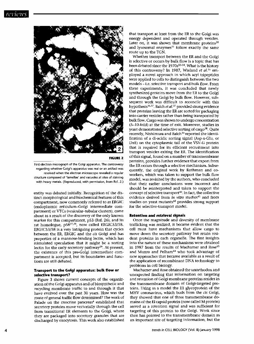
FIGURE 2
First electron micrograph of the Golgi apparatus. The controversy
regarding whether Colgi’s apparatus was real or an artifact was
resolved when the electron microscope revealed a regular
structure composed of ‘lamellae’ and vacuoles at sites of staining
with heavy metals. (Reproduced, with permission, from Ref. 2.)
entity was debated initially. Recognition of the dis- tinct morphological and biochemical features of this compartment, now commonly referred to as ERGIC (endoplasmic reticulum-Golgi intermediate com- partment) or VTCs (vesicular-tubular clusters), came about as a result of the discovery of the only known marker for this compartment, p53 (Ref. 26), and its rat homologue, p5827,28, now called ERGIC53/58. ERGIC53/58 is a very intriguing protein that cycles between the ER, ERGIC and the cis Golgi and has properties of a mannose-binding lectin, which has stimulated speculation that it might be a sorting lectin for the early secretory pathwayz9. At present, the existence of this pre-Golgi intermediate com- partment is accepted, but its boundaries and func- tions are still debated.
Transport to the Golgi apparatus: bulk flow or selective transport?
Figure 3 shows current concepts of the organiz- ation of the Golgi apparatus and of biosynthetic and recycling membrane traffic to and through it that have evolved over the past 30 years. How was the route of general traffic flow determined? The work of Palade on the exocrine pancreas4 established that secretory proteins move vectorially through the cell from transitional ER elements to the Golgi, where they are packaged into secretory granules that are discharged by exocytosis. This work also established
that transport at least from the ER to the Golgi was energy dependent and operated through vesicles. Later on, it was shown that membrane proteins30 and lysosomal enzymes3i follow exactly the same route up to the TGN.
Whether transport between the ER and the Golgi is selective or occurs by bulk flow is a topic that has been debated since the 1970s32-34. What is the history of this controversy? In 1987, Wieland et a1.35 em- ployed a novel approach in which acyl tripeptides were applied to cells to distinguish between the two models - i.e. selective transport and bulk flow. From these experiments, it was concluded that newly synthesized proteins move from the ER to the Golgi and through the Golgi by bulk flow. However, sub- sequent work was difficult to reconcile with this hypothesis36,37. Balch et aZ.37 provided strong evidence that proteins leaving the ER are sorted for packaging into carrier vesicles rather than being transported by bulk flow. Cargo was shown to undergo concentration (S-lo-fold) at the time of exit. Moreover, studies in yeast demonstrated selective sorting of cargo38. Quite recently, Nishimura and Balch39 reported the identi- fication of a d&acidic sorting signal (Asp-x-Glu, or DxE) on the cytoplasmic tail of the VSV-G protein that is required for its efficient recruitment ,into transport vesicles exiting the ER. The identification of this signal, found on a number of transmembrane proteins, provides further evidence that export from the ER occurs through a selective mechanism. Subse- quently, the original work by Rothman and co- workers, which was taken to support the bulk flow model, was revisited by the authors, who concluded that their earlier conclusions were incorrect and should be reinterpreted and taken to support the concept of selective transport40. In fact, the collective evidence derived from in vitro studies41 and from studies on yeast mutants42 provides strong support for the selective-transport model.
Retention and retrieval signals Once the magnitude and diversity of membrane
trafficking was realized, it became evident that the cell must have mechanisms that allow cargo to move down the secretory pathway but retain resi- dent proteins in each organelle. The first insights into the nature of these mechanisms were obtained in 1987 from the results of Machamer and Rose43 and Munro and Pelham44 who took advantage of new approaches that became available as a result of the application of recombinant DNA technology to problems in cell biology.
Machamer and Rose obtained the unorthodox and unexpected finding that information on targeting and retention of Golgi membrane proteins resides in the transmembrane domain of Golgi-targeted pro- teins. Using as a model the El glycoprotein of the MHV coronavirus, which buds from the cis GO@, they showed that one of three transmembrane do- mains of the EI capsid protein (now called M protein) served as a retention signal and was sufficient for targeting of this protein to the Go@. Work since then has pointed to the transmembrane domain as an important site of targeting information, but the
trends in CELL BIOLOGY (Vol. 8) January 1998

TABLE 1 - SOME IMPORTANT MILESTONES IN GOLGI RESEARCH
Year
1898
1954
1957
1961
1964
1966
1967-1975
1969
1971
1973-l 981
1977
1980
1981-1983
1984
1985
1986
1987
1988
1990
1991
1993-l 994
1994
Event
Discovery of the Colgi apparatus
First electron microscopy (EM) description of the Colgi apparatus
Cisternal maturation model of Colgi transport
Compartmentalization: regional distribution of enzymes
Involvement in sulfation
Involvement in glycosylation: glucose incorporation
Role in secretory pathway defined and vesicular transport documented
Incorporation of mannose in endoplasmic reticulum (ER), galactose in Golgi
Calactosyltransferase as a biochemical marker for the Golgi apparatus
GERL concept
Role of mannose 6-phosphate in lysosomal enzyme sorting by Golgi
Demonstration of recycling plasma membrane to Golgi
Introduction of glycosidase (endo H) treatment to assess transport
Topology of N-glycosylation
lmmunocytochemical localization of galactosyltransferase to tram Golgi
Reconstitution in vitro of transport within Golgi stack
Description of 15” block and cargo accumulation in pre-Golgi intermediate compartment
Regulated vs. constitutive secretory pathways
Description of 20” block and cargo accumulation in trons-Golgi network (TGN)
Transmembrane domain required for retention of resident Golgi proteins
KDEL retrieval signal for resident ER proteins
Involvement of small GTP-binding proteins in vesicular transport
Heterotrimeric G-proteins implicated in traffic control
Reconstitution in vitro of ER-to-Golgi transport
Isolation of ER-Golgi intermediate compartment (ERGIC)
Application of brefeldin A to study Golgi-ER transport
Phosphoinositide 3-kinase implicated in control of Golgi traffic
Discovery of COPI coat
Demonstration of role of Gai3 in traffic control
Demonstration that ER-to-Golgi transport is selective
Discovery of COPII coated vesicles
COPI functions in Golgi-to-ER retrograde transport
precise mechanism involved is still the subject of yet another Golgi controversy. In 1993, two models were introduced that now dominate the field: the kin- recognition model and the bilayer-mediated sorting model (reviewed in Ref. 45). The kin-recognition model holds that resident proteins of a particular cisterna interact to form large hetero-oligomers that prevent resident proteins from entering transport vesicles. The bilayer-mediated sorting model proposes that the length of the transmembrane domain is the crucial factor in sorting resident Golgi proteins, which have shorter transmembrane domains than those of the plasma membrane. It is proposed that Golgi membrane proteins are retained because they
Discoverer(s)
Golgi
Dalton and Felix
G rasse
Novikoff and Goldfischer
Godman and Lane
Neutra and Leblond
Palade, Jamieson and coworkers
Whur, Herscovics and Leblond
6. Fleischer et ol.; Morre et ol.
Novikoff and Novikoff
Sly, Neufeld, Kornfeld, Jourdian
Herzog and Farquhar
Strous and Lodish
Dunphy and Rothman
Roth and Berger
Rothman et al.
Saraste and Kuismanen
Moore and Kelly
Griffiths and Simons
Machamer and Rose
Munro and Pelham
Salminen and Novick
Melanson et ol.
Becker and Balch
Schweizer et a/.
Lippincott-Schwartz et al.
Herman and Emr; Schu et al.
Duden et al.; Seratini et ol.; Waters et al.
Stow et al.
Balch et al.; Mizuno and Singer; Rexach et al.
Barlow et al.
Letourneur et ol.
are excluded from cholesterol-rich regions of Golgi membranes destined for the plasma membrane. At present, the only aspect that appears to be agreed upon is the importance of the transmembrane do- main in retention of Golgi-resident proteins.
The other key finding was that of Pelham46 and colleagues, who demonstrated that receptor-mediated retrieval mechanisms are used for retention of resident ER proteins. They showed that, if resident proteins of the ER lumen escape and move down the secretory pathway, they are retrieved by a specialized C-terminal KDEL (or closely related) signal that is recognized by KDEL receptors located in the cis Golgi or ERGIC and transported back to the ER. Some ER membrane
trends in CELL BIOLOGY (Vol. 8) January 1998 5

TGN
tram
cis WV
ERGIC
TE
FIGURE 3
Diagram depicting the major routes of vesicular traffic to and through the Colgi apparatus along the exocytic (l-6) and endocytic (7-l 2)
pathways. Exocytic pathways: secretory proteins, membrane glycoproteins and lysosomal enzymes are synthesized on polyribosomes
and translocated to the endoplasmic reticulum [ER; (l)] where they undergo cotranslational and posttranslocational processing. They
exit the ER via COPII vesicles (2), which serve to shuttle them to the ER-Golgi intermediate compartment (ERGIC, or vesicular-tubular
clusters, VTCs). From there, they could be transported to the cis-Colgi network (CCN) via COPI-coated vesicles (3), but this is still
controversial. COPI-coated vesicles also function in retrograde transport (Golgi to ER) (4). Subsequently, either the proteins traverse
the Colgi cisternae one by one via vesicular carriers (5) or transport occurs by cisternal maturation. Retrograde transport is also
assumed to take place between the stacked cisternae (6). Sorting occurs in the trons cisterna or Irons-Colgi network (TGN).
Lysosomal enzymes bind to mannose 6-phosphate receptors in the Golgi, are packaged into clathrin-coated vesicles in the TCN, and
delivered either to early (7) or late (7’) endosomes. Membrane and secretory proteins are also sorted in the TGN and delivered by
exocytosis along the regulated secretory pathway via secretion granules (8) or along the constitutive pathway (9). In polarized
secretory cells, a separate pathway exists for delivery of vesicles to the basolateral domain (10). Endocytic pathways: the
best-characterized endocytic pathway is receptor-mediated endocytosis through clathrin-coated vesicles budding from either the
apical domain (11) or the basolateral domain (11 ‘). Many receptors (low-density lipoprotein, transferrin) recycle back to the plasma
membrane (PM) from early endosomes (12), whereas many ligands are transported from early to late endosomes to reach lysosomes
(13). Other pathways include uptake in nonclathrin-coated vesicles (14) or by caveolae (15). Mannose 6-phosphate receptors shuttle
between the Golgi and endosomes. Abbreviation: TE, transitional ER element. (Adapted, with permission, from Ref. SO.)
6
proteins possess either a KKXX (type I) or XXRR Role of the Golgi in sorting and packaging (type II) retrieval signal that binds to COP1 (Ref. 41). A role for the Golgi apparatus in the formation Also, a tyrosine-based sorting signal is used for re- of secretion granules was suggested as long ago as trieval of a TGN protein, TGN38, from the plasma the 1920s based on observations on glandular cells membrane. Thus, the existence of both retention by light microscopy. Direct evidence for the role of and retrieval signals for resident ER and Golgi pro- the Golgi in concentration and packaging of secre- teins has been validated. tory proteins was provided by the work of Palade’s
trends in CELL BIOLOGY (Vol. 8) january 1998
laboratory on the exocrine pancreas4 and was sub- stantiated subsequently by work on many other cell types during the golden era of electron microscopy in the 1960s and 1970s. At about the same time, it was recognized, on the basis of acid phosphatase enzyme cytochemistry, that Iysosomal enzymes are transported to endosomes in coated vesicles that bud from the tram or exit side of the Golgi14. An impor- tant milestone in understanding Golgi sorting func- tions was the recognition in 1985 by Moore and Kellyl” of the existence of two distinct secretory path- ways, regulated and constitutive, and the demon- stration that secretory proteins are selectively sorted from membrane proteins, with the former packaged into dense-cored granules and the latter into consti- tutive secretory vesicles in the truns Golgi. Thus, it became evident that sorting of Golgi cargo for dis- tribution to other cellular sites occurred in the last cistema of the stack, which had a distinct morphology and was given several names including GERL, tmns Golgi reticulum and puns-Golgi network or TGN (Ref. 16). Among these, only TGN has survived.
Later on, it was recognized that there are two types of constitutive secretory vesicles destined for differ- ent plasmalemmal domains that bud from the TGN. Originally identified in polarized epithelial cells48,4g, they were more recently found also to exist in non- polarized cells. Thus, it became apparent that newly synthesized proteins are sorted in the TGN and selectively packaged into different containers. Four such containers have so far been recognized and well characterized: secretion granules, two types of con- stitutive secretory vesicles, and clathrin-coated ves- icles carrying lysosomal enzymes. Several other puta- tive vesicle populations (e.g. ~200 vesicles and AP-3 vesicles) have been described, but exactly what they are doing remains controversial (reviewed in Ref. SO).
The first sorting signal: mannose i-phosphate A landmark discovery familiar to most cell biologists
was the discovery of the mechanism of sorting of lyso- somal enzymes through the mannose 6-phosphate (M6P) sorting signal. This came about as the result of work in the late 1970s from several laboratories, including those of Kaplan and Sly, and Kornfeld and Jourdian (see Ref. 51 for a review), demonstrating that lysosomal enzymes possess phosphorylated mannose residues recognized by M6P receptors in the TGN, leading to their selective removal from the exocytic pathway. This discovery demonstrated that sorting inside the cell occurs by a process resembling receptor-mediated endocytosis at the plasma mem- brane and became the paradigm that has guided thinking about mechanisms of intracellular sorting to this day. Surprisingly, although more than 20 years have passed since the discovery of the first intra- cellular sorting signal, the mechanisms of sorting at the TGN for biosynthetic products other than lyso- somal enzymes are still poorly understood. Sort- ing into regulated granules is believed to occur by aggregation and sorting of at least some proteins into constitutive vesicles by tyrosine-based signals. In addition, a unique mechanism was suggested re- cently by Simons and Ikonens2 for sorting membrane
trends in CELL BIOLOGY (Vol. 8) January 1998
proteins destined for the apical domain of the plasma membrane involving lipid rafts and a galactose- binding protein, VIP36.
A continuing controversy: does transport across the Colgi stack occur by cisternal maturation or vesicular transport?
Since the 1960s and 197Os, it has been clear that cargo moves across the Golgi stack, but the mecha- nism by which this takes place has been debated for over 40 years. Two models have been proposed. The first was the maturation or cistemal progression model introduced in the 1950ss3, which visualizes the Golgi as a kind of bottling station: cisternae are formed on the entry or cis face and move sequentially towards the exit or tram face, where they are used up in packaging. This concept has been championed by MorreS4 and Leblonds5 among others.
New information obtained during the 197Os, es- pecially the demonstration of the distinctive com- position of Golgi compartments, the faster turnover of secretory versus membrane proteins and the exist- ence of recycling, was difficult to reconcile with the maturation model. The vesicular transport/stationary cisterna model, introduced in 1981 (Ref. 1 l), was de- signed to take into account this new information. It held that each cisterna (or set of cistemae) constitutes a separate compartment of distinctive composition and that transport from one cistema to another occurs through vesicles. The vesicular transport/stationary cisterna model gained wide support based on results obtained from in vitro systems, and was championed by Rothman and coworkers, whose in vitro experi- ments were interpreted as reconstituting anterograde vesicular transport40*56r57. As a result, the vesicular transport model has dominated the field for over 15 years. Recently, however, it has been seriously challenged5s as the result of several developments, including the failure to identify t-SNARES (see below) associated with intra-Golgi transport, the failures9 until recently60 to detect secretory proteins in Golgi vesicles, and the difficulties in applying the vesicu- lar transport model to the maturation of complex structures such as algal scale@, procollagen fibrilss5 and casei# that are found across the Golgi stack.
Once again, new data and resurrection of some old data have stimulated a re-examination of existing models. As a result, the maturation model in modi- fied form, which is actually a hybrid between the maturation and vesicular transport models, is enjoy- ing a strong comeback. It holds that anterograde transport occurs by cisternal maturation coupled with retrograde vesicular transport of Go@ enzymes. There are many aspects of this modified cisternal progression model that appear attractive, but its key features - anterograde transport of individual cister- nae, retrograde transport of Golgi enzymes, and de novo formation of cis Golgi cisternae - remain to be demonstrated convincingly. Similarly, a key fea- ture of the stationary cistemae model-the existence of anterograde vesicular transport - has been seri- ously challengeds8. Therefore, it is safe to say that neither the vesicular transport model nor the modi- fied maturation model for anterograde transport is

completely proven and that the mechanism of trans- port across the stack remains controversial. As long as the evidence is conflicting or not entirely con- vincing, this controversy will continue. The lessons learned from Golgi history suggest that it will take new approaches and convergent information from different quarters to design universally accepted, accurate models.
Recyling, retrograde transport and tubules The likely existence of recycling of membranes
involved in transport to and through the Golgi was predicted at the time of the discovery of vesicular trafficking4. The first convincing evidence for reutiliz- ation or recycling of Go&i-derived membranes was obtained using particulate, electron-dense tracers (dextrans and cationized ferritin) to mark recovery of membranes of secretion granules, their recycling to the trans Golgi, and reutilization in packaging of secretory proteins 63,64. It may seem hard to believe, but this, too, was the subject of controversy because at that time it was assumed, based on the studies with the fluid-phase marker horseradish peroxidase, that plasma membrane proteins recycle through endosomes and that there was no access to the Golgi from the plasma membrane. Doubts lingered until the demonstration65 that, when plasma membrane proteins are desialylated at the cell surface, they can be resialylated during recycling through the Golgi. In the meantime, it became evident that, as the half-life of most membrane proteins is rather long (l-3 days), recycling must occur at all steps in intracellular transport’l and that transport between the Golgi and other compartments to which its cargo is delivered (lysosomes, plasma membrane, endosomes) involves both anterograde and retrograde pathways (see Fig. 3).
Until recently, however, most studies focused on anterograde transport, but studies with brefeldin A (BFA) focused attention on retrograde transport and brought to light the potential role of tubules as well as vesicles in transport from the Golgi to the ER66. When cells were treated with BFA, tubules formed through which Golgi components relocated to the ER. Tubules were also seen as components of the ERGIC and VTCs in normal cells. Recent work in which cargo is tagged with green fluorescent protein (GFP) suggests a wider involvement of tubules in anterograde transport both between the ER and Golgi and between the Golgi and the plasma mem- brane than had been appreciated until now(j7.
Mechanisms of vesicle budding, targeting and fusion
Since the realization that proteins could be trans- ported to and through the Golgi by vesicular trans- port, investigators have been intrigued to know how vesicles bud and recognize and reach their target. Progress in this arena was slow and was not linked to a single discovery or event. Significant insights have been obtained only in the past four years as a result of the convergence of information derived from three seemingly disparate sources: biochemical stud- ies on Golgi transport in vitro40r57, analysis of yeast set mutants42,6s-70 and characterization of synaptic
vesicle proteins71,72. It became evident that vesicles involved in transport at different steps along the exocytic pathway have common features in terms of their fusion machinery but apparently specific tar- geting equipment. Most have prominent coats of which there are at least three types - COPII, COP1 and clathrin (Fig. 3) - plus several less-well-characterized new candidates, including AP-3 and p20073f74. COP1 and COP11 vesicles are involved in transport between the ER and Golgi, and clathrin-coated vesicles are of two types: those involved in transport of lysosomal enzymes between the TGN and endosomes and those involved in receptor-mediated endocytosis at the plasma membrane. The model for formation of all these vesicles was provided by clathrin-coated vesicles73,74. Based on this model, vesicle budding is thought to be initiated by the assembly of the protein coat, triggering assembly of elaborate membrane and cytosolic protein complexes, unique for each vesicle population, that drive vesicle budding and fission. Once formed, cargo proteins, cargo receptors and membrane proteins required for target recognition are included in the vesicle.
How do vesicles recognize and fuse with their ap- propriate target? The prevailing working model is the SNARE hypothesis introduced in 1993 (Ref. 75) when it was realized that synaptic vesicles and intra-Golgi transport vesicles have similar components. Accord- ing to this model, pairs of integral vesicle and target membrane proteins (v- and t-SNARES), with specific family members assigned to different stations, ensure docking to the appropriate membrane receptor40f57. Soluble cytosolic proteins - N-ethylmaleimide- sensitive fusion protein (NSF) and soluble NSF attach- ment proteins (SNAPS) - serve as common fusion machinery for different vesicle relays. Rab proteins are believed to check the fidelity of the membrane- fusion and -targeting event. Recognition by specific v- and t-SNARES is assumed to apply to most but not all vesicular transport steps along both the exocytic and the endocytic pathways. An exception is repre- sented by the vesicles involved in apical delivery of Golgi-derived proteins in polarized secretory cells where glycolipids and glycans appear to be involveds2.
Several controversies currently pervade this area of Golgi research. First, it is debated whether COP1 vesicles are involved in both anterograde and retro- grade transport between the ER and Golgi or only in retrograde transport. Second, it is questioned whether COP1 vesicles are involved at all in anterograde intra-Golgi transport. Indeed, as indicated earlier, the very concept of anterograde vesicular transport within the Golgi is currently being contesteds8. Third, the SNARE hypothesis has recently been attacked by some as being too simplistic, which is probably the case. One has only to remember that synaptic ves- icles contain dozens of proteins72 and the function of relatively few of these has been established. More- over, from the work in yeast, we begin to have an inventory of many of the genes involved in sorting and in vesicle formation and targeting. In the case of COP11 vesicles, for example, at least nine proteins besides SNARES are proposed to be involved in sort- ing, coat assembly and vesicle budding42.
trends in CELL BIOLOGY (Vol. 8) January 1998
How are Colgi functions and Colgi traffic regulated?
This question has only begun to be tackled in the past 5-10 years and was made possible by availability of in vitro transport assays in both mammalian cells and yeast and by analysis of yeast mutants. Evidence is accumulating that small GTPases of the Ras super- family, heterotrimeric G proteins, and phospho- inositides, are involved. Most is known about the small GTPases. Their involvement in vesicular traffic was first indicated in 1987 when Salminen and Novick76 discovered a small Rab GTPase, Sec4p, which is required for transport from the Golgi to the cell surface in yeast. This finding was extended very rapidly, and subsequently a large (20-30) family of small GTPases, the Rabs, was discovered, each with a characteristic distribution in the ce1170,77,78. Of these, RablA and Rab2 are located on ERGIC (VTCs) and the cis Golgi, and Rab6 is located in the truns Golgi. RablA and another small GTPase, Sarlp, were shown to be essential for ER-to-Golgi transport. Rabs are believed to carry out a proofreading function, check- ing that each vesicle fuses with the appropriate tar- get ‘O,“. Originally it was believed that each Rab was dedicated to a specific step in transport, but now there are more Rabs than defined steps.
Heterotrimeric G proteins were first implicated in control of vesicular trafficking by the finding of Melancon et ~1.‘~ that AlF-, a specific inhibitor of heterotrimeric G proteins, inhibited ER-to-Go&i trans- port in vitro. Indirect evidence suggested the involve- ment of trimeric G proteins in virtually every trans- port step80,81. Subsequently, several G proteins, notably Gai382-s4 and GCXS and GaqB4, were localized to Golgi membranes. The most direct findings documenting involvement of G proteins in Golgi traffic control were those of Stow et aLB2, who showed that over- expression of Gai3 inhibited processing of a secretory protein, heparan sulfate proteoglycan, presumably by inhibition of transport through the Golgi. The precise role of G proteins-whether in coat assembly, sorting, vesicle budding or vesicle fusion - remains unknown. Trimeric G proteins are well known to regulate signalling at the plasma membrane by inter- action with various effecters. The presence of trimeric G proteins on intracellular membranes, especially those of the Golgi apparatus, suggests that they could have similar functions inside the cell. However, it is not clear whether the effects occur via classical or nonclassical G-protein pathways.
The first indication of a role for phosphoinositides in vesicular trafficking came from the discovery by Emr and coworkers85,86 of the yeast VPS34 gene, whose product is required for transport of vacuolar proteins from the Golgi to the vacuole, which is a lysosome equivalent. Vps34p was shown to possess extensive homology with bovine phosphoinositide 3-kinase. Indirect evidence was obtained subse- quently, by use of the inhibitor wortmannin, for the involvement of a Vps34p-like protein in transport from the TGN to the Golgi in mammalian cellss7,88. The mechanism by which phosphoinositides control vesicular traffic has not been established, but recent evidence suggests that they could regulate the activity
trends in CELL BIOLOGY (Vol. 8) January 1998
of proteins with diverse functions such as dynamin, AP-2 and guanine-nucleotide exchange factorss9. In short, delineation of mechanisms for control of vesicular traffic is in its infancy and provides prob- lems of sufficient magnitude to keep an army of investigators busy for perhaps another 100 years.
Future perspectives The first century of the history of the Golgi com-
plex has been one of great progress, yet it has also been fraught with controversy. It was not until 1954 that the Golgi itself was accepted as a bona fide organ- elle. Since that time, controversy has surrounded the Golgi, and it continues to this day. What lessons can be learned from past history? First, it is clear that prob lems are not resolved by heated discussions, intro- duction of premature dogmas, intimidation or sup- pression of alternative points of view. Second, from the Golgi milestones reviewed here, it is evident that controversies that have arisen were often resolved by information that came about as a result of new technical developments such as the introduction of the electron microscope in the 195Os, of enzyme cyto- chemistry, autoradiography and cell fractionation in the 1960s and 197Os, and of recombinant DNA tech- nology, in vitro assays and yeast mutants in the 1980s and 1990s. Third, only when the evidence is strong, and complementary approaches converge and lead to the same conclusions, can models be formulated that stand the test of time.
References 1 Colgi, C. (1898) Arch. Ital. Go/. 30, 60-71 2 Dalton, A. 1. and Felix, M. D. (1954) Am. 1. Anat. 94, 171-l 87 3 Sjostrand, F. S. and Hanzon, V. (1954) Exp. Cell Res. 7, 415-429 4 Palade, C. (1975) Science 89, 347-358 5 Fleischer, B., Fleischer, 5. and Ozawa, H. (1969) j. Cell Biol.
43,59-59 6 Morre, D. I., Merlin, L. and Keenan, T. (1969) B&hem.
Biophys. Res. Commun. 37, 813-819 7 Neutra, M. and Leblond, C. P. (1966) /. Cell Biol. 30, 137-l 50 8 Whur, P., Herscovics, A. and Leblond, C. P. (1969) ). Cell Biol.
43,289-311 9 Codman, C. C. and Lane, N. (1964) /. Cell Biol. 21, 353-366
10 Novikoff, A. and Goldfischer, S. (1961) Proc. Nat/. Acad. Sci. U. 5. A. 47, 802-810
11 Farquhar, M. C. and Palade, C. E. (1981) ). Cell Biol. 91, 77s-103s 12 Friend, D. 5. (1969) 1. Cell Biol. 41, 269-279 13 Bainton, D. F. and Farquhar, M. C. (1966) /. Cell Biol. 28,277-301 14 Friend, D. 5. and Farquhar, M. C. (1967) /. Cell Biol. 35, 357-376 15 Novikoff, P. M. et a/. (1971) 1. Cell Biol. 50, 859-886 16 Griffiths, C. and Simons, K. (1986) Science 234,438-443 17 Kornfeld, R. and Kornfeld, 5. (1985) Annu. Rev. Biochem. 54,
631-664 18 Roth, 1. (1997) in The Go/g; Apparatus (Berger, E. G. and
Roth, J., eds), pp. 131-l 61, Birkhluser 19 Roth, J. and Berger, E. G. (1982) 1. Cell Biol. 92, 223-229 20 Roth, J. et ol. (1985) Cell 43, 287-295 21 Dunphy, W. G. et a/. (1981) Proc. Nat/. Acod. Sci. U. 5. A. 78,
7453-7457 22 Goldberg, D. E. and Kornfeld, 5. (1983) 1. Biol. Chem. 258,
3159-3165 23 Velasco, A. et al. (1993) /. Cell Biol. 122, 39-51 24 Varki, A. P. (1998) Trends Cell Biol. 8, 34-40

25 Saraste, 1. and Kuismanen, E. (1984) Cell 38, 535-549 26 Schweizer, A. et al. (1988) j. Cell Biol. 107, 1643-l 653 27 Saraste, J., Palade, G. E. and Farquhar, M. G. (1987) 1. Cell Biol.
105,2021-2029 28 Lahtinen, U. et al. (1996) 1, Biol. Chem. 271, 40314d37 29 Itin, C. et al. (1996) Mol. ho/. Cell 7, 48H93 30 Bergmann, I. E., Tokuyasu, K. T. and Singer, 5. J. (1981) Proc.
Not/. Acad. Sci. U. 5. A. 78, 17461750 31 Kornfeld, 5. and Mellman, I. (1989) Annu. Rev. Cell Biol. 5,
483-525 32 Balch, W. E. and Farquhar, M. C. (1995) Trends Cell Biol. 5, 16-l 9 33 Griffiths, C. et al. (1995) Trends Cell Biol. 5, 9-l 3 34 Singer, S. J. (1995) Trends Cell Biol. 5, 14-l 5 35 Wieland, F. T. et al. (1987) Cell 50, 289-300 36 Mizuno, M. and Singer, 5. J. (1993) Proc. Nat/. Acad. Sci.
U. S. A. 90,5732-5736 37 Balch, W. E. et al. (1994) Cell 76, 841-852 38 Rexach, M. F., Latterich, M. and Schekman, R. W. (1994) 1. Cell
Biol. 126, 1133-l 148 39 Nishimura, N. and Balch, W. E. (1997) Science277, 556-558 40 Rothman, J. E. and Wieland, F. T. (1996) Science 272, 227-234 41 Bannykh, S. I., Nishimura, N. and Balch, W. E. (1998) Trends
Cell Biol. 8, 21-25 42 Bednarek, 5. Y., Orci, L. and Schekman, R. (1996) Trends Cell
Biol. 6, 468-473 43 Machamer, C. E. and Rose, J. K. (1987) 1. Cell Biol. 105,
1205-l 214 44 Munro, 5. and Pelham, H. R. B. (1987) Cell48, 899-907 45 Munro, 5. (1998) Trends Cell Biol. 8, 1 l-l 5 46 Pelham, H. R. B. (1989) Annu. Rev. Cell Biol. 5, l-23 47 Moore, H. P. and Kelly, R. B. (1985) /. Cell Biol. 101, 1773-l 781 48 Simons, K. and Fuller, 5. D. (1985) Annu. Rev. Cell Biol. 1, 243-288 49 Rodriguez-Boulan, E. (1983) in Modem Cell tliology (Satir, B., ed.),
pp. 119-l 70, Alan R. Liss 50 Farquhar, M. C. and Hauri, H-P. (1997) in The Colgi Apparatus
(Berger, E. G. and Roth, I., eds), pp. 63-129, Birkhluser 51 Kornfeld, S. (1992) Annu. Rev. Biocbem. 61, 307-330
Acknowledgements 52 Simons, K. and Ikonen, E. (1997) Nature 387, 569-572
The authors are 53 Grass& P-P. (1957) C. R. Acad. Sci. 245, 1278-l 281
supported by NIH 54 Morn?, D. I., Kartenback, 1. and Franke, W. W. (1979) Biochim.
grants CA58689 Biophys. Acta 559, 71-l 52 and ~~17780 55 Leblond, C. P. and Inoue, S. (1989) Am. 1. Anat. 185, 367-390 (to M. G. F.). 56 Rothman, 1, E. and Orci, L. (1992) Nature 355, 409-415
57 Rothman, j. E. (1994) Nature 372,55-61 58 Pelham, H. R. B. (1998) Trends Cell Biol. 8, 4549 59 Dahan, S. et al. (1994) /, Cell Biol. 127, 1859-l 869 60 Orci, L. et al. (1997) Cell90, 335-349 61 Becker, B., Bolinger, B. and Melkonian, M. (1995) Trends Cell
Biol. 5, 305-307 62 Clermont, Y. et a/. (1993) Anat. Rec. 235, 363-373 63 Herzog, V. and Farquhar, M. C. (1977) Proc. Nat/. Acad. Sci.
U. 5. A. 74,5073-5077 64 Farquhar, M. C. (1978) /. Cell Biol. 77, R35-R42 65 Snider, M. D. and Rogers, 0. C. (1986) 1. Cell Biol. 103, 265-275 66 Lippincott-Schwartz, j. et a/. (1990) Cell 60, 821-836 67 Lippincott-Schwartz, j., Cole, N. and Presley, 1. (1998) Trends
Cell Biol. 8, l&20 68 Pryer, N. K., Wuestehube, L. 1. and Schekman, R. (1992) Annu.
Rev. Biochem. 61,471-516 69 Schekman, R. and Orci, L. (1996) Science 271, 1526-l 533 70 Ferro-Novick, 5. and Novick, P. (1993) Annu. Rev. Cell Biol. 9,
575-599 71 Bennett, M. K. and Scheller, R. H. (1994) Annu. Rev. Biochem.
63,63-l 00 72 Stidhof, T. C. (1995) Nature 375,645-653 73 Brodsky, F. M. (1997) Trends Cell Biol. 7, 175-l 79 74 Robinson, M. S. (1997) Trends Cell Biol. 7, 99-l 02 75 %llner, T. et al. (1993) Nature 362, 318-324 76 Salminen, A. and Novick, P. 1. (1987) Cell 49, 527-538 77 Nuoffer, C. and Balch, W. E. (1994) Annu. Rev. Biochem. 63,
949-990 78 Novick, P. and Zerial, M. (1997) Curr. Opin. Cell Biol. 9, 496-504 79 Melancon, P. et a/. (1987) Cell 51, 1053-l 062 80 Stow, 1. L. (1995) Curr. Opin. Nephrol. Hypertens. 4, 421-425 81 Helms, 1. B. (1995) FEBS lett. 369, 84-88 82 Stow, 1. L. et a/. (1991) 1, Cell Biol. 114, 1113-l 124 83 Wilson, 8. S., Komuro, M. and Farquhar, M. G. (1994)
Endocrinology 134,2 3 3-244 84 Denker, S. et a/. (1996) j. Cell Biol. 133, 1027-l 040 85 Herman, P. K. and Emr, 5. D. (1990) Mol. Cell. Biol. 10,
6742-6754 86 Schu, P. V. et a/. (1993) Science 260, 88-91 87 Brown, W. 1. et a/. (1995) I. Cell Biol. 130, 781-796 88 Davidson, H. W. (1995) j. Cell Biol. 130, 797-805 89 Roth, M. G. and Sternweis, P. C. (1997) Curr. Opin. Cell Biol. 9,
519-526
Golgi centenary meeting
To celebrate the Golgi centenary, there will be a special meeting later this year in Pavia, Italy, the place where Colgi carried out the majority of his investigations.
Structure and function of the Golgi complex: state of the art 100 years after Camillo Colgi’s discovery 19-23 September 1998 University of Pavia, Italy
Organizers: Kathryn Howell, Albert0 Luini, Antonieta de Matteis and Alexander Minorov
The invited speakers will incfude: W. Balch, V. Bankaitis, 1. Bergeron, 1. Bonifacino, P. de Camilli, S. Emr, M. Farquhar, B. Click, H-P. Hauri, W. Hong, K. Howell, T. Kreis, J. Lippincott-Schwartz, V. Malhotra, P. Melancon, 1. Meldolesi, D. Morr6, 1. Morrow, 5. Munro,
P. Novick, G. Palade, H. Pelham, A. Rambourg, J. Rothman, R. Schekman, K. Simons, A. Staehelin, C. van Meer, G. Warren, M. C. Waters, F. Wieland and M. Zerial.
For information contact: MS Anna Cavallo
Secretariat, Colgi centenary meeting Consorzio Mario Negri Sud, Via Nazionale, 66030 S. Maria lmbaro (Chieti), Italy Tel: +39 872 570 338 l Fax: +39 872 578 240 l E-mail: [email protected]
10 trends in CELL BIOLOGY (Vol. 8) January 1998