Structural Basis for Arl1-Dependent Targeting of Homodimeric GRIP Domains to the Golgi Apparatus
12
Molecular Cell, Vol. 12, 863–874, October, 2003, Copyright 2003 by Cell Press Structural Basis for Arl1-Dependent Targeting of Homodimeric GRIP Domains to the Golgi Apparatus elle (Donaldson and Jackson, 2000). For example, Arf1 recruits COP I vesicle coats to Golgi membranes, and the distantly related Sar1 recruits COP II coats to the Bojana Panic, 1 Olga Perisic, 1 Dmitry B. Veprintsev, 2 Roger L. Williams, 1 and Sean Munro 1, * 1 MRC Laboratory of Molecular Biology endoplasmic reticulum (ER). Arf1 also binds directly to the Golgi-localized protein GGA1, an adaptor that re- 2 Centre for Protein Engineering MRC Centre cruits cargo into Golgi-derived clathrin-coated vesicles. Arf1 has a number of close relatives, including the Hills Road Cambridge CB2 2QH plasma membrane-localized Arf6 that modulates the ac- tin cytoskeleton and endocytosis via generation of United Kingdom PtdIns(4,5)P 2 (Krauss et al., 2003; Niedergang et al., 2003). Beyond this, there is a large number of more distantly related proteins known as Arf-like (Arl) Summary GTPases that share structural features with Arf1 and Sar1, but whose functions are less well understood Golgins are large coiled-coil proteins that play a role in Golgi structure and vesicle traffic. The Arf-like GTPase (Clark et al., 1993; Pasqualato et al., 2002). There are at least ten Arls in humans which all share Arl1 regulates the translocation of GRIP domain-con- taining golgins to Golgi membranes. We report here with the Arfs an N-terminal amphipathic helix and, in most cases, a consensus sequence for N-myristoylation the 1.7 A ˚ resolution structure of human Arl1-GTP in a complex with the GRIP domain of golgin-245. The (Antonny et al., 1997; Pasqualato et al., 2002). The func- tion of most Arl proteins is unclear, but their importance structure reveals that the GRIP domain consists of an S-shaped arrangement of three helices. The domain is suggested by the observations that mutation of Arl1 in Drosophila is zygotically lethal (Tamkun et al., 1991) forms a homodimer that binds two Arl1-GTPs using two helices from each monomer. The structure is con- and that deletion in mice of Arf-related protein (ARFRP1 or ARP) causes embryonic lethality (Mueller et al., 2002). sistent with golgin-245 forming parallel coiled-coils and suggests how Arl1-GTP/GRIP complexes interact Arl2 has been implicated in tubulin assembly and micro- tubule polymerization, but its effector in this process is with Golgi membranes via the N termini of Arl1-GTP and the C-terminal tails of the GRIP domains. In cells, not known (Bhamidipati et al., 2000). In addition, Arl2- GTP binds to the phosphodiesterase (PDE) subunit, bivalent association with Arl1-GTP would increase residence time of the golgins on Golgi membranes. which has been proposed to act as a transport factor for the PDE catalytic subunits, and other prenylated Despite no conservation of sequence, topology, or even helical direction, several other effectors form proteins including GTPases (Hanzal-Bayer et al., 2002). Of the other Arls, perhaps the best understood is Arl1, similar interactions with small GTPases via a pair of helices, suggesting a common structural basis for which localized to the Golgi apparatus of both mamma- lian cells and yeast (Lowe et al., 1996; Gangi Setty et effector recognition. al., 2003). Arl1 is myristoylated, and mutation of the N-terminal myristoylation site abrogates Golgi targeting Introduction (Lee et al., 1997; Lu et al., 2001). Yeast two-hybrid screens for potential effectors of human Ar1l identified Small GTPases of the Ras superfamily control a wide range of cellular events and are divided into the Ras, two Golgi-localized proteins, golgin-97 and golgin-245 (also termed p230) (Fritzler et al., 1995; Erlich et al., 1996; Rho, Ran, Rab, and Arf classes (Takai et al., 2001). For all classes, a cycle of GTP binding and hydrolysis is Lu et al., 2001; Van Valkenburgh et al., 2001). These are two members of a set of large coiled-coil proteins that linked to a conformational change which alters interac- tion with effectors (Vetter and Wittinghofer, 2001). The have been found on the Golgi apparatus and which are often referred to as “golgins.” The golgins have been Arf class of GTPases is distinguished from the others by an N-terminal amphipathic helix, typically myristoylated, implicated in Golgi formation and maintenance as well that mediates interaction with membranes (Chavrier and as in vesicular transport. Golgin-97 and golgin-245, Goud, 1999). In addition, the Arf GTPases undergo a along with GCC88 and GCC185, are peripheral mem- “front-back” change so that in the GTP-bound form the brane proteins that share a C-terminal GRIP domain. amphipathic helix is displaced from a hydrophobic This 50 residue domain is both necessary and suffi- pocket in the GTPase and instead acts to stabilize inter- cient for targeting to the Golgi apparatus and is con- action with lipid bilayers (Antonny et al., 1997; Pasqua- served in animals, fungi, plants, and protozoa (Barr, lato et al., 2002). Genetic and biochemical evidence has 1999; Kjer-Nielsen et al., 1999a; Munro and Nichols, shown that Arf GTPases play central roles in both mem- 1999; McConville et al., 2002). Arl1 is required for tar- brane traffic and cytoskeletal organization. Activation geting GRIP domain proteins to the Golgi apparatus in of Arf GTPases is usually controlled by exchange factors both yeast and mammalian cells and has been shown resident on specific organelles, with the GTP-bound to bind directly to the GRIP domain in a GTP-dependent GTPase then recruiting specific effectors to that organ- manner (Gangi Setty et al., 2003; Panic et al., 2003). Arl1 appears to be predominantly localized to Golgi mem- branes, and relocation of Arl1-GTP to a different organ- *Correspondence: [email protected]
-
Upload
independent -
Category
Documents
-
view
0 -
download
0
Transcript of Structural Basis for Arl1-Dependent Targeting of Homodimeric GRIP Domains to the Golgi Apparatus
Molecular Cell, Vol. 12, 863–874, October, 2003, Copyright 2003 by Cell Press
Structural Basis for Arl1-Dependent Targeting ofHomodimeric GRIP Domains to the Golgi Apparatus
elle (Donaldson and Jackson, 2000). For example, Arf1recruits COP I vesicle coats to Golgi membranes, andthe distantly related Sar1 recruits COP II coats to the
Bojana Panic,1 Olga Perisic,1
Dmitry B. Veprintsev,2 Roger L. Williams,1
and Sean Munro1,*1MRC Laboratory of Molecular Biology endoplasmic reticulum (ER). Arf1 also binds directly to
the Golgi-localized protein GGA1, an adaptor that re-2 Centre for Protein EngineeringMRC Centre cruits cargo into Golgi-derived clathrin-coated vesicles.
Arf1 has a number of close relatives, including theHills RoadCambridge CB2 2QH plasma membrane-localized Arf6 that modulates the ac-
tin cytoskeleton and endocytosis via generation ofUnited KingdomPtdIns(4,5)P2 (Krauss et al., 2003; Niedergang et al.,2003). Beyond this, there is a large number of moredistantly related proteins known as Arf-like (Arl)SummaryGTPases that share structural features with Arf1 andSar1, but whose functions are less well understoodGolgins are large coiled-coil proteins that play a role in
Golgi structure and vesicle traffic. The Arf-like GTPase (Clark et al., 1993; Pasqualato et al., 2002).There are at least ten Arls in humans which all shareArl1 regulates the translocation of GRIP domain-con-
taining golgins to Golgi membranes. We report here with the Arfs an N-terminal amphipathic helix and, inmost cases, a consensus sequence for N-myristoylationthe 1.7 A resolution structure of human Arl1-GTP in
a complex with the GRIP domain of golgin-245. The (Antonny et al., 1997; Pasqualato et al., 2002). The func-tion of most Arl proteins is unclear, but their importancestructure reveals that the GRIP domain consists of an
S-shaped arrangement of three helices. The domain is suggested by the observations that mutation of Arl1in Drosophila is zygotically lethal (Tamkun et al., 1991)forms a homodimer that binds two Arl1-GTPs using
two helices from each monomer. The structure is con- and that deletion in mice of Arf-related protein (ARFRP1or ARP) causes embryonic lethality (Mueller et al., 2002).sistent with golgin-245 forming parallel coiled-coils
and suggests how Arl1-GTP/GRIP complexes interact Arl2 has been implicated in tubulin assembly and micro-tubule polymerization, but its effector in this process iswith Golgi membranes via the N termini of Arl1-GTP
and the C-terminal tails of the GRIP domains. In cells, not known (Bhamidipati et al., 2000). In addition, Arl2-GTP binds to the phosphodiesterase (PDE) � subunit,bivalent association with Arl1-GTP would increase
residence time of the golgins on Golgi membranes. which has been proposed to act as a transport factorfor the PDE catalytic subunits, and other prenylatedDespite no conservation of sequence, topology, or
even helical direction, several other effectors form proteins including GTPases (Hanzal-Bayer et al., 2002).Of the other Arls, perhaps the best understood is Arl1,similar interactions with small GTPases via a pair of
� helices, suggesting a common structural basis for which localized to the Golgi apparatus of both mamma-lian cells and yeast (Lowe et al., 1996; Gangi Setty eteffector recognition.al., 2003). Arl1 is myristoylated, and mutation of theN-terminal myristoylation site abrogates Golgi targetingIntroduction(Lee et al., 1997; Lu et al., 2001). Yeast two-hybridscreens for potential effectors of human Ar1l identifiedSmall GTPases of the Ras superfamily control a wide
range of cellular events and are divided into the Ras, two Golgi-localized proteins, golgin-97 and golgin-245(also termed p230) (Fritzler et al., 1995; Erlich et al., 1996;Rho, Ran, Rab, and Arf classes (Takai et al., 2001). For
all classes, a cycle of GTP binding and hydrolysis is Lu et al., 2001; Van Valkenburgh et al., 2001). These aretwo members of a set of large coiled-coil proteins thatlinked to a conformational change which alters interac-
tion with effectors (Vetter and Wittinghofer, 2001). The have been found on the Golgi apparatus and which areoften referred to as “golgins.” The golgins have beenArf class of GTPases is distinguished from the others by
an N-terminal amphipathic helix, typically myristoylated, implicated in Golgi formation and maintenance as wellthat mediates interaction with membranes (Chavrier and as in vesicular transport. Golgin-97 and golgin-245,Goud, 1999). In addition, the Arf GTPases undergo a along with GCC88 and GCC185, are peripheral mem-“front-back” change so that in the GTP-bound form the brane proteins that share a C-terminal GRIP domain.amphipathic helix is displaced from a hydrophobic This �50 residue domain is both necessary and suffi-pocket in the GTPase and instead acts to stabilize inter- cient for targeting to the Golgi apparatus and is con-action with lipid bilayers (Antonny et al., 1997; Pasqua- served in animals, fungi, plants, and protozoa (Barr,lato et al., 2002). Genetic and biochemical evidence has 1999; Kjer-Nielsen et al., 1999a; Munro and Nichols,shown that Arf GTPases play central roles in both mem- 1999; McConville et al., 2002). Arl1 is required for tar-brane traffic and cytoskeletal organization. Activation geting GRIP domain proteins to the Golgi apparatus inof Arf GTPases is usually controlled by exchange factors both yeast and mammalian cells and has been shownresident on specific organelles, with the GTP-bound to bind directly to the GRIP domain in a GTP-dependentGTPase then recruiting specific effectors to that organ- manner (Gangi Setty et al., 2003; Panic et al., 2003). Arl1
appears to be predominantly localized to Golgi mem-branes, and relocation of Arl1-GTP to a different organ-*Correspondence: [email protected]
Molecular Cell864
elle causes relocation of a GRIP golgin (Lu and Hong, structures of the GDP-bound forms of all Arf family mem-bers so far examined, including that of yeast Arl1p (Amor2003).
Although the precise function of the GRIP golgins is et al., 2001). This helix is displaced in the GTP-boundforms of other Arf family GTPases to expose the hy-still unclear, golgins such as p115 and GM130 have
been shown to participate in both tethering of transport drophobic face for association with membranes, and itsremoval was necessary for crystallization of GTP-boundvesicles to Golgi membranes prior to fusion and in stack-
ing cisternae to create the higher order structure of the forms of Arf1p and Sar1p (Bi et al., 2002; Shiba et al.,2003). The GTPase active site mutant Q71L was incorpo-Golgi (Pfeffer, 1999; Shorter and Warren, 2002; Barr and
Short, 2003; Gillingham and Munro, 2003). In yeast, dele- rated into Arl1 to ensure that the protein accumulatedin the GTP-bound form. Complexes were purified bytion of either Arl1p or Imh1p, the only GRIP domain
protein, results in missorting of vacuolar proteins and, in affinity chromatography and gel filtration in the presenceof Mg2� and GTP.much more severe growth phenotypes when combined
with loss of Ypt6p, another GTPase that acts in thisprocess (Li and Warner, 1996; Tsukada et al., 1999; Bon- Overall Structure of the Arl1-GTP/GRIPangelino et al., 2002). Such phenotypes would be consis- Domain Complextent with a role in tethering vesicles returning from endo- The complex of the human golgin-245 GRIP domain withsomes back to the trans Golgi. It seems likely that both human Arl1(Q71L) crystallized in space group P21 withvesicle tethering and cisternal stacking roles will require unit cell dimensions a � 72.5, b � 89.7, c � 72.5, � �that the golgins are accurately targeted to specific parts 110.7. There are four Arl1(Q71L)-GTP/GRIP domainof the Golgi apparatus. Golgin-245 is found on the trans complexes in the asymmetric unit. The structure wasface of the Golgi apparatus (Kooy et al., 1992), and the solved using Se-Met multiple anomalous dispersionGRIP domains of all four human GRIP domain golgins (MAD) data (Table 1). There are only minor differencesare sufficient to confer this compartment-specific tar- in conformation among the four Arl1/GRIP complexesgeting (Luke et al., 2003). in the asymmetric unit.
In this paper, we report a 1.7 A resolution X-ray struc- The structure reveals that the GRIP domain forms ature of a complex between a human GRIP domain and homodimer, with each GRIP domain bound to an Arl1-GTP-bound Arl1. The structure reveals that the GRIP GTP (Figure 2). The resulting dyad symmetric arrange-domain is a dimer, allowing two Arl1 proteins to interact ment of the Arl1 proteins leaves their N termini facing thesimultaneously with a golgin. To our knowledge, such same direction so that the two N-terminal amphipathica dyad-symmetrical interaction of a small GTPase with helices absent from the structure would be able to inter-an effector has not been described previously. The inter- act simultaneously with a lipid bilayer. The overall extentaction interface between Arl1 and the GRIP domain of the residues in the GRIP domain that participate inshares features with those formed between several dimerization or Arl1 binding corresponds well to theother Ras-like GTPases and their effectors, despite a 40–50 residue region conserved among different GRIPrange of effector topologies. The specificity of the inter- domains, and specifically to the 45 residue portion ofactions is dictated by packing restraints at the interface golgin-245 that is sufficient for some Golgi targetingwith the GTPase. (gray bar in Figure 1C; Kjer-Nielsen et al., 1999b). Previ-
ous mutagenesis studies have also identified a numberof residues in the GRIP domain that are required forResults and DiscussionGolgi targeting (summarized in Figure 1C). In a previousstructural analysis of yeast Arl1p-GDP in the absence ofPreparation and Crystallization of an Arl1-GTP/GRIPthe GRIP domain, the protein formed an intermolecularDomain Complexdisulfide bond in the crystal via Cys 80, although thisThe GRIP domain was initially defined on the basis ofbond is unlikely to occur in the reducing environmentrelated sequences present at the C termini of severalof the cytoplasm (Amor et al., 2001). In the Arl1-GTP/Golgi coiled-coil proteins (Barr, 1999; Kjer-Nielsen et al.,GRIP crystal, this cysteine residue is buried in the Arl1/1999a; Munro and Nichols, 1999). There are four GRIPGRIP interface (see below), which has prevented suchdomain-containing proteins in humans and Drosophila,artifactual disulfide bond formation.and one in fungi and plants (Figure 1A). Direct binding
has previously been reported between recombinantforms of the GRIP domain of S. cerevisiae Imh1p and Structure of Arl1(Q71L)-GTP
Like other Arf family GTPases, the GTP-bound Arl1Arl1p-GTP. It has also been reported that human Arl1pinteracts with golgin-245 and golgin-97 in a yeast two- (Q71L) overall fold consists of six � strands surrounded
by five � helices (Figure 2). Comparison of the humanhybrid assay (Lu et al., 2001; Van Valkenburgh et al.,2001). E. coli-expressed forms of all four human GRIP Arl1(Q71L)-GTP with the previously reported yeast Arl1-
GDP (Amor et al., 2001), shows that the region from 40domains bind directly to human Arl1 in a GTP-dependentfashion (Figure 1B), suggesting that GRIP domains de- to 82 changes conformation upon GTP binding. This is
consistent with other members of the Arf family GTPasesfined by sequence may all share the capacity to bind toArl1p-GTP. (Goldberg, 1998; Pasqualato et al., 2001). The region
from 51 to 67 (strands �2 and �3) has been referred toThe C-terminal 59 residues of human golgin-245 en-compassing the GRIP domain was coexpressed in E. as the “interswitch” region (Pasqualato et al., 2002) and
is located between switch 1 (residues 40–50) and switchcoli with human Arl1 lacking the N-terminal 14 residues.This N-terminal region is very likely to form an amphi- 2 (residues 68–82). In the GTP-bound form of Arl1, the
interswitch region has moved two residues with respectpathic helix, as such a helix has been observed in the

Structure of the Arl1-GTP/GRIP Domain Complex865
Figure 1. Domain Structure and Sequence Alignment of GRIP Domain-Containing Proteins
(A) Schematic representation of the four human GRIP domain proteins, and their homologs in Drosophila melanogaster (Dm) and Caenorhabditiselegans (Ce). Also shown is Imh1p, the single GRIP domain protein of Saccharomyces cerevisiae (Sc). Sequence homology suggests that thegene encoding the C. elegans homolog of golgin-245 probably comprises two adjacent predicted genes in the genome (F59A2.2 and F59A2.6).There is a fifth protein in the human genome that ends with a GRIP domain, Ran binding protein 2� (RanBP2�), but it appears to be theproduct of a recent fusion of duplications of the genes for RanBP2 and GCC185, and its significance is unclear.(B) Binding of the GRIP domains from the indicated human golgins to Arl1. Extracts of E. coli expressing the His6-tagged GRIP domains ofgolgin-245, golgin-97, GCC88, and GCC185 (C-terminal 58, 78, 62, and 110 residues, respectively) were applied to glutathione beads loadedwith either human GST-Arl1 with mutations to lock it in the GDP (T31N) or GTP (Q71L) bound forms (upper panels), or GST-Arl1 loaded withGDP or the nonhydrolysable analog GMPPNP (GTP*) as described previously (lower panels) (Panic et al., 2003). After washing, bound proteinswere eluted with SDS sample buffer and analyzed by gel electrophoresis and Coomassie blue staining.(C) Alignment of the C termini of the indicated human GRIP domain proteins and their homologs in other species. Sequences were alignedwith Clustal W, and residues identical, or related, in seven or more of the sequences are indicated by black or gray, respectively (BoxShade),and tryptophans are marked in orange. A schematic of the structure of the golgin-245 GRIP domain is shown above the alignment with theinteracting residues marked as indicated. The residue numbering for golgin-245 is based on that of Swissprot accession Q13439 (isoform 4)(Erlich et al., 1996). Residues in the GRIP domains of golgin-245 or golgin-97 that have been mutated in previous studies are indicated withcircles (filled, loss of Golgi targeting; open, normal targeting; half filled, reduced targeting) (Barr, 1999; Kjer-Nielsen et al., 1999a; Munro andNichols, 1999; Lu and Hong, 2003). The gray bar indicates the minimal portion of golgin-245 that shows some Golgi targeting (Kjer-Nielsenet al., 1999b). Species as in (A), or Candida albicans (Ca), Oryza sativa (Os), and Trypanosoma brucei (Tb).
to the rest of the � sheet. This register shift is character- features in the switch/interswitch regions that can ac-count for the specificity of the Arl1-GTP/GRIP interac-istic of the Arf family GTPases and has the effect of
communicating conformational changes in the switch tion (see below).regions with the other side of the molecule so that theN-terminal amphipathic helix swings away from the GRIP Domain Dimerization
The GRIP domain consists of three antiparallel � helicesbody of the GTPase upon GTP binding (Hanzal-Bayeret al., 2002; Pasqualato et al., 2002). arranged in an S-shaped configuration (Figures 2 and
3). The surface of the S-shaped domain is slightlyIn Arl1-GTP, switch 1 has a conformation that enablesThr 48 to act as a ligand of the nucleotide-associated curved, and the convex face interacts with the Arl1 while
the concave face pairs with another GRIP domain in aMg2�. GTP binding also results in a conformationalchange of switch 2, enabling the 67-DXXG-70 motif to parallel �-helical “handshake.” The interaction between
GRIP domains in the crystal is extensive (2282 A2 ofcoordinate the �-phosphate of the GTP. These confor-mational changes are characteristic of nucleotide- solvent-accessible area is buried) and overwhelmingly
hydrophobic, with only a single intermolecular hydrogendependent structural rearrangements in the Ras super-family of GTPases (Vetter and Wittinghofer, 2001). While bond (Arg 2179 to carbonyl oxygen of Leu 2202; Figure
3A). The extensive interface between the GRIP domainsthe overall fold of the Arl1(Q71L) is similar to the GTP-bound forms of Arf1 and Arl2, there are local surface suggests that this homodimeric interaction is probably

Molecular Cell866
Table 1. Data Collection, Structure Determination, and Refinement Statistics
Data collection and MAD phasing statistics
Data Set Peakc Inflectionc Remotec
Resolution 1.7 A 1.9 A 1.9 ACompleteness (last shell) 98.7 (92.3) 100.0 (98.8) 99.9 (100.0)Rmerge
a 0.062 0.057 0.065Redundancy 3.6 3.7 3.7�I/� (last shell) 13.9 (1) 16.6 (2.5) 11.7 (1.5)
Phasing statistics
Phasing power (iso)b — 0.78 1.1Phasing power (anom)b 1.48 1.42 1.2Se sites found/expected 39/48FOM after SHARP 0.43FOM after SOLOMON 0.79
Refinement statistics
Resolution 67–1.7 AProtein atoms 7148Waters 266Rcryst
d 0.22Rfree
d (% data used) 0.25 (3.4)Rmsd from idealitye
Bonds 0.014 AAngles 1.5
Dihedrals 6.0
a Rmerge � �hkl�i|Ii(hkl) � �I(hkl)|/�hkl�iIi(hkl).b The phasing power is defined as the ratio of the r.m.s. value of the heavy atom structure factor amplitudes to the r.m.s. value of the lack-of-closure error.c Data sets were collected at ESRF beamline ID14-4 at 12.6612, 12.6590, and 13.2 keV for the peak, inflection, and remote data sets, respectively.d Rcryst and Rfree � �|Fobs � Fcalc|/�Fobs; Rfree calculated with the percentage of the data shown in parentheses.e Rms deviations for bond angles and lengths in regard to Engh and Huber parameters.
present in solution, and this was confirmed in two ways. Interaction between Arl1-GTP and the GRIP DomainAll of the contacts that the GRIP domain makes withFirst, versions of the GRIP domain were expressed in
E. coli with either His6 or MBP tags. The MBP form could Arl1 are via GRIP helices �1 and �2, with �1 formingmost of the switch 1 interactions and �2 forming mostbe precipitated with Ni2�-NTA-agarose only if it was
coexpressed with the His6-tagged species, or mixed of the switch 2 interactions (Figure 4). Interaction withthe Arl1 switch/interswitch regions in the crystal struc-with it after lysis (Figure 3B). Second, when the GRIP
domain was subjected to equilibrium centrifugation, ture provides a rational explanation for the GTP-depen-dent binding of the GRIP domain seen in vitro and inmonomers and dimers were found to be in equilibrium
in solution, with the sedimentation profile indicating a vivo. The only residue in the GRIP domain that interactswith both switch 1 and switch 2 is the very well con-Kd for dimerization of about 2.5 M (Figure 3C).
Examination of the dimer interface shows that helix served Tyr 2177. Mutation of this residue to alanine ingolgin-245 and several other GRIP domains has been�1 of the GRIP domain has a pivotal role. This helix has
a hydrophobic face consisting of residues Phe 2175, shown to result in loss of Golgi targeting in mammals,yeast, and protozoa (Kjer-Nielsen et al., 1999a; MunroLeu 2178, Leu 2182, and Tyr 2185 that lines up along
the dyad axis and primarily interacts with helices �2 and and Nichols, 1999; McConville et al., 2002). Moreover,the same residue is required for Arl1 binding by the GRIP�3 and connecting loops from the other half of the dimer
(Figure 3A). Many of the residues that comprise the inter- domain of the yeast protein Imh1p and for yeast two-hybrid interaction between the GRIP domain of golgin-face are well conserved in evolution, with Phe or Tyr
equivalent to Tyr 2185 of golgin-245 present in almost 245 and human Arl1 (Van Valkenburgh et al., 2001; Panicet al., 2003). Tyr 2177 protrudes into a pocket on theall GRIP domains (Figure 1C). This residue, near the
C-terminal end of helix �1, makes hydrophobic contacts surface of the Arl1 that is lined by a cluster of hydropho-bic residues from the interswitch and switch 2 (Phe 51,with Tyr 2185 and Met 2186 from the dyad-related mono-
mer. The importance of these interactions is under- Leu 66, Ile 74, Tyr 77, and Tyr 81; Figure 4). This “selectiv-ity pocket” appears to be one important determinant ofscored by mutations in two conserved residues in helix
�1 having been previously reported to result in loss of the specificity of the Arl1-GTP/GRIP interaction. At thebottom of this selectivity pocket, the OH of Tyr 2177Golgi targeting of GRIP domains (Barr, 1999; Kjer-Niel-
sen et al., 1999a). These mutations are Y2185A in golgin- forms a hydrogen bond with the OH of Tyr 81 in switch2. Although a tyrosine at the position equivalent to Tyr245 and K699A in golgin-97 (equivalent to Arg 2179 in
golgin-245, which forms extensive hydrophobic con- 2177 is conserved in all GRIP domains, mutagenesisindicates that the hydrogen bond formed with Tyr 81 istacts as well as an intermolecular hydrogen bond).

Structure of the Arl1-GTP/GRIP Domain Complex867
Figure 2. Structure of the Human Arl1/Gol-gin-245 GRIP Domain Complex
(A) Ribbon diagram of the Arl1(Q71L)-GTP/Mg2�/GRIP domain complex. The golgin-245GRIP domain forms a homodimer (one GRIPmolecule is colored yellow, the other orange),with each GRIP domain binding one Arl1 mol-ecule. Arl1 is shown in green, with switch 1residues highlighted in blue, switch 2 resi-dues in cyan, and interswitch in red. The GTPis shown as balls-and-sticks with Mg2� as agray sphere.(B) A view of Arl1/GRIP complex approxi-mately perpendicular to the view shown in (A).
not essential, as substitution of Tyr 2177 with Phe did Arl1), presumably cramping entry of the critical GRIPTyr 2177. Other residues in the interface are likely tonot significantly affect Golgi localization (Kjer-Nielsen
et al., 1999a). This suggests that the hydrophobic inter- contribute to selectivity for Ar1l-GTP. For example, Arl1Cys 80 makes a hydrogen bond to GRIP Thr 2200 andactions of the Tyr 2177 with the selectivity pocket are
paramount for Arl1-GTP/GRIP binding. In the GDP com- is well conserved in Arl1 from different species (Figure4C). In contrast, a histidine is conserved at this positionplexes of both Arls and Arfs, the selectivity pocket is
occupied by the phenylalanine at residue 51 in Arl1 or in the Arf proteins, and a C80H mutation in Arl1 elimi-nates a yeast two-hybrid interaction with the golgin-245its equivalent. This, and other structural changes in the
switch regions, presumably prevents binding of the GRIP domain (Lu and Hong, 2003), presumably becauseof the steric effects of the bulkier histidine residue.GRIP domain to Arl1-GDP.
Just as comparison of Arl1-GTP with Arf1-GTP sug-gests differences that could account for effector selec-Selectivity of the Golgin GRIP Domains for Arl1-GTP
The golgin-245 GRIP domain binds selectively to Arl1- tivity, comparison of the GRIP domain with the Arf1effector domain N-GAT shows differences that couldGTP and not to other Arf-family GTPases such as Arfs1-6
and Arl2 (Van Valkenburgh et al., 2001; Lu and Hong, account for GTPase selectivity. The presence of a GRIPtyrosine (Tyr 2177) that occupies the selectivity pocket2003). Comparison of the Arl1-GTP/GRIP structure with
that of Arf1-GTP (Shiba et al., 2003) shows that the appears to be a unique GRIP domain feature. Althougha variety of folds have been observed for various Arfswitch 2 region involved in effector binding may account
for much of the effector selectivity. In particular, a hy- and Arl effectors, in all cases except for the GRIP do-mains either an isoleucine or leucine from the effectordrophobic selectivity pocket on Arl1 wide enough to
accommodate GRIP Tyr 2177 appears to be a unique occupies the selectivity pocket of the GTPase. For ex-ample, Ile 197 in the N-GAT domain occupies a positionfeature of Arl1. In Arf1, the residues lining the selectivity
pocket are identical to those of Arl1 except for Leu 77 in space analogous to Tyr 2177 of golgin-245 GRIP, butthe N-GAT isoleucine makes a shallower contact withof Arf1, corresponding to Tyr 77 in Arl1. In Arf1, Leu 77
fills in much of the selectivity pocket because the two Arf1 (Shiba et al., 2003).The only other structure of an Arl/effector complex� methyl groups of the leucine protrude into the pocket
due to sp3 hybridization at the � carbon, whereas the that has been reported is that of Arl2 with PDE� (Hanzal-Bayer et al., 2002). In this complex, the effector makessp2 hybridization at the �-carbon of the tyrosine in Arl1
creates more space in the pocket. All of the Arf family extensive interactions with switch 1 of the Arl2 (849 A2
buried in the switch 1/PDE� interface) and forms anGTPases have a Leu at residue 77, whereas only Arl1,Arl2, and Arl3 have a tyrosine at this position (Figure intermolecular � sheet similar to Ras/effector com-
plexes (Nassar et al., 1995; Pacold et al., 2000). The4B). Although Arl2 has this tyrosine, the opposite cornerof the selectivity pocket in Arl2 is partially filled in by a PDE� effector domain makes only limited contacts with
switch 2 (338 A2 buried in the switch 2/PDE� interface).leucine (residue 73 of Arl2, corresponding to Ile 74 of

Molecular Cell868
In contrast, the GRIP domain makes a much more exten-sive interface with switch 2 than with switch 1 (730 A2
buried in the switch 2/GRIP interface versus 308 A2 forthe switch 1 interface out of a total of 1329 A2 buried inthe Arl1/GRIP interface).
A Shared Mode of GTPase-Effector RecognitionSeveral effector domains of Ras superfamily GTPasesare helical, including N-GAT domains, Arfaptin2, the pro-tein kinase N (PKN) ACC finger, and from this work,GRIP domains (Maesaki et al., 1999; Tarricone et al.,2001; Collins et al., 2003; Shiba et al., 2003). Structuralcomparison of the complexes between these effectorsand their GTPases shows a shared mode of interaction.For each of the effectors, antiparallel helices contacthydrophobic patches formed by switch/interswitch re-gions of the GTPase (Figure 5). Because the vast major-ity of the interactions are with residues in the effectorhelices and not in the connecting loops, and because theinteractions are principally nondirectional hydrophobicinteractions between side chains, there are no con-straints on the connecting topology or even the directionof the effector helices. Thus, the direction of the equiva-lent helices is swapped N- to C terminus in N-GAT andArfaptin2 relative to GRIP, and the connection betweenthe two helices is swapped from one end of the helicesto the other in N-GAT relative to Arfaptin2 (Figure 5B).A more distant variant is the complex between Rab3Aand rabphilin-3A, where the effector lacks one of thetwo helices, and instead a short motif in rabphilin-3Amakes additional contacts outside of the Rab3A switchregions (Ostermeier and Brunger, 1999). This plasticmode of interaction of these helical effectors with theirGTPases leads to a striking structural mimicry that wascompletely unsuspected from sequence alignments.
The C-Terminal Tail of the GRIP DomainAlthough the GRIP domain is located near the C terminusof all proteins in which it has been found, in many casesthe domain is followed by an extension of typically 5–30residues before the end of the protein. Despite beingheterogeneous in length and sequence, and indeed notpresent in all GRIP domain proteins, there are indica-
Figure 3. The Golgin-245 GRIP Domain Homodimerizes in the Crys- tions that these C-terminal tails may be functionally im-tal and in Solution portant. First, some of the tails have shared sequence(A) GRIP homodimer interface. Each GRIP domain from a dyad- features, most notably one or two tryptophan residues,symmetric homodimer is colored in rainbow colors, from blue (N which in some cases are interspersed with additionalterminus) to red (C terminus). Two residues from the homodimer
bulky hydrophobic residues (Figure 1C). Second, Golgiinterface are highlighted: Tyr 2185, which forms a hydrophobic inter-targeting of the golgin-97 GRIP domain was reportedaction, and Arg 2179, which makes a hydrogen bond to the carbonylto be reduced when the tryptophan in the tail (Trp 744)oxygen of Leu 2202. Mutation of either of these residues results in
loss of Golgi targeting (Kjer-Nielsen et al., 1999a; Lu and Hong, 2003). was mutated to alanine (Barr, 1999). Human golgin-245(B) GRIP domain homodimerization as shown by coprecipitation of occurs in two splice isoforms that differ only in theseHis6-tagged and MBP-tagged GRIP domains. Cytosol from E. coli tail residues (Erlich et al., 1996). In the isoform usedcoexpressing the two proteins from a single polycistronic plasmid
for these studies, the C-terminal seven residues are(lane 1), expressing only MBP-GRIP (lane 2), expressing only His6-SWLRSSS, but inclusion of an extra exon results in theseGRIP (lane 3), or a mixture of cytosols expressing the two proteinsbeing replaced with FTSPRSGIF, with the latter versionindividually (lane 4) was bound to Ni-NTA beads. Imidazole eluates
were analyzed by SDS gel electrophoresis and Coomassie staining. being represented by 3 of the 20 ESTs for human golgin-(C) The oligomerization state of the His6-GRIP domain derived by 245 in the current GenBank database. The significanceanalytical ultracentrifugation is best described by a monomer-dimer of this is unclear, as Golgi targeting of the two forms wasequilibrium with a Kd � 2.5 � 0.9 M. The results are presented
apparently indistinguishable (Kjer-Nielsen et al., 1999b),as a plot of the observed oligomerization state (i.e., the apparentbut similar alternative splicing is seen in ESTs from micemolecular weight divided by the molecular weight of the GRIP mono-and rats, suggesting that it has been conserved at leastmer) as a function of protein concentration.through recent evolution. As with golgin-97, mutation

Structure of the Arl1-GTP/GRIP Domain Complex869
Figure 4. The Arl1-GTP/GRIP Interface
(A) GRIP domain helices �1 and �2 are positioned against the switch 1 and switch 2 regions of Arl1. The side chains of the Arl1 and GRIPresidues involved in the interaction are shown as balls-and-sticks. The GRIP domain residues are orange, with the conserved Tyr 2177 ingreen. The Arl1 switch 1 residues are shown in blue, switch 2 residues in cyan, interswitch in red, and the rest of Arl1 in light green.(B) Tyr 2177, a residue that is strictly conserved among GRIP domains, slots into the Arl1 hydrophobic selectivity pocket. In the upper panel,the Arl1 molecular surface is colored gray, with switch and interswitch regions colored as in (A). The backbone of the GRIP monomer is shownas an orange worm, with the side chain of Tyr 2177 in yellow. In the lower panel, a cut through the Arl1 surface shows a view from the bottomof the selectivity pocket and highlights three of the Arl1 residues lining the pocket.(C) Alignment of the switch region of Arl1 from various species, and those of the indicated human small GTPases. Residues identical or relatedin ten or more of the sequences are shaded black or gray, respectively. Species are as in Figure 1, or Neurospora crassa (Nc) and Arabidopsisthaliana (At). The structure of Arl1 in the Arl1-GTP/GRIP crystal is shown schematically, and the residues in Arl1 that interact with the GRIPdomain are shaded in purple. The effector binding residues in Arf1, RhoA, and Rac are also boxed in purple, based on the structures of Arf1/N-GAT, RhoA/PKN (contact 2), and Rac/Arfaptin2 (Maesaki et al., 1999; Tarricone et al., 2001; Shiba et al., 2003).
of Trp 2223 did not prevent the Golgi targeting of the that the W2223A mutation reduced the half-time for ex-change between the Golgi and cytoplasmic pools fromSWLRSSS form of the GRIP domain, but reduced the
Golgi-specific signal (Figure 6A). Moreover, photo- �35 s to �13 s (Figure 6B). This is also consistent withthe observation that Golgi targeting of the golgin-245bleaching of the cytoplasmic pool of GFP-GRIP revealed

Molecular Cell870
Figure 5. Comparison of the Arl1/GRIP Complex with Other GTPase/�-Helical Effector Complexes
(A) For comparison, the Arl1 from Arl1/GRIP complex was superimposed on the GTPase of each of the indicated GTPase/effector complexes.Both effectors are shown next to the ribbon diagram of Arl1 (gray), with switch 1 (blue), switch 2 (cyan), and interswitch (red). The GRIPdomain is shown as an orange ribbon, while the N-GAT, PKN effector domain (contact 2), or Arfaptin2 is shown as tan ribbon. For simplicity,only a portion of Arfaptin2 is shown (helices A and B from molecule A).(B) Schematic diagrams of helical pairs from different effectors interacting with switch 1 and switch 2 regions of their cognate GTPases.
GRIP domain was reduced by truncation at residue ure 6C). Insertion of the N-terminal myristoyl group andamphipathic helix of Arl1 into the lipid bilayer would2218, or mutation of residues Arg 2219–Leu 2224 to six
alanines (Kjer-Nielsen et al., 1999b). orient the GRIP domain so that the C-terminal tail isadjacent to the bilayer surface. Formation of an � helixThe tail region of the golgin-245 GRIP domain is not
well resolved in the crystal structure, with helix �3 end- might then accompany insertion of the tryptophan andother hydrophobic residues into the lipid interfacial re-ing at residues 2220–2226 for three of the four domains
in the asymmetric unit. However, in the case of the fourth gion (Figure 6C). Such an interaction would be analo-gous to the insertion of hydrophobic residues that ac-GRIP domain, the tail lies against the surface of an adja-
cent Arl1-GTP/GRIP dimer, and although the side chains, companies the interaction of FYVE or PX domains withspecific lipid head groups (Misra et al., 2001; Karatha-including that of Trp 2223, cannot be resolved, it is clear
that the backbone forms an � helix all the way to the nassis et al., 2002; Stahelin et al., 2002). The interactionmay serve to stabilize membrane attachment or to orientC terminus at residue 2228. While this intermolecular
interaction is almost certainly a consequence of the the domain to facilitate interaction with free Arl1-GTPdiffusing in the plane of the bilayer.crystal environment, it indicates that the apparently un-
structured C terminus has a propensity to form an �helix. A possible alternative inducer of a helical confor- Conclusions
The GRIP domain is responsible for targeting four gol-mation is suggested by considering the likely orientationof the Arl1-GTP/GRIP complex on the lipid bilayer (Fig- gins to the membranes of the trans Golgi. This targeting
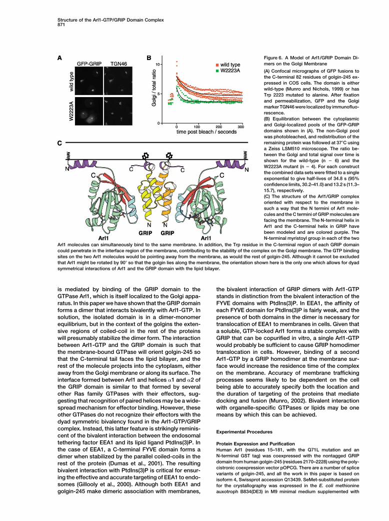
Structure of the Arl1-GTP/GRIP Domain Complex871
Figure 6. A Model of Arl1/GRIP Domain Di-mers on the Golgi Membrane
(A) Confocal micrographs of GFP fusions tothe C-terminal 82 residues of golgin-245 ex-pressed in COS cells. The domain is eitherwild-type (Munro and Nichols, 1999) or hasTrp 2223 mutated to alanine. After fixationand permeabilization, GFP and the Golgimarker TGN46 were localized by immunofluo-rescence.(B) Equilibration between the cytoplasmicand Golgi-localized pools of the GFP-GRIPdomains shown in (A). The non-Golgi poolwas photobleached, and redistribution of theremaining protein was followed at 37C usinga Zeiss LSM510 microscope. The ratio be-tween the Golgi and total signal over time isshown for the wild-type (n � 6) and theW2223A mutant (n � 4). For each constructthe combined data sets were fitted to a singleexponential to give half-lives of 34.8 s (95%confidence limits, 30.2–41.0) and 13.2 s (11.3–15.7), respectively.(C) The structure of the Arl1/GRIP complexoriented with respect to the membrane insuch a way that the N termini of Arl1 mole-cules and the C termini of GRIP molecules arefacing the membrane. The N-terminal helix inArl1 and the C-terminal helix in GRIP havebeen modeled and are colored purple. TheN-terminal myristoyl group in each of the two
Arl1 molecules can simultaneously bind to the same membrane. In addition, the Trp residue in the C-terminal region of each GRIP domaincould penetrate in the interface region of the membrane, contributing to the stability of the complex on the Golgi membrane. The GTP bindingsites on the two Arl1 molecules would be pointing away from the membrane, as would the rest of golgin-245. Although it cannot be excludedthat Arl1 might be rotated by 90 so that the golgin lies along the membrane, the orientation shown here is the only one which allows for dyadsymmetrical interactions of Arl1 and the GRIP domain with the lipid bilayer.
is mediated by binding of the GRIP domain to the the bivalent interaction of GRIP dimers with Arl1-GTPstands in distinction from the bivalent interaction of theGTPase Arl1, which is itself localized to the Golgi appa-
ratus. In this paper we have shown that the GRIP domain FYVE domains with PtdIns(3)P. In EEA1, the affinity ofeach FYVE domain for PtdIns(3)P is fairly weak, and theforms a dimer that interacts bivalently with Arl1-GTP. In
solution, the isolated domain is in a dimer-monomer presence of both domains in the dimer is necessary fortranslocation of EEA1 to membranes in cells. Given thatequilibrium, but in the context of the golgins the exten-
sive regions of coiled-coil in the rest of the proteins a soluble, GTP-locked Arl1 forms a stable complex withGRIP that can be copurified in vitro, a single Arl1-GTPwill presumably stabilize the dimer form. The interaction
between Arl1-GTP and the GRIP domain is such that would probably be sufficient to cause GRIP homodimertranslocation in cells. However, binding of a secondthe membrane-bound GTPase will orient golgin-245 so
that the C-terminal tail faces the lipid bilayer, and the Arl1-GTP by a GRIP homodimer at the membrane sur-face would increase the residence time of the complexrest of the molecule projects into the cytoplasm, either
away from the Golgi membrane or along its surface. The on the membrane. Accuracy of membrane traffickingprocesses seems likely to be dependent on the cellinterface formed between Arl1 and helices �1 and �2 of
the GRIP domain is similar to that formed by several being able to accurately specify both the location andthe duration of targeting of the proteins that mediateother Ras family GTPases with their effectors, sug-
gesting that recognition of paired helices may be a wide- docking and fusion (Munro, 2002). Bivalent interactionwith organelle-specific GTPases or lipids may be onespread mechanism for effector binding. However, these
other GTPases do not recognize their effectors with the means by which this can be achieved.dyad symmetric bivalency found in the Arl1-GTP/GRIPcomplex. Instead, this latter feature is strikingly reminis-
Experimental Procedurescent of the bivalent interaction between the endosomaltethering factor EEA1 and its lipid ligand PtdIns(3)P. In Protein Expression and Purification
Human Arl1 (residues 15–181, with the Q71L mutation and anthe case of EEA1, a C-terminal FYVE domain forms aN-terminal GST tag) was coexpressed with the nontagged GRIPdimer when stabilized by the parallel coiled-coils in thedomain from human golgin-245 (residues 2170–2228) using the poly-rest of the protein (Dumas et al., 2001). The resultingcistronic coexpression vector pOPCG. There are a number of splicebivalent interaction with PtdIns(3)P is critical for ensur-variants of golgin-245, and all the work in this paper is based on
ing the effective and accurate targeting of EEA1 to endo- isoform 4, Swissprot accession Q13439. SeMet-substituted proteinsomes (Gillooly et al., 2000). Although both EEA1 and for the crystallography was expressed in the E. coli methionine
auxotroph B834(DE3) in M9 minimal medium supplemented withgolgin-245 make dimeric association with membranes,

Molecular Cell872
amino acids, seleno-(L)-methionine, and vitamins as described pre- column (Amersham Pharmacia) equilibrated in 20 mM Tris-HCl (pH7.5, 4C) and 100 mM NaCl. The protein was loaded into six-sectorviously (Karathanassis et al., 2002). Cells were grown to OD600 � 1
at 37C and then induced with 0.3 mM IPTG at 17C for 12 hr. Cells 12 mm path length cells at three different concentrations: 8 M, 60 M, and 220 M. The samples were centrifuged until they reachedwere lysed with a French press in buffer A (50 mM Tris-HCl [pH 7.5,
4C], 100 mM NaCl, 5 mM MgCl2, 10 mM �-mercaptoethanol, and equilibrium as judged by the changes in the subsequent scans.Speeds were 32,000, 38,000, and 45,000 rpm. Data were analyzed0.2 mM GTP). After ultracentrifugation, the complex was bound
to glutathione-Sepharose (Amersham Biosciences), washed with using UltraSpin software (www.mrc-cpe.cam.ac.uk).buffer A, and cleaved on resin using 0.125 mg of tobacco etch virusprotease (TEV) per 10 mg of complex for 12 hr at 4C. After cleavage, Immunofluorescence
COS cells were transfected using FuGene (Roche), split onto glassArl1 retained an N-terminal GSHM linker sequence. The complexwas further purified by gel filtration on Superdex 75 16/60 equili- slides, and fixed 30 hr post transfection with 4% paraformaldehyde,
0.1% glutaraldehyde. Cells were permeabilized with 0.5% (v/v) Tri-brated in buffer B (20 mM Tris [pH 7.5, 4C], 100 mM NaCl, 1 mMMgCl2, 5 mM DTT, and 10 M GTP). The complex was concentrated ton X-100 in PBS, blocked with 20% (v/v) fetal calf serum/0.25%
(v/v) Tween 20 in PBS, and probed with rabbit TGN46 antibodiesto 11 mg/ml and snap frozen in liquid nitrogen.(Prescott et al., 1997) and Alexa568 anti-rabbit antibodies (MolecularProbes). Images were obtained on a BioRad Radiance confocal mi-Crystallizationcroscope.Initial crystals were obtained at 17C by vapor diffusion in sitting
drops made by mixing 100 nl protein and 100 nl reservoir fromAcknowledgmentscrystallization screen Wizard I, condition 1 (Emerald BioStructures)
in 96 well crystallization plates (Corning). Final conditions were opti-We thank Joanne McCarthy for assistance with data collection atmized using hair seeding in hanging drops made by mixing 1 lESRF beamline ID14-4, Ben Nichols for assistance with obtainingprotein with 1 l of reservoir solution consisting of 20% PEG 3350and analyzing photobleaching data, and Phil Evans, David Owen,and 0.2 M Tris-HCl (pH 8.5). After the crystals had reached their fulland Katja Roper for critical reading of the manuscript.size, the reservoir solution was exchanged for one containing 31%
PEG 3350, and drops were incubated over this for approximately 2Received: July 25, 2003days at 17C. Crystals were frozen in cryoprotectant (31% PEGRevised: September 3, 20033350, 0.2 M Tris-HCl [pH 8.5]).Accepted: September 5, 2003Published: October 23, 2003Data Collection, Phasing, and Model Refinement
For data collection, crystals were frozen in a nitrogen gas stream,Referencesand data were collected at 100 K. Multiple anomalous dispersion
(MAD) data sets were collected at ESRF beamline ID14-4 usingAbrahams, J.P., and Leslie, A.G.W. (1996). Methods used in thean ADSC CCD detector. Prior to data collection, a fluorescencestructure determination of bovine mitochondrial F1 ATPase. Actaspectrum for the crystal was obtained, and three data sets wereCrystallogr. D 52, 30–42.collected at wavelengths corresponding to the fluorescence peak,Amor, J.C., Horton, J.R., Zhu, X., Wang, Y., Sullards, C., Ringe, D.,inflection, and a high energy remote. Table 1 lists statistics for dataCheng, X., and Kahn, R.A. (2001). Structures of yeast ARF2 andcollection. Fifty putative Se sites were located using the programARL1: distinct roles for the N terminus in the structure and functionSnB (Turner et al., 1998; Weeks and Miller, 1999), and subsequentof ARF family GTPases. J. Biol. Chem. 276, 42477–42484.refinement with autoSHARP (de La Fortelle and Bricogne, 1997)
resulted in 39 Se sites (Table 1). Solvent flattening was carried out Antonny, B., Beraud-Dufour, S., Chardin, P., and Chabre, M. (1997).with SOLOMON (Abrahams and Leslie, 1996), using a solvent con- N-terminal hydrophobic residues of the G protein ADP-ribosylationtent of 30.4% as optimized by SHARP. An initial model was built factor-1 insert into membrane phospholipids upon GDP to GTPusing Arp/warp (Perrakis et al., 1999) and refined by alternating exchange. Biochemistry 36, 4675–4684.rounds of refinement with REFMAC5 (CCP4, 1994) and manual re- Barr, F.A. (1999). A novel Rab6-interacting domain defines a familybuilding with the program O (Jones et al., 1991). Final statistics for of Golgi-targeted coiled-coil proteins. Curr. Biol. 9, 381–384.the 1.7 A resolution model are given in Table 1. There are no residues
Barr, F.A., and Short, B. (2003). Golgins in the structure and dynam-in the disallowed regions of the Ramachandran plot, and 95% ofics of the Golgi apparatus. Curr. Opin. Cell Biol. 15, 405–413.residues are in the most favored regions as defined by PROCHECK.Bhamidipati, A., Lewis, S.A., and Cowan, N.J. (2000). ADP ribosyla-Residues 2171–2220 are ordered for each of the GRIP domains intion factor-like protein 2 (Arl2) regulates the interaction of tubulin-the asymmetric unit. The C-terminal residue ordered differs for eachfolding cofactor D with native tubulin. J. Cell Biol. 149, 1087–1096.of the GRIP domains in the asymmetric unit: 2220, 2221, 2226,
and 2228. Bi, X., Corpina, R.A., and Goldberg, J. (2002). Structure of the Sec23/24-Sar1 pre-budding complex of the COPII vesicle coat. Nature419, 271–277.Coprecipitation Assays
His6-tagged GRIP (golgin-245 residues 2170–2228) and MBP-tagged Bonangelino, C.J., Chavez, E.M., and Bonifacino, J.S. (2002). Geno-GRIP were produced in E. coli C41(DE3) either by coexpression mic screen for vacuolar protein sorting genes in Saccharomycesfrom a polycistronic pOPC plasmid, with MBP-GRIP in the first cas- cerevisiae. Mol. Biol. Cell 13, 2486–2501.sette and His6-GRIP in the second, or by expressing individually CCP4 (Collaborative Computing Project Number 4) (1994). A suiteMBP-GRIP (from pOPTM plasmid, encoding an N-terminal MBP- of programs for protein crystallography. Acta Crystallogr. D 50,tag) or His6-GRIP (from pOPTH plasmid, encoding an N-terminal 760–763.MetAlaHis6Met-tag). Cells were grown in 2xTY/Amp to OD600 � 1 at
Chavrier, P., and Goud, B. (1999). The role of ARF and Rab GTPases37C, and then induced with 0.3 mM IPTG at 17C for 12 hr. Cell
in membrane transport. Curr. Opin. Cell Biol. 11, 466–475.pellets were lysed in PBS and bound to Ni-NTA (QIAGEN) resin,
Clark, J., Moore, L., Krasinskas, A., Way, J., Battey, J., Tamkun, J.,washed with PBS, and eluted with buffer containing 0.3 M imidazole,and Kahn, R.A. (1993). Selective amplification of additional membersand aliquots were run on 20% Phast SDS gels (Amersham Biosci-of the ADP-ribosylation factor (ARF) family: cloning of additionalences).human and Drosophila ARF-like genes. Proc. Natl. Acad. Sci. USA90, 8952–8956.Analytical UltracentrifugationCollins, B.M., Watson, P.J., and Owen, D.J. (2003). The structure ofSedimentation equilibrium was performed on a Beckman Optimathe GGA1-GAT domain reveals the molecular basis for ARF bindingXLI ultracentrifuge using a Ti-60 rotor and interference and ab-and membrane association of GGAs. Dev. Cell 4, 321–332.sorbance at 280 and 230 nm, at 10C. His6-GRIP domain from human
golgin-245 expressed in E. coli C41(DE3) was purified by Ni2�-affinity de La Fortelle, E., and Bricogne, G. (1997). Maximum-likelihoodheavy-atom parameter refinement for multiple isomorphous re-chromatography followed by gel filtration on a Superdex 75 16/60
Structure of the Arl1-GTP/GRIP Domain Complex873
placement and multiwavelength anomalous diffraction methods. Golgi structure and function by ARF-like protein 1 (Arl1). J. Cell Sci.114, 4543–4555.Methods Enzymol. 276, 472–494.
Donaldson, J.G., and Jackson, C.L. (2000). Regulators and effectors Luke, M.R., Kjer-Nielsen, L., Brown, D.L., Stow, J.L., and Gleeson,P.A. (2003). GRIP domain-mediated targeting of two new coiled-coilof the ARF GTPases. Curr. Opin. Cell Biol. 12, 475–482.proteins, GCC88 and GCC185, to subcompartments of the trans-Dumas, J.J., Merithew, E., Sudharshan, E., Rajamani, D., Hayes,Golgi network. J. Biol. Chem. 278, 4216–4226.S., Lawe, D., Corvera, S., and Lambright, D.G. (2001). Multivalent
endosome targeting by homodimeric EEA1. Mol. Cell 8, 947–958. Maesaki, R., Ihara, K., Shimizu, T., Kuroda, S., Kaibuchi, K., andHakoshima, T. (1999). The structural basis of Rho effector recogni-Erlich, R., Gleeson, P.A., Campbell, P., Dietzsch, E., and Toh, B.H.tion revealed by the crystal structure of human RhoA complexed(1996). Molecular characterization of trans-Golgi p230. A humanwith the effector domain of PKN/PRK1. Mol. Cell 4, 793–803.peripheral membrane protein encoded by a gene on chromosome
6p12–22 contains extensive coiled-coil �-helical domains and a McConville, M.J., Ilgoutz, S.C., Teasdale, R.D., Foth, B.J., Matthews,A., Mullin, K.A., and Gleeson, P.A. (2002). Targeting of the GRIPgranin motif. J. Biol. Chem. 271, 8328–8337.domain to the trans-Golgi network is conserved from protists toFritzler, M.J., Lung, C.C., Hamel, J.C., Griffith, K.J., and Chan, E.K.animals. Eur. J. Cell Biol. 81, 485–495.(1995). Molecular characterization of Golgin-245, a novel Golgi com-
plex protein containing a granin signature. J. Biol. Chem. 270, 31262– Misra, S., Miller, G.J., and Hurley, J.H. (2001). Recognizing phospha-tidylinositol 3-phosphate. Cell 107, 559–562.31268.
Gangi Setty, S.R., Shin, M.E., Yoshino, A., Marks, M.S., and Burd, Mueller, A.G., Moser, M., Kluge, R., Leder, S., Blum, M., Buttner, R.,Joost, H.G., and Schurmann, A. (2002). Embryonic lethality causedC.G. (2003). Golgi recruitment of GRIP domain proteins by Arf-like
GTPase 1 (Arl1p) is regulated by the Arf-like GTPase 3 (Arl3p). Curr. by apoptosis during gastrulation in mice lacking the gene of theADP-ribosylation factor-related protein 1. Mol. Cell. Biol. 22, 1488–Biol. 13, 401–404.1494.Goldberg, J. (1998). Structural basis for activation of ARF GTPase:
mechanisms of guanine nucleotide exchange and GTP-myristoyl Munro, S. (2002). Organelle identity and the targeting of peripheralmembrane proteins. Curr. Opin. Cell Biol. 14, 506–514.switching. Cell 95, 237–248.
Gillingham, A.K., and Munro, S. (2003). Long coiled-coil proteins Munro, S., and Nichols, B.J. (1999). The GRIP domain: a novel Golgi-targeting domain found in several coiled-coil proteins. Curr. Biol.and membrane traffic. Biochim. Biophys. Acta 1641, 71–85.9, 377–380.Gillooly, D.J., Morrow, I.C., Lindsay, M., Gould, R., Bryant, N.J.,
Gaullier, J.M., Parton, R.G., and Stenmark, H. (2000). Localization Nassar, M., Horn, G., Herrmann, C., Scherer, A., McCormick, F., andWittinghofer, A. (1995). The 2.2 A crystal structure of the Ras bindingof phosphatidylinositol 3-phosphate in yeast and mammalian cells.
EMBO J. 19, 4577–4588. domain of the serine/threonine kinase c-Raf1 in complex with Rap1Aand a GTP analog. Nature 375, 554–560.Hanzal-Bayer, M., Renault, L., Roversi, P., Wittinghofer, A., and Hil-
lig, R.C. (2002). The complex of Arl2-GTP and PDE delta: from struc- Niedergang, F., Colucci-Guyon, E., Dubois, T., Raposo, G., andChavrier, P. (2003). ADP ribosylation factor 6 is activated and con-ture to function. EMBO J. 21, 2095–2106.trols membrane delivery during phagocytosis in macrophages. J.Jones, T.A., Zou, J.-Y., Cowan, S.W., and Kjeldgaard, M. (1991).Cell Biol. 161, 1143–1150.Improved methods for building protein models in electron density
maps and the location of errors in these models. Acta Crystallogr. Ostermeier, C., and Brunger, A.T. (1999). Structural basis of Rabeffector specificity: crystal structure of the small G protein Rab3AA 47, 110–119.complexed with the effector domain of rabphilin-3A. Cell 96,Karathanassis, D., Stahelin, R.V., Bravo, J., Perisic, O., Pacold, C.M.,363–374.Cho, W., and Williams, R.L. (2002). Binding of the PX domain of
p47phox to phosphatidylinositol 3,4-bisphosphate and phosphatidic Pacold, M.E., Suire, S., Perisic, O., Lara-Gonzalez, S., Davis, C.T.,Walker, E.H., Hawkins, P.T., Stephens, L., Eccleston, J.F., and Wil-acid is masked by an intramolecular interaction. EMBO J. 21, 5057–
5068. liams, R.L. (2000). Crystal structure and functional analysis of Rasbinding to its effector phosphoinositide 3-kinase �. Cell 103,Kjer-Nielsen, L., Teasdale, R.D., van Vliet, C., and Gleeson, P.A.931–943.(1999a). A novel Golgi-localisation domain shared by a class of
coiled- coil peripheral membrane proteins. Curr. Biol. 9, 385–388. Panic, B., Whyte, J.R., and Munro, S. (2003). The ARF-like GTPasesArl1p and Arl3p act in a pathway that interacts with vesicle-tetheringKjer-Nielsen, L., van Vliet, C., Erlich, R., Toh, B.H., and Gleeson, P.A.factors at the Golgi apparatus. Curr. Biol. 13, 405–410.(1999b). The Golgi-targeting sequence of the peripheral membrane
protein p230. J. Cell Sci. 112, 1645–1654. Pasqualato, S., Menetrey, J., Franco, M., and Cherfils, J. (2001). Thestructural GDP/GTP cycle of human Arf6. EMBO Rep. 2, 234–238.Kooy, J., Toh, B.H., Pettitt, J.M., Erlich, R., and Gleeson, P.A. (1992).
Human autoantibodies as reagents to conserved Golgi components. Pasqualato, S., Renault, L., and Cherfils, J. (2002). Arf, Arl, Arp andSar proteins: a family of GTP binding proteins with a structuralCharacterization of a peripheral, 230 kDa compartment-specific
Golgi protein. J. Biol. Chem. 267, 20255–20263. device for ‘front-back’ communication. EMBO Rep. 3, 1035–1041.
Perrakis, A., Morris, R., and Lamzin, V.S. (1999). Automated proteinKrauss, M., Kinuta, M., Wenk, M.R., De Camilli, P., Takei, K., andHaucke, V. (2003). ARF6 stimulates clathrin/AP-2 recruitment to syn- model building combined with iterative structure refinement. Nat.
Struct. Biol. 6, 458–463.aptic membranes by activating phosphatidylinositol phosphate ki-nase type I�. J. Cell Biol. 162, 113–124. Pfeffer, S.R. (1999). Transport-vesicle targeting: tethers before
SNAREs. Nat. Cell Biol. 1, E17–E22.Lee, F.J.S., Huang, C.F., Yu, W.L., Buu, L.M., Lin, C.Y., Huang, M.C.,Moss, J., and Vaughan, M. (1997). Characterization of an ADP-ribo- Prescott, A.R., Lucocq, J.M., James, J., Lister, J.M., and Ponnamba-sylation factor-like 1 protein in Saccharomyces cerevisiae. J. Biol. lam, S. (1997). Distinct compartmentalization of TGN46 and �1,4-Chem. 272, 30998–31005. galactosyltransferase in HeLa cells. Eur. J. Cell Biol. 72, 238–246.Li, B., and Warner, J.R. (1996). Mutation of the Rab6 homolog of Shiba, T., Kawasaki, M., Takatsu, H., Nogi, T., Matsugaki, N., Igara-Saccharomyces cerevisiae, YPT6, inhibits both early Golgi function shi, N., Suzuki, M., Kato, R., Nakayama, K., and Wakatsuki, S. (2003).and ribosome biosynthesis. J. Biol. Chem. 271, 16813–16819. Molecular mechanism of membrane recruitment of GGA by ARF in
lysosomal protein transport. Nat. Struct. Biol. 10, 386–393.Lowe, S.L., Wong, S.H., and Hong, W. (1996). The mammalian ARF-like protein 1 (Arl1) is associated with the Golgi complex. J. Cell Sci. Shorter, J., and Warren, G. (2002). Golgi architecture and inheri-109, 209–220. tance. Annu. Rev. Cell Dev. Biol. 18, 379–420.Lu, L., and Hong, W. (2003). Interaction of Arl1-GTP with GRIP do- Stahelin, R.V., Long, F., Diraviyam, K., Bruzik, K.S., Murray, D., andmains recruits autoantigens Golgin-97 and Golgin-245/p230 onto Cho, W. (2002). Phosphatidylinositol-3-phosphate induces thethe Golgi. Mol. Biol. Cell 14, 3767–3781. membrane penetration of the FYVE domains of Vps27p and Hrs. J.
Biol. Chem. 277, 26379–26388.Lu, L., Horstmann, H., Ng, C., and Hong, W. (2001). Regulation of

Molecular Cell874
Takai, Y., Sasaki, T., and Matozaki, T. (2001). Small GTP bindingproteins. Physiol. Rev. 81, 153–208.
Tamkun, J.W., Kahn, R.A., Kissinger, M., Brizuela, B.J., Rulka, C.,Scott, M.P., and Kennison, J.A. (1991). The arflike gene encodes anessential GTP binding protein in Drosophila. Proc. Natl. Acad. Sci.USA 88, 3120–3124.
Tarricone, C., Xiao, B., Justin, N., Walker, P.A., Rittinger, K., Gamblin,S.J., and Smerdon, S.J. (2001). The structural basis of Arfaptin-mediated cross-talk between Rac and Arf signaling pathways. Na-ture 411, 215–219.
Tsukada, M., Will, E., and Gallwitz, D. (1999). Structural and func-tional analysis of a novel coiled-coil protein involved in Ypt6 GTPase-regulated protein transport in yeast. Mol. Biol. Cell 10, 63–75.
Turner, M.A., Yuan, C.-S., Borchardt, R.T., Hershfield, M.S., Smith,G.D., and Howell, L.L. (1998). Structure determination of seleno-methionyl S-adenosylhomocysteine hydrolase using data at a singlewavelength. Nat. Struct. Biol. 5, 369–376.
Van Valkenburgh, H., Shern, J.F., Sharer, J.D., Zhu, X., and Kahn,R.A. (2001). ADP-ribosylation factors (ARFs) and ARF-like 1 (ARL1)have both specific and shared effectors: characterizing ARL1 bind-ing proteins. J. Biol. Chem. 276, 22826–22837.
Vetter, I.R., and Wittinghofer, A. (2001). The guanine nucleotide bind-ing switch in three dimensions. Science 294, 1299–1304.
Weeks, C.M., and Miller, R. (1999). The design and implementationof SnB v2.0. J. Appl. Crystallogr. 32, 120–124.
Accession Numbers
The coordinates of the Arl1-GTP/GRIP domain complex have beendeposited in the Protein Data Bank under the ID code 1UPT.