Supplemental Data GGA and Arf Proteins Modulate Retrovirus Assembly and Release
14
Molecular Cell, Volume 30 Supplemental Data GGA and Arf Proteins Modulate Retrovirus Assembly and Release Anjali Joshi, Himanshu Garg, Kunio Nagashima, Juan S. Bonifacino, and Eric O. Freed SUPPLEMENTAL EXPERIMENTAL PROCEDURES Reagents: siRNAs against GGAs, Arfs 1-5 and Tsg101 have been described elsewhere and were obtained from Qiagen as custom-synthesized oligos (Ghosh et al., 2003; Hewitt et al., 2002; Puertollano and Bonifacino, 2004; Volpicelli-Daley et al., 2005). Arf6 siRNA was obtained as a validated siRNA from Qiagen. HIV immunoglobulin (HIV-Ig) was obtained from the NIH AIDS Research and Reference Reagent Program. Anti-EIAV horse serum was kindly provided by Dr. R. Montelaro (University of Pittsburgh). The following Abs or antisera were obtained from the indicated sources: goat anti-MLV Gag p30, ViroMed Biosafety Laboratories, Camden, NJ; anti-HIV-1 p17 and p24, Advanced Biotechnologioes Inc, Columbia, MD; anti-Myc (A-14, 9E10) and HA (HA-7, Y11), Sigma or Santa Cruz Biotechnologies; horseradish peroxidase (HRP)-conjugated anti- mouse Ig, Amersham Biosciences (Uppsala, Sweden); anti-GGA1, a kind gift from Dr. R. Kahn (Emory University, Atlanta); anti-GGA2, GGA3 and AP-3, BD Biosciences; mouse anti-Tsg101 (C-2); Santa Cruz Biotechnologies; mouse anti-GM130, Transduction Laboratories (Lexington, Ky.); and sheep anti-TGN46, Serotec (Oxford, United Kingdom). Molecular clones expressing full-length GGAs have been described (Dell'Angelica et al., 2000; Puertollano and Bonifacino, 2004). The aggresome marker GFP-250 was kindly provided by Dr. E. S. Sztul (University of Alabama at Birmingham,
-
Upload
independent -
Category
Documents
-
view
0 -
download
0
Transcript of Supplemental Data GGA and Arf Proteins Modulate Retrovirus Assembly and Release

Molecular Cell, Volume 30
Supplemental Data
GGA and Arf Proteins Modulate
Retrovirus Assembly and Release Anjali Joshi, Himanshu Garg, Kunio Nagashima, Juan S. Bonifacino, and Eric O. Freed
SUPPLEMENTAL EXPERIMENTAL PROCEDURES
Reagents: siRNAs against GGAs, Arfs 1-5 and Tsg101 have been described elsewhere
and were obtained from Qiagen as custom-synthesized oligos (Ghosh et al., 2003; Hewitt
et al., 2002; Puertollano and Bonifacino, 2004; Volpicelli-Daley et al., 2005). Arf6
siRNA was obtained as a validated siRNA from Qiagen. HIV immunoglobulin (HIV-Ig)
was obtained from the NIH AIDS Research and Reference Reagent Program. Anti-EIAV
horse serum was kindly provided by Dr. R. Montelaro (University of Pittsburgh). The
following Abs or antisera were obtained from the indicated sources: goat anti-MLV Gag
p30, ViroMed Biosafety Laboratories, Camden, NJ; anti-HIV-1 p17 and p24, Advanced
Biotechnologioes Inc, Columbia, MD; anti-Myc (A-14, 9E10) and HA (HA-7, Y11),
Sigma or Santa Cruz Biotechnologies; horseradish peroxidase (HRP)-conjugated anti-
mouse Ig, Amersham Biosciences (Uppsala, Sweden); anti-GGA1, a kind gift from Dr.
R. Kahn (Emory University, Atlanta); anti-GGA2, GGA3 and AP-3, BD Biosciences;
mouse anti-Tsg101 (C-2); Santa Cruz Biotechnologies; mouse anti-GM130, Transduction
Laboratories (Lexington, Ky.); and sheep anti-TGN46, Serotec (Oxford, United
Kingdom). Molecular clones expressing full-length GGAs have been described
(Dell'Angelica et al., 2000; Puertollano and Bonifacino, 2004). The aggresome marker
GFP-250 was kindly provided by Dr. E. S. Sztul (University of Alabama at Birmingham,

Birmingham). Mammalian expression constructs for WT and dominant-active Arf
proteins were kindly provided by Dr. K. Nakayama (Kyoto University, Kyoto, Japan)
and J. Donaldson (NIH, Bethesda, MD). The construction of EIAV Gag expression
plasmids (pPRE-Gag), (Patnaik et al., 2002) containing WT (YPDL) and PTAP late
domains have been described (Li et al., 2002; Shehu-Xhilaga et al., 2004). The full-
length Tsg101 expression vector (TSG-F), (Goila-Gaur et al., 2003) and the HIV-1
molecular clone pNL4-3 (Adachi et al., 1986) have been described previously. The
Fyn10deltaMA Gag chimera, provided by Dr. A. Ono (University of Michigan, Ann
Arbor, Michigan), was constructed by fusing the 10 N-terminal residues of Fyn directly
to the CA domain of a MA-deleted pNL4-3 derivative (Chukkapalli et al., 2008). The
WT MLV expression vector was obtained from the NIH AIDS Research and Reference
Reagent Program. Intracellular PI(4,5)P2 levels were monitored by using a GFP
derivative fused to the N-terminal pleckstrin homology (PH) domain derived from
phospholipase C δ1 (PH-GFP), a kind gift of Dr. T. Balla (NIH, Bethesda, MD) (Varnai
and Balla, 1998). BFA was purchased from Calbiochem.
Immunofluorescence and EM. For fluorescence microscopy, cells were plated and
transfected in 8-well chamber slides (Nunc). Transfected cells were fixed 24-48 h
posttransfection for 20 min with freshly prepared formaldehyde buffer (3.7%
formaldehyde in 100 mM sodium phosphate buffer) and permeabilized with 0.1% Triton
X-100 in phosphate buffered saline (PBS). Following incubation with appropriate
primary and secondary Abs, cover slips were mounted with Aqua Poly Mount
(Polysciences Inc, Warrington, PA). Cells were examined and images acquired using a

Delta Vision RT deconvolution microscope. When dual staining with different Abs, the
extent of colocalization between two markers was determined by using the softWoRx co-
localization tool, with R = Pearson coefficient of correlation. For EM analysis, 24-48 h
posttransfection, cells were washed once with PBS and fixed with buffer containing 2%
glutaraldehyde and 0.1M sodium cacodylate. Transfected cell samples were prepared for
EM as described (Freed et al., 1994).

Figure S1. GGA depletion leads to an increase in HIV-1 release from MDMs. (A) Cells
were transfected with pNL4-3 along with the indicated siRNAs using an Amaxa
electroporator. Cell and virus lysates were resolved by SDS-PAGE, blotted onto
membranes and probed with HIV-Ig. Bands were quantified using the Alpha Innnotech
digital imager. Data ± SD; n = 3. Efficiency of siRNA-mediated depletion of GGAs in
MDMs, determined by immunoblot analysis using Abs to detect endogenous GGAs or
Tsg101, is shown in the right panel. (B) GGA overexpression inhibits HIV-1 production
in MDMs. Cells were transfected with pNL4-3 along with control vector or GGA or

Tsg101 (Tsg-F) expression plasmids using an Amaxa electroporator. Cell and viral
lysates were prepared and analyzed as described above. Data ± SD; n = 3. Lower gel
panel shows GGA (lanes 2-5) and Tsg101 (lane 6) expression detected by anti-Myc or
anti-HA WB, respectively.

Figure S2. Effect of GGA overexpression and BFA on AP-3 localization. (A) HeLa cells
transfected with Myc-tagged GGA expression constructs were immunostained with anti-
Myc (red) and anti-AP-3 (green) Abs. (B) HeLa cells were treated with 20 µg/ml BFA
for 1 h. Cells were either fixed immediately post-treatment or allowed to recover for 1 h
(BFA washout) and were then immunostained for endogenous AP-3 (green). (C) Effect
of BFA treatment on HIV-1 particle production. HeLa cells transfected with pNL4-3
were metabolically labeled with [35S]Met/Cys in the presence or absence of 20 µg/ml
BFA. Quantitation of virus release efficiency; ± SD , n=3.

Figure S3. Overexpression of the GAT domain alone inhibits pNL4-3/PR- particle
production. HeLa cells were transfected with the PR-defective molecular clone pNL4-
3/PR- along with vectors expressing full-length GGA3 or the indicated GGA domains at a
5:1 DNA ratio. Virus release was determined by radioimmunoprecipitation assay with
HIV-Ig (left panel) and quantitation was obtained by phosphorimager analysis (top right
panel), normalized to individual GGA fragment expression levels (lower right panel)
determined by anti-Myc Western blot (WB) followed by quantitation with an Alpha
Innnotech digital imager. Pr55 indicates position of the HIV-1 Gag precursor Pr55Gag.
This experiment was performed twice, with similar results in both assays.

Figure S4. A GGA3 mutant defective in Arf binding does not inhibit EIAV particle
production. HeLa cells were transfected with the EIAV Gag expression vector along with
either WT GGA expression plasmids or their Arf-binding-defective (NA) counterparts.
Virus release was determined by radioimmunoprecipitation assay, and quantitation was
obtained by phosphorimager analysis. Position of the EIAV Gag precursor Pr55Gag
(Pr55) is indicated.
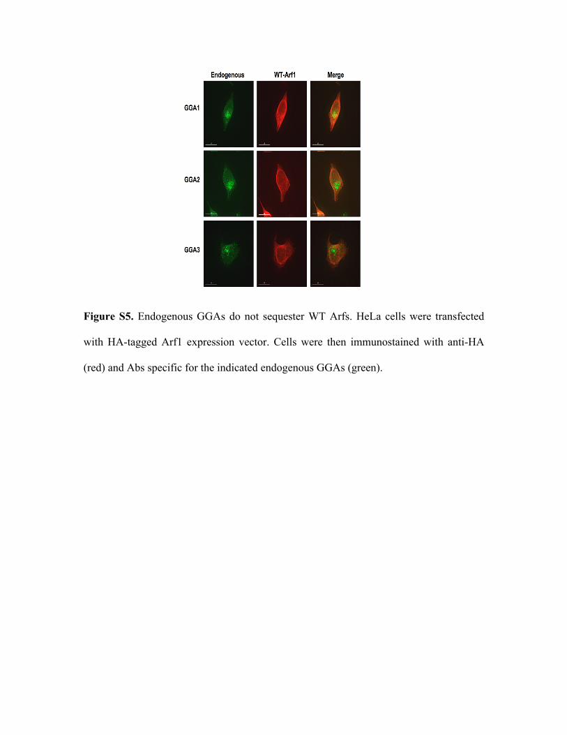
Figure S5. Endogenous GGAs do not sequester WT Arfs. HeLa cells were transfected
with HA-tagged Arf1 expression vector. Cells were then immunostained with anti-HA
(red) and Abs specific for the indicated endogenous GGAs (green).

Figure S6. Disruption of class I or II Arfs does not induce a detectable shift in PI(4,5)P2
localization. HeLa cells were transfected with PH-GFP, a probe for cellular PI(4,5)P2,
along with vectors expressing HA-tagged dominant-active isoforms of either Class I and
II (Arf1-5) or class III (Arf6) Arfs. Cells were fixed, immunostained with anti-HA Ab,
and analyzed by fluorescence microscopy. PH-GFP signal is in green, anti-HA signal in
red. Bottom panel shows PH-GFP localization in cells not transfected with an Arf
expression vector.

Figure S7. Inhibition of virus production by Arf disruption is not due to decreased Gag
expression. (A) HeLa cells were transfected with decreasing amounts of pNL4-3 (lanes 1-
4) or pNL4-3 along with the indicated dominant-active Arf expression vectors or Arf
siRNAs (lanes 5-8). Virus release efficiency was calculated after
radioimmunoprecipitation with HIV-Ig and phosphorimager analysis as described in the
Figure 1 legend. Red inset depicts the Gag expression levels that correspond to those in
the Arf1QL- and Arf3QL-transfected samples. In this experiment, Gag expression levels
in the pNL4-3 titration spanned a 6-fold range whereas the dominant-active Arfs and Arf
siRNAs had only a 2-fold effect on Gag expression levels. (B) HeLa cells were
transfected with pNL4-3/PR- along with the indicated dominant-active Arfs or Arf
siRNAs. Virus release efficiency was determined as described above.

Figure S8. Schematic representation of host proteins involved in HIV-1 assembly and
release. At the bottom of the figure is depicted Pr55Gag, with major domains matrix
(MA), capsid (CA), nucleocapsid (NC), and p6 and spacer peptides SP1 (1) and SP2 (2)
indicated (Adamson and Freed, 2007). The MA domain has been reported to interact
with clathrin adaptor protein complexes AP-1, AP-2, and AP-3 (Batonick et al., 2005;
Camus et al., 2007; Dong et al., 2005). Interaction of Gag with membranes takes place in
cholesterol-enriched lipid rafts (Ono and Freed, 2005) via the phospholipid PI(4,5)P2
(Ono et al., 2004; Saad et al., 2006). Association between Gag and the Env glycoprotein
complex has been reported to require TIP47 (Lopez-Verges et al., 2006). The GGA and

Arf proteins, demonstrated here to modulate virus budding and regulate Gag trafficking
to the plasma membrane, respectively, are shown at various subcellular locations. Virus
budding requires components of the ESCRT machinery of which ESCRT-I (whose
Tsg101 subunit binds the p6 domain of Gag), ESCRT-III, and the ESCRT-associated
factor Alix are shown (Fujii et al., 2007). At the lower right is illustrated the
sequestration of endogenous Arf proteins by exogenously expressed GGAs. TGN, trans-
Golgi network; MVB, multivesicular body. Modified from Future HIV Research, Joshi
and Freed, 2007 (Joshi and Freed, 2007) with permission of Future Medicine Ltd., and
from the cover of the Cold Spring Harbor 2006 Retroviruses abstract book, courtesy W.
Sundquist and H.-Y. Chung, with permission.
REFERENCES Adachi, A., Gendelman, H. E., Koenig, S., Folks, T., Willey, R., Rabson, A., and Martin, M. A. (1986). Production of acquired immunodeficiency syndrome-associated retrovirus in human and nonhuman cells transfected with an infectious molecular clone. J Virol 59, 284-291. Adamson, C. S., and Freed, E. O. (2007). Human immunodeficiency virus type 1 assembly, release, and maturation. Adv Pharmacol 55, 347-387. Batonick, M., Favre, M., Boge, M., Spearman, P., Honing, S., and Thali, M. (2005). Interaction of HIV-1 Gag with the clathrin-associated adaptor AP-2. Virology 342, 190-200. Camus, G., Segura-Morales, C., Molle, D., Lopez-Verges, S., Begon-Pescia, C., Cazevieille, C., Schu, P., Bertrand, E., Berlioz-Torrent, C., and Basyuk, E. (2007). The clathrin adaptor complex AP-1 binds HIV-1 and MLV Gag and facilitates their budding. Mol Biol Cell 18, 3193-3203. Chukkapalli, V., Hogue, I. B., Boyko, V., Hu, W.-S., and Ono, A. (2008). Interaction between HIV-1 Gag matrix domain and phosphatidylinositol-(4,5)-bisphosphate is essential for efficient Gag-membrane binding. J Virol in press. Dell'Angelica, E. C., Puertollano, R., Mullins, C., Aguilar, R. C., Vargas, J. D., Hartnell, L. M., and Bonifacino, J. S. (2000). GGAs: a family of ADP ribosylation factor-binding proteins related to adaptors and associated with the Golgi complex. J Cell Biol 149, 81-94.

Dong, X., Li, H., Derdowski, A., Ding, L., Burnett, A., Chen, X., Peters, T. R., Dermody, T. S., Woodruff, E., Wang, J. J., and Spearman, P. (2005). AP-3 directs the intracellular trafficking of HIV-1 Gag and plays a key role in particle assembly. Cell 120, 663-674. Freed, E. O., Orenstein, J. M., Buckler-White, A. J., and Martin, M. A. (1994). Single amino acid changes in the human immunodeficiency virus type 1 matrix protein block virus particle production. J Virol 68, 5311-5320. Fujii, K., Hurley, J. H., and Freed, E. O. (2007). Beyond Tsg101: the role of Alix in 'ESCRTing' HIV-1. Nat Rev Microbiol 5, 912-916. Ghosh, P., Griffith, J., Geuze, H. J., and Kornfeld, S. (2003). Mammalian GGAs act together to sort mannose 6-phosphate receptors. J Cell Biol 163, 755-766. Goila-Gaur, R., Demirov, D. G., Orenstein, J. M., Ono, A., and Freed, E. O. (2003). Defects in human immunodeficiency virus budding and endosomal sorting induced by TSG101 overexpression. J Virol 77, 6507-6519. Hewitt, E. W., Duncan, L., Mufti, D., Baker, J., Stevenson, P. G., and Lehner, P. J. (2002). Ubiquitylation of MHC class I by the K3 viral protein signals internalization and TSG101-dependent degradation. Embo J 21, 2418-2429. Joshi, A., and Freed, E. O. (2007). HIV-1 Gag trafficking. Future HIV Ther 1, 427-438. Li, F., Chen, C., Puffer, B. A., and Montelaro, R. C. (2002). Functional replacement and positional dependence of homologous and heterologous L domains in equine infectious anemia virus replication. J Virol 76, 1569-1577. Lopez-Verges, S., Camus, G., Blot, G., Beauvoir, R., Benarous, R., and Berlioz-Torrent, C. (2006). Tail-interacting protein TIP47 is a connector between Gag and Env and is required for Env incorporation into HIV-1 virions. Proc Natl Acad Sci U S A 103, 14947-14952. Ono, A., Ablan, S. D., Lockett, S. J., Nagashima, K., and Freed, E. O. (2004). Phosphatidylinositol (4,5) bisphosphate regulates HIV-1 Gag targeting to the plasma membrane. Proc Natl Acad Sci U S A 101, 14889-14894. Ono, A., and Freed, E. O. (2005). Role of lipid rafts in virus replication. Adv Virus Res 64, 311-358. Patnaik, A., Chau, V., Li, F., Montelaro, R. C., and Wills, J. W. (2002). Budding of equine infectious anemia virus is insensitive to proteasome inhibitors. J Virol 76, 2641-2647. Puertollano, R., and Bonifacino, J. S. (2004). Interactions of GGA3 with the ubiquitin sorting machinery. Nat Cell Biol 6, 244-251. Saad, J. S., Miller, J., Tai, J., Kim, A., Ghanam, R. H., and Summers, M. F. (2006). Structural basis for targeting HIV-1 Gag proteins to the plasma membrane for virus assembly. Proc Natl Acad Sci U S A 103, 11364-11369. Shehu-Xhilaga, M., Ablan, S., Demirov, D. G., Chen, C., Montelaro, R. C., and Freed, E. O. (2004). Late domain-dependent inhibition of equine infectious anemia virus budding. J Virol 78, 724-732. Varnai, P., and Balla, T. (1998). Visualization of phosphoinositides that bind pleckstrin homology domains: calcium- and agonist-induced dynamic changes and relationship to myo-[3H]inositol-labeled phosphoinositide pools. J Cell Biol 143, 501-510. Volpicelli-Daley, L. A., Li, Y., Zhang, C. J., and Kahn, R. A. (2005). Isoform-selective effects of the depletion of ADP-ribosylation factors 1-5 on membrane traffic. Mol Biol Cell 16, 4495-4508.


![Arf nucleotide binding site opener [ARNO] promotes sequential activation of Arf6, Cdc42 and Rac1 and insulin secretion in INS 832/13 β-cells and rat islets](https://static.fdokumen.com/doc/165x107/6316194efc260b7102104d00/arf-nucleotide-binding-site-opener-arno-promotes-sequential-activation-of-arf6.jpg)