
Lung Adenocarcinoma Originates from Retrovirus Infection of Proliferating Type 2 Pneumocytes during...
12
Lung Adenocarcinoma Originates from Retrovirus Infection of Proliferating Type 2 Pneumocytes during Pulmonary Post-Natal Development or Tissue Repair Claudio Murgia 1 , Marco Caporale 1,2 , Ousman Ceesay 1 , Gabriella Di Francesco 2 , Nicola Ferri 2 , Vincenzo Varasano 3 , Marcelo de las Heras 4 , Massimo Palmarini 1 * 1 Medical Research Council – University of Glasgow Centre for Virus Research, Institute of Infection, Inflammation and Immunity, College of Medical, Veterinary and Life Sciences, University of Glasgow, United Kingdom, 2 Istituto G. Caporale, Teramo, Italy, 3 Dipartimento di Scienze Cliniche Veterinarie, Facolta’ di Medicina Veterinaria, Universita’ di Teramo, Italy, 4 Facultad de Veterinaria, Universidad de Zaragoza, Zaragoza, Spain Abstract Jaagsiekte sheep retrovirus (JSRV) is a unique oncogenic virus with distinctive biological properties. JSRV is the only virus causing a naturally occurring lung cancer (ovine pulmonary adenocarcinoma, OPA) and possessing a major structural protein that functions as a dominant oncoprotein. Lung cancer is the major cause of death among cancer patients. OPA can be an extremely useful animal model in order to identify the cells originating lung adenocarcinoma and to study the early events of pulmonary carcinogenesis. In this study, we demonstrated that lung adenocarcinoma in sheep originates from infection and transformation of proliferating type 2 pneumocytes (termed here lung alveolar proliferating cells, LAPCs). We excluded that OPA originates from a bronchioalveolar stem cell, or from mature post-mitotic type 2 pneumocytes or from either proliferating or non-proliferating Clara cells. We show that young animals possess abundant LAPCs and are highly susceptible to JSRV infection and transformation. On the contrary, healthy adult sheep, which are normally resistant to experimental OPA induction, exhibit a relatively low number of LAPCs and are resistant to JSRV infection of the respiratory epithelium. Importantly, induction of lung injury increased dramatically the number of LAPCs in adult sheep and rendered these animals fully susceptible to JSRV infection and transformation. Furthermore, we show that JSRV preferentially infects actively dividing cell in vitro. Overall, our study provides unique insights into pulmonary biology and carcinogenesis and suggests that JSRV and its host have reached an evolutionary equilibrium in which productive infection (and transformation) can occur only in cells that are scarce for most of the lifespan of the sheep. Our data also indicate that, at least in this model, inflammation can predispose to retroviral infection and cancer. Citation: Murgia C, Caporale M, Ceesay O, Di Francesco G, Ferri N, et al. (2011) Lung Adenocarcinoma Originates from Retrovirus Infection of Proliferating Type 2 Pneumocytes during Pulmonary Post-Natal Development or Tissue Repair. PLoS Pathog 7(3): e1002014. doi:10.1371/journal.ppat.1002014 Editor: Michael Emerman, Fred Hutchinson Cancer Research Center, United States of America Received November 22, 2010; Accepted February 4, 2011; Published March 31, 2011 Copyright: ß 2011 Murgia et al. This is an open-access article distributed under the terms of the Creative Commons Attribution License, which permits unrestricted use, distribution, and reproduction in any medium, provided the original author and source are credited. Funding: This study was supported by the Biotechnology and Biological Sciences Research Council (BBSRC; www.bbsrc.ac.uk), the Wellcome Trust (www. wellcome.ac.uk), the Istituto G. Caporale (www.izs.it) and the Italian Ministry of Health (Ministero della Salute; www.salute.gov.it). The funders had no role in study design, data collection and analysis, decision to publish, or preparation of the manuscript. Competing Interests: The authors have declared that no competing interests exist. * E-mail: [email protected] Introduction Retroviruses have been instrumental in understanding the genetic basis and the fundamental molecular mechanisms leading to cancer [1]. Studies on the pathogenesis of retrovirus induced malignancies have also contributed to our understanding of the cells that give origin to cancer and the role played by stem and progenitor cells in these processes [2]. The ‘‘cancer stem cell’’ (CSC) hypothesis postulates that cancer is initiated and sustained by adult stem cells [3–4]. A growing body of experimental evidence is supporting the presence of CSCs in haematological malignancies and in some solid tumours. However, the presence and significance of CSCs is object of considerable debate particularly in slow turnover organs such as the lungs [5–7]. Identifying the cells that give origin to cancer is critical both to understand the basic carcinogenetic processes but also to devise appropriate therapeutic strategies. Most retroviruses induce transformation of hematopoietic cells but there are a few notable exceptions causing sarcomas, nephroblastomas, mammary carcinomas, nasal and lung adeno- carcinomas in a variety of animal species [8]. Ovine pulmonary adenocarcinoma (OPA) is a naturally occurring (and experimen- tally inducible) lung cancer of sheep caused by a retrovirus known as Jaagsiekte sheep retrovirus (JSRV) [9–11]. OPA is a common disease of sheep in most geographical areas of the world. Interestingly, the disease shares several clinical and histological features with some forms of human lung adenocarcinomas. Therefore, OPA represents an excellent animal model with great potential to contribute significantly to our understanding of retroviral pathogenesis, lung tumorigenesis and pulmonary biology [9,12–13]. JSRV is the only oncogenic virus that causes a naturally occurring lung adenocarcinoma. Interestingly, in contrast to the overwhelming majority of oncogenic retroviruses, JSRV is a replication-competent virus that possesses a structural protein (the viral envelope, Env) that acts as a dominant oncoprotein [14–16]. Expression of the JSRV Env is sufficient to induce cell transformation in vitro in a variety of cell lines [13–15,17–22] and importantly in vivo in both experimental mice PLoS Pathogens | www.plospathogens.org 1 March 2011 | Volume 7 | Issue 3 | e1002014
-
Upload
independent -
Category
Documents
-
view
2 -
download
0
Transcript of Lung Adenocarcinoma Originates from Retrovirus Infection of Proliferating Type 2 Pneumocytes during...

Lung Adenocarcinoma Originates from RetrovirusInfection of Proliferating Type 2 Pneumocytes duringPulmonary Post-Natal Development or Tissue RepairClaudio Murgia1, Marco Caporale1,2, Ousman Ceesay1, Gabriella Di Francesco2, Nicola Ferri2, Vincenzo
Varasano3, Marcelo de las Heras4, Massimo Palmarini1*
1 Medical Research Council – University of Glasgow Centre for Virus Research, Institute of Infection, Inflammation and Immunity, College of Medical, Veterinary and Life
Sciences, University of Glasgow, United Kingdom, 2 Istituto G. Caporale, Teramo, Italy, 3 Dipartimento di Scienze Cliniche Veterinarie, Facolta’ di Medicina Veterinaria,
Universita’ di Teramo, Italy, 4 Facultad de Veterinaria, Universidad de Zaragoza, Zaragoza, Spain
Abstract
Jaagsiekte sheep retrovirus (JSRV) is a unique oncogenic virus with distinctive biological properties. JSRV is the only viruscausing a naturally occurring lung cancer (ovine pulmonary adenocarcinoma, OPA) and possessing a major structuralprotein that functions as a dominant oncoprotein. Lung cancer is the major cause of death among cancer patients. OPA canbe an extremely useful animal model in order to identify the cells originating lung adenocarcinoma and to study the earlyevents of pulmonary carcinogenesis. In this study, we demonstrated that lung adenocarcinoma in sheep originates frominfection and transformation of proliferating type 2 pneumocytes (termed here lung alveolar proliferating cells, LAPCs). Weexcluded that OPA originates from a bronchioalveolar stem cell, or from mature post-mitotic type 2 pneumocytes or fromeither proliferating or non-proliferating Clara cells. We show that young animals possess abundant LAPCs and are highlysusceptible to JSRV infection and transformation. On the contrary, healthy adult sheep, which are normally resistant toexperimental OPA induction, exhibit a relatively low number of LAPCs and are resistant to JSRV infection of the respiratoryepithelium. Importantly, induction of lung injury increased dramatically the number of LAPCs in adult sheep and renderedthese animals fully susceptible to JSRV infection and transformation. Furthermore, we show that JSRV preferentially infectsactively dividing cell in vitro. Overall, our study provides unique insights into pulmonary biology and carcinogenesis andsuggests that JSRV and its host have reached an evolutionary equilibrium in which productive infection (andtransformation) can occur only in cells that are scarce for most of the lifespan of the sheep. Our data also indicate that,at least in this model, inflammation can predispose to retroviral infection and cancer.
Citation: Murgia C, Caporale M, Ceesay O, Di Francesco G, Ferri N, et al. (2011) Lung Adenocarcinoma Originates from Retrovirus Infection of Proliferating Type 2Pneumocytes during Pulmonary Post-Natal Development or Tissue Repair. PLoS Pathog 7(3): e1002014. doi:10.1371/journal.ppat.1002014
Editor: Michael Emerman, Fred Hutchinson Cancer Research Center, United States of America
Received November 22, 2010; Accepted February 4, 2011; Published March 31, 2011
Copyright: � 2011 Murgia et al. This is an open-access article distributed under the terms of the Creative Commons Attribution License, which permitsunrestricted use, distribution, and reproduction in any medium, provided the original author and source are credited.
Funding: This study was supported by the Biotechnology and Biological Sciences Research Council (BBSRC; www.bbsrc.ac.uk), the Wellcome Trust (www.wellcome.ac.uk), the Istituto G. Caporale (www.izs.it) and the Italian Ministry of Health (Ministero della Salute; www.salute.gov.it). The funders had no role in studydesign, data collection and analysis, decision to publish, or preparation of the manuscript.
Competing Interests: The authors have declared that no competing interests exist.
* E-mail: [email protected]
Introduction
Retroviruses have been instrumental in understanding the
genetic basis and the fundamental molecular mechanisms leading
to cancer [1]. Studies on the pathogenesis of retrovirus induced
malignancies have also contributed to our understanding of the
cells that give origin to cancer and the role played by stem and
progenitor cells in these processes [2]. The ‘‘cancer stem cell’’
(CSC) hypothesis postulates that cancer is initiated and sustained
by adult stem cells [3–4]. A growing body of experimental
evidence is supporting the presence of CSCs in haematological
malignancies and in some solid tumours. However, the presence
and significance of CSCs is object of considerable debate
particularly in slow turnover organs such as the lungs [5–7].
Identifying the cells that give origin to cancer is critical both to
understand the basic carcinogenetic processes but also to devise
appropriate therapeutic strategies.
Most retroviruses induce transformation of hematopoietic cells
but there are a few notable exceptions causing sarcomas,
nephroblastomas, mammary carcinomas, nasal and lung adeno-
carcinomas in a variety of animal species [8]. Ovine pulmonary
adenocarcinoma (OPA) is a naturally occurring (and experimen-
tally inducible) lung cancer of sheep caused by a retrovirus known
as Jaagsiekte sheep retrovirus (JSRV) [9–11]. OPA is a common
disease of sheep in most geographical areas of the world.
Interestingly, the disease shares several clinical and histological
features with some forms of human lung adenocarcinomas.
Therefore, OPA represents an excellent animal model with great
potential to contribute significantly to our understanding of
retroviral pathogenesis, lung tumorigenesis and pulmonary biology
[9,12–13].
JSRV is the only oncogenic virus that causes a naturally occurring
lung adenocarcinoma. Interestingly, in contrast to the overwhelming
majority of oncogenic retroviruses, JSRV is a replication-competent
virus that possesses a structural protein (the viral envelope, Env) that
acts as a dominant oncoprotein [14–16]. Expression of the JSRV Env
is sufficient to induce cell transformation in vitro in a variety of cell lines
[13–15,17–22] and importantly in vivo in both experimental mice
PLoS Pathogens | www.plospathogens.org 1 March 2011 | Volume 7 | Issue 3 | e1002014

models and in lambs [23–24]. Thus, productive virus infection and
cell transformation are mutually dependent in OPA and this creates
an ‘‘evolutionary dilemma’’ as, at face value, abundant viral
replication is entirely dependent on tumor development in the host.
The JSRV Env is believed to induce cell transformation via the
activation of a variety of signal transduction pathways including the
PI-3K/Akt and Ras-MEK-MAPK [13,20,22,25–27].
Experimentally, intratracheal inoculation of concentrated JSRV
viral particles in young lambs induces OPA in the overwhelming
majority of animals with a very short incubation period (varying
from a few weeks to a few months) [28–29]. There is a clear age-
dependent susceptibility to experimentally induced OPA in lambs
while it is not possible (or extremely difficult) to reproduce the
disease in adult sheep [29]. These data suggest that there is a
different availability of the target cells of JSRV transformation in
animals of a different age. The age-susceptibility to OPA induction
does not appear to be related to expression of the receptor in target
cells or to a differential immune response towards the virus.
Indeed, the cellular receptor for the virus (Hyaluronidase-2, Hyal-
2) is ubiquitously expressed [16,29] and this virus can infect several
cell types in vitro and in vivo [30–33]. In addition, JSRV naturally or
experimentally-infected animals do not mount a significant
immune response, likely as a result of tolerance induced by
expression of JSRV-related endogenous retroviruses (enJSRVs)
which are present in the genome of all domestic and wild sheep
[34–37].
In OPA affected sheep, abundant expression of JSRV proteins
are confined to the tumor cells although viral RNA and DNA can
be detected by sensitive PCR assays in a variety of cells of the
lymphoreticular system [30–31,38]. In sheep naturally infected
with JSRV and with no neoplastic lesions, JSRV can be detected
only in lymphoid tissues [39]. OPA tumours, similar to some
human lung adenocarcinomas, are formed by secretory cells of
the distal pulmonary tract; predominantly alveolar type 2
pneumocytes and less commonly the non-ciliated bronchial cells
of the terminal bronchioli (Clara cells; see note at the end of the
text on the usage of this term) [40–42]. Interestingly, a putative
bronchioalveolar stem cell (BASC) has been identified in mice
lungs although its presence in other species, including humans,
has not been established with certainty [43]. It has been proposed
that BASCs have the capacity to originate both Clara cells and
type 2 pneumocytes and to be the cell origin of lung
adenocarcinoma in mice in response to oncogenic K-ras [43].
However the significance of BASCs in physiological and
pathological processes and the origin of lung adenocarcinoma
are under debate [44–45].
In order to identify the target cells of JSRV infection and
transformation we performed a series of in vivo studies in
experimentally infected lambs and adult sheep. Furthermore, we
derived a JSRV-based vector in order to assess the ability of this
virus to infect non-dividing cells in vitro. In this study we identified
the cells target of JSRV infection and transformation and provide
important insights into lung biology, pulmonary carcinogenesis
and retroviral pathogenesis.
Methods
Ethics statementAll experimental procedures carried out in this study are
included in Project Licence 60/3905 approved by the Home
Office of the United Kingdom in accordance to the ‘‘animals
(scientific procedures) act 1986’’. Experiments carried out at the
Istituto G. Caporale were also detailed in protocol number 3315
approved by the Italian Ministry of Health (Ministero della Salute)
in accordance with Council Directive 86/609/EEC of the
European Union.
Virus stock preparationViral stocks used in all these experiments were produced in rat
208F.JSRV21 cells as already described [46]. Briefly, 208F.JSRV21
derive from 208F cells [47] stably transfected with a plasmid
expressing the JSRV21 infectious molecular clone [11].
208F.JSRV21 cells were plated at 80% confluence and superna-
tants were collected after 24, 48 and 72 h. Virus was concentrated
by ultracentrifugation [3006] as previously described [11] and
resuspended in 16TNE buffer (100mM NaCl, 10 mM Tris,
1 mM EDTA). The infectious titer for JSRV cannot be easily
calculated in vitro, because of the lack of a convenient tissue culture
system for this virus. In order to infect animals with the same
amount of JSRV, pellets from various virus preparations were
pooled into a single stock, divided into 1 ml aliquots and stored at
280uC until use. In all the experiments described below, each
animal received the same amount of virus stock. In a related study,
the same JSRV preparation used here, induced OPA in 4 of 4
experimentally infected lambs within 5 months after inoculation
(Caporale and Palmarini, unpublished).
In vivo studiesAnimal studies were performed at the Istituto G. Caporale
(Teramo, Italy) and at the University of Glasgow. Prior to
experimental infections all animals were anaesthetised with
sodium pentobarbital anesthesia, and all efforts were made to
minimize suffering. To facilitate the detection of infected cells,
JSRV (1 ml) was inoculated directly into the accessory bronchus of
the cranial lobe of the right lung by fiber-optic bronchoscopy.
Sheep used in this study were females between 3 and 5 year old of
either bergamasca cross-breed (study 1, 2 & 4) or blackface breed
(study 3) unless otherwise indicated. Three independent studies
were performed as follow.
Study 1: Age related susceptibility to JSRV infection. Four
2-day old lambs and 4 adult sheep were anesthetized and inoculated
with JSRV, as described above. Two animals were used as mock
Author Summary
The identification of cells that give origin to cancer iscritical in order to design effective therapeutic strategies.To this end, the early stages of cancer are the mostinformative but they are seldom associated with clinicalsymptoms and therefore pass unnoticed in humanpatients. Studies on animal tumors are invaluable to thisresearch area. In this study, we determined the cellsoriginating an infectious lung cancer of sheep (ovinepulmonary adenocarcinoma, OPA) that is similar to someforms of human pulmonary adenocarcinoma. OPA iscaused by a virus known as Jaagsiekte sheep retrovirus(JSRV). We show that OPA is caused by JSRV infection ofproliferating type 2 pneumocytes (lung alveolar prolifer-ating cells, LAPCs). We show that young animals possessabundant LAPCs and are highly susceptible to JSRVinfection while healthy adult sheep exhibit a relativelylow number of LAPCs and are resistant to OPA induction.However, adult sheep were susceptible to JSRV infectionwhen the presence of LAPCs was stimulated by inductionof a mild injury to the respiratory epithelium. Thus, ourstudy identifies the cells originating lung adenocarcinomain OPA and shows that inflammation to the respiratoryepithelium can predispose to retrovirus infection andcancer.
Pathogenesis of Retrovirus-Induced Lung Cancer
PLoS Pathogens | www.plospathogens.org 2 March 2011 | Volume 7 | Issue 3 | e1002014

inoculated controls. Ten days post infection animals were euthanized,
the lungs removed from the thoracic cavity and examined for the
presence of macroscopic lesions. Samples from respectively 8 (in
lambs) and 16 (in sheep) regions from the cranial lobe were collected
and fixed overnight in 10% buffered formalin and embedded in
paraffin. Tissue sections were examined by immunohistochemistry
and immunofluorescence as described below.
Study 2: Bronchioalveolar proliferation in lambs and
adult sheep. Lung tissues were collected at post-mortem from
adult sheep (n = 2) and 4 lambs (2–4 day old). Tissues were
collected from 4 different lobes of the lungs and fixed overnight in
10% buffered formalin. Tissues were examined for bronchiolar
alveolar cell proliferation from ten sections from each animal as
described below.
Study 3: Induction of mild lung injury in adult sheep. Mild
lung injury was induced in adult sheep using 3- methylindole (3MI,
Sigma). Four adult sheep were divided in two groups. All animals
were weighted and fasted 12 hours before dosing. Group 1 received
0.25 g/kg body weight of 3MI (Sigma) dissolved in 50 ml of corn oil
(Sigma) and administrated using a stomach tube attached to a
syringe. Group II served as control and received a similar amount of
corn oil. After 48 hours all animals were euthanized and lung tissue
were collected for histological and immunofluorescence analysis to
assess the injury and cell proliferation.
Study 4: Infection of adult sheep with or without lung
injury. Ten adult sheep were divided in two groups of 5 animals
each. Group 1 received 0.25 g 3MI/kg body wt as described
above. Group 2 served as control and received a similar amount of
corn oil. After 48 h all animals from group 1 and 2 were infected
with JSRV as described above and euthanized 10 days post-
infection. Samples of lung tissues were collected at post-mortem
and processed as above.
Naturally occurring OPA tumour samplesFormalin-fixed, paraffin-embedded OPA tumour samples from
naturally occurring (n = 6) and experimentally induced (n = 2)
cases were obtained from the Department of Veterinary
Pathology, University of Zaragoza. All tumour samples were
previously diagnosed as JSRV positive by immunohistochemistry
as already described [23,38,48]. Four serial sections for each
tumour were analysed by immunofluorescence as described below.
Immunofluorescence and immunohistochemistryTissue sections were deparaffinised and hydrated using standard
procedures. Antigen retrieval was performed using citrate buffer
(pH6) and pressure cooker heating. To quench endogenous
peroxidase, sections were incubated in 3% H2O2 diluted in methanol
or PBS for 30 minutes. Sections were incubated overnight at 4uCwith the following primary antibodies: polyclonal rabbit anti pro-SP-
C (Seven Hills Bioregagents or Chemicon, dilution 1:4000),
monoclonal mouse anti Ki67 (DAKO, 1:2000), mouse monoclonal
anti JSRV Env (1:200, kindly provided by Dusty Miller) [24,49]. For
CC10 detection we used either a polyclonal rabbit (Proteintech) or
mouse (Dundeecell products) antisera generated against full length
recombinant bovine CC10. Mouse CC10 was detected using goat
anti-mouse CC10 clone T18 (Santa Cruz; 1:200). Immunofluores-
cence detection was performed using the following labelled secondary
antibodies: goat anti-mouse Alexa488, donkey anti-rabbit Alexa-555,
donkey anti-rabbit Alexa 488. SP-C was detected using horseradish
peroxidase (HRP)-conjugated donkey anti-rabbit secondary antibody
(1:6000) by tyramide signal amplification (TSA; Perkin-Elmer Life
Science Products) while Ki67 was detected using donkey anti-mouse
Alexa488 or Alexa-555. Slides were mounted with medium
containing DAPI (Vectashield; Vector Laboratories). Immunohisto-
chemistry was performed with Dako supervision system (DAKO) and
slides were counterstained with haematoxylin. Confocal images were
analysed and merged using Image-pro analyser 7 software (Media-
Cybernetics). Histological images were captured using cell‘D software
(Olympus). Proliferation analysis was performed by counting SP-C/
Ki67 double positive cells in the entire 10 lung sections for each
animal using a Leica TCS SP2 confocal microscope. Numbers of
double positive cells were normalized to the sectioned area using
Image-pro analyser 7 Software. Bronchiolar cell proliferation was
determined by counting the number of CC10+/Ki67+ cells in 100
terminal bronchioli for each animal.
PlasmidsThe JSRV-based vector employed in this study was derived
from the JSRV21 infectious molecular clone pCMV2JS21 [11] and
was termed pCJS-EfGFP-mC. Most of the JSRV gag and pol have
been deleted and replaced by a cassette containing the promoter of
the human elongation factor 1 a (EF1a) driving the enhanced
green fluorescent protein (eGFP). The EF1a-eGFP cassette was
derived from pDRIVE5-GFP-3 (InvivoGen). In addition, pCJS-
EfGFP-mC also contains the woodchuck hepatitis post-transcrip-
tional regulatory element (WPRE; before the env splice acceptor)
Figure 1. Phenotype of tumor cells in naturally occurring OPA.Immunofluorescence of lung tumor sections from sheep affected byOPA. Panel A–C show tumor sections analyzed by confocal microscopyusing antibodies towards SP-C (showed in red), the JSRV Env (showed ingreen) and the appropriate secondary antibodies as described inMaterials and Methods. Panels D–F show tumor sections analyzed byconfocal microscopy using antibodies towards CC10 (showed in red),and the JSRV Env (showed in green). Nuclei were stained with DAPI andare shown in blue. Arrows in panel E indicates a CC10+ cells where JSRVEnv expression is not detectable. The insert in panel E shows a largermagnification of the area indicated by the arrows. Scale bars:A,F = 75 mm, B = 43 mm, C = 34 mm, D = 89 mm, E = 29 mm.doi:10.1371/journal.ppat.1002014.g001
Pathogenesis of Retrovirus-Induced Lung Cancer
PLoS Pathogens | www.plospathogens.org 3 March 2011 | Volume 7 | Issue 3 | e1002014

[50–51] derived from pCCLcPPTPGKEGFPLTRH1shSOD1
(Addgene Inc.). In pCJS-EfGFP-mC, the JSRV env has also been
deleted and replaced with the cDNA expressing the mCherry
fluorescent protein, followed by two copies of the Mason-Pfizer
constitutive transport element (CTE) [52–53]. The packaging
plasmid pCMVGPP-MX-4CTE expresses the JSRV Gag, Pro and
Pol genes and derives from plasmid pGPP-MX by the addition of
3 additional CTE copies. pGPP-MX has been already described
[23]. pCMV-SX2.JS-env expresses the JSRV Env under the
control of the CMV immediate early promoter and was derived
from the pSX2.Jenv (a gift by Dusty Miller) [33,54]. pCDNA3-
HA-Sam68 is an expression plasmid for the RNA binding protein
Sam68 and was a gift from David Shalloway [55]. Plasmids
pCSGW-GFP (HIV-based vector), p8.2 and pMD.G have been
described previously [56].
Cells293T cells and sheep choroid plexus (SCP) cells were grown in
Dulbecco’s modified Eagle’s medium and Iscove’s modified
Dulbecco’s medium respectively supplemented with 10% fetal
bovine serum at 37uC, with 5% CO2 and 95% humidity.
JSRV vector productionParticles of a JSRV-based viral vector (JS-EeGFP-mCherry)
were produced by co-transfecting 293T cells with pCJS-EfGFP-
mC, pCMVGPP-MX-4CTE, pCMV-SX2.JS-env and pCDNA3-
HA-Sam68 plasmids essentially as described previously [23]. Viral
particles were collected from supernatants of transfected cells, 24
and 48 h post-transfection, filtered through 0.45 mm filters
(Millipore) and concentrated [2006] by ultracentrifugation as
described previously [23]. A lentiviral vector (HIV-GFP) was used
as control and prepared exactly as above by co-transfecting 293T
cells with pCSGW-GFP, p8.2 and pMD.G.
In vitro vector transductionTarget cells synchronization was established by culturing SCP
cells in the presence of 0.2% fetal bovine serum (FBS) for 72 h.
Synchronized SCP cells were then seeded at 56104 cells/well in 6
well plates and treated for 25 h with 5 mg aphidicolin (Sigma).
Target cells were infected with serial dilutions of the JSRV or
HIV-based vector in presence of polybrene [57,58]. Transduction
controls included infection with heat-treated vector preparations
(65uC/309). 12 h post-infection, cells were washed three times with
phosphate-buffered saline and incubated for further 48 h in the
presence or absence of aphidicolin. Viral titers were expressed as
fluorescence forming foci/ml and were determined by counting
foci of GFP positive cells 48 h post-infection. Cellular DNA
content was determined by staining cells with 7-Aminoactinomy-
cin D (7AAD, Invitrogen) and measuring fluorescence in a
Beckman Coulter flow cytometer. SCP cells were harvested by
trypsinization and incubated for 1 h with 25 mg/ml 7AAD, 0.03%
saponin (Sigma) and 1% BSA (Sigma). Cells were then transferred
in 500 ml of 16PBS and the proportion of cells in G0/G1, S and
G2/M phases was estimated using expo32 software (Beckman
Coulter) and counting 20000 events.
Results
Phenotype of naturally occurring OPA tumoursUltrastructural, histological and immunophenotyping studies
have shown that OPA tumours, similarly to some forms of human
adenocarcinomas, are formed by type 2 pneumocytes and to a
Figure 2. JSRV infection in lambs and adult sheep. (A) Schematic diagram of the sheep lungs. Experimental inoculations were performedadministering JSRV by bronchoscopy directly into the accessory bronchus. Tissue samples (1 for each lamb and 2 for each adult sheep) were collectedfrom eight areas (delimited by red lines dotted lines in the panel) of the right cranial lobe. (B) Graph showing the mean number of JSRV Env+ clustersper animal as detected by immunohistochemistry in four lambs and four adult sheep (error bars indicate 6 SD) 10 days post-infection. (C–E)Immunohistochemistry of JSRV Env+ cells in lung sections of adult sheep (C) and lambs (D–E) 10 days post infection. Env expression (characterized bythe intra cytoplasmic dark brown colour) was detected in all experimentally infected lambs but not in adult sheep. (F–I) Phenotype of JSRV infectedcells in experimentally infected lambs after 10 day post-infection. Panels F–G show lung sections analyzed by confocal microscopy using antibodiestowards SP-C (showed in red) and the JSRV Env (showed in green). Nuclei were stained with DAPI and are shown in blue. Arrows indicate JSRV Env+cells. Panel H–I show lung sections analyzed by confocal microscopy using antibodies towards CC10 (showed in red) and the JSRV Env (showed ingreen). Inserts show a larger magnification of the area indicated by the arrows. Scale bars in C = 200 mm; D–E = 100 mm; F–G = 47 mm; H = 30 mm;H = 75 mm.doi:10.1371/journal.ppat.1002014.g002
Pathogenesis of Retrovirus-Induced Lung Cancer
PLoS Pathogens | www.plospathogens.org 4 March 2011 | Volume 7 | Issue 3 | e1002014

lesser extent by Clara cells [40,42,59–62]. No data are available in
the literature on whether JSRV is expressed in both these cell types
in the OPA tumours.
Here, we analysed by immunofluorescence and confocal micros-
copy serial tumor sections collected from six sheep with late stages of
naturally occurring OPA and two lambs with experimentally induced
disease, in order to characterize both the phenotype of the cells
forming the neoplasm and viral expression. Type 2 pneumocytes and
Clara cells can be easily identified by the expression of surfactant
protein-C (SP-C) and the Clara cell 10 protein (CC10) respectively
[63–64]. As expected, our confocal microscopy analysis revealed that
all the neoplastic foci were composed mainly by SP-C+ cells (Fig. 1).
In all cases the SP-C+ cells co-expressed the JSRV Env that was
localized mainly at the apical surface of the cell (Fig. 1A–C). Despite
multiple optical serial section (z stacks images) were analysed for each
section, we found that the majority of tumor lesions were formed by
cells that did not express CC10 (Fig. 1D). Areas with CC10+ cells
were detected in 2 of the 6 natural OPA tumours analyzed. However,
in both of these cases CC10+ positive cells did not show clear
expression of the JSRV Env (Fig. 1E–F).
Age susceptibility to JSRV infection and transformationExperimentally, OPA can be easily induced in lambs but not in
adults [28–29,65]. The incubation period of experimentally
induced OPA is directly related to the age of the infected animals
[29]. These data can be explained by hypothesizing a differential
abundance of the cell targets for viral infection in lambs compared
to adult sheep. Alternatively, the target cells for JSRV infection
may be present both in lambs and in adults but only in the former,
infection is able to progress to neoplastic transformation. In order
to begin to address this issue we experimentally infected four
newborn lambs and four adult sheep with JSRV and analysed
virus-infected cells 10 days post-infection.
Virus was inoculated directly in the accessory bronchus via
bronchoscopy in order to facilitate subsequent detection (Fig. 2A).
Animals were euthanized 10 days post-infection and lung samples
collected from either 8 (in lambs) or 16 (in adult sheep) regions of
the cranial lobe of the lungs to maximise the chances of detecting a
small number of virus infected cells and in order to compensate the
differences in size between the lambs and adult lungs.
We detected JSRV infected cells by immunohistochemistry
using monoclonal antibodies against the viral Env [24,49]. We
were not able to detect any JSRV-infected cells in all the sections
derived from the adult sheep used in this experiment (Fig. 2B–C).
In contrast, all sections analyzed from each lamb showed JSRV-
infected cells (Fig. 2B, D–E). On average, in each lamb we
detected 32 clusters of JSRV infected cells ranging in size from 1 to
36 cells (mean 4.966.5) with some of them clearly displaying a
neoplastic phenotype. Overall these data strongly suggest that the
age related susceptibility to OPA is due to the ability of JSRV to
Figure 3. Number of proliferating SP-C+ and CC10+ cells in healthy lambs and adult sheep. (A) Analysis of proliferating type 2pneumocytes was performed by counting SP-C/Ki67 double positive cells in 2–4 day-old lambs (n = 4) and adult sheep (n = 2) by confocal microscopyas described in Materials and Methods. 10 sections for each animal were analysed using by confocal microscopy and numbers of double positive cellswere normalized to the sectioned area. Results shown are the average numbers of SP-C+/Ki67+ (6 SD) per section for both groups of animals. (B)Representative image of a lung section from a 2 day old lamb analyzed by confocal microscopy using antibodies towards SP-C (showed in red) andKi67 (showed in green). Nuclei were stained with DAPI and are shown in blue. Note that Ki67 is a nuclear marker and therefore positive signal appearsin turquoise in the merged image. Arrows indicate SP-C+/Ki67+ cells. (C) Analysis of proliferating Clara cells was performed by counting the number ofCC10+/Ki67+ cells in 100 terminal bronchioli per each animal as indicated in Panel A. Results shown are the average numbers of CC-10+/Ki67+ (6 SD)per 100 terminal bronchioli for both groups of animals. (D) Representative image of a lung section from a 2 day old lamb analyzed by confocalmicroscopy using antibodies towards CC-10 (showed in green) and Ki67 (showed in red). Nuclei were stained with DAPI and are shown in blue.Arrows indicate CC-10+/Ki67+ cells.doi:10.1371/journal.ppat.1002014.g003
Pathogenesis of Retrovirus-Induced Lung Cancer
PLoS Pathogens | www.plospathogens.org 5 March 2011 | Volume 7 | Issue 3 | e1002014
infect cells that are much more abundant in the lungs of lambs
compared to adult sheep.
We then characterized the phenotype of viral infected cells in
the lungs of experimentally infected lambs. We analyzed by
immunofluorescence and confocal microscopy lung sections
incubated with both antibodies towards SP-C or CC10 and the
JSRV Env. In all cases, JSRV Env+ cells were also SP-C+ (Fig. 2F,
G). We were not able to detect any JSRV Env+ cell that was also
CC10+. Some early neoplastic lesions were observed in the
respiratory bronchioli but in these cases they were always CC10
negative (Fig. 2H–I). Overall, the data obtained in experimentally
infected lambs at the early stages of viral infection are in
accordance with the observations made in naturally occurring
OPA cases and indicate that cells of the type 2 pneumocytes
lineage are infected and transformed by JSRV.
Age-dependent susceptibility to JSRV infection correlateswith the presence of proliferating type 2 pneumocytes
So far, our results showed that lambs are more susceptible to
experimentally induced OPA due to the ability of the virus to infect
type 2 pneumocytes in lambs but not in adults. Obviously, mature
type 2 pneumocytes are present abundantly both in lambs and adults.
Therefore, we reasoned that JSRV was able to infect a sub-population
of SP-C+ that was abundantly present in lambs but not in adult sheep.
The normal developed lung is a relatively quiescent organ, with low
levels of cell division in the bronchioalveolar epithelium [66]. For a
variety of mammals, lungs are not yet mature at birth but continue to
develop during a period (‘‘alveolar’’ stage) where the number of alveoli
increases dramatically [67–68]. Thus, we hypothesised that JSRV
infected lung alveolar proliferating cells instead of post-mitotic type 2
pneumocytes. In order to test this hypothesis, we first analysed by
immunofluorescence the mitotic status of type 2 pneumocytes and
Clara cells in lambs and adults sheep lungs using antibodies towards
the proliferation marker Ki67 [69] in conjunction with either antisera
towards SP-C or CC10 (Fig. 3). We found that proliferating type 2
pneumocytes (SP-C+/Ki67+), addressed here as lung alveolar
proliferating cells (LAPCs), were up to 50 times more abundant in
newborn lambs compared to adult sheep (p,0.001) (Fig. 3A–B). Also
proliferating Clara cell (CC10+/Ki67+) in the terminal bronchioli
were more abundant in lambs compared to adult sheep. We detected
94.5639 CC10+/Ki67+ per 100 terminal bronchioli in lambs while
there were only 5.562.1 CC10+/Ki67+ per 100 terminal bronchioli
in adult sheep (p = 0.004) (Fig. 3C–D).
A subset of SP-C+/CC10+ putative pulmonary stem cells
(known as bronchioalveolar stem cells or BASCs) was identified
at the bronchioalveolar junction in mice [43]. We analysed the
localization of the proliferating Clara cells in the terminal
bronchioli of lambs and sheep and found that they were not
localised in a specific area of the terminal bronchioli but randomly
distributed. In addition, we could not detect SP-C+/CC-10+
double-positive cells by confocal microscopy in either lambs or
adult sheep, while we were able to identify cells with this
phenotype in mice (Fig. S1).
Lung injury renders adult animals susceptible to JSRVinfection and transformation
So far our data suggested that the presence of LAPCs in lambs is
the main factor determining the susceptibility of young animals to
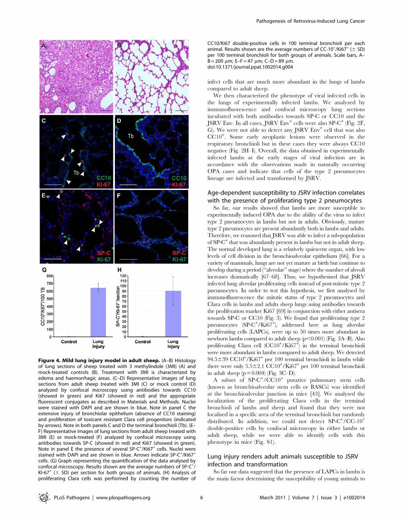
Figure 4. Mild lung injury model in adult sheep. (A–B) Histologyof lung sections of sheep treated with 3 methylindole (3MI) (A) andmock-treated controls (B). Treatment with 3MI is characterized byedema and haemorrhagic areas. (C–D) Representative images of lungsections from adult sheep treated with 3MI (C) or mock control (D)analyzed by confocal microscopy using antibodies towards CC10(showed in green) and Ki67 (showed in red) and the appropriatefluorescent conjugates as described in Materials and Methods. Nucleiwere stained with DAPI and are shown in blue. Note in panel C theextensive injury of bronchiolar epithelium (absence of CC10 staining)and proliferation of toxicant resistant Clara cell progenitors (indicatedby arrows). Note in both panels C and D the terminal bronchioli (Tb). (E–F) Representative images of lung sections from adult sheep treated with3MI (E) or mock-treated (F) analyzed by confocal microscopy usingantibodies towards SP-C (showed in red) and Ki67 (showed in green).Note in panel E the presence of several SP-C+/Ki67+ cells. Nuclei werestained with DAPI and are shown in blue. Arrows indicate SP-C+/Ki67+
cells. (G) Graph representing the quantification of the data analysed byconfocal microscopy. Results shown are the average numbers of SP-C+/KI-67+ (6 SD) per section for both groups of animals. (H) Analysis ofproliferating Clara cells was performed by counting the number of
CC10/Ki67 double-positive cells in 100 terminal bronchioli per eachanimal. Results shown are the average numbers of CC-10+/Ki67+ (6 SD)per 100 terminal bronchioli for both groups of animals. Scale bars, A–B = 200 mm; E–F = 47 mm; C–D = 89 mm.doi:10.1371/journal.ppat.1002014.g004
Pathogenesis of Retrovirus-Induced Lung Cancer
PLoS Pathogens | www.plospathogens.org 6 March 2011 | Volume 7 | Issue 3 | e1002014

JSRV infection as opposed to the resistance observed by adult
sheep. Indeed, in the adult lungs, the proliferation rate of the
respiratory epithelium is very low [68]. However, the lung has a
significant reparative capability and after an injury the LAPC
proliferate and play an important role in the tissue regenerative
process. We therefore reasoned that we would be able to render
adult sheep susceptible to experimental JSRV infection by
previous induction of a mild lung injury that would stimulate
LAPCs. 3MI is an organ-selective pneumotoxicant that affects
specifically type I pneumocytes and bronchiolar epithelial (Clara)
cells and it is especially effective in ruminants [69,70]. Here, to
assess the ability of 3MI to induce lung injury and repair we
exposed two sheep to this pneumotoxicant and we then assessed
lung injury after 48 hours. Histological examination showed
diffuse pulmonary edema with scattered hemorrhagic foci
(Fig. 4A–B). Next, we assessed the proliferation status of type 2
pneumocytes and Clara cells by verifying co-expression of SP-C or
CC10 with the proliferating marker Ki67 by immunofluorescence
as described above (Fig. 4C–F). The number of SP-C+/Ki67+ cells
was 90 fold higher in sheep after lung injury as opposed to normal
control sheep (p,0.001) (Fig. 4H). The examination of the
terminal bronchioli in sheep after 3MI administration revealed
that almost 100% of terminal bronchioli contained CC10+/Ki67+
(Fig. 4G). The total number of CC10+/Ki67+ cells was more than
100 fold higher in adult sheep after lung injury compared to
healthy controls (p = 0.009) (Fig. 4H). Also in adult sheep after
lung injury we were not able to identify any SP-C+/CC10+
double-positive cells (data not shown).
Overall, the data presented above indicate that the number of
LAPCs, that we identified as target cells of JSRV infection,
increase dramatically after mild lung injury. In order to determine
whether lung injury may render adult sheep susceptible to JSRV
infection, we treated five sheep with 3MI and after 48 h we
infected them with JSRV (Group I). Five additional sheep were
infected with JSRV without pre-treatment with 3MI (Group II). 10
days after infection animals were euthanized (Fig. 5A). As
expected, post-mortem examination revealed no signs of lesions
attributed to lung injury. In each animal, the presence of JSRV
infection was assessed in 15 sections collected from the cranial lobe
by immunohistochemistry. JSRV Env expression was only
detected in lung cells of animals that were infected after treatment
with 3MI (Fig. 5B, D–F). On average, 10 clusters of JSRV Env+
cells (ranging from 1 to 80 cells) were detected in each animal
while no JSRV infected cell was detected in those animals that
were infected without 3MI pre-treatment (Fig. 5C).
By immunofluorescence and confocal microscopy we found that
all JSRV infected cells were SP-C positive (Fig. 6A–C). None of
the JSRV Env+ cells were CC10+ (Fig. 6D–F), despite the high
number of proliferating Clara cells induced by 3MI and the
presence of numerous infected cells localized in the terminal
bronchioli.
JSRV preferentially infects dividing cells in vitroOur data have shown that JSRV infects LAPCs but not the
overwhelming majority of type 2 pneumocytes which divide very
slowly. These data could be explained mechanistically by the fact
that the majority of retroviruses, with the exception of lentiviruses
[71], infect more efficiently cells that are in mitosis [72–73]. The
proliferation rate of type 2 pneumocytes is very low in adults under
normal conditions. On the other hand the higher proliferative rate
Figure 5. Induction of mild lung injury renders adult sheep susceptible to JSRV infection. (A) Schematic diagram of the study design. Tenadult sheep were randomly allocated in two groups. Sheep in Group I (red bar) were dosed 3MI (red arrow) as described in Materials and Methodswhile Group II served as control. After 2 days all animals in both groups were infected with JSRV (black arrow) and euthanized 10 days post infection.(B) Graph showing the mean number of JSRV Env+ clusters per animal as detected by immunohistochemistry in sheep of Group I and Group II 10 dayspost-infection (6 SD). (C) Graph showing the number of cells forming each cluster of JSRV Env+ cells in sheep of Group I. (D–F)Immunohistochemistry of lung sections of adult sheep of group II (D) and group I (E–F, pre-treated with 3MI before JSRV infection) 10 dayspost-infection as described in Materials and Methods. Env expression (characterized by the intra cytoplasmic dark brown colour) was detected insheep of Group I but not in sheep of Group II. Scale bars, D = 200 mm; E–F = 100 mm.doi:10.1371/journal.ppat.1002014.g005
Pathogenesis of Retrovirus-Induced Lung Cancer
PLoS Pathogens | www.plospathogens.org 7 March 2011 | Volume 7 | Issue 3 | e1002014

Pathogenesis of Retrovirus-Induced Lung Cancer
PLoS Pathogens | www.plospathogens.org 8 March 2011 | Volume 7 | Issue 3 | e1002014

of LAPCs during post-natal development or tissue repair in the
adult would facilitate JSRV infection. Experiments with JSRV in
vitro are hindered by the lack of a convenient tissue culture system
for the propagation of this virus [32]. Therefore, we constructed a
convenient JSRV-derived viral vector (JS-EeGFP-mCherry) in
order to easily quantify JSRV infection in proliferating and non-
proliferating cells. JS-eGFP-mCherry was derived by transiently
transfecting 293T cells with (i) a packaging plasmid (pGPP-MX-
4CTE) devoid of the JSRV packaging signal (Y) and expressing
the viral Gag, Pro and Pol; (ii) a plasmid providing the JSRV Env
in trans (pC-ML-JSenv, also devoided of Y), and (iii) the packaged
JSRV vector (pCJS-EFGFP-MC) that upon infection and
integration expresses eGFP under the control of an internal
promoter (Fig. 7A). JS-eGFP-mCherry viral particles were then
used to infect synchronized SCP cells in the presence or absence of
a drug that, at the concentration used in this study, arrests cells in
the G1 phase (aphidicolin) (Fig. 7B). Consistently, JS-eGFP-
mCherry was able to transduce actively dividing SCP cells
approximately 200 times more efficiently (p = 0.002) than the
same cells where mitosis was arrested with aphidicolin while only
minor differences between treated and untreated cells were
observed with the lentivirus vector HIV-GFP (Fig. 7C).
Discussion
In this study we have investigated the pathogenesis of a unique
virus-induced lung adenocarcinoma and obtained data that have a
broad significance in pulmonary biology, carcinogenesis and
retroviral pathogenesis. Most adenocarcinomas in humans display
cells expressing type 2 pneumocytes or Clara cell markers but it is
not completely clear whether the neoplasm arises from a stem cell
that is able to differentiate into both cell types, or from a
committed progenitor or from the fully differentiated cell
compartments [74]. In this study, we identified the target cells of
JSRV infection and transformation in vivo as proliferating cells of
the type 2 pneumocytes lineage (SP-C+/Ki67+, LAPC). In
addition, we showed that the age-related susceptibility to
experimental OPA induction is directly related to the abundance
of LAPCs. Importantly, induction of mild injury to the respiratory
epithelium increased dramatically the number of LAPCs in adult
sheep and rendered these animals susceptible to JSRV infection
and transformation. We have not found evidence that CC10+/
Ki67+ cells are infected and transformed by JSRV. Furthermore,
we found that the CC10+ cells that are found in a proportion of
late stages OPA tumours are not expressing JSRV proteins and
Figure 7. Infectivity of JSRV in proliferating and mitotic arrested cell. (A) Schematic representation of the experimental design. A JSRV-based vector was derived by transfecting 293T with pJS-EFGFP-MC, pGPP-MX-4CTE and pC-MLV-JSenv as described in the materials and methods.The resulting vector, JS-eGFP, was then used to infect synchronized choroid plexus cells (CP) cultures in the presence or absence of aphidicolin. (B)Histograms showing the DNA content of CP cells with or without Aphidicolin (5 mg/ml for 24 hours). The DNA content was measured by 7AADstaining and flow-cytometry analysis and provides an indication of the cell cycle. The x and y axis represent the relative DNA content and the cellcounts. (C) Graph showing the transduction efficiency (expressed as fluorescence forming foci/ml) of JS-eGFP in proliferating or mitotic arrested CP asdescribed in Materials and Methods.doi:10.1371/journal.ppat.1002014.g007
Figure 6. Phenotype of JSRV infected cells in adult sheep with lung injury. Representative images of lung sections from adult sheep pre-treated with 3MI before experimental JSRV infection. (A–C) Sections were analyzed by confocal microscopy using antibodies towards SP-C (showed inred) and the JSRV Env (showed in green). Nuclei were stained with DAPI and are shown in blue. Arrows indicate autofluorescent red blood cells (RBC).(D–F) Sections analyzed as above using antibodies towards CC10 (showed in red) and the JSRV Env (showed in green). Nuclei were stained with DAPIand are shown in blue. Scale bars, A–B = 47 mm; C = 89 mm; D = 26 mm; E = 33 mm and F = 25 mm.doi:10.1371/journal.ppat.1002014.g006
Pathogenesis of Retrovirus-Induced Lung Cancer
PLoS Pathogens | www.plospathogens.org 9 March 2011 | Volume 7 | Issue 3 | e1002014

may therefore not be true tumour cells, at least in the cases we
examined.
Our data provide important consideration for pulmonary biology
and carcinogenesis. We infer from our study that at least in sheep,
type 2 pneumocytes and Clara cells have two distinct populations of
proliferating progenitor cells committed to the alveolar and the
bronchiolar lineages. From this study, we cannot determine whether
the LAPCs are progenitor committed solely to type 2 or type 1
pneumocytes. We showed that lung adenocarcinoma can originate
from an alveolar proliferating cell of the alveolar lineage, rather than
from a bronchioalveolar stem cell postulated to originate both type 2
pneumocytes and Clara cells. Studies in mice have identified a
population of putative stem cells that are both SP-C+ and CC10+
(bronchioalveolar stem cells, BASCs) located at the bronchioalveolar
duct junction [43]. Based on in vitro analysis, BASCs were
hypothesised to give rise to Clara cells, alveolar type 2 cells and be
the cell originating lung adenocarcinoma [43]. On the other hand,
studies using genetic lineage-labelling experiments in mice, support-
ed a model where bronchioli and alveoli are maintained and
repaired distinctively by Clara cells and LAPCs respectively [44,75].
The presence of BASCs in humans has not been confirmed and in
general the biological relevance of BASCs is object of debate [44–
45]. In our study, by confocal microscopy, we have not been able to
detect SP-C+/CC10+ in sheep while we were able to detect cells with
this phenotype in mice (Fig. S1). We cannot rule out the presence of
a rare bronchioalveolar stem cell (SP-C+/CC10+) able to differen-
tiate in both type 2 pneumocytes and Clara cell progenitors in sheep.
We also cannot rule out the presence in sheep of phenotypically
uncharacterized pulmonary stem cells. However, if these cells exist in
the sheep, they are very rare and unlike LAPCs they do not appear
to play a major role in OPA. Interestingly, from the anatomical and
histological point of view the human lungs are more comparable to
the sheep lungs as opposed to the mice lungs [76–77].
We showed with experiments in vitro that JSRV, similarly to
other retroviruses, infects preferentially cells in active mitosis.
These experiments provide a mechanistic explanation to the
observation that JSRV infects readily LAPCs but not mature type
2 pneumocytes.
As mentioned before, JSRV is a unique oncogenic virus as it
possesses the viral Env (a structural protein) that behaves as a
functional dominant oncoprotein both in vitro and in vivo. In
general, viral oncoproteins are non structural proteins whose
expression is not linked to productive infection. It would be
detrimental from an evolutionary point of view of the virus, to
have productive viral infection and carcinogenesis as strictly
mutually dependent events (viral replication would in this case lead
to the death of the infected host). Onset of lung adenocarcinoma in
JSRV-infected animals could therefore be viewed as either
‘‘accidental’’ (similarly to other retrovirus-induced tumors) or
‘‘essential’’ in order to allow virus spread among susceptible hosts.
Although these two alternative hypotheses are not necessarily
mutually exclusive, the data obtained in this study and
accumulated over the years on JSRV/OPA, strongly suggest that
tumor induction plays an important part in the evolutionary
strategies used by the virus to persist in the sheep population. In
previous studies we have shown that development of OPA in the
field occurs only in a minority of the JSRV-infected sheep [39].
On the other hand, animals with OPA produce lung secretions
containing abundant amounts of infectious JSRV particles that
pour freely from the nostrils of the affected sheep [41,78–79]. The
data from this paper strongly suggests that clinical OPA develops
in natural conditions as a result of viral infection only when LAPCs
are available to the virus: in young lambs during post-natal
development or in the presence of an injury to the bronchioalveo-
lar epithelium. Importantly, as mentioned in the introduction,
JSRV proteins are detected readily only in the tumour cells of
OPA affected animals (and in the LAPCs as shown in this study)
[38] although low levels of virus infection and protein expression
are detectable in cells of the lymphoreticular system of animals
with or without clinical OPA. We and others have shown that the
JSRV LTRs are the main determinants regulating the tight cell-
specific expression pattern displayed by this virus. The JSRV
LTRs contains lung-specific enhancer binding motifs that are
preferentially active in cell lines derived from transformed type 2
pneumocytes [80–83]. In addition, in transgenic mice, reporter
gene expression driven by the JSRV LTR has been detected
specifically in type 2 pneumocytes [84]. Thus, JSRV-host
equilibrium has been reached by a combination of factors. JSRV
has evolved a structural protein that is a powerful oncoprotein but
only when expressed at high levels in the LAPCs, which are
relatively rare cells in the adult healthy sheep. Therefore, JSRV
has a limited window of opportunity to infect the target cells of the
host that allow high level of viral expression (and that can be
consequently transformed). At the same time, onset of lung
adenocarcinoma in a minority of the infected animals allows an
amplification of the cells that can produce infectious virus and
therefore it is a likely evolutionary mechanism that helps JSRV to
persist in the population.
It is important to note that in natural conditions, sheep with
OPA present consistently a variety of other parasitic, bacterial or
viral infections [9]. Classically, these infections were considered as
‘‘secondary’’ to JSRV infection. We suggest instead that in the
adult, the induction of an injury to the respiratory epithelium by
various pathogens substantially increases the number of LAPCs
and renders adult sheep susceptible to JSRV-induced transforma-
tion, similarly to what we have shown experimentally in this study
with the pneumotoxicant 3MI. Thus, inflammation induced by
different pathogens is the ‘‘primary’’ event for OPA induction. It is
feasible that in animals already infected with JSRV the virus
present in lymphoreticular cells is able to spread to injured tissues
where it can infect and transform alveolar progenitor cells actively
involved in repairing the epithelium.
In conclusion, this work provided unique insights into
pulmonary physiology, lung cancer, and retrovirus pathogenesis
and is another telling example where viruses have helped us to
understand fundamental aspects of host biology.
Supporting Information
Figure S1 Detection of SP-C and CC10 in cells of the
bronchioalveolar duct junctions of sheep and mice lungs. A.
Representative images of lung sections from adult mice. Sections
were analyzed by confocal microscopy using antibodies towards
CC-10 (green) and SP-C (red). CC10+/SPC+ double positive cells
bronchioalveolar stem cells (BASCs) are located at the bronch-
ioalveolar duct junction (BADJ). Arrows point to BASC cells.
Nuclei were stained with DAPI and are shown in blue. Scale bar,
75 mm. B. Representative image of lung sections from adult sheep.
Sections were analyzed by confocal microscopy using antibodies
towards CC-10 (green) and SP-C (red). Nuclei were stained with
DAPI and are shown in blue. No SP-C/CC10 double-positive
cells are detectable. Scale bar, 62 mm.
(TIF)
Acknowledgments
We would like to thank Henny Martineau for immunohistochemistry and
Brigid Hogan for useful suggestions. We are grateful to Luigi D’Innocenzo
Pathogenesis of Retrovirus-Induced Lung Cancer
PLoS Pathogens | www.plospathogens.org 10 March 2011 | Volume 7 | Issue 3 | e1002014
and Doriano Ferrari for excellent animal care and to Sandro Martella and
Federica Lopes for sample collection and processing. In addition, we thank
Dusty Miller for generously providing some of the reagents used in this
study. We also thank Camille Huser and Gillian Borland for FACS
analysis. CM holds a Clinical Intermediate Fellowship by the Wellcome
Trust.
Note
The term ‘‘Clara’’ cell is widely used in the literature to describe the non-
ciliated, secretory cells in the respiratory epithelium of the distal airways.
This term is an eponym in honor of his discoverer, Max Clara. Recent
studies have shown that Max Clara was an active member of the Nazi
regime. The tissue used for the original study on the Clara cell derived
from a prisoner executed by the Nazi ‘‘justice system’’ [85]. We are in favor
of abandoning the use of this eponym. We reluctantly used the term Clara
cell in this paper as we are not aware of any new terminology officially
proposed and adopted by the scientific community specializing in this field.
Author Contributions
Conceived and designed the experiments: CM MC MP. Performed the
experiments: CM MC OC GDF NF VV MdlH. Analyzed the data: CM
MC MdlH MP. Wrote the paper: CM MC MP.
References
1. Coffin JM, Hughes SH, Varmus HE (1997) Retroviruses. New York: ColdSpring Harbor Laboratory Press. 843 p.
2. Banerjee P, Crawford L, Samuelson E, Feuer G (2010) Hematopoietic stem cellsand retroviral infection. Retrovirology 7: 8.
3. Clarke MF, Dick JE, Dirks PB, Eaves CJ, Jamieson CH, et al. (2006) Cancer
stem cells–perspectives on current status and future directions: AACR Workshop
on cancer stem cells. Cancer Res 66: 9339–9344.
4. Reya T, Morrison SJ, Clarke MF, Weissman IL (2001) Stem cells, cancer, andcancer stem cells. Nature 414: 105–111.
5. Hill RP (2006) Identifying cancer stem cells in solid tumors: case not proven.Cancer Res 66: 1891–1895; discussion 1890.
6. Visvader JE, Lindeman GJ (2008) Cancer stem cells in solid tumours:
accumulating evidence and unresolved questions. Nat Rev Cancer 8: 755–768.
7. Zhou BB, Zhang H, Damelin M, Geles KG, Grindley JC, et al. (2009) Tumour-
initiating cells: challenges and opportunities for anticancer drug discovery. NatRev Drug Discov 8: 806–823.
8. Rosenberg N, Jolicoeur P (1997) Retroviral pathogenesis. In: Coffin JM,Hughes S, Varmus HE, eds. Retroviruses. New York: Cold Spring Harbor
laboratory Press. 475585 p.
9. Fan H (2003) Jaagsiekte sheep retrovirus and lung cancer. Berlin: Springer-Verlag.
10. Palmarini M (2007) A veterinary twist on pathogen biology. PLoS Pathog 3: e12.
11. Palmarini M, Sharp JM, De las Heras M, Fan H (1999) Jaagsiekte sheepretrovirus is necessary and sufficient to induce a contagious lung cancer in sheep.
J Virol 73: 6964–6972.
12. Palmarini M, Fan H (2001) Retrovirus-induced ovine pulmonary adenocarci-
noma, an animal model for lung cancer. J Natl Cancer Inst 93: 1603–1614.
13. Liu SL, Miller AD (2007) Oncogenic transformation by the jaagsiekte sheep
retrovirus envelope protein. Oncogene 26: 789–801.
14. Allen TE, Sherrill KJ, Crispell SM, Perrott MR, Carlson JO, et al. (2002) Thejaagsiekte sheep retrovirus envelope gene induces transformation of the avian
fibroblast cell line DF-1 but does not require a conserved SH2 binding domain.
J Gen Virol 83: 2733–2742.
15. Maeda N, Palmarini M, Murgia C, Fan H (2001) Direct transformation ofrodent fibroblasts by jaagsiekte sheep retrovirus DNA. Proc Natl Acad Sci U S A
98: 4449–4454.
16. Rai SK, Duh FM, Vigdorovich V, Danilkovitch-Miagkova A, Lerman MI, et al.
(2001) Candidate tumor suppressor HYAL2 is a glycosylphosphatidylinositol(GPI)-anchored cell-surface receptor for jaagsiekte sheep retrovirus, the envelope
protein of which mediates oncogenic transformation. Proc Natl Acad Sci U S A98: 4443–4448.
17. Liu SL, Duh FM, Lerman MI, Miller AD (2003) Role of virus receptor Hyal2 inoncogenic transformation of rodent fibroblasts by sheep betaretrovirus env
proteins. J Virol 77: 2850–2858.
18. Liu SL, Miller AD (2005) Transformation of madin-darby canine kidney
epithelial cells by sheep retrovirus envelope proteins. J Virol 79: 927–933.
19. Maeda N, Inoshima Y, Fruman DA, Brachmann SM, Fan H (2003)Transformation of mouse fibrobalsts by jaagsiekte sheep retrovirus envelope
does not require phosphatidylinositol 3-kinase. J Virol 77: 9951–9959.
20. Palmarini M, Maeda N, Murgia C, De-Fraja C, Hofacre A, et al. (2001) A
phosphatidylinositol 3-kinase docking site in the cytoplasmic tail of the Jaagsiektesheep retrovirus transmembrane protein is essential for envelope-induced
transformation of NIH 3T3 cells. J Virol 75: 11002–11009.
21. Zavala G, Pretto C, Chow YH, Jones L, Alberti A, et al. (2003) Relevance of Akt
phosphorylation in cell transformation induced by Jaagsiekte sheep retrovirus.Virology 312: 95–105.
22. Johnson C, Sanders K, Fan H (2010) Jaagsiekte sheep retrovirus transformation
in Madin-Darby canine kidney epithelial cell three-dimensional culture. J Virol
84: 5379–5390.
23. Caporale M, Cousens C, Centorame P, Pinoni C, De las Heras M, et al. (2006)Expression of the Jaagsiekte sheep retrovirus envelope glycoproteins is sufficient
to induce lung tumor in sheep. J Virol 80: 8030–8037.
24. Wootton SK, Halbert CL, Miller AD (2005) Sheep retrovirus structural protein
induces lung tumours. Nature 434: 904–907.
25. Maeda N, Fu W, Ortin A, de las Heras M, Fan H (2005) Roles of the Ras-MEK-
mitogen-activated protein kinase and phosphatidylinositol 3-kinase-Akt-mTOR
pathways in Jaagsiekte sheep retrovirus-induced transformation of rodent
fibroblast and epithelial cell lines. J Virol 79: 4440–4450.
26. Liu SL, Lerman MI, Miller AD (2003) Putative phosphatidylinositol 3-kinase
(PI3K) binding motifs in ovine betaretrovirus Env proteins are not essential forrodent fibroblast transformation and PI3K/Akt activation. J Virol 77:
7924–7935.
27. De Las Heras M, Ortin A, Benito A, Summers C, Ferrer LM, et al. (2006) In-situ
demonstration of mitogen-activated protein kinase Erk 1/2 signalling pathway in
contagious respiratory tumours of sheep and goats. J Comp Pathol 135: 1–10.
28. Sharp JM, Angus KW, Gray EW, Scott FM (1983) Rapid transmission of sheep
pulmonary adenomatosis (jaagsiekte) in young lambs. Brief report. Arch Virol78: 89–95.
29. Salvatori D, Gonzalez L, Dewar P, Cousens C, de las Heras M, et al. (2004)Successful induction of ovine pulmonary adenocarcinoma in lambs of different
ages and detection of viraemia during the preclinical period. J Gen Virol 85:3319–3324.
30. Holland MJ, Palmarini M, Garcia-Goti M, Gonzalez L, McKendrick I, et al.
(1999) Jaagsiekte retrovirus is widely distributed both in T and B lymphocytesand in mononuclear phagocytes of sheep with naturally and experimentally
acquired pulmonary adenomatosis. J Virol 73: 4004–4008.
31. Palmarini M, Holland MJ, Cousens C, Dalziel RG, Sharp JM (1996) Jaagsiekte
retrovirus establishes a disseminated infection of the lymphoid tissues of sheepaffected by pulmonary adenomatosis. J Gen Virol 77: 2991–2998.
32. Palmarini M, Sharp JM, Lee C, Fan H (1999) In vitro infection of ovine cell lines
by jaagsiekte sheep retrovirus (JSRV). J Virol 73: 10070–10078.
33. Rai SK, DeMartini JC, Miller AD (2000) Retrovirus vectors bearing jaagsiekte
sheep retrovirus Env transduce human cells by using a new receptor localized tochromosome 3p21.3. J Virol 74: 4698–4704.
34. Arnaud F, Caporale M, Varela M, Biek R, Chessa B, et al. (2007) A paradigmfor virus-host coevolution: sequential counter-adaptations between endogenous
and exogenous retroviruses. PLoS Pathog 3: e170.
35. Arnaud F, Varela M, Spencer TE, Palmarini M (2008) Coevolution of
endogenous Betaretroviruses of sheep and their host. Cell Mol Life Sci 65:
3422–3432.
36. Ortin A, Minguijon E, Dewar P, Garcia M, Ferrer LM, et al. (1998) Lack of a
specific immune response against a recombinant capsid protein of Jaagsiektesheep retrovirus in sheep and goats naturally affected by enzootic nasal tumour
or sheep pulmonary adenomatosis. Vet Immunol Immunopathol 61: 229–237.
37. Chessa B, Pereira F, Arnaud F, Amorim A, Goyache F, et al. (2009) Revealing
the history of sheep domestication using retrovirus integrations. Science 324:532–536.
38. Palmarini M, Dewar P, De las Heras M, Inglis NF, Dalziel RG, et al. (1995)
Epithelial tumour cells in the lungs of sheep with pulmonary adenomatosis aremajor sites of replication for Jaagsiekte retrovirus. J Gen Virol 76: 2731–2737.
39. Caporale M, Centorame P, Giovannini A, Sacchini F, Di Ventura M, et al.(2005) Infection of lung epithelial cells and induction of pulmonary
adenocarcinoma is not the most common outcome of naturally occurring JSRVinfection during the commercial lifespan of sheep. Virology 338: 144–153.
40. Platt JA, Kraipowich N, Villafane F, DeMartini JC (2002) Alveolar type II cells
expressing jaagsiekte sheep retrovirus capsid protein and surfactant proteins arethe predominant neoplastic cell type in ovine pulmonary adenocarcinoma. Vet
Pathol 39: 341–352.
41. De las Heras M, Gonzalez L, Sharp JM (2003) Pathology of ovine pulmonary
adenocarcinoma. Curr Top Microbiol Immunol 275: 25–54.
42. Beytut E, Sozmen M, Erginsoy S (2009) Immunohistochemical detection of
pulmonary surfactant proteins and retroviral antigens in the lungs of sheep withpulmonary adenomatosis. J Comp Pathol 140: 43–53.
43. Kim CF, Jackson EL, Woolfenden AE, Lawrence S, Babar I, et al. (2005)
Identification of bronchioalveolar stem cells in normal lung and lung cancer.Cell 121: 823–835.
44. Rawlins EL, Okubo T, Xue Y, Brass DM, Auten RL, et al. (2009) The role ofScgb1a1+ Clara cells in the long-term maintenance and repair of lung airway,
but not alveolar, epithelium. Cell Stem Cell 4: 525–534.
45. Giangreco A, Arwert EN, Rosewell IR, Snyder J, Watt FM, et al. (2009) Stem
cells are dispensable for lung homeostasis but restore airways after injury. ProcNatl Acad Sci U S A 106: 9286–9291.
Pathogenesis of Retrovirus-Induced Lung Cancer
PLoS Pathogens | www.plospathogens.org 11 March 2011 | Volume 7 | Issue 3 | e1002014

46. Caporale M, Arnaud F, Mura M, Golder M, Murgia C, et al. (2009) The signal
peptide of a simple retrovirus envelope functions as a posttranscriptionalregulator of viral gene expression. J Virol 83: 4591–4604.
47. Quade K (1979) Transformation of mammalian cells by avian myelocytomatosis
virus and avian erythroblastosis virus. Virology 98: 461–465.48. Palmarini M, Cousens C, Dalziel RG, Bai J, Stedman K, et al. (1996) The
exogenous form of Jaagsiekte retrovirus is specifically associated with acontagious lung cancer of sheep. J Virol 70: 1618–1623.
49. Wootton SK, Metzger MJ, Hudkins KL, Alpers CE, York D, et al. (2006) Lung
cancer induced in mice by the envelope protein of jaagsiekte sheep retrovirus(JSRV) closely resembles lung cancer in sheep infected with JSRV. Retrovirology
3: 94.50. Donello JE, Loeb JE, Hope TJ (1998) Woodchuck hepatitis virus contains a
tripartite posttranscriptional regulatory element. J Virol 72: 5085–5092.51. Schwenter F, Deglon N, Aebischer P (2003) Optimization of human
erythropoietin secretion from MLV-infected human primary fibroblasts used
for encapsulated cell therapy. J Gene Med 5: 246–257.52. Bray M, Prasad S, Dubay JW, Hunter E, Jeang KT, et al. (1994) A small
element from the Mason-Pfizer monkey virus genome makes humanimmunodeficiency virus type 1 expression and replication Rev-independent.
Proc Natl Acad Sci U S A 91: 1256–1260.
53. Zolotukhin AS, Valentin A, Pavlakis GN, Felber BK (1994) Continuouspropagation of RRE(-) and Rev(-)RRE(-) human immunodeficiency virus type 1
molecular clones containing a cis-acting element of simian retrovirus type 1 inhuman peripheral blood lymphocytes. J Virol 68: 7944–7952.
54. Miller AD, Chen F (1996) Retrovirus packaging cells based on 10A1 murineleukemia virus for production of vectors that use multiple receptors for cell entry.
J Virol 70: 5564–5571.
55. Zaffran S, Astier M, Gratecos D, Semeriva M (1997) The held out wings (how)Drosophila gene encodes a putative RNA-binding protein involved in the
control of muscular and cardiac activity. Development 124: 2087–2098.56. Naldini L, Blomer U, Gallay P, Ory D, Mulligan R, et al. (1996) In vivo gene
delivery and stable transduction of nondividing cells by a lentiviral vector.
Science 272: 263–267.57. Bahnson AB, Dunigan JT, Baysal BE, Mohney T, Atchison RW, et al. (1995)
Centrifugal enhancement of retroviral mediated gene transfer. J Virol Methods54: 131–143.
58. O’Doherty U, Swiggard WJ, Malim MH (2000) Human immunodeficiency virustype 1 spinoculation enhances infection through virus binding. J Virol 74:
10074–10080.
59. DeMartini JC, Rosadio RH, Sharp JM, Russell HI, Lairmore MD (1987)Experimental coinduction of type D retrovirus-associated pulmonary carcinoma
and lentivirus-associated lymphoid interstitial pneumonia in lambs. J NatlCancer Inst 79: 167–177.
60. Nisbet DI, Mackay JM, Smith W, Gray EW (1971) Ultrastructure of sheep
pulmonary adenomatosis (Jaagsiekte). J Pathol 103: 157–162.61. Payne AL, Verwoerd DW (1984) A scanning and transmission electron
microscopy study of jaagsiekte lesions. Onderstepoort J Vet Res 51: 1–13.62. Perk K, Hod I, Nobel TA (1971) Pulmonary adenomatosis of sheep (jaagsiekte).
I. Ultrastructure of the tumor. J Natl Cancer Inst 46: 525–537.63. Plopper CG, Hyde DM, Buckpitt AR (1997) Clara cells. In: Crystal RG,
West JB, Weibel ER, Barnes PJ, eds. The Lung: Scientific Foundations.
Philadelphia: Lippincott-Raven. pp 517–533.64. Whitsett JA, Glasser SW (1998) Regulation of surfactant protein gene
transcription. Biochim Biophys Acta 1408: 303–311.65. Verwoerd DW, Williamson AL, De Villiers EM (1980) Aetiology of jaagsiekte:
transmission by means of subcellular fractions and evidence for the involvement
of a retrovirus. Onderstepoort J Vet Res 47: 275–280.
66. Kauffman SL (1980) Cell proliferation in the mammalian lung. Int Rev ExpPathol 22: 131–191.
67. Zeltner TB, Burri PH (1987) The postnatal development and growth of the
human lung. II. Morphology. Respir Physiol 67: 269–282.
68. Zeltner TB, Caduff JH, Gehr P, Pfenninger J, Burri PH (1987) The postnatal
development and growth of the human lung. I. Morphometry. Respir Physiol
67: 247–267.
69. Kubbutat MH, Key G, Duchrow M, Schluter C, Flad HD, et al. (1994) Epitope
analysis of antibodies recognising the cell proliferation associated nuclear antigen
previously defined by the antibody Ki-67 (Ki-67 protein). J Clin Pathol 47:524–528.
70. Bradley BJ, Carlson JR, Dickinson EO (1978) 3-methylindole-induced
pulmonary edema and emphysema in sheep. Am J Vet Res 39: 1355–1358.
71. Lewis PF, Emerman M (1994) Passage through mitosis is required for
oncoretroviruses but not for the human immunodeficiency virus. J Virol 68:
510–516.
72. Miller DG, Adam MA, Miller AD (1990) Gene transfer by retrovirus vectors
occurs only in cells that are actively replicating at the time of infection. Mol Cell
Biol 10: 4239–4242.
73. Roe T, Reynolds TC, Yu G, Brown PO (1993) Integration of murine leukemia
virus DNA depends on mitosis. Embo J 12: 2099–2108.
74. Rosai JaS LH (1995) Conditional Clara cell ablation reveals a self-renewing
progenitor function of pulmonary neuroendocrine cells Atlas of Tumor
Pathology. Washington DC: Armed Forces Institute of Pathology.
75. Rawlins EL, Okubo T, Que J, Xue Y, Clark C, et al. (2008) Epithelial stem/
progenitor cells in lung postnatal growth, maintenance, and repair. Cold Spring
Harb Symp Quant Biol 73: 291–295.
76. Scheerlinck JP, Snibson KJ, Bowles VM, Sutton P (2008) Biomedical
applications of sheep models: from asthma to vaccines. Trends Biotechnol 26:
259–266.
77. Harris A (1997) Towards an ovine model of cystic fibrosis. Hum Mol Genet 6:
2191–2194.
78. Sharp JM, DeMartini JC (2003) Natural history of JSRV in sheep. Curr TopMicrobiol Immunol 275: 55–79.
79. Cousens C, Thonur L, Imlach S, Crawford J, Sales J, et al. (2009) Jaagsiekte
sheep retrovirus is present at high concentration in lung fluid produced by ovinepulmonary adenocarcinoma-affected sheep and can survive for several weeks at
ambient temperatures. Res Vet Sci 87: 154–156.
80. McGee-Estrada K, Fan H (2006) In Vivo and In Vitro Analysis of FactorBinding Sites in Jaagsiekte Sheep Retrovirus Long Terminal Repeat Enhancer
Sequences: Roles of HNF-3, NF-I, and C/EBP for Activity in Lung Epithelial
Cells. J Virol 80: 332–341.
81. McGee-Estrada K, Palmarini M, Fan H (2002) HNF-3ß is a critical factor for
the expression of the Jaagsiekte sheep retrovirus (JSRV) long terminal repeat in
type II pneumocytes but not in clara cells. Virology 292: 87–97.
82. McGee-Estrada K, Palmarini M, Hallwirth C, Fan H (2005) A Moloney murine
leukemia virus driven by the Jaagsiekte sheep retrovirus enhancers shows
enhanced specificity for infectivity in lung epithelial cells. Virus Genes 31:257–263.
83. Palmarini M, Datta S, Omid R, Murgia C, Fan H (2000) The long terminalrepeats of Jaagsiekte sheep retrovirus (JSRV) are preferentially active in type II
pneumocytes. J Virol 74: 5776–5787.
84. Dakessian RM, Fan H (2008) Specific in vivo expression in type II pneumocytesof the Jaagsiekte sheep retrovirus long terminal repeat in transgenic mice.
Virology 372: 398–408.
85. Winkelmann A, Noack T (2010) The Clara cell: a ‘‘Third Reich eponym’’? EurRespir J 36: 722–727.
Pathogenesis of Retrovirus-Induced Lung Cancer
PLoS Pathogens | www.plospathogens.org 12 March 2011 | Volume 7 | Issue 3 | e1002014