The enigma of eugregarine epicytic folds: where gliding motility originates?
28
The enigma of eugregarine epicytic folds: where gliding motility originates? Valigurová et al. Valigurová et al. Frontiers in Zoology 2013, 10:57 http://www.frontiersinzoology.com/content/10/1/57
-
Upload
independent -
Category
Documents
-
view
1 -
download
0
Transcript of The enigma of eugregarine epicytic folds: where gliding motility originates?

The enigma of eugregarine epicytic folds: wheregliding motility originates?Valigurová et al.
Valigurová et al. Frontiers in Zoology 2013, 10:57http://www.frontiersinzoology.com/content/10/1/57

Valigurová et al. Frontiers in Zoology 2013, 10:57http://www.frontiersinzoology.com/content/10/1/57
RESEARCH Open Access
The enigma of eugregarine epicytic folds: wheregliding motility originates?Andrea Valigurová1*, Naděžda Vaškovicová2, Naďa Musilová1 and Joseph Schrével3
Abstract
Background: In the past decades, many studies focused on the cell motility of apicomplexan invasive stages asthey represent a potential target for chemotherapeutic intervention. Gregarines (Conoidasida, Gregarinasina) are aheterogeneous group that parasitize invertebrates and urochordates, and are thought to be an early branchinglineage of Apicomplexa. As characteristic of apicomplexan zoites, gregarines are covered by a complicated pellicle,consisting of the plasma membrane and the closely apposed inner membrane complex, which is associated with anumber of cytoskeletal elements. The cell cortex of eugregarines, the epicyte, is more complicated than that ofother apicomplexans, as it forms various superficial structures.
Results: The epicyte of the eugregarines, Gregarina cuneata, G. polymorpha and G. steini, analysed in the presentstudy is organised in longitudinal folds covering the entire cell. In mature trophozoites and gamonts, each epicyticfold exhibits similar ectoplasmic structures and is built up from the plasma membrane, inner membrane complex,12-nm filaments, rippled dense structures and basal lamina. In addition, rib-like myonemes and an ectoplasmicnetwork are frequently observed. Under experimental conditions, eugregarines showed varied speeds and paths ofsimple linear gliding. In all three species, actin and myosin were associated with the pellicle, and this actomyosincomplex appeared to be restricted to the lateral parts of the epicytic folds. Treatment of living gamonts withjasplakinolide and cytochalasin D confirmed that actin actively participates in gregarine gliding. Contributions togliding of specific subcellular components are discussed.
Conclusions: Cell motility in gregarines and other apicomplexans share features in common, i.e. a three-layeredpellicle, an actomyosin complex, and the polymerisation of actin during gliding. Although the general architectureand supramolecular organisation of the pellicle is not correlated with gliding rates of eugregarines, an increase incytoplasmic mucus concentration is correlated. Furthermore, our data suggest that gregarines utilize severalmechanisms of cell motility and that this is influenced by environmental conditions.
Keywords: Actin, Cytochalasin D, Epicyte, Epicytic folds, Eugregarine, Glideosome, Gliding motility, Jasplakinolide,Mucus, Myosin, Pellicle
IntroductionApicomplexans are one of the most successful and di-verse groups of eukaryotic unicellular parasites that ex-hibit unique adaptations to life in a wide spectrum ofvertebrate and invertebrate hosts. Many cause major hu-man diseases, i.e. malaria, toxoplasmosis, coccidiosis andcryptosporidiosis. Because apicomplexan diseases are stillproblematic, therapeutic research focuses either on para-sitic structures or metabolic pathways which might serve
* Correspondence: [email protected] of Botany and Zoology, Faculty of Science, Masaryk University,Kotlářská 2, 611 37 Brno, Czech RepublicFull list of author information is available at the end of the article
© 2013 Valigurová et al.; licensee BioMed CenCreative Commons Attribution License (http:/distribution, and reproduction in any medium
as drug targets. The cytoskeleton of these parasites has be-come a focus for drug development because it plays animportant role in various life processes, e.g., locomotion,division, invasion and formation of parasite cell polarity[1]. This is especially true of invasive stages of Toxoplasmagondii and Plasmodium falciparum [2,3].Infective stages of Apicomplexa are characterised by an
apical complex of organelles as well as a complicated cellcortex consisting of cortical alveoli, i.e., flattened vesicleslimited by a membrane and packed into a continuouslayer (inner membrane complex), underlying the plasmamembrane. The inner membrane complex (IMC) has mi-cropores and connects numerous cytoskeletal elements
tral Ltd. This is an Open Access article distributed under the terms of the/creativecommons.org/licenses/by/2.0), which permits unrestricted use,, provided the original work is properly cited.

Valigurová et al. Frontiers in Zoology 2013, 10:57 Page 2 of 27http://www.frontiersinzoology.com/content/10/1/57
that include an actomyosin complex, microtubules anda network of intermediate filamentous proteins. Theinvasive zoites of Apicomplexa are motile and use actin-based gliding for host invasion and tissue traversal. Thisgliding mechanism called ‘glideosome’ was first describedfor Toxoplasma [4] and has been extended as a concept tosporozoites of Plasmodium [5] and other apicomplexans[6]. In Toxoplasma and Plasmodium, myosin A is linkedto the IMC and probably interacts with subpellicularmicrotubules. The head of myosin A moves along theactin filament, which is connected to a cell adhesion mo-lecule (TRAP in Plasmodium spp. or TgMIC2 in T. gondii)via a tetrameric aldolase [6].As deep-branching apicomplexan parasites of inverte-
brates and urochordates, gregarines (Gregarinasina) aregenerally thought to be of no economic importance.Recent analyses, however, indicate a close affinity of grega-rines with species of Cryptosporidium [7,8] that parasitizehumans. Most eugregarine gamonts are covered by apellicle folded in numerous longitudinal epicytic folds(e.g., Gregarina, Lecudina) [9-11] and exhibit glidingmotility [12-14]. Using a laser trap and bead trans-location, King and Sleep [15] have described glidingas an irregular, erratic process. Some marine gregarines(e.g. archigregarines) possess regular sets of subpellicularmicrotubules under the pellicle [16,17] and typicallydisplay a pendular or rolling movement. In contrast,urosporidians that evolved as free-floating parasites withinthe host tissue move by pulsation of their body wall corre-sponding to the so-called peristaltic motility. The possiblefunction of epicytic folds has been often discussed, and itis generally thought that they increase the surface area fornutrient acquisition and facilitate actomyosin-based gli-ding motility. The involvement of actin- and myosin-likeproteins in gregarine cell motility has been previouslyreported [18-20]. Several electron microscopic studieshave revealed the typical organisation of epicytic folds ineugregarines [10,11,21-23]. These suggest that there areundulating epicytic folds located between those that donot move [24-26], but the exact mechanism of motilityremains unclear. Therefore, it is imperative that struc-tural observations be integrated with biochemical and mo-lecular data for the actin and myosins of Gregarina species[20,27]. Of the three myosin genes so far characterisedin Gregarina polymorpha, myosins A (93 kDa) and B(96 kDa) belong to the class of myosin (XIV) that isrestricted to the phylum Apicomplexa [28] and my-osin F (222 kDa) to the class XII [27,29]. King andSleep [15] estimated that the number of myosin headsat the site of interaction in Gregarina gamonts to be inexcess of 200 and showed that the gliding rate in a gianteugregarine Porospora gigantea is four times higher (up to60 μm/s) than the speed of myosin movement along theactin filaments of a muscle sarcomere. In spite of a few
freeze-etching studies that focus on the supramolecularcell organisation of some Gregarina species [11,22,30],the precise location of the actomyosin complex is asyet unknown. However, the TRAP or TgMIC2 moleculesthat are in contact with the substrate suggest that theconcept of a glideosome might help shed light on therole of the mucus [12] in the gliding mechanism ofgregarines [31-33].Laboratory-reared colony of the mealworm Tenebrio
molitor parasitized by three species of Gregarina haspermitted the comparison of gliding by G. cuneata, G.polymorpha and G. steini under identical environmentalconditions. This study was performed so that the re-spective roles of the apical and lateral parts of theepicytic folds in apicomplexan zoite gliding could bediscerned. Our intent was to evaluate the presumptiveinvolvement of specific subcellular components such asthe 12-nm filaments, rippled dense structures [11], andmucus in eugregarine gliding motility [32,33] using boththe experimental and morphological approaches.
ResultsLight microscopic observations on gregarine movementbehaviour and gliding motilityThe pellicle (epicyte) appeared as a thick but transparentlayer of even width covering the entire gregarine (Figures 1A,2A and 3A). Longitudinal striations that were easily recog-nisable in the pellicle corresponded to the epicytic folds.During gliding, the shape of the cell varied by species. InG. cuneata and G. polymorpha, the changes of directionduring gliding seemed to be controlled by protomerite ac-tivity. In G. polymorpha the protomerite and deutomeritewere very flexible. The bending of the protomerite maytake place in any plane, but in G. polymorpha it was some-times so extensive that the axis of the protomerite (oreven with the one-third of the deutomerite) came to forma right or even acute angle with that of the deutomerite.This behaviour was especially noted when gamonts en-countered barriers in their gliding path. In G. polymorpha,a partial pulling of the protomerite into the deutomerite,so that the corresponding pellicle became pleated, couldbe observed. A slight bending of the protomerite could bedetected also in G. cuneata, but the angle between theplanes of protomerite and deutomerite was only obtuse.In contrast, the protomerite of G. steini did not show anychanges during gliding; only a slight bending of thegamont deutomerite, usually in its posterior half, wasobservable when turning to the side. In syzygies of allspecies, the satellite seemed to be passive and justfollowed the path given by the obviously active primite,and this path corresponded to a forward unidirectionalgliding. In all three species, the gliding locomotion ofsingle and associated gamonts was usually discontinuouswith multiple stops and frequent changes of direction, and
Valigurová et al. Frontiers in Zoology 2013, 10:57 Page 3 of 27http://www.frontiersinzoology.com/content/10/1/57
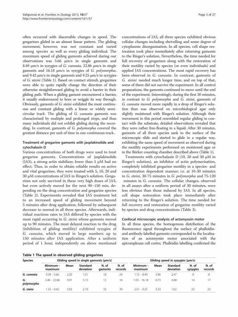
often occurred with discernible changes in speed. Thegregarines glided in an almost linear pattern. The glidingmovement, however, was not constant and variedamong species as well as every gliding individual. Themaximum speed of gliding gamonts achieved during ourobservations was 5.66 μm/s in single gamonts and8.49 μm/s in syzygies of G. cuneata, 22.86 μm/s in singlegamonts and 16.18 μm/s in syzygies of G. polymorpha,and 9.42 μm/s in single gamonts and 9.25 μm/s in syzygiesof G. steini (Table 1). Based on contact stimuli, gregarineswere able to quite rapidly change the direction of theirotherwise straightforward gliding to avoid a barrier in theirgliding path. When a gliding gamont encountered a barrier,it usually endeavoured to bore or wriggle its way through.Obviously, gamonts of G. steini exhibited the most continu-ous and constant gliding with a linear or widely semi-circular track. The gliding of G. cuneata gamonts wascharacterised by multiple and prolonged stops, and thusmany individuals did not exhibit gliding during the recor-ding. In contrast, gamonts of G. polymorpha covered thegreatest distance per unit of time in one continuous track.
Treatment of gregarine gamonts with jasplakinolide andcytochalasin DVarious concentrations of both drugs were used to treatgregarine gamonts. Concentrations of jasplakinolide(JAS), a strong actin stabilizer, lower than 5 μM had noeffect. Thus, in order to obtain reliable results on activeand vital gregarines, they were treated with 5, 10, 20 and30 μM concentrations of JAS in Ringer’s solution. Grega-rines not only survived in these very high doses of JAS,but even actively moved for the next 90–150 min, de-pending on the drug concentration and gregarine species(Table 2). Experiments revealed that JAS treatment ledto an increased speed of gliding movement beyond5 minutes after drug application, followed by subsequentdecrease to normal in all three species. Afterwards, indi-vidual reactions rates to JAS differed by species with themost rapid occurring in G. steini whose gamonts movedup to 90 minutes. The most delayed reaction to the drug(inhibition of gliding motility) exhibited syzygies ofG. cuneata, which moved in large numbers up to150 minutes after JAS application. After a uniformperiod of 1 hour, independently on above mentioned
Table 1 The speed in observed gliding gregarines
Species Gliding speed in single gamonts (μm/s)
Minimum -maximum
Mean Standarddeviation
N. ofgamonts
N.reco
G. cuneata 0.38 - 5.66 2.20 1.51 18 2
G.polymorpha
3.46 - 22.86 9.91 5.15 12 1
G. steini 1.32 - 9.42 5.02 2.19 33 3
concentrations of JAS, all three species exhibited obviouscellular changes including shrivelling and some degree ofcytoplasmic disorganisation. In all species, cell shape res-toration took place immediately after returning gamontsto the Ringer’s solution. Nevertheless, the time needed forfull recovery of gregarines along with the restoration oftheir motility varied by species (or even individuals) andapplied JAS concentrations. The most rapid recovery hasbeen observed in G. cuneata. In contrast, gamonts ofG. steini needed much longer time, and on top of that,some of them did not survive the experiment. In all controlpreparations, the gamonts continued to move until the endof the experiment. Interestingly, during the first 30 minutes,in contrast to G. polymorpha and G. steini, gamonts ofG. cuneata moved more rapidly in a drop of Ringer’s solu-tion than was observed on microbiological agar onlyslightly moistened with Ringer’s solution. Although theirmovement in this period resembled regular gliding in con-tact with the substrate, detailed observations revealed thatthey were rather free-floating in a liquid. After 30 minutes,gamonts of all three species sank to the surface of themicroscopic slide and started to glide in a regular way,exhibiting the same speed of movement as observed duringthe motility experiments performed on moistened agar orin the Bürker counting chamber described above (Table 1).Treatments with cytochalasin D (10, 20 and 30 μM in
Ringer’s solution), an inhibitor of actin polymerisation,completely inhibited gregarine motility in a species- andconcentration dependent manner; i.e. at 10-30 minutesin G. steini, 30-75 minutes in G. polymorpha and 75-120minutes in G. cuneata. The cellular changes, observedin all assays after a uniform period of 30 minutes, wereless obvious than those induced by JAS. In all species,cell shape restoration took place immediately afterreturning to the Ringer’s solution. The time needed forfull recovery and restoration of gregarine motility variedby species and drug concentrations (Table 3).
Confocal microscopic analysis of actomyosin motorIn all three species, the homogenous distribution of thefluorescence signal throughout the surface of phalloidin-and antibody-labelled gamonts corresponded to the localisa-tion of an actomyosin motor associated with theapicomplexan cell cortex. Phalloidin labelling confirmed the
Gliding speed in syzygies (μm/s)
ofrds
Minimum -maximum
Mean Standarddeviation
N. ofsyzygies
N. ofrecords
4 1.15 - 8.49 3.96 2.47 8 8
6 1.05 - 16.18 6.73 4.08 14 17
9 2.51 - 9.25 5.33 1.62 23 23

Table 2 The treatment of living gregarines with jasplakinolide
Species/JAS concentration Gregarina cuneata Gregarina polymorpha Gregarina steini
5 μM JAS 10 μM JAS 20 μM JAS 30 μM JAS 5 μM JAS 10 μM JAS 20 μM JAS 30 μM JAS 5 μM JAS 10 μM JAS 20 μM JAS 30 μM JAS
Changes/time left afterdrug application(in minutes)
Initial increase of glidingspeed
≥ 5 min+ ≥ 5 min+ ≥ 5 min++ ≥ 5 min++ ≥ 5 min+ ≥ 5 min+ ≥ 5 min++ ≥ 5 min++ ≥ 5 min+ ≥ 5 min+ ≥ 5 min++ ≥ 5 min++
Decrease of gliding speedto the normal rate
≥ 90 min+ ≥ 20 min++ ≥ 20 min++ ≥ 20 min++ ≥ 90 min+ ≥ 20 min++ ≥ 20 min++ ≥ 20 min++ ≥ 90 min+ ≥ 20 min++ ≥ 20 min++ ≥ 20 min++
Further decrease of glidingspeed
≥ 100 min+ ≥ 30 min++ ≥ 30 min++ ≥ 30 min++ ≥ 100 min+ ≥ 30 min++ ≥ 30 min++ ≥ 30 min++ ≥ 100 min+ ≥ 30 min+ ≥ 30 min++ ≥ 30 min++
Cellular changes(shrivelling)
≥ 60 min ≥ 60 min ≥ 60 min ≥ 60 min ≥ 60 min ≥ 60 min ≥ 60 min ≥ 60 min ≥ 60 min ≥ 60 min ≥ 60 min ≥ 60 min
Complete stoppage ofgliding motility
≤ 150 min ≤ 140 min ≤ 100 min ≤ 100 min ≤ 150 min ≤ 140 min ≤ 100 min ≤ 100 min ≤ 130 min ≤ 120 min ≤ 90 min ≤ 90 min
Recovery of cell shapeafter washing in Ringer’ssolution
≤ 1 min ≤ 1 min ≤ 1 min ≤ 1 min ≤ 1 min ≤ 1 min ≤ 1 min ≤ 1 min ≤ 1 min ≤ 1 min ≤ 1 min ≤ 1 min
Full recovery of motilityafter washing in Ringer’ssolution
≥ 5 min ≥ 5 min ≥ 5 min ≥ 5 min ≤ 10 min ≥ 10 min ≥ 10 min ≥ 10 min ≤ 30 min ≥ 30 min ≥ 30 min ≥ 30 min
The symbol + indicates an intensity of observed change ranging from + to +++ when compared among gregarine species and JAS concentrations. Applied only if the listed phenomenon showed significant differencesin its intensity.
Valigurováet
al.Frontiersin
Zoology2013,10:57
Page4of
27http://w
ww.frontiersinzoology.com
/content/10/1/57

Table 3 The treatment of living gregarines with cytochalasin D
Species/Cytochalasin D concentration Gregarina cuneata Gregarina polymorpha Gregarina steini
10 μM CytD 20 μM CytD 30 μM CytD 10 μM CytD 20 μM CytD 30 μM CytD 10 μM CytD 20 μM CytD 30 μM CytD
Changes/time left after drug application (in minutes)
Initial decrease of gliding speed ≥ 5 min+ ≥ 5 min++ ≥ 5 min+++ ≥ 5 min+ ≥ 5 min++ ≥ 5 min+++ ≥ 5 min+ ≥ 5 min++ ≥ 5 min+++
Further decrease of gliding speed ≥ 20 min+ ≥ 10 min++ ≥ 10 min+++ ≥ 15 min+ ≥ 10 min++ ≥ 10 min+++ ≥ 10 min+ ≥ 10 min++ ≥ 10 min+++
Cellular changes (shrivelling) ≥ 30 min ≥ 30 min ≥ 30 min ≥ 30 min ≥ 30 min ≥ 30 min ≥ 30 min ≥ 30 min ≥ 30 min
Complete stoppage of gliding motility ≤ 120 min ≤ 90 min ≤ 75 min ≤ 75 min ≤ 60 min ≤ 30 min ≤ 30 min ≤ 20 min ≤ 10 min
Recovery after washing in Ringer’s solution ≤ 1 min ≤ 1 min ≤ 1 min ≤ 1 min ≤ 1 min ≤ 1 min ≤ 1 min ≤ 1 min ≤ 1 min
Full recovery after washing in Ringer’s solution ≥ 5 min ≤ 10 min ≤ 10 min ≥ 5 min ≤ 10 min ≤ 10 min ≥ 5 min ≥ 10 min ≥ 10 min
The symbol + indicates an intensity of observed change ranging from + to +++ when compared between gregarine species and concentrations of cytochalasin D. Applied only if the listed phenomenon showedsignificant differences in its intensity.
Valigurováet
al.Frontiersin
Zoology2013,10:57
Page5of
27http://w
ww.frontiersinzoology.com
/content/10/1/57

Figure 1 Actin and myosin in Gregarina cuneata gamonts. A. Gamonts in syzygy; primite (p), satellite (s). LM, transmitted light. B. Localisationof F-actin in a gamont; nucleus (asterisk), septum (arrowhead) between protomerite and deutomerite. CLSM, phalloidin-TRITC. C-D. Localisation ofF-actin in gamonts (previously associated in syzygy) treated for 10 minutes with 10 μM JAS. Intense labelling is restricted to the cortex andcytoplasmic F-actin aggregations; septum (arrowhead), nucleus (asterisk). Figure C shows a primite. CLSM (left) and merged CLSM/transmittedlight (right), phalloidin-TRITC. Figure D shows a satellite. CLSM, phalloidin-TRITC. E. The deutomerite of a gamont treated for 10 minutes with10 μM JAS. F-actin localisation corresponds to the cortex and nucleus (asterisk). Upper two figures show the gamont middle plane; lower figure shows thecortex in the area of epicytic folds. Merged CLSM/transmitted light (upper) and CLSM, phalloidin-TRITC. F. F-actin localisation in a gamont treated for 150minutes with 10 μM JAS. Note the decreased labelling of cell cortex and septum (arrowhead), and formation of numerous cytoplasmic aggregations ofF-actin; nucleus (asterisk). CLSM, phalloidin-TRITC. G. The deutomerite of a gamont treated for 150 minutes with 10 μM JAS. F-actin labelling is restricted tothe cortex in the area of epicytic folds (lower); cytoplasmic F-actin aggregations (upper). CLSM, phalloidin-TRITC. H. Actin localisation in previouslyassociated gamonts; septum (arrowheads). CLSM, IFA. I. Actin localisation in a gamont ghost; nucleus (asterisk), septum (arrowhead). CLSM, IFA. J. Myosinlabelling in a maturing gamont. CLSM, IFA. K. Myosin labelling in a mature gamont is restricted to the cortex, but not to the septum (arrowhead). MergedCLSM/transmitted light, IFA. L. Labelling of myosin in a gamont cortex shows a pattern of longitudinal rows; septum (arrowhead). CLSM, IFA. Figures H, Iand J show merged FITC (antibody) and rhodamine (counterstaining with Evans blue) fluorescence channels.
Valigurová et al. Frontiers in Zoology 2013, 10:57 Page 6 of 27http://www.frontiersinzoology.com/content/10/1/57
presence of filamentous actin (F-actin) in the gregarine cellcortex, the fibrillar septum separating the protomerite fromthe deutomerite and the area of the nucleus (Figures 1B, 2Band 3B). After treatment with 10 μM JAS for 10 minutes,when gregarines glided with increased speed, the F-actinstaining became more bright and confined to the cell
cortex, the septum and the perinuclear space (Figures 1C-E,2C-E and 3C-D). Higher magnification revealed numeroustransverse and oblique actin filaments in the area of epicyticfolds (Figures 1E, 2E and 3D). In addition, several aggrega-tions of F-actin were observed in the cytoplasm of G.cuneata protomerite and deutomerite (Figure 1C-D). This

Figure 2 Actin and myosin in Gregarina polymorpha gamonts. A. Gamonts in syzygy; primite (p), satellite (s). LM, transmitted light. B. F-actinlocalisation in a gamont; nucleus (asterisk), septum (arrowhead). CLSM, phalloidin-TRITC. C-D. F-actin localisation in gamonts (previously associated insyzygy) treated for 10 minutes with 10 μM JAS. The more intense labelling is restricted to the cortex; septum (arrowhead), nucleus (asterisk). Figure Cshows a primite. CLSM (left) and merged CLSM/transmitted light (right), phalloidin-TRITC. Figure D shows a satellite. CLSM, phalloidin-TRITC. E. Thedeutomerite of a gamont treated for 10 minutes with 10 μM JAS. F-actin localisation corresponds to the cortex; nucleus (asterisk). Upper two figuresshow the gamont middle plane; lower figure shows the cortex in the area of epicytic folds. Merged CLSM/transmitted light (upper) and CLSM,phalloidin-TRITC. F. F-actin localisation in a gamont treated for 150 minutes with 10 μM JAS, showing decreased labelling of cortex and septum(arrowhead). Numerous cytoplasmic F-actin aggregations give the labelling homogenous appearance. CLSM, phalloidin-TRITC. G. The deutomeriteof a gamont treated for 150 minutes with 10 μM JAS; rosette-like aggregations of F-actin (arrows), nucleus (asterisk). Merged CLSM/transmitted light(upper) and CLSM (lower), phalloidin-TRITC. H. Actin localisation in a gamont; nucleus (asterisk), septum (arrowhead). The indistinct labelling (green)is more evident in the cortex covering the anterior part of cell. CLSM, IFA. I–J. Myosin localisation in a gamont. The labelling is restricted to the cortex,with a pattern of longitudinal rows; septum (arrowhead). CLSM, IFA. K. The anterior part of the gamont shown in J. Myosin localisation is restricted tothe cortex, but not to the septum (arrowhead). Merged CLSM/transmitted light, IFA. L. Myosin localisation in a gamont ghost; septum (arrowhead).CLSM, IFA. Figures H, J and L show merged FITC (antibody) and rhodamine (counterstaining with Evans blue) fluorescence channels.
Valigurová et al. Frontiers in Zoology 2013, 10:57 Page 7 of 27http://www.frontiersinzoology.com/content/10/1/57
was accompanied by an obvious decrease of diffuse F-actinin all analysed gregarines. The localisation of F-actin did notsignificantly change even after 150 minutes incubation in10 μM JAS, when gregarines completely stopped their
movement and showed obvious cellular changes, but the in-tensity of cell cortex labelling significantly decreased andnumerous small aggregations of F-actin appeared in the cellcytoplasm (Figures 1F-G, 2F-G and 3E). The transverse

Figure 3 Actin and myosin in Gregarina steini gamonts. A. Gamonts associated in syzygy; primite (p), satellite (s). LM, transmitted light.B. Localisation of F-actin in three single gamonts; nucleus (asterisk), septum (arrowheads). CLSM, phalloidin-TRITC. C. Localisation of F-actin ingamonts associated in syzygy treated for 10 minutes with 10 μM JAS. The intense labelling is restricted to the cortex, septum (arrowheads) andnucleus (asterisks). CLSM (left) and merged CLSM/transmitted light (right), phalloidin-TRITC. D. The deutomerite of a gamont treated for 10minutes with 10 μM JAS. The localisation of F-actin corresponds to the cortex. Upper two figures show the gamont middle plane; lower figure isa view of the cortex in the area of epicytic folds. Merged CLSM/transmitted light (upper) and CLSM, phalloidin-TRITC. E. Localisation of F-actin ingamonts associated in syzygy treated for 150 minutes with 10 μM JAS. Note the decreased labelling of cell cortex and septum (arrowheads);nucleus (asterisks). Numerous indistinct, small cytoplasmic aggregations of F-actin give the labelling a more homogenous appearance. CLSM, phalloidin-TRITC. F. Actin localisation in gamonts associated in syzygy; nucleus (asterisks). The septum (arrowhead) exhibits an intense labelling. CLSM, IFA. G.Myosin labelling in gamonts associated in syzygy; septum (arrowhead). CLSM, IFA. H. An optical plane of the gamont cortex showing the distribution ofmyosin corresponding to the epicytic folds; septum area (arrowhead). CLSM, IFA. I. Myosin localisation in a single gamont. The labelling is restricted tothe cortex and exhibits a pattern of longitudinal rows. A slight labelling in the septum area is shown (arrowhead). Merged CLSM/transmitted light (left)and CLSM (right), IFA. Figures F, G and H show merged FITC (antibody) and rhodamine (counterstaining with Evans blue) fluorescence channels.
Valigurová et al. Frontiers in Zoology 2013, 10:57 Page 8 of 27http://www.frontiersinzoology.com/content/10/1/57
actin filaments in epicytic folds appeared to fuse into a homo-geneous layer. In G. polymorpha, rosette-like aggregations ofF-actin were situated in the cell periphery (Figure 2G).Gamonts stained with the specific anti-actin anti-
body (known to recognise the actin in Toxoplasma and
Plasmodium) exhibited a similar actin localisation, how-ever, in comparison with the phalloidin-stained specimens,only slight labelling of actin was associated with thecell cortex and the septum in G. cuneata (Figure 1H–I)and G. polymorpha (Figure 2H). Gamonts of G. steini

Valigurová et al. Frontiers in Zoology 2013, 10:57 Page 9 of 27http://www.frontiersinzoology.com/content/10/1/57
exhibited a higher intensity of actin labelling with thisantibody, and gamonts associated in syzygy labelled with adifferent intensity in that a higher concentration of actinwas observed in the primite (Figure 3F).Similarly to actin, the myosin was restricted to the cell
cortex (Figures 1J–L, 2I–L and 3G–I), but no specific la-belling corresponding to the septum was observed. Whenfocussing on the gregarine surface, the organisation of my-osin exhibited a pattern of longitudinal rows correspond-ing to the epicytic folds (Figures 1L, 2I–J and 3H-I). Theprimites and satellites in syzygies (Figure 3G) exhibitedmore or less identical intensity of labelling.The results of immunofluorescent labelling using an anti-
α-tubulin antibody were negative (data not shown) inagreement with the absence of subpellicular microtubules.
Mucus sheddingOnly phase contrast microscopy was able to showmucus shedding and trail formation of gamonts glidingon agar (Figure 4A–F). The most evident mucous trailswere that of associated (Figure 4C) or single (Figure 4D)gamonts of G. polymorpha due to the distance travelledby them, which left long and regular mucous paths. Incontrast, G. cuneata (Figure 4A) and G. steini (Figure 4E)gliding gamonts exhibited irregular and short pathscontaining greater accumulations of mucus, especiallywhen these species were avoiding barriers or alteringdirection (Figure 4B and F).The mucous substances were visualized by Alcian blue
staining within the gamont cytoplasm (Figure 4G–I).The most intense labelling occurred in the deutomeritecytoplasm of G. polymorpha (Figure 4H). In contrast toG. cuneata (Figure 4G) and G. steini (Figure 4I), thisgregarine showed an increased volume of mucus in theprotomerite cytoplasm, which might be correlated withthe increased motility of the G. polymorpha protomerite.
Ultrastructural features of the pellicleThe pellicle in all the species was organised in numerouslongitudinal narrow folds, which were more raised in thedeutomerite region than in the protomerite. The epicyticfolds of the deutomerite were more or less undulated,depending on the species. Similarly, the number ofepicytic folds per square micrometer varied among spe-cies, and their number did not significantly changed inthe course of gamont growth (Table 4). The gamontsG. cuneata were covered by almost linear folds and nu-merous mucus-like drops were often present in the groovesseparating them (Figure 5A–F). Occasionally, rings of un-dulated folds could be observed, especially in the posteriorhalf of the gamont deutomerite (Figure 5E). The pellicleappeared to be slightly helically coiled along the gregarinelongitudinal axis, resulting in a helical course of epicyticfolds (Figure 5A, the syzygy on the right). The pellicle
of G. polymorpha gamonts exhibited zones of almostlinear folds alternating with zones of much undulatedfolds; however, surprisingly when considering the results ofthe Alcian blue staining, only a few mucus drops could bedetected (Figure 6A–E). The epicytic folds of G. steini wereundulated in a more regular pattern than in G. polymorpha,and numerous mucus-like drops covered the entire surfaceof the gamonts (Figure 7A–G). Depending on the species,more or less evident constriction could be found at theinterface between the protomerite and deutomerite; how-ever, no interruption of folds, running from the gamontapical to its posterior end, was present in this area(Figures 5A, 6A and 7A). Syzygies were caudo-frontal,i.e. the posterior end of the primite deutomerite wasjoined with the apical part of the satellite protomerite(Figures 5A, 6A and 7A). The connection area appearedas a collar-like junction composed of modified epicyticfolds of the primite deutomerite meshing in a gear-likemanner with the folds of the sucker-like apical regionof the satellite (Figures 5B–C, 6B–C and 7B–D). InG. cuneata, two or more satellites were often found to beassociated with one primite (Figure 5A and E). In somecases, a large primite was associated with several tiny satel-lites (up to four satellites associated with one primite wereobserved). Occasionally, three individuals of G. cuneatawere seen to be associated in a row, the last one of whichwas the smallest.The folded pellicle was three-layered, composed of the
superficial plasma membrane covering the entire grega-rine and a middle lucent region, underlined by two dis-tinct and tightly apposed membranes, i.e., external andinternal cytomembrane, forming the inner membranecomplex (IMC). Epicytic folds emerged from the periph-eral ectoplasm bounding the endoplasm. Three types ofassociated structures were constantly present in eachfold: an internal lamina, 12-nm filaments and rippleddense structures. The internal lamina, running under theIMC, not only linked the bases of epicytic folds but alsobifurcated just beneath each fold, and its thinner part ex-tended to the individual folds (Figures 5G, 6G and 7I).The localisation along with the organisation of internallamina suggest its function in the stabilisation of indivi-dual folds as well their interconnection. The thickness ofthe internal lamina varied by species; i.e., 50–75 nm inG. polymorpha (Figure 6F–G), 17–30 nm in G. cuneata(Figure 5G–H) and 8–11 nm in G. steini (Figure 7H–I).The compact organisation of the internal lamina usuallydisappeared when reaching the region of 12-nm filaments.In fact, the 12-nm filaments seemed to be embedded inthe widened area of the internal lamina (Figures 5G, 6Gand 7I). The 12-nm filaments, exhibiting the propertiesof intermediate filaments, ran under the IMC alongthe longitudinal axis of each fold. Their numbers variedby species and developmental stage, i.e., in gamonts of

Figure 4 Gliding motility and mucus. A–F. Mucous trail (arrowheads) left behind gliding gregarines; syzygy of Gregarina cuneata (A, B),syzygy (C) and single gamont (D) of Gregarina polymorpha, syzygy of Gregarina steini (E, F). Note the increased mucus shedding by the syzygy ofG. cuneata exhibiting rotary movement (B). G–I. Light micrographs showing the volume of mucus in gamonts of G. cuneata (G), G. polymorpha(H) and G. steini (I) revealed with Alcian blue staining at low pH.
Table 4 The number of epicytic folds in deutomerite of gamonts
Species G. cuneata G. polymorpha G. steini
Gamont N. Number offolds/μm2
Deutomeriteperimeter (μm)
Number offolds/μm2
Deutomeriteperimeter (μm)
Number offolds/μm2
Deutomeriteperimeter (μm)
1 4.5 53.4 4.3 94.3 5.6 31.4
2 4.4 98.1 4.2 103.7 6.3 34.6
3 3.9 113.1 4.9 106.8 6.3 37.7
4 3.6 114.0 4.3 110.0 5.9 58.0
5 3.2 150.9 4.5 132.0 5.0 66.0
6 3.3 163.4 3.6 144.5 4.0 113.1
Valigurová et al. Frontiers in Zoology 2013, 10:57 Page 10 of 27http://www.frontiersinzoology.com/content/10/1/57

Figure 5 Pellicle architecture in Gregarina cuneata gamonts. A. Gamonts associated in syzygy; primite (p), satellite (s). SEM. The syzygy on theright is composed of one primite (p) and two satellites (s1, s2). B. Higher magnification of the junction between the posterior end of the primitedeutomerite (d) and the apical part of the satellite protomerite (p). SEM. C. A detail of the junction (arrow) between folded pellicles covering theprimite deutomerite (def) and satellite protomerite (pef). SEM. D. Organisation of linear epicytic folds covering the deutomerite. SEM. E. Highermagnification of the junction between the primite (p) and two satellites (s1, s2) shown in panel A. SEM. F. A higher magnification of deutomeriteepicytic folds (ef); grooves (g) between folds, mucus drops (*). SEM. G. Cross section of deutomerite epicytic folds; grooves (g) with mucus (*)between folds (ef), 12-nm filaments (arrowhead), inner membrane complex (imc), internal lamina (double arrowhead), plasma membrane (pm),rippled dense structures (white arrowhead), unknown dense structure (arrow). TEM. H. Cross section of deutomerite epicytic folds (ef); internallamina (double arrowhead), rib-like myonemes (arrowheads). TEM. I. Detailed view of an epicytic fold in cross section revealing filamentousconnections (arrowheads) localised between the plasma membrane (pm) and inner membrane complex (imc). TEM. J. Cross section showing theorganisation of the deutomerite pellicle; ectoplasmic network (arrows), epicytic folds (ef), duct (*), rib-like myonemes (arrowheads). The insectshows the micropore located in the groove between two epicytic folds. TEM. K. Organisation of the pellicle and ectoplasmic network (n) duringgregarine movement; epicytic folds (ef), deutomerite (d), protomerite (p). TEM. L. Higher magnification of a septum (arrow) separating theprotomerite (p) from the deutomerite (d); epicytic folds (ef). Note the ectoplasmic network (n) connected to the septum. TEM.
Valigurová et al. Frontiers in Zoology 2013, 10:57 Page 11 of 27http://www.frontiersinzoology.com/content/10/1/57
G. cuneata up to 7 (Figure 5G) and in G. polymorphaup to 10 filaments were observed (Figure 6G), while inG. steini only 2–4 filaments could be seen (Figure 7I).Their number increased with fold maturation, i.e., new
rising folds contained fewer filaments than the olderand evidently higher epicytic folds. Rippled dense struc-tures, located between the plasma membrane and IMC,appeared as electron-dense, triangle-shaped structures

Figure 6 Pellicle architecture in Gregarina polymorpha gamonts. A. Gamonts associated in syzygy; primite (p), satellite (s). SEM. B. Highermagnification of the junction between the posterior end of the primite deutomerite (d) and apical part of the satellite protomerite (p). SEM.C. Detailed view of the junction (arrows) between folded pellicles covering the primite deutomerite (def) and the satellite protomerite (pef). SEM.D. Organisation of undulated epicytic folds covering the deutomerite. SEM. E. Higher magnification of deutomerite epicytic folds (ef); grooves (g)between folds. SEM. F. Cross section of the deutomerite pellicle; epicytic folds (ef), grooves (g), internal lamina (double arrowhead), rib-likemyonemes (arrowhead). TEM. G. Cross section of deutomerite epicytic folds; 12-nm filaments (arrowhead), inner membrane complex (imc),internal lamina (double arrowhead), plasma membrane (pm), rippled dense structures (white arrowhead), unknown dense structure (arrow). TEM.H. Detailed view of epicytic folds in cross section revealing filamentous connections (arrowheads) localised between the plasma membrane (pm)and inner membrane complex (imc). TEM. I. Pellicle organisation revealed in a mechanically ruptured gamont; cytoplasm (c) with ectoplasmicnetwork, epicytic folds (ef). SEM. J. The view of an ectoplasmic face (*) of a pellicle separated from the gamont cytoplasm; epicytic folds (ef). SEM.K. The septum (arrow) separating the protomerite (p) from the deutomerite (d); epicytic folds (ef). TEM.
Valigurová et al. Frontiers in Zoology 2013, 10:57 Page 12 of 27http://www.frontiersinzoology.com/content/10/1/57
with base lying on the external cytomembrane and medianrunning in between two adjacent 12-nm filaments. Theirnumber varied by species and developmental stages,in correlation with the number of 12-nm filaments(Figures 5G, 6G and 7I). Unknown dense and usuallyhalf-moon-shaped structures underlined the 12-nm
filaments at their cytoplasmic face in all gregarines. Thisstructure achieved its maximum length in G. polymorpha,in which its ends were in obvious contact with the internallamina extending to the top of the fold (Figure 6G). InG. cuneata, this structure was evidently shorter andthicker (Figure 5G), and in G. steini it was shortest and in

Figure 7 Pellicle architecture in Gregarina steini gamonts. A. Gamonts associated in syzygy; primite (p), satellite (s). SEM. B. Highermagnification of the junction between the posterior end of the primite deutomerite (d) and apical part of the satellite protomerite (p). SEM.C, D. Detailed views of the junction (arrow) between folded pellicles covering the primite deutomerite (def) and satellite protomerite (pef). SEM.E, F. Organisation of more and less undulated epicytic folds covering the deutomerite. SEM. G. Higher magnification of deutomerite epicytic folds(ef); grooves (g) between folds, pore-like structure (arrow). SEM. H. Organisation of the deutomerite pellicle in cross section; epicytic folds (ef),grooves with mucus (*), internal lamina (double arrowhead). TEM. I. Cross section of deutomerite epicytic folds. Note the filamentous connections(white arrows) localised between the plasma membrane (pm) and inner membrane complex (imc); 12-nm filaments (arrowhead), rippled densestructures (white arrowhead), unknown dense structure (arrow). TEM. J. The septum (arrow) and ectoplasmic network (n); deutomerite (d),epicytic folds (ef), protomerite (p). TEM. K. Golgi apparatus in deutomerite cytoplasm. TEM. L. The duct (arrow) opening outwards to the groovebetween epicytic folds. TEM.
Valigurová et al. Frontiers in Zoology 2013, 10:57 Page 13 of 27http://www.frontiersinzoology.com/content/10/1/57
some sections it was even reversed with its ends facingthe apical top of the fold (Figure 7I). As the internallamina lacked its typical compact look in this area, itis possible that the mentioned half-mooned structuresrepresent its component. Careful analysis of the pelliclecovering the lateral part of epicytic folds revealed novel
thin filamentous connections interconnecting the IMCand the plasma membrane (Figures 5I, 6H and 7I).In addition to structures restricted to the epicytic
folds, an ectoplasmic network and rib-like myonemes,present to some degree in the deutomerite ectoplasm ofall studied species, could be observed in some ultrathin

Valigurová et al. Frontiers in Zoology 2013, 10:57 Page 14 of 27http://www.frontiersinzoology.com/content/10/1/57
sections. The ectoplasmic network contacting the basesof the epicytic folds was most prominent in fully ma-tured gamonts of G. cuneata (Figure 5J–L), especially atthe septum periphery or in areas with an obviouslypleated pellicle (Figure 5K). This local pleating of thepellicle seemed to be the result of gregarine movement.The rib-like myonemes, running perpendicularly to thelongitudinal axis of the gregarine and located beneath thedeutomerite ectoplasm, were very distinct in gamontsof G. cuneata (Figure 5H and J) and G. polymorpha(Figure 6F), but were hard to detect and often absentin G. steini. The septum separating the protomerite fromthe deutomerite was well developed in all three species(Figures 5L, 6K and 7J). The micropores, interruptingthe IMC, were sometimes seen in the grooves betweenthe folds of G. cuneata (Figure 5J). In addition, ducts,appearing as elongated dense sacs passing through thepellicle and opening outwards, were often present in theectoplasm of G. cuneata and G. steini gamonts (Figures 5Jand 7L).
Membranes as exposed by freeze etchingThe freeze-etching technique confirmed the presence ofthree fracture planes in the pellicle of all analysed grega-rines, corresponding to the three membranes, i.e., twocytomembranes (IMC) underlying the plasma mem-brane. The general aspects of the epicytic folds in freeze-fracture are shown in Figures 8, 9 and 10, and theirarchitecture corresponded to the TEM observations. Atthe tip of each fold, double linear rows of tightly alignedintramembranous particles (IMP) were observed in bothfractures of the external and internal cytomembranes.These rows seemed to correspond to the apical site ofthe 12-nm filaments and to the base of the triangle-shaped, rippled dense structures. The number of theselines roughly coincided with the number of 12-nm fila-ments. The freeze-etching approach proved to be astrong tool for visualisation of structures that are hardlydocumented on TEM micrographs including roundmicropores (110–130 nm in diameter) at the base ofthe grooves between the folds (Figures 8A, 8G, 9E, 9Gand 10E) or smaller pores (35–50 nm in diameter) ran-domly distributed on the base or on the lateral side of thefolds (Figures 8A–D, 8F-G, 9A–E, 9G, 10A, 10C and10E–F), ducts and cisternae often connected these poresor to the pellicle (Figures 8B, 8E, 8H, 9F–G and10A), as well as mucus drops often present in the groovesbetween the folds of G. cuneata (Figure 8B) and G. steini(Figure 10A–D and F).The densities of the IMP for the fracture faces of the
plasma membrane and both cytomembranes aresummarised in Table 5. The density of the IMP in mem-branes differed in analysed gregarines; in general, thevalues in G. polymorpha were significantly lower in
comparison to G. cuneata and G. steini. In G. cuneataand G. polymorpha, the IMP density in the IMC waslower than in the plasma membrane. In G. steini, how-ever, the IMP density in the plasma membrane waslower than those in the IMC. The IMP in all three mem-branes showed a high variability in their size distribution(see histograms in Figures 11, 12 and 13); nevertheless,only particles in a range of 6–14 nm were included instatistical calculations to obtain data comparable withthose previously published on other apicomplexans.Membranes forming the pellicle in G. cuneata, especiallythe plasma membrane, were the most extraordinarygiven the wide range of the IMP size along with its dis-tribution (Figure 11).
DiscussionGliding movement is a feature observed in a wide rangeof unicellular organisms including diatoms, flagellates,apicomplexan zoites, and gregarines. The speed of theseorganisms varies along with the special locomotivestructures and is affected mostly by their physiologicalstatus and surrounding environmental conditions. Thereported motility rates in Apicomplexa are usually in therange of 1–10 μm/s, and the maximal rate was observedin gregarines [34]. To minimise a potential effect of differ-ent environmental conditions on gregarine motility in thisstudy, we took advantage of a naturally mixed infectionwith three Gregarina species occurring in the intestine oflarval mealworms kept under laboratory conditions. Inaddition, the experimental part of this work, including thelight microscopic observations on gliding and treatmentof living gamonts with JAS and cytochalasin D, has beenperformed on suspensions consisting of all three species(often from a single host). Göhre [35] states that grega-rines parasitising the intestine of larval T. molitor are dis-tributed based on intestinal pH, i.e., G. cuneata inhabits apart of the intestine with pH 4.5–5.5, and G. steini(pH 5.5–8.2) inhabits a part of the intestine together withG. polymorpha (pH 6.4–7.5). Therefore, it could beexpected that the mixed suspensions of gamonts are notpreferable for motility assays, nevertheless, pH-restrictedlocalisation of gregarines in mealworms applies only to at-tached trophozoites [36] not to gamonts that are usuallyfound in luminal part of the mesenteron. During our ob-servations, eugregarines isolated from one host were glid-ing at speeds ranging from 0.38 to 22.86 μm/s, while thehighest speed was reached by gamonts of G. polymorphaand the lowest by G. cuneata. To explain these evidentdifferences study focussed on structures that were gene-rally considered to be responsible for gregarine gliding -epicytic folds and mucus. Longitudinal folds formed bythe pellicle represent the most conspicuous feature diffe-rentiating eugregarine trophozoites and gamonts from theother apicomplexans. The presence of the swellings along

Figure 8 Pellicle organisation in Gregarina cuneata gamonts as revealed by the freeze-etching. A. The general view of fractured epicyticfolds; cytoplasm (c), micropore (encircled), cytoplasm of folds (*), groove (g), EF face of external cytomembrane (Ee), EF face of internalcytomembrane (Ei), IMP alignments (arrowhead), PF face of external cytomembrane (Pe), PF face of internal cytomembrane (Pi), plasmamembrane (arrow), pores (white arrows). B. The base of the epicytic folds; cytoplasm of folds (*), deutomerite cytoplasm (c), duct (encircled),grooves (g), mucus (x), PF face of the external cytomembrane (Pe), pores (white arrows). C, D. The fracture of the epicytic fold; cytoplasm (c),EF of the external cytomembrane (Ee), EF face of the internal cytomembrane (Ei), PF face of the external cytomembrane (Pe), PF face of theinternal cytomembrane (Pi), pores (white arrows), IMP alignments (arrowheads). E. The base of the fold with a duct opening outwards (arrow) tothe groove; deutomerite cytoplasm (c), EF of the external cytomembrane (Ee), PF face of the external cytomembrane (Pe), PF face of the internalcytomembrane (Pi). F. The longitudinal fracture of the fold; cytoplasm (c), cytoplasm of fold (*), PF face of the external cytomembrane (Pe),PF face of the internal cytomembrane (Pi), pores (white arrows). G. The grooves (g) between the folds (ef); cytoplasm (c), mucus (x), numerouspores (some of them are shown by white arrows), PF face of the external cytomembrane (Pi). The inset shows a micropore and a pore of smallersize. H. A detail of the grove between folds (ef) showing the part of micropore (arrow) with vesicle (v); deutomerite cytoplasm (c). I. The top ofthe fold; EF face of the plasma membrane (Ep), IPM alignments (arrowheads), PF face of the external cytomembrane (Pe). The arrowhead in thecircle shows the direction of shadowing.
Valigurová et al. Frontiers in Zoology 2013, 10:57 Page 15 of 27http://www.frontiersinzoology.com/content/10/1/57
the longitudinal epicytic folds of gliding gregarines sug-gests that these structures, pushed against the substrate,may provide the force for gliding [33], and the mucusseems to enhance the efficiency of their interaction with
the substrate to produce gregarine forward movement. Itis important, however, to mention that gregarine gamontsin performed experiments were able to move without anycontact with the substrate when put in a drop of Ringer’s

Figure 9 Pellicle organisation in Gregarina polymorpha gamonts as revealed by the freeze-etching. A. The general view of fracturedepicytic folds; cytoplasm of folds (*), deutomerite cytoplasm (c), EF face of external cytomembrane (Ee), IMP alignments (arrowhead), groove (g),PF face of external cytomembrane (Pe), PF face of internal cytomembrane (Pi), pores (white arrows). B. The fractured epicytic fold; deutomeritecytoplasm (c), EF face of the internal cytomembrane (Ei), groove (g), PF face of the external cytomembrane (Pe), pores (white arrows). C. Thefracture of the lower epicytic fold; cytoplasm of folds (*), deutomerite cytoplasm (c), EF face of the internal cytomembrane (Ei), EF face of theplasma membrane (Ep), IMP alignments (arrowhead), PF face of the internal cytomembrane (Pi), pore (white arrow). D. The fracture of higherepicytic folds; cytoplasm of folds (*), deutomerite cytoplasm (c), EF of the internal cytomembrane (Ei), EF of the plasma membrane (Ep), groove(g), IMP alignments (arrowhead), PF face of the internal cytomembrane (Pi), pore (white arrow). The inset shows the IMP alignments located onthe EF face of the external cytomembrane (white arrowhead) and on the PF of the internal cytomembrane (arrowhead) at the top of the epicyticfold. E. The grooves (g) between epicytic folds with numerous pores (some of them shown by white arrows); cytoplasm of folds (*), deutomeritecytoplasm (c), EF face of the internal cytomembrane (Ei), PF face of the external cytomembrane (Pe). Note the large opened micropore (arrow).The inset shows a micropore and three pores of different sizes. F. The base of the epicytic fold (ef) with a duct (encircled); cytoplasm (c). G. Thebase of the epicytic folds showing the micropore connected to a vesicle (encircled); cytoplasm (c), pores (white arrows). The arrowhead in thecircle shows the direction of shadowing.
Valigurová et al. Frontiers in Zoology 2013, 10:57 Page 16 of 27http://www.frontiersinzoology.com/content/10/1/57
solution, i.e., for approximately 30 min, they were floatingin a liquid until sinking to the surface of microscopic slide.These observations conflict with the supposed need oftheir contact with some solid matter [37]. Although some
correlations can be found between speed and the numberof epicytic folds, i.e., gamonts of G. cuneata are quippedby fewer folds (3.2–4.5) per square micrometer than smallgamonts of G. steini (4.0–6.3) or similarly-sized gamonts

Figure 10 Pellicle organisation in Gregarina steini gamonts as revealed by the freeze-etching. A–C. Fractured epicytic folds; cytoplasmof epicytic folds (*), deutomerite cytoplasm (c), ducts (encircled), EF face of the external cytomembrane (Ee), EF face of the internalcytomembrane (Ei), EF face of the plasma membrane (Ep), groove (g), mucus (x), PF face of the external cytomembrane (Pe), PF face of theinternal cytomembrane (Pi), PF face of the plasma membrane (Pp), pores (white arrows). D. General view of the epicytic folds showing thegrooves (g) with mucus drops (x); cytoplasm of epicytic folds (*), deutomerite cytoplasm (c). E. The cytoplasmic face of the grooves betweenepicytic folds with micropores and numerous pores (some of them shown by white arrows); cytoplasm of epicytic folds (*), EF face of the internalcytomembrane (Ei), PF face of the external cytomembrane (Pe). The inset shows the detailed view of micropore and four pores of different sizes.F. Fractured epicytic folds showing their IMP alignments (arrowheads) located on the PF of the internal cytomembrane (Pi) and on the EF faceof the external cytomembrane (Ee); cytoplasm of epicytic folds (*), EF face of internal cytomembrane (Ei), grooves (g), PF face of externalcytomembrane (Pe), pores (white arrows). The inset shows EF of the plasma membrane (Ep) with mucus drops (x). The arrowhead in the circleshows the direction of shadowing.
Valigurová et al. Frontiers in Zoology 2013, 10:57 Page 17 of 27http://www.frontiersinzoology.com/content/10/1/57
of G. polymorpha (3.6–4.9), these differences were onlyslight and we do not consider them to significantly influ-ence the gliding rate. More important, however, seem to beundulations of folds documented by SEM. In accordancewith Vávra and Small [24], the folds of glutaraldehyde-fixedG. cuneata were almost linear, forming occasional rings of
undulated folds, while the pellicle of G. polymorpha formedzones of undulated as well as almost linear folds, and thefolds in G. steini were undulated in quite a regular pattern.Microscopic observations indicate that the lateral undula-tions of folds arise during gliding. Nevertheless, the ques-tion of whether epicytic folds might be responsible for

Table 5 A protein particle density in the fracture faces of the membranes of studied gregarines
Species Gregarina cuneata Gregarina polymorpha Gregarina steini
Membrane Face Density = number ofparticles/μm2 (± SE)
Kp Density = number ofparticles/μm2 (± SE)
Kp Density = number ofparticles/μm2 (± SE)
Kp
Plasma membrane PF 2244 ± 283 0.81 1446 ± 158 0.59 2265 ± 154 1.27
EF 2770 ± 96 2473 ± 147 1783 ± 233
Externalcytomembrane
PF 1420 ± 190 1.13 602 ± 265 0.70 2588 ± 189 0.68
EF 1260 ± 211 863 ± 202 3820 ± 211
Internalcytomembrane
PF 1993 ± 253 1.33 1276 ± 200 1.57 2339 ± 132 1.24
EF 1502 ± 273 814 ± 246 1886 ± 274
The size of IMP is in range 6-14 nm.Kp partition coefficient defined as the ratio of number of particles per μm2 in the PF face/number of particles per μm2 in the EF face.SE standard error.
Valigurová et al. Frontiers in Zoology 2013, 10:57 Page 18 of 27http://www.frontiersinzoology.com/content/10/1/57
gliding in gregarines can be satisfactorily answered onlyafter careful analysis of all subcellular components formingthe complicated pellicle in gregarines.
The ‘glideosome’ concept and eugregarine glidingFirst, it must be highlighted that no similar structuresresembling epicytic folds can be found on the surfaceof the zoite stage (e.g., sporozoites in gregarines orPlasmodium, tachyzoites of Toxoplasma), which areused to illustrate the mechanism of gliding motility inapicomplexans. The concept of the ‘glideosome’ [4] de-scribes apicomplexan zoites as actively entering host cellsand moving by a substrate-dependent gliding motility,which requires coordinated interactions between parasitesurface adhesins and its cytoskeleton. This machinery isconsidered an unusual form of eukaryotic locomotion.The so-called actomyosin motor, which is generally as-sumed to be embedded between the plasma membraneand the IMC, consists of immobilised unconventionalmyosins, short actin stubs, and TRAP-family invasins.This motor is expected to be oriented by subpellicular mi-crotubules [38]. Micronemal proteins, inserted into theplasma membrane, are carried along the IMC by themotor and interact with the parasite substrate, or associatewith a GPI-anchored protein interacting with the sub-strate, resulting in gliding [38].Considering the possible application of this concept
for eugregarine gliding, the first striking inconsistency isthe fact that in the eugregarines analysed here, there areno subpellicular microtubules in the epicytic folds (con-firmed also by negative results of immunolabelling).Thus, a question arises concerning the real motor in theirmotility. It could be expected that enigmatic 12-nm fila-ments, running under the IMC and exhibiting the proper-ties of intermediate filaments [34,39], could support theactomyosin motor in a similar way. Longitudinal arraysof IMP found in the area of the 12-nm filaments andthe rippled dense structures [11,22,30,40] are compar-able to the lines of higher particle density overlaying thesubpellicular microtubules in Eimeria or Plasmodium
sporozoites [41,42]. Our data show, however, that thenumber of 12-nm filaments does not influence the speedof gregarine gliding (up to 7 filaments in G. cuneata vs. 10filaments in G. polymorpha and maximally 4 filaments inG. steini), but rather seem to control the direction ofmovement. Indeed, despite folds equipped by a low num-ber of 12-nm filaments, gamonts of G. steini glided withrelatively high speed, but their gliding path was ratherwidely semi-circular than linear. The question remainswhether apical rippled dense structures, with their base lo-cated at the external cytomembrane, serve as supportingelements interconnecting 12-nm filaments and the plasmamembrane. Such speculation is supported by the existenceof filamentous interconnections occasionally observed be-tween their tips and the plasma membrane in ultrathinsections [39]. The half-moon-shaped dense structureunderlining the 12-nm filaments could play the role of a‘skeleton’ reinforcing the tips of folds, which contact thesubstrate during gliding.The apicomplexan gliding motility relies on the dy-
namic turnover of actin, the polymerisation of which iscontrolled by a number of regulators. A general modelfor the organisation of the apicomplexan actomyosinmotor depicts actin filaments lying in the space betweenthe parasite IMC and plasma membrane, parallel to theplasma membrane [43]. In gregarines, actin is generallyexpected to be localised in the epicytic folds [20,27].Using a specific anti-actin antibody known to recognisethe actin in Toxoplasma and Plasmodium, actin in all threegregarine species was localised. The unusual nature ofapicomplexan actin, where its unpolymerised form seemsto have an increased potential to form filaments relative tovertebrate actin, is discussed elsewhere [44]. In contrast toother eukaryotic cells, which maintain comparable amountsof globular and F-actin, in Toxoplasma more than 97% ofactin is found in its globular form and in Plasmodium,where more F-actin may be recovered, the filamentousfraction appears to represent a collection of short poly-mers [27]. The apparent lack of visible, stable filaments inapicomplexans, however, does not fit eugregarines, in

Figure 11 Histograms illustrating the IMP size distribution in Gregarina cuneata; protoplasmic (PF) and exoplasmic fracture (EF) facesof the plasma membrane (PM), external cytomembrane (EC), internal cytomembrane (IC). The mean diameter (nm) of IMP, with itsstandard error, is given for each. Ordinate - number of particles; abscissa - particle diameter in nm.
Valigurová et al. Frontiers in Zoology 2013, 10:57 Page 19 of 27http://www.frontiersinzoology.com/content/10/1/57
which the phalloidin labelling revealed that the presenceof F-actin does not require filament-stabilising drugs[45,46]. Still, the results of JAS (a toxin that stabilizes actinfilaments and induces actin polymerisation) and cytochala-sin D (disrupts actin filaments and inhibits actin polymer-isation) treatments are the clear evidence of an essential
role of actin in eugregarine gliding. Both probes aremembrane-permeable probes and thus suitable for exa-mining actin dynamics in living cells, and are known todisrupt Toxoplasma motility and invasion [1]. The treat-ment of Toxoplasma tachyzoites with 1–2 μM JASinhibited their gliding and cell invasion, but once the drug

Figure 12 Histograms illustrating the IMP size distribution in Gregarina polymorpha; protoplasmic (PF) and exoplasmic fracture (EF)faces of the plasma membrane (PM), external cytomembrane (EC), internal cytomembrane (IC). The mean diameter (nm) of IMP, with itsstandard error, is given for each. Ordinate - number of particles; abscissa - particle diameter in nm.
Valigurová et al. Frontiers in Zoology 2013, 10:57 Page 20 of 27http://www.frontiersinzoology.com/content/10/1/57
was removed the parasites were able to invade hostcells [43]. Furthermore, JAS treatment increased the rateof Toxoplasma gliding [43], indicating that filaments arerate-limiting for motility and also caused frequent re-versals of direction [3]. In Eimeria sporozoites, the inhibi-tor of actin polymerisation, cytochalasin B, reversibly
inhibited the gliding; nevertheless, the bending was onlyslightly less [47]. In agreement with these studies, thetreatment of living gregarines with JAS and cytochalasinD suspended their gliding motility, and they were able torecover after returning to normal physiological conditionsin insect saline. In spite of high doses of both probes used

Figure 13 Histograms illustrating the IMP size distribution in Gregarina steini; protoplasmic (PF) and exoplasmic fracture (EF) faces ofthe plasma membrane (PM), external cytomembrane (EC), internal cytomembrane (IC). The mean diameter (nm) of IMP, with the standarderror, is given for each. Ordinate - number of particles; abscissa - particle diameter in nm.
Valigurová et al. Frontiers in Zoology 2013, 10:57 Page 21 of 27http://www.frontiersinzoology.com/content/10/1/57
in this study, prolonged incubations were necessary to in-hibit gregarine gliding completely. Similar to Toxoplasmatachyzoites [43], JAS application led to an initial in-creased gliding activity, which gradually decreased untilcomplete blocking. In contrast, reversals or changes ofgliding direction and apical protrusion were not observed
in gregarine gamonts. Interestingly, an enhanced depo-sition of actin, resembling an apical protrusion, occurredon the apical end of the migrating trophozoites ofeugregarine Ascogregarina [48].High concentrations of JAS can increase the density of
actin filaments adjacent the plasma membrane [49].

Valigurová et al. Frontiers in Zoology 2013, 10:57 Page 22 of 27http://www.frontiersinzoology.com/content/10/1/57
Despite studies reporting competitive binding of JASand phalloidin with F-actin [50], this work also revealedan increase in F-actin labelling in the cell cortex after ashort treatment of living gamonts with JAS (correspon-ding to the period of their increased motility). Theseobservations are supported by another study reportingthat washing of cells before fixation and staining withphalloidin-TRITC to remove JAS revealed brighter stain-ing of F-actin [51]. Prolonged treatment with JAS (untilcomplete inhibition of gliding), however, resulted in anobvious decrease in F-actin labelling. These data concurwith studies reporting that treatment of living cells withJAS causes a redistribution of their actin cytoskeleton,formation of F-actin aggregates and cell shape change,and can result in a patchy appearance of cortical actin[52]. Observations on gregarines during cytochalasin Dtreatment are supported by another study in whichgliding was inhibited by cytochalasin B [53]. With regardsto the Ca++ activation of the actomyosin system, it isinteresting that the anti-psychotic drug trifluoperazinecan inhibit gregarine gliding. This observation sug-gests that the calcium-binding protein calmodulin mightaffect motility even though extracellular Ca++ is notrequired [53].Although the localisation of actin seems to be more
diffuse in gregarines, myosin seems to be organised inlongitudinal rows corresponding to epicytic folds similarto observations in G. blaberae [54]. The micrographsobtained with the anti-myosin antibody (smooth andskeletal, the whole antiserum from Sigma-Aldrich, CzechRepublic) showed similar localization of myosin as obtainedby Heintzelman with specific antibodies directed againstthe myosins A, B and F [20,27]. Nevertheless, the commer-cial antibody used in this study is developed for immuno-fluorescence and in the absence of conclusive results fromWestern Blotting this point needs further investigation.The presence of previously unreported tiny filamentousconnections observed in some ultrathin sections betweenthe plasma membrane and the IMC suggests that the acto-myosin complex could be restricted to the lateral parts ofthe epicytic folds and, thus, this could be the source of thelateral undulations described above.
Subcortical filamentous structuresIn the course of environmental adaptations, the cortexof eugregarines became very rigid and, hence, they lost thewriggling ability of archigregarines. Nonetheless, whenconsidering active movements of the protomerite, the cel-lular plasticity must be relatively high. Indeed, additionalbut very prominent forms of motility, such as bending,curving or shortening of the longitudinal axis and intensemovements of the protomerite, commonly observed ineugregarines, were attributed to the presence of contrac-tile elements entitled ‘myocyte’ as early as one hundred
years ago [37]. A network of intermediate filamentousproteins can be often found associated with the gregarinecortex. Early studies reporting the filamentous characterof eugregarine ‘myonemes’ suggest that they may consistof actin microfilaments [55], and recent studies confirmedthem to be actin-rich [20]. An additional robust popu-lation of actin filaments, forming a series of annular(rib-like) myonemes girding the cell cortex, was reportedfrom G. polymorpha but not seen in other apicomplexans[20,27]. Actin and myosin A were detected in both theepicytic folds and rib-like myonemes, while myosin B wasexclusively restricted to folds [20]. Later on, a WD40repeat-containing myosin designed myosin F, unique tothe Apicomplexa and associated with rib-like myonemes,was reported in G. polymorpha [27]. Interestingly, thebending of the protomerite that is expected to be relatedto the ectoplasmic network and rib-like myonemes oc-curred in G. polymorpha and G. cuneata gamonts, whichare indeed proven to possess these structures, while thestiff gamonts of G. steini missing the rib-like myonemeson ultrathin sections showed no shape changes duringtheir rapid gliding. Despite the work reporting myosin-and actin-like proteins restricted to the vegetative stagesof G. blaberae [54], the presence of these proteins wasdocumented in both the gamonts as well as the trophozo-ites of Gregarina representatives [45,46]. Nevertheless, wedo not exclude that there may exist some correlationbetween the abundance or form of actin and gregarinedevelopmental stage.
Shedding of mucous materialThe next point that is worth noting with regard to theglideosome is the lack of micronemes in gregarinegamonts. Eugregarine gliding resembles the gliding mo-tility in sporozoites of Plasmodium [56]. The trail left behindgliding ookinetes of Plasmodium was shown to correspondto the release of the Pbs25 and the circumsporozoitethrombospondin-related protein (CTRP) [57]. The materialobserved in the trail left after gliding eugregarines is gen-erally designated as mucus, but more detailed biochemicalanalyses are needed to determine the exact composition.It could be expected the origin of this mucous material isrelated to numerous Golgi apparatuses present in thecytoplasm of gregarines [46,58,59]. The longest mucoustrail was left behind gamonts of G. polymorpha. Similarly,Alcian blue staining at low pH, which proved to be veryhelpful to visualise mucous substances (i.e., glycosamino-glycans) [59,60], showed the highest amount of mucousmaterial in the cytoplasm of G. polymorpha and lowest inG. cuneata. The results of this staining, however, conflictwith the seemingly mucus-free surface of G. polymorphaand the abundant mucus-like drops covering the surfaceof G. cuneata, supported by the observations on the secre-tion of a mucus-like material in the grooves between

Valigurová et al. Frontiers in Zoology 2013, 10:57 Page 23 of 27http://www.frontiersinzoology.com/content/10/1/57
G. cuneata folds [24]. Accepting the possibility that theincreased production of mucus allows gregarines to glidewith higher speeds, the differences in its viscosity couldbe the reason for mentioned observations on mucussecretion. Nevertheless, the origin of these drops re-mains unclear as no reliable conclusions about the che-mical composition could be drawn from electronmicroscopic observations. Furthermore, the way of mucussecretion is not clear. It could be expected that openings orpore-like structures observed in the grooves between thefolds might be related to mucus. Freeze-etching data fur-ther supported these speculations by the demonstrationof numerous micropores in the pellicle lined by a col-lar and often in connections with some cisternae, vesi-cles or ducts, similar to those observed in G. garnhami[22]. Generally, the micropore is defined as an organ-elle formed by the apicomplexan pellicle, which iscomposed of two concentric rings (in transverse se-ction), the inner of which corresponds with an inva-gination of the outer pellicle membrane. Microporesare assumed to have a feeding function but their realfunction is still poorly understood. A typical microporewas documented in ultrathin sections of the pellicle ofG. cuneata (Figure 5J), and we assume that this struc-ture corresponds to the pores revealed by the freeze-etching. Similar structures were observed in the pellicleof trophozoites of another Gregarina species [22,61] orin the pellicle of Plasmodium ookinete [62]. In addition,numerous tiny pore-like structures, located at the baseand lateral side of epicytic folds, were found on thefractured faces of membranes, especially on the proto-plasmic faces of IMC. The typical organisation of theproteins forming these structures proved them as pores.
Intramembranous particlesWe did not find any correlation between IMP density inmembranes forming the pellicle and the gregarine glid-ing rate. Although the density of IMP, as well as the Kp,in studied gregarines differs, the overall values are
Table 6 Densities of IMP (particles/μm2) in different apicompl
Species Plasma membrane
EF PF
Gregarina cuneata 2770 ± 96 2244 ± 283 14
Gregarina polymorpha 2473 ± 147 1446 ± 158 6
Gregarina steini 1783 ± 233 2265 ± 154 25
Gregarina blaberae1 977 ± 235 1469 ± 233 2
Eimeria nieschulzi2 218 ± 21 648 ± 73 23
Plasmodium knowlesi3 185 ± 25 2198 ± 528 17
The size of IMP is in range 6-14 nm.1Values taken from Schrével et al. [11].2Values taken from Dubremetz and Topier [41].3Values taken from McLaren et al. [63].
highest in G. steni and lowest in G. polymorpha, both ofwhich glide at high speed. Only IMP with their size ran-ging from 6 to 14 nm were used for statistical evaluationof their densities in order to get data comparable withthose already published on other apicomplexans (Table 6).Most conspicuous differences in IMP densities can beseen between the values reported for IMC of G. garnhami[11] and our data, especially when considering that bothstudies focused on representatives of the same genus. Itmust be highlighted, however, that the statistical values dif-fer considerably when including all visible IMP. A magnifi-cation of 56,000X used for statistics in this study allowsvisualisation of tiny IMP of 1 nm in diameter. That is whywe included histograms illustrating the IMP size distribu-tion in all analysed membranes to show their size variabilityamong species as well as the frequency of particles withtheir sizes beyond this range. Considering the frequency ofparticles beyond the size range of 6–14 nm, especially in G.cuneata, it is questionable if this range set in previous stu-dies really offers reliable data on IMP densities. An exampleis the differences in Kp for membranes when consideringall detectable IMP in contrast to statistical values calculatedfor a size range 6–14 nm shown in Table 5; i.e., the Kp forthe G. cuneata plasma membrane is 0.91 (in contrast to0.81 for a set range of 6–14 nm), for the external cytomem-brane is 0.93 (1.13) and for the internal cytomembrane is1.49 (1.33); Kp for the G. polymorpha plasma membrane is1.69 (0.59), for the external cytomembrane is 0.38 (0.70)and for the internal cytomembrane is 1.41 (1.57); and theKp for the G. steini plasma membrane is 1.02 (1.27), for theexternal cytomembrane is 0.93 (0.68) and for the internalcytomembrane is 0.90 (1.24).
ConclusionsNeither the general architectural features of the pellicle, in-cluding the number of epicytic folds or its subcellular com-ponents, nor the supramolecular organisation of theplasma membrane and IMC (density of IMP and their Kp)correlate with a gliding rate in eugregarines. Phalloidin and
exans
External cytomembrane Internal cytomembrane
PF EF EF PF
20 ± 190 1260 ± 211 1502 ± 273 1993 ± 253
02 ± 265 863 ± 202 814 ± 246 1276 ± 200
88 ± 189 3820 ± 211 1886 ± 274 2339 ± 132
85 ± 39 133 ± 34 158 ± 72 297 ± 33
60 ± 133 29 ± 7 146 ± 31 1780 ± 97
511 ± 228 38 ± 15 48 ± 28 574 ± 200

Valigurová et al. Frontiers in Zoology 2013, 10:57 Page 24 of 27http://www.frontiersinzoology.com/content/10/1/57
antibody labelling repeatedly confirmed the presence ofactin and myosin restricted to the cell cortex. Moreover,the reaction of gregarines to the application of JAS and cy-tochalasin D serves as indirect proof of the importance ofactin dynamic polymerisation during gregarine gliding. Thelocation of the actomyosin complex seems to be restrictedto the lateral parts of the epicytic folds rather than to theirtips, as the number of 12-nm filaments and rippled densestructures running along their length does not influencethe speed of gliding. The results of Alcian blue stainingalong with the mucous trail left behind gliding gamonts arethe proof that the increased load of mucus in the cyto-plasm correlates with gliding rate, as shown in G.polymorpha vs. G. cuneata, however, the viscosity of themucus of the seemingly mucus-free surface of G.polymorpha needs further investigation. It is also worthyto highlight that despite the basic concept describing asubstrate-dependent gliding in gregarines [31], for someperiod subsequent to drugs application to the cell suspen-sion, gamonts were free-floating in a liquid lacking anycontact with the substrate but with a significantly higherrate than exhibited during regular gliding.Gregarines retained some ancestral features and are con-
sidered to be deep-branching apicomplexans. They evolvedan enormous morphological and ecological diversity, andexhibit unique and novel adaptations to surrounding envi-ronment. Various gregarines parasitizing terrestrial andmarine invertebrates not only exhibit diverse modes of loco-motion (e.g., gliding in eugregarines with well-developedepicytic folds vs. bending, rolling or coiling known frommarine archigregarines that probably evolved from hyper-trophic zoite and retained subpellicular microtubules [9,17],and finally peristalsis-like movements observed inurosporidians), but even might use several mechanisms ofcell motility depending on their actual physiological andenvironmental conditions. An understanding of themechanism of gregarine motility and host cell invasionwould offer significant insights into the parasitic strategyof apicomplexan parasites and evolution of obligate intracel-lular parasitism from free-living photosynthetic ancestors.
Materials and methodsMaterial collectionLarvae of the yellow mealworm, Tenebrio molitor Linnaeus,1758 (Coleoptera, Tenebrionidae) infected with gregarineswere obtained from colonies maintained in our laboratory.Gamonts of Gregarina cuneata, G. polymorpha and G. steiniwere collected from the intestinal lumen of naturally infectedlarvae. As all three eugregarine species can be simultaneouslypresent in the larvae of T. molitor, experimental infections oflarvae previously sterilised of gregarines were performedusing infective oocysts in order to obtain a model infectedwith a single species for electron microscopic analyses.
Protocols concerning experimental infection and followingdissection of infected larvae are described elsewhere [8,45].
Light microscopic observations on gliding motilitySingle gamonts and gamonts associated in syzygy wereremoved from the host and placed on glass slides inMinimum Essential Medium [enriched with 3% bovinefoetal serum with penicillin, streptomycin, amphotericinB and L-glutamine] (Sigma-Aldrich, Czech Republic). In-cubation in this medium increased the viability of grega-rines after isolation from host intestine. Light microscopic(LM) observations of gliding movement, orientation, andconformational changes were made. Results were con-firmed by observations on gregarines incubated in Ringer’sinsect physiological solution (pH 7.2) [64]. For speed cal-culations, short video records were taken using Bürkercounting chamber. Individual cell speeds (in micrometersper second) were calculated from individual gregarinetracks by measurements of distances between initial andfinal positions covered over the time interval. The intervalbetween selected recorded positions was normalised to1 second using the ImageJ2x software developed at theNational Institutes of Health.For observations on mucus shedding, living gamonts
were put on microscopic slides covered by a thinlayer of microbiological agar, slightly moistened withRinger’s solution and observed using phase contrastmicroscopy.For treatment of gregarines with jasplakinolide (JAS;
Invitrogen, Czech Republic) and cytochalasin D (Invitrogen,Czech Republic), living gamonts of G. cuneata, G. steiniand G. polymorpha (a mixture of suspensions obtainedfrom the guts of several hosts) were put on single cavitymicroscope glass slides with a drop of JAS or cytochalasinD in Ringer’s insect physiological solution (pH 7.2). TheJAS was reconstituted in dimethyl sulfoxide (DMSO) toprepare a 1 mM stock solution and diluted in Ringer’s solu-tion to prepare final working concentrations (5, 10, 20 and30 μM JAS). Similarly, the 1 mM stock solution of cytocha-lasin D in DMSO was diluted in Ringer’s solution to obtainworking concentrations (10, 20 and 30 μM cytochalasin D).Control assays of living gregarines were performed in pureRinger’s solution as well as corresponding concentrations ofDMSO in Ringer’s solution.Cell suspensions, squash and/or wet smear prepara-
tions were investigated with the use of a motorisedOlympus microscope BX61 equipped with OlympusDP71 digital camera and software (Olympus Stream Mo-tion version 1.5.1).
Mucus staining with alcian blueLiving gamonts were centrifuged at 10 000 × g for30 minutes and subsequently fixed in freshly prepared

Valigurová et al. Frontiers in Zoology 2013, 10:57 Page 25 of 27http://www.frontiersinzoology.com/content/10/1/57
4% paraformaldehyde in phosphate buffered saline (PBS).After washing in 0.2 M PBS, cell suspensions were stainedwith Alcian blue (pH 1.3) for 1 hour, rinsed with 0.1 Mhydrochloric acid and washed again in PBS [59]. For lightmicroscopic analyses, the drops of stained cell suspensionin PBS were dropped onto microscopic slides and coveredby a cover slip.
Electron microscopyCell suspensions were fixed overnight at 4°C in freshlyprepared 2.5–3% glutaraldehyde in 0.2 M cacodylatebuffer (pH 7.4). Procedures for sample processing fortransmission (TEM) and scanning electron microscopy(SEM) follow Valigurová et al. [8,45]. Observations weremade using a JEOL 1010 TEM and JEOL JSM-7401 F -Field emission scanning microscope.
Freeze etchingCell suspensions were fixed overnight at 4°C in freshlyprepared 3% glutaraldehyde in 0.2 M cacodylate buffer(pH 7.4) followed by cryoprotection with 20% glycerol(w) treatment, concentrated on glass slides by usingtweezers and put on the gold carrier. Specimens werethen frozen in melting liquid nitrogen (-210°C), and forone cycle three carriers were mounted on a gold stand(subcooled in liquid nitrogen). Using the manipulator,the gold stand was transported into the working chamber(Freeze-etching system device, BAF 060 BAL-TEC) cooledto a temperature of -100°C at a pressure of 10-5 Pa.Subsequently, the specimens were cut and fracturedwith a microtome knife, etched (ice sublimation) for2 minutes, and replicas were prepared by vacuum-deposition of platinum (the angle of evaporation was45°, the thickness of layer 2.4 nm) and carbon (90°angle of evaporation, 22.2-nm thick layer) onto thefrozen, fractured surface. The replicas were cleanedwith 7% sodium hypochlorite and chromo-sulphuricacid to remove all the biological material, and washedin distilled water. The pieces of replica were mountedon copper grids and examined under a transmissionelectron microscope (Morgagni 268 D, FEI). Statisticalevaluation of intramembranous particles (IMP) per aunit area (1 μm2) and histograms illustrating the IMPsize distribution were done in ACC (Adaptive ContrastControl) developed by the Institute of Mathematics, Facultyof Mechanical Engineering, University of Technology, Brno.The nomenclature follows that proposed in Branton et al.[65] and used in Schrével et al. [11].
Confocal laser scanning microscopyCell suspensions were washed in 0.2 M PBS, fixedfor 15 minutes at room temperature in freshly pre-pared 4% paraformaldehyde in 0.2 M PBS, washed,and permeabilised for 15 minutes in 0.1-0.5% Triton
X-100 (Sigma-Aldrich, Czech Republic). Protocols fordirect staining of filamentous actin with phalloidin–tetramethylrhodamine B isothiocyanate (phalloidin-TRITC;Sigma-Aldrich, Czech Republic), as well as indirect im-munofluorescent antibody (IFA) staining using the rabbitanti-myosin antibody (smooth and skeletal, the wholeantiserum, Sigma-Aldrich, Czech Republic), the mousemonoclonal IgG anti-actin antibody that was raisedagainst Dictyostelium actin (provided by Prof. DominiqueSoldati-Favre) and mouse monoclonal anti-α-tubulin anti-body (Clone B-5-1-2, Sigma-Aldrich, Czech Republic)follow Valigurová et al. [45,46]. Similarly, living gamontstreated for 10 and 150 minutes with 10 μM JAS werebriefly washed in 0.2 M PBS and fixed for subsequent phal-loidin labelling. Preparations were observed and docu-mented using an Olympus IX80 microscope equipped witha laser-scanning FluoView 500 confocal unit (OlympusFluoView 4.3 software).
AbbreviationsAct: Actin; CLSM: Confocal laser scanning microscopy; DMSO: Dimethylsulfoxide; EF: Exoplasmic fracture; F-act: Filamentous actin; FITC: Fluoresceinisothiocyanate; IFA: Indirect immunofluorescent assay; IMC: Inner membranecomplex; IMP: Intramembranous particles; JAS: Jasplakinolide; Kp: Partitioncoefficient; LM: Light microscopy; Myo: Myosin; PF: Protoplasmic fracture;PBS: Phosphate buffered saline; SE: Standard error; SEM: Scanning electronmicroscopy; TEM: Transmission electron microscopy;TRITC: Tetramethylrhodamine B isothiocyanate.
Competing interestsThe authors declare that they have no competing interests.
Authors’ contributionsAV conceived and designed the study, carried out the research, performedthe experiments and microscopic analyses, and wrote the manuscript.JS contributed substantially to the conception and design of the study,interpretation of experimental data and to the writing of the manuscript.NV implemented freeze-etching techniques, performed related statisticalanalyses and data interpretation. NM assisted in the laboratory work, materialcollection and acquisition of data. All authors read and approved the finalmanuscript.
AcknowledgementsThe authors are greatly indebted to the members of the Laboratory ofElectron Microscopy (Institute of Parasitology, Academy of Sciences of theCzech Republic, in České Budějovice) for technical assistance. Many thanks toMSc. Veronika Toporcerová for help with preparation of some of thescanning electron micrographs. Financial support was provided by theproject GPP506/10/P372 from the Czech Science Foundation. Travelexpenses were partially supported by a Czech-French bilateral projectMEB021127 (Barrande 2011-2012) from the Ministry of Education, Youth andSports of the Czech Republic. The first author acknowledges support fromthe Department of Botany and Zoology, Faculty of Science, MasarykUniversity, towards the preparation of this manuscript.
Author details1Department of Botany and Zoology, Faculty of Science, Masaryk University,Kotlářská 2, 611 37 Brno, Czech Republic. 2Department of Biophysics, Facultyof Medicine, Masaryk University, Kamenice 3, 625 00 Brno, Czech Republic.3Muséum National d’Histoire Naturelle, UMR 7245 CNRS/MNHN, CP 52, 61rue Buffon, 75231 Paris Cedex 05, France.
Received: 19 January 2013 Accepted: 24 August 2013Published: 22 September 2013

Valigurová et al. Frontiers in Zoology 2013, 10:57 Page 26 of 27http://www.frontiersinzoology.com/content/10/1/57
References1. Morrissette NS, Sibley LD: Cytoskeleton of apicomplexan parasites.
Microbiol Mol Biol Rev 2002, 66:21–38.2. Kappe SHI, Buscaglia CA, Bergman LW, Coppens I, Nussenzweig V:
Apicomplexan gliding motility and host cell invasion: overhauling themotor model. Trends Parasitol 2004, 20:13–16.
3. Wetzel DM, Hakansson S, Hu K, Roos D, Sibley LD: Actin filamentpolymerization regulates gliding motility by apicomplexan parasites.Mol Biol Cell 2003, 14:396–406.
4. Opitz C, Soldati D: ‘The glideosome’: a dynamic complex powering glidingmotion and host cell invasion by Toxoplasma gondii. Mol Microbiol 2002,45:597–604.
5. Kappe SHI, Buscaglia CA, Nussenzweig V: Plasmodium sporozoitemolecular cell biology. Annu Rev Cell Dev Biol 2004, 20:29–59.
6. Santos JM, Lebrun M, Daher W, Soldati D, Dubremetz JF: Apicomplexancytoskeleton and motors: key regulators in morphogenesis, cell division,transport and motility. Int J Parasitol 2009, 39:153–162.
7. Barta JR, Thompson RCA: What is Cryptosporidium? Reappraisingits biology and phylogenetic affinities. Trends Parasitol 2006,22:463–468.
8. Valigurová A, Hofmannová L, Koudela B, Vávra J: An ultrastructuralcomparison of the attachment sites between Gregarina steini andCryptosporidium muris. J Eukaryotic Microbiol 2007, 54:495–510.
9. Schrével J, Goldstein S, Kuriyama R, Prensier G, Vávra J: Biology ofgregarines and their host-parasite interactions. In The gregarines: the earlybranching Apicomplexa. Volume 1st edition. Edited by Desportes I, SchrévelJ. Leiden, Netherlands: BRILL; 2013:26–196.
10. Vivier E, Devauchelle G, Petitprez A, Porchet-Hennere E, Prensier G, Schrével J,Vinckier D: Observations on comparative cytology in sporozoans. Part 1.The sperficial structures in vegetative forms. Protistologica 1970, 6:127–150.
11. Schrével J, Caigneaux E, Gros D, Philippe M: The 3 cortical membranes ofthe gregarines. 1. Ultrastructural organization of Gregarina blaberae.J Cell Sci 1983, 61:151–174.
12. Schewiakoff W: Über die Ürsache der fortschreitenden Bewegung derGregarinen. Z wiss Zool 1894, 58:340–354.
13. Mühl D: Beitrag zur Kenntnis der Morphologie und Physiologie derMehlwurm-Gregarinen. Arch Protistenkd 1921, 43:361–414.
14. Richter IE: Bewegungsphysiologische Untersuchungen an polycystidenGregarinen unter Anwendung des Mikrozeitrafferfilmes. Protoplasma1959, 51:197–241.
15. King C, Sleep J: Modelling the mechanism of gregarine gliding usingbead translocation. J Eukaryot Microbiol 2005, 52:7S–27S.
16. Vivier E, Schrével J: Etude au microscope electronique dune gregarine dugenre Selenidium parasite de Sabellaria alveolata L. J De Microscopie 1964,3:651–670.
17. Schrével J: Biological and ultrastructural observations on Selenidiidae andtheir relation to systematics of gregarines. J Protozool 1971, 18:448–470.
18. Baines I, King CA: Demonstration of actin in the protozoan Gregarina.Cell Biol Int Reports 1989, 13:679–686.
19. Ghazali M, Philippe M, Deguercy A, Gounon P, Gallo JM, Schrével J: Actin andspectrin-like (Mr = 260-240 000) proteins in gregarines. Biol Cell 1989, 67:173–184.
20. Heintzelman MB: Actin and myosin in Gregarina polymorpha. Cell MotilCytoskeleton 2004, 58:83–95.
21. Reger JF: The fine structure of the gregarine Pyxinoides balani parasitic inthe barnacle Balanus tintinnabulum. J Protozool 1967, 14:488–497.
22. Walker MH, Lane NJ, Lee WM: Freeze-fracture studies on the pellicleof the eugregarine, Gregarina garnhami (Eugregarinida, Protozoa).J Ultrastruct Res 1984, 88:66–76.
23. Korn H, Ruhl H: Comparative study of ultrastructure of Gregarina polymorphaand Gregarina cuneata (Sporozoa, Gregarinida). Z Parasitenkd - Parasitol Res1972, 39:285–301.
24. Vávra J, Small EB: Scanning electron microscopy of gregarines (Protozoa,Sporozoa) and its contribution to the theory of gregarine movement.J Protozool 1969, 16:745–757.
25. Valigurová A, Koudela B: Morphological analysis of the cellular, interactionsbetween the eugregarine Gregarina garnhami (Apicomplexa) and theepithelium of its host, the desert locust Schistocerca gregaria. Eur J Protistol2008, 44:197–207.
26. Vivier E: L’ organisation ultrastructurale corticale de la grégarine Lecudinapellucida; ses rapports avec l’ alimentation et la locomotion. J Protozool1968, 15:230–246.
27. Heintzelman MB, Mateer MJ: GpMyoF, a WD40 repeat-containing myosin associatedwith the myonemes of Gregarina polymorpha. J Parasitol 2008, 94:158–168.
28. Heintzelman MB, Schwartzman JD: Characterization of myosin-A andmyosin-C: two class XIV unconventional myosins from Toxoplasmagondii. Cell Motil Cytoskeleton 1999, 44:58–67.
29. Foth BJ, Goedecke MC, Soldati D: New insights into myosin evolution andclassification. Proc Nat Acad Sci U S A 2006, 103:3681–3686.
30. Dallai R, Talluri MV: Freeze-fracture study of the gregarine trophozoite:I the top of the epicyte folds. Boll Zool 1983, 50:235–244.
31. King CA: Cell-surface interaction of the protozoan gregarina withconcanavalin-a beads - implications for models of gregarine gliding.Cell Biol Int Rep 1981, 5:297–305.
32. Walker MH, Mackenzie C, Bainbridge SP, Orme C: Study of the structure andgliding movement of Gregarina garnhami. J Protozool 1979, 26:566–574.
33. Mackenzie C, Walker MH: Substrate contact, mucus, and eugregarinegliding. J Protozool 1983, 30:3–8.
34. King CA: Cell motility of sporozoan protozoa. Parasitol Today 1988, 4:315–319.35. Göhre E: Untersuchungen über den plasmatischen Feinbau der
Gregarinen, mit besonderer Berücksichtigung der Sexualitätsverhältnisse.Arch Protistenkd 1943, 96:295–324.
36. Weiser J: Nemoci hmyzu. Praha: Nakladatelství Československé akademie věd;1966.
37. Crawley H: The Movements of gregarines. Proc Acad Nat Sci Philadelphia1905, 57:89–99.
38. Dubremetz JF, Garcia-Reguet N, Conseil V, Fourmaux MN: Apicalorganelles and host-cell invasion by Apicomplexa. Int J Parasitol1998, 28:1007–1013.
39. Schrével J, Philippe M: The gregarines. In Parasitic Protozoa second edition.Vol. 4th edition. Edited by Kreier JP. San Diego, United States: AcademicPress, Inc. edition; 1993:133–245.
40. Dallai R, Talluri MV: Evidence for septate junctions in the syzygy of theprotozoan Gregarina plymorpha (Protozoa, Apicomplexa). J Cell Sci 1988,89:217–224.
41. Dubremetz JF, Torpier G: Freeze fracture study of the pellicle of aneimerian sporozoite (Protozoa, Coccidia). J Ultrastruct Res 1978, 62:94–109.
42. Dubremetz JF, Torpier G, Maurois P, Prensier G, Sinden R: Structure de lapellicule du sporozoite de Plasmodium yoelii: étude par cryofracture.C R Acad Sci Paris 1979, Ser. D:623–626.
43. Shaw MK, Tilney LG: Induction of an acrosomal process in Toxoplasmagondii: visualization of actin filaments in a protozoan parasite. Proc NatAcad Sci U S A 1999, 96:9095–9099.
44. Olshina MA, Wong W, Baum J: Holding back the microfilamentu - structuralinsights into actin and the actin-monomer-binding proteins of apicomplexanparasites. Iubmb Life 2012, 64:370–377.
45. Valigurová A, Michalková V, Koudela B: Eugregarine trophozoite detachmentfrom the host epithelium via epimerite retraction: fiction or fact? Int JParasitol 2009, 39:1235–1242.
46. Valigurová A: Sophisticated adaptations of Gregarina cuneata (Apicomplexa)feeding stages for epicellular parasitism. PLoS ONE 2012, 7:e42606.
47. Russell DG, Sinden RE: The role of the cytoskeleton in the motility ofcoccidian sporozoites. J Cell Sci 1981, 50:345–359.
48. Chen WJ, Fan-Chiang MH: Directed migration of Ascogregarinataiwanensis (Apicomplexa: Lecudinidae) in its natural host Aedesalbopictus (Diptera: Culicidae). J Eukaryot Microbiol 2001, 48:537–541.
49. Bubb MR, Spector I, Beyer BB, Fosen KM: Effects of jasplakinolide on thekinetics of actin polymerization. An explanation for certain in vivoobservations. J Biol Chem 2000, 275:5163–5170.
50. Bubb MR, Senderowicz AM, Sausville EA, Duncan KL, Korn ED:Jasplakinolide, a cytotoxic natural product, induces actin polymerizationand competitively inhibits the binding of phalloidin to F-actin. J BiolChem 1994, 269:14869–14871.
51. Ayscough KR: Endocytosis and the development of cell polarity in yeastrequire a dynamic F-actin cytoskeleton. Curr Biol 2000, 10:1587–1590.
52. Posey SC, Bierer BE: Actin stabilization by jasplakinolide enhances apoptosisinduced by cytokine deprivation. J Biol Chem 1999, 274:4259–4265.
53. King CA, Lee K: Effect of trifluoperazine and calcium ions on gregarinegliding. Experientia 1982, 38:1051–1052.
54. Ghazali M, Schrével J: Myosin-like protein (M(r)175,000) in Gregarinablaberae. J Eukaryotic Microbiol 1993, 40:345–354.
55. Hildebrand HF: Electron-microscopic investigation on evolution stagesof trophozoite of Didymophyes gigantea (Sporozoa, Gregarinida). 3.

Valigurová et al. Frontiers in Zoology 2013, 10:57 Page 27 of 27http://www.frontiersinzoology.com/content/10/1/57
The fine structure of the epicyte with emphasis on the contractileelements. Z Parasitenkd 1980, 64:29–46.
56. Vanderberg JP: Studies on the motility of Plasmodium sporozoites.J Protozool 1974, 21:527–537.
57. Lecona AN, Rodriguez MH, Argotte RS, Alvarado A, Rodriguez MC: Plasmodiumberghei ookinetes glide and release Pbs25 and circumsporozoitethrombospondin-related protein on solid surface substrata. J Parasitol2010, 96:216–218.
58. Desportes I: Ultrastructure et développement des grégarines du genreStylocephalus. Ann Sci Nat Zool Paris 1969, 12:31–96.
59. Schrével J: Polysaccharides of cell-surface of gregarines (Protozoa Parasites).1. Ultrastructure and cytochemistry. J Microscopy-Oxford 1972, 15:21–40.
60. Schrével J, Gros D, Monsigny M: Cytochemistry of cell glycoconjugates.Prog Histochem Cytochem 1981, 14:1–269.
61. Talluri MV, Dallai R: Freeze-fracture study of the gregarine trophozoite: II.Evidence of “rosette” organization on cytomembranes in relation withmicropore structure. Boll Zool 1983, 50:247–256.
62. Raibaud A, Lupetti P, Paul RE, Mercati D, Brey PT, Sinden RE, Heuser JE,Dallai R: Cryofracture electron microscopy of the ookinete pellicle ofPlasmodium gallinaceum reveals the existence of novel pores in thealveolar membranes. J Struct Biol 2001, 135:47–57.
63. McLaren DJ, Bannister LH, Trigg PI, Butcher GA: Freeze fracture studies onthe interaction between the malaria parasite and the host erythrocyte inPlasmodium knowlesi infections. Parasitology 1979, 79:125–139.
64. Belton P, Grundfest H: Potassium activation and K spikes in muscle fibersof the mealworm larva (Tenebrio molitor). Am J Physiol 1962, 203:588–594.
65. Branton D, Bullivant S, Gilula NB, Karnovsky MJ, Moor H, Muhlethaler K,Northcote DH, Packer K, Satir B, Satir P, et al: Freeze etching nomenclature.Science 1975, 190:54–56.
doi:10.1186/1742-9994-10-57Cite this article as: Valigurová et al.: The enigma of eugregarine epicyticfolds: where gliding motility originates? Frontiers in Zoology 2013 10:57.
Submit your next manuscript to BioMed Centraland take full advantage of:
• Convenient online submission
• Thorough peer review
• No space constraints or color figure charges
• Immediate publication on acceptance
• Inclusion in PubMed, CAS, Scopus and Google Scholar
• Research which is freely available for redistribution
Submit your manuscript at www.biomedcentral.com/submit