Complete Genome Sequence and Comparative Genomics of Shigella flexneri Serotype 2a Strain 2457T
Upload
independentCategory
view
2download
0
Standards in Genomic Sciences (2011) 5:30-40 DOI:10.4056/sigs.2154937
The Genomic Standards Consortium
Complete genome sequence of the gliding, heparinolytic Pedobacter saltans type strain (113T)
Konstantinos Liolios1, Johannes Sikorski2, Meagan Lu3, Matt Nolan1, Alla Lapidus 1, Susan Lucas1, Nancy Hammon1, Shweta Deshpande1, Jan-Fang Cheng1, Roxanne Tapia1,3, Cliff Han1,3, Lynne Goodwin1,3, Sam Pitluck1, Marcel Huntemann1, Natalia Ivanova1, Ioanna Pagani1, Konstantinos Mavromatis1, Galina Ovchinikova1, Amrita Pati1, Amy Chen4, Krishna Palaniappan4, Miriam Land1,5, Loren Hauser1,5, Evelyne-Marie Brambilla2, Oleg Kotsyurbenko6,7, Manfred Rohde8, Brian J. Tindall2, Birte Abt2, Markus Göker2, John C. Detter1,3, Tanja Woyke1, James Bristow1, Jonathan A. Eisen1,9, Victor Markowitz4, Philip Hugenholtz1,10, Hans-Peter Klenk2, and Nikos C. Kyrpides1*
1 DOE Joint Genome Institute, Walnut Creek, California, USA 2 DSMZ - German Collection of Microorganisms and Cell Cultures GmbH, Braunschweig,
Germany 3 Los Alamos National Laboratory, Bioscience Division, Los Alamos, New Mexico, USA 4 Biological Data Management and Technology Center, Lawrence Berkeley National
Laboratory, Berkeley, California, USA 5 Oak Ridge National Laboratory, Oak Ridge, Tennessee, USA 6 Technical University of Braunschweig, Institute for Microbiology, Braunschweig, Germany 7 Lomonosov Moscow State University, Biological Department, Moscow, Russia 8 HZI – Helmholtz Centre for Infection Research, Braunschweig, Germany 9 University of California Davis Genome Center, Davis, California, USA 10 Australian Centre for Ecogenomics, School of Chemistry and Molecular Biosciences, The
University of Queensland, Brisbane, Australia
*Corresponding author: Nikos C. Kyrpides
Keywords: strictly aerobic, gliding motility, Gram-negative, heparinolytic, mesophilic, che-moorganotrophic, Sphingobacteriaceae, GEBA
Pedobacter saltans Steyn et al. 1998 is one of currently 32 species in the genus Pedobacter within the family Sphingobacteriaceae. The species is of interest for its isolated location in the tree of life. Like other members of the genus P. saltans is heparinolytic. Cells of P. saltans show a peculiar gliding, dancing motility and can be distinguished from other Pedobacter strains by their ability to utilize glycerol and the inability to assimilate D-cellobiose. The ge-nome presented here is only the second completed genome sequence of a type strain from a member of the family Sphingobacteriaceae to be published. The 4,635,236 bp long genome with its 3,854 protein-coding and 67 RNA genes consists of one chromosome, and is a part of the Genomic Encyclopedia of Bacteria and Archaea project.
Introduction Strain 113T (= DSM 12145 = LMG 10337 = NBRC 100064) is the type strain of the species Pedobac-ter saltans [1], one of currently 32 validly named species in the genus Pedobacter [2]. We prefer to use here the strain designation ‘113’ as originally published by Steyn et al. in 1992 [3] and as also shown in the LMG online catalogue [4] and in the StrainInfo database [5] over the designation ‘LMG 10337T’ which was later used for the description
of the species by the same authors [1]. The genus name is derived from the Latinized Greek word 'pedon' meaning 'the ground, earth' and the Neo-Latin word 'bacter' meaning 'rod', yielding 'Pedobacter', the 'rod from soil' [1]. The species epithet is derived from the Latin word 'saltare' meaning 'to dance', yielding 'saltans', referring to the gliding motility of the strain' [1]. P. saltans strain 113T was isolated from soil in Iceland;

Liolios et al.
http://standardsingenomics.org 31
several more strains belonging to the species were isolated from soil in Iceland, Belgium (Brussels) and Germany (Rüdesheim) [1]. Members of the genus Pedobacter were isolated from various en-vironments including different soils [1,6-10], wa-ter [11-13], a nitrifying inoculum [14], glaciers [15,16], fish [1] and compost [17]. Here we present a summary classification and a set of fea-tures for P. saltans strain 113T, together with the description of the complete genome sequence and the genome annotation.
Classification and features A representative genomic 16S rRNA sequence of strain 113T was compared using NCBI BLAST [18,19] under default settings (e.g., considering only the high-scoring segment pairs (HSPs) from the best 250 hits) with the most recent release of the Greengenes database [20] and the relative fre-quencies of taxa and keywords (reduced to their stem [21]) were determined, weighted by BLAST scores. The most frequently occurring genera were Pedobacter (53.4%), Sphingobacterium (33.3%), Mucilaginibacter (5.0%), Flavobacterium (4.1%) and 'Sphingoterrabacterium' (2.1%) (95 hits in total). Regarding the two hits to sequences from members of the species, the average identity within HSPs was 99.7%, whereas the average cov-erage by HSPs was 97.6%. Regarding the 20 hits to sequences from other members of the genus, the average identity within HSPs was 92.8%, whereas the average coverage by HSPs was 84.1%. Among all other species, the one yielding the highest score was Pedobacter lentus (EF446146), which corresponded to an identity of 93.2% and an HSP coverage of 93.4%. (Note that the Greengenes da-tabase uses the INSDC (= EMBL/NCBI/DDBJ) an-notation, which is not an authoritative source for nomenclature or classification.) The highest-scoring environmental sequence was HM008274 ('anodic biomass air-cathode single chamber mi-crobial fuel cell clone 9week.anode.2'), which showed an identity of 94.6% and an HSP coverage of 83.8%. The most frequently occurring key-words within the labels of environmental samples which yielded hits were 'skin' (8.6%), 'fossa' (4.2%), 'poplit' (2.2%), 'soil' (2.2%) and 'forearm, volar' (2.0%) (152 hits in total). Interestingly, sev-eral of the most frequent keywords relate to a mammalian or clinical habitats, which may allude to some yet unknown ecological features of
P. saltans, taking into account that all known iso-lates are from soil in different countries [1]. How-ever, environmental samples which yielded hits of a higher score than the highest scoring species were not found. Figure 1 shows the phylogenetic neighborhood of P. saltans in a 16S rRNA based tree. The sequences of the four 16S rRNA gene copies in the genome differ from each other by up to one nucleotide, and differ by up to three nucleotides from the pre-viously published 16S rRNA sequence (AJ438173). The cells of P. saltans are short rods (0.5 × 0.7-1.0 µm) with rounded or slightly tapering ends (Fig-ure 2) [1]. Three of the four strains belonging to P. saltans were described as being motile via gliding [1]. P. saltans cells strain Gram-negative and are non-spore-forming (Table 1). Strain 113T is strict-ly aerobic and chemoorganotrophic [1]. Colonies on modified TSA are smooth, light yellow to yel-low, translucent, round, 2-5 mm in diameter, con-vex to slightly umbonate with entire margins [1]. On nutrient agar colonies are smooth, yellow, round, 2-4 mm in diameter, convex with entire to scalloped margins [1]. The temperature range for growth is normally between 5°C and 30°C [1]. The biochemical features and antibiotic resistance of P. saltans has been described previously [1]. Strain 113T produces acetoin from sodium pyruvate, de-grades chondroitin sulfate and hydrolyzes aescu-lin. It grows on heparin, which is degraded by in-ducible enzymes. Good growth occurs on nutrient agar or on regular or modified TSA. P. saltans does not produce H2S from thiosulfate and does not grow on MacConkey agar [1]. P. saltans can be dif-ferentiated phenotypically from other Pedobacter species by its inability to assimilate D-cellobiose and the ability to utilize glycerol. The organism does not reduce nitrate [1].
Chemotaxonomy The cell wall of the members of the genus Pedobac-ter contain sphingolipids and menaquinone-7 as the predominant menaquinone system [11-13]. Strain 113T contains the following fatty acids: iso-C15:0 (31.4%), C16:1ω7c (19.6%), iso-C17:0 3-OH (12.7%), iso-C15:0 2-OH (8.9%), iso-C17:1ω9c (6.6%), C16:0 (4.0%), anteiso-C15:0 (2.9%), iso-C15:0 3-OH (2.8%), C15:0 (1.4%), C15:1ω6c (1.4%), and C16:1ω7c (19.6%) which are acids typical of the genus. It also contains traces of C14:0, C16:1ω5c, and C16:0 3-OH [1].

Pedobacter saltans type strain (113T)
32 Standards in Genomic Sciences
Figure 1. Phylogenetic tree highlighting the position of P. saltans relative to the other type strains within the genus Pe-dobacter. The tree was inferred from 1,402 aligned characters [22,23] of the 16S rRNA gene sequence under the max-imum-likelihood (ML) criterion [24] and rooted with the type strain of the type species of the family Sphingobacteria-ceae [25]. The branches are scaled in terms of the expected number of substitutions per site. Numbers adjacent to the branches are support values from 550 ML bootstrap replicates [26] (left) and from 1,000 maximum-parsimony bootstrap replicates [27] (right) if larger than 60%. Lineages with type strain genome sequencing projects registered in GOLD [28] as unpublished are marked with one asterisk, those listed as published with two asterisks [29]. Note that the taxon se-lection used in this figure does not allow conclusions about the monophyly of the genus Pedobacter. In an expanded analysis also including the genera Mucilaginibacter and Nubsella (data not shown), neither the Kishino-Hasegawa test as implemented in PAUP* [27] in conjunction with the maximum-parsimony criterion nor the Shimodaira-Hasegawa test as implemented in RAxML [24] in conjunction with the ML criterion indicated a significant difference between the respective globally best tree and the best tree after constraining for the monophyly of all four genera. (See, e.g. chapter 21 in [30] for an in-depth description of such paired-site tests.)
Figure 2. Scanning electron micrograph of P. saltans strain 113T.

Liolios et al.
http://standardsingenomics.org 33
Table 1. Classification and general features of P. saltans strain 113T according to the MIGS recommendations [31] and the NamesforLife database [2]
MIGS ID Property Term Evidence code
Current classification
Domain Bacteria TAS [32]
Phylum “Bacteroidetes” TAS [33]
Class “Sphingobacteria” TAS [34]
Order “Sphingobacteriales” TAS [34]
Family Sphingobacteriaceae TAS [1]
Genus Pedobacter TAS [1,11,12,14]
Species Pedobacter saltans TAS [1]
Type strain 113 TAS [1,3]
Gram stain negative TAS [1]
Cell shape short rods with rounded, slightly tapering ends TAS [1]
Motility gliding TAS [1]
Sporulation none TAS [1]
Temperature range 5°C–30°C TAS [1]
Optimum temperature not reported
Salinity not reported
MIGS-22 Oxygen requirement strictly aerobic TAS [1]
Carbon source carbohydrates, some alcohols and glycosides TAS [1]
Energy metabolism chemoorganotroph TAS [1]
MIGS-6 Habitat soil TAS [1]
MIGS-15 Biotic relationship free-living NAS
MIGS-14 Pathogenicity none NAS
Biosafety level 1 TAS [35]
Isolation soil TAS [3]
MIGS-4 Geographic location Iceland TAS [1,3]
MIGS-5 Sample collection time 1992 or before TAS [1,3]
MIGS-4.1 Latitude not reported
MIGS-4.2 Longitude not reported
MIGS-4.3 Depth not reported
MIGS-4.4 Altitude not reported
Evidence codes - TAS: Traceable Author Statement (i.e., a direct report exists in the literature); NAS: Non-traceable Author Statement (i.e., not directly observed for the living, isolated sample, but based on a generally accepted property for the species, or anecdotal evidence). These evidence codes are from of the Gene Ontol-ogy project [36].
Genome sequencing and annotation Genome project history This organism was selected for sequencing on the basis of its phylogenetic position [37], and is part of the Genomic Encyclopedia of Bacteria and Arc-haea project [38]. The genome project is depo-sited in the Genome OnLine Database [28] and the
complete genome sequence is deposited in Gen-Bank. Sequencing, finishing and annotation were performed by the DOE Joint Genome Institute (JGI). A summary of the project information is shown in Table 2.

Pedobacter saltans type strain (113T)
34 Standards in Genomic Sciences
Table 2. Genome sequencing project information MIGS ID Property Term MIGS-31 Finishing quality Finished
MIGS-28 Libraries used Tree genomic libraries: one 454 pyrosequence standard library, one 454 PE library (7.7 kb insert size), one Illumina library
MIGS-29 Sequencing platforms Illumina GAii, 454 GS FLX Titanium MIGS-31.2 Sequencing coverage 645.0 × Illumina; 19.5 × pyrosequence MIGS-30 Assemblers Newbler version 2.3, Velvet version 0.7.63, phrap version SPS - 4.24 MIGS-32 Gene calling method Prodigal 1.4, GenePRIMP INSDC ID CP002545 Genbank Date of Release March 2, 2011 GOLD ID Gc01673 NCBI project ID 49337 Database: IMG-GEBA 649633082 MIGS-13 Source material identifier DSM 12145 Project relevance Tree of Life, GEBA
Growth conditions and DNA isolation P. saltans 113T (DSM 12145), was grown in DSMZ medium 605 (Nutrient agar (Oxoid CM3)) [39] at 28°C. DNA was isolated from 0.5-1 g of cell paste using Jetflex Genomic DNA Purification Kit (GE-NOMED 600100), modified by 1 hour incubation at 58°C with 20 µl proteinase for improved cell lysis. DNA is available through the DNA Bank Network [40].
Genome sequencing and assembly The genome was sequenced using a combination of Illumina and 454 sequencing platforms. All general aspects of library construction and se-quencing can be found at the JGI website [41]. Py-rosequencing reads were assembled using the Newbler assembler (Roche). The initial Newbler assembly consisting of 44 contigs in two scaffolds was converted into a phrap [42] assembly by mak-ing fake reads from the consensus, to collect the read pairs in the 454 paired end library. Illumina sequencing data (6,233.8 Mb) was assembled with Velvet [43] and the consensus sequences were shredded into 1.5 kb overlapped fake reads and assembled together with the 454 data. The 454 draft assembly was based on 112.7 Mb 454 draft data and all of the 454 paired end data. Newbler parameters are -consed -a 50 -l 350 -g -m -ml 20. The Phred/Phrap/Consed software package [42] was used for sequence assembly and quality as-sessment in the subsequent finishing process. Af-ter the shotgun stage, reads were assembled with parallel phrap (High Performance Software, LLC).
Possible mis-assemblies were corrected with gapResolution [41], Dupfinisher [44], or sequenc-ing cloned bridging PCR fragments with subclon-ing. Gaps between contigs were closed by editing in Consed, by PCR and by Bubble PCR primer walks (J.-F. Chang, unpublished). A total of 205 additional reactions were necessary to close gaps and to raise the quality of the finished sequence. Illumina reads were also used to correct potential base errors and increase consensus quality using a software Polisher developed at JGI [45]. The error rate of the completed genome sequence is less than 1 in 100,000. Together, the combination of the Illumina and 454 sequencing platforms pro-vided 664.5 × coverage of the genome. The final assembly contained 205,963 pyrosequence and 82,382,711 Illumina reads.
Genome annotation Genes were identified using Prodigal [46] as part of the Oak Ridge National Laboratory genome an-notation pipeline, followed by a round of manual curation using the JGI GenePRIMP pipeline [47]. The predicted CDSs were translated and used to search the National Center for Biotechnology In-formation (NCBI) non-redundant database, Uni-Prot, TIGR-Fam, Pfam, PRIAM, KEGG, COG, and In-terPro databases. Additional gene prediction anal-ysis and functional annotation was performed within the Integrated Microbial Genomes - Expert Review (IMG-ER) platform [48].

Liolios et al.
http://standardsingenomics.org 35
Genome properties The genome consists of a 4,635,236 bp long chro-mosome with a G + C content of 36.6% (Figure 3 and Table 3). Of the 3,921 genes predicted, 3,854 were protein-coding genes, and 67 RNAs; 62 pseudogenes were also identified. The majority of
the protein-coding genes (64.8%) were assigned a putative function while the remaining ones were annotated as hypothetical proteins. The distribu-tion of genes into COGs functional categories is presented in Table 4.
Figure 3. Graphical circular map of genome. From outside to the center: Genes on forward strand (color by COG categories), Genes on reverse strand (color by COG categories), RNA genes (tRNAs green, rRNAs red, other RNAs black), GC content, GC skew.

Pedobacter saltans type strain (113T)
36 Standards in Genomic Sciences
Table 3. Genome Statistics Attribute Value % of Total Genome size (bp) 4,635,236 100.00% DNA coding region (bp) 4,149,395 89.52% DNA G+C content (bp) 1,695,689 36.58% Number of replicons 1 Extrachromosomal elements 0 Total genes 3,921 100.00% RNA genes 67 1.71% rRNA operons 4 Protein-coding genes 3,854 98.29% Pseudo genes 62 1.58% Genes with function prediction 2,539 64.75% Genes in paralog clusters 87 2.22% Genes assigned to COGs 2,644 67.43% Genes assigned Pfam domains 2,757 70.31% Genes with signal peptides 1,646 41.98% Genes with transmembrane helices 898 22.90% CRISPR repeats 0
Table 4. Number of genes associated with the general COG functional categories
Code value %age Description J 158 5.5 Translation, ribosomal structure and biogenesis A 0 0.0 RNA processing and modification K 175 6.1 Transcription L 142 5.0 Replication, recombination and repair B 1 0.0 Chromatin structure and dynamics D 26 0.9 Cell cycle control, cell division, chromosome partitioning Y 0 0.0 Nuclear structure V 55 1.9 Defense mechanisms T 146 5.1 Signal transduction mechanisms M 278 9.7 Cell wall/membrane/envelope biogenesis N 11 0.4 Cell motility Z 0 0.0 Cytoskeleton W 0 0.0 Extracellular structures U 47 1.6 Intracellular trafficking, secretion, and vesicular transport O 106 3.7 Posttranslational modification, protein turnover, chaperones C 157 5.5 Energy production and conversion G 282 9.8 Carbohydrate transport and metabolism E 172 6.0 Amino acid transport and metabolism F 69 2.4 Nucleotide transport and metabolism H 128 4.5 Coenzyme transport and metabolism I 86 3.0 Lipid transport and metabolism P 195 6.8 Inorganic ion transport and metabolism Q 41 1.4 Secondary metabolites biosynthesis, transport and catabolism R 355 12.4 General function prediction only S 238 8.3 Function unknown - 1,277 32.6 Not in COGs
Liolios et al.
http://standardsingenomics.org 37
Insights into the genome sequence An estimate of the overall similarity between Pe-dobacter heparinus and P. saltans [1] was generat-ed with the GGDC-Genome-to-Genome Distance Calculator [49,50]. This system calculates the dis-tances by comparing the genomes to obtain high-scoring segment pairs (HSPs) and interfering dis-tances from a set of three formulae (1, HSP length
/ total length; 2, identities / HSP length; 3, identi-ties / total length). The comparison of P. heparinus and P. saltans revealed that an average of only 4.7% of the two genomes are covered with HSPs. The identity within these HSPs was 82.3%, whe-reas the identity over the whole genome was 3.8%.
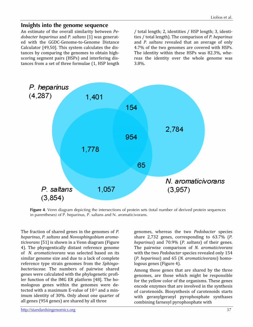
Figure 4. Venn diagram depicting the intersections of protein sets (total number of derived protein sequences in parentheses) of P. heparinus, P. saltans and N. aromaticivorans.
The fraction of shared genes in the genomes of P. heparinus, P. saltans and Novosphingobium aroma-ticivorans [51] is shown in a Venn diagram (Figure 4). The phyogentically distant reference genome of N. aromaticivorans was selected based on its similar genome size and due to a lack of complete reference type strain genomes from the Sphingo-bacteriaceae. The numbers of pairwise shared genes were calculated with the phylogenetic profi-ler function of the IMG ER platform [48]. The ho-mologous genes within the genomes were de-tected with a maximum E-value of 10-5 and a min-imum identity of 30%. Only about one quarter of all genes (954 genes) are shared by all three
genomes, whereas the two Pedobacter species share 2,732 genes, corresponding to 63.7% (P. heparinus) and 70.9% (P. saltans) of their genes. The pairwise comparison of N. aromaticivorans with the two Pedobacter species revealed only 154 (P. heparinus) and 65 (N. aromaticivorans) homo-logous genes (Figure 4). Among those genes that are shared by the three genomes, are those which might be responsible for the yellow color of the organisms. These genes encode enzymes that are involved in the synthesis of carotenoids. Biosynthesis of carotenoids starts with geranylgeranyl pyrophosphate synthases combining farnesyl pyrophosphate with

Pedobacter saltans type strain (113T)
38 Standards in Genomic Sciences
C5 isoprenoid units to C20-molecules, geranylge-ranyl pyrophosphate. The phytoene synthase cata-lyzes the condensation of two geranylgeranyl py-rophosphate molecules followed by the removal of diphosphate and a proton shift leading to the for-mation of phytoene. Sequential desaturation steps are catalyzed by phytoene desaturase followed by cyclization of the ends of the molecules catalyzed by the lycopene cyclase [52]. Genes encoding ly-copene cyclases (Phep_2088, Pedsa_2222, Sa-ro_1817) and phytoene synthases (Phep_2092, Pedsa_2218, Saro_1814) were identified in the genomes. In the two Pedobacter species, genes coding for phytoene desaturases (Phep_2093, Pedsa_2217) were also identified. A carotene hy-droxylase gene (Saro_1168) was only identified in the genome of N. aromaticivorans. As the two Pedobacter species are known for their ability to degrade heparin, it is not surprising that the genomes encode several heparinase encoding
genes: seven (P. saltans) and five (P. heparinus) heparinases, were identified, whereas N. aromati-civorans encodes only one heparinase. Fucoidan degradation was not determined expe-rimentally, but is assumed as both P. saltans and P. heparinus have genes for eleven and ten α-fucosidases respectively. In addition, 12 (P. sal-tans) and 18 (P. heparinus) α-sulfatases genes were identified, whereas N. aromaticivorans con-tains only five α-sulfatases and no α-fucosidase genes. Experimental evidence for the fucoidan hy-drolysis in Pedobacter has not been found, but for Mucilaginibacter paludis and M. gracilis, which are also members of the family Sphingobacteriaceae, have been experimentally confirmed to exhibit fucoidan degradation [53]. Moreover, Sakai et al. [54] reported the existence of intracellular α-L-fucosidases and sulfatases, which enable ‘F. fucoi-danolyticus’ to degrade fucoidan.
Acknowledgements We would like to gratefully acknowledge the help of Helga Pomrenke (DSMZ) for growing P. saltans cul-tures. This work was performed under the auspices of the US Department of Energy Office of Science, Biologi-cal and Environmental Research Program, and by the University of California, Lawrence Berkeley National Laboratory under contract No. DE-AC02-05CH11231,
Lawrence Livermore National Laboratory under Con-tract No. DE-AC52-07NA27344, and Los Alamos Na-tional Laboratory under contract No. DE-AC02-06NA25396, UT-Battelle and Oak Ridge National La-boratory under contract DE-AC05-00OR22725, as well as German Research Foundation (DFG) INST 599/1-2.
References 1. Steyn PL, Segers P, Vancanneyt M, Sandra P, Ker-
sters K, Joubert JJ. Classification of heparinolytic bacteria into a new genus, Pedobacter, compris-ing four species: Pedobacter heparinus comb. nov., Pedobacter piscium comb. nov., Pedobacter africanus sp. nov. and Pedobacter saltans sp. nov. Proposal of the family Sphingobacteriaceae. Int J Syst Bacteriol 1998; 48:165-177. PubMed doi:10.1099/00207713-48-1-165
2. Garrity G. NamesforLife. BrowserTool takes ex-pertise out of the database and puts it right in the browser. Microbiol Today 2010; 37:9.
3. Steyn PL, Pot B, Segers P, Kesters K, Joubert JJ. Some novel aerobic heparin-degrading bacterial isolates. Syst Appl Microbiol 1992; 15:137-143.
4. BCCM/LMG bacteria catalogue: http://bccm.belspo.be/db/lmg_species_details.php
5. StrainInfo website: http://www.straininfo.net/strains/45464
6. Jeon Y, Kim JM, Park JH, Lee SH, Seong CN, Lee SS, Jeon CO. Pedobacter oryzae sp.nov., isolated
from rice paddy soil. Int J Syst Evol Microbiol 2009; 59:2491-2495. PubMed doi:10.1099/ijs.0.005710-0
7. Zhang DC, Schinner F, Margesin R. Pedobacter bauzanensis sp. nov., isolated from soil. Int J Syst Evol Microbiol 2010; 60:2592-2595. PubMed doi:10.1099/ijs.0.018903-0
8. Yoon JH, Kang SJ, Park S, Oh TK. Pedobacter len-tus sp.nov. and Pedobacter terricola sp. nov., iso-lated from soil. Int J Syst Evol Microbiol 2007; 57:2089-2095. PubMed doi:10.1099/ijs.0.65146-0
9. Luo X, Wang Z, Dai J, Zhang L, Li J, Tang Y, Wang Y, Fang C. Pedobacter glucosidilyticus sp.nov., isolated from dry riverbed soil. Int J Syst Evol Microbiol 2010; 60:229-233. PubMed doi:10.1099/ijs.0.008060-0
10. Yali T, Wang Y, Ji S, Zhang K, Dai J, Zhang L, Peng F, Fang C. Pedobacter xinjiangensis sp. nov., from the Desert, Xinjiang. Int J Syst Evol Microbiol 2010; 20:397-402.

Liolios et al.
http://standardsingenomics.org 39
11. Hwang CY, Choi DH, Cho BC. Pedobacter roseus sp. nov., isolated from a hypertrophic pond, and emended description of the genus Pedobacter. Int J Syst Evol Microbiol 2006; 56:1831-1836. PubMed doi:10.1099/ijs.0.64045-0
12. Gallego V, Garcia MT, Ventosa A. Pedobacter aquatilis sp. nov., isolated from drinking water, and emended description of the genus Pedobac-ter. Int J Syst Evol Microbiol 2006; 56:1853-1858. PubMed doi:10.1099/ijs.0.64176-0
13. Joung Y, Kim H, Joh K. Pedobacter yonginense sp.nov., isolated from a mesotrophic artificial lake in Korea. J Microbiol 2010; 48:536-540. PubMed doi:10.1007/s12275-010-0010-4
14. Vanparys B, Heylen K, Lebbe L, de Vos P. Pedo-bacter caeni sp. nov., a novel species isolated from a nitrifying inoculum. Int J Syst Evol Micro-biol 2005; 55:1315-1318. PubMed doi:10.1099/ijs.0.63635-0
15. Margesin R, Spröer C, Schumann P, Schinner F. Pedobacter cryoconitis sp.nov., a facultative psychrophile from alpine glacier cryoconite. Int J Syst Evol Microbiol 2003; 53:1291-1296. PubMed doi:10.1099/ijs.0.02436-0
16. Shivaji S, Chaturvedi P, Reddy GSN, Suresh K. Pedobacter himalayensis sp.nov., from the Hamta glacier located in the Himalayan mountain ranges of India. Int J Syst Evol Microbiol 2005; 55:1083-1088. PubMed doi:10.1099/ijs.0.63532-0
17. Lee HG, Kim SG, Im WT, Oh HM, Lee ST. Pedo-bacter composti sp. nov., isolated from compost. Int J Syst Evol Microbiol 2009; 59:345-349. PubMed doi:10.1099/ijs.0.003061-0
18. Altschul SF, Gish W, Miller W, Myers E, Lipman D. Basic local alignment search tool. J Mol Biol 1990; 215:403-410. PubMed
19. Korf I, Yandell M, Bedell J. BLAST, O'Reilly, Se-bastopol, 2003
20. DeSantis TZ, Hugenholtz P, Larsen N, Rojas M, Brodie EL, Keller K, Huber T, Dalevi D, Hu P, Andersen GL. Greengenes, a chimera-checked 16S rRNA gene database and workbench compat-ible with ARB. Appl Environ Microbiol 2006; 72:5069-5072. PubMed doi:10.1128/AEM.03006-05
21. Porter MF. An algorithm for suffix stripping Pro-gram: electronic library and information systems 1980; 14:130-137.
22. Lee C, Grasso C, Sharlow MF. Multiple sequence alignment using partial order graphs. Bioinformat-
ics 2002; 18:452-464. PubMed doi:10.1093/bioinformatics/18.3.452
23. Castresana J. Selection of conserved blocks from multiple alignments for their use in phylogenetic analysis. Mol Biol Evol 2000; 17:540-552. PubMed
24. Stamatakis A, Hoover P, Rougemont J. A rapid bootstrap algorithm for the RAxML web-servers. Syst Biol 2008; 57:758-771. PubMed doi:10.1080/10635150802429642
25. Fitch WM. Toward defining the course of evolu-tion: minimum change for a specific tree topolo-gy. Syst Zool 1971; 20:406-416. doi:10.2307/2412116
26. Pattengale ND, Alipour M, Bininda-Emonds ORP, Moret BME, Stamatakis A. How many bootstrap replicates are necessary? Lect Notes Comput Sci 2009; 5541:184-200. doi:10.1007/978-3-642-02008-7_13
27. Swofford DL. PAUP*: Phylogenetic Analysis Us-ing Parsimony (*and Other Methods), Version 4.0 b10. Sinauer Associates, Sunderland, 2002.
28. Liolios K, Chen IM, Mavromatis K, Tavernarakis N, Hugenholtz P, Markowitz VM, Kyrpides NC. The Genomes On Line Database (GOLD) in 2009: status of genomic and metagenomic projects and their associated metadata. Nucleic Acids Res 2010; 38:D346-D354. PubMed doi:10.1093/nar/gkp848
29. Han C, Spring S, Lapidus A, Glavina Del Rio T, Tice H, Copeland A, Cheng JF, Lucas S, Chen F, Nolan M, et al. Complete genome sequence of Pedobacter heparinus type strain (HIM 762-3T). Stand Genomic Sci 2009; 1:54-62. PubMed doi:10.4056/sigs.22138
30. Felsenstein J. Inferring phylogenies. Sinauer Asso-ciates Inc., Sunderland, Massachusetts 2004.
31. Field D, Garrity G, Gray T, Morrison N, Selengut J, Sterk P, Tatusova T, Thomson N, Allen MJ, An-giuoli SV, et al. The minimum information about a genome sequence (MIGS) specification. Nat Biotechnol 2008; 26:541-547. PubMed doi:10.1038/nbt1360
32. Woese CR, Kandler O, Wheelis ML. Towards a natural system of organisms: proposal for the do-mains Archaea, Bacteria, and Eucarya. Proc Natl Acad Sci USA 1990; 87:4576-4579. PubMed doi:10.1073/pnas.87.12.4576
33. Garrity GM, Holt JG. The Road Map to the Ma-nual. In: Garrity GM, Boone DR, Castenholz RW (eds), Bergey's Manual of Systematic Bacteriology,

Pedobacter saltans type strain (113T)
40 Standards in Genomic Sciences
Second Edition, Volume 1, Springer, New York, 2001, p. 119-169.
34. Garrity GM, Holt JG. Taxonomic outline of the Archaea and Bacteria. In: Garrity GM, Boone DR, Castenholz RW (eds), Bergey's Manual of Syste-matic Bacteriology, Second Edition, Volume 1, Springer, New York, 2001, p. 155-166.
35. BAuA. Classification of bacteria and archaea in risk groups. TRBA 2010; 466:165.
36. Ashburner M, Ball CA, Blake JA, Botstein D, But-ler H, Cherry JM, Davis AP, Dolinski K, Dwight SS, Eppig JT, et al. Gene Ontology: tool for the unification of biology. Nat Genet 2000; 25:25-29. PubMed doi:10.1038/75556
37. Klenk HP, Göker M. En route to a genome-based classification of Archaea and Bacteria? Syst Appl Microbiol 2010; 33:175-182. PubMed doi:10.1016/j.syapm.2010.03.003
38. Wu D, Hugenholtz P, Mavromatis K, Pukall R, Dalin E, Ivanova NN, Kunin V, Goodwin L, Wu M, Tindall BJ, et al. A phylogeny-driven genomic encyclopaedia of Bacteria and Archaea. Nature 2009; 462:1056-1060. PubMed doi:10.1038/nature08656
39. List of growth media used at DSMZ: http://www.dsmz.de/microorganisms/media_list.php.
40. Gemeinholzer B, Dröge G, Zetzsche H, Haszpru-nar G, Klenk HP, Güntsch A, Berendsohn WG, Wägele JW. The DNA Bank Network: the start from a German initiative. Biopreservation and Biobanking 2011; 9:51-55. doi:10.1089/bio.2010.0029
41. JGI website. http://www.jgi.doe.gov
42. The Phred/Phrap/Consed software package. http://www.phrap.com
43. Zerbino DR, Birney E. Velvet: algorithms for de novo short read assembly using de Bruijn graphs. Genome Res 2008; 18:821-829. PubMed doi:10.1101/gr.074492.107
44. Han C, Chain P. Finishing repeat regions auto-matically with Dupfinisher. In: Proceeding of the 2006 international conference on bioinformatics & computational biology. Arabnia HR, Valafar H (eds), CSREA Press. June 26-29, 2006: 141-146.
45. Lapidus A, LaButti K, Foster B, Lowry S, Trong S, Goltsman E. POLISHER: An effective tool for us-
ing ultra short reads in microbial genome assem-bly and finishing. AGBT, Marco Island, FL, 2008.
46. Hyatt D, Chen GL, LoCascio PF, Land ML, Larimer FW, Hauser LJ. Prodigal: prokaryotic gene recognition and translation initiation site identification. BMC Bioinformatics 2010; 11:119. PubMed doi:10.1186/1471-2105-11-119
47. Pati A, Ivanova NN, Mikhailova N, Ovchinnikova G, Hooper SD, Lykidis A, Kyrpides NC. Gene-PRIMP: a gene prediction improvement pipeline for prokaryotic genomes. Nat Methods 2010; 7:455-457. PubMed doi:10.1038/nmeth.1457
48. Markowitz VM, Ivanova NN, Chen IMA, Chu K, Kyrpides NC. IMG ER: a system for microbial ge-nome annotation expert review and curation. Bio-informatics 2009; 25:2271-2278. PubMed doi:10.1093/bioinformatics/btp393
49. Auch AF, von Jan M, Klenk HP, Göker M. Digital DNA-DNA hybridization for microbial species delineation by means of genome-to-genome se-quence comparison. Stand Genomic Sci 2010; 2:117-134. PubMed doi:10.4056/sigs.531120
50. Auch AF, Klenk HP, Göker M. Standard operating procedure for calculating genome-to-genome dis-tances based on high-scoring segment pairs. Stand Genomic Sci 2010; 2:142-148. PubMed doi:10.4056/sigs.541628
51. Takeuchi M, Hamama K, Hiraishi A. Proposal of the genus Sphingomonas sensu stricto and three new genera, Sphingobium, Novosphingobium and Sphingopyxis, on the basis of phylogenetic and chemotaxonomic analyses. Int J Syst Evol Mi-crobiol 2001; 51:1405-1417. PubMed
52. Sandmann G. Carotenoid biosynthesis and bio-technological application. Arch Biochem Biophys 2001; 385:4-12. PubMed doi:10.1006/abbi.2000.2170
53. Pankratov TA, Tindall BJ, Liesack W, Dedysh SN. Mucilaginibacter paludis gen. nov., sp. nov. and Mucilaginibacter gracilis sp. nov., pectin-, xylan- and laminarin-degrading members of the family Sphingobacteriaceae from acidic Sphagnum peat bog. Int J Syst Evol Microbiol 2007; 57:2349-2354. PubMed doi:10.1099/ijs.0.65100-0
54. Sakai T, Ishizuka K, Kato I. Isolation and characte-rization of fucoidan-degrading marine bacterium. Mar Biotechnol 2003; 5:409-416. PubMed doi:10.1007/s10126-002-0118-6
Copyright © 2022 FDOKUMEN