The complete genome sequence of the avian pathogen Mycoplasma gallisepticum strain Rlow
10
The complete genome sequence of the avian pathogen Mycoplasma gallisepticum strain R low Leka Papazisi, 1,2 Timothy S. Gorton, 1,2 Gerald Kutish, 3 Philip F. Markham, 4 Glenn F. Browning, 4 Di Kim Nguyen, 5 Steven Swartzell, 6 Anup Madan, 7 Greg Mahairas 6 and Steven J. Geary 1,2 Correspondence Steven J. Geary [email protected] 1,2 Center of Excellence for Vaccine Research 1 and Department of Pathobiology and Veterinary Science 2 , The University of Connecticut, Storrs, CT 06269-3089, USA 3 Plum Island Animal Disease Center, US Department of Agriculture, Greenport, NY 11944, USA 4 Department of Veterinary Science, University of Melbourne, Parkville, Victoria 3010, Australia 5,6 Department of Medicine, University of Washington 5 and Regulome Corporation 6 , Seattle, WA 98195, USA 7 The Institute for Systems Biology, Seattle, WA 98103-8904, USA Received 16 April 2003 Revised 9 June 2003 Accepted 16 June 2003 The complete genome of Mycoplasma gallisepticum strain R low has been sequenced. The genome is composed of 996 422 bp with an overall G+C content of 31 mol%. It contains 742 putative coding DNA sequences (CDSs), representing a 91 % coding density. Function has been assigned to 469 of the CDSs, while 150 encode conserved hypothetical proteins and 123 remain as unique hypothetical proteins. The genome contains two copies of the rRNA genes and 33 tRNA genes. The origin of replication has been localized based on sequence analysis in the region of the dnaA gene. The vlhA family (previously termed pMGA) contains 43 genes distributed among five loci containing 8, 2, 9, 12 and 12 genes. This family of genes constitutes 10?4% (103 kb) of the total genome. Two CDSs were identified immediately downstream of gapA and crmA encoding proteins that share homology to cytadhesins GapA and CrmA. Based on motif analysis it is predicted that 80 genes encode lipoproteins and 149 proteins contain multiple transmembrane domains. The authors have identified 75 proteins putatively involved in transport of biomolecules, 12 transposases, and a number of potential virulence factors. The completion of this sequence has spawned multiple projects directed at defining the biological basis of M. gallisepticum. INTRODUCTION Phylogenetic analyses indicate that mycoplasmas (class Mollicutes) have undergone a degenerative evolution from related, low G+C content, Gram-positive eubacteria (Rogers et al., 1985; Woese et al., 1980). The reduction of the mycoplasma genome has resulted in the loss of the cell wall and has limited the biosynthetic capabilities of these organisms. As a consequence of this loss of biosynthetic machinery, mycoplasmas are obligate parasites and rely on the uptake of many essential molecules from their environment. Mycoplasmas have long been considered model systems for defining the minimal set of genes required for a living cell (Morowitz, 1984). For this reason, it was not surprising when Mycoplasma genitalium (580 kb) was selected as one of the first targets for complete genome sequencing (Fraser et al., 1995). Since this initial report, the genomes of four additional mycoplasmas have been sequenced, Mycoplasma pneumoniae (816 kb; Dandekar et al., 2000; Himmelreich et al., 1996), Ureaplasma urealyticum (752 kb; Glass et al., 2000), Mycoplasma pulmonis (964 kb; Chambaud et al., 2001) and Mycoplasma penetrans (1358 kb; Sasaki et al., 2000). Theoretical and experimental approaches have estimated the minimum number of essential mycoplasma genes to be between 265 and 350 (Hutchison et al., 1999; Mushegian & Koonin, 1996). Abbreviations: CDS, coding DNA sequence; COGs, conserved ortho- logous groups. The GenBank accession number for the sequence reported in this paper is AE015450. The online version of this paper (at http://mic.sgmjournals.org) contains a supplementary table showing additional information concerning the features and homologies of the M. gallisepticum strain R low genome. 0002-6427 G 2003 SGM Printed in Great Britain 2307 Microbiology (2003), 149, 2307–2316 DOI 10.1099/mic.0.26427-0
Transcript of The complete genome sequence of the avian pathogen Mycoplasma gallisepticum strain Rlow

The complete genome sequence of the avianpathogen Mycoplasma gallisepticum strain Rlow
Leka Papazisi,1,2 Timothy S. Gorton,1,2 Gerald Kutish,3 Philip F. Markham,4
Glenn F. Browning,4 Di Kim Nguyen,5 Steven Swartzell,6 Anup Madan,7
Greg Mahairas6 and Steven J. Geary1,2
Correspondence
Steven J. Geary
1,2Center of Excellence for Vaccine Research1 and Department of Pathobiology and VeterinaryScience2, The University of Connecticut, Storrs, CT 06269-3089, USA
3Plum Island Animal Disease Center, US Department of Agriculture, Greenport, NY 11944,USA
4Department of Veterinary Science, University of Melbourne, Parkville, Victoria 3010, Australia
5,6Department of Medicine, University of Washington5 and Regulome Corporation6, Seattle,WA 98195, USA
7The Institute for Systems Biology, Seattle, WA 98103-8904, USA
Received 16 April 2003
Revised 9 June 2003
Accepted 16 June 2003
The complete genome ofMycoplasma gallisepticum strain Rlow has been sequenced. The genome
is composed of 996422 bp with an overall G+C content of 31 mol%. It contains 742
putative coding DNA sequences (CDSs), representing a 91% coding density. Function has been
assigned to 469 of the CDSs, while 150 encode conserved hypothetical proteins and 123
remain as unique hypothetical proteins. The genome contains two copies of the rRNA genes and
33 tRNA genes. The origin of replication has been localized based on sequence analysis in the
region of the dnaA gene. The vlhA family (previously termed pMGA) contains 43 genes distributed
among five loci containing 8, 2, 9, 12 and 12 genes. This family of genes constitutes 10?4%
(103 kb) of the total genome. Two CDSs were identified immediately downstream of gapA and
crmA encoding proteins that share homology to cytadhesins GapA and CrmA. Based on motif
analysis it is predicted that 80 genes encode lipoproteins and 149 proteins contain multiple
transmembrane domains. The authors have identified 75 proteins putatively involved in transport of
biomolecules, 12 transposases, and a number of potential virulence factors. The completion of
this sequence has spawned multiple projects directed at defining the biological basis of
M. gallisepticum.
INTRODUCTION
Phylogenetic analyses indicate that mycoplasmas (classMollicutes) have undergone a degenerative evolution fromrelated, low G+C content, Gram-positive eubacteria(Rogers et al., 1985; Woese et al., 1980). The reduction ofthe mycoplasma genome has resulted in the loss of the cellwall and has limited the biosynthetic capabilities of theseorganisms. As a consequence of this loss of biosyntheticmachinery, mycoplasmas are obligate parasites and rely
on the uptake of many essential molecules from theirenvironment.
Mycoplasmas have long been considered model systems fordefining the minimal set of genes required for a living cell(Morowitz, 1984). For this reason, it was not surprisingwhen Mycoplasma genitalium (580 kb) was selected asone of the first targets for complete genome sequencing(Fraser et al., 1995). Since this initial report, the genomes offour additional mycoplasmas have been sequenced,Mycoplasma pneumoniae (816 kb; Dandekar et al., 2000;Himmelreich et al., 1996), Ureaplasma urealyticum(752 kb; Glass et al., 2000), Mycoplasma pulmonis(964 kb; Chambaud et al., 2001) andMycoplasma penetrans(1358 kb; Sasaki et al., 2000). Theoretical and experimentalapproaches have estimated the minimum number ofessential mycoplasma genes to be between 265 and 350(Hutchison et al., 1999; Mushegian & Koonin, 1996).
Abbreviations: CDS, coding DNA sequence; COGs, conserved ortho-logous groups.
The GenBank accession number for the sequence reported in thispaper is AE015450.
The online version of this paper (at http://mic.sgmjournals.org) containsa supplementary table showing additional information concerningthe features and homologies of the M. gallisepticum strain Rlow genome.
0002-6427 G 2003 SGM Printed in Great Britain 2307
Microbiology (2003), 149, 2307–2316 DOI 10.1099/mic.0.26427-0

Mycoplasma gallisepticum is an avian pathogen involvedin chronic respiratory disease in chickens resulting inconsiderable economic losses in poultry production.Infection with this bacterium is spread by aerosol exposureand egg transmission. Outbreaks spread rapidly throughflocks, establish chronic infections, and are difficult tocontrol with antimicrobial therapy. The chronic nature ofmycoplasma infections demonstrates a failure of the hostimmune system to deal effectively with these organisms.Antigenic variation of surface proteins allows M. gallisep-ticum to evade the host’s immune response through thegeneration of escape variants (Glew et al., 2000; Gorton &Geary, 1997; Levisohn et al., 1995). Intracellular invasionand survival within eukaryotic cells byM. gallisepticummaycontribute to this organism’s resistance to the host’simmune response and antimicrobial therapy (Winneret al., 2000).
M. gallisepticum colonizes the respiratory system ofchickens, but has also been isolated from the reproductiveorgans, brain and eyes of several avian species. Cytadherenceto the epithelial surfaces of these host tissues is arequirement for successful colonization. Research into themolecular mechanisms ofM. gallisepticum cytadherence hasidentified a coordinate action between the primarycytadhesin, GapA, and at least one cytadherence-relatedmolecule, CrmA (Papazisi et al., 2002).
Whole-genome sequencing establishes a solid foundationfrom which to begin a myriad of experimental projectsdirected at examining the pathogenic mechanisms of anorganism. Here we present the complete sequence and initialanalysis of the M. gallisepticum genome.
METHODS
Construction of M. gallisepticum genomic library. A clonalisolate, designated Rlowc2, of the virulent M. gallisepticum strain Rlow
was chosen for sequencing. M. gallisepticum genomic DNA was pre-pared by the method of Hempstead (1990). Genomic DNA (4 mg)was sheared using a Hydroshear device (GeneMachines) to a meansize of 3–5 kb. Sheared ends were repaired using T4 DNA kinaseand Klenow fragment of DNA polymerase (New England Biolabs).The reactions were stopped and the resulting repaired fragmentspurified using Qiaquick (Qiagen) columns according to the manu-facturer’s instructions. Plasmid pBlueScript SKII(+) (Stratagene)was used as the vector and prepared by digestion with EcoRV (NewEngland Biolabs), treatment with alkaline phosphatase and electro-phoresis in an agarose gel (0?6%). A single discrete band wasexcised from the gel and purified using a Gene Clean Column(Qbiogene). Ligation reactions were prepared using a 2 : 1 insert tovector ratio and T4 DNA ligase (New England Biolabs), thentransformed into Escherichia coli DH10B Max competent cells(Invitrogen). Plates with 1000–3000 recombinant clones were subse-quently picked and arrayed into Genetix square-well 384-well platescontaining SOB medium (Difco) supplemented with 20% (v/v)glycerol. Following overnight (18 h) incubation these plates weresealed and stored at 280 uC until further use.
Sequencing and assembly. Each quadrant of a stock 384-wellplate was freshly inoculated into a deep-well 96-well plate contain-ing 1 ml 26 YT medium (Difco) supplemented with 100 mg
carbenicillin ml21. These inoculated plates were shaken at250 r.p.m. for 14 h at 37 uC followed by centrifugation at3000 r.p.m. for 10 min. Plasmid preparations were processed auto-matically using an Eppendorf PerfectPrep-96 VAC RoboticWorkstation. Sequencing reactions were carried out using 1 ml DNA(350 ng), 4 pmol forward and reverse primers (221M13 andM13Reverse) and 3 ml ABI Prism Big Dye Terminators (AppliedBiosystems/Perkin Elmer) in a total volume of 5 ml. Reactions werecarried out in the ABI GeneAmp PCR System 9700 (AppliedBiosystems/Perkin Elmer). The DNA from sequencing reactions wasprecipitated with 2-propanol, resuspended in 10 ml sterile distilledH2O, and loaded onto an ABI 3700 capillary electrophoresis sequen-cer. Sequence data were assembled and analysed using the Phred/Phrap/Consed assembly package (Ewing & Green, 1998; Ewing et al.,1998; Gordon et al., 1998) and CAP3 (Huang & Madan, 1999).CAP3 uses forward and reverse sequence reads from the same plas-mid sequences to place constraints on assembled sequence and isuseful to break false joins and assure correct sequence assembly(Huang & Madan, 1999). Additional reactions were performed toclose gaps in the assembly. Custom primers were designed near gapsand sequence was obtained from plasmid clones that spanned thegaps. Where no clone was available to span the gap, PCR productswere generated using custom primers on genomic DNA and the sub-sequent PCR products were sequenced and added to the assembly.Sequencing coverage, of both strands, was 116 (or 22 reads perkb), done to an error rate less than 1 per 10 kb.
The sequence data have been submitted to the NCBI database underaccession number AE015450.
Identification of CDSs and annotation. Coding DNA sequences(CDSs), of at least 195 nt in length, were initially identified usingORF Finder software (with genetic code 4) provided by the NationalCenter for Biotechnology information (NCBI; http://www.ncbi.nlm.nih.gov/). GLIMMER (Delcher et al., 1999) was trained to recognize apotential CDS whose translated product had at least 33 amino acidsand did not overlap more than 30 nt with neighbouring CDSs.BLAST (Zhang et al., 1998) and FASTA (Pearson, 1999) searches wereperformed on the NCBI non-redundant protein databases, con-served orthologous groups (COGs) database (Tatusov et al., 2001),and proteins from the mycoplasma genomes sequenced to date. ACDS was assigned a designation based on homology when its trans-lated product presented a BLAST score of at least 100 in the COGdatabase and 150 in the non-redundant database. Scores of 300 ormore were considered definitive. MSP-Crunch (Sonnhammer &Durbin, 1994) was used to filter low-complexity sequences in orderto eliminate spurious homologies. FAStRNA (http://bioweb.pasteur.fr/seqanal/interfaces/fastrna.html) (el-Mabrouk & Lisacek,1996) was used for finding tRNA coding sequences. The PROSITE(Falquet et al., 2002), Pfam (Bateman et al., 2000), BLOCKS(Henikoff et al., 1999) and TIGERfam (http://www.tigr.org/) data-bases were searched to identify motifs and other shared features ofproteins. Specific motif searches were performed against the M. galli-septicum genome using FUZZPRO from the Jemboss software package(http://www.hgmp.mrc.ac.uk/Software/EMBOSS/Jemboss/index.html).Protein topology and location were predicted using TMPred(Hofmann & Stoffel, 1993), MEMSAT (Jones et al., 1994), PSORT
(http://psort.nibb.ac.jp/) and SAPS (Brendel et al., 1992).
RESULTS AND DISCUSSION
General features of the genome
The general features of the M. gallisepticum strain Rlow
genome are shown in Fig. 1 and Table 1. The online versionof this paper (at http://mic.sgmjournals.org) contains a
2308 Microbiology 149
L. Papazisi and others

supplementary table with additional information concern-ing the features and homologies of the genome.The genomeis composed of 996 422 bp with an overall G+C content of31 mol%. It contains 742 putative coding DNA sequences(CDSs), representing a 91% coding density. Function hasbeen assigned to 469 of the CDSs, while 150 are conservedhypothetical proteins and 123 remain as hypotheticalproteins. The mean CDS length was 1206 nt(108–5928 nt) and the mean CDS G+C content was32 mol% (17–45 mol%). The mean G+C content of the
third nucleotide position in codons was 24%. The predictedcodon usage within M. gallisepticum varies with statisticalsignificance from the other mycoplasma genomes sequencedto date, with the exception of M. genitalium (data notshown). A set of 33 tRNA genes was identified, correspond-ing to all amino acids. A single polypeptide release factor(prfA) was identified, consistent with the use of only UAAandUAG as stop codons. Themean occurrence of the aminoacid tryptophan was 3?6 per CDS and the usage of thecodons TGA and TGG was 2?8 and 0?8 per CDS,
Fig. 1. Circular representation of the M. gallisepticum strain Rlow genome. This figure was generated using GenVision(DNAStar).
http://mic.sgmjournals.org 2309
M. gallisepticum complete genome sequence

Table 1. General features of Mycoplasma species
M. gallisepticum M. genitalium* M. pneumoniaeD M. pulmonisd U. urealyticum§ M. penetrans||
Genome size (bp) 996 422 580 074 816 394 963 879 751 719 1 358 633
G+C content (mol%) 31?5 32 40 26?6 25?5 25?7
Sets of 5S 16S and 23S rRNA 2 1 1 1 2 1
tRNA 33 36 37 29 30 30
Total no. of predicted CDSs 742 484 689 782 614 1038
Functional category in COGs
[J] Translation, ribosomal structure and biogenesis 103 14% 99 20% 100 15% 102 13% 102 17% 108 10%
[K] Transcription 14 2% 14 3% 14 2% 18 2% 18 3% 23 2%
[L] DNA replication, recombination and repair 53 7% 41 8% 57 8% 72 9% 56 9% 78 8%
[D] Cell division and chromosome partitioning 7 1% 5 1% 5 1% 4 1% 4 1% 11 1%
[O] Posttranslational modification, protein turnover, chaperones 33 4% 20 4% 20 3% 18 2% 19 3% 26 3%
[M] Cell envelope biogenesis, outer membrane 8 1% 10 2% 10 1% 5 1% 4 1% 13 1%
[N] Cell motility and secretion 9 1% 11 2% 12 2% 13 2% 15 2% 12 1%
[P] Inorganic ion transport and metabolism 14 2% 17 4% 17 2% 15 2% 28 5% 22 2%
[T] Signal transduction mechanisms 4 1% 4 1% 4 1% 3 0% 4 1% 4 0%
[C] Energy production and conversion 28 4% 20 4% 20 3% 29 4% 17 3% 31 3%
[G] Carbohydrate transport and metabolism 25 3% 24 5% 34 5% 58 7% 14 2% 50 5%
[E] Amino acid transport and metabolism 21 3% 15 3% 24 3% 23 3% 20 3% 26 3%
[F] Nucleotide transport and metabolism 25 3% 21 4% 21 3% 24 3% 21 3% 39 4%
[H] Coenzyme metabolism 11 1% 13 3% 14 2% 12 2% 9 1% 12 1%
[I] Lipid metabolism 11 1% 8 2% 9 1% 8 1% 8 1% 15 1%
[Q] Secondary metabolites biosynthesis, transport and catabolism 13 2% 4 1% 5 1% 10 1% 7 1% 20 2%
[R] General function prediction only 51 7% 44 9% 48 7% 49 6% 41 7% 95 9%
[S] Function unknown 26 4% 14 3% 15 2% 26 3% 22 4% 25 2%
(–) not in COGs 286 39% 100 21% 260 38% 293 37% 205 33% 428 41%
*Fraser et al. (1995).
DHimmelreich et al. (1996); Dandekar et al. (2000).
dChambaud et al., (2001).
§Glass et al. (2000).
||Sasaki et al. (2002).
2310
Micro
biology
149
L.Papazisi
andothers

respectively. As previously reported (Chen & Finch, 1989;Gorton et al., 1995; Scamrov & Beabealashvilli, 1991), theM. gallisepticum genome contains two copies of the rRNAgenes. One set is organized as an operon, with adjacent 16S,23S and 5S genes; and a second copy of the 16S rRNA genelies 221 kb upstream of the 23S and 5S rRNA genes.
Origin of replication
The origin of replication (oriC) for most bacteria is locatedin the region of the dnaA gene. Comparative analysis of theoriC regions of sequenced mycoplasma genomes predictsputative DnaA boxes in the area surrounding the dnaA gene(Cordova et al., 2002). Functional mollicute oriC regionshave been identified in Spiroplasma citri and M. pulmonis(Cordova et al., 2002; Ye et al., 1994).
In silico analysis localized the predicted oriC region ofM. gallisepticum based on several criteria. First, despite anapparent overall lack of conserved gene order in themollicute oriC region (Cordova et al., 2002), the gene orderin the oriC region does appear to be conserved within thephylogenetic cluster containing M. pneumoniae, M. genita-lium and M. gallisepticum (Cordova et al., 2002; Hilbertet al., 1996; Zou & Dybvig, 2002). These three mycoplasmashave the gyrA, gyrB, dnaJ, dnaN and soj genes upstream ofthe dnaA gene, and ABC transporter genes, rpl34 andrpnA, downstream of dnaA. As inM. pneumoniae, there is aunique hypothetical coding region immediately upstreamof the dnaA gene in M. gallisepticum. Second, we identifiedputative DnaA boxes in the vicinity of the dnaA gene(Fig. 2). The region around dnaA was searched for 9 ntlong sequences resembling a consensus sequence (59-TTWTMHAMA-39) based on DnaA box sequences fromM. pulmonis, M. capricolum and M. pneumoniae(Chambaud et al., 2001; Cordova et al., 2002; Hilbertet al., 1996; Zou & Dybvig, 2002). Finally, the regionbetween dnaN and soj contains higher than average AT basepair frequency (80%) and contains AT-rich repeats. Thesecriteria are considered to be characteristic of the origin ofreplication in prokaryotes (Baker & Wickner, 1992).
vlhA gene family
Perhaps the best-described example of a multi-gene familyin mycoplasmas is that encoding the VlhA or pMGAlipoproteins (Baseggio et al., 1996; Liu et al., 1998;Markhamet al., 1993). It has been well established thatM. gallisepticumgenerally expresses a single member of the family at any onetime (Glew et al., 1995) and that the specific gene expressedcan be influenced by growth in the presence of cognateantibody (Markham et al., 1998). The probable role of thisfamily in generating antigenic variation has been demon-strated in infected chickens, with both phase variationdemonstrated during the acute stages of disease, andantigenic switching during the chronic stages (Glew et al.,2000). These findings have led to the suggestion that theprincipal function of this family is to generate antigenicdiversity and hence facilitate immune evasion duringchronic infections. These genes formed the largest para-logous gene family in the M. gallisepticum genome.
To establish consistency, and in accordance with standardnomenclature, we have annotated this family of genes asvlhA (Noormohammadi et al., 1998). This family contains43 genes, constituting a total of 103 kb or 10?4% of thegenome. The 43 vlhA genes are distributed among five locicontaining 8, 2, 9, 12 and 12 genes, respectively, and havebeen numbered according to their locus and position (e.g.vlhA1.01) (Fig. 3). Of the 43 genes, 38 possess the signaturevlhA gene features, which include a GAA repeat motif 59 of aGTG start codon and conserved regions flanking the startcodon (Markham et al., 1994). The sequence identity amongthese 38 genes ranges between 41 and 99%. Five of the genes(1.05, 2.01, 2.02, 5.01 and 5.02) possess sequence homologyto vlhA but lack the vlhA signature motifs. The genes withineach of the five loci are in the same transcriptionalorientation, with the exception of vlhA1.05. In addition tothe lack of vlhA signature motifs, a putative transposase(MGA_0073) has been identified adjacent to vlhA1.05,suggesting that rearrangements by a transposon mayaccount for the differences observed in this region. Five ofthe genes (vlhA1.01, 1.08, 3.01, 4.03 and 5.01) have been
Fig. 2. Organization of the putative origin of replication of M. gallisepticum. The black triangles represent putative DnaA boxeswith 9 out of 9 matches to the consensus sequence 59-TTWTMHAMA-39.
http://mic.sgmjournals.org 2311
M. gallisepticum complete genome sequence
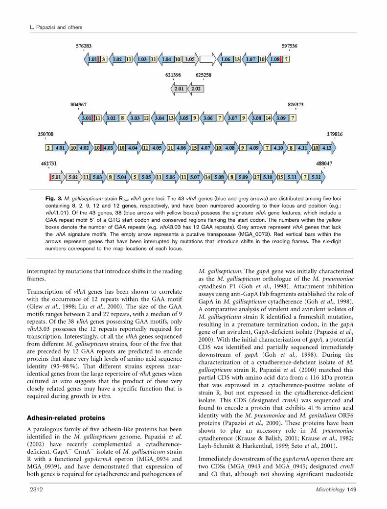
interrupted bymutations that introduce shifts in the readingframes.
Transcription of vlhA genes has been shown to correlatewith the occurrence of 12 repeats within the GAA motif(Glew et al., 1998; Liu et al., 2000). The size of the GAAmotifs ranges between 2 and 27 repeats, with a median of 9repeats. Of the 38 vlhA genes possessing GAA motifs, onlyvlhA3.03 possesses the 12 repeats reportedly required fortranscription. Interestingly, of all the vlhA genes sequencedfrom different M. gallisepticum strains, four of the five thatare preceded by 12 GAA repeats are predicted to encodeproteins that share very high levels of amino acid sequenceidentity (95–98%). That different strains express near-identical genes from the large repertoire of vlhA genes whencultured in vitro suggests that the product of these veryclosely related genes may have a specific function that isrequired during growth in vitro.
Adhesin-related proteins
A paralogous family of five adhesin-like proteins has beenidentified in the M. gallisepticum genome. Papazisi et al.(2002) have recently complemented a cytadherence-deficient, GapA2 CrmA2 isolate of M. gallisepticum strainR with a functional gapAcrmA operon (MGA_0934 andMGA_0939), and have demonstrated that expression ofboth genes is required for cytadherence and pathogenesis of
M. gallisepticum. The gapA gene was initially characterizedas the M. gallisepticum orthologue of the M. pneumoniaecytadhesin P1 (Goh et al., 1998). Attachment inhibitionassays using anti-GapA Fab fragments established the role ofGapA in M. gallisepticum cytadherence (Goh et al., 1998).A comparative analysis of virulent and avirulent isolates ofM. gallisepticum strain R identified a frameshift mutation,resulting in a premature termination codon, in the gapAgene of an avirulent, GapA-deficient isolate (Papazisi et al.,2000). With the initial characterization of gapA, a potentialCDS was identified and partially sequenced immediatelydownstream of gapA (Goh et al., 1998). During thecharacterization of a cytadherence-deficient isolate of M.gallisepticum strain R, Papazisi et al. (2000) matched thispartial CDS with amino acid data from a 116 kDa proteinthat was expressed in a cytadherence-positive isolate ofstrain R, but not expressed in the cytadherence-deficientisolate. This CDS (designated crmA) was sequenced andfound to encode a protein that exhibits 41% amino acididentity with the M. pneumoniae and M. genitalium ORF6proteins (Papazisi et al., 2000). These proteins have beenshown to play an accessory role in M. pneumoniaecytadherence (Krause & Balish, 2001; Krause et al., 1982;Layh-Schmitt & Harkenthal, 1999; Seto et al., 2001).
Immediately downstream of the gapAcrmA operon there aretwo CDSs (MGA_0943 and MGA_0945; designated crmBand C) that, although not showing significant nucleotide
Fig. 3. M. gallisepticum strain Rlow vlhA gene loci. The 43 vlhA genes (blue and grey arrows) are distributed among five locicontaining 8, 2, 9, 12 and 12 genes, respectively, and have been numbered according to their locus and position (e.g.:vlhA1.01). Of the 43 genes, 38 (blue arrows with yellow boxes) possess the signature vlhA gene features, which include aGAA repeat motif 59 of a GTG start codon and conserved regions flanking the start codon. The numbers within the yellowboxes denote the number of GAA repeats (e.g. vlhA3.03 has 12 GAA repeats). Grey arrows represent vlhA genes that lackthe vlhA signature motifs. The empty arrow represents a putative transposase (MGA_0073). Red vertical bars within thearrows represent genes that have been interrupted by mutations that introduce shifts in the reading frames. The six-digitnumbers correspond to the map locations of each locus.
2312 Microbiology 149
L. Papazisi and others

homology, are predicted to encode proteins sharinghomology to GapA and CrmA. Analysis shows that themajority of the similarity lies within the C-terminal region ofthese four proteins. A fifth paralogue (MGA_1117) sharessimilarity with the N-terminus of GapA and CrmB.
Membrane-associated proteins
Motif analysis of theM. gallisepticum genome has predicteda large repertoire of membrane-associated proteins. Of theseproteins, 149 containmultiple transmembrane domains and18 possess ten or more transmembrane domains. These 18proteins include molecules involved in amino acid transport(PotE), phosphate transport (Pts) and protein translocation(SecY), as well as five conserved hypothetical and twounique hypothetical proteins. Based on the prokaryoticmembrane lipoprotein lipid attachment site motif(PROSITE PS00013), 80 CDSs are predicted to encodelipoproteins (10?8% of all CDSs). The second largestparalogous gene family in M. gallisepticum consists of 24ATP-binding proteins belonging to the ABC transportersuperfamily. This family accounts for nearly one-third of the75 proteins predicted to be involved in transport ofbiomolecules.
Components of the membrane protein secretion andtranslocation pathways were found in M. gallisepticum.The Sec pathway of M. gallisepticum consists of SecA(MGA_670), SecE (MGA_0474), SecY (MGA_0740), YidC(MGA_0631), trigger factor (Tig, MGA_1297) and DnaK(MGA_0279). The signal recognition particle pathwaycontains FtsY (MGA_0919) and Ffh (MGA_1143).Although the Sec pathway is incomplete when comparedto the secretion pathway in E. coli (Cao & Saier, 2003;Driessen et al., 2001; van Wely et al., 2001), these proteinsare consistent with the minimal set of secretion andtranslocation proteins proposed by Mushegian & Koonin(1996).
Signal peptidase I (LepB; SPase I) was identified in M.gallisepticum (MGA_1091) and was found to possess twomotifs similar (one mismatch) to PROSITE SPase Imotifs (PS00501; PS00761). The only other knownSPase I orthologue in mycoplasmas is in M. pulmonis(MYPU_6300). Phylogenetic analysis using FASTA showsthat these two mycoplasma signal peptidases clusterseparately from other prokaryotic LepB proteins (data notshown).
Prolipoprotein signal peptidase (LspA; SPase II) was alsoidentified in M. gallisepticum (MGA_0997). Orthologuesof this protein have been identified in all mycoplasmassequenced to date. Although these proteins have beenannotated as ‘signal peptidase type 2’ based on the homo-logy that they share with their prokaryotic homologues,all of the mycoplasma LspAs appear to form a divergentphylogenetic cluster using FASTA analysis (data not shown).Additionally, the SPase II motif for all mycoplasmas is
similar, but not identical, to the PROSITE SPase II motif(data not shown).
A potential cleavage motif (PXRX0–5SS, with 1 mismatch;for review see Krause, 1998) was found in GapA, 26 of theVlhA proteins, and three high-molecular-mass proteins(MGA_0306, MGA_0928 and MGA_0205), which havehomology to M. pneumoniae proteins HMW1, HMW3 andP200, respectively.
Collectively, these findings indicate that post-translationalmodification of membrane proteins inM. gallisepticum, andperhaps other mycoplasma species, occurs in a divergentmanner from that of other prokaryotes.
Transposases
Twelve putative transposases were identified within theM. gallisepticum genome. Ten of these have homology totransposases found in both Gram-positive and Gram-negative bacteria, while two (MGA_1109 and MGA_1329)have homology with the transposases found in M. mycoidessubsp. mycoides Small Colony type and M. hyopneumoniae.In eight cases the presence of a transposase was found tobe associated with the rearrangement or disruption of aneighbouring gene. An example of this is seen in therearrangement found in the vlhA locus 1 (Fig. 3). Six of theputative transposases have a motif similar to PROSITEPS01007 ‘transposase/mutator’ (one mismatch). However,it remains to be proven if any of the 12 M. gallispticumpredicted transposases are part of any active mobile element.
PvpA
PvpA is a putative haemagglutinin that undergoes phase-variable expression and exhibits size variation amongstrains of M. gallisepticum (Boguslavsky et al., 2000; Liuet al., 2001; Yogev et al., 1994). Analysis of the pvpA genefrom the genomic sequence revealed a duplication of 37 ntcompared to the M. gallisepticum strain R sequence sub-mitted as GenBank accession number AF224059 (Boguslavskyet al., 2000). The region encompassing the pvpA gene hasbeen amplified by PCR and sequenced to confirm thisduplication. The 37 nt duplication results in a frameshiftand predicted premature termination of PvpA expression.
Virulence factors
Putative virulence factors inM. gallisepticum were identifiedbased on protein motif analysis. Nine of the 20 proteaseswere found to possess between one and five transmembranedomains; three of them appear to be zinc metalloproteases.In addition, three nucleases and a lipase-like (GDSL motif)protein were found to possess between one and fourtransmembrane domains. We found 133 membrane-associated proteins containing putative lectin-bindingmotifs (Elgavish & Shaanan, 1997; Loris et al., 1998); 51of them are lipoproteins and 34 belonged to the VlhA family.This suggests that there are a number of membrane-associated proteins that may bind sugar moieties for the
http://mic.sgmjournals.org 2313
M. gallisepticum complete genome sequence

purpose of nutrient uptake or cytadherence. Two pro-teins, MGA_0090 and MGA_0091, showed similarity tophospholipid-binding proteins. These findings indicate thatM. gallisepticum, in addition to GapA, CrmA and the VlhAs,has a wide array of proteins predicted to be involved inbinding biomolecules. A motif similar to the aerolysin motif(PROSITE PS00274, one mismatch) was found in fourmembrane proteins: GapA (MGA_0934), two hypotheticals(MGA_0226 and MGA_1162) and VlhA5.04 (MGA_1243).Although this remains speculative, it may help to explain thereported invasiveness of GapA+ variants (Much et al., 2002;Winner et al., 2000). Proteins MGA_0313 and MGA_0931were found to contain a motif similar to the staphylococcal/streptococcal pyrogenic exotoxin signaturemotif (PROSITEPS00277). A number of heat-shock proteins were identified,including GroEL (MGA_0152), GroES (MGA_0153),Clp (MGA_0178), DnaK (MGA_0279), GrpE (MGA_1232),and seven DnaJ-class proteins (MGA_0617, MGA_0877,MGA_0885, MGA_1131, MGA_1135, MGA_1228,MGA_1324). The number of DnaJ-class proteins is higherthan in other mycoplasmas (e.g. M. pneumoniae andM. genitalium each have three DnaJ-class proteins).
Regulatory proteins
The reduction of the mycoplasma genome has resulted inbiosynthetic limitations affecting alterations in regulatorypathways. No component of any ‘classical’ bacterial two-component regulatory systemwas found inM. gallisepticum.However, a search for motifs identified several potentialregulatory proteins. It is known that regulatory proteins thatbind DNA possess helix–turn–helix (HTH) motifs. Wefound 21 proteins (excluding sigma 70, recombinases,helicases, and other enzymes involved in nucleic acid basemodification) that contained HTH motifs similar to thePROSITE AraC (PS00041), LysR (PS00044), GntR(PS00043) and LuxR (PS00622) motifs. Another protein,MGA_1295, was found to share homology with the Fur-likeclass of regulatory proteins involved in iron/zinc uptake(Escolar et al., 1998). Two multi-transmembrane proteins,MGA_1037 and MGA_0337, were found to share homologywith chemoreceptors. Amotif similar to the PROSITE sigma54 interaction domain (PS00675) was found in severalproteins, including PhnL and HatB (MGA_0655 andMGA_1018, respectively, ATPases that are components oftwo different ABC transporter sytems), UvrC (MGA_1269;recombinase), OppF (MGA_0218 and MGA_0230;oligopeptide/dipeptide uptake transporter), Gmk(MGA_0462; guanylate kinase) and a HprK (MGA_0599;predicted serine kinase). This finding indicates that, in theabsence of identified alternative sigma factors in myco-plasmas, proteins exhibiting similar function may exist.
Conclusions
M. gallisepticum strain R contains a 996 kb genome with 742CDSs. Among the 742 CDSs, function has been assigned to469 genes, while approximately one-third of the genes
remain undefined in terms of function. Nearly 17% of thegenes appear to be unique to M. gallisepticum.
We identified all of the members of the pMGA family, andhave renamed this family vlhA in accordance with standardnomenclature. The vlhA family, totalling 43 genes, consti-tutes the largest paralogous set of genes in M. gallisepticum.Our analysis has identified three putative adhesin-relatedmolecules that share homology with the GapA and CrmAcytadherence molecules of M. gallisepticum. We have foundthat 10% of the CDSs are putative lipoproteins, and nearly20% of the CDSs contain multiple transmembranedomains. With 24 CDSs, ABC transporter moleculesmake up the second-largest paralogous family in M.gallisepticum. A large percentage of the M. gallisepticumgenome is devoted to membrane-associated molecules.
The completion of this sequence has spawned multipleprojects directed at defining the biological basis of M.gallisepticum. We have developed microarray chips coveringthe entire genome of M. gallisepticum and are currentlyperforming experiments to analyse gene expression patterns.Experiments are currently under way to examine theproteome profile of M. gallisepticum. This sequence isgreatly aiding our transposon mutagenesis studies ofmycoplasma pathogenicity.
Themost salient feature arising from this sequencing projectis the large number (36%) of CDSs annotated as unique(126) or conserved (151) hypothetical, a total of 277 CDSswhose functions remain speculative at this time. Thisemphasizes the vast amount we do not know about the innerworkings of this pathogen. Further analysis of these CDSsand the functions of the proteins that they encode will addsignificantly to our ever-expanding body of knowledgeregarding virulence mechanisms in M. gallisepticum.
ACKNOWLEDGEMENTS
This work was supported by USDA grant 58-1940-0-007 (S. J. G.) andthe Egg Industry and Chicken Meat Programmes of the RuralIndustries Research and Development Corporation, Australia(G. F. B.). This work was also supported by the Center of Excellencefor Vaccine Research (CEVR #83).
REFERENCES
Baker, T. A. & Wickner, S. H. (1992). Genetics and enzymology ofDNA replication in Escherichia coli. Annu Rev Genet 26, 447–477.
Baseggio, N., Glew, M. D., Markham, P. F., Whithear, K. G. &Browning, G. F. (1996). Size and genomic location of the pMGAmultigene family of Mycoplasma gallisepticum. Microbiology 142,1429–1435.
Bateman, A., Birney, E., Durbin, R., Eddy, S. R., Howe, K. L. &Sonnhammer, E. L. (2000). The Pfam protein families database.Nucleic Acids Res 28, 263–266.
Boguslavsky, S., Menaker, D., Lysnyansky, I., Liu, T., Levisohn, S.,Rosengarten, R., Garcia, M. & Yogev, D. (2000). Molecularcharacterization of the Mycoplasma gallisepticum pvpA gene which
2314 Microbiology 149
L. Papazisi and others

encodes a putative variable cytadhesin protein. Infect Immun 68,
3956–3964.
Brendel, V., Bucher, P., Nourbakhsh, I. R., Blaisdell, B. E. & Karlin, S.(1992). Methods and algorithms for statistical analysis of protein
sequences. Proc Natl Acad Sci U S A 89, 2002–2006.
Cao, T. B. & Saier, M. H., Jr (2003). The general protein secretory
pathway: phylogenetic analyses leading to evolutionary conclusions.
Biochim Biophys Acta 1609, 115–125.
Chambaud, I., Heilig, R., Ferris, S. & 9 other authors (2001). Thecomplete genome sequence of the murine respiratory pathogen
Mycoplasma pulmonis. Nucleic Acids Res 29, 2145–2153.
Chen, X. & Finch, L. R. (1989). Novel arrangement of rRNA genes in
Mycoplasma gallisepticum: separation of the 16S gene of one set from
the 23S and 5S genes. J Bacteriol 171, 2876–2878.
Cordova, C. M., Lartigue, C., Sirand-Pugnet, P., Renaudin, J., Cunha,R. A. & Blanchard, A. (2002). Identification of the origin of
replication of the Mycoplasma pulmonis chromosome and its use in
oriC replicative plasmids. J Bacteriol 184, 5426–5435.
Dandekar, T., Huynen, M., Regula, J. T. & 10 other authors (2000).Re-annotating the Mycoplasma pneumoniae genome sequence:
adding value, function and reading frames. Nucleic Acids Res 28,
3278–3288.
Delcher, A. L., Harmon, D., Kasif, S., White, O. & Salzberg, S. L.(1999). Improved microbial gene identification with GLIMMER.
Nucleic Acids Res 27, 4636–4641.
Driessen, A. J., Manting, E. H. & van der Does, C. (2001). Thestructural basis of protein targeting and translocation in bacteria.
Nat Struct Biol 8, 492–498.
Elgavish, S. & Shaanan, B. (1997). Lectin-carbohydrate interactions:different folds, common recognition principles. Trends Biochem Sci
22, 462–467.
el-Mabrouk, N. & Lisacek, F. (1996). Very fast identification of RNA
motifs in genomic DNA. Application to tRNA search in the yeast
genome. J Mol Biol 264, 46–55.
Escolar, L., Perez-Martin, J. & de Lorenzo, V. (1998). Binding of the
fur (ferric uptake regulator) repressor of Escherichia coli to arrays of
the GATAAT sequence. J Mol Biol 283, 537–547.
Ewing, B. & Green, P. (1998). Base-calling of automated sequencer
traces using phred. II. Error probabilities. Genome Res 8, 186–194.
Ewing, B., Hillier, L., Wendl, M. C. & Green, P. (1998). Base-calling of
automated sequencer traces using phred. I. Accuracy assessment.
Genome Res 8, 175–185.
Falquet, L., Pagni, M., Bucher, P., Hulo, N., Sigrist, C. J., Hofmann, K.& Bairoch, A. (2002). The PROSITE database, its status in 2002.
Nucleic Acids Res 30, 235–238.
Fraser, C. M., Gocayne, J. D., White, O. & 26 other authors (1995).The minimal gene complement of Mycoplasma genitalium. Science
270, 397–403.
Glass, J. I., Lefkowitz, E. J., Glass, J. S., Heiner, C. R., Chen, E. Y. &Cassell, G. H. (2000). The complete sequence of the mucosal
pathogen Ureaplasma urealyticum. Nature 407, 757–762.
Glew, M. D., Markham, P. F., Browning, G. F. & Walker, I. D. (1995).Expression studies on four members of the pMGA multigene family
in Mycoplasma gallisepticum S6. Microbiology 141, 3005–3014.
Glew, M. D., Baseggio, N., Markham, P. F., Browning, G. F. &Walker, I. D. (1998). Expression of the pMGA genes of Mycoplasma
gallisepticum is controlled by variation in the GAA trinucleotide
repeat lengths within the 59 noncoding regions. Infect Immun 66,
5833–5841.
Glew, M. D., Browning, G. F., Markham, P. F. & Walker, I. D. (2000).pMGA phenotypic variation in Mycoplasma gallisepticum occurs in
vivo and is mediated by trinucleotide repeat length variation. Infect
Immun 68, 6027–6033.
Goh, M. S., Gorton, T. S., Forsyth, M. H., Troy, K. E. & Geary, S. J.(1998). Molecular and biochemical analysis of a 105 kDa
Mycoplasma gallisepticum cytadhesin (GapA). Microbiology 144,
2971–2978.
Gordon, D., Abajian, C. & Green, P. (1998). Consed: a graphical tool
for sequence finishing. Genome Res 8, 195–202.
Gorton, T. S. & Geary, S. J. (1997). Antibody-mediated selection of a
Mycoplasma gallisepticum phenotype expressing variable proteins.
FEMS Microbiol Lett 155, 31–38.
Gorton, T. S., Goh, M. S. & Geary, S. J. (1995). Physical mapping of
the Mycoplasma gallisepticum S6 genome with localization of selected
genes. J Bacteriol 177, 259–263.
Hempstead, P. G. (1990). An improved method for the rapid
isolation of chromosomal DNA from Mycoplasma spp. Can
J Microbiol 36, 59–61.
Henikoff, S., Henikoff, J. G. & Pietrokovski, S. (1999). Blocks+: a
non-redundant database of protein alignment blocks derived from
multiple compilations. Bioinformatics 15, 471–479.
Hilbert, H., Himmelreich, R., Plagens, H. & Herrmann, R. (1996).Sequence analysis of 56 kb from the genome of the bacterium
Mycoplasma pneumoniae comprising the dnaA region, the atp operon
and a cluster of ribosomal protein genes. Nucleic Acids Res 24, 628–
639.
Himmelreich, R., Hilbert, H., Plagens, H., Pirkl, E., Li, B. C. &
Herrmann, R. (1996). Complete sequence analysis of the genome
of the bacterium Mycoplasma pneumoniae. Nucleic Acids Res 24,
4420–4449.
Hofmann, K. & Stoffel, W. (1993). TMbase – a database of membrane
spanning protein segments. Biol Chem Hoppe-Seyler 347, 166–173.
Huang, X. & Madan, A. (1999). CAP3: a DNA sequence assembly
program. Genome Res 9, 868–877.
Hutchison, C. A., Peterson, S. N., Gill, S. R., Cline, R. T., White, O.,
Fraser, C. M., Smith, H. O. & Venter, J. C. (1999). Global trans-
poson mutagenesis and a minimal Mycoplasma genome. Science
286, 2165–2169.
Jones, D. T., Taylor, W. R. & Thornton, J. M. (1994). A model
recognition approach to the prediction of all-helical membrane
protein structure and topology. Biochemistry 33, 3038–3049.
Krause, D. C. (1998). Mycoplasma pneumoniae cytadherence:
organization and assembly of the attachment organelle. Trends
Microbiol 6, 15–18.
Krause, D. C. & Balish, M. F. (2001). Structure, function, and
assembly of the terminal organelle of Mycoplasma pneumoniae. FEMS
Microbiol Lett 198, 1–7.
Krause, D. C., Leith, D. K., Wilson, R. M. & Baseman, J. B. (1982).
Identification of Mycoplasma pneumoniae proteins associated with
hemadsorption and virulence. Infect Immun 35, 809–817.
Layh-Schmitt, G. & Harkenthal, M. (1999). The 40- and 90-kDa
membrane proteins (ORF6 gene product) of Mycoplasma pneumo-
niae are responsible for the tip structure formation and P1
(adhesin) association with the Triton shell. FEMS Microbiol Lett
174, 143–149.
Levisohn, S., Rosengarten, R. & Yogev, D. (1995). In vivo variation
of Mycoplasma gallisepticum antigen expression in experimentally
infected chickens. Vet Microbiol 45, 219–231.
Liu, L., Payne, D. M., van Santen, V. L., Dybvig, K. & Panangala, V. S.(1998). A protein (M9) associated with monoclonal antibody-
mediated agglutination of Mycoplasma gallisepticum is a member of
the pMGA family. Infect Immun 66, 5570–5575.
http://mic.sgmjournals.org 2315
M. gallisepticum complete genome sequence

Liu, L., Dybvig, K., Panangala, V. S., van Santen, V. L. & French, C. T.(2000). GAA trinucleotide repeat region regulates M9/pMGA geneexpression in Mycoplasma gallisepticum. Infect Immun 68, 871–876.
Liu, T., Garcia, M., Levisohn, S., Yogev, D. & Kleven, S. H. (2001).Molecular variability of the adhesin-encoding gene pvpA amongMycoplasma gallisepticum strains and its application in diagnosis.J Clin Microbiol 39, 1882–1888.
Loris, R., Hamelryck, T., Bouckaert, J. & Wyns, L. (1998). Legumelectin structure. Biochim Biophys Acta 1383, 9–36.
Markham, P. F., Glew, M. D., Whithear, K. G. & Walker, I. D. (1993).Molecular cloning of a member of the gene family that encodespMGA, a hemagglutinin of Mycoplasma gallisepticum. Infect Immun61, 903–909.
Markham, P. F., Glew, M. D., Sykes, J. E., Bowden, T. R., Pollocks,T. D., Browning, G. F., Whithear, K. G. & Walker, I. D. (1994). Theorganisation of the multigene family which encodes the major cellsurface protein, pMGA, of Mycoplasma gallisepticum. FEBS Lett 352,347–352.
Markham, P. F., Glew, M. D., Browning, G. F., Whithear, K. G. &Walker, I. D. (1998). Expression of two members of the pMGA genefamily of Mycoplasma gallisepticum oscillates and is influenced bypMGA-specific antibodies. Infect Immun 66, 2845–2853.
Morowitz, H. J. (1984). The completeness of molecular biology. IsrJ Med Sci 20, 750–753.
Much, P., Winner, F., Stipkovits, L., Rosengarten, R. & Citti, C.(2002). Mycoplasma gallisepticum: influence of cell invasiveness onthe outcome of experimental infection in chickens. FEMS ImmunolMed Microbiol 34, 181–186.
Mushegian, A. R. & Koonin, E. V. (1996). A minimal gene set forcellular life derived by comparison of complete bacterial genomes.Proc Natl Acad Sci U S A 93, 10268–10273.
Noormohammadi, A. H., Markham, P. F., Duffy, M. F., Whithear,K. G. & Browning, G. F. (1998). Multigene families encoding themajor hemagglutinins in phylogenetically distinct mycoplasmas.Infect Immun 66, 3470–3475.
Papazisi, L., Troy, K. E., Gorton, T. S., Liao, X. & Geary, S. J. (2000).Analysis of cytadherence-deficient, GapA-negative Mycoplasmagallisepticum strain R. Infect Immun 68, 6643–6649.
Papazisi, L., Frasca, S., Jr, Gladd, M., Liao, X., Yogev, D. & Geary,S. J. (2002). GapA and CrmA coexpression is essential forMycoplasma gallisepticum cytadherence and virulence. InfectImmun 70, 6839–6845.
Pearson, W. R. (1999). Flexible similarity searching with the FASTA3program package. In Bioinformatics Methods and Protocols,pp. 185–219. Totowa, NJ: Humana Press.
Rogers, M. J., Simmons, J., Walker, R. T. & 8 other authors (1985).Construction of the mycoplasma evolutionary tree from 5S rRNAsequence data. Proc Natl Acad Sci U S A 82, 1160–1164.
Sasaki, Y., Ishikawa, J., Yamashita, A. & 8 other authors (2002).The complete genomic sequence of Mycoplasma penetrans, anintracellular bacterial pathogen in humans. Nucleic Acids Res 30,5293–5300.
Scamrov, A. & Beabealashvilli, R. (1991). Mycoplasma gallisepticumstrain S6 genome contains three regions hybridizing with 16 S rRNAand two regions hybridizing with 23S and 5S rRNA. FEBS Lett 291,71–74.
Seto, S., Layh-Schmitt, G., Kenri, T. & Miyata, M. (2001).Visualization of the attachment organelle and cytadherence proteinsof Mycoplasma pneumoniae by immunofluorescence microscopy.J Bacteriol 183, 1621–1630.
Sonnhammer, E. L. & Durbin, R. (1994). A workbench for large-scalesequence homology analysis. Comput Appl Biosci 10, 301–307.
Tatusov, R. L., Natale, D. A., Garkavtsev, I. V. & 7 other authors(2001). The COG database: new developments in phylogeneticclassification of proteins from complete genomes. Nucleic Acids Res29, 22–28.
van Wely, K. H., Swaving, J., Freudl, R. & Driessen, A. J. (2001).Translocation of proteins across the cell envelope of Gram-positivebacteria. FEMS Microbiol Rev 25, 437–454.
Winner, F., Rosengarten, R. & Citti, C. (2000). In vitro cell invasionof Mycoplasma gallisepticum. Infect Immun 68, 4238–4244.
Woese, C. R., Maniloff, J. & Zablen, L. B. (1980). Phylogeneticanalysis of the mycoplasmas. Proc Natl Acad Sci U S A 77,494–498.
Ye, F., Renaudin, J., Bove, J. M. & Laigret, F. (1994). Cloning andsequencing of the replication origin (oriC) of the Spiroplasma citrichromosome and construction of autonomously replicating artificialplasmids. Curr Microbiol 29, 23–29.
Yogev, D., Menaker, D., Strutzberg, K., Levisohn, S., Kirchhoff, H.,Hinz, K. H. & Rosengarten, R. (1994). A surface epitope undergoinghigh-frequency phase variation is shared by Mycoplasmagallisepticum and Mycoplasma bovis. Infect Immun 62, 4962–4968.
Zhang, Z., Schaffer, A. A., Miller, W., Madden, T. L., Lipman, D. J.,Koonin, E. V. & Altschul, S. F. (1998). Protein sequencesimilarity searches using patterns as seeds. Nucleic Acids Res 26,3986–3990.
Zou, N. & Dybvig, K. (2002). DNA replication, repair and hostresponse. In Molecular Biology and Pathogenicity of Mycoplasmas,pp. 303–321. Edited by S. Razin & R. Herrmann. New York:Kluwer/Plenum.
2316 Microbiology 149
L. Papazisi and others