Host-Pathogen Adhesion as the Basis of Innovative ...
23
HAL Id: hal-03331223 https://hal.archives-ouvertes.fr/hal-03331223 Submitted on 1 Sep 2021 HAL is a multi-disciplinary open access archive for the deposit and dissemination of sci- entific research documents, whether they are pub- lished or not. The documents may come from teaching and research institutions in France or abroad, or from public or private research centers. L’archive ouverte pluridisciplinaire HAL, est destinée au dépôt et à la diffusion de documents scientifiques de niveau recherche, publiés ou non, émanant des établissements d’enseignement et de recherche français ou étrangers, des laboratoires publics ou privés. Distributed under a Creative Commons Attribution| 4.0 International License Host-Pathogen Adhesion as the Basis of Innovative Diagnostics for Emerging Pathogens Alex van Belkum, Carina Almeida, Benjamin Bardiaux, Sarah Barrass, Sarah Butcher, Tuğçe Çaykara, Sounak Chowdhury, Rucha Datar, Ian Eastwood, Adrian Goldman, et al. To cite this version: Alex van Belkum, Carina Almeida, Benjamin Bardiaux, Sarah Barrass, Sarah Butcher, et al.. Host- Pathogen Adhesion as the Basis of Innovative Diagnostics for Emerging Pathogens. Diagnostics, MDPI, 2021, 11 (7), pp.1259. 10.3390/diagnostics11071259. hal-03331223
-
Upload
khangminh22 -
Category
Documents
-
view
3 -
download
0
Transcript of Host-Pathogen Adhesion as the Basis of Innovative ...

HAL Id: hal-03331223https://hal.archives-ouvertes.fr/hal-03331223
Submitted on 1 Sep 2021
HAL is a multi-disciplinary open accessarchive for the deposit and dissemination of sci-entific research documents, whether they are pub-lished or not. The documents may come fromteaching and research institutions in France orabroad, or from public or private research centers.
L’archive ouverte pluridisciplinaire HAL, estdestinée au dépôt et à la diffusion de documentsscientifiques de niveau recherche, publiés ou non,émanant des établissements d’enseignement et derecherche français ou étrangers, des laboratoirespublics ou privés.
Distributed under a Creative Commons Attribution| 4.0 International License
Host-Pathogen Adhesion as the Basis of InnovativeDiagnostics for Emerging Pathogens
Alex van Belkum, Carina Almeida, Benjamin Bardiaux, Sarah Barrass, SarahButcher, Tuğçe Çaykara, Sounak Chowdhury, Rucha Datar, Ian Eastwood,
Adrian Goldman, et al.
To cite this version:Alex van Belkum, Carina Almeida, Benjamin Bardiaux, Sarah Barrass, Sarah Butcher, et al.. Host-Pathogen Adhesion as the Basis of Innovative Diagnostics for Emerging Pathogens. Diagnostics,MDPI, 2021, 11 (7), pp.1259. �10.3390/diagnostics11071259�. �hal-03331223�
diagnostics
Review
Host-Pathogen Adhesion as the Basis of Innovative Diagnosticsfor Emerging Pathogens
Alex van Belkum 1,* , Carina Almeida 2, Benjamin Bardiaux 3 , Sarah V. Barrass 4, Sarah J. Butcher 4,Tugçe Çaykara 5, Sounak Chowdhury 6, Rucha Datar 7, Ian Eastwood 8, Adrian Goldman 4,9, Manisha Goyal 1,Lotta Happonen 6, Nadia Izadi-Pruneyre 3, Theis Jacobsen 3, Pirjo H. Johnson 9, Volkhard A. J. Kempf 10,Andreas Kiessling 9, Juan Leva Bueno 9, Anchal Malik 9, Johan Malmström 6 , Ina Meuskens 11, Paul A. Milner 9,Michael Nilges 3, Nicole Pamme 12 , Sally A. Peyman 9 , Ligia R. Rodrigues 13 , Pablo Rodriguez-Mateos 12,Maria G. Sande 13, Carla Joana Silva 5, Aleksandra Cecylia Stasiak 14 , Thilo Stehle 14, Arno Thibau 10,Diana J. Vaca 10 and Dirk Linke 11,*
�����������������
Citation: van Belkum, A.; Almeida,
C.; Bardiaux, B.; Barrass, S.V.; Butcher,
S.J.; Çaykara, T.; Chowdhury, S.;
Datar, R.; Eastwood, I.; Goldman, A.;
et al. Host-Pathogen Adhesion as the
Basis of Innovative Diagnostics for
Emerging Pathogens. Diagnostics
2021, 11, 1259. https://doi.org/
10.3390/diagnostics11071259
Academic Editors: Xavier
Muñoz-Berbel, Michele Dei and
Miguel A. Pellitero
Received: 20 April 2021
Accepted: 21 June 2021
Published: 14 July 2021
Publisher’s Note: MDPI stays neutral
with regard to jurisdictional claims in
published maps and institutional affil-
iations.
Copyright: © 2021 by the authors.
Licensee MDPI, Basel, Switzerland.
This article is an open access article
distributed under the terms and
conditions of the Creative Commons
Attribution (CC BY) license (https://
creativecommons.org/licenses/by/
4.0/).
1 BioMérieux, Open Innovation & Partnerships, 38390 La Balme Les Grottes, France;[email protected]
2 Biomode, 4715-330 Braga, Portugal; [email protected] Institut Pasteur, Structural Biology and Chemistry, 75724 Paris, France; [email protected] (B.B.);
[email protected] (N.I.-P.); [email protected] (T.J.); [email protected] (M.N.)4 Department of Biological Sciences, University of Helsinki, 00014 Helsinki, Finland;
[email protected] (S.V.B.); [email protected] (S.J.B.); [email protected] (A.G.)5 Centre for Nanotechnology and Smart Materials, 4760-034 Vila Nova de Famalicão, Portugal;
[email protected] (T.Ç.); [email protected] (C.J.S.)6 Division of Infection Medicine, Department of Clinical Sciences, Lund University, 22242 Lund, Sweden;
[email protected] (S.C.); [email protected] (L.H.); [email protected] (J.M.)7 BioMérieux, Microbiology R&D, 38390 La Balme Les Grottes, France; [email protected] Eluceda, Burnley BB11 5UB, UK; [email protected] School of Biomedical Sciences, University of Leeds, Leeds LS2 9JT, UK; [email protected] (P.H.J.);
[email protected] (A.K.); [email protected] (J.L.B.); [email protected] (A.M.);[email protected] (P.A.M.); [email protected] (S.A.P.)
10 Institute for Medical Microbiology and Infection Control, University Hospital, Goethe-University,60596 Frankfurt am Main, Germany; [email protected] (V.A.J.K.); [email protected] (A.T.);[email protected] (D.J.V.)
11 Department of Biosciences, University of Oslo, 0316 Oslo, Norway; [email protected] School of Mathematics and Physical Sciences, University of Hull, Hull HU6 7RX, UK;
[email protected] (N.P.); [email protected] (P.R.-M.)13 CEB—Centre of Biological Engineering, University of Minho, 4710-057 Braga, Portugal;
[email protected] (L.R.R.); [email protected] (M.G.S.)14 Interfaculty Institute of Biochemistry, University of Tübingen, 72076 Tübingen, Germany;
[email protected] (A.C.S.); [email protected] (T.S.)* Correspondence: [email protected] (A.v.B.); [email protected] (D.L.)
Abstract: Infectious diseases are an existential health threat, potentiated by emerging and re-emergingviruses and increasing bacterial antibiotic resistance. Targeted treatment of infectious diseases re-quires precision diagnostics, especially in cases where broad-range therapeutics such as antibioticsfail. There is thus an increasing need for new approaches to develop sensitive and specific in vitrodiagnostic (IVD) tests. Basic science and translational research are needed to identify key microbialmolecules as diagnostic targets, to identify relevant host counterparts, and to use this knowledge indeveloping or improving IVD. In this regard, an overlooked feature is the capacity of pathogens toadhere specifically to host cells and tissues. The molecular entities relevant for pathogen–surfaceinteraction are the so-called adhesins. Adhesins vary from protein compounds to (poly-)saccharidesor lipid structures that interact with eukaryotic host cell matrix molecules and receptors. Such interac-tions co-define the specificity and sensitivity of a diagnostic test. Currently, adhesin-receptor bindingis typically used in the pre-analytical phase of IVD tests, focusing on pathogen enrichment. Furtherexploration of adhesin–ligand interaction, supported by present high-throughput “omics” technolo-gies, might stimulate a new generation of broadly applicable pathogen detection and characterizationtools. This review describes recent results of novel structure-defining technologies allowing for
Diagnostics 2021, 11, 1259. https://doi.org/10.3390/diagnostics11071259 https://www.mdpi.com/journal/diagnostics
Diagnostics 2021, 11, 1259 2 of 22
detailed molecular analysis of adhesins, their receptors and complexes. Since the host ligands evolveslowly, the corresponding adhesin interaction is under selective pressure to maintain a constantreceptor binding domain. IVD should exploit such conserved binding sites and, in particular, usethe human ligand to enrich the pathogen. We provide an inventory of methods based on adhesionfactors and pathogen attachment mechanisms, which can also be of relevance to currently emergingpathogens, including SARS-CoV-2, the causative agent of COVID-19.
Keywords: adhesin; receptor; infectious diseases; diagnostics
1. Introduction
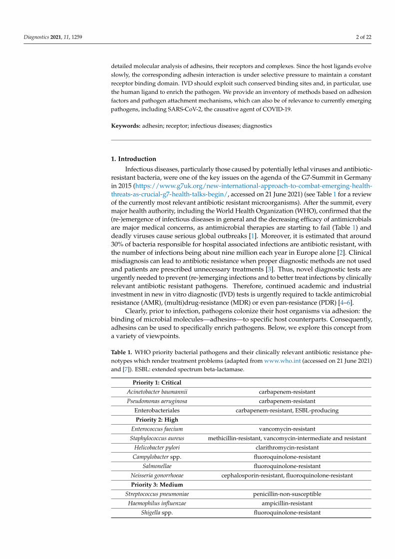
Infectious diseases, particularly those caused by potentially lethal viruses and antibiotic-resistant bacteria, were one of the key issues on the agenda of the G7-Summit in Germanyin 2015 (https://www.g7uk.org/new-international-approach-to-combat-emerging-health-threats-as-crucial-g7-health-talks-begin/, accessed on 21 June 2021) (see Table 1 for a reviewof the currently most relevant antibiotic resistant microorganisms). After the summit, everymajor health authority, including the World Health Organization (WHO), confirmed that the(re-)emergence of infectious diseases in general and the decreasing efficacy of antimicrobialsare major medical concerns, as antimicrobial therapies are starting to fail (Table 1) anddeadly viruses cause serious global outbreaks [1]. Moreover, it is estimated that around30% of bacteria responsible for hospital associated infections are antibiotic resistant, withthe number of infections being about nine million each year in Europe alone [2]. Clinicalmisdiagnosis can lead to antibiotic resistance when proper diagnostic methods are not usedand patients are prescribed unnecessary treatments [3]. Thus, novel diagnostic tests areurgently needed to prevent (re-)emerging infections and to better treat infections by clinicallyrelevant antibiotic resistant pathogens. Therefore, continued academic and industrialinvestment in new in vitro diagnostic (IVD) tests is urgently required to tackle antimicrobialresistance (AMR), (multi)drug-resistance (MDR) or even pan-resistance (PDR) [4–6].
Clearly, prior to infection, pathogens colonize their host organisms via adhesion: thebinding of microbial molecules—adhesins—to specific host counterparts. Consequently,adhesins can be used to specifically enrich pathogens. Below, we explore this concept froma variety of viewpoints.
Table 1. WHO priority bacterial pathogens and their clinically relevant antibiotic resistance phe-notypes which render treatment problems (adapted from www.who.int (accessed on 21 June 2021)and [7]). ESBL: extended spectrum beta-lactamase.
Priority 1: Critical
Acinetobacter baumannii carbapenem-resistant
Pseudomonas aeruginosa carbapenem-resistant
Enterobacteriales carbapenem-resistant, ESBL-producing
Priority 2: High
Enterococcus faecium vancomycin-resistant
Staphylococcus aureus methicillin-resistant, vancomycin-intermediate and resistant
Helicobacter pylori clarithromycin-resistant
Campylobacter spp. fluoroquinolone-resistant
Salmonellae fluoroquinolone-resistant
Neisseria gonorrhoeae cephalosporin-resistant, fluoroquinolone-resistant
Priority 3: Medium
Streptococcus pneumoniae penicillin-non-susceptible
Haemophilus influenzae ampicillin-resistant
Shigella spp. fluoroquinolone-resistant

Diagnostics 2021, 11, 1259 3 of 22
1.1. Microbial Adhesion, Colonization and Host Infection
The severity of an infectious disease depends on the level of invasiveness and theextent of host cell and tissue damage caused by the pathogen involved [8]. A varying degreeof virulence can be observed across different pathogens and among different strains of asingle pathogen [9]. Virulence is a complicated concept in microbial pathogenesis since itdepends not only on the infectious agent but also on host cell susceptibility [10]. Adhesion isat the heart of virulence: it plays the initial and decisive role in colonization and subsequentinfection (Figure 1) [11–14]. Bacterial, viral and parasitic pathogens use adhesins to bindto individual host cells and establish interactions with host molecules, thereby initiatingcolonization. The exact nature of the interaction between pathogen surface moleculesand cell receptors defines the cellular or tissue specificity. Such interactions can invokemechanisms of immune evasion, as adhesion can directly result in the modulation of thehost immune response [15]. Finally, not all adhesins have yet been identified, let alonecharacterized; and, in general, the precise role of adhesins in tissue tropism needs furtherstudy. Different bacterial species may target similar or different host ligands using differenttypes of adhesins. At the same time, an individual bacterial species typically harborsmultiple adhesion systems with different molecular targets. Understanding and exploitingthe molecular basis of pathogen adhesion and the resulting adhesion behavior of wholecells will lead to new formats of diagnostic testing. We propose that cross-disciplinary andtranslational research is needed to improve our fundamental understanding of adhesionbiology, and to translate this knowledge into novel detection strategies based on host–pathogen interaction [16].
Diagnostics 2021, 11, x FOR PEER REVIEW 3 of 23
1.1. Microbial Adhesion, Colonization and Host Infection The severity of an infectious disease depends on the level of invasiveness and the
extent of host cell and tissue damage caused by the pathogen involved [8]. A varying de-gree of virulence can be observed across different pathogens and among different strains of a single pathogen [9]. Virulence is a complicated concept in microbial pathogenesis since it depends not only on the infectious agent but also on host cell susceptibility [10]. Adhesion is at the heart of virulence: it plays the initial and decisive role in colonization and subsequent infection (Figure 1) [11–14]. Bacterial, viral and parasitic pathogens use adhesins to bind to individual host cells and establish interactions with host molecules, thereby initiating colonization. The exact nature of the interaction between pathogen sur-face molecules and cell receptors defines the cellular or tissue specificity. Such interactions can invoke mechanisms of immune evasion, as adhesion can directly result in the modu-lation of the host immune response [15]. Finally, not all adhesins have yet been identified, let alone characterized; and, in general, the precise role of adhesins in tissue tropism needs further study. Different bacterial species may target similar or different host ligands using different types of adhesins. At the same time, an individual bacterial species typically har-bors multiple adhesion systems with different molecular targets. Understanding and ex-ploiting the molecular basis of pathogen adhesion and the resulting adhesion behavior of whole cells will lead to new formats of diagnostic testing. We propose that cross-discipli-nary and translational research is needed to improve our fundamental understanding of adhesion biology, and to translate this knowledge into novel detection strategies based on host–pathogen interaction [16].
Figure 1. Adhesion of Bartonella henselae to human cells. B. henselae (strain Marseille) bacteria (light blue) in an early stage infection process (30 min) to human HeLa-229 cells (red). Adhesion to host Figure 1. Adhesion of Bartonella henselae to human cells. B. henselae (strain Marseille) bacteria (light
blue) in an early stage infection process (30 min) to human HeLa-229 cells (red). Adhesion to hostcells is mediated by specific interactions between B. henselae surface proteins and components of thehost extracellular matrix including molecules such as fibronectin or collagen. Scale bar: 8 µm.
Diagnostics 2021, 11, 1259 4 of 22
1.2. Current State of Infectious Disease Diagnostics
The core technologies used by the routine microbiology laboratory are still mostlymicroscopy- and culture-based. Immunological tests detecting pathogen-specific antigensand antibodies are also routinely used in diagnostics. Furthermore, over the past years,matrix-assisted laser desorption ionization-time of flight mass spectrometry (MALDI-TOFMS) and molecular (nucleic acid-targeting) testing have been introduced successfully inthe routine clinical microbiology laboratory (for some recent reviews see [17,18]).
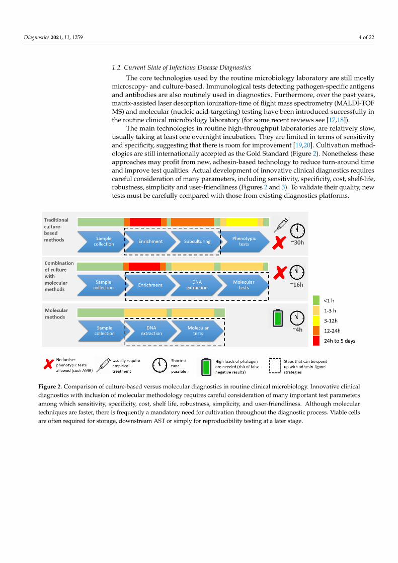
The main technologies in routine high-throughput laboratories are relatively slow,usually taking at least one overnight incubation. They are limited in terms of sensitivityand specificity, suggesting that there is room for improvement [19,20]. Cultivation method-ologies are still internationally accepted as the Gold Standard (Figure 2). Nonetheless theseapproaches may profit from new, adhesin-based technology to reduce turn-around timeand improve test qualities. Actual development of innovative clinical diagnostics requirescareful consideration of many parameters, including sensitivity, specificity, cost, shelf-life,robustness, simplicity and user-friendliness (Figures 2 and 3). To validate their quality, newtests must be carefully compared with those from existing diagnostics platforms.
Diagnostics 2021, 11, x FOR PEER REVIEW 4 of 23
cells is mediated by specific interactions between B. henselae surface proteins and components of the host extracellular matrix including molecules such as fibronectin or collagen. Scale bar: 8 μm.
1.2. Current State of Infectious Disease Diagnostics The core technologies used by the routine microbiology laboratory are still mostly
microscopy- and culture-based. Immunological tests detecting pathogen-specific antigens and antibodies are also routinely used in diagnostics. Furthermore, over the past years, matrix-assisted laser desorption ionization-time of flight mass spectrometry (MALDI-TOF MS) and molecular (nucleic acid-targeting) testing have been introduced successfully in the routine clinical microbiology laboratory (for some recent reviews see [17,18]).
The main technologies in routine high-throughput laboratories are relatively slow, usually taking at least one overnight incubation. They are limited in terms of sensitivity and specificity, suggesting that there is room for improvement [19,20]. Cultivation meth-odologies are still internationally accepted as the Gold Standard (Figure 2). Nonetheless these approaches may profit from new, adhesin-based technology to reduce turn-around time and improve test qualities. Actual development of innovative clinical diagnostics re-quires careful consideration of many parameters, including sensitivity, specificity, cost, shelf-life, robustness, simplicity and user-friendliness (Figures 2 and 3). To validate their quality, new tests must be carefully compared with those from existing diagnostics plat-forms.
Figure 2. Comparison of culture-based versus molecular diagnostics in routine clinical microbiology. Innovative clinical diagnostics with inclusion of molecular methodology requires careful consideration of many important test parameters among which sensitivity, specificity, cost, shelf life, robustness, simplicity, and user-friendliness. Although molecular techniques are faster, there is frequently a mandatory need for cultivation throughout the diagnostic process. Viable cells are often required for storage, downstream AST or simply for reproducibility testing at a later stage.
Figure 2. Comparison of culture-based versus molecular diagnostics in routine clinical microbiology. Innovative clinicaldiagnostics with inclusion of molecular methodology requires careful consideration of many important test parametersamong which sensitivity, specificity, cost, shelf life, robustness, simplicity, and user-friendliness. Although moleculartechniques are faster, there is frequently a mandatory need for cultivation throughout the diagnostic process. Viable cellsare often required for storage, downstream AST or simply for reproducibility testing at a later stage.
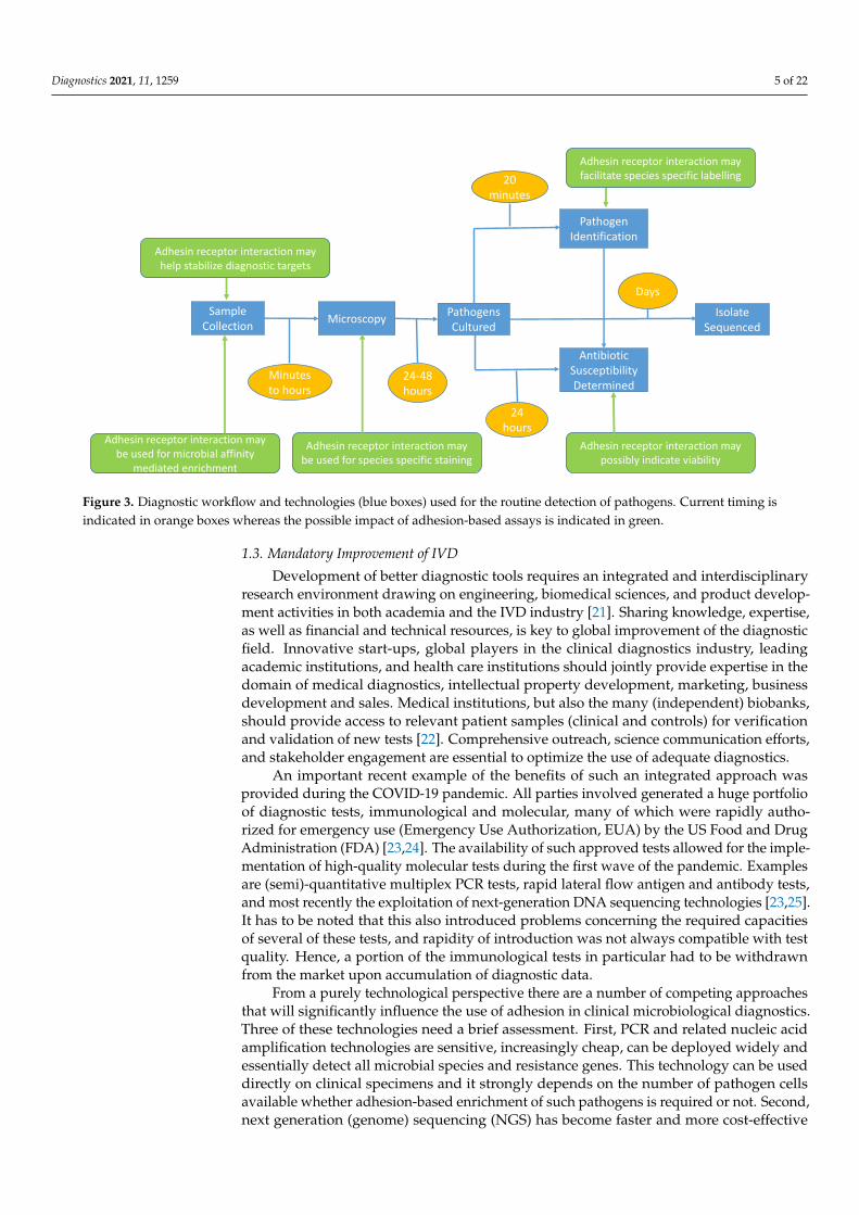
Diagnostics 2021, 11, 1259 5 of 22Diagnostics 2021, 11, x FOR PEER REVIEW 5 of 23
Figure 3. Diagnostic workflow and technologies (blue boxes) used for the routine detection of pathogens. Current timing is indi-cated in orange boxes whereas the possible impact of adhesion-based assays is indicated in green.
1.3. Mandatory Improvement of IVD Development of better diagnostic tools requires an integrated and interdisciplinary
research environment drawing on engineering, biomedical sciences, and product devel-opment activities in both academia and the IVD industry [21]. Sharing knowledge, exper-tise, as well as financial and technical resources, is key to global improvement of the di-agnostic field. Innovative start-ups, global players in the clinical diagnostics industry, leading academic institutions, and health care institutions should jointly provide expertise in the domain of medical diagnostics, intellectual property development, marketing, busi-ness development and sales. Medical institutions, but also the many (independent) bi-obanks, should provide access to relevant patient samples (clinical and controls) for veri-fication and validation of new tests [22]. Comprehensive outreach, science communication efforts, and stakeholder engagement are essential to optimize the use of adequate diag-nostics.
An important recent example of the benefits of such an integrated approach was pro-vided during the COVID-19 pandemic. All parties involved generated a huge portfolio of diagnostic tests, immunological and molecular, many of which were rapidly authorized for emergency use (Emergency Use Authorization, EUA) by the US Food and Drug Ad-ministration (FDA) [23,24]. The availability of such approved tests allowed for the imple-mentation of high-quality molecular tests during the first wave of the pandemic. Exam-ples are (semi)-quantitative multiplex PCR tests, rapid lateral flow antigen and antibody tests, and most recently the exploitation of next-generation DNA sequencing technologies [23,25]. It has to be noted that this also introduced problems concerning the required ca-pacities of several of these tests, and rapidity of introduction was not always compatible with test quality. Hence, a portion of the immunological tests in particular had to be with-drawn from the market upon accumulation of diagnostic data.
From a purely technological perspective there are a number of competing approaches that will significantly influence the use of adhesion in clinical microbiological diagnostics. Three of these technologies need a brief assessment. First, PCR and related nucleic acid amplification technologies are sensitive, increasingly cheap, can be deployed widely and essentially detect all microbial species and resistance genes. This technology can be used directly on clinical specimens and it strongly depends on the number of pathogen cells
Sample Collection Microscopy Pathogens
Cultured
Antibiotic Susceptibility Determined
Pathogen Identification
Isolate Sequenced
Minutes to hours
24-48 hours
24 hours
20 minutes
Days
Adhesin receptor interaction may help stabilize diagnostic targets
Adhesin receptor interaction may be used for species specific staining
Adhesin receptor interaction may facilitate species specific labelling
Adhesin receptor interaction may possibly indicate viability
Adhesin receptor interaction may be used for microbial affinity
mediated enrichment
Figure 3. Diagnostic workflow and technologies (blue boxes) used for the routine detection of pathogens. Current timing isindicated in orange boxes whereas the possible impact of adhesion-based assays is indicated in green.
1.3. Mandatory Improvement of IVD
Development of better diagnostic tools requires an integrated and interdisciplinaryresearch environment drawing on engineering, biomedical sciences, and product develop-ment activities in both academia and the IVD industry [21]. Sharing knowledge, expertise,as well as financial and technical resources, is key to global improvement of the diagnosticfield. Innovative start-ups, global players in the clinical diagnostics industry, leadingacademic institutions, and health care institutions should jointly provide expertise in thedomain of medical diagnostics, intellectual property development, marketing, businessdevelopment and sales. Medical institutions, but also the many (independent) biobanks,should provide access to relevant patient samples (clinical and controls) for verificationand validation of new tests [22]. Comprehensive outreach, science communication efforts,and stakeholder engagement are essential to optimize the use of adequate diagnostics.
An important recent example of the benefits of such an integrated approach wasprovided during the COVID-19 pandemic. All parties involved generated a huge portfolioof diagnostic tests, immunological and molecular, many of which were rapidly autho-rized for emergency use (Emergency Use Authorization, EUA) by the US Food and DrugAdministration (FDA) [23,24]. The availability of such approved tests allowed for the imple-mentation of high-quality molecular tests during the first wave of the pandemic. Examplesare (semi)-quantitative multiplex PCR tests, rapid lateral flow antigen and antibody tests,and most recently the exploitation of next-generation DNA sequencing technologies [23,25].It has to be noted that this also introduced problems concerning the required capacitiesof several of these tests, and rapidity of introduction was not always compatible with testquality. Hence, a portion of the immunological tests in particular had to be withdrawnfrom the market upon accumulation of diagnostic data.
From a purely technological perspective there are a number of competing approachesthat will significantly influence the use of adhesion in clinical microbiological diagnostics.Three of these technologies need a brief assessment. First, PCR and related nucleic acidamplification technologies are sensitive, increasingly cheap, can be deployed widely andessentially detect all microbial species and resistance genes. This technology can be useddirectly on clinical specimens and it strongly depends on the number of pathogen cellsavailable whether adhesion-based enrichment of such pathogens is required or not. Second,next generation (genome) sequencing (NGS) has become faster and more cost-effective
Diagnostics 2021, 11, 1259 6 of 22
over recent decades, and this is likely to continue. Its performance will soon equal orbetter that of amplification testing. This suggests that NGS will be routinely used in themicrobiology lab. Finally, there is an increase in the rapid availability of high-affinityspecific binding reagents other than functional adhesins, such as (monoclonal) antibodies,adhirons and aptamers [26,27]. These have the advantage that they share a basic molecularstructure, rendering them suitable for “plug and play” diagnostic applications using thesame platforms. If a good diagnostic platform has also been developed, essentially allbinding reagents can be applied. Adhesion based assays will have to compete with testsbased upon the three concepts mentioned above.
In the following sections, we describe the major interactions between microbial ad-hesins and host ligands or receptors. We try to define what further structural biology studiesare needed, and how these might be useful in the development of novel diagnostic tests.
2. Microbial Adhesin–Receptor Pairs
The initial interaction between pathogens and their hosts is defined at the molecularlevel by the selective interaction of pathogen adhesins with their host receptors. Thisspecific interplay can be exploited in various stages of the classical microbiological diag-nostic workflow. To do so, we must extend our understanding of the basic principles ofpathogen adhesion so we can apply adhesion assays in the initial capture and enrichmentof (complete or parts of) pathogens [28]. All microbial adhesion molecules are surfaceexposed structures, but their expression may depend on physiological parameters such asenvironmental temperature, growth stage or availability of nutrients [29,30]. Understand-ing precisely how microbial adhesins interact with their host receptors poses challengesbecause the receptor may, for instance, be part of a structurally complex cellular mem-brane [31]. Site-directed mutagenesis and adhesion assays with whole cells or purifiedadhesins have shed light on basic aspects of the binding interactions [32].
The emergence of SARS-CoV-2 has made it clear that not all adhesins have been iden-tified yet. New ones can be detected using nucleic acid sequencing strategies and searchesfor new structures that are homologous to known adhesins. Otherwise, proteomic researchcan be applied to detect cellular surface proteins that provide adhesive characteristics.Random knock-out mutagenesis can also generate cells deficient in adhesion and reversegenetics then allows the functional analysis of the genes and proteins involved. Biophysicaltechnologies can be used to define adhesin structures (see sections below).
2.1. Viral Adhesion Processes
Viruses, with relatively small genomes, have a limited repertoire of adhesin structuresper individual viral lineage, although an individual adhesin structure is repeated frequentlyon a single virion. It has to be noted that viruses that are becoming endemic or pandemicexist, even in single hosts, as a species swarm with differing receptor affinities. Recentexamples include the SARS-CoV-2 variants such as the B.1.1.7 (α-variant, UK), B.1.351(β-variant, South Africa), P.1 (γ-variant, Brazilian) and B.617.2 (δ-variant, India) variants ofconcern [33]. The viral surface is normally quite homogenous allowing for fewer possiblereceptor specificities [34]. Viral interactions with glycan-based receptors are frequent buttypically have affinities in the mM range, while interactions with protein receptors areusually of higher affinity [35–39]. For example, human coronavirus NL63 (HCoV-NL63)uses heparan sulfate proteoglycans (HSPGs) as the initial host receptor. The membraneprotein (M) of HCoV-NL63 mediates this attachment to HSPGs and is not spike (S) protein-dependent. It was recently shown that the M protein is also an important player duringthe early stages of HCoV-NL63 infection, thereby identifying a new adhesin for thisvirus [40]. Both fungi and viruses exploit a variety of immune modulators to achievehost colonization [41,42]. Recent examples of human receptors relevant for SARS-CoV-2adhesion are described in Box 1. The emergence of new viruses will undoubtedly lead tothe identification of new viral adhesins.
Diagnostics 2021, 11, 1259 7 of 22
Box 1. Adhesion of SARS-CoV-2, the causative agent of COVID-19.
The severe acute respiratory syndrome coronavirus-2 (SARS-CoV-2) causes coronavirus disease2019 (COVID-19). The precise mechanisms of disease are still incompletely understood [43] andreplicating virus particles can be observed in a variety of host tissues. Nonetheless, two key hostreceptors have been identified: angiotensin-converting enzyme 2 (ACE2) [44,45] and liver/lymphnode-specific intracellular adhesion molecule-3 grabbing non-integrin (L-SIGN) [46,47]. The crystalstructure for ACE2 complexed with the receptor binding domain (RBD) of the SARS-CoV-2 spikeprotein was solved [48,49]. The binary complex showed clear conservation as compared to similarcomplexes for the original SARS-CoV-1 virus. This hints at functional conservation of the process ofACE2 binding, but also at the possibility of immunological cross-influences between SARS-CoV-1and SARS-CoV-2 antibodies. Other forms of structure-based strategies would be the use of the RBDas a subunit vaccine [50] or as a target for inhibition by possible compounds. Neuropilin recognizesa furin cleavage on the SARS-CoV-2 spike protein and is a key target in the development oftherapeutics against COVID-19. Recently, X-ray structure-based studies of the neuropilin complexedto the S fragment of the spike protein indicated potentially important design opportunities fortherapeutic compounds [13,14]. In addition, virtual drug screening and actual high-throughputscreening of compound libraries has been exploited for the key SARS-CoV-2 protease MPRO, whichled to the successful identification of potential antiviral drugs [51]. Despite a wide variety ofnew tests [23], formats based on anti-adhesive strategies have not yet been developed for thispriority pathogen.
2.2. Modes of Bacterial Adhesion
The nature of the bacterial adhesion molecule varies from distinct organelles suchas flagellae or fimbriae to surface exposed, cell wall- or cell membrane-attached proteins,lipids, and sugar (poly- or oligo-saccharide) moieties [52]. Adhesins, especially proteina-ceous ones, are often repetitive in primary structure, either by repeating similar domainswithin a protein chain or by polymerizing subunits into long fibrous structures [53,54].Most bacterial adhesins tend to bind to host structures that are also often structurally repet-itive and ubiquitously distributed, including extracellular matrix (ECM) components suchas collagen, fibronectin, or glycoprotein receptors that harbor repeating carbohydrate units.
In certain bacterial adhesins, such as the trimeric autotransporter adhesins (TAAs) [55],the individual binding affinities can be very low (0.1–0.5 M). In this context, binding ofpathogens to host cell surfaces is accomplished by avidity, like ‘Velcro’ on a shoe: thethree-dimensional arrangement of multiple weak binding sites leads to tight and henceeffective binding [56–58]. TAAs are can be divided into three domains; a membraneanchored β-barrel domain, a stalk domain and a head domain [55,59,60]. The head domain,once assembled, then adheres to the host ECM via, for example, collagen, vitronectin orfibronectin [58]. Recent work showed that different adhesins bind differently to ECMcomponents and that binding is dramatically influenced by shear forces [56,61–63]. Ingeneral, improving our understanding of adhesin–receptor interaction requires moredetailed insights into their structural aspects [64].
2.3. Adhesion Diversity and Evolution
Surface exposed adhesion domains are external moieties and exposed parts of theproteins may be subject to strong environmental selection and possible natural adapta-tion [65,66]. Hence, the evolution of pathogens is critically linked to the variation inadhesins and their receptor affinities [67], potentially allowing for the colonization of novelhosts. Evolutionary changes in adhesins and ligands can be easily identified by NGScombined with quantitative MS-based proteomics, a combination referred to as proteo-genomics [68–71]. For instance, conserved peptide sequences (conserved at least within thesame species) can be used to perform quantification of species–specific peptides in complex(patient-derived) samples [72–74]. The combination of these two methods thus facilitatesthe correlation of genotypes with adhesion-related phenotypes. There is a continued needfor the characterization of additional adhesin–ligand pairs to define conservation of theadhesin or ligand between microbial species.

Diagnostics 2021, 11, 1259 8 of 22
3. Structural Analysis of Adhesin–Ligand Pairs
There are few structures of bacterial adhesins complexed with their ligands, eventhough there are many of virus-receptor complexes [35,36]. This may be due to the low-affinity/high-avidity binding of bacterial adhesins, leading to many different complexesand frequent non-specific aggregation. This is problematic because (a) it makes it hardto define the biologically relevant interactions and (b) structural techniques, even cryo-electron microscopy, depend on having a small number (<10) of different conformationsand complexes in a single experiment. The modular repetitive structure of some bacterialadhesins and of their host receptors (collagen, laminin, fibronectin, etc.) hampers thedetermination of their structure and specific interactions by standard methods. Nonetheless,we believe that structural investigations will contribute to the design of better adhesinconstructs. These could in return serve as diagnostic tools, as vaccine components, andpotentially to develop anti-adhesive drugs.
Technological and Methodological Developments
NMR spectroscopy, X-ray crystallography, cryo-electron microscopy (cryoEM; see [75,76]for reviews), and mass spectrometry (MS) can be used to characterize the individual adhesinbinding domains or their receptors in molecular detail. CryoEM and, to a lesser extent,X-ray crystallography are the best methods for higher-order assemblies. Furthermore,cross-linking MS (XL–MS) and hydrogen-deuterium exchange MS (HDX–MS) are beingincreasingly used to determine structural constraints between interacting proteins, proteincomplexes, and their binding interfaces [77–81]. Such constraints can facilitate bindingoptimization, improvement of ligand design, and thus improved capture for pre-analyticaldiagnostic steps [82–88].
Nano-biosensors for miniaturized detection of adhesion, (cryo)EM for visualizationof adhesion and identification of molecular partners, X-ray crystallography for the defi-nition of global adhesin structure and more generic tools like NMR for local informationon binding partners, and advanced bioinformatics all play essential roles in the furtheroptimization of structure determination and translational applications [89–91]. Integrativemethods for structure determination of adhesin complexes and for defining their clinicalrelevance have been shown to be useful [82,83,85,87,92]. For example, structure analysis ofYersinia enterocolitica YadA helped to further the understanding of interleukin-1 expressionby epithelial cells [93]. The structure of the Escherichia coli immunoglobulin binding pro-teins (Eibs) showed how they are involved in entero–hemorrhagic pathogenicity [94,95].Another innovative tool that was developed for use in molecular recognition applicationswas the use of “adhirons” to help stabilize complexes and to gain structural information(e.g., [96]). Adhirons are non-antibody scaffold binding proteins [97]. Well-characterizedadhirons display low-nanomolar affinity and high specificity for defined proteins andselectively recognize their target molecules.
4. Microbial Adhesion and Future High-Throughput DiagnosticMicrobiology Technology
Exploiting the adhesion capacity of pathogens for in vitro diagnostics is a relativelynew concept [98,99], but adhesion-based principles can be applied at various stages of thediagnostic process: for specific staining of bacteria, for enhanced species identification, forantimicrobial susceptibility testing, or ultimately maybe even for in vivo therapy (Figure 3).Therefore, prerequisites for further translation into clinical practice are bottom-up researchin adhesion from a clinical research perspective, the definition of molecular structure–function relationships, and the design and development of new diagnostic tests anddevices [100].
4.1. Adhesin-Based Sample Processing in Microbial Diagnostics
Bacterial or viral detection and identification is a complicated multi-step processstarting with the collection of clinical samples of diverse origin (blood, sputum, saliva,
Diagnostics 2021, 11, 1259 9 of 22
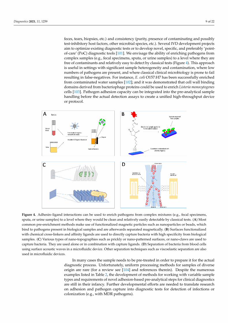
feces, tears, biopsies, etc.) and consistency (purity, presence of contaminating and possiblytest-inhibitory host factors, other microbial species, etc.). Several IVD development projectsaim to optimize existing diagnostic tests or to develop novel, specific, and preferably ‘point-of-care’ (PoC) diagnostic tools [101]. We envisage the ability of enriching pathogens fromcomplex samples (e.g., fecal specimens, sputa, or urine samples) to a level where they arefree of contaminants and relatively easy to detect by classical tests (Figure 4). This approachis useful in settings with significant sample heterogeneity and contamination, where lownumbers of pathogens are present, and where classical clinical microbiology is prone to failresulting in false-negatives. For instance, E. coli O157:H7 has been successfully enrichedfrom contaminated water samples [102]; and it was demonstrated that cell wall bindingdomains derived from bacteriophage proteins could be used to enrich Listeria monocytogenescells [103]. Pathogen adhesion capacity can be integrated into the pre-analytical samplehandling before the actual detection assays to create a unified high-throughput deviceor protocol.
Diagnostics 2021, 11, x FOR PEER REVIEW 9 of 23
diagnostic process: for specific staining of bacteria, for enhanced species identification, for antimicrobial susceptibility testing, or ultimately maybe even for in vivo therapy (Figure 3). Therefore, prerequisites for further translation into clinical practice are bottom-up re-search in adhesion from a clinical research perspective, the definition of molecular struc-ture–function relationships, and the design and development of new diagnostic tests and devices [100].
4.1. Adhesin-Based Sample Processing in Microbial Diagnostics Bacterial or viral detection and identification is a complicated multi-step process
starting with the collection of clinical samples of diverse origin (blood, sputum, saliva, feces, tears, biopsies, etc.) and consistency (purity, presence of contaminating and possibly test-inhibitory host factors, other microbial species, etc.). Several IVD development pro-jects aim to optimize existing diagnostic tests or to develop novel, specific, and preferably ‘point-of-care’ (PoC) diagnostic tools [101]. We envisage the ability of enriching pathogens from complex samples (e.g., fecal specimens, sputa, or urine samples) to a level where they are free of contaminants and relatively easy to detect by classical tests (Figure 4). This approach is useful in settings with significant sample heterogeneity and contamination, where low numbers of pathogens are present, and where classical clinical microbiology is prone to fail resulting in false-negatives. For instance, E. coli O157:H7 has been success-fully enriched from contaminated water samples [102]; and it was demonstrated that cell wall binding domains derived from bacteriophage proteins could be used to enrich Lis-teria monocytogenes cells [103]. Pathogen adhesion capacity can be integrated into the pre-analytical sample handling before the actual detection assays to create a unified high-throughput device or protocol.
Figure 4. Adhesin–ligand interactions can be used to enrich pathogens from complex mixtures (e.g., fecal specimens, sputa, or urine samples) to a level where they would be clean and relatively easily detectable by classical tests. (A) Most common pre-enrichment methods make use of func-tionalized magnetic particles such as nanoparticles or beads, which bind to pathogens present in biological samples and are afterwards separated magnetically. (B) Surfaces functionalized with chemical cross-linkers and affinity ligands are used to directly capture bacteria with high specific-ity from biological samples. (C) Various types of nano-topographies such as prickly or nano-pat-terned surfaces, or nano-claws are used to capture bacteria. They are used alone or in combination with capture ligands. (D) Separation of bacteria from blood cells using surface acoustic waves in a
Figure 4. Adhesin–ligand interactions can be used to enrich pathogens from complex mixtures (e.g., fecal specimens,sputa, or urine samples) to a level where they would be clean and relatively easily detectable by classical tests. (A) Mostcommon pre-enrichment methods make use of functionalized magnetic particles such as nanoparticles or beads, whichbind to pathogens present in biological samples and are afterwards separated magnetically. (B) Surfaces functionalizedwith chemical cross-linkers and affinity ligands are used to directly capture bacteria with high specificity from biologicalsamples. (C) Various types of nano-topographies such as prickly or nano-patterned surfaces, or nano-claws are used tocapture bacteria. They are used alone or in combination with capture ligands. (D) Separation of bacteria from blood cellsusing surface acoustic waves in a microfluidic device. Other separation techniques such as viscoelastic separation are alsoused in microfluidic devices.
In many cases the sample needs to be pre-treated in order to prepare it for the actualdiagnostic process. Unfortunately, uniform processing methods for samples of diverseorigin are rare (for a review see [104] and references therein). Despite the numerousexamples listed in Table 2, the development of methods for working with variable sampletypes and requirements of novel adhesion-based pre-analytical steps for clinical diagnosticsare still in their infancy. Further developmental efforts are needed to translate researchon adhesion and pathogen capture into diagnostic tests for detection of infections orcolonization (e.g., with MDR pathogens).
Diagnostics 2021, 11, 1259 10 of 22
Progress has been made with certain receptors and bacterial ligands, however. Man-nose binding lectin is a host receptor capable of signaling or sensing pathogens exposingmannose at their outer cell surface, and can thus be used to capture a variety of micro-bial species [105,106]. If mannose binding lectin is attached to a solid surface, mannose-presenting pathogens can be captured on the surface [107]. This approach allows highlysensitive detection of pathogens from a clinical sample at the capturing surface and isapplicable in a variety of downstream classical and molecular diagnostic methods. It hasbeen successfully applied in sepsis testing in experimental animals, where an extra-corporalblood-cleansing device was developed to detect pathogens circulating in their blood [108].
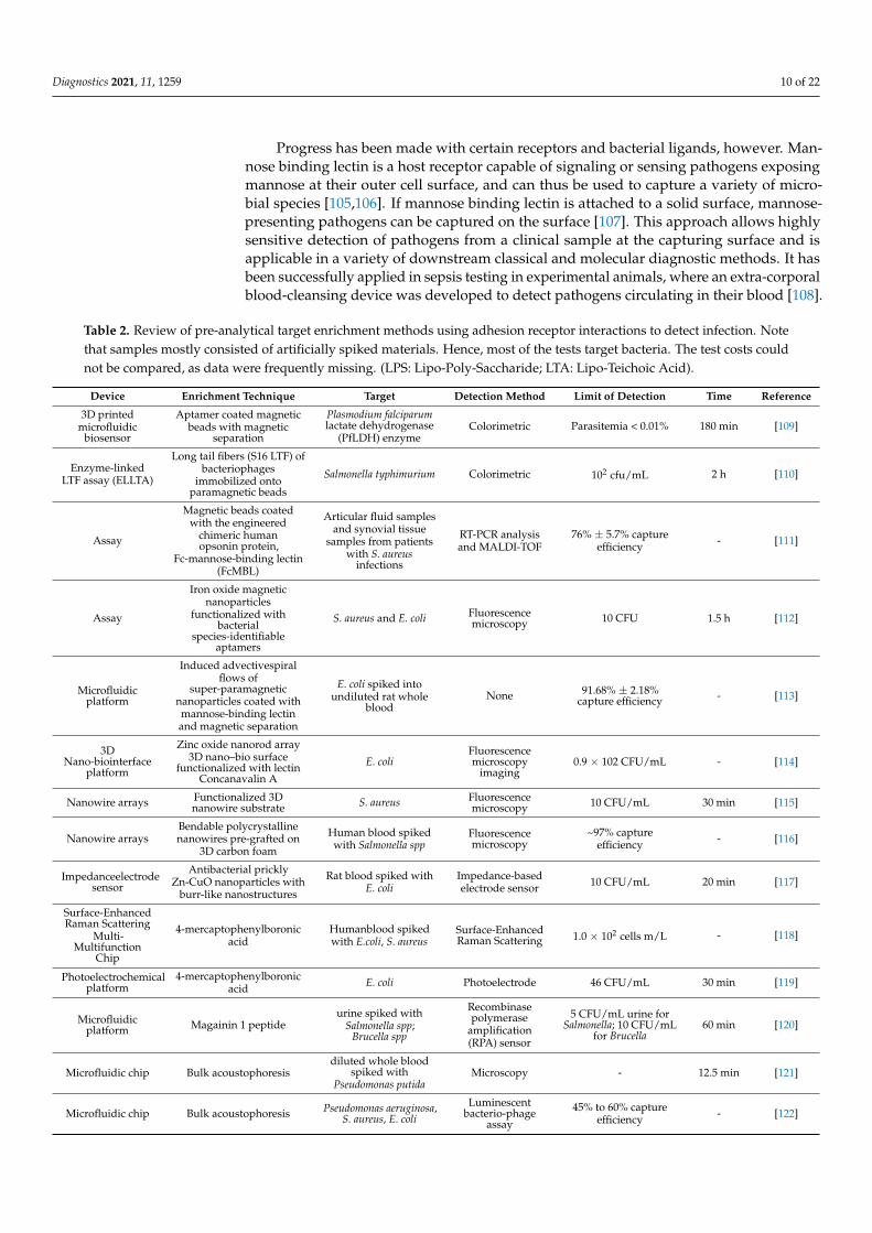
Table 2. Review of pre-analytical target enrichment methods using adhesion receptor interactions to detect infection. Notethat samples mostly consisted of artificially spiked materials. Hence, most of the tests target bacteria. The test costs couldnot be compared, as data were frequently missing. (LPS: Lipo-Poly-Saccharide; LTA: Lipo-Teichoic Acid).
Device Enrichment Technique Target Detection Method Limit of Detection Time Reference
3D printedmicrofluidic
biosensor
Aptamer coated magneticbeads with magnetic
separation
Plasmodium falciparumlactate dehydrogenase
(PfLDH) enzymeColorimetric Parasitemia < 0.01% 180 min [109]
Enzyme-linkedLTF assay (ELLTA)
Long tail fibers (S16 LTF) ofbacteriophages
immobilized ontoparamagnetic beads
Salmonella typhimurium Colorimetric 102 cfu/mL 2 h [110]
Assay
Magnetic beads coatedwith the engineered
chimeric humanopsonin protein,
Fc-mannose-binding lectin(FcMBL)
Articular fluid samplesand synovial tissue
samples from patientswith S. aureus
infections
RT-PCR analysisand MALDI-TOF
76% ± 5.7% captureefficiency - [111]
Assay
Iron oxide magneticnanoparticles
functionalized withbacterial
species-identifiableaptamers
S. aureus and E. coli Fluorescencemicroscopy 10 CFU 1.5 h [112]
Microfluidicplatform
Induced advectivespiralflows of
super-paramagneticnanoparticles coated withmannose-binding lectinand magnetic separation
E. coli spiked intoundiluted rat whole
bloodNone 91.68% ± 2.18%
capture efficiency - [113]
3DNano-biointerface
platform
Zinc oxide nanorod array3D nano–bio surface
functionalized with lectinConcanavalin A
E. coliFluorescencemicroscopy
imaging0.9 × 102 CFU/mL - [114]
Nanowire arrays Functionalized 3Dnanowire substrate S. aureus Fluorescence
microscopy 10 CFU/mL 30 min [115]
Nanowire arraysBendable polycrystallinenanowires pre-grafted on
3D carbon foam
Human blood spikedwith Salmonella spp
Fluorescencemicroscopy
~97% captureefficiency - [116]
Impedanceelectrodesensor
Antibacterial pricklyZn-CuO nanoparticles with
burr-like nanostructures
Rat blood spiked withE. coli
Impedance-basedelectrode sensor 10 CFU/mL 20 min [117]
Surface-EnhancedRaman Scattering
Multi-Multifunction
Chip
4-mercaptophenylboronicacid
Humanblood spikedwith E.coli, S. aureus
Surface-EnhancedRaman Scattering 1.0 × 10² cells m/L - [118]
Photoelectrochemicalplatform
4-mercaptophenylboronicacid E. coli Photoelectrode 46 CFU/mL 30 min [119]
Microfluidicplatform Magainin 1 peptide
urine spiked withSalmonella spp;
Brucella spp
Recombinasepolymerase
amplification(RPA) sensor
5 CFU/mL urine forSalmonella; 10 CFU/mL
for Brucella60 min [120]
Microfluidic chip Bulk acoustophoresisdiluted whole blood
spiked withPseudomonas putida
Microscopy - 12.5 min [121]
Microfluidic chip Bulk acoustophoresis Pseudomonas aeruginosa,S. aureus, E. coli
Luminescentbacterio-phage
assay
45% to 60% captureefficiency - [122]

Diagnostics 2021, 11, 1259 11 of 22
Table 2. Cont.
Device Enrichment Technique Target Detection Method Limit of Detection Time Reference
Microfluidiccapillaric circuit
Antibody-functionalizedmicrobeads
synthetic urine spikedwith E. coli
Fluorescencemicroscopy 1.2 × 10² CFU/mL 7 min [123]
Microfluidic chip
Pillar-assistedself-assemblymicroparticlesNano- filter for
E. coli from samples Fluorescencemicroscopy
capture efficiencyof 93% - [124]
Reusablesupramolecular
platform
Multilayered film andβ-cyclodextrin (β-CD)
derivatives modified withmannose
Type I fimbriae E. coliand lectin proteins
Fluorescencemicroscopy
Capture efficiencyof 93% - [125]
Photonic PCRon a chip
Gravity-driven cellenrichment E. coli Photonic PCR on a
chip 103 CFU/mL 10 min [126]
Enzyme-linkedlectin sorbent
assay (ELLecSA)Fc-mannose-binding lectin
Bacteria, fungi, virus,parasites. LPS, LTAfrom Gram-negativeand Gram-positivebacteria, as well as
lipo-arabino-mannan(LAM) and
phosphatidyl-inositolmannoside from M.
tuberculosis
Scanning electronmicroscopy - <1 h [114]
Fluorometric assay
Two distinct terminalphosphate-labeled LPS
specific aptamers attachedonto Zr-MOFs to fabricatethe magnetic core-shell for
magnetic separation
Acinetobacter baumanniiin blood samples
Fluorescent signalamplification by
fluorescenceprobes
10 cfu/mL ~2.5 h [127]
A second example concerns protein A (SpA), a surface protein expressed by Staphy-lococcus aureus and other species of coagulase positive staphylococci [128]. SpA has highaffinity for the Fc region of IgG antibodies. When immobilized to a solid support, SpAcan be used to affinity purify Langerhans cells expressing receptors for the Fc portion ofIgG (Fc-IgG), thus generating clean specimens that are well suited for various formats ofimmune detection [129]. Binding via the Fc part supports the proper presentation of theantigen-binding sites of not only natural but also monoclonal antibodies [108,130]. SpA hasproven to be an important biotechnological tool not only in the development of immunetests, but also for the purification and concentration of a variety of human and animalantibodies [131]. However, recent work has shown that SpA does not bind all antibodiesuniformly well, an issue that must be kept in mind when developing SpA-mediated proto-cols [132]. With a similar approach, a 50 amino acid residue termed SAP peptide has beenderived from M protein, one of the major virulence factors of Streptococcus pyogenes. Thiscan be used to enrich IgA from biological mixtures [133].
4.2. Target Enrichment Technology
The development of diagnostic tools requires detailed testing in artificially spiked and“real-life” samples from clinical, environmental, and industrial origins. Institutional orcommercially available pathogen strain collections should be used to define the naturalvariation in the adhesins and the effect of such variation on adhesion efficiency and,hence, test quality [134]. This could even work for an unknown pathogen if the adhesinin question were reasonably well-conserved. The SpA of a S. non-aureus strain or anew Coronavirus spike protein are significant examples. The question in novel sampleenrichment approaches is whether there is a need for test devices that can capture most ifnot all clinically relevant pathogens or whether sequential testing would be a better option.In any case, new technologies should preferably allow the direct enrichment of pathogensfrom low-titer samples.
5. Clinical–Diagnostic Application of Pathogen Adhesion Tests
High content proteomics [135], electrochemical biosensors [136,137], lanthanide-basedfluorescent up-conversion particle assays for detection of adhesin–ligand binding [138],

Diagnostics 2021, 11, 1259 12 of 22
and In Situ Hybridization (ISH) using peptide nucleic acid (PNA) probes and nano-biosensors [139] are only four examples of complex experimental technologies to identifyadhesin–receptor interactions, all of which can be translated into new IVD test formats.Such tests can be used in translational research to simplify and accelerate pathogen iden-tification and/or characterization processes (Table 2), which lists tests that are in usesummarizing the core competencies and technologies used in these tests.
Currently used physical test platforms range from microfluidic biosensors and nanowiresto more classical Raman spectroscopy-, electrochemical- or PCR-based equipment. Further-more, enrichment platforms frequently utilize nanorods or -wires when straightforwardanalytical signal detection is required (pathogen present or absent as the final test result).When, after the initial adhesion test, follow-up testing is required in a more preparativemanner, magnetic beads are by far the most common approach. The entities to be detectedcan vary from intact cells, through simple enzymes, to the products of nucleic acid ampli-fication reactions. Of note, nucleic acids rarely play a role in microbial adhesion thoughbiofilms contain relatively large amounts of these molecules and are thought to functionas adhesins under biofilm conditions [140]. Still, in diagnosis, nucleic acids are usuallyemployed because they efficiently hybridize to other nucleic acids. These concepts arebeyond the scope of the current review.
The most commonly used procedures at the level of detection are coloration, fluores-cence measurement, and detection of amplified nucleic acid. The most frequent modelorganisms used are E. coli and S. aureus, representatives of Gram-negative and Gram-positive pathogens, respectively. All methods described generate results in 7 min to 3 h,show high sensitivity (to about ten colony-forming units at their most sensitive) and are ofgreat quality; still, their implementation into routine use is sparse. Table 2 and referencestherein summarize studies that have successfully demonstrated the relevance of adhesionfor improved microbiological testing.
5.1. Biosensor-Based Pathogen Detection
Biosensors, analytical devices that detect and quantify biomolecules or cells, are com-posed of three elements: the bioreceptor (allowing binding of the analyte), a transducer(translating the signal into analytical data), and the display set-up [141]. Their advantagesare miniaturization, rapidity, mass-production, low cost, high specificity, and automation.This is especially true for electrochemical biosensors where screen-printing of electrodes(SPE) has allowed further miniaturization [142]. Biosensors can be integrated into microflu-idic platforms allowing efficient, rapid, portable testing, and permitting reduced volumesof analyte and waste [143]. The basic biosensor consists of a bioreceptor molecule attachedto a transducer surface. Upon analyte binding, a recordable change at the transducersurface is measured, usually electrochemical, optical, or mechanical. Different types ofbio-receptors are employed for pathogen detection, where antibodies are currently theGold standard [144]. Other biosensor receptors include, for instance, lectins and bacterio-phages or subunits thereof [145]. Most biosensors rely on antibodies or DNA, but the useof adhesins and ECM proteins as receptors for biomolecules or pathogens is increasing.
There is promising biosensor-mediated research for different microbial pathogens.E. coli strain ORN178 can be detected through the binding of type-1 fimbriae to α-D-mannoseby attaching the sugar to a nanomechanical cantilever in the biosensor [146]. Upon binding,a change in the resonance frequency of the cantilever is generated. To quantify the adheredbacteria, standard curves displaying the resonance frequencies of the cantilever againstbacterial numbers were developed. Biosensors have been developed to study the bindingbetween E. coli and ECM proteins in the presence of polysaccharides [147] in a modelsystem, using surface plasmon resonance (SPR) for the inhibition of collagen- and laminin-mediated E. coli binding using poly-sulfated polysaccharides. In this work, the bindingof pathogenic E. coli O157:H7 was also evaluated. SPR and electrochemical impedancespectroscopy (EIS) have been used for rapid detection of E. coli through lectin binding
Diagnostics 2021, 11, 1259 13 of 22
using concanavalin A immobilized as a self-assembled monolayer onto a gold electrodesurface. These biosensors could be used for screening bacterial load in water samples [148].
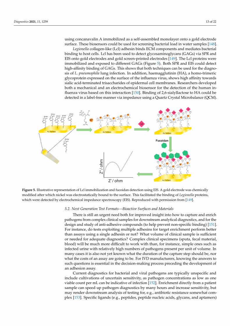
Legionella collagen-like (Lcl) adhesin binds ECM components and mediates bacterialbinding to host cells. Lcl has been used to detect glycosaminoglycans (GAGs) via SPR andEIS onto gold electrodes and gold screen-printed electrodes [149]. The Lcl proteins wereimmobilized and exposed to different GAGs (Figure 5). Both SPR and EIS could detecthigh-affinity binding of GAGs. This shows that both techniques can be used for the diagno-sis of L. pneumophila lung infection. In addition, haemagglutinin (HA), a homo-trimericglycoprotein expressed on the surface of the influenza virus, shows high affinity towardssialic acid-terminated trisaccharides of epidermal cell membranes. Researchers developedboth a mechanical and an electrochemical biosensor for the detection of the human in-fluenza virus based on this interaction [150]. Binding of 2,6-sialyllactose to HA could bedetected in a label-free manner via impedance using a Quartz Crystal Microbalance (QCM).
Diagnostics 2021, 11, x FOR PEER REVIEW 14 of 23
Figure 5. Illustrative representation of Lcl immobilization and fucoidan detection using EIS. A gold electrode was chemi-cally modified after which nickel was electrostatically bound to the surface. This facilitated the binding of Legionella pro-teins, which were detected by electrochemical impedance spectroscopy (EIS). Reproduced with permission from [149].
5.2. Next Generation Test Formats—Bioactive Surfaces and Materials There is still an urgent need both for improved insight into how to capture and enrich
pathogens from complex clinical samples for downstream analytical diagnostics, and for the design and study of anti-adhesive compounds (to help prevent non-specific binding) [151]. For instance, do tests exploiting multiple adhesins for target enrichment perform better than assays using a single adhesin or not? What volume of clinical sample is suffi-cient or needed for adequate diagnostics? Complex clinical specimens (sputa, fecal mate-rial, blood) will be much more difficult to work with than, for instance, simple ones such as infected urine with relatively high numbers of pathogens present per unit of volume. In many cases it is also not yet known what the duration of the capture step should be, nor what the costs of an assay are going to be. For IVD manufacturers, knowing the an-swers to such questions is essential in the decision-making process preceding the devel-opment of an adhesion assay.
Current diagnostics for bacterial and viral pathogens are typically unspecific and in-clude cultivations of uncertain sensitivity, as pathogen concentrations as low as one viable count per mL can be indicative of infection [152]. Enrichment directly from a patient sam-ple can speed up pathogen diagnostics by many hours and increase sensitivity, but may render downstream analysis of testing for, e.g., antibiotic resistance somewhat complex [153]. Specific ligands (e.g., peptides, peptide nucleic acids, glycans, and aptamers) that bind to ECM, membrane components, or capsids of (a) given pathogen(s) can be immobi-lized onto the surface of materials used for fabrication of diagnostic devices, such as pep-tide or nucleic acid arrays, microbeads, membranes, and even electrodes using a paper format. Such bioactive surfaces provide sufficiently high and well-controlled binding ca-pacities for ligands, intact cells, and cellular extracts. The surface characteristics prevent denaturation of the immobilized ligands, allow for convenient and efficient immobiliza-tion techniques, and are preferably reversible to allow regeneration. Such surfaces should prevent non-specific interactions, i.e., be anti-adhesive or anti-fouling or even prevent in-fection, which is particularly relevant for intra-corporeal devices [154]. A recurring prob-lem is the significant fraction of bacteria that is accidentally lost by non-specific binding in miniaturized devices and microfluidics due to their high surface area to volume ratios. Thus, antifouling properties are of greater importance [155]. Furthermore, fouling by non-specific biomolecules can also hinder sensitivity and selectivity and result in false-positive or -negative readings [156]. Improved materials can be obtained by physico–chemical modification of the surface [157], grafting [158,159], coating [160], surface topography
Figure 5. Illustrative representation of Lcl immobilization and fucoidan detection using EIS. A gold electrode was chemicallymodified after which nickel was electrostatically bound to the surface. This facilitated the binding of Legionella proteins,which were detected by electrochemical impedance spectroscopy (EIS). Reproduced with permission from [149].
5.2. Next Generation Test Formats—Bioactive Surfaces and Materials
There is still an urgent need both for improved insight into how to capture and enrichpathogens from complex clinical samples for downstream analytical diagnostics, and for thedesign and study of anti-adhesive compounds (to help prevent non-specific binding) [151].For instance, do tests exploiting multiple adhesins for target enrichment perform betterthan assays using a single adhesin or not? What volume of clinical sample is sufficientor needed for adequate diagnostics? Complex clinical specimens (sputa, fecal material,blood) will be much more difficult to work with than, for instance, simple ones such asinfected urine with relatively high numbers of pathogens present per unit of volume. Inmany cases it is also not yet known what the duration of the capture step should be, norwhat the costs of an assay are going to be. For IVD manufacturers, knowing the answers tosuch questions is essential in the decision-making process preceding the development ofan adhesion assay.
Current diagnostics for bacterial and viral pathogens are typically unspecific andinclude cultivations of uncertain sensitivity, as pathogen concentrations as low as oneviable count per mL can be indicative of infection [152]. Enrichment directly from a patientsample can speed up pathogen diagnostics by many hours and increase sensitivity, butmay render downstream analysis of testing for, e.g., antibiotic resistance somewhat com-plex [153]. Specific ligands (e.g., peptides, peptide nucleic acids, glycans, and aptamers)
Diagnostics 2021, 11, 1259 14 of 22
that bind to ECM, membrane components, or capsids of (a) given pathogen(s) can beimmobilized onto the surface of materials used for fabrication of diagnostic devices, suchas peptide or nucleic acid arrays, microbeads, membranes, and even electrodes using apaper format. Such bioactive surfaces provide sufficiently high and well-controlled bindingcapacities for ligands, intact cells, and cellular extracts. The surface characteristics preventdenaturation of the immobilized ligands, allow for convenient and efficient immobilizationtechniques, and are preferably reversible to allow regeneration. Such surfaces shouldprevent non-specific interactions, i.e., be anti-adhesive or anti-fouling or even prevent infec-tion, which is particularly relevant for intra-corporeal devices [154]. A recurring problemis the significant fraction of bacteria that is accidentally lost by non-specific binding inminiaturized devices and microfluidics due to their high surface area to volume ratios.Thus, antifouling properties are of greater importance [155]. Furthermore, fouling by non-specific biomolecules can also hinder sensitivity and selectivity and result in false-positiveor -negative readings [156]. Improved materials can be obtained by physico–chemicalmodification of the surface [157], grafting [158,159], coating [160], surface topographymodification [161] and surfactant adsorption [162]. At present, the development of novelmulti-functional materials to capture pathogens from biological samples combining graft-ing with plasma or UV treatments will help integrate the pre-enrichment methods intofull diagnostics workflows [163–165]. Overall, this approach can lead to fast, specific,and efficient pathogen trapping strategies, reduced sample processing times, and bettersensitivity for downstream detection techniques. The inverse process, anti-adhesion, canbe used to develop “clean” materials (see Box 2).
Box 2. Anti-Adhesion.
The development of anti-adhesive materials with minimal fouling, based on ‘grafting-from’ ap-proaches, may be useful to inhibit adhesion. The development of ‘anti-ligands’ to inhibit adhesionprovides additional therapeutic approaches. Pilicides, which inhibit the first steps in biofilmformation for E. coli, constitute a special category of such anti-ligands [166,167], and these arebeing considered as alternative therapeutic approaches in, for instance, bacterial urinary tractinfections [168]. The target of pilicides is the biofilm, but some also show surprising anti-pilinactivity, thereby tackling infections from two fundamentally different angles [169]. Their diagnosticvalue is limited at this stage although detection of pilicide activity could indicate early stage biofilmformation. Altogether this could provide important tools for the prevention of (nosocomial) infec-tions in general. Simultaneous detection of multiple pathogens may thus be a distinct possibility.This will facilitate new formats for syndrome testing, where all possible pathogens involved in acertain type of infection can be detected at the same time and ruled in or out simultaneously (e.g.,gastro–intestinal or respiratory infections) [170–172]. This has clear benefits both in terms of cost,morbidity, and even mortality, as the faster the correct pathogen(s) are identified, the faster thecorrect treatment can be given.
6. Conclusions
The IVD workflows show the overall state of readiness for innovation in clinicalmicrobiology (Figures 3 and 4). Expansion of the existing portfolio of IVD tests is a must,and innovative adhesion-based assays have been proposed. Some of these are alreadyaccepted for routine diagnostic use, but there are still many procedures in developmentthat require additional validation, verification and, in the end, user acceptance as valuableIVD tools. Acceptance of such tests will improve overall public health status by helpingcontrol the spread of pathogens and allowing for personalized treatment. We believethat the application of integrative approaches, including bioinformatics, quantitative andstructural proteomics and structure modeling, will improve the understanding of thepathogenesis of infectious diseases in general [173]. Understanding the sequential andstructural determinants of adhesion will further drive the translational aspects, leadingto the design of novel tests. New technologies, as described here, will play a key rolein facilitating this essential phase of test design [174–177]. Cost effective scale-up and

Diagnostics 2021, 11, 1259 15 of 22
application of adhesins to commercially relevant sensor-activated testing systems needs tobe implemented in the developmental cycles exploited by commercial companies.
COVID-19 has, at last, made the general public aware of the global impact of infectiousdiseases and the need for and relevance of rapid diagnostics [178]. We must seize themoment: this current appreciation of the value of infectious disease testing should be usedto push for affordable, high-quality, rapid diagnostic tests for all infectious diseases tobe available worldwide. We believe that adhesion research will contribute to this, sinceit defines fundamental new processes that allow identification and enrichment of newbinding partner molecules. Such molecules can then be implemented in IVD using thecontinuously expanding experimental toolbox to allow sensitive detection of molecularbinding events. The combination of accelerated detection and identification of microbialadhesins and the technological capabilities defines a prosperous future for this field ofin vitro diagnostics.
Author Contributions: Conceptualization, A.v.B. and D.L.; validation, all other authors; writing—original draft preparation, A.v.B. and D.L.; writing—review and editing, all authors; writing, finalreview, A.G. All authors have read and agreed to the published version of the manuscript.
Funding: This research was funded by the European Union’s Horizon 2020 research and innovationprogram in a project named Viral and Bacterial Adhesin Network Training (ViBrANT) under MarieSkłodowska-Curie Grant Agreement No. 765042. A.G. also acknowledges support from the BBSRC(grant number BB/M021610/1).
Institutional Review Board Statement: Not applicable.
Informed Consent Statement: Not applicable.
Acknowledgments: The authors gratefully thank Jürgen Berger and Katharina Hipp (both MaxPlanck-Institute for Developmental Biology, Tübingen, Germany) for the electron microscopy dis-played in Figure 1.
Conflicts of Interest: A.v.B., M.G. and R.D. are employees of bioMérieux, a company designing,developing, and marketing tests in the domain of infectious diseases. The company was not involvedin the design of the current study and the opinions expressed are those of the authors and may bedifferent from formal company opinions and policies. Carina Almeida (Biomode, Braga, Portugal)and Ian Eastwood (Eluceda, UK) also have commercial affiliations. The remaining authors declarethat the research was conducted in the absence of any commercial or financial relationships thatcould be construed as a potential conflict of interest.
References1. Markwell, A.; Mitchell, R.; Wright, A.L.; Brown, A.F. Clinical and ethical challenges for emergency departments during
communicable disease outbreaks: Can lessons from Ebola Virus Disease be applied to the COVID-19 pandemic? Emerg. Med.Australas. 2020, 32, 520–524. [CrossRef]
2. Suetens, C.; Latour, K.; Kärki, T.; Ricchizzi, E.; Kinross, P.; Moro, M.L.; Jans, B.; Hopkins, S.; Hansen, S.; Lyytikäinen, O.; et al.Prevalence of healthcare-associated infections, estimated incidence and composite antimicrobial resistance index in acute carehospitals and long-term care facilities: Results from two European point prevalence surveys, 2016 to 2017. Eurosurveillance2018, 23. [CrossRef]
3. Chokshi, A.; Sifri, Z.; Cennimo, D.; Horng, H. Global Contributors to Antibiotic Resistance. J. Glob. Infect. Dis. 2019, 11, 36–42.[CrossRef]
4. Zumla, A.; Memish, Z.A.; Maeurer, M.; Bates, M.; Mwaba, P.; Al-Tawfiq, J.A.; Denning, D.W.; Hayden, F.G.; Hui, D.S. Emergingnovel and antimicrobial-resistant respiratory tract infections: New drug development and therapeutic options. Lancet. Infect. Dis.2014, 14, 1136–1149. [CrossRef]
5. Sloots, T.P.; Nissen, M.D.; Ginn, A.N.; Iredell, J.R. Rapid identification of pathogens using molecular techniques. Pathology 2015,47, 191–198. [CrossRef]
6. Pinsky, B.A.; Hayden, R.T. Cost-Effective Respiratory Virus Testing. J. Clin. Microbiol. 2019, 57. [CrossRef]7. Tacconelli, E.; Carrara, E.; Savoldi, A.; Harbarth, S.; Mendelson, M.; Monnet, D.L.; Pulcini, C.; Kahlmeter, G.; Kluytmans, J.;
Carmeli, Y.; et al. Discovery, research, and development of new antibiotics: The WHO priority list of antibiotic-resistant bacteriaand tuberculosis. Lancet Infect. Dis. 2018, 18, 318–327. [CrossRef]
Diagnostics 2021, 11, 1259 16 of 22
8. Yamazaki, K.; Kashimoto, T.; Kado, T.; Akeda, Y.; Yoshioka, K.; Kodama, T.; Yamamoto, M.; Okamura, M.; Kakuda, T.; Ueno, S.Chemotactic invasion in deep soft tissue by Vibrio vulnificus is essential for the progression of necrotic lesions. Virulence 2020,11, 840–848. [CrossRef] [PubMed]
9. Singh, V.; Phukan, U.J. Interaction of host and Staphylococcus aureus protease-system regulates virulence and pathogenicity. Med.Microbiol. Immunol. 2019, 208, 585–607. [CrossRef] [PubMed]
10. Comunian, S.; Dongo, D.; Milani, C.; Palestini, P. Air Pollution and COVID-19: The Role of Particulate Matter in the Spread andIncrease of COVID-19’s Morbidity and Mortality. Int. J. Environ. Res. Public. Health. 2020, 17, 4487. [CrossRef] [PubMed]
11. Speziale, P.; Arciola, C.R.; Pietrocola, G. Fibronectin and Its Role in Human Infective Diseases. Cells 2019, 8, 1516. [CrossRef]12. Duell, B.L.; Su, Y.C.; Riesbeck, K. Host-pathogen interactions of nontypeable Haemophilus influenzae: From commensal to pathogen.
FEBS Lett. 2016, 590, 3840–3853. [CrossRef]13. Daly, J.L.; Simonetti, B.; Klein, K.; Chen, K.E.; Williamson, M.K.; Antón-Plágaro, C.; Shoemark, D.K.; Simón-Gracia, L.; Bauer, M.;
Hollandi, R.; et al. Neuropilin-1 is a host factor for SARS-CoV-2 infection. Science 2020, 370, 861–865. [CrossRef]14. Cantuti-Castelvetri, L.; Ojha, R.; Pedro, L.D.; Djannatian, M.; Franz, J.; Kuivanen, S.; van der Meer, F.; Kallio, K.; Kaya, T.;
Anastasina, M.; et al. Neuropilin-1 facilitates SARS-CoV-2 cell entry and infectivity. Science 2020, 370, 856–860. [CrossRef]15. Pietrocola, G.; Arciola, C.R.; Rindi, S.; Montanaro, L.; Speziale, P. Streptococcus agalactiae Non-Pilus, Cell Wall-Anchored Proteins:
Involvement in Colonization and Pathogenesis and Potential as Vaccine Candidates. Front. Immunol. 2018, 9, 602. [CrossRef][PubMed]
16. Thomas, R.J. Receptor mimicry as novel therapeutic treatment for biothreat agents. Bioeng. Bugs. 2010, 1, 17–30. [CrossRef]17. Miller, M.B.; Atrzadeh, F.; Burnham, C.A.; Cavalieri, S.; Dunn, J.; Jones, S.; Mathews, C.; McNult, P.; Meduri, J.; Newhouse, C.;
et al. Clinical Utility of Advanced Microbiology Testing Tools. J. Clin. Microbiol. 2019, 57. [CrossRef]18. Dubourg, G.; Lamy, B.; Ruimy, R. Rapid phenotypic methods to improve the diagnosis of bacterial bloodstream infections:
Meeting the challenge to reduce the time to result. Clin. Microbiol. Infect. 2018, 24, 935–943. [CrossRef]19. Vandenberg, O.; Durand, G.; Hallin, M.; Diefenbach, A.; Gant, V.; Murray, P.; Kozlakidis, Z.; van Belkum, A. Consolidation of
Clinical Microbiology Laboratories and Introduction of Transformative Technologies. Clin. Microbiol. Rev. 2020, 33. [CrossRef]20. Boonham, N.; Kreuze, J.; Winter, S.; van der Vlugt, R.; Bergervoet, J.; Tomlinson, J.; Mumford, R. Methods in virus diagnostics:
From ELISA to next generation sequencing. Virus. Res. 2014, 186, 20–31. [CrossRef]21. Han, D.; Li, Z.; Li, R.; Tan, P.; Zhang, R.; Li, J. mNGS in clinical microbiology laboratories: On the road to maturity. Crit. Rev.
Microbiol. 2019, 45, 668–685. [CrossRef]22. van Belkum, A.; Bachmann, T.T.; Lüdke, G.; Lisby, J.G.; Kahlmeter, G.; Mohess, A.; Becker, K.; Hays, J.P.; Woodford, N.;
Mitsakakis, K.; et al. Developmental roadmap for antimicrobial susceptibility testing systems. Nat. Rev. Microbiol. 2019, 17, 51–62.[CrossRef]
23. Vandenberg, O.; Martiny, D.; Rochas, O.; van Belkum, A.; Kozlakidis, Z. Considerations for diagnostic COVID-19 tests. Nat. Rev.Microbiol. 2021, 19, 171–183. [CrossRef] [PubMed]
24. Mitchell, S.L.; St George, K.; Rhoads, D.D.; Butler-Wu, S.M.; Dharmarha, V.; McNult, P.; Miller, M.B. Understanding, Verifying,and Implementing Emergency Use Authorization Molecular Diagnostics for the Detection of SARS-CoV-2 RNA. J. Clin. Microbiol.2020, 58. [CrossRef]
25. Andryukov, B.G.; Besednova, N.N.; Romashko, R.V.; Zaporozhets, T.S.; Efimov, T.A. Label-Free Biosensors for Laboratory-BasedDiagnostics of Infections: Current Achievements and New Trends. Biosensors 2020, 10, 11. [CrossRef]
26. Bedford, R.; Tiede, C.; Hughes, R.; Curd, A.; McPherson, M.J.; Peckham, M.; Tomlinson, D.C. Alternative reagents to antibodies inimaging applications. Biophys. Rev. 2017, 9, 299–308. [CrossRef] [PubMed]
27. Huang, J.; Chen, X.; Fu, X.; Li, Z.; Huang, Y.; Liang, C. Advances in Aptamer-Based Biomarker Discovery. Front. Cell. Dev. Biol.2021, 9, 659760. [CrossRef]
28. Sharon, N.; Ofek, I. Identification of receptors for bacterial lectins by blotting techniques. Methods Enzymol. 1995, 253, 91–98.[CrossRef] [PubMed]
29. Kitts, G.; Giglio, K.M.; Zamorano-Sánchez, D.; Park, J.H.; Townsley, L.; Cooley, R.B.; Wucher, B.R.; Klose, K.E.; Nadell, C.D.;Yildiz, F.H.; et al. A Conserved Regulatory Circuit Controls Large Adhesins in Vibrio cholerae. MBio 2019, 10. [CrossRef]
30. Wang, C.; Chen, W.; Xia, A.; Zhang, R.; Huang, Y.; Yang, S.; Ni, L.; Jin, F. Carbon Starvation Induces the Expression ofPprB-Regulated Genes in Pseudomonas aeruginosa. Appl. Environ. Microbiol. 2019, 85. [CrossRef]
31. Sequeira, S.; Kavanaugh, D.; MacKenzie, D.A.; Šuligoj, T.; Walpole, S.; Leclaire, C.; Gunning, A.P.; Latousakis, D.; Willats,W.G.T.; Angulo, J.; et al. Structural basis for the role of serine-rich repeat proteins from Lactobacillus reuteri in gut microbe-hostinteractions. Proc. Natl. Acad. Sci. USA 2018, 115, E2706–E2715. [CrossRef] [PubMed]
32. Loukachevitch, L.V.; Bensing, B.A.; Yu, H.; Zeng, J.; Chen, X.; Sullam, P.M.; Iverson, T.M. Structures of the Streptococcus sanguinisSrpA Binding Region with Human Sialoglycans Suggest Features of the Physiological Ligand. Biochemistry 2016, 55, 5927–5937.[CrossRef]
33. Peacock, T.P.; Penrice-Randal, R.; Hiscox, J.A.; Barclay, W.S. SARS-CoV-2 one year on: Evidence for ongoing viral adaptation. J.Gen. Virol. 2021, 102. [CrossRef]
34. Sherman, M.B.; Smith, H.Q.; Smith, T.J. The Dynamic Life of Virus Capsids. Viruses 2020, 12, 618. [CrossRef]35. Neu, U.; Bauer, J.; Stehle, T. Viruses and sialic acids: Rules of engagement. Curr. Opin. Struct. Biol. 2011, 21, 610–618. [CrossRef]
Diagnostics 2021, 11, 1259 17 of 22
36. Stroh, L.J.; Stehle, T. Glycan Engagement by Viruses: Receptor Switches and Specificity. Annu. Rev. Virol. 2014, 1, 285–306.[CrossRef] [PubMed]
37. Hiatt, A.; Bohorova, N.; Bohorov, O.; Goodman, C.; Kim, D.; Pauly, M.H.; Velasco, J.; Whaley, K.J.; Piedra, P.A.; Gilbert, B.E.; et al.Glycan variants of a respiratory syncytial virus antibody with enhanced effector function and in vivo efficacy. Proc. Natl. Acad.Sci. USA 2014, 111, 5992–5997. [CrossRef] [PubMed]
38. Imamura, T.; Okamoto, M.; Nakakita, S.; Suzuki, A.; Saito, M.; Tamaki, R.; Lupisan, S.; Roy, C.N.; Hiramatsu, H.; Sugawara, K.E.;et al. Antigenic and receptor binding properties of enterovirus 68. J. Virol. 2014, 88, 2374–2384. [CrossRef]
39. Horiguchi, Y.; Goda, T.; Matsumoto, A.; Takeuchi, H.; Yamaoka, S.; Miyahara, Y. Direct and label-free influenza virus detectionbased on multisite binding to sialic acid receptors. Biosens. Bioelectron. 2017, 92, 234–240. [CrossRef]
40. Naskalska, A.; Dabrowska, A.; Szczepanski, A.; Milewska, A.; Jasik, K.P.; Pyrc, K. Membrane Protein of Human CoronavirusNL63 Is Responsible for Interaction with the Adhesion Receptor. J. Virol. 2019, 93. [CrossRef]
41. Serrano-Gómez, D.; Domínguez-Soto, A.; Ancochea, J.; Jimenez-Heffernan, J.A.; Leal, J.A.; Corbí, A.L. Dendritic cell-specificintercellular adhesion molecule 3-grabbing nonintegrin mediates binding and internalization of Aspergillus fumigatus conidia bydendritic cells and macrophages. J. Immunol. 2004, 173, 5635–5643. [CrossRef]
42. Wang, Q.C.; Feng, Z.H.; Nie, Q.H.; Zhou, Y.X. DC-SIGN: Binding receptors for hepatitis C virus. Chin. Med. J. 2004, 117, 1395–1400.[PubMed]
43. Li, H.; Liu, L.; Zhang, D.; Xu, J.; Dai, H.; Tang, N.; Su, X.; Cao, B. SARS-CoV-2 and viral sepsis: Observations and hypotheses.Lancet 2020, 395, 1517–1520. [CrossRef]
44. Zhou, P.; Yang, X.L.; Wang, X.G.; Hu, B.; Zhang, L.; Zhang, W.; Si, H.R.; Zhu, Y.; Li, B.; Huang, C.L.; et al. A pneumonia outbreakassociated with a new coronavirus of probable bat origin. Nature 2020, 579, 270–273. [CrossRef]
45. Hamming, I.; Timens, W.; Bulthuis, M.L.; Lely, A.T.; Navis, G.; van Goor, H. Tissue distribution of ACE2 protein, the functionalreceptor for SARS coronavirus. A first step in understanding SARS pathogenesis. J. Pathol. 2004, 203, 631–637. [CrossRef]
46. Jeffers, S.A.; Tusell, S.M.; Gillim-Ross, L.; Hemmila, E.M.; Achenbach, J.E.; Babcock, G.J.; Thomas, W.D., Jr.; Thackray, L.B.;Young, M.D.; Mason, R.J.; et al. CD209L (L-SIGN) is a receptor for severe acute respiratory syndrome coronavirus. Proc. Natl.Acad. Sci. USA 2004, 101, 15748–15753. [CrossRef]
47. Amraie, R.; Napoleon, M.A.; Yin, W.; Berrigan, J.; Suder, E.; Zhao, G.; Olejnik, J.; Gummuluru, S.; Muhlberger, E.; Chitalia, V.; et al.CD209L/L-SIGN and CD209/DC-SIGN act as receptors for SARS-CoV-2 and are differentially expressed in lung and kidneyepithelial and endothelial cells. bioRxiv 2020. [CrossRef]
48. Lan, J.; Ge, J.; Yu, J.; Shan, S.; Zhou, H.; Fan, S.; Zhang, Q.; Shi, X.; Wang, Q.; Zhang, L.; et al. Structure of the SARS-CoV-2 spikereceptor-binding domain bound to the ACE2 receptor. Nature 2020, 581, 215–220. [CrossRef]
49. Shang, J.; Ye, G.; Shi, K.; Wan, Y.; Luo, C.; Aihara, H.; Geng, Q.; Auerbach, A.; Li, F. Structural basis of receptor recognition bySARS-CoV-2. Nature 2020, 581, 221–224. [CrossRef] [PubMed]
50. Qi, X.; Ke, B.; Feng, Q.; Yang, D.; Lian, Q.; Li, Z.; Lu, L.; Ke, C.; Liu, Z.; Liao, G. Construction and immunogenic studies of a mFcfusion receptor binding domain (RBD) of spike protein as a subunit vaccine against SARS-CoV-2 infection. Chem. Commun. 2020,56, 8683–8686. [CrossRef]
51. Jin, Z.; Du, X.; Xu, Y.; Deng, Y.; Liu, M.; Zhao, Y.; Zhang, B.; Li, X.; Zhang, L.; Peng, C.; et al. Structure of M(pro) from SARS-CoV-2and discovery of its inhibitors. Nature 2020, 582, 289–293. [CrossRef]
52. Viela, F.; Mathelié-Guinlet, M.; Viljoen, A.; Dufrêne, Y.F. What makes bacterial pathogens so sticky? Mol. Microbiol. 2020,113, 683–690. [CrossRef]
53. Kwon, H.; Squire, C.J.; Young, P.G.; Baker, E.N. Autocatalytically generated Thr-Gln ester bond cross-links stabilize the repetitiveIg-domain shaft of a bacterial cell surface adhesin. Proc. Natl. Acad. Sci. USA 2014, 111, 1367–1372. [CrossRef] [PubMed]
54. Wrobel, A.; Ottoni, C.; Leo, J.C.; Gulla, S.; Linke, D. The repeat structure of two paralogous genes, Yersinia ruckeri invasin (yrInv)and a “Y. ruckeri invasin-like molecule”, (yrIlm) sheds light on the evolution of adhesive capacities of a fish pathogen. J. Struct.Biol. 2018, 201, 171–183. [CrossRef] [PubMed]
55. Linke, D.; Riess, T.; Autenrieth, I.B.; Lupas, A.; Kempf, V.A. Trimeric autotransporter adhesins: Variable structure, commonfunction. Trends Microbiol. 2006, 14, 264–270. [CrossRef] [PubMed]
56. Müller, N.F.; Kaiser, P.O.; Linke, D.; Schwarz, H.; Riess, T.; Schäfer, A.; Eble, J.A.; Kempf, V.A. Trimeric autotransporter adhesin-dependent adherence of Bartonella henselae, Bartonella quintana, and Yersinia enterocolitica to matrix components and endothelialcells under static and dynamic flow conditions. Infect. Immun. 2011, 79, 2544–2553. [CrossRef]
57. Weidensdorfer, M.; Chae, J.I.; Makobe, C.; Stahl, J.; Averhoff, B.; Müller, V.; Schürmann, C.; Brandes, R.P.; Wilharm, G.;Ballhorn, W.; et al. Analysis of Endothelial Adherence of Bartonella henselae and Acinetobacter baumannii Using a Dynamic HumanEx Vivo Infection Model. Infect. Immun. 2015, 84, 711–722. [CrossRef]
58. Meuskens, I.; Saragliadis, A.; Leo, J.C.; Linke, D. Type V Secretion Systems: An Overview of Passenger Domain Functions. Front.Microbiol. 2019, 10, 1163. [CrossRef]
59. Koiwai, K.; Hartmann, M.D.; Linke, D.; Lupas, A.N.; Hori, K. Structural Basis for Toughness and Flexibility in the C-terminalPassenger Domain of an Acinetobacter Trimeric Autotransporter Adhesin. J. Biol. Chem. 2016, 291, 3705–3724. [CrossRef]
60. Bassler, J.; Hernandez Alvarez, B.; Hartmann, M.D.; Lupas, A.N. A domain dictionary of trimeric autotransporter adhesins. Int. J.Med. Microbiol. 2015, 305, 265–275. [CrossRef]
Diagnostics 2021, 11, 1259 18 of 22
61. Shin, P.K.; Pawar, P.; Konstantopoulos, K.; Ross, J.M. Characteristics of new Staphylococcus aureus-RBC adhesion mechanismindependent of fibrinogen and IgG under hydrodynamic shear conditions. Am. J. Physiol. Cell. Physiol. 2005, 289, C727–C734.[CrossRef]
62. Reichhardt, C.; Wong, C.; Passos da Silva, D.; Wozniak, D.J.; Parsek, M.R. CdrA Interactions within the Pseudomonas aeruginosaBiofilm Matrix Safeguard It from Proteolysis and Promote Cellular Packing. MBio 2018, 9. [CrossRef]
63. Foster, T.J. The remarkably multifunctional fibronectin binding proteins of Staphylococcus aureus. Eur. J. Clin. Microbiol. Infect. Dis.2016, 35, 1923–1931. [CrossRef] [PubMed]
64. Stones, D.H.; Krachler, A.M. Fatal attraction: How bacterial adhesins affect host signaling and what we can learn from them. Int.J. Mol. Sci. 2015, 16, 2626–2640. [CrossRef]
65. Adrian, J.; Bonsignore, P.; Hammer, S.; Frickey, T.; Hauck, C.R. Adaptation to Host-Specific Bacterial Pathogens Drives RapidEvolution of a Human Innate Immune Receptor. Curr. Biol. 2019, 29, 616–630.e5. [CrossRef] [PubMed]
66. Mortimer, T.D.; Annis, D.S.; O’Neill, M.B.; Bohr, L.L.; Smith, T.M.; Poinar, H.N.; Mosher, D.F.; Pepperell, C.S. Adaptation in aFibronectin Binding Autolysin of Staphylococcus saprophyticus. mSphere 2017, 2. [CrossRef] [PubMed]
67. Gutiérrez, R.; Markus, B.; Carstens Marques de Sousa, K.; Marcos-Hadad, E.; Mugasimangalam, R.C.; Nachum-Biala, Y.; Hawlena,H.; Covo, S.; Harrus, S. Prophage-Driven Genomic Structural Changes Promote Bartonella Vertical Evolution. Genome. Biol. Evol.2018, 10, 3089–3103. [CrossRef] [PubMed]
68. Lamelas, A.; Harris, S.R.; Röltgen, K.; Dangy, J.P.; Hauser, J.; Kingsley, R.A.; Connor, T.R.; Sie, A.; Hodgson, A.; Dougan, G.; et al.Emergence of a new epidemic Neisseria meningitidis serogroup A Clone in the African meningitis belt: High-resolution picture ofgenomic changes that mediate immune evasion. MBio 2014, 5, e01974-14. [CrossRef]
69. Diderrich, R.; Kock, M.; Maestre-Reyna, M.; Keller, P.; Steuber, H.; Rupp, S.; Essen, L.O.; Mösch, H.U. Structural Hot SpotsDetermine Functional Diversity of the Candida glabrata Epithelial Adhesin Family. J. Biol. Chem. 2015, 290, 19597–19613. [CrossRef][PubMed]
70. Chattopadhyay, S.; Tchesnokova, V.; McVeigh, A.; Kisiela, D.I.; Dori, K.; Navarro, A.; Sokurenko, E.V.; Savarino, S.J. Adaptiveevolution of class 5 fimbrial genes in enterotoxigenic Escherichia coli and its functional consequences. J. Biol. Chem. 2012,287, 6150–6158. [CrossRef]
71. Lorenz, S.C.; Monday, S.R.; Hoffmann, M.; Fischer, M.; Kase, J.A. Plasmids from Shiga Toxin-Producing Escherichia coli Strainswith Rare Enterohemolysin Gene (ehxA) Subtypes Reveal Pathogenicity Potential and Display a Novel Evolutionary Path. Appl.Environ. Microbiol. 2016, 82, 6367–6377. [CrossRef]
72. Sjöholm, K.; Kilsgård, O.; Teleman, J.; Happonen, L.; Malmström, L.; Malmström, J. Targeted Proteomics and Absolute ProteinQuantification for the Construction of a Stoichiometric Host-Pathogen Surface Density Model. Mol. Cell. Proteom. 2017,16, S29–S41. [CrossRef] [PubMed]
73. Karlsson, C.; Malmström, L.; Aebersold, R.; Malmström, J. Proteome-wide selected reaction monitoring assays for the humanpathogen Streptococcus pyogenes. Nat. Commun. 2012, 3, 1301. [CrossRef]
74. Karlsson, C.A.Q.; Järnum, S.; Winstedt, L.; Kjellman, C.; Björck, L.; Linder, A.; Malmström, J.A. Streptococcus pyogenes Infectionand the Human Proteome with a Special Focus on the Immunoglobulin G-cleaving Enzyme IdeS. Mol. Cell. Proteom. 2018,17, 1097–1111. [CrossRef]
75. Lawson, C.L.; Chiu, W. Comparing cryo-EM structures. J. Struct. Biol. 2018, 204, 523–526. [CrossRef]76. Earl, L.A.; Falconieri, V.; Subramaniam, S. Microbiology catches the cryo-EM bug. Curr. Opin. Microbiol. 2018, 43, 199–207.
[CrossRef]77. Hauri, S.; Khakzad, H.; Happonen, L.; Teleman, J.; Malmström, J.; Malmström, L. Rapid determination of quaternary protein
structures in complex biological samples. Nat. Commun. 2019, 10, 192. [CrossRef]78. Bastos, V.A.; Gomes-Neto, F.; Rocha, S.L.G.; Teixeira-Ferreira, A.; Perales, J.; Neves-Ferreira, A.G.C.; Valente, R.H. The interaction
between the natural metalloendopeptidase inhibitor BJ46a and its target toxin jararhagin analyzed by structural mass spectrometryand molecular modeling. J. Proteom. 2020, 221, 103761. [CrossRef] [PubMed]
79. Slavata, L.; Chmelik, J.; Kavan, D.; Filandrová, R.; Fiala, J.; Rosulek, M.; Mrázek, H.; Kukacka, Z.; Vališ, K.; Man, P.; et al. MS-BasedApproaches Enable the Structural Characterization of Transcription Factor/DNA Response Element Complex. Biomolecules 2019,9, 535. [CrossRef] [PubMed]
80. Seacrist, C.D.; Kuenze, G.; Hoffmann, R.M.; Moeller, B.E.; Burke, J.E.; Meiler, J.; Blind, R.D. Integrated Structural Modelingof Full-Length LRH-1 Reveals Inter-domain Interactions Contribute to Receptor Structure and Function. Structure 2020, 28,830–846.e9. [CrossRef]
81. Zhang, M.M.; Beno, B.R.; Huang, R.Y.; Adhikari, J.; Deyanova, E.G.; Li, J.; Chen, G.; Gross, M.L. An Integrated Approach forDetermining a Protein-Protein Binding Interface in Solution and an Evaluation of Hydrogen-Deuterium Exchange Kinetics forAdjudicating Candidate Docking Models. Anal. Chem. 2019, 91, 15709–15717. [CrossRef] [PubMed]
82. López-Castilla, A.; Thomassin, J.L.; Bardiaux, B.; Zheng, W.; Nivaskumar, M.; Yu, X.; Nilges, M.; Egelman, E.H.;Izadi-Pruneyre, N.; Francetic, O. Structure of the calcium-dependent type 2 secretion pseudopilus. Nat. Microbiol. 2017,2, 1686–1695. [CrossRef]
83. Bardiaux, B.; de Amorim, G.C.; Luna Rico, A.; Zheng, W.; Guilvout, I.; Jollivet, C.; Nilges, M.; Egelman, E.H.; Izadi-Pruneyre, N.;Francetic, O. Structure and Assembly of the Enterohemorrhagic Escherichia. Coli. Type 4 Pilus. Structure 2019, 27, 1082–1093.e5.[CrossRef]
Diagnostics 2021, 11, 1259 19 of 22
84. Bardiaux, B.; Cordier, F.; Brier, S.; López-Castilla, A.; Izadi-Pruneyre, N.; Nilges, M. Dynamics of a type 2 secretion systempseudopilus unraveled by complementary approaches. J. Biomol. NMR 2019, 73, 293–303. [CrossRef] [PubMed]
85. Patwardhan, A.; Ashton, A.; Brandt, R.; Butcher, S.; Carzaniga, R.; Chiu, W.; Collinson, L.; Doux, P.; Duke, E.; Ellisman, M.H.;et al. A 3D cellular context for the macromolecular world. Nat. Struct. Mol. Biol. 2014, 21, 841–845. [CrossRef]
86. He, L.; Bardiaux, B.; Ahmed, M.; Spehr, J.; König, R.; Lünsdorf, H.; Rand, U.; Lührs, T.; Ritter, C. Structure determination ofhelical filaments by solid-state NMR spectroscopy. Proc. Natl. Acad. Sci. USA 2016, 113, E272–E281. [CrossRef] [PubMed]
87. Ferber, M.; Kosinski, J.; Ori, A.; Rashid, U.J.; Moreno-Morcillo, M.; Simon, B.; Bouvier, G.; Batista, P.R.; Müller, C.W.; Beck, M.; et al.Automated structure modeling of large protein assemblies using crosslinks as distance restraints. Nat. Methods 2016, 13, 515–520.[CrossRef]
88. Ramboarina, S.; Garnett, J.A.; Zhou, M.; Li, Y.; Peng, Z.; Taylor, J.D.; Lee, W.C.; Bodey, A.; Murray, J.W.; Alguel, Y.; et al. Structuralinsights into serine-rich fimbriae from Gram-positive bacteria. J. Biol. Chem. 2010, 285, 32446–32457. [CrossRef]
89. Olano, J.P.; Walker, D.H. Diagnosing emerging and reemerging infectious diseases: The pivotal role of the pathologist. Arch.Pathol. Lab. Med. 2011, 135, 83–91. [CrossRef]
90. Ramanan, P.; Bryson, A.L.; Binnicker, M.J.; Pritt, B.S.; Patel, R. Syndromic Panel-Based Testing in Clinical Microbiology. Clin.Microbiol. Rev. 2018, 31. [CrossRef] [PubMed]
91. Özenci, V.; Patel, R.; Ullberg, M.; Strålin, K. Demise of Polymerase Chain Reaction/Electrospray Ionization-Mass Spectrometry asan Infectious Diseases Diagnostic Tool. Clin. Infect. Dis. 2018, 66, 452–455. [CrossRef]
92. Campos, M.; Francetic, O.; Nilges, M. Modeling pilus structures from sparse data. J. Struct. Biol. 2011, 173, 436–444. [CrossRef]93. Schmid, Y.; Grassl, G.A.; Buhler, O.T.; Skurnik, M.; Autenrieth, I.B.; Bohn, E. Yersinia enterocolitica adhesin A induces production
of interleukin-8 in epithelial cells. Infect. Immun. 2004, 72, 6780–6789. [CrossRef]94. Leo, J.C.; Goldman, A. The immunoglobulin-binding Eib proteins from Escherichia coli are receptors for IgG Fc. Mol. Immunol.
2009, 46, 1860–1866. [CrossRef]95. Leo, J.C.; Łyskowski, A.; Hattula, K.; Hartmann, M.D.; Schwarz, H.; Butcher, S.J.; Linke, D.; Lupas, A.N.; Goldman, A. The
structure of E. coli IgG-binding protein D suggests a general model for bending and binding in trimeric autotransporter adhesins.Structure 2011, 19, 1021–1030. [CrossRef]
96. Kearney, K.J.; Pechlivani, N.; King, R.; Tiede, C.; Phoenix, F.; Cheah, R.; Macrae, F.L.; Simmons, K.J.; Manfield, I.W.; Smith, K.A.;et al. Affimer proteins as a tool to modulate fibrinolysis, stabilize the blood clot, and reduce bleeding complications. Blood 2019,133, 1233–1244. [CrossRef]
97. Tiede, C.; Tang, A.A.; Deacon, S.E.; Mandal, U.; Nettleship, J.E.; Owen, R.L.; George, S.E.; Harrison, D.J.; Owens, R.J.;Tomlinson, D.C.; et al. Adhiron: A stable and versatile peptide display scaffold for molecular recognition applications. ProteinEng. Des. Sel. 2014, 27, 145–155. [CrossRef]
98. Peña-Bahamonde, J.; Nguyen, H.N.; Fanourakis, S.K.; Rodrigues, D.F. Recent advances in graphene-based biosensor technologywith applications in life sciences. J. Nanobiotechnol. 2018, 16, 75. [CrossRef]
99. Elahi, N.; Kamali, M.; Baghersad, M.H.; Amini, B. A fluorescence Nano-biosensors immobilization on Iron (MNPs) and gold(AuNPs) nanoparticles for detection of Shigella spp. Mater. Sci. Eng. C Mater. Biol. Appl. 2019, 105, 110113. [CrossRef]
100. Govender, V.S.; Ramsugit, S.; Pillay, M. Mycobacterium tuberculosis adhesins: Potential biomarkers as anti-tuberculosis therapeu-tic and diagnostic targets. Microbiology 2014, 160, 1821–1831. [CrossRef]
101. Peeling, R.W.; Mabey, D. Point-of-care tests for diagnosing infections in the developing world. Clin. Microbiol. Infect. 2010,16, 1062–1069. [CrossRef]
102. He, X.; Zhou, L.; He, D.; Wang, K.; Cao, J. Rapid and ultrasensitive E. coli O157:H7 quantitation by combination of ligandmagneticnanoparticles enrichment with fluorescent nanoparticles based two-color flow cytometry. Analyst 2011, 136, 4183–4191. [CrossRef]
103. Kretzer, J.W.; Lehmann, R.; Schmelcher, M.; Banz, M.; Kim, K.P.; Korn, C.; Loessner, M.J. Use of high-affinity cell wall-bindingdomains of bacteriophage endolysins for immobilization and separation of bacterial cells. Appl. Environ. Microbiol. 2007,73, 1992–2000. [CrossRef] [PubMed]
104. Pilecky, M.; Schildberger, A.; Orth-Höller, D.; Weber, V. Pathogen enrichment from human whole blood for the diagnosis ofbloodstream infection: Prospects and limitations. Diagn. Microbiol. Infect. Dis. 2019, 94, 7–14. [CrossRef] [PubMed]
105. Auriti, C.; Prencipe, G.; Moriondo, M.; Bersani, I.; Bertaina, C.; Mondì, V.; Inglese, R. Mannose-Binding Lectin: BiologicCharacteristics and Role in the Susceptibility to Infections and Ischemia-Reperfusion Related Injury in Critically Ill Neonates. J.Immunol. Res. 2017, 2017, 7045630. [CrossRef] [PubMed]
106. Unione, L.; Gimeno, A.; Valverde, P.; Calloni, I.; Coelho, H.; Mirabella, S.; Poveda, A.; Arda, A.; Jimenez-Barbero, J. Glycans inInfectious Diseases. A Molecular Recognition Perspective. Curr. Med. Chem. 2017, 24, 4057–4080. [CrossRef] [PubMed]
107. Turner, M.W. The role of mannose-binding lectin in health and disease. Mol. Immunol. 2003, 40, 423–429. [CrossRef]108. Kangwa, M.; Yelemane, V.; Ponnurangam, A.; Fernández-Lahore, M. An engineered Staphylococcal Protein A based ligand:
Production, characterization and potential application for the capture of Immunoglobulin and Fc-fusion proteins. Protein Expr.Purif. 2019, 155, 27–34. [CrossRef] [PubMed]
109. Templier, V.; Roux, A.; Roupioz, Y.; Livache, T. Ligands for label-free detection of whole bacteria on biosensors: A review.Trac-Trend. Anal. Chem. 2016, 79, 71–79. [CrossRef]
110. Dunne, M.; Loessner, M.J. Modified Bacteriophage Tail Fiber Proteins for Labeling, Immobilization, Capture, and Detection ofBacteria. Methods Mol. Biol. 2019, 1918, 67–86. [CrossRef]

Diagnostics 2021, 11, 1259 20 of 22
111. Bicart-See, A.; Rottman, M.; Cartwright, M.; Seiler, B.; Gamini, N.; Rodas, M.; Penary, M.; Giordano, G.; Oswald, E.; Super, M.;et al. Rapid Isolation of Staphylococcus aureus Pathogens from Infected Clinical Samples Using Magnetic Beads Coated withFc-Mannose Binding Lectin. PLoS ONE 2016, 11, e0156287. [CrossRef]
112. Wang, J.; Wu, H.; Yang, Y.; Yan, R.; Zhao, Y.; Wang, Y.; Chen, A.; Shao, S.; Jiang, P.; Li, Y.Q. Bacterial species-identifiable magneticnanosystems for early sepsis diagnosis and extracorporeal photodynamic blood disinfection. Nanoscale 2017, 10, 132–141.[CrossRef]
113. Jung, S.H.; Hahn, Y.K.; Oh, S.; Kwon, S.; Um, E.; Choi, S.; Kang, J.H. Advection Flows-Enhanced Magnetic Separation forHigh-Throughput Bacteria Separation from Undiluted Whole Blood. Small 2018, 14, e1801731. [CrossRef]
114. Zheng, L.; Wan, Y.; Qi, P.; Sun, Y.; Zhang, D.; Yu, L. Lectin functionalized ZnO nanoarrays as a 3D nano-biointerface for bacterialdetection. Talanta 2017, 167, 600–606. [CrossRef]
115. Li, Y.Q.; Zhu, B.; Li, Y.; Leow, W.R.; Goh, R.; Ma, B.; Fong, E.; Tang, M.; Chen, X. A synergistic capture strategy for enhanceddetection and elimination of bacteria. Angew. Chem. Int. Ed. Engl. 2014, 53, 5837–5841. [CrossRef]
116. Liu, L.; Chen, S.; Xue, Z.; Zhang, Z.; Qiao, X.; Nie, Z.; Han, D.; Wang, J.; Wang, T. Bacterial capture efficiency in fluid bloodstreamimproved by bendable nanowires. Nat. Commun. 2018, 9, 444. [CrossRef]
117. Wu, R.; Ma, Y.; Pan, J.; Lee, S.H.; Liu, J.; Zhu, H.; Gu, R.; Shea, K.J.; Pan, G. Efficient capture, rapid killing and ultrasensitivedetection of bacteria by a nano-decorated multi-functional electrode sensor. Biosens. Bioelectron. 2018, 101, 52–59. [CrossRef][PubMed]
118. Sheng, E.Z.; Lu, Y.X.; Xiao, Y.; Li, Z.X.; Wang, H.S.; Dai, Z.H. Simultaneous and ultrasensitive detection of three pesticides using asurface-enhanced Raman scattering-based lateral flow assay test strip. Biosens. Bioelectron. 2021, 181. [CrossRef]
119. Yang, Z.Q.; Wang, Y.; Zhang, D. An integrated multifunctional photoelectrochemical platform for simultaneous capture, detection,and inactivation of pathogenic bacteria. Sensor. Actuat. B-Chem. 2018, 274, 228–234. [CrossRef]
120. Dao, T.N.T.; Lee, E.Y.; Koo, B.; Jin, C.E.; Lee, T.Y.; Shin, Y. A microfluidic enrichment platform with a recombinase polymeraseamplification sensor for pathogen diagnosis. Anal. Biochem. 2018, 544, 87–92. [CrossRef] [PubMed]
121. Ohlsson, P.; Petersson, K.; Augustsson, P.; Laurell, T. Acoustic impedance matched buffers enable separation of bacteria fromblood cells at high cell concentrations. Sci. Rep. 2018, 8, 9156. [CrossRef]
122. Dow, P.; Kotz, K.; Gruszka, S.; Holder, J.; Fiering, J. Acoustic separation in plastic microfluidics for rapid detection of bacteria inblood using engineered bacteriophage. Lab. Chip. 2018, 18, 923–932. [CrossRef]
123. Olanrewaju, A.O.; Ng, A.; DeCorwin-Martin, P.; Robillard, A.; Juncker, D. Microfluidic Capillaric Circuit for Rapid and FacileBacteria Detection. Anal. Chem. 2017, 89, 6846–6853. [CrossRef]
124. AbdElFatah, T.; Jalali, M.; Mahshid, S. A nanosurface microfluidic device for capture and detection of bacteria. In Proceedings ofthe IEEE Life Sciences Conference (LSC), Montreal, QC, Canada, 28–30 October 2018; pp. 167–170.
125. Qu, Y.; Wei, T.; Zhan, W.; Hu, C.; Cao, L.; Yu, Q.; Chen, H. A reusable supramolecular platform for the specific capture and releaseof proteins and bacteria. J. Mater. Chem B 2017, 5, 444–453. [CrossRef] [PubMed]
126. Cho, B.; Lee, S.H.; Song, J.; Bhattacharjee, S.; Feng, J.; Hong, S.; Song, M.; Kim, W.; Lee, J.; Bang, D.; et al. Nanophotonic CellLysis and Polymerase Chain Reaction with Gravity-Driven Cell Enrichment for Rapid Detection of Pathogens. ACS. Nano 2019,13, 13866–13874. [CrossRef] [PubMed]
127. Zheng, L.B.; Qi, P.; Zhang, D. DNA-templated fluorescent silver nanoclusters for sensitive detection of pathogenic bacteria basedon MNP-DNAzyme-AChE complex. Sensor. Actuat. B-Chem. 2018, 276, 42–47. [CrossRef]
128. Zeltzer, P.M.; Seeger, R.C. Microassay using radioiodinated protein A from Staphylococcus aureus for antibodies bound to cellsurface antigens of adherent tumor cells. J. Immunol. Methods 1977, 17, 163–175. [CrossRef]
129. Schuler, G.; Auböck, J.; Linert, J. Enrichment of epidermal Langerhans cells by immunoadsorption to Staphylococcus aureus cells. J.Immunol. 1983, 130, 2008–2010.
130. Fishman, J.B.; Berg, E.A. Protein A and Protein G Purification of Antibodies. Cold. Spring. Harb. Protoc. 2019, 2019. [CrossRef]131. Rigi, G.; Ghaedmohammadi, S.; Ahmadian, G. A comprehensive review on staphylococcal protein A (SpA): Its production and
applications. Biotechnol. Appl. Biochem. 2019, 66, 454–464. [CrossRef]132. Cronin, U.P.; Girardeaux, L.; O’Meara, E.; Wilkinson, M.G. Protein A-Mediated Binding of Staphylococcus spp. to Antibodies
in Flow Cytometric Assays and Reduction of This Binding by Using Fc Receptor Blocking Reagent. Appl. Environ. Microbiol.2020, 86. [CrossRef] [PubMed]
133. Sandin, C.; Linse, S.; Areschoug, T.; Woof, J.M.; Reinholdt, J.; Lindahl, G. Isolation and detection of human IgA using astreptococcal IgA-binding peptide. J. Immunol. 2002, 169, 1357–1364. [CrossRef]
134. Gullsby, K.; Olsen, B.; Bondeson, K. Molecular Typing of Mycoplasma pneumoniae Strains in Sweden from 1996 to 2017 and theEmergence of a New P1 Cytadhesin Gene, Variant 2e. J. Clin. Microbiol. 2019, 57. [CrossRef]
135. Röst, H.L.; Rosenberger, G.; Navarro, P.; Gillet, L.; Miladinovic, S.M.; Schubert, O.T.; Wolski, W.; Collins, B.C.; Malmström, J.;Malmström, L.; et al. OpenSWATH enables automated, targeted analysis of data-independent acquisition MS data. Nat. Biotechnol.2014, 32, 219–223. [CrossRef] [PubMed]
136. Bu, S.; Wang, K.; Li, Z.; Wang, C.; Hao, Z.; Liu, W.; Wan, J. An electrochemical biosensor based on methylene blue-loadednanocomposites as signal-amplifying tags to detect pathogenic bacteria. Analyst 2020, 145, 4328–4334. [CrossRef] [PubMed]
137. Cesewski, E.; Johnson, B.N. Electrochemical biosensors for pathogen detection. Biosens. Bioelectron. 2020, 159, 112214. [CrossRef]
Diagnostics 2021, 11, 1259 21 of 22
138. Zhang, Y.; Das, G.K.; Vijayaragavan, V.; Xu, Q.C.; Padmanabhan, P.; Bhakoo, K.K.; Selvan, S.T.; Tan, T.T. “Smart” theranosticlanthanide nanoprobes with simultaneous up-conversion fluorescence and tunable T1-T2 magnetic resonance imaging contrastand near-infrared activated photodynamic therapy. Nanoscale 2014, 6, 12609–12617. [CrossRef]
139. Erickson, D.; Mandal, S.; Yang, A.H.; Cordovez, B. Nanobiosensors: Optofluidic, electrical and mechanical approaches tobiomolecular detection at the nanoscale. Microfluid. Nanofluid. 2008, 4, 33–52. [CrossRef] [PubMed]
140. Okshevsky, M.; Meyer, R.L. The role of extracellular DNA in the establishment, maintenance and perpetuation of bacterialbiofilms. Crit. Rev. Microbiol. 2015, 41, 341–352. [CrossRef]
141. Metkar, S.K.; Girigoswami, K. Diagnostic biosensors in medicine—A review. Biocatal. Agr. Biotech. 2019, 17, 271–283. [CrossRef]142. Taleat, Z.; Khoshroo, A.; Mazloum-Ardakani, M. Screen-printed electrodes for biosensing: A review (2008–2013). Microchim. Acta
2014, 181, 865–891. [CrossRef]143. Mairhofer, J.; Roppert, K.; Ertl, P. Microfluidic systems for pathogen sensing: A review. Sensors 2009, 9, 4804–4823. [CrossRef]144. Birch, J.R.; Racher, A.J. Antibody production. Adv. Drug. Deliv. Rev. 2006, 58, 671–685. [CrossRef]145. Leva-Bueno, J.; Peyman, S.A.; Millner, P.A. A review on impedimetric immunosensors for pathogen and biomarker detection.
Med. Microbiol. Immunol. 2020, 209, 343–362. [CrossRef] [PubMed]146. Tzen, T.R.J.; Cheng, Y.Y.R.; Saeidpourazar, R.; Aphale, S.S.; Jalili, N. Adhesin-Specific Nanomechanical Cantilever Biosensors for
Detection of Microorganisms. J. Heat. Trans-T Asme. 2011, 133. [CrossRef]147. Medina, M.B. Biosensor studies of the binding of extracellular matrix components with immobilized Escherichia coli O157: H7
and inhibition by polysulfated polysaccharides. Biotechnol. Lett. 2002, 24, 77–84. [CrossRef]148. Jantra, J.; Kanatharana, P.; Asawatreratanakul, P.; Hedström, M.; Mattiasson, B.; Thavarungkul, P. Real-time label-free affinity
biosensors for enumeration of total bacteria based on immobilized concanavalin A. J. Environ. Sci. Health Part A 2011, 46, 1450–1460.[CrossRef] [PubMed]
149. Su, H.; Li, S.; Terebiznik, M.; Guyard, C.; Kerman, K. Biosensors for the Detection of Interaction between Legionella pneumophilaCollagen-Like Protein and Glycosaminoglycans. Sensors 2018, 18, 668. [CrossRef]
150. Hai, W.; Goda, T.; Takeuchi, H.; Yamaoka, S.; Horiguchi, Y.; Matsumoto, A.; Miyahara, Y. Specific Recognition of Human InfluenzaVirus with PEDOT Bearing Sialic Acid-Terminated Trisaccharides. ACS Appl. Mater. Interfaces 2017, 9, 14162–14170. [CrossRef]
151. Shimetani, N. Potential of Next-generation POCT in Infectious Disease Rapid Test. Med. Mycol. J. 2017, 58, J91–J94. [CrossRef]152. Kretzer, J.W.; Schmelcher, M.; Loessner, M.J. Ultrasensitive and Fast Diagnostics of Viable Listeria Cells by CBD Magnetic
Separation Combined with A511::luxAB Detection. Viruses 2018, 10, 626. [CrossRef]153. Saab, M.E.; Muckle, C.A.; Stryhn, H.; McClure, J.T. Comparison of culture methodology for the detection of methicillin-resistant
Staphylococcus pseudintermedius in clinical specimens collected from dogs. J. Vet. Diagn. Investig. 2018, 30, 93–98. [CrossRef][PubMed]
154. Burtscher, S.; Krieg, P.; Killinger, A.; Al-Ahmad, A.; Seidenstücker, M.; Latorre, S.H.; Bernstein, A. Thin Degradable Coatings forOptimization of Osteointegration Associated with Simultaneous Infection Prophylaxis. Materials 2019, 12, 3495. [CrossRef]
155. Boardman, A.K.; Allison, S.; Sharon, A.; Sauer-Budge, A.F. Comparison of anti-fouling surface coatings for applications inbacteremia diagnostics. Anal. Methods 2013, 5, 273–280. [CrossRef] [PubMed]
156. van Andel, E.; de Bus, I.; Tijhaar, E.J.; Smulders, M.M.J.; Savelkoul, H.F.J.; Zuilhof, H. Highly Specific Binding on AntifoulingZwitterionic Polymer-Coated Microbeads as Measured by Flow Cytometry. ACS. Appl. Mater. Interfaces 2017, 9, 38211–38221.[CrossRef]
157. Swar, S.; Máková, V.; Stibor, I. Effectiveness of Diverse Mesoporous Silica Nanoparticles as Potent Vehicles for the Drug L-DOPA.Materials 2019, 12, 202. [CrossRef]
158. Santos, M.R.E.; Mendonca, P.V.; Almeida, M.C.; Branco, R.; Serra, A.C.; Morais, P.V.; Coelho, J.F.J. Increasing the AntimicrobialActivity of Amphiphilic Cationic Copolymers by the Facile Synthesis of High Molecular Weight Stars by Supplemental Activatorand Reducing Agent Atom Transfer Radical Polymerization. Biomacromolecules 2019, 20, 1146–1156. [CrossRef] [PubMed]
159. Zhang, B.; Braun, B.M.; Skelly, J.D.; Ayers, D.C.; Song, J. Significant Suppression of Staphylococcus aureus Colonization onIntramedullary Ti6Al4V Implants Surface-Grafted with Vancomycin-Bearing Polymer Brushes. ACS. Appl. Mater. Interfaces 2019,11, 28641–28647. [CrossRef]
160. Perez-Alvarez, L.; Ruiz-Rubio, L.; Azua, I.; Benito, V.; Bilbao, A.; Vilas-Vilela, J.L. Development of multiactive antibacterialmultilayers of hyaluronic acid and chitosan onto poly(ethylene terephthalate). Eur. Polym. J. 2019, 112, 31–37. [CrossRef]
161. Arisoy, F.D.; Kolewe, K.W.; Homyak, B.; Kurtz, I.S.; Schiffman, J.D.; Watkins, J.J. Bioinspired Photocatalytic Shark-Skin Surfaceswith Antibacterial and Antifouling Activity via Nanoimprint Lithography. Acs. Appl. Mater. Inter. 2018, 10, 20055–20063.[CrossRef] [PubMed]
162. Ceresa, C.; Tessarolo, F.; Maniglio, D.; Tambone, E.; Carmagnola, I.; Fedeli, E.; Caola, I.; Nollo, G.; Chiono, V.; Allegrone, G.; et al.Medical-Grade Silicone Coated with Rhamnolipid R89 Is Effective against Staphylococcus spp. Biofilms. Molecules 2019, 24, 3843.[CrossRef]
163. Wu, Z.; Willing, B.; Bjerketorp, J.; Jansson, J.K.; Hjort, K. Soft inertial microfluidics for high throughput separation of bacteriafrom human blood cells. Lab. Chip. 2009, 9, 1193–1199. [CrossRef] [PubMed]
164. Pratt, E.D.; Huang, C.; Hawkins, B.G.; Gleghorn, J.P.; Kirby, B.J. Rare Cell Capture in Microfluidic Devices. Chem. Eng. Sci. 2011,66, 1508–1522. [CrossRef]
Diagnostics 2021, 11, 1259 22 of 22
165. Shangguan, J.; Li, Y.; He, D.; He, X.; Wang, K.; Zou, Z.; Shi, H. A combination of positive dielectrophoresis driven on-lineenrichment and aptamer-fluorescent silica nanoparticle label for rapid and sensitive detection of Staphylococcus aureus. Analyst2015, 140, 4489–4497. [CrossRef]
166. Pinkner, J.S.; Remaut, H.; Buelens, F.; Miller, E.; Åberg, V.; Pemberton, N.; Hedenström, M.; Larsson, A.; Seed, P.; Waksman, G.;et al. Rationally designed small compounds inhibit pilus biogenesis in uropathogenic bacteria. Proc. Natl. Acad. Sci. USA 2006,103, 17897–17902. [CrossRef] [PubMed]
167. Qvortrup, K.; Hultqvist, L.D.; Nilsson, M.; Jakobsen, T.H.; Jansen, C.U.; Uhd, J.; Andersen, J.B.; Nielsen, T.E.; Givskov, M.; Tolker-Nielsen, T. Small Molecule Anti-biofilm Agents Developed on the Basis of Mechanistic Understanding of Biofilm Formation.Front. Chem. 2019, 7, 742. [CrossRef]
168. Zalewska-Piatek, B.M.; Piatek, R.J. Alternative treatment approaches of urinary tract infections caused by uropathogenicEscherichia coli strains. Acta Biochim. Pol. 2019, 66, 129–138. [CrossRef]
169. Chorell, E.; Pinkner, J.S.; Bengtsson, C.; Banchelin, T.S.; Edvinsson, S.; Linusson, A.; Hultgren, S.J.; Almqvist, F. Mappingpilicide anti-virulence effect in Escherichia coli, a comprehensive structure-activity study. Bioorg. Med. Chem. 2012, 20, 3128–3142.[CrossRef]
170. Drancourt, M.; Michel-Lepage, A.; Boyer, S.; Raoult, D. The Point-of-Care Laboratory in Clinical Microbiology. Clin. Microbiol.Rev. 2016, 29, 429–447. [CrossRef] [PubMed]
171. Zhan, Z.; Guo, J.; Xiao, Y.; He, Z.; Xia, X.; Huang, Z.; Guan, H.; Ling, X.; Li, J.; Diao, B.; et al. Comparison of BioFire FilmArraygastrointestinal panel versus Luminex xTAG Gastrointestinal Pathogen Panel (xTAG GPP) for diarrheal pathogen detection inChina. Int. J. Infect. Dis. 2020, 99, 414–420. [CrossRef]
172. Buchan, B.W.; Windham, S.; Balada-Llasat, J.M.; Leber, A.; Harrington, A.; Relich, R.; Murphy, C.; Dien Bard, J.; Naccache, S.;Ronen, S.; et al. Practical Comparison of the BioFire FilmArray Pneumonia Panel to Routine Diagnostic Methods and PotentialImpact on Antimicrobial Stewardship in Adult Hospitalized Patients with Lower Respiratory Tract Infections. J. Clin. Microbiol.2020, 58. [CrossRef] [PubMed]
173. Chowdhury, S.; Happonen, L.; Khakzad, H.; Malmström, L.; Malmström, J. Structural proteomics, electron cryo-microscopy andstructural modeling approaches in bacteria-human protein interactions. Med. Microbiol. Immunol. 2020, 209, 265–275. [CrossRef]
174. Galvan, D.D.; Parekh, V.; Liu, E.; Liu, E.L.; Yu, Q. Sensitive Bacterial Detection via Dielectrophoretic-Enhanced Mass TransportUsing Surface-Plasmon-Resonance Biosensors. Anal. Chem. 2018, 90, 14635–14642. [CrossRef] [PubMed]
175. Prozeller, D.; Morsbach, S.; Landfester, K. Isothermal titration calorimetry as a complementary method for investigatingnanoparticle-protein interactions. Nanoscale 2019, 11, 19265–19273. [CrossRef]
176. Niether, D.; Wiegand, S. Thermophoresis of biological and biocompatible compounds in aqueous solution. J. Phys. Condens.Matter 2019, 31, 503003. [CrossRef] [PubMed]
177. Philo, J.S. Is any measurement method optimal for all aggregate sizes and types? AAPS. J. 2006, 8, E564–E571. [CrossRef][PubMed]
178. Vankadari, N.; Wilce, J.A. Emerging WuHan (COVID-19) coronavirus: Glycan shield and structure prediction of spike glycoproteinand its interaction with human CD26. Emerg. Microbes. Infect. 2020, 9, 601–604. [CrossRef]