Adhesion formation after intracapsular myomectomy with or without adhesion barrier
6
Adhesion formation after intracapsular myomectomy with or without adhesion barrier Andrea Tinelli, M.D., a Antonio Malvasi, M.D., b Marcello Guido, Ph.D., c Daniel Alberto Tsin, M.D., d Gernot Hudelist, M.D., e Brad Hurst, M.D., f Michael Stark, Ph.D., M.D., g,h and Liselotte Mettler, M.D., Ph.D. i a Department of Obstetrics and Gynecology, Vito Fazzi Hospital, Lecce, Italy; b Department of Obstetrics and Gynecology, Santa Maria Hospital, Bari, Italy; c Laboratory of Hygiene, Department of Biologic and Environmental Sciences and Technologies, Faculty of Sciences, University of Salento, Lecce, Italy; d Department of Minimally Invasive Surgery, The Mount Sinai Hospital of Queens, Long Island City, New York; e Department of Obstetrics and Gynecology, Wilhelminen Hospital, University of Vienna, Vienna, Austria; f Assisted Reproduction Center, Carolinas Medical Center, Charlotte, North Carolina; g New European Surgical Academy, Berlin, Germany; h USP Hospital, Mallorca, Spain; and i Kiel School of Gynecologic Endoscopy, Department of Obstetrics and Gynecology, University Hospitals Schleswig-Holstein, Kiel, Germany Objective: To show the prevention of adhesion formation by placing an absorbable adhesion barrier after intracapsular myomectomy. Design: Prospective blinded observational study. Setting: University-affiliated Hospitals. Patient(s): Patients R18 years old with single or multiple uterine fibroids removed by laparoscopic or abdominal intracapsular myomectomy. Intervention(s): A total of 694 women undergoing laparoscopic or abdominal myomectomy were randomized for placement of oxidized regenerated cellulose absorbable adhesion barrier to the uterine incision or for control subjects without barriers. The presence of adhesions was assessed in 546 patients who underwent subsequent surgery. Main Outcome Measure(s): The primary and secondary outcomes of the analysis were the presence and severity of adhesions for four groups: laparotomy with barrier, laparotomy without barrier, laparoscopy with barrier, and laparoscopy without barrier. Result(s): There was a higher rate of adhesions in laparotomy without barrier (28.1%) compared with laparoscopy with no barrier (22.6%), followed by laparotomy with barrier (22%) and laparoscopy with barrier (15.9%). Additionally, the type of adhesions were different, filmy and organized were predominant with an adhesion barrier, and cohesive adhesions were more common without an adhesion barrier. Conclusion(s): Oxidized regenerated cellulose reduces postsurgical adhesions. Cohesive adhesions reduction was noted in laparoscopy. (Fertil Steril Ò 2011;95:1780–5. Ó2011 by American Society for Reproductive Medicine.) Key Words: Adhesion, intracapsular myomectomy, laparoscopy, uterine leyomyoma, fibroid, adhesion prevention, Interceed, oxidized regenerated cellulose, fertility, pelvic pain, adhesiolysis Postoperative adhesions of pelvic or abdominal surgery is a known complication (1). Adhesions may include bowel obstruction, chronic pelvic pain, and infertility in women (2, 3). An absorbable barrier composed of oxidized regenerated cellulose (ORC; Interceed, Johnson & Johnson, New Brunswick, NJ), has been shown to reduce adhesion formation (4) in both animal models (5–7) and human clinical trials (8, 9). This adhesion barrier is placed on the surgical site. It helps to prevent postoperative adhesions by protecting and separating the surfaces of denuded peritoneum where adhesions are likely to form, and it dissolves as healing occurs. ORC increases the tissue plasminogen activator (tPA)/plasminogen activator inhibitor (PAI) 1 ratio in fibroblasts isolated from adhesion tissues (10) and reduces the inflammatory response by acting on macrophages, another potential mechanism to decrease adhesion formation (11). One study reported that 55%–97% of women undergoing pelvic surgery form adhesions; several investigators reported that adhesions after myomectomy by laparotomy occurred in all patients (12). Currently, laparoscopy is commonly used in myomectomies. ORC was used in laparoscopic myomectomy trials with beneficial results (13, 14). The present prospective randomized study was conducted to determine if ORC reduces the risk of adhesions after laparoscopic as well as abdominal intracapsular myomectomy (15, 16). MATERIALS AND METHODS Six hundred ninety-four women R18 years old underwent laparoscopic or abdominal single or multiple intracapsular myomectomy (15, 16). Patients underwent myomectomy for the following associated symptoms: pelvic pain, menorrhagia, and growth of fibroids, veri- fied by ultrasound. Some women requested myomectomy because of infertility. Exclusion criteria for the investigation were: previous uterine or pelvic surgery, previous abdominal general surgery, presurgical treatment with GnRH analogues (17, 18), gynecologic malignancy, pregnancy, the use of any instillation, such as 32% Received August 27, 2010; revised November 19, 2010; accepted December 22, 2010; published online January 22, 2011. A.T. has nothing to disclose. A.M. has nothing to disclose. M.G. has noth- ing to disclose. D.A.T. has nothing to disclose. G.H. has nothing to disclose. B.H. has nothing to disclose. M.S. has nothing to disclose. L.M. has nothing to disclose. Reprint requests: Andrea Tinelli, M.D., Division of Experimental Researches on Endoscopic Surgery, Imaging, Minimally Invasive Therapy and Tech- nology, Department of Obstetrics and Gynecology, Vito Fazzi Hospital, P.zza Muratore, 73100 Lecce, Italy (E-mail: [email protected]). Fertility and Sterility â Vol. 95, No. 5, April 2011 0015-0282/$36.00 Copyright ª2011 American Society for Reproductive Medicine, Published by Elsevier Inc. doi:10.1016/j.fertnstert.2010.12.049 1780
-
Upload
moscowstate -
Category
Documents
-
view
1 -
download
0
Transcript of Adhesion formation after intracapsular myomectomy with or without adhesion barrier

Received
Decemb
A.T. has no
ing to d
disclose
L.M. has
Reprint req
on Endo
nology,
P.zza M
FC
1780
Adhesion formation after intracapsular myomectomywith or without adhesion barrierAndrea Tinelli, M.D.,a Antonio Malvasi, M.D.,b Marcello Guido, Ph.D.,c Daniel Alberto Tsin, M.D.,d
Gernot Hudelist, M.D.,e Brad Hurst, M.D.,f Michael Stark, Ph.D., M.D.,g,h and Liselotte Mettler, M.D., Ph.D.i
a Department of Obstetrics and Gynecology, Vito Fazzi Hospital, Lecce, Italy; b Department of Obstetrics and Gynecology, Santa
Maria Hospital, Bari, Italy; c Laboratory of Hygiene, Department of Biologic and Environmental Sciences and Technologies,
Faculty of Sciences, University of Salento, Lecce, Italy; d Department of Minimally Invasive Surgery, The Mount Sinai
Hospital of Queens, Long Island City, New York; e Department of Obstetrics and Gynecology, Wilhelminen Hospital,
University of Vienna, Vienna, Austria; f Assisted Reproduction Center, Carolinas Medical Center, Charlotte, North Carolina;g New European Surgical Academy, Berlin, Germany; h USP Hospital, Mallorca, Spain; and i Kiel School of Gynecologic
Endoscopy, Department of Obstetrics and Gynecology, University Hospitals Schleswig-Holstein, Kiel, Germany
Objective: To show the prevention of adhesion formation by placing an absorbable adhesion barrier afterintracapsular myomectomy.Design: Prospective blinded observational study.Setting: University-affiliated Hospitals.Patient(s): PatientsR18 years old with single or multiple uterine fibroids removed by laparoscopic or abdominalintracapsular myomectomy.Intervention(s): A total of 694 women undergoing laparoscopic or abdominal myomectomy were randomized forplacement of oxidized regenerated cellulose absorbable adhesion barrier to the uterine incision or for controlsubjects without barriers. The presence of adhesions was assessed in 546 patients who underwent subsequentsurgery.Main Outcome Measure(s): The primary and secondary outcomes of the analysis were the presence and severity ofadhesions for four groups: laparotomy with barrier, laparotomy without barrier, laparoscopy with barrier, andlaparoscopy without barrier.Result(s): There was a higher rate of adhesions in laparotomy without barrier (28.1%) compared with laparoscopywith no barrier (22.6%), followed by laparotomy with barrier (22%) and laparoscopy with barrier (15.9%).Additionally, the type of adhesions were different, filmy and organized were predominant with an adhesion barrier,and cohesive adhesions were more common without an adhesion barrier.Conclusion(s): Oxidized regenerated cellulose reduces postsurgical adhesions. Cohesive adhesions reduction wasnoted in laparoscopy. (Fertil Steril� 2011;95:1780–5. �2011 by American Society for Reproductive Medicine.)
Key Words: Adhesion, intracapsular myomectomy, laparoscopy, uterine leyomyoma, fibroid, adhesion prevention,Interceed, oxidized regenerated cellulose, fertility, pelvic pain, adhesiolysis
Postoperative adhesions of pelvic or abdominal surgery is a knowncomplication (1). Adhesions may include bowel obstruction,chronic pelvic pain, and infertility in women (2, 3). An absorbablebarrier composed of oxidized regenerated cellulose (ORC;Interceed, Johnson & Johnson, New Brunswick, NJ), has beenshown to reduce adhesion formation (4) in both animal models(5–7) and human clinical trials (8, 9). This adhesion barrier isplaced on the surgical site. It helps to prevent postoperativeadhesions by protecting and separating the surfaces of denudedperitoneum where adhesions are likely to form, and it dissolves ashealing occurs. ORC increases the tissue plasminogen activator(tPA)/plasminogen activator inhibitor (PAI) 1 ratio in fibroblasts
August 27, 2010; revised November 19, 2010; accepted
er 22, 2010; published online January 22, 2011.
thing to disclose. A.M. has nothing to disclose. M.G. has noth-
isclose. D.A.T. has nothing to disclose. G.H. has nothing to
. B.H. has nothing to disclose. M.S. has nothing to disclose.
nothing to disclose.
uests: Andrea Tinelli, M.D., Division of Experimental Researches
scopic Surgery, Imaging, Minimally Invasive Therapy and Tech-
Department of Obstetrics and Gynecology, Vito Fazzi Hospital,
uratore, 73100 Lecce, Italy (E-mail: [email protected]).
ertility and Sterility� Vol. 95, No. 5, April 2011opyright ª2011 American Society for Reproductive Medicine, P
isolated from adhesion tissues (10) and reduces the inflammatoryresponse by acting on macrophages, another potential mechanismto decrease adhesion formation (11). One study reported that55%–97% of women undergoing pelvic surgery form adhesions;several investigators reported that adhesions after myomectomyby laparotomy occurred in all patients (12). Currently, laparoscopyis commonly used in myomectomies. ORCwas used in laparoscopicmyomectomy trials with beneficial results (13, 14). The presentprospective randomized study was conducted to determine if ORCreduces the risk of adhesions after laparoscopic as well asabdominal intracapsular myomectomy (15, 16).
MATERIALS AND METHODSSix hundredninety-fourwomenR18years old underwent laparoscopicor abdominal single or multiple intracapsular myomectomy (15, 16).
Patients underwent myomectomy for the following associatedsymptoms: pelvic pain, menorrhagia, and growth of fibroids, veri-fied by ultrasound. Some women requested myomectomy becauseof infertility. Exclusion criteria for the investigation were: previousuterine or pelvic surgery, previous abdominal general surgery,presurgical treatment with GnRH analogues (17, 18), gynecologicmalignancy, pregnancy, the use of any instillation, such as 32%
0015-0282/$36.00ublished by Elsevier Inc. doi:10.1016/j.fertnstert.2010.12.049
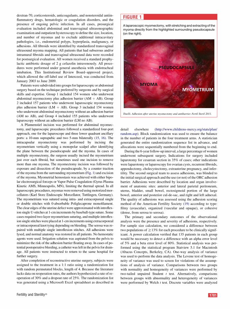
FIGURE 1
A laparoscopicmyomectomy, with stretching and extracting of the
myoma directly from the highlighted surrounding pseudocapsule(on the right).
Tinelli. Adhesion after uterine myomectomy and antibarrier. Fertil Steril 2011.
dextran-70, corticosteroids, anticoagulants, and nonsteroidal antiin-flammatory drugs, hematologic or coagulation disorders, and thepresence of ongoing pelvic infection. In all cases, presurgicalevaluation included abdominal and transvaginal ultrasonographicexamination and outpatient hysteroscopy to define the size, location,and number of myomas and to exclude additional intracavitarypathologies, i.e., endometrial polyps, hyperplasia, malignancy, oradhesions. All fibroids were identified by standardized transvaginalultrasound myoma mapping. All patients that had subserous and/orintramural fibroids and transvaginal ultrasound data were recordedfor postsurgical evaluation. All women received a standard prophy-lactic antibiotic dosage of 2 g cefazolin intravenously. All proce-dures were performed under general anesthesia with endotrachealintubation. This Institutional Review Board–approved project,which allowed the off-label use of Interceed, was conducted fromJanuary 2003 to June 2009.
Patientswere subdivided into groups for laparoscopic or abdominalsurgery based on the technique preferred by surgeons and by surgicalskills and expertise. Group 1 included 154 women who underwentabdominal myomectomy plus adhesion barrier (AM þ AB). Group2 included 157 patients who underwent laparoscopic myomectomyplus adhesion barrier (LM þ AB). Group 3 included 154 womenwho underwent abdominal myomectomy without an adhesion barrier(AM no AB), and Group 4 included 155 patients who underwentlaparoscopy without an adhesion barrier (LM no AB).
A Pfannenstiel incision was performed for abdominal myomec-tomy, and laparoscopic procedures followed a standardized four-portapproach, one for the laparoscope and three lower quadrant ancillaryports: a 10-mm suprapubic and two 5-mm bilaterally (15, 16). Theintracapsular myomectomy was performed by incising themyometrium vertically using a monopolar scalpel after identifyingthe plane between the pseudocapsule and the myoma. In cases ofmultiple myomectomy, the surgeon usually incised the myometriumjust over each fibroid, but sometimes used one incision to removemore than one myoma. The myomectomy incision was followed byexposure and dissection of the pseudocapsule, by a counter tractionof the myoma from the surrounding myometrium (Fig. 1) and excisionof the myoma. Myometrial hemostasis was achieved with either bipo-lar electrosurgical forceps or Vapor Pulse Coagulation (Gyrus PlasmaKinetic AMS, Minneapolis, MN), limiting the thermal spread. In alllaparoscopic procedures,myomaswere removed usingmotorizedmor-cellators (Karl Storz Endoscopic Morcellator, Tuttlingen, Germany).The myometrium was sutured using intra- and extracorporeal singleor double stitches with 0-absorbable Poliglecaprone monofilament.The clear edges of the uterine defectwere approximatedwith introflex-ion single U-stitches at 1-cm increments by baseball-type suture. Somecases required two-layer myometrium suturing, andmultiple introflex-ion single stitcheswereplaced at 1-cm increments using extracorporealor intracorporeal knot tying depending on bleeding. The serosa was re-paired with multiple single introflexion stitches. All adhesions werelysed, and normal anatomy was restored in all patients. No hemostaticagents were used. Irrigation solution was aspirated from the pelvis tominimize the risk of the adhesion barrier floating away. In cases of po-tential postoperative bleeding, a catheterwas left in the pelvis for drain-age. All patients were instructed to return to the same hospital forfurther surgery.
After completion of reconstructive uterine surgery, subjects wereassigned to the treatment in a 1:1 ratio using a randomization listwith random permutated blocks, length of 4. Because the literaturelacks data on reoperation rates, the authors hypothesized a rate of re-operation of 30% and a dropout rate of 25%. The randomization listwas generated using a Microsoft Excel spreadsheet as described in
Fertility and Sterility�
detail elsewhere (http://www.childrens-mercy.org/stats/plan/random.asp). Block randomization was used to ensure the balancein the number of patients in the four treatment arms. A statisticiangenerated the entire randomization sequence list in advance, andallocations were sequentially numbered from the beginning to end.
During the 6-year follow-up interval, a large percentage ofwomenunderwent subsequent surgery. Indications for surgery includedlaparotomy for cesarean section in 35% of cases; other indicationswere laparotomy or laparoscopy for ovarian cysts, recurrent fibroids,appendectomy, cholecystectomy, extrauterine pregnancy, and infer-tility. The second surgical team to assess adhesions, was blinded tothe initial surgical approach and the use (or not) of the ORC adhesionbarrier. Adhesions were described by location and organ involve-ment of anatomic sites: anterior and lateral parietal peritoneum,uterus, bladder, small bowel, rectosigmoid portion of the largebowel, anterior and posterior cul-de-sac, and right and left adnexal.The quality of adhesions was assessed using the adhesion scoringmethod of the American Fertility Society (19) according to type:filmy (avascular), organized (vascular and opaque), or cohesive(dense, from serosa to serosa).
The primary and secondary outcomes of the observationalanalysis were the presence and severity of adhesions, respectively.For sample size calculation, we considered a difference betweentwo populations ofR13% for each procedure to be clinically signif-icant. A power calculation verified that 135 patients in each groupwould be necessary to detect a difference with an alpha error levelof 5% and a beta error level of 80%. Statistical analysis was per-formed using the statistical program Statview 5.1 for Macintosh(Abacus Concepts, Berkeley, CA). One-way analysis of variancewas used to perform the data analysis. The Levene test of homoge-neity of variance was used to screen for violations of the assump-tions of analysis of variance. Comparisons between two groupswith normality and homogeneity of variances were performed bytwo-tailed unpaired Student t test. Alternatively, comparisonsbetween groups with abnormality and heterogeneity of varianceswere performed by Welch t test. Discrete variables were analyzed
1781
ue
a b b a
with the chi-squared test. A P value of < .05 was considered to bestatistically significant.
lusAB
LaparotomywithoutAB
II(n
[138)
Pvalue
GroupIII(n
[135)
GroupIV
(n[
137)
Pval
2�
7.5
NSa
28.9
�9.2
30.1
�6.8
NS
9�
0.3
NSb
22.2
�0.9
22.5
�0.1
NS
3�
0.9
NSb
1.2
�0.7
1.4
�0.1
NS
6�
0.2
NSa
2.4
�1.7
2.5
�0.8
NS
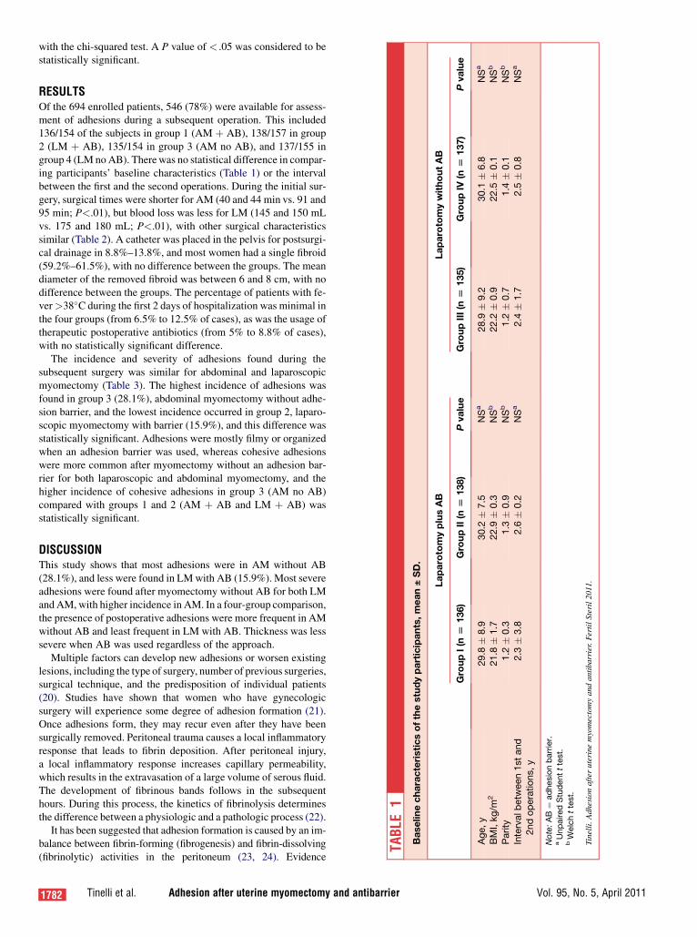
RESULTSOf the 694 enrolled patients, 546 (78%) were available for assess-ment of adhesions during a subsequent operation. This included136/154 of the subjects in group 1 (AM þ AB), 138/157 in group2 (LM þ AB), 135/154 in group 3 (AM no AB), and 137/155 ingroup 4 (LM noAB). Therewas no statistical difference in compar-ing participants’ baseline characteristics (Table 1) or the intervalbetween the first and the second operations. During the initial sur-gery, surgical times were shorter for AM (40 and 44 min vs. 91 and95 min; P<.01), but blood loss was less for LM (145 and 150 mLvs. 175 and 180 mL; P<.01), with other surgical characteristicssimilar (Table 2). A catheter was placed in the pelvis for postsurgi-cal drainage in 8.8%–13.8%, and most women had a single fibroid(59.2%–61.5%), with no difference between the groups. The meandiameter of the removed fibroid was between 6 and 8 cm, with nodifference between the groups. The percentage of patients with fe-ver>38�C during the first 2 days of hospitalization was minimal inthe four groups (from 6.5% to 12.5% of cases), as was the usage oftherapeutic postoperative antibiotics (from 5% to 8.8% of cases),with no statistically significant difference.
The incidence and severity of adhesions found during thesubsequent surgery was similar for abdominal and laparoscopicmyomectomy (Table 3). The highest incidence of adhesions wasfound in group 3 (28.1%), abdominal myomectomy without adhe-sion barrier, and the lowest incidence occurred in group 2, laparo-scopic myomectomy with barrier (15.9%), and this difference wasstatistically significant. Adhesions were mostly filmy or organizedwhen an adhesion barrier was used, whereas cohesive adhesionswere more common after myomectomy without an adhesion bar-rier for both laparoscopic and abdominal myomectomy, and thehigher incidence of cohesive adhesions in group 3 (AM no AB)compared with groups 1 and 2 (AM þ AB and LM þ AB) wasstatistically significant.
TABLE
1Baselinecharacteristicsofthestudyparticipants,mean±SD.
Laparotomyp
GroupI(n
[136)
Group
Age,y
29.8
�8.9
30.
BMI,kg/m
221.8
�1.7
22.
Parity
1.2
�0.3
1.
Intervalb
etw
een1stand
2ndoperations,y
2.3
�3.8
2.
Note:AB¼
adhesionbarrier.
aUnpairedStudentttest.
bWelchttest.
Tinelli.Adhesionafteruterine
myomectomyand
antibarrier.FertilSteril2011.
DISCUSSIONThis study shows that most adhesions were in AM without AB(28.1%), and less were found in LMwith AB (15.9%). Most severeadhesions were found after myomectomy without AB for both LMandAM, with higher incidence in AM. In a four-group comparison,the presence of postoperative adhesions were more frequent in AMwithout AB and least frequent in LM with AB. Thickness was lesssevere when AB was used regardless of the approach.
Multiple factors can develop new adhesions or worsen existinglesions, including the type of surgery, number of previous surgeries,surgical technique, and the predisposition of individual patients(20). Studies have shown that women who have gynecologicsurgery will experience some degree of adhesion formation (21).Once adhesions form, they may recur even after they have beensurgically removed. Peritoneal trauma causes a local inflammatoryresponse that leads to fibrin deposition. After peritoneal injury,a local inflammatory response increases capillary permeability,which results in the extravasation of a large volume of serous fluid.The development of fibrinous bands follows in the subsequenthours. During this process, the kinetics of fibrinolysis determinesthe difference between a physiologic and a pathologic process (22).
It has been suggested that adhesion formation is caused by an im-balance between fibrin-forming (fibrogenesis) and fibrin-dissolving(fibrinolytic) activities in the peritoneum (23, 24). Evidence
1782 Tinelli et al. Adhesion after uterine myomectomy and antibarrier Vol. 95, No. 5, April 2011

TABLE 2Initial surgery characteristics of the 546 study participants.
Laparotomy plus AB Laparotomy without AB
Group I(n [ 136)
Group II(n [ 138) P value
Group III(n [ 135)
Group IV(n [ 137) P value
Patients with single fibroid, n (%) 82 (60.2) 85 (61.5) NSa 80 (59.2) 83 (60.5) NSa
Dimension of single fibroid, cm 7 � 0.7 6 � 1.5 NSb 8 � 0.4 7 � 1.3 NSb
Women with catheter to drain
(removed in 1st day), n (%)
12 (8.8) 16 (11.5) NSa 15 (11.1) 19 (13.8) NSa
Total operative surgical time, min 44 � 9.3 95 � 4.7 < .01b 40 � 6.2 91 � 5.3 < .01b
Intrasurgical
blood loss, mL
180 � 3.7 150 � 4.2 < .01b 175 � 8.1 145 � 7.7 < .01b
Fever, n (%)c 12 (12.5) 9 (6.5) NSa 15 (11.1) 11 (8) NSa
Therapeutic postoperativeantibiotics administration,
no. of patients (%)
9 (6.6) 7 (5) NSa 12 (8.8) 8 (5.8) NSa
Note: AB ¼ adhesion barrier.a c2 test.b Welch t test.c Patients with fever >38�C after 24 h and for the first 2 days of hospitalization.
Tinelli. Adhesion after uterine myomectomy and antibarrier. Fertil Steril 2011.
suggests that a posttraumatic insufficiency in peritoneal fibrinolyticactivity caused by decreased tPA and increased PAI-1 and PAI-2 per-mits the deposited fibrin to become organized into permanent adhe-sions (25). This theory is supported by the observation that womenwith endometriosis have significantly higher amounts of peritonealfluid and adhesions, presumably due to an endometriosis-inducedlow-grade sterile inflammation of the peritoneal cavity (26, 27). Aprospective study in humans adds further weight to the hypothesisthat adhesions are caused by an insufficiency in peritonealfibrinolytic activity, and authors also conclude that fibrinolysis issignificantly enhanced 1 week after surgery (28).
Two studies showed that adhesions are not reduced when ORC isapplied on bleeding or oozing surfaces (29, 30), but other studieshave not supported those observations. An investigation comparedthe efficacy of Interceed with ORC alone. At second-look laparos-copy, there was no significant difference in adhesions in comparing
TABLE 3Adhesion characteristics of the 546 study participants.
Laparotomy plus A
Group I(n [ 136)
Group II(n [ 138)
Women with adhesions, n (%) 30 (22) 22 (15.9)
Filmy adhesions
(avascular), n (%)
11 (8) 10 (7.2)
Organized adhesions
(vascular and opaque), n (%)
9 (6.6) 7 (5)
Cohesive adhesions
(serosa to serosa), n (%)
6 (4.4) 5 (3.6)
Note: AB ¼ adhesion barrier.a c2 test with 1 DF.
Tinelli. Adhesion after uterine myomectomy and antibarrier. Fertil Steril 2011.
Fertility and Sterility�
ORC plus heparin (52.5%, 21/40) or ORC alone (65%, 26/40) (31).Another study showed that ORC significantly reduced the incidenceand extent of adhesions: Adhesion-free outcomes were 1.5–2.5 timesmore at sites treatedwith theABduring laparotomy (32). Studies haveshown a further reduction in adhesions with laparoscopy comparedwith traditional laparotomy (33, 34). The American Society ofReproductive Medicine (ASRM) Practice Committee recommendsthat efforts to minimize adhesion formation should be used,includingminimally invasive techniques and adhesiolysis agents (35).
Experts recommend meticulous procedures, physical separationof denuded or damaged peritoneal surfaces, and placing adhesiolysisagents (36, 37).
Literature reports that the surgical restoration of the uterine anat-omy after myomectomy is accompanied by fewer adhesions duringsecond-look surgery (38). Thus, the restoration of the proper anatomicrelationships of the pseudocapsule surrounding the myoma (15, 39),
B Laparotomy without AB
P valueaGroup III(n [ 135)
Group IV(n [ 137) P valuea
NS 38 (28.1) 31 (22.6) NS
NS 10 (7.4) 8 (5.8) NS
NS 12 (8.8) 11 (8) NS
NS 16 (11.8) 12 (8.7) NS
1783

should prevent excessive bleeding when removing single or multiplefibroids (15, 16). In fact, the secondary outcomes of our analysis ofsurgical data showed that blood loss was less for LM, even thoughthe surgical times were shorter for AM. This is probably due to themagnification of endoscope vision, by a superior visualization of thepseudocapsule fibers and blood vessels surrounding the myoma (15,39), enabling the surgeon to delicately dissect it and to selectivelycoagulate and cut. Laparoscopic myomectomy is an alternative tolaparotomy in many cases. Laparoscopy advantages are shorthospitalization, decreased need for postoperative analgesia, and lessintraoperative blood loss (40).
Adding tissue separation and protection during the healingprocess by applying the biocompatible and absorbable ORC ABadherent to traumatized and weeping surfaces (41) further protectsagainst new and recurrent adhesions.
Limitations and biases of this study include the lack ofa structured second-look surgery, a loss of follow-up for patientswho did not undergo a subsequent surgery, and the noncomparisonof this technique between the groups. However, the authors couldnot propose a standardized second-look laparoscopy in the study
1784 Tinelli et al. Adhesion after uterine myomectomy and
protocol for many reasons. Therefore, a longer study interval wasneeded to recruit and follow subjects to have a satisfactory numberof patients who eventually required subsequent surgery.
Because intra-abdominal adhesions are caused by a variety offactors, the combination of laparoscopic intracapsular myomec-tomy, which limits tissue damage, and the use of an absorbableadhesion barrier is an effective means to minimize adhesion forma-tion. The authors strongly support the ASRM Practice Committeerecommendation that that efforts to prevent adhesions should beimplemented (35).
CONCLUSIONAdhesion formation after uterine surgery remains a challenge,even with the benefit of adhesion prevention by synthetic barriers(42). Continued efforts are required to improve surgical techniquesand antiadhesion agents. In this study, the combined method oflaparoscopic intracapsular myomectomy plus ORC absorbableAB reduced the incidence of cohesive postsurgical adhesions.
REFERENCES
1. Ellis H, Moran BJ, Thompson JN, Parker MC,
Wilson MS, Menzies D, et al. Adhesion-related
hospital readmissions after abdominal and pelvic
surgery: a retrospective cohort study. Lancet 1999;
353:1476–80.
2. Trimbos-Kemper TCM, Trimbos JB, van Hall EV.
Adhesion formation in tubal surgery: results of the
eight day laparoscopy in 188 patients. Fertil Steril
1985;43:395–400.
3. Bronson RA, Wallace EE. Lysis of periadnexal
adhesions for correction of infertility. Fertil Steril
1977;28:613–9.
4. Interceed (TC7) Adhesion Barrier Study Group.
Prevention of postsurgical adhesions by Interceed
(TC7), an absorbable adhesion barrier:
a prospective, randomized multicenter clinical
study. Fertil Steril 1989;51:933–8.
5. Linsky CB, Diamond MP, Cunningham TJ,
Constantine B, DeCherney AH, DiZerega GS.
Adhesion reduction in the rabbit uterine horn model
using an absorbable barrier, TC7. J Reprod Med
1987;32:17–20.
6. Steinleitner A, Lopez G, Suarez M, Lambert H.
An evaluation of Flowgel as an intraperitoneal
barrier material for the prevention of
postsurgical adhesion reformation. Fertil Steril
1992;57:305–8.
7. Montz FJ, Monk BJ, Lacy SM. Effectiveness of two
barriers at inhibiting post-radical pelvic surgery ad-
hesions. Gynecol Oncol 1993;48:247–51.
8. Nordic Adhesion Prevention Study Group. The
efficacy of Interceed (TC7) for prevention of
reformation of postoperative adhesions on ovaries,
fallopian tubes, and fimbriae in microsurgical
operations for fertility: a multicenter study. Fertil
Steril 1995;63:709–14.
9. Franklin RR, Ovarian Adhesion Study Group.
Reduction of ovarian adhesions by the use of
Interceed. Obstet Gynecol 1995;86:335–40.
10. Gago LA, Saed G, Elhammady E, Diamond MP.
Effect of oxidized regenerated cellulose (Interceed)
on the expression of tissue plasminogen activator
and plasminogen activator inhibitor-1 in human peri-
toneal fibroblasts and mesothelial cells. Fertil Steril
2006;86(Suppl 4):1223–7.
11. Reddy S, Santanam N, Reddy PP. Interaction of
Interceed oxidized regenerated cellulose with
macrophages: a potential mechanism by which
Interceed may prevent adhesions. Am J Obstet
Gynecol 1997;177:1315–20; discussion 1320–1.
12. Tsuji S, Takahashi K, Yomo H, Fujiwara M, Kita N,
Takebayashi K, et al. Effectiveness of antiadhesion
barriers in preventing adhesion after myomectomy
in patients with uterine leiomyoma. Eur J Obstet
Gynecol Reprod Biol 2005;123:224–48.
13. Pados G, Camus M, De Munck L, Devroey P.
Laparoscopic application of Interceed (TC7). Hum
Reprod 1992;7:1141–3.
14. Hurst BS, Matthews ML, Marshburn PB.
Laparoscopic myomectomy for symptomatic uterine
myomas. Fertil Steril 2005;83:1–23.
15. Tinelli A, Malvasi A, Rahimi S, Negro R,
Cavallotti C, Vergara D, et al. Myoma
pseudocapsule: a new-old endocrino-anatomical en-
tity to exploit in uterine myomectomy. Endocrinol
Gynecol 2009;25:661–7.
16. Tinelli A, Malvasi A, Cavallotti C, Hudelist G,
Tsin DA, Schollmeyer T, et al. Single and multiple
intracapsular laparoscopic myomectomy. an
institutional experience. J Laparoendosc Adv Surg
Tech A 2010;20:705–11.
17. Zimbris L, Hedon B, Lafrargue F. Myomectomie
parcoelioscopie. J Obstet Gynecol 1994;2:
219–23.
18. Cohen D, Mazur MT, Jozefczyk MA, Badawy SZA.
Hyalinization and cellular changes in uterine
leiomyomata after gonadotropin releasing hormone
agonist therapy. J Reprod Med 1994;39:377–80.
19. The American Fertility Society classifications of ad-
nexal adhesions, distal tubal occlusion, tubal occlu-
sion secondary to tubal ligation, tubal pregnancies,
m€ullerian anomalies and intrauterine adhesions. Fer-
til Steril 1988;49:944–55.
20. Diamond MP, Freeman ML. Clinical implications of
postsurgical adhesions. Hum Reprod Update
2001;7567–76.
21. Ellis H, Crowe A. Medico-legal consequences of
post-operative intra-abdominal adhesions. Int J Surg
2009;7:187–91.
22. Liakakos T, lthomakos N, Fine PM, Dervenis C,
Young RC. Peritoneal adhesions: etiology,
pathophysiology, and clinical significance. Recent
advances in prevention and management. Dig Surg
2001;18:260–73.
antibarrier
23. DiZerega GS, Campeau JD. Peritoneal repair and
post-surgical adhesion formation. Biochemical
events in peritoneal tissue repair. HumReprod Update
2001;7:547–55.
24. Holmdahl L. The role of fibrinolysis in adhesion
formation. Eur J Surg 1997;577(Suppl):24–31.
25. Ivarsson ML, Bergstrom M, Eriksson E, Risberg B,
Holmdahl L. Tissue markers as predictors of
postoperative adhesions. Br J Surg 1998;85:1549–54.
26. KoninckxPR,Kennedy SH,BarlowDH. Pathogenesis
of endometriosis: the role of peritoneal fluid. Gynecol
Obstet Invest 1999;47(Suppl 1):23–33.
27. LebovicDI,MuellerMD, TaylorRN. Immunobiology
of endometriosis. Fertil Steril 2001;75:1–10.
28. Hellebrekers BW, Emeis JJ, Kooistra T, Trimbos JB,
Moore NR, Zwinderman KH, et al. A role for the
fibrinolytic system in postsurgical adhesion
formation. Fertil Steril 2005;83:122–9.
29. Linksy CB, Diamond PM, DeCherney AH,
DiZerega GS, Cunningham T. Effect of blood on
the efficacy of barrier adhesion reduction in the
rabbit uterine horn model. Infertility 1988;11:
273–80.
30. Wiseman DM, Kamp LF, Saferstein L, Linsky CB,
Gottlick LE, Diamond MP. Improving the efficacy
of Interceed Barrier in the presence of blood using
thrombin, heparin or a blood insensitive barrier,
modified Interceed (TC7). Prog Clin Biol Res
1993;381:205–12.
31. Reid RL, Hahn PM, Spence JEH, Tulandi T,
Yupze AA, Wiseman DM. A randomized clinical
trial of oxidized regenerated cellulose adhesion
barrier (Interceed, TC7) alone or in combination
with heparin. Fertil Steril 1997;67:24–9.
32. Wiseman DM, Trout JR, Franklin RR, Diamond MP.
Metaanalysis of the safety and efficacy of an adhesion
barrier (Interceed TC7) in laparotomy. J Reprod Med
1999;44:325–31.
33. Luciano AA, Maier DB, Nulsen JC, Whitman GF,
Koch EI. A comparative study of postoperative
adhesion formation following laser surgery by
laparoscopy versus laparotomy in the rabbit model.
Obstet Gynecol 1989;74:220–4.
34. Luciano AA, Montanino-Oliva M. Comparison of
postoperative adhesion formation laparoscopy
versus laparotomy. Infertil Reprod Med Clin North
Am 1994;5:437–44.
Vol. 95, No. 5, April 2011

35. Practice Committee of the American Society of Re-
productive Medicine. Pathogenesis, consequences,
and control of peritoneal adhesions in gynecologic
surgery. Fertil Steril 2007;88:21–6.
36. Wallwiener M, Brucker S, Hierlemann H,
Brochhausen C, Solomayer E, Wallwiener C.
Innovative barriers for peritoneal adhesion
prevention: liquid or solid? A rat uterine horn
model. Fertil Steril 2006;86:1266–76.
37. Freije WA, DeCherney A. Preventing adhesions in
gynecologic surgery. OBG Manag 2001;5:39–49.
Fertility and Sterility�
38. Nezhat C, Nezhat F, Silfen SL, Schaffer N, Evans D.
Laparoscopic myomectomy. Obstet Gynaecol
1992;36:275–80.
39. de Falco M, Staibano S, Mascolo M, Mignogna C,
Improda L, Ciociola F, et al. Leiomyoma
pseudocapsule after pre-surgical treatment with gona-
dotropinreleasing hormone agonists: relationship be-
tween clinical features and immunohistochemical
changes. Eur J Obstet Gynecol Reprod Biol
2009;144:44–7.
40. Falcone T, Bedaiwy MA. Minimally invasive
management of uterine fibroids. Curr Opin Obstet
Gynaecol 2002;14:401–7.
41. Azziz R, Interceed Adhesion Barrier Study Group II.
Microsurgery alone or with Interceed adhesion
barrier for pelvic sidewall adhesion re-formation.
Surg Gynecol Obstet 1993;177:135–9.
42. Ahmad G, Duffy JM, Farquhar C, Vail A,
Vandekerckhove P, Watson A, et al. Barrier agents for
adhesion prevention after gynaecological surgery.
Cochrane Database Syst Rev 2008;(2):CD000475.
1785