Novel precipitated fluorescent substrates for the screening of cellulolytic microorganisms

APPLIED AND ENVIRONMENTAL MICROBIOLOGY, June 2007, p. 3536–3546 Vol. 73, No. 110099-2240/07/$08.00�0 doi:10.1128/AEM.00225-07Copyright © 2007, American Society for Microbiology. All Rights Reserved.
Genome Sequence of the Cellulolytic Gliding BacteriumCytophaga hutchinsonii�†
Gary Xie,1,2 David C. Bruce,1,2 Jean F. Challacombe,1,2 Olga Chertkov,1,2 John C. Detter,1,2
Paul Gilna,1,2 Cliff S. Han,1,2 Susan Lucas,2,4 Monica Misra,1 Gerald L. Myers,1Paul Richardson,2,5 Roxanne Tapia,1,2 Nina Thayer,1 Linda S. Thompson,1,2
Thomas S. Brettin,1,2 Bernard Henrissat,3 David B. Wilson,6and Mark J. McBride7*
Los Alamos National Laboratory, Bioscience Division, Los Alamos, New Mexico1; Department of Energy Joint Genome Institute,Walnut Creek, California2; Architecture et Fonction des Macromolecules Biologiques, UMR 6098, Centre National de la
Recherche Scientifique, and Universities of Aix-Marseille I and II, Marseille, France3; Lawrence Livermore National Laboratory,Livermore, California4; Lawrence Berkeley National Laboratory, Berkeley, California5; Cornell University,
Ithaca, New York6; and University of Wisconsin—Milwaukee, Milwaukee, Wisconsin7
Received 29 January 2007/Accepted 23 March 2007
The complete DNA sequence of the aerobic cellulolytic soil bacterium Cytophaga hutchinsonii, whichbelongs to the phylum Bacteroidetes, is presented. The genome consists of a single, circular, 4.43-Mbchromosome containing 3,790 open reading frames, 1,986 of which have been assigned a tentative function.Two of the most striking characteristics of C. hutchinsonii are its rapid gliding motility over surfaces andits contact-dependent digestion of crystalline cellulose. The mechanism of C. hutchinsonii motility is notknown, but its genome contains homologs for each of the gld genes that are required for gliding of thedistantly related bacteroidete Flavobacterium johnsoniae. Cytophaga-Flavobacterium gliding appears to benovel and does not involve well-studied motility organelles such as flagella or type IV pili. Many genesthought to encode proteins involved in cellulose utilization were identified. These include candidateendo-�-1,4-glucanases and �-glucosidases. Surprisingly, obvious homologs of known cellobiohydrolaseswere not detected. Since such enzymes are needed for efficient cellulose digestion by well-studied cellu-lolytic bacteria, C. hutchinsonii either has novel cellobiohydrolases or has an unusual method of celluloseutilization. Genes encoding proteins with cohesin domains, which are characteristic of cellulosomes, wereabsent, but many proteins predicted to be involved in polysaccharide utilization had putative D5 domains,which are thought to be involved in anchoring proteins to the cell surface.
Cellulose is probably the most abundant biopolymer onearth (5). The �-1,4-linked glucose chains of cellulose formhighly ordered crystalline fibrils that are insoluble and rela-tively recalcitrant to degradation. Cellulolytic bacteria producea suite of enzymes that act synergistically to convert crystallinecellulose to glucose (38, 62). The cellulolytic bacteria that havebeen studied most use either of two strategies to digest cellu-lose. Some, such as aerobic actinomycetes belonging to thegenera Streptomyces and Thermobifida, secrete soluble extra-cellular cellulolytic enzymes that usually contain carbohydratebinding modules (CBMs) that anchor them to the substrate(71). These enzymes attack the insoluble substrate and releaseglucose, cellobiose, and short oligomers that are taken up andmetabolized by the cells. In contrast, anaerobic bacteria of thegenera Clostridium and Ruminococcus produce multiproteincomplexes, called cellulosomes, that often remain attached tocells (7, 9, 19, 50, 62). Cellulosomes contain cellulose-bindingproteins, enzymes involved in the hydrolysis of cellulose and of
other polysaccharides, and scaffolding proteins that hold themultiprotein complex together. Cellulosomes often anchor thebacteria to their substrate and result in localized release ofsugars that are taken up by the cells.
Cytophaga hutchinsonii (Fig. 1), the type species of the genusCytophaga, is an abundant aerobic cellulolytic soil bacterium(37, 55, 64, 72). C. hutchinsonii is a gram-negative bacteriumthat belongs to the phylum Bacteroidetes (also known as theCytophaga-Flavobacterium-Bacteroides group). C. hutchinsoniiutilizes very few substrates as sole carbon and energy sources.Besides cellulose, its only known substrates are cellobiose andglucose (37). Wild strains generally use these soluble sugarspoorly and exhibit a preference for crystalline cellulose. C.hutchinsonii requires direct contact with cellulose for efficientdigestion, and most of the cellulolytic enzymes appear to becell associated (16, 47, 64). Another unusual feature of cellu-lose degradation by C. hutchinsonii is that reducing sugars suchas glucose and cellobiose do not accumulate in the mediumwhen it digests cellulose (64). This is probably not simply theresult of efficient uptake and metabolism of these sugars, sinceincubation of cells under anaerobic conditions, which shouldinterfere with these processes, does not result in accumulationof reducing sugars.
C. hutchinsonii was selected for genome sequencing forseveral reasons. First, biochemical and physiological studies
* Corresponding author. Mailing address: Department of BiologicalSciences, University of Wisconsin—Milwaukee, 3209 N. MarylandAve., 181 Lapham Hall, Milwaukee, WI 53211. Phone: (414) 229-5844.Fax: (414) 229-3926. E-mail: [email protected].
† Supplemental material for this article may be found at http://aem.asm.org/.
� Published ahead of print on 30 March 2007.
3536

suggested that it might use a novel strategy for celluloseutilization. Second, techniques to genetically manipulate C.hutchinsonii are available (44). Most cellulolytic bacteriahave resisted genetic analysis, so the development of thesetechniques promises new insights into bacterial celluloseutilization. Third, C. hutchinsonii exhibits a form of rapidgliding motility whose mechanism remains unexplained de-spite decades of study (43, 64, 72). Gliding may help tofacilitate cellulose digestion, since gliding cells align them-selves with and move along cellulose fibers as they digestthem (64, 72). Finally, few genome sequences have beenreported for members of the phylum Bacteroidetes, and noneare closely related to C. hutchinsonii. This paper highlightsnovel features of the C. hutchinsonii genome, with particularemphasis on genes and proteins likely to be involved incellulose utilization and gliding motility.
MATERIALS AND METHODS
Sequencing of the C. hutchinsonii genome. The random shotgun method wasused to sequence the genome of C. hutchinsonii ATCC 33406. Large (40 kb)-,medium (8 kb)-, and small (3 kb)-insert random libraries were partially se-quenced, with an average success rate of 90% and an average high-quality readlength of 634 nucleotides. Sequences were assembled with parallel phrap (HighPerformance Software, LLC). Possible misassemblies were corrected with Dup-finisher (C. F. Han and P. Chain, presented at the 2006 International Conferenceon Bioinformatics and Computational Biology, Las Vegas, NV, 26 to 29 June2006) or by analysis of transposon insertions in bridge clones. Gaps betweencontigs were closed by editing, custom primer walking, or PCR amplification.The completed genome sequence of C. hutchinsonii contains 105,896 reads,
achieving an average of 15-fold sequence coverage per base, with an error rate of�1 in 100,000.
Annotation. Gene predictions were obtained using Glimmer, and tRNAs wereidentified using tRNAScan-SE. Basic analyses of the gene predictions wereperformed by comparing coding sequences against the Pfam, BLOCKS, andProdom databases. Protein localizations were predicted with PSORTb (21), andlipoproteins were identified using LipoP (33). A team of annotators added genedefinitions and functional classes, using BLAST results and information from thePfam (63; http://pfam.janelia.org/index.html), BLOCKS (26), Prodom (59), andSMART (58) databases. Metabolic pathways were constructed using MetaCyc asa reference data set (14). Genes encoding candidate glycoside hydrolases weredetected with routines used for updates of the Carbohydrate Active Enzymes(CAZY) database (18) at http://www.cazy.org/CAZY/. Detailed informationabout the genome properties and genome annotation can be obtained from theJGI Integrated Microbial Genomes website (40) at http://img.jgi.doe.gov/pub/main.cgi.
Comparative analysis. To obtain a list of orthologs from bacteroidete ge-nomes, a perl script that determines bidirectional best hits was written. Genes gand h are considered orthologs if h is the best BLASTP hit for g and vice versa,with E values of �10�15. A gene is considered strain specific if it has no hits withan E value of 10�5 or less.
Phylogenetic trees. Prealigned 16S rRNA sequences were derived from theRibosomal Database Project site (17; http://rdp.cme.msu.edu/index.jsp). Manualalignment adjustments were made as needed, with the assistance of the BioEditmultiple alignment tool of Hall (http://www.mbio.ncsu.edu/BioEdit/bioedit.html). The refined multiple alignment was used as input for the generation of aphylogenetic tree, using the program package PHYLIP (20). The fastDNAml(52) and nucleic acid sequence maximum likelihood (51) methods with defaultsettings were employed to obtain character-based trees. Both the fastDNAmland maximum likelihood trees yielded similar clusters and arrangements of taxawithin them.
Electron microscopy. Cells for scanning electron microscopy were grown onWhatman filter paper on DMA agar as previously described (44). Cells werefixed with 2.5% glutaraldehyde, 0.1% OsO4 in 10 mM HEPES buffer, pH 7.2, for30 min at 25°C. Fixed cells were washed twice in 10 mM HEPES buffer, postfixedin 1% OsO4, and washed twice with distilled water. Cells were dehydrated withethanol and critical point dried with CO2. Samples were mounted, sputter coatedwith 60% gold–40% palladium, and viewed with a Hitachi S-570 scanning elec-tron microscope.
Nucleotide sequence accession number. The sequences of C. hutchinsonii canbe accessed using GenBank accession number CP000383.
RESULTS AND DISCUSSION
General genome features. The genome of C. hutchinsoniiconsists of a single, circular, 4,433,218-bp chromosome that hasa GC content of 38.85% (see Fig. S1 in the supplementalmaterial). A total of 3,790 protein-encoding genes are pre-dicted, 1,986 of which have been assigned a tentative function.The GC skew allowed prediction of the site of the origin ofreplication to near nucleotide position 4,038,464. Surprisingly,ribosomal genes, tRNA genes, and typical replication-relatedgenes, such as dnaA, dnaN, recF, and gyrA, which are oftenlocated near the origin of replication in bacteria, were notlocated near this site. Three rRNA operons were identified inthe genome. Analysis of the 16S rRNA sequences confirmedthat C. hutchinsonii belongs to the phylum Bacteroidetes (Fig.2). It is a member of the class Sphingobacteria and is onlydistantly related to well-studied bacteroidetes, such as mem-bers of the genera Bacteroides, Porphyromonas, Prevotella (allfrom class Bacteroidetes), and Flavobacterium (class Flavobac-teria).
Cellulose utilization. One of the most distinctive features ofC. hutchinsonii is its ability to rapidly digest crystalline cellu-lose. Cellulose utilization by most well-studied microorganismsinvolves the activities of multiple enzymes, including endo-�-1,4-glucanases, cellobiohydrolases (also called exo-�-1,4-glu-
FIG. 1. Scanning electron micrograph of C. hutchinsonii cells di-gesting cellulose filter paper. Bar, 10 �m.
VOL. 73, 2007 C. HUTCHINSONII GENOME ANALYSIS 3537

canases), and �-glucosidases, that act synergistically to convertcrystalline cellulose to glucose (39, 70) (Fig. 3). Endo-�-1,4-glucanases attack the cellulose chain at exposed internal gly-cosidic bonds. They nick the long polymeric substrate andprovide sites of attack for cellobiohydrolases, which releasecellobiose from the exposed ends. Members of one class ofcellobiohydrolases attack the nonreducing end of the polymer,whereas members of the second class attack the reducing end(4). �-Glucosidases hydrolyze cellobiose and short cellodextrinoligomers to glucose. Analysis of the amino acid sequences ofcellulolytic enzymes allows them to be grouped into a limitednumber of glycoside hydrolase families (27–29). Examinationof the C. hutchinsonii genome identified many genes encodingpossible cellulolytic enzymes. These include possible endo-�-1,4-glucanases belonging to glycoside hydrolase families GH5and GH9 and �-glucosidases belonging to GH3 (Table 1).Genes encoding proteins with similarity to known cellobiohy-drolases, which are typically members of family GH6 or GH48in bacteria (23, 71), were not detected. Since cellobiohydro-lases are needed for efficient cellulose utilization by otherorganisms, C. hutchinsonii either has novel cellobiohydrolasesor uses an unusual method of cellulose utilization that does notrequire such enzymes. Some GH9 endo-�-1,4-glucanases areprocessive (22). The presence of processive endoglucanasescould account for the ability of C. hutchinsonii to digest crys-talline cellulose in the apparent absence of cellobiohydrolases.However, none of the C. hutchinsonii enzymes display highlevels of similarity to experimentally characterized processiveendoglucanases, so further experimental evidence is needed todetermine this possibility. The apparent absence of cellobio-hydrolases may partially explain why reducing sugars do notaccumulate in the medium when cells of C. hutchinsonii digestcellulose (64). All four of the candidate �-glucosidases arepredicted to reside in the periplasm, and three of them arepredicted lipoproteins (Table 1). The periplasmic location ofthese enzymes may also limit the amount of reducing sugarsreleased beyond the confines of the cell. In addition to en-zymes involved in cellulose utilization, C. hutchinsonii has
genes predicted to encode glycohydrolases involved in the di-gestion of hemicelluloses, such as xylan (Table 2), and genesencoding �-glycosidases of uncertain specificity (Table 3). Theremaining predicted glycohydrolases (Table 4) are likely to beinvolved in peptidoglycan turnover and utilization of intracel-lular storage compounds. Genes encoding predicted pectatelyases (Table 5) and genes encoding polysaccharide esterases,some of which are predicted to be involved in xylan utilization(Table 6), were also identified. C. hutchinsonii does not growon xylan or pectin as a sole carbon and energy source but mayuse xylanases, pectate lyases, and polysaccharide esterases togain access to cellulose in plant material. A similar situationhas been observed for the cellulolytic anaerobes Clostridiumthermocellum and Clostridium cellulovorans (6).
Recognizable CBMs are found on many of the glycohydro-lases (Tables 2 and 3), but surprisingly, none of the proteinsrelated to endo-�-1,4-glucanases contain such a module (Table1). The predicted endo-�-1,4-glucanases may have novelCBMs or may interact with other CBM-containing proteins toform multiprotein complexes that function in cellulose diges-tion. Equally surprising is that no genes encoding proteins withtype A CBMs, the only ones known to bind to crystallinecellulose (11), were detected in the entire genome. Cells of C.hutchinsonii do bind to crystalline cellulose and actively glidealong the fibers (Fig. 1). Presumably, components of the mo-tility machinery are involved in these interactions. The attach-ment of cells and movement along the cellulose fibers maymake dedicated CBM domains on the cellulolytic enzymesunnecessary. If type A CBMs were present, they might evenprevent gliding and thus decrease the ability of the cells toaccess and digest cellulose.
Many of the C. hutchinsonii proteins with possible roles in
FIG. 2. 16S rRNA-based phylogenetic tree of the phylum Bacte-roidetes derived from 16S rRNA gene sequence data. The tree wasconstructed using the fastDNAml method (20, 51, 52). Species withcomplete genome sequences that were used in this study are indicatedin bold. Bar, 0.1 nucleotide substitution per site.
FIG. 3. Enzymes involved in cellulose breakdown in well-studiedcellulolytic bacteria. C. hutchinsonii lacks obvious homologs of knowncellobiohydrolases.
3538 XIE ET AL. APPL. ENVIRON. MICROBIOL.

polysaccharide utilization are large and have complex multido-main structures (Tables 1, 2, 3, 5, and 7). In addition to theglycohydrolase and CBM domains discussed above, other do-mains present on some of the predicted proteins include do-mains with immunoglobulin-like folds, including group 2 bac-terial immunoglobulin-like domains (BIG_2; Pfam number
PF02368), N-terminal immunoglobulin-like domains of cellu-lase (CelD_N; Pfam number PF02927), immunoglobulin I-setdomains (IG; Pfam number PF00047), fibronectin type 3-likedomains (FN3; Pfam number PF00041), and polycystic kidneydisease protein-like domains (PKD; Pfam number PF000801).Parallel � helix repeat domains (PbH1; SMART database
TABLE 1. Predicted C. hutchinsonii glycohydrolases that may be involved in cellulose digestion
Gene Predicted function of gene producta Predicted localizationb Mol massc
(kDa) Modular structured
CHU_0778 Related to endo-�-1,4-glucanase Unknown 81.7 CelD_N-GH9CHU_1107 Candidate endo-�-1,4-glucanase Extracellular 135.2 GH5-X1-PKD-PKD-FN3CHU_1280 Candidate endo-�-1,4-glucanase Unknown 66.2 CelD_N-GH9CHU_1335 Related to endo-�-1,4-glucanases Extracellular/outer membrane 207.3 GH9-PKD-PKD-PKD-
BIG-BIG-BIG-BIG-BIG-BIG-BIG-BIG-BIG-BIG-BIG-D5
CHU_1336 Related to endo-�-1,4-glucanases Extracellular 105.3 GH9-PKD-PKD-PKDCHU_1655 Candidate endo-�-1,4-glucanase Extracellular/outer membrane 92.6 CelD_N-GH9CHU_1727 Related to endo-�-1,4-glucanases Unknown 67.9 GH5CHU_2103 Candidate endo-�-1,4-glucanase Extracellular 38.8 GH5CHU_2235 Distantly related to endo-�-1,4-glucanases Unknown 64.2 GH9CHU_2268 Candidate �-glucosidase Periplasmic lipoprotein 83.7 GH3CHU_2273 Candidate �-glucosidase Periplasmic lipoprotein 89.7 GH3CHU_3577 Candidate �-glucosidase Periplasmic lipoprotein 83.2 GH3CHU_3784 Candidate �-glucosidase Periplasmic 81.8 GH3
a Assigned by routines used for updating the CAZY database (http://www.cazy.org/CAZY/), using the following criteria. Typically, �70% amino acid identityto a protein domain with a biochemically determined function at the time of analysis resulted in “candidate” status. A 30% to 70% amino acid identity to aprotein domain with known function resulted in “related to” status. Less than 30% amino acid identity to a protein domain with known function resulted in“distantly related to” status. Because the threshold of similarity that correlates with a change of substrate specificity is variable from one glycoside hydrolasefamily to another, the criteria were tightened or loosened appropriately for several families. All analyses were conducted domain by domain because of themodular structure of many of the proteins.
b Predicted using the default settings of PSORTb (21). Predicted lipoproteins were identified using LipoP (33).c Molecular mass of primary product of translation, including any predicted signal peptide.d Modular structure is indicated by the following abbreviations: BIG, bacterial immunoglobulin-like domain group 2 (Pfam number PF02368); CelD_N,
N-terminal immunoglobulin-like domain of cellulase (Pfam number PF02927); D5, carboxy-terminal domain of Rhodothermus marinus xylanases predicted tobe involved in attachment to the cell surface (34); FN3, fibronectin type 3 domain (Pfam number PF00041); GH, glycoside hydrolase (number indicates family),as assigned by CAZY; PKD, polycystic kidney disease protein PKD1 (Pfam number PF00801); and X1, conserved domain of unknown function.
TABLE 2. Predicted C. hutchinsonii glycohydrolases that may be involved in hemicellulose digestion
Gene Predicted function ofgene producta
Predictedlocalizationb
Mol massc
(kDa) Modular structured
CHU_1155 Candidate xyloglucanase Extracellular 132.2 GH74-PKD-PKD-PKD-PKD-PKD-D5CHU_1239 Candidate acetylxylan esterase/
xylanaseExtracellular/outer
membrane148.1 CE6-X1-GH10-X1-CBM4-PKD-PKD-
PKD-PKD-D5CHU_1240 Candidate xylanase/esterase Extracellular/outer
membrane183.0 CE4-GH8-CBM9-PKD-PKD-PKD-
PKD-PKD-PKD-X1-D5CHU_2041 Candidate acetylxylan esterase/
�-xylosidase/�-L-arabinofuranosidase
Extracellular/outermembrane
166.6 CE6-GH43-X1-X1-CBM9-PKD-PKD-PKD-PKD-X1-D5
CHU_2042 Candidate xylanase Extracellular/outermembrane
127.4 GH5-X1-CBM9-PKD-PKD-PKD-PKD-X1-D5
CHU_2043 Candidate xylanase Extracellular/outermembrane
128.5 GH10-X1-CBM9-PKD-PKD-PKD-PKD-PKD-X1-D5
CHU_2044 Related to �-xylosidases/�-L-arabinofuranosidases
Extracellular/outermembrane
154.4 GH43-CBM6-X1-CBM9-PKD-PKD-PKD-X1-D5
CHU_2105 Candidate xylanase Unknown 76.3 CBM4-GH10CHU_2379 Candidate xylanase Extracellular/outer
membrane119.5 GH11-CBM9-PKD-PKD-PKD-PKD-
PKD-X1-D5CHU_0353 Candidate �-mannanase Extracellular 86.6 CBM6-GH26-PKD-PKD-D5
a Assigned by routines used for updating the CAZY database (http://www.cazy.org/CAZY/), using the criteria indicated in Table 1.b Predicted using the default settings of PSORTb (21). Predicted lipoproteins were identified using LipoP (33).c Molecular mass of primary product of translation, including any predicted signal peptide.d Modular structure is indicated by the following abbreviations: CE, carbohydrate esterase (number indicates family), as assigned by CAZY; CBM, carbohydrate
binding module (number indicates family), as assigned by CAZY; D5, carboxy-terminal domain of Rhodothermus marinus xylanases predicted to be involved inattachment to the cell surface (34); GH, glycoside hydrolase (number indicates family), as assigned by CAZY; PKD, polycystic kidney disease protein PKD1 (Pfamnumber PF00801); and X1, conserved domain of unknown function.
VOL. 73, 2007 C. HUTCHINSONII GENOME ANALYSIS 3539

number SM00710) and novel conserved domains (X1, X3,X100, and Y98) were also identified. The functions of thesedomains are not known, but possibilities include binding tocellulose, binding to other components of the cellulolytic ma-chinery, or interaction with the cell surface. Eighteen of theproteins listed in Tables 1, 2, 3, 5, 6, and 7 have carboxy-terminaldomains that are similar to the D5 domains of Rhodothermusmarinus cell-associated xylanolytic enzymes (34). The R. mari-nus D5 domains have been postulated to be involved in attach-ment to the cell surface. Carboxy-terminal D5 domains are alsofound on 39 additional C. hutchinsonii proteins (Fig. 4). Thesemay also be part of the cell-bound polysaccharide utilizationmachinery of C. hutchinsonii or may have other functions.Experimental evidence indicates that C. hutchinsonii requiresdirect contact with cellulose for efficient digestion and that itscellulolytic enzymes are primarily cell associated (47, 64), and
some of the domains described above may be involved in thislocalization. Cellulolytic members of the gram-positive generaClostridium and Ruminococcus also produce cell-associatedcellulases. These bacteria have multiprotein cellulolytic com-plexes, called cellulosomes, composed of scaffoldin proteinswith multiple cohesin domains that bind to complementarydockerin domains on the cellulolytic enzymes (6). The high-affinity cohesin-dockerin interactions result in the formation ofthe large cell surface cellulosomal structures. C. hutchinsoniidoes not encode obvious scaffoldin proteins or proteins withrecognizable cohesin or dockerin domains but may have cel-lulosome-like structures composed of different cellulolyticmultienzyme complexes.
Other predicted cell-associated proteins that may be in-volved in cellulose utilization include homologs of the Bacte-roides thetaiotaomicron outer membrane starch utilization pro-
TABLE 3. Predicted �-glycosidases of uncertain specificity
Gene Predicted function ofgene producta Predicted localizationb Mol massc
(kDa) Modular structured
CHU_0013 Candidate �-glycosidase Periplasmic 110.0 GH3-BLCHU_0961 Candidate �-glycosidase Extracellular 151.0 CBM4-CelD_N-GH9-X3-Y98-
Y98-Y98-Y98-Y98-Y98CHU_0981 Candidate �-glycosidase Unknown 37.5 GH16CHU_1075 Candidate �-glycosidase Extracellular/outer membrane 274.2 GH8-repeat-repeatCHU_1842 Candidate �-glycosidase Lipoprotein 62.3 GH5CHU_2149 Candidate �-glycosidase Extracellular/outer membrane 249.7 GH5-PKD-PKD-PKD-PKD-PKD-
PKD-PKD-D5CHU_2802 Candidate �-glycosidase Extracellular 40.1 GH16CHU_2852 Candidate �-glycosidase Extracellular/outer membrane 119.6 GH8-X1-X1-X1-X1-X1CHU_3441 Related to �-glycosidases Extracellular/outer membrane 294.1 GH8-repeatCHU_3727 Candidate �-glycosidase Extracellular 126.8 GH8-X1-CBM9-Y98-Y98-Y98-D5CHU_3811 Candidate �-glycosidase Cytoplasmic 53.5 GH1
a Assigned by routines used for updating the CAZY database (http://www.cazy.org/CAZY/), using the criteria indicated in Table 1.b Predicted using the default settings of PSORTb (21). Predicted lipoproteins were identified using LipoP (33).c Molecular mass of primary product of translation, including any predicted signal peptide.d Modular structure is indicated by the following abbreviations: BL, �-lactamase (Pfam number PF00144); CelD_N, N-terminal immunoglobulin-like domain of
cellulase; CBM, carbohydrate binding module (number indicates family), as assigned by CAZY; D5, carboxy-terminal domain of Rhodothermus marinus xylanasespredicted to be involved in attachment to the cell surface (34); GH, glycoside hydrolase (number indicates family), as assigned by CAZY; PKD, polycystic kidney diseaseprotein PKD1 (Pfam number PF00801); and X1, X3, and Y98, conserved domains of unknown function.
TABLE 4. C. hutchinsonii glycohydrolases with predicted functions unrelated to cellulose or hemicellulose digestion
Gene Predicted function of gene producta Predicted localizationb Mol massc
(kDa) Modular structured
CHU_0026 Related to peptidoglycan lytic transglycosylases (10) Unknown 55.1 GH23-LysM-LysMCHU_0157 Related to peptidoglycan lytic transglycosylases (10) Unknown 36.5 GH23CHU_2792 Related to peptidoglycan lytic transglycosylases (10) Periplasmic 56.3 SBP-GH23CHU_3030 Related to peptidoglycan lytic transglycosylases (10) Lipoprotein 65.9 GH23CHU_0470 Candidate �-N-acetylglucosaminidase Lipoprotein 42.9 GH3CHU_0399 Distantly related to glucoamylases Unknown 68.5 GH15CHU_0801 Candidate �-amylase Cytoplasmic 46.4 GH57CHU_0803 Candidate �-glucosidase Cytoplasmic 92.8 GH31CHU_0959 Candidate �-glycosidase Cytoplasmic 60.3 GH13CHU_1345 Candidate glycogen branching enzyme Unknown 79.1 GH13CHU_2409 Candidate �-amylase Cytoplasmic 56.7 GH13CHU_2602 Candidate �-glycosidase; possible maltooligosyl
trehalose synthaseUnknown 105.6 GH13
CHU_2290 Candidate 4-�-glucanotransferase Cytoplasmic 58.0 GH77
a Assigned by routines used for updating the CAZY database (http://www.cazy.org/CAZY/), using the criteria indicated in Table 1.b Predicted using the default settings of PSORTb (21). Predicted lipoproteins were identified using LipoP (33).c Molecular mass of primary product of translation, including any predicted signal peptide.d Modular structure is indicated by the following abbreviations: GH, glycoside hydrolase (number indicates family), as assigned by CAZY; LysM, lysin motif (SMART
database number SM00257); and SBP, solute-binding protein Bac 3 (Pfam number PF00497).
3540 XIE ET AL. APPL. ENVIRON. MICROBIOL.

teins SusC and SusD. B. thetaiotaomicron is an anaerobicinhabitant of the human large intestine and is a distant relativeof C. hutchinsonii. It does not digest cellulose but does utilizestarch. Like those of C. hutchinsonii, the B. thetaiotaomicronpolysaccharide-degrading enzymes are primarily cell associ-ated (2, 57). Several genes that are required for starch utiliza-tion have been identified, and the encoded proteins have beencharacterized (54, 57, 60, 61). SusC and SusD are involved inbinding starch on the cell surface and in transport of oligomersacross the outer membrane. Homologs of SusC and SusD arecommon in the phylum Bacteroidetes and include the cell sur-face proteins RagA and RagB, respectively, of Porphyromonasgingivalis (24). Analysis of the C. hutchinsonii genome revealedthe presence of two genes related to susC (CHU_0546 andCHU_0553) and two genes related to susD (CHU_0547 andCHU_0554). Since C. hutchinsonii does not utilize extracellu-lar starch and the only polysaccharide that it is known to use iscellulose, we speculate that the proteins encoded by thesegenes may be involved in binding and utilization of cellulose orproducts of cellulose digestion.
Transport. C. hutchinsonii has 184 genes predicted to beinvolved in transport. Of particular interest are genes po-tentially involved in transport of the products of cellulosehydrolysis. Transporters of the phosphoenol-pyruvate:sugarphosphotransferase system are absent from C. hutchinsonii,
but six genes encoding putative sugar transport permeasesof the major facilitator superfamily are present (CHU_0773,CHU_0325, CHU_0960, CHU_1068, CHU_1656, andCHU_3606). The predicted transport proteins are membersof the fucose:H� symporter family, which has well-charac-terized members that transport fucose, glucose, and galac-tose, among other sugars. These proteins may be involved intransport of the products released during cellulose diges-tion. Another transporter that may play a role in celluloseutilization is the putative gluconate transporter encoded byCHU_1403. The remaining small-molecule transporters arepredicted to function in transport of amino acids and inor-ganic ions across the cytoplasmic membrane and in cellenvelope biogenesis. Genes encoding ExbB, ExbD, 4 TonB-like proteins, 15 TonB-dependent outer membrane recep-tors, and a TolC-like protein were also identified and mayplay roles in transport across the outer membrane.
Translocation of proteins across the cytoplasmic membraneis apparently mediated by SecA, SecE, SecY, SecDF, SecG,YidC, and YajC of the Sec system and by TatA and TatC of thetwin-arginine transport system. The major components in-volved in type II secretion (GspD, GspE, GspF, and GspG) arealso present and, presumably, mediate protein translocationacross the outer membrane. Possible ABC transporters thatmay transport specific proteins (type I transport) and auto-
TABLE 5. Predicted C. hutchinsonii polysaccharide lyases
Gene Predicted function of gene producta Predicted localizationb Mol massc
(kDa) Modular structured
CHU_0455 Distantly related to pectatelyases
Unknown 62.0 PL-PbH1-PbH1-PbH1-PbH1-PbH1-PbH1-PbH1-PbH1-PbH1
CHU_1157 Candidate rhamnogalacturonanlyase
Extracellular/outer membrane 132.1 PL11-PKD-PKD-PKD-PKD-PKD-PKD-D5
CHU_1162 Candidate pectate lyase Extracellular 100.6 PL1-X1-PKD-PKD-PKD-PKD-PKD-D5CHU_2148 Candidate polysaccharide lyase Unknown 40.0 PL14
a Assigned by routines used for updating the CAZY database (http://www.cazy.org/CAZY/), using the criteria indicated in Table 1.b Predicted using the default settings of PSORTb (21). Predicted lipoproteins were identified using LipoP (33).c Molecular mass of primary product of translation, including any predicted signal peptide.d Modular structure is indicated by the following abbreviations: D5, carboxy-terminal domain of Rhodothermus marinus xylanases predicted to be involved in
attachment to the cell surface (34); PbH1, parallel beta-helix repeats (SMART database number SM00710); PKD, polycystic kidney disease protein PKD1 (Pfamnumber PF00801); and PL, polysaccharide lyase (number indicates family), as assigned by CAZY.
TABLE 6. Predicted C. hutchinsonii carbohydrate esterases
Gene Predicted function of gene producta Predicted localizationb Mol massc
(kDa) Modular structured
CHU_0603 Related to polysaccharide deacetylases Cytoplasmic membrane 43.1 CE4CHU_0697 Related to polysaccharide deacetylases Unknown 36.4 CE4CHU_0721 Related to acetylxylan esterases Lipoprotein 44.2 CE2CHU_1037 Candidate UDP-3-0-acyl N-
acetylglucosamine deacetylaseUnknown 51.3 CE11
CHU_2040 Related to esterases Extracellular 107.2 CE1-X1-CBM9-PKD-PKD-PKD-X1-D5CHU_2113 Related to GlcNAc deacetylases Cytoplasmic membrane 29.3 CE4CHU_2175 Related to GlcNAc deacetylases Cytoplasmic 26.0 CE4CHU_2408 Related to esterases Unknown 35.6 CE1CHU_3337 Related to acetylxylan esterases Lipoprotein 44.4 CE2
a Assigned by routines used for updating the CAZY database (http://www.cazy.org/CAZY/), using the criteria indicated in Table 1.b Predicted using the default settings of PSORTb (21). Predicted lipoproteins were identified using LipoP (33).c Molecular mass of primary product of translation, including any predicted signal peptide.d Modular structure is indicated by the following abbreviations: CE, carbohydrate esterase (number indicates family), as assigned by CAZY; CBM, carbohydrate
binding module (number indicates family), as assigned by CAZY; D5, carboxy-terminal domain of Rhodothermus marinus xylanases predicted to be involved inattachment to the cell surface (34); PKD, polycystic kidney disease protein PKD1 (Pfam number PF00801); and X1, conserved domain of unknown function.
VOL. 73, 2007 C. HUTCHINSONII GENOME ANALYSIS 3541
transporters that may facilitate transport of themselves orother proteins across the outer membrane (type V transport)were also identified. The C. hutchinsonii genome encodes 194predicted lipoproteins, and genes encoding machinery for theirprocessing (lgt, lspA, and lnt) and translocation (lolA, lolD, andlolE) are also present.
Metabolism of glucose. C. hutchinsonii carries out aerobicrespiration of glucose to CO2 (47, 64), and genome analysisindicated the presence of a complete Embden-Meyerhof-Par-nas pathway and tricarboxylic acid cycle. Genes encoding eachof the NADH dehydrogenase subunits, cytochrome c, cyto-chrome c oxidase, and components of ATP synthase were alsopresent, accounting for the ability of C. hutchinsonii to carryout aerobic respiration of glucose. C. hutchinsonii also mayhave the ability to import and metabolize gluconate. In addi-tion to the apparent gluconate transporter mentioned above,the protein encoded by CHU_1404 is predicted to exhibit glu-conokinase and 6-phosphogluconate dehydrogenase activitiesand is likely to be involved in gluconate metabolism (see Fig.S2 in the supplemental material). As indicated above, C.hutchinsonii appears to carry enzymes involved in digestion ofxylan but does not use xylan as a sole source of carbon andenergy. C. hutchinsonii also fails to grow on common compo-nents of xylan, such as xylose and arabinose (37, 64), andanalysis of the genome revealed the apparent absence of trans-porters for these sugars and of key enzymes needed for theirmetabolism, including arabinose isomerase, xylose isomerase,ribulokinase, and xylulokinase. Transporters and enzymes in-volved in the utilization of galacturonic acid were also appar-ently lacking, explaining the inability to grow on pectin despitethe presence of genes encoding candidate pectate lyases.
Biosynthetic capabilities. C. hutchinsonii grows on minimalmedia with filter paper cellulose or glucose as the sole carbonand energy source and thus has the ability to synthesize all ofits organic components from these substrates. As expected,analysis of the genome sequence revealed genes encoding bio-synthetic enzymes needed to synthesize the 20 common aminoacids and to synthesize nucleotides, fatty acids, and vitaminsand coenzymes, such as biotin, folic acid, lipoic acid, nicotinicacid, pantothenic acid, pyradoxine, riboflavin, and thiamine.Genes encoding enzymes for the synthesis of heme and mena-quinone were also identified.
Signal transduction and regulation of gene expression. C.hutchinsonii carries a variety of proteins that are predicted toregulate gene expression in response to external or internalstimuli. Sigma factors include an RpoD (�70) homolog, a �54
homolog, and 19 sigma factors belonging to the extracytoplas-mic function (ECF) subfamily. C. hutchinsonii RpoD is similarin size (32.7 kDa) and sequence to other bacteroidete RpoDproteins and is much smaller than Escherichia coli �70. Like theother bacteroidete RpoD proteins, C. hutchinsonii RpoD lacksregions found in most nonbacteroidete RpoD proteins, such asthe N-terminal region 1.1 and the segment between regions 1.2and 2.1 (68). The novel structure of bacteroidete RpoD sigmafactors may account for some of the unusual features of bac-teroidete promoters that have been studied (8). The presenceof large numbers of ECF sigma factors is a common propertyof members of the phylum Bacteroidetes (35). ECF sigma fac-tors in other bacteria regulate the expression of genes withextracytoplasmic function. C. hutchinsonii has 53 predicted his-tidine kinase two-component signal transduction proteins, 34predicted response regulatory proteins, and 7 proteins thatcontain both histidine kinase and response regulatory domains.C. hutchinsonii is predicted to carry 17 cyclic nucleotide-bind-ing proteins and probably uses cyclic AMP to regulate geneexpression and enzymatic activities. In contrast, it does notappear to use the common bacterial signaling molecule cyclicdi-GMP. Most bacteria carry proteins with GGDEF domains,involved in the synthesis of cyclic di-GMP, and proteins withEAL domains, thought to function as phosphodiesterases forcyclic di-GMP turnover (56). Analysis of the C. hutchinsoniigenome indicated the complete absence of genes encodingproteins with these domains. GGDEF and EAL domain pro-teins were also absent from the other members of the phylumBacteroidetes with complete genome sequence data that weanalyzed, including B. thetaiotaomicron, Bacteroides fragilisYCH46, Porphyromonas gingivalis, Prevotella intermedia, andTannerella forsythensis.
Gliding motility. C. hutchinsonii cells cannot swim in liquid,but they attach to and move along cellulose fibers as they digestthem (64) (Fig. 1). The mechanism of this gliding motility isnot known. Gliding cells of C. hutchinsonii move at speeds ofup to 5 �m/second over glass surfaces and occasionally reversetheir direction of movement. Cells also attach to the glass by
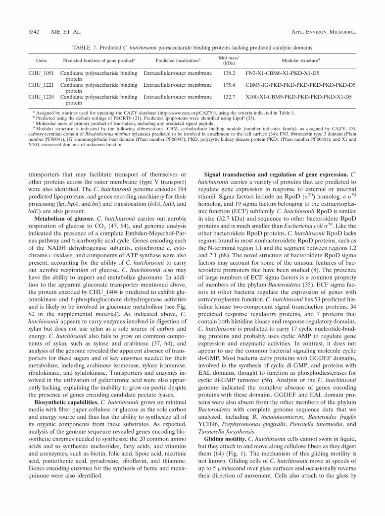
TABLE 7. Predicted C. hutchinsonii polysaccharide binding proteins lacking predicted catalytic domains
Gene Predicted function of gene producta Predicted localizationb Mol massc
(kDa) Modular structured
CHU_1051 Candidate polysaccharide bindingprotein
Extracellular/outer membrane 138.2 FN3-X1-CBM6-X1-PKD-X1-D5
CHU_1221 Candidate polysaccharide bindingprotein
Extracellular/outer membrane 175.4 CBM9-IG-PKD-PKD-PKD-PKD-PKD-PKD-D5
CHU_1238 Candidate polysaccharide bindingprotein
Extracellular/outer membrane 132.7 X100-X1-CBM9-PKD-PKD-PKD-PKD-X1-D5
a Assigned by routines used for updating the CAZY database (http://www.cazy.org/CAZY/), using the criteria indicated in Table 1.b Predicted using the default settings of PSORTb (21). Predicted lipoproteins were identified using LipoP (33).c Molecular mass of primary product of translation, including any predicted signal peptide.d Modular structure is indicated by the following abbreviations: CBM, carbohydrate binding module (number indicates family), as assigned by CAZY; D5,
carboxy-terminal domain of Rhodothermus marinus xylanases predicted to be involved in attachment to the cell surface (34); FN3, fibronectin type 3 domain (Pfamnumber PF00041); IG, immunoglobulin I-set domain (Pfam number PF00047); PKD, polycystic kidney disease protein PKD1 (Pfam number PF00801); and X1 andX100, conserved domains of unknown function.
3542 XIE ET AL. APPL. ENVIRON. MICROBIOL.

one pole and rotate in place at frequencies of about 2 revolu-tions per second. The ability to glide over surfaces may provideC. hutchinsonii cells with a selective advantage for utilization ofinsoluble cellulose. Gliding motility may allow cells to migratealong cellulose fibers in search of regions that are most ame-nable to enzymatic attack and to penetrate deep into the cel-lulose matrix as the substrate is digested. In some other bac-teria, flagella and type IV pili allow cells to move over surfaces(25, 41). Electron microscopic analyses have failed to identifythese organelles on cells of C. hutchinsonii, and analysis of thegenome also failed to identify genes encoding critical compo-
nents of flagella and type IV pili, suggesting that C. hutchin-sonii gliding motility relies on other machinery.
Gliding motility is characteristic of many members of thephylum Bacteroidetes (42). The mechanism of gliding motilityhas been studied extensively for one bacteroidete, Flavobacte-rium johnsoniae, which is distantly related to C. hutchinsonii(Fig. 2). Sixteen genes (gldA, gldB, gldD, gldF, gldG, gldH, gldI,gldJ, gldK, gldL, gldM, gldN, sprA, sprB, sprC, and sprD) in-volved in F. johnsoniae gliding motility have been identified (1,12, 13, 30–32, 45, 46). gldA, gldF, and gldG are thought toencode components of an ATP-binding cassette transporter
FIG. 4. Multiple sequence alignment of C-terminal domains in C. hutchinsonii proteins, found by BLASTP similarity searches using D5 ofRhodothermus marinus xylanase Xyn10A and R. marinus mannanase Man26A as queries. Black shading indicates amino acid identity, and grayshading indicates amino acid similarity. A threshold for shading was set at 60% of the consensus.
VOL. 73, 2007 C. HUTCHINSONII GENOME ANALYSIS 3543

that is required for gliding (1, 30). The functions of the othermotility proteins are less certain. In a current model of F.johnsoniae gliding, the Gld proteins comprise the gliding mo-tor, which propels cell surface adhesins comprised of Spr pro-teins. C. hutchinsonii has homologues of each of the gld and sprgenes, and these are likely to be involved in cell movement.Disruption of C. hutchinsonii gldG by homologous recombina-tion with a plasmid containing an internal fragment of the generesulted in a loss of motility, implicating C. hutchinsonii gldG ingliding (C. Guntur and M. J. McBride, unpublished data).
Gliding motility is not confined to the bacteroidetes, andrecent evidence indicates that there are multiple, geneticallyunrelated mechanisms for bacterial gliding (42, 43). Myxococ-cus xanthus has two gliding motility systems. The S motilitysystem relies on extension and retraction of type IV pili to pullcells, whereas the mechanism of the A motility system is not yetcertain (48, 65, 69, 73). Mycoplasma mobile appears to use athird gliding motility system composed of cytoskeletal proteinsand large cell surface proteins (49). Most F. johnsoniae and C.hutchinsonii gld genes have no obvious homologs in the ge-nomes of M. xanthus and M. mobile (12), suggesting that Cy-tophaga-Flavobacterium gliding motility is distinct from myxo-bacterial or mycoplasma gliding.
F. johnsoniae does not digest cellulose, but it does digestanother insoluble polysaccharide, chitin. Surprisingly, all mu-tations that eliminate gliding also eliminate the ability of F.johnsoniae to digest chitin (12, 13, 15, 45, 46). The connectionbetween motility and chitin digestion is not understood. Per-haps cells glide along the insoluble polymer until they reachregions amenable to digestion. Alternatively, the motility ma-chinery, which can move large particles over the cell surface(53), may be needed to move oligomers to sites where they canbe digested further or taken into the cell. The role, if any, ofmotility in C. hutchinsonii cellulose digestion remains to beexplored. The regular arrangement of gliding cells on cellulosefibers (Fig. 1) suggests that motility may position the cell-associated cellulolytic machinery so that digestion of the insol-uble polymer is optimized.
All motile bacteria that have been studied control theirmotility apparatus to move in a favorable direction. This typ-ically involves a complex signal transduction system, such asthe E. coli chemotaxis system, that senses the environment andaffects the functioning of the motility apparatus. C. hutchinso-nii presumably controls its motility machinery in response toexternal stimuli, and it has genes encoding proteins that aresimilar to E. coli CheY (CHU_2082), CheB (CHU_2078), andCheR (CHU_2079). These genes are clustered together on thegenome, supporting the idea that their products may functiontogether. C. hutchinsonii CheB is unusual in that it lacks theN-terminal response regulatory domain that is found in mostCheB proteins. Homologs of cheB and cheR are also found inother bacteroidetes that exhibit gliding motility, such as F.johnsoniae and Tenacibaculum sp. strain MED152, but are notfound in the nonmotile bacteroidetes B. thetaiotaomicronBPI5482, B. fragilis YCH46 or NCTC9343, P. gingivalis W83, P.intermedia 17, and T. forsythensis. C. hutchinsonii has anothergene, CHU_1237, that encodes a protein with similarity toboth CheB and CheR. This protein has a CheB-like domain atthe amino terminus followed by a CheR-like domain, a PASdomain, and a histidine kinase domain at the carboxy terminus.
The CheB-like domain lacks the response regulator domain thatis present in most CheB proteins. Genes encoding other expectedcomponents of a bacterial chemotaxis system, such as homologsof methyl-accepting chemotaxis proteins (MCPs) and of CheA,CheW, and CheZ, are apparently lacking. The absence of obviousMCPs is surprising since CheB (methyltransferase) and CheR(methylesterase) are expected to modify MCPs. The C. hutchin-sonii CheB and CheR homologs may modify novel chemotaxisproteins or may have roles unrelated to motility and chemotaxis.The region surrounding C. hutchinsonii cheB, cheR, and cheYcontains several genes whose products are likely to be involved insignal transduction. These include an apparent histidine kinasewith a PAS domain (CHU_2084), two proteins that each containhistidine kinase and response regulatory domains (CHU_2077and CHU_2081), and a large protein (CHU_2080) that containsa CHASE domain, a GAF domain, a histidine kinase domain,and a response regulatory domain. PAS, CHASE, and GAF do-mains are commonly found in sensory or signal transduction pro-teins (3, 66, 74) and could have roles in C. hutchinsonii tacticresponses.
Comparative analysis of bacteroidete genomes. The com-plete genome sequences of five members of the phylum Bacte-roidetes, namely, C. hutchinsonii, B. thetaiotaomicron BPI5482, B.fragilis, P. gingivalis W83, and P. intermedia 17, were analyzed. B.thetaiotaomicron, B. fragilis, P. gingivalis, and P. intermedia arenonmotile anaerobic members of the class Bacteroidetes, and eachtypically inhabits animal digestive tracts. C. hutchinsonii is anaerobic bacterium of the class Sphingobacteria that is commonlyfound in soil. Given these differences in phylogeny and lifestyle, itis not surprising that there are many features of the C. hutchin-sonii genome that set it apart from the others. Direct comparisonof the complete nucleotide sequences, using NUCmer and dot-plot (36) analyses, revealed no recognizable synteny between C.hutchinsonii and the other bacteroidetes. Five hundred fifty-fivecore bacteroidete genes (requiring bidirectional best hits as aminimum criterion) were shared between the five organismstested, i.e., C. hutchinsonii, B. thetaiotaomicron, P. gingivalis, P.intermedia, and T. forsynthensis. C. hutchinsonii, unlike the otherbacteria, appears to have a complete tricarboxylic acid cycle anda complete respiratory electron transport chain, as expected fromits demonstrated ability to carry out aerobic respiration. Otherdifferences are noted in Table S1 and Fig. S2 in the supplementalmaterial.
The complete genome sequence of C. hutchinsonii providesa window on the inner workings of this common cellulolytic soilbacterium. The mechanism of cellulose utilization employed bycells of C. hutchinsonii appears to differ from that employed bycellulosome-containing anaerobic clostridia (7, 9) or by aerobiccellulolytic bacteria, such as Thermobifida fusca and Sacchar-ophagus degradans, which employ soluble cellulases (67, 71).The apparent absence of cellobiohydrolases and type A CBMsalso sets C. hutchinsonii apart from most well-studied cellulo-lytic microorganisms. We hypothesize that C. hutchinsonii usescell surface endoglucanases to attack the insoluble cellulosefibers. Soluble oligomers may be transported actively acrossthe outer membrane with the assistance of SusC-like andSusD-like proteins, with further digestion occurring in theperiplasm and cytoplasm. An efficient surface motility systemallows the bacterium to attach to and migrate along its insol-uble substrate as it digests it. The genome sequence data and
3544 XIE ET AL. APPL. ENVIRON. MICROBIOL.

the availability of techniques to genetically manipulate C.hutchinsonii will allow experiments to probe the cellulolyticand motility systems of this common but poorly understoodbacterium.
ACKNOWLEDGMENTS
This work was supported by the U.S. Department of Energy undercontract no. W-7405-ENG-36 and by a UWM-RGI award to M.M.
We thank M. Burmeister and H. Owen for assistance with scanningelectron microscopy and E. Leadbetter for constructive comments onthe manuscript.
REFERENCES
1. Agarwal, S., D. W. Hunnicutt, and M. J. McBride. 1997. Cloning and char-acterization of the Flavobacterium johnsoniae (Cytophaga johnsonae) glidingmotility gene, gldA. Proc. Natl. Acad. Sci. USA 94:12139–12144.
2. Anderson, K. L., and A. A. Salyers. 1989. Biochemical evidence that starchbreakdown by Bacteroides thetaiotaomicron involves outer membrane starch-binding sites and periplasmic starch-degrading enzymes. J. Bacteriol. 171:3192–3198.
3. Aravind, L., and C. P. Ponting. 1997. The GAF domain: an evolutionary linkbetween diverse phototransducing proteins. Trends Biochem. Sci. 22:458–459.
4. Barr, B. K., Y. L. Hsieh, B. Ganem, and D. B. Wilson. 1996. Identification oftwo functionally different classes of exocellulases. Biochemistry 35:586–592.
5. Bayer, E., and R. Lamed. 1992. The cellulose paradox: pollutant par excel-lence and/or a reclaimable natural resource? Biodegradation 3:171–188.
6. Bayer, E. A., J. P. Belaich, Y. Shoham, and R. Lamed. 2004. The cellulo-somes: multienzyme machines for degradation of plant cell wall polysaccha-rides. Annu. Rev. Microbiol. 58:521–554.
7. Bayer, E. A., L. J. W. Shimon, Y. Shoham, and R. Lamed. 1998. Cellulo-somes—structure and ultrastructure. J. Struct. Biol. 124:221–234.
8. Bayley, D. P., E. R. Rocha, and C. J. Smith. 2000. Analysis of cepA and otherBacteroides fragilis genes reveals a unique promoter structure. FEMS Micro-biol. Lett. 193:149–154.
9. Beguin, P., and M. Lemaire. 1996. The cellulosome: an exocellular, multi-protein complex specialized in cellulose degradation. Crit. Rev. Biochem.Mol. Biol. 31:201–236.
10. Blackburn, N. T., and A. J. Clarke. 2001. Identification of four families ofpeptidoglycan lytic transglycosylases. J. Mol. Evol. 52:78–84.
11. Boraston, A. B., D. N. Bolam, H. J. Gilbert, and G. J. Davies. 2004. Carbo-hydrate-binding modules: fine-tuning polysaccharide recognition. Biochem.J. 382:769–781.
12. Braun, T. F., M. K. Khubbar, D. A. Saffarini, and M. J. McBride. 2005.Flavobacterium johnsoniae gliding motility genes identified by mariner mu-tagenesis. J. Bacteriol. 187:6943–6952.
13. Braun, T. F., and M. J. McBride. 2005. Flavobacterium johnsoniae GldJ is alipoprotein that is required for gliding motility. J. Bacteriol. 187:2628–2637.
14. Caspi, R., H. Foerster, C. A. Fulcher, R. Hopkinson, J. Ingraham, P. Kaipa,M. Krummenacker, S. Paley, J. Pick, S. Y. Rhee, C. Tissier, P. Zhang, andP. D. Karp. 2006. MetaCyc: a multiorganism database of metabolic pathwaysand enzymes. Nucleic Acids Res. 34:D511–D516.
15. Chang, L. Y. E., J. L. Pate, and R. J. Betzig. 1984. Isolation and character-ization of nonspreading mutants of the gliding bacterium Cytophaga johnso-nae. J. Bacteriol. 159:26–35.
16. Chang, W. T. H., and D. W. Thayer. 1977. The cellulase system of a Cyto-phaga species. Can. J. Microbiol. 23:1285–1292.
17. Cole, J. R., B. Chai, R. J. Farris, Q. Wang, S. A. Kulam, D. M. McGarrell,G. M. Garrity, and J. M. Tiedje. 2005. The Ribosomal Database Project(RDP-II): sequences and tools for high-throughput rRNA analysis. NucleicAcids Res. 33:D294–D296.
18. Coutinho, P. M., and B. Henrissat. 1999. Carbohydrate-active enzymes: anintegrated database approach, p. 3–12. In H. J. Gilbert, G. Davies, B. Hen-rissat, and B. Svensson (ed.), Recent advances in carbohydrate bioengineer-ing. The Royal Society of Chemistry, Cambridge, United Kingdom.
19. Doi, R. H., M. Goldstein, S. Hashida, J. S. Park, and M. Takagi. 1994. TheClostridium cellulovorans cellulosome. Crit. Rev. Microbiol. 20:87–93.
20. Felsenstein, J. 1997. An alternating least squares approach to inferringphylogenies from pairwise distances. Syst. Biol. 46:101–111.
21. Gardy, J. L., M. R. Laird, F. Chen, S. Rey, C. J. Walsh, M. Ester, and F. S. L.Brinkman. 2005. PSORTb v. 2.0: expanded prediction of bacterial proteinsubcellular localization and insights gained from comparative proteomeanalysis. Bioinformatics 21:617–623.
22. Gilad, R., L. Rabinovich, S. Yaron, E. A. Bayer, R. Lamed, H. J. Gilbert, andY. Shoham. 2003. CelI, a noncellulosomal family 9 enzyme from Clostridiumthermocellum, is a processive endoglucanase that degrades crystalline cellu-lose. J. Bacteriol. 185:391–398.
23. Gilkes, N. R., D. Kilburn, E. Kwan, S. Mansfield, J. Saddler, H. Shen, H.
Stalbrand, and A. Warren. 1999. Recent insights into the activities of cello-biohydrolases from Cellulomonas fimi and other cellulolytic microorganisms,p. 24–34. In K. Ohmiya, K. Hayashi, K. Sakka, Y. Kobayashi, S. Karita, andT. Kimura (ed.), Genetics, biochemistry and ecology of cellulose degrada-tion. Uni Publishers Co., Tokyo, Japan.
24. Hanley, S. A., J. Aduse-Opoku, and M. A. Curtis. 1999. A 55-kilodaltonimmunodominant antigen of Porphyromonas gingivalis W50 has arisen viahorizontal gene transfer. Infect. Immun. 67:1157–1171.
25. Harshey, R. M. 1994. Bees aren’t the only ones: swarming in gram-negativebacteria. Mol. Microbiol. 13:389–394.
26. Henikoff, S., and J. G. Henikoff. 1994. Protein family classification based onsearching a database of blocks. Genomics 19:97–107.
27. Henrissat, B. 1991. A classification of glycosyl hydrolases based on aminoacid sequence similarities. Biochem. J. 280:309–316.
28. Henrissat, B., and A. Bairoch. 1993. New families in the classification ofglycosyl hydrolases based on amino acid sequence similarities. Biochem. J.293:781–788.
29. Henrissat, B., and A. Bairoch. 1996. Updating the sequence-based classifi-cation of glycosyl hydrolases. Biochem. J. 316:695–696.
30. Hunnicutt, D. W., M. J. Kempf, and M. J. McBride. 2002. Mutations inFlavobacterium johnsoniae gldF and gldG disrupt gliding motility and inter-fere with membrane localization of GldA. J. Bacteriol. 184:2370–2378.
31. Hunnicutt, D. W., and M. J. McBride. 2001. Cloning and characterization ofthe Flavobacterium johnsoniae gliding motility genes gldD and gldE. J. Bac-teriol. 183:4167–4175.
32. Hunnicutt, D. W., and M. J. McBride. 2000. Cloning and characterization ofthe Flavobacterium johnsoniae gliding motility genes gldB and gldC. J. Bac-teriol. 182:911–918.
33. Juncker, A. S., H. Willenbrock, G. von Heijne, H. Nielsen, S. Brunak, and A.Krogh. 2003. Prediction of lipoprotein signal peptides in gram-negative bac-teria. Protein Sci. 12:1652–1662.
34. Karlsson, E. N., M. A. Hachem, S. Ramchuran, H. Costa, O. Holst, S. F.Svenningsen, and G. O. Hreggvidsson. 2004. The modular xylanase Xyn10Afrom Rhodothermus marinus is cell-attached, and its C-terminal domain hasseveral putative homologues among cell-attached proteins within the phylumBacteroidetes. FEMS Microbiol. Lett. 241:233–242.
35. Kill, K., T. T. Binnewies, T. Sicheritz-Ponten, H. Willenbrock, P. F. Hallin,T. M. Wassenaar, and D. W. Ussery. 2005. Genome update: sigma factors in240 bacterial genomes. Microbiology 151:3147–3150.
36. Kurtz, S., A. Phillippy, A. L. Delcher, M. Smoot, M. Shumway, C. Antonescu,and S. L. Salzberg. 2004. Versatile and open software for comparing largegenomes. Genome Biol. 5:R12.
37. Larkin, J. M. 1989. Nonphotosynthetic, nonfruiting gliding bacteria, p. 2010–2138. In J. T. Staley, M. P. Bryant, N. Pfennig, and J. G. Holt (ed.), Bergey’smanual of systematic bacteriology, vol. 3. Williams and Wilkins, Baltimore,MD.
38. Leschine, S. B. 1995. Cellulose degradation in anaerobic environments.Annu. Rev. Microbiol. 49:399–426.
39. Lynd, L. R., P. J. Weimer, W. H. van Zyl, and I. S. Pretorius. 2002. Microbialcellulose utilization: fundamentals and biotechnology. Microbiol. Mol. Biol.Rev. 66:506–577.
40. Markowitz, V. M., F. Korzeniewski, K. Palaniappan, E. Szeto, G. Werner, A.Padki, X. Zhao, I. Dubchak, P. Hugenholtz, I. Anderson, A. Lykidis, K.Mavromatis, N. Ivanova, and N. C. Kyrpides. 2006. The integrated microbialgenomes (IMG) system. Nucleic Acids Res. 34:D344–D348.
41. Mattick, J. S. 2002. Type IV pili and twitching motility. Annu. Rev. Micro-biol. 56:289–314.
42. McBride, M. J. 2001. Bacterial gliding motility: multiple mechanisms for cellmovement over surfaces. Annu. Rev. Microbiol. 55:49–75.
43. McBride, M. J. 2004. Cytophaga-Flavobacterium gliding motility. J. Mol.Microbiol. Biotechnol. 7:63–71.
44. McBride, M. J., and S. A. Baker. 1996. Development of techniques togenetically manipulate members of the genera Cytophaga, Flavobacterium,Flexibacter, and Sporocytophaga. Appl. Environ. Microbiol. 62:3017–3022.
45. McBride, M. J., and T. F. Braun. 2004. GldI is a lipoprotein that is requiredfor Flavobacterium johnsoniae gliding motility and chitin utilization. J. Bac-teriol. 186:2295–2302.
46. McBride, M. J., T. F. Braun, and J. L. Brust. 2003. Flavobacteriumjohnsoniae GldH is a lipoprotein that is required for gliding motility andchitin utilization. J. Bacteriol. 185:6648–6657.
47. Middleton, K. S. 1963. Studies on the cellulose-decomposing soil cytophagas.M.A. thesis. University of California-Berkeley, Berkeley.
48. Mignot, T., J. W. Shaevitz, P. L. Hartzell, and D. R. Zusman. 2007. Evidencethat focal adhesion complexes power bacterial gliding motility. Science 315:853–856.
49. Miyata, M. 2005. Gliding motility of mycoplasmas: the mechanism cannot beexplained by current biology, p. 137–163. In A. Blanchard and G. Browning(ed.), Mycoplasmas: pathogenesis, molecular biology and emerging strate-gies for control. Horizon Scientific Press, Norwich, United Kingdom.
50. Morrison, M., and J. Miron. 2000. Adhesion to cellulose by Ruminococcusalbus: a combination of cellulosomes and Pil-proteins? FEMS Microbiol.Lett. 185:109–115.
VOL. 73, 2007 C. HUTCHINSONII GENOME ANALYSIS 3545

51. Navidi, W. C., G. A. Churchill, and A. von Haeseler. 1991. Methods forinferring phylogenies from nucleic acid sequence data by using maximumlikelihood and linear invariants. Mol. Biol. Evol. 8:128–143.
52. Olsen, G. J., H. Matsuda, R. Hagstrom, and R. Overbeek. 1994. fastDNAmL: atool for construction of phylogenetic trees of DNA sequences using maxi-mum likelihood. Comput. Appl. Biosci. 10:41–48.
53. Pate, J. L., and L.-Y. E. Chang. 1979. Evidence that gliding motility inprokaryotic cells is driven by rotary assemblies in the cell envelopes. Curr.Microbiol. 2:59–64.
54. Reeves, A. R., J. N. D’Elia, J. Frias, and A. A. Salyers. 1996. A Bacteroidesthetaiotaomicron outer membrane protein that is essential for utilization ofmaltooligosaccharides and starch. J. Bacteriol. 178:823–830.
55. Reichenbach, H. 1992. The order Cytophagales, p. 3631–3675. In A. Balows,H. G. Truper, M. Dworkin, W. Harder, and K. M. Schleifer (ed.), Theprokaryotes. Springer-Verlag, New York, NY.
56. Romling, U., M. Gomelsky, and M. Y. Galperin. 2005. C-di-GMP: the dawn-ing of a novel bacterial signaling system. Mol. Microbiol. 57:629–639.
57. Salyers, A. A., A. Reeves, and J. D’Elia. 1996. Solving the problem of how toeat something as big as yourself: diverse bacterial strategies for degradingpolysaccharides. J. Ind. Microbiol. 17:470–476.
58. Schultz, J., F. Milpetz, P. Bork, and C. P. Ponting. 1998. SMART, a simplemodular architecture research tool: identification of signaling domains. Proc.Natl. Acad. Sci. USA 95:5857–5864.
59. Servant, F., C. Bru, S. Carrere, E. Courcelle, J. Gouzy, D. Peyruc, and D.Kahn. 2002. ProDom: automated clustering of homologous domains. Brief.Bioinform. 3:246–251.
60. Shipman, J. A., J. E. Berleman, and A. A. Salyers. 2000. Characterization offour outer membrane proteins involved in binding starch to the cell surfaceof Bacteroides thetaiotaomicron. J. Bacteriol. 182:5365–5372.
61. Shipman, J. A., K. H. Cho, H. A. Siegel, and A. A. Salyers. 1999. Physiolog-ical characterization of susG, an outer membrane protein essential for starchutilization by Bacteroides thetaiotaomicron. J. Bacteriol. 181:7206–7211.
62. Singh, A., and K. Hayashi. 1995. Microbial cellulases: protein architecture,molecular properties, and biosynthesis. Adv. Appl. Microbiol. 40:1–44.
63. Sonnhammer, E. L., S. R. Eddy, and R. Durbin. 1997. Pfam: a comprehen-sive database of protein domain families based on seed alignments. Proteins28:405–420.
64. Stanier, R. Y. 1942. The cytophaga group: a contribution to the biology ofmyxobacteria. Bacteriol. Rev. 6:143–196.
65. Sun, H., D. R. Zusman, and W. Shi. 2000. Type IV pilus of Myxococcusxanthus is a motility apparatus controlled by the frz chemosensory system.Curr. Biol. 10:1143–1146.
66. Taylor, B. L., and I. B. Zhulin. 1999. PAS domains: internal sensors ofoxygen, redox potential, and light. Microbiol. Mol. Biol. Rev. 63:479–506.
67. Taylor, L. E. N., B. Henrissat, P. M. Coutinho, N. A. Ekborg, S. W.Hutcheson, and R. M. Weiner. 2006. Complete cellulase system in the ma-rine bacterium Saccharophagus degradans strain 2-40T. J. Bacteriol. 188:3849–3861.
68. Vingadassalom, D., A. Kolb, C. Mayer, T. Rybkine, E. Collatz, and I.Podglajen. 2005. An unusual primary sigma factor in the Bacteroidetesphylum. Mol. Microbiol. 56:888–902.
69. Wall, D., and D. Kaiser. 1999. Type IV pili and cell motility. Mol. Microbiol.32:1–10.
70. Warren, R. A. 1996. Microbial hydrolysis of polysaccharides. Annu. Rev.Microbiol. 50:183–212.
71. Wilson, D. B. 1992. Biochemistry and genetics of actinomycete cellulases.Crit. Rev. Biotechnol. 12:45–63.
72. Winogradsky, S. 1929. E’tudes sur la microbiologie du sol. Ann. Inst. Pasteur43:549–633.
73. Wolgemuth, C., E. Hoiczyk, D. Kaiser, and G. Oster. 2002. How myxobac-teria glide. Curr. Biol. 12:369–377.
74. Zhulin, I. B., A. N. Nikolskaya, and M. Y. Galperin. 2003. Common extra-cellular sensory domains in transmembrane receptors for diverse signaltransduction pathways in bacteria and archaea. J. Bacteriol. 185:285–294.
3546 XIE ET AL. APPL. ENVIRON. MICROBIOL.
Copyright © 2022 FDOKUMEN