Application of a suite of 16S rRNA-specific oligonucleotide probes designed to investigate bacteria...
10
Microbiology (1 996), 142, 1097-1 106 Printed in Great Britain Application of a suite of 16s rRNA-specific oligonucleotide probes designed to investigate bacteria of the phylum cytophaga-f lavobacter- bacteroides in the natural environment Werner Manz,’t Rudolf Amann,’ Wolfgang Ludwig,’ Marc Vancanneyt’ and Karl-Heinz Schleiferl Author for correspondence: Werner Manz. Tel: +49 30 314 25831. Fax: +49 30 314 73461. e-mail : [email protected] Lehrstuhl fur Mikrobiologief Technische Universitat Munchen, D- 80290 Munchen, Germany Laboratorium voor Microbiologie, universiteit Gent, Belgium We designed a panel of four 16s rRNA-targeted oligonucleotide probes specific for bacteria of the phylum cytophaga-flavobacter-bacteroides (CFB). Probes CF319a and CF319b are targeted to members Of the flavobacteria-cytophaga group and the genus Porphyromonas, whereas probe BAC303 has a target region characteristic for the genera Prevote//a and Bacteroides within the bacteroides group. The probe FFE8b was developed for species-specif ic hybridizations with Flavobacterium ferrugineum. All probes were designed by computer-assisted sequence analysis and compared to all currently accessible 16s and 235 rRNA sequences. The oligonucleotides were further evaluated by whole-cell and non-radioactive dot-blot hybridization against reference strains of the CFB phylum and other major lineages of Bacteria. The newly developed probes were used together with other higher-order probes t o analyse the structure and community composition in complex environments. In activated sludge samples, members of the f lavobacteria-cytophaga group were revealed by in situ hybridization as important constituents of sludge flocs and characteristic colonizers of filamentous bacteria. By application of fluorescent probe BAC303, members of the genera Bacteroides and Prewotella could be visualized without prior cultivation as an important part of the human faecal microf lora. Keywords : 16s rRNA-targeted oligonucleotide probes, in sitzt hybridization, phylum cytophaga-flavobacter-bacteroides, flavobacteria-cytophaga group, bacteroides group INTRODUCTION The phylum cytophaga-flavobacter-bacteroides (CFB) is one of the major lineages (phyla) of Bacteria (Woese, 1987). Members of the flavobacteria-cytophaga group within the CFB phylum are frequently isolated from many Bacteroides species that form the second major group within the CFB phylum account for approximately 30 % of all faecal isolates from the normal human colon microflora (Moore & Holdeman, 1974; Holdeman et al., 1976). They are also the most important anaerobic bacteria associated with human infections (Salyers, 1984). natural and man-made ecosystems (soil, fresh and marine waters, clinical specimens, air-conditioning, sewage-treat- ment plants) and exhibit a broad range of phenotypic diversity. By their ability to degrade macromolecules such as cellulose, agar and chitin they are of considerable practical importance. The Gram-negative, anaerobic Despite the known importance of the CFB phylum, the lack of characteristic bacterial morphologies and physio- logies has hindered the establishment of a valid system of classification for this phenotypically complex taxon. Since the first description of the genus Cytophaga (Winogradsky, 1929) and the ‘ colour genus ’, Flavobacterium (Bergey et al., ............................................................................................................................................................ *Present address: Fachgebiet Okologie der Mikroorganisrnen, Tech- nische Universitat Berlin, D-10587 Berlin, Germany. Abbreviation : CFB, cytophaga-f lavobacter-bacteroides. 1923) it has proven difficult to define valid criterTa for the phenotypic differentiation of the genera Flavobacterium and Cytophaga. Several chemotaxonomic approaches based on pigmentation (Reichenbach et al., 19Sl), respiratory 0002-0270 0 1996 SGM 1097
-
Upload
mpi-bremen -
Category
Documents
-
view
0 -
download
0
Transcript of Application of a suite of 16S rRNA-specific oligonucleotide probes designed to investigate bacteria...

Downloaded from www.microbiologyresearch.org by
IP: 54.237.57.119
On: Fri, 20 May 2016 10:29:03
Microbiology (1 996), 142, 1097-1 106 Printed in Great Britain
Application of a suite of 16s rRNA-specific oligonucleotide probes designed to investigate bacteria of the phylum cytophaga-f lavobacter- bacteroides in the natural environment
Werner Manz,’t Rudolf Amann,’ Wolfgang Ludwig,’ Marc Vancanneyt’ and Karl-Heinz Schleiferl
Author for correspondence: Werner Manz. Tel: +49 30 314 25831. Fax: +49 30 314 73461. e-mail : [email protected]
Lehrstuhl fur Mikrobiologief Technische Universitat Munchen, D- 80290 Munchen, Germany
Laboratorium voor Microbiologie, universiteit Gent, Belgium
We designed a panel of four 16s rRNA-targeted oligonucleotide probes specific for bacteria of the phylum cytophaga-flavobacter-bacteroides (CFB). Probes CF319a and CF319b are targeted to members O f the flavobacteria-cytophaga group and the genus Porphyromonas, whereas probe BAC303 has a target region characteristic for the genera Prevote//a and Bacteroides within the bacteroides group. The probe FFE8b was developed for species-specif ic hybridizations with Flavobacterium ferrugineum. All probes were designed by computer-assisted sequence analysis and compared to all currently accessible 16s and 235 rRNA sequences. The oligonucleotides were further evaluated by whole-cell and non-radioactive dot-blot hybridization against reference strains of the CFB phylum and other major lineages of Bacteria. The newly developed probes were used together with other higher-order probes t o analyse the structure and community composition in complex environments. In activated sludge samples, members of the f lavobacteria-cytophaga group were revealed by in situ hybridization as important constituents of sludge flocs and characteristic colonizers of filamentous bacteria. By application of fluorescent probe BAC303, members of the genera Bacteroides and Prewotella could be visualized without prior cultivation as an important part of the human faecal microf lora.
Keywords : 16s rRNA-targeted oligonucleotide probes, in sitzt hybridization, phylum cytophaga-flavobacter-bacteroides, flavobacteria-cytophaga group, bacteroides group
INTRODUCTION
The phylum cytophaga-flavobacter-bacteroides (CFB) is one of the major lineages (phyla) of Bacteria (Woese, 1987). Members of the flavobacteria-cytophaga group within the CFB phylum are frequently isolated from many
Bacteroides species that form the second major group within the CFB phylum account for approximately 30 % of all faecal isolates from the normal human colon microflora (Moore & Holdeman, 1974; Holdeman e t al., 1976). They are also the most important anaerobic bacteria associated with human infections (Salyers, 1984).
natural and man-made ecosystems (soil, fresh and marine waters, clinical specimens, air-conditioning, sewage-treat- ment plants) and exhibit a broad range of phenotypic diversity. By their ability to degrade macromolecules such as cellulose, agar and chitin they are of considerable practical importance. The Gram-negative, anaerobic
Despite the known importance of the CFB phylum, the lack of characteristic bacterial morphologies and physio- logies has hindered the establishment of a valid system of classification for this phenotypically complex taxon. Since the first description of the genus Cytophaga (Winogradsky, 1929) and the ‘ colour genus ’, Flavobacterium (Bergey e t al.,
............................................................................................................................................................
*Present address: Fachgebiet Okologie der Mikroorganisrnen, Tech- nische Universitat Berlin, D-10587 Berlin, Germany.
Abbreviation : CFB, cytophaga-f lavobacter-bacteroides.
1923) it has proven difficult to define valid criterTa for the phenotypic differentiation of the genera Flavobacterium and Cytophaga. Several chemotaxonomic approaches based on pigmentation (Reichenbach e t al., 19Sl), respiratory
0002-0270 0 1996 SGM 1097

Downloaded from www.microbiologyresearch.org by
IP: 54.237.57.119
On: Fri, 20 May 2016 10:29:03
W. M A N Z a n d OTHERS
quinone systems (Mannheim, 1981 ; Reichenbach, 1989) and cellular fatty acid composition (Oyaizu & Komagata, 1981) showed many kinds of relationship between the genera Flavobacterizlm and Cytophaga, but could not provide a reliable basis for classification of this taxon.
Nowadays the comparative analysis of phylogenetic marker molecules which provide a large set of definable criteria at the molecular and genotypic level (Ludwig & Schleifer, 1994) allows the valid phylogenetic affiliation of prokaryotes. Molecular approaches using DNA-DNA (Callies & Mannheim, 1980) and DNA-rRNA hybridi- zations (Bauwens & de Ley, 1981), studies on the basis of oligonucleotide cataloguing (Fox etal., 1977) and compari- sons of 16s rRNA sequences have revealed the natural relationships within the CFB phylum (Gherna & Woese, 1992) and between these organisms and bacteria from other taxa (Woese e t al., 1990a, b).
Analysis of spzcific signature regions within the 16s rRNA has provided more detailed insights into the relationships between members of the CFB phylum. The two primary groups, bacteroides and flavobacteria- cytophaga, have been subdivided by Gherna & Woese (1992) into five major phylogenetic subgroups : bacteroides, cytophaga, flavobacter, sphingobacter and saprospira; the latter four subgroups constitute the flavobacteria-cytophaga group. Paster e t al. (1 994) divided the bacteroides (sub)group into three clusters, named by the genera Prevotella, Bacteroides and Porpbyromonas. The species Flavobacterizlm ferrtdgineztm has been removed from the genus Flavobacteritdm (Holmes e t al., 1984) and an outlying taxonomic status has been accorded to this organism (Gherna & Woese, 1992).
Signature regions of the 16s rRNA also provide potential target sites for rRNA-targeted oligonucleotide hybrid- ization probes (Giovannoni e t al., 1988; Schleifer e t al., 1993). Such probes can be designed on all taxonomic levels ranging from domains (Amann e t al., 1990a; Stahl & Amann, 1991) and other higher taxa (Manz etal., 1992; Burggraf e t al. , 1994 ; Roller e t al. , 1994) to the species and subspecies level (Amann e t al., 1990b; Ehrmann e t al., 1992). Fluorescently labelled probes have been success- fully applied for identification of hitherto uncultured bacteria (Amann e t al., 1991 ; Spring e t al., 1992) and for elucidating the community structure of diverse microbial consortia (Amann e t al., 1992; Wagner e t al., 1993; Manz etal. , 1993, 1994).
The aim of this work was the development of higher- order probes for the specific characterization of bacteria belonging to the CFB phylum and, in view of its outlying taxonomic status, for Flavobacteritdm ferrtdginezlm. These probes are important parts of a comprehensive panel of rRNA-targeted oligonucleotide probes intended to fa- cilitate the rapid analysis of microbial community struc- tures by in sitn hybridizations. Fluorescent derivatives of the newly developed probes specific for the flavobacteria- cytophaga group were successfully used in in sitzt hybrid- ization experiments to examine the occurrence and spatial arrangement of these organisms within activated sludge flocs. One of the probes was used for identification and
characterization of members of the genera Bacteroides and Prevotella in the human faecal flora without prior anaerobic cultivation.
METHODS
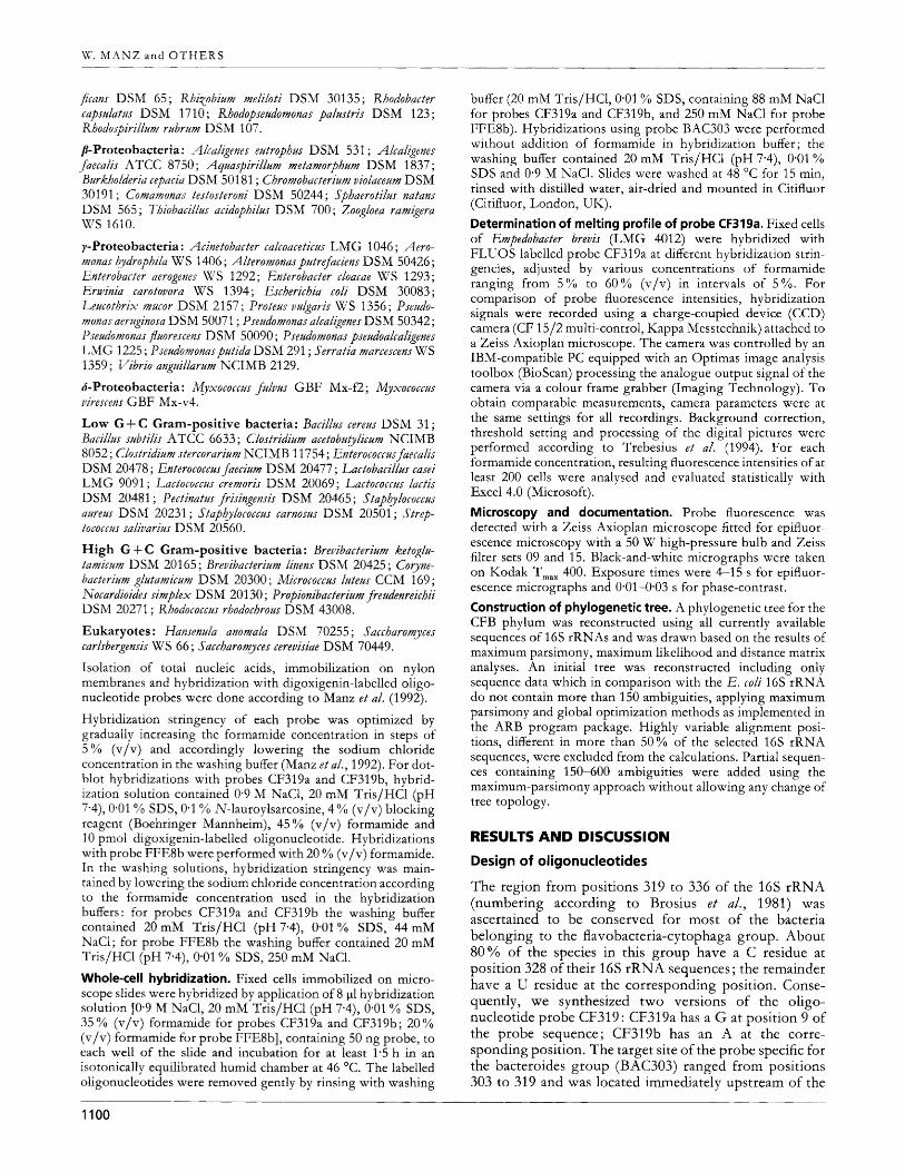
Organisms and culture conditions. Strain numbers and sources of bacteria belonging to the CFB phylum investigated in this study are listed in Table 1. Strains were cultured in media and under culture conditions as recommended in the corresponding catalogues of the DSM (Deutsche Sammlung von Mikro- organismen und Zellkulturen, Braunschweig, FRG), the GBF (Gesellschaft fur Biotechnologische Forschung, Braunschweig, FRG) and the LMG (Laboratorium voor Microbiologie, Universiteit Gent, Belgium).
Cell fixation. Cells in the exponential phase were harvested by centrifugation (2 min, 5000g) and carefully suspended in 1 ml sterile phosphate-buffered saline at pH 7.4 (PBS : 130 mM NaC1, 10 mM Na,HPO,/NaH,PO,). Pure cultures and activated sludge samples were fixed for at least 3 h by addition of 3 vols 4% (w/v) paraformaldehyde at 4 "C. After washing once with PBS, cells were stored in a 1 : 1 mixture of PBS and 96 % (v/v) ethanol at -20 "C. Fixed cells were spotted on precleaned, gelatin-coated [0*01 % KCr(SO,),, 0.1 % gelatin] microscope slides (Paul Marienfeld, Bad Mergentheim, FRG), air-dried and dehydrated in 50, 80, and 96 % (v/v) ethanol (3 min each).
Faecal specimens. Stool samples were obtained from two healthy adults. To obtain the bacterial fraction approximately 0-5 g faeces was suspended evenly in 1 ml sterile PBS at pH 7.4, harvested by centrifugation (2 min, 5000 g), carefully resus- pended and washed twice in PBS. The cell suspension was fixed for at least 3 h by addition of 3 vols 4 % (w/v) paraformaldehyde at 4 "C. After washing once with PBS, cells were stored in a 1 : 1 mixture of PBS and 96 % (v/v) ethanol at - 20 OC. Preparation of cell smears was done as described above.
Oligonucleotide probes. An alignment of 16s rRNA sequences from 89 members of the CFB phylum and representatives of other major lineages of the Bacteria obtained from the Technical University Munich data collection (Dr W. Ludwig) was screened for signatures characteristic for the flavobacteria- cytophaga and the bacteroides group within the CFB phylum, as well as Flavobacterizlm ferrzlgineum. Currently available data sets of about 3000 complete or almost complete 16s and 23s rRNA sequences were checked by the newly developed ARB software package (Technical University Munich, Munich, FRG).
Two oligonucleotides specific for the flavobacteria-cytophaga group (probes CF319a and CF319b), one complementary to a characteristic region of the bacteroides group (probe BAC303) and one specific to 16s rRNA of Flavobacterinm ferrzlgineum (probe FFE8b) were designed. Probe sequences and target positions corresponding to positions in the Escberichia coli 16s rRNA (Brosius e t al., 1981) are listed in Table 2. An alignment of probes CF319a, CF319b and BAC303 with the complemen- tary regions of the 16s rRNAs from representative organisms of the five main subgroups of the CFB phylum and representative non-target organisms characteristic of other phyla is given in Fig. 1. Probe EUB338, complementary to a region of the 16s rRNA conserved in the domain Bacteria (Amann e t al., 1990a) was used as a positive control. Oligonucleotides were syn- thesized with a C6-TFA aminolinker [6-(trifluoroacety1amino)- hexyl-(2-cyanoethyl)-(N,N-diisopropyl)-phosphoramidite] at the 5'-end (MW'G Biotech). Labelling with tetramethyl- rhodamine-5-isothiocyanate (TRITC ; Molecular Probes) or 5(6)-carboxyfluorescein-N-hydroxysuccinimide ester (FLUOS ; Boehringer Mannheim) was performed as described pre-
1098

Downloaded from www.microbiologyresearch.org by
IP: 54.237.57.119
On: Fri, 20 May 2016 10:29:03
r R N A probes for cytophaga-flavobacter-bacteroides
Table 1. Target organisms, sources, phylogeny and results of whole-cell and dot-blot hybridizations
~~
Organism Source* Phylogenyt Hybridization signal with probe3
BAC303 CF319a CF319b FFE8b
Bacteroides vulgatzts Bacteroides distasonis Bacteroides eggerthii Bacteroides fragilis Bacteroides ovatus Bacteroides thetaiotaomicron Prevoteila loescheii Bergtyella xoohelcum Bergtyella xooheicum Ch yseobacterium gleum Chyseobacterium indologenes Chyseobacterium indologenes Cytophaga htltcbinsonii Cytophaga johnsonae Cy t ophaga yohnsonae Cytophaga ulkinosa Empedobacter brevis Empedobacter brevis Flavobacterium aqtlatile Flavobacterium ferrugineum Flavobacteriztm odoratum Flavobacterium odoratum Flexibacter columnaris Flexitbrix dorotbeae Ha liscomenobacter (ydrossis R iemerella anatipestfer Riemerella anatipestfer Yaprospira grandis Sphingobacterium heparinum Sphingobacteriztm mixntae Yphingobacterium spiritivorum Sphingobacterium spiritivorum Yporocytophaga myxococcoides Taxeobacter ocellatus Weekseila virosa K’eeksella virosa
ATCC 8482T ATCC 8503T ATCC 27754T ATCC 25285T ATCC 8483T ATCC 2914gT ATCC 15930T LMG 8351T LMG 8352 LMG 8334 LMG 8336 LMG 8337T LMG 10844T LMG 1341T LMG 1342 LMG 3809T LMG 401 1 LMG 4012 LMG 400gT LMG 4021T LMG 1233T LMG 4028 LMG 13035 DSM 6795T DSM l l O O T LMG 1 1054T LMG 11602 DSM 2844 LMG 4024T LMG 8340T LMG 8347T LMG 8348 LMG 8393T GBF Txol LMG 8349 LMG 8350
B + B + B + B + B + B + B
CF CF - CF -
CF CF -
CF CF -
CF CF CF -
CF CF CF -
CF - CF -
CF CF CF -
CF CF CF -
CF CF CF CF CF CF - CF -
CF
-
-
-
-
-
-
-
-
-
-
-
-
-
-
-
-
-
-
* A superscript T denotes a type strain. Culture collections are given in full in Methods.
t B, bacteroides group ; CF, flavobacteria-cytophaga group.
$ +, Strong hybridization signal; -, no hybridization signal.
viously (Amann e t al., 1990b). For dot-blot hybridizations, probes were labelled with digoxigenin following the protocols of Zarda e t al. (1991). Nucleic acid extraction and dot-blot hybridization. For further evaluation of probe specificities whole-cell hybridizations and dot-blot analyses were performed using cells and nucleic acids of 36 different strains of organisms representing major sub- groups within the CFB phylum (Table 1). The strains listed below were used as negative controls for whole-cell and dot- blot hybridizations (culture collections : ATCC, American Type Culture Collection, Rockville, MD, USA ; CCM, Czechoslovak Collection of Microorganisms, Brno, CSFR ; DSM, Deutsche Sammlung von Mikroorganismen und Zellkulturen,
~ ~~~~
Braunschweig, FRG ; GBF, Gesellschaft fur Biotechnologische Forschung, Braunschweig, FRG ; LMG, Laboratorium voor Microbiologie, Universiteit Gent, Belgium ; NCIMB, The National Collections of Industrial and Marine Bacteria, Torry Research Station, Aberdeen, Scotland, UK ; WS, Institut fur Mikrobiologie, Forschungszentrum fur Milch und Lebensmittel, Technical University Munich, Freising Weihen- stephan, FRG). a-Proteobacteria: Agrobacterium tumefdciens ATCC 23308; Apospirillnm amaponense DSM 2787 ; ATospirillum brasilense DSM 1690 ; Axospirillum halopraefeerens DSM 3675 ; BradyrhiTobitim
japonicum DSM 30131 ; Brevzndimonas diminuta DSM 1635; Magnetospirillnm gryphiswaldense D S M 636 1 ; Paracocczls denitri-
1099
Downloaded from www.microbiologyresearch.org by
IP: 54.237.57.119
On: Fri, 20 May 2016 10:29:03
W. M A N 2 a n d OTHERS ~~
ficans DSM 65; RhiTobium meliloti DSM 30135; Rhodobacter capsulatus DSM 1710; Rbodopseztdomonas palustris DSM 123; Rhodospirilhm rubrum DSM 107.
p-Proteobacteria: Ahahgenes eutrophus DSM 531 ; Alcaligenes faecalis ATCC 8750 ; Aguaspirillztm metamorphum DSM 1837 ; Burkholderia cepacia DSM 501 81 ; Cbromobacterium violaceum DSM 301 91 ; Comamonas testosteroni DSM 50244; Sphaerotilus natans DSM 565; Thobacillus acidopbilus DSM 700; Zoogloea ramigera WS 1610.
y-Proteobacteria : Acinetobacter calcoaceticus LMG 1046 ; Aero- monas h_ydrophila W S 1406 ; Alteromonas putrefaciens DSM 50426 ; Enterobacter aerogenes KJS 1292 ; Enterobacter cloacae WS 1293 ; Erwinia carotovora WS 1394; Escbericbia coli DSM 30083 ; Leucothrix mucor DSM 2157; Proteus vulgaris WS 1356; Pseudo- monas aeruginosa DSM 50071 ; Pseudomonas alcaligenes DSM 50342 ; Pseudomonas j'uorescens DSM 50090 ; Psezddomonas pseudoalcaligenes LMG 1225 ; Pseudomonasputida DSM 291 ; Serratia marcescens WS 1359; Vibrio anguillarum NCIMB 2129.
S-Proteobacteria : Myxococcus fulvus GBF Mx-f2 ; Myxococcus virescens GBF Mx-v4.
Low G + C Gram-positive bacteria: Bacillus cereus DSM 31 ; Bacillus subtilis ATCC 6633 ; Clostridium acetobut_ylicum NCIMB 8052 ; Clostridium stercorarium NCIMB 1 1754 ; Enterococcus faecalis DSM 20478 ; Enterococczts faeciztm DSM 20477 ; Lactobacillzts casei LMG 9091 ; Lactococcus cremoris DSM 20069 ; Lactococcus lactis DSM 20481 ; Pectinatus frisingensis DSM 20465 ; Stapbylococcus aureus DSM 20231 ; Stapbylococcus carnosus DSM 20501 ; Strep- tococcus salivarius DSM 20560.
High G + C Gram-positive bacteria : Brevibacterium ketoglu- tamicum DSM 20165 ; Brevibacterium linens DSM 20425 ; Coyne- bacterium glutamicum DSM 20300 ; Micrococcus luteus CCM 169 ; Nocardioides simplex D SM 201 30 ; Propionibacterium freudenreicbii DSM 20271 ; Rhodococcus rhodocbrous DSM 43008.
Eukaryotes : Hamenula anomala DSM 70255 ; Saccbaromyces carlsbergensis W S 66 ; Saccbaromyces cerevisiae DSM 70449.
Isolation of total nucleic acids, immobilization on nylon membranes and hybridization with digoxigenin-labelled oligo- nucleotide probes were done according to Manz e t al. (1992).
Hybridization stringency of each probe was optimized by gradually increasing the formamide concentration in steps of 5 % (v/v) and accordingly lowering the sodium chloride concentration in the washing buffer (Manz etal., 1992). For dot- blot hybridizations with probes CF319a and CF319b, hybrid- ization solution contained 0.9 M NaC1, 20 mM Tris/HCl (pH 7*4), 0.01 % SDS, 0.1 YO N-lauroylsarcosine, 4 YO (v/v) blocking reagent (Boehringer Mannheim), 45 % (v/v) formamide and 10 pmol digoxigenin-labelled oligonucleotide. Hybridizations with probe FFE8b were performed with 20 YO (v/v) formamide. In the washing solutions, hybridization stringency was main- tained by lowering the sodium chloride concentration according to the formamide concentration used in the hybridization buffers: for probes CF319a and CF319b the washing buffer contained 20 mM Tris/HCl (pH 7-4), 0.01 % SDS, 44 mM NaCl; for probe FFE8b the washing buffer contained 20 mM Tris/HCl (pH 7.4), 0.01 YO SDS, 250 mM NaC1.
Whole-cell hybridization. Fixed cells immobilized on micro- scope slides were hybridized by application of 8 p1 hybridization solution [On9 M NaC1, 20 mM Tris/HCl (pH 7.4), 0.01 % SDS, 35 % (v/v) formamide for probes CF319a and CF319b; 20 % (v/v) formamide for probe FFE8b1, containing 50 ng probe, to each well of the slide and incubation for at least 1.5 h in an isotonically equilibrated humid chamber at 46 "C. The labelled oligonucleotides were removed gently by rinsing with washing
buffer (20 mM Tris/HCl, 0.01 % SDS, containing 88 mM NaCl for probes CF319a and CF319b, and 250 mM NaCl for probe FFE8b). Hybridizations using probe BAC303 were performed without addition of formamide in hybridization buffer ; the washing buffer contained 20 mM Tris/HCl (pH 7-4), 0.01 % SDS and 0.9 M NaC1. Slides were washed at 48 *C for 15 min, rinsed with distilled water, air-dried and mounted in Citifluor (Citifluor, London, UIC).
Determination of melting profile of probe CF319a. Fixed cells of Empedobacter brevis (LMG 4012) were hybridized with FLUOS-labelled probe CF319a at different hybridization strin- gencies, adjusted by various concentrations of formamide ranging from 5 YO to 60 '3'0 (v/v) in intervals of 5 YO. For comparison of probe fluorescence intensities, hybridization signals were recorded using a charge-coupled device (CCD) camera (CF 15/2 multi-control, Kappa Messtechnik) attached to a Zeiss Axioplan microscope. The camera was controlled by an IBM-compatible PC equipped with an Optimas image analysis toolbox (BioScan) processing the analogue output signal of the camera via a colour frame grabber (Imaging Technology). To obtain comparable measurements, camera parameters were at the same settings for all recordings. Background correction, threshold setting and processing of the digital pictures were performed according to Trebesius e t al. (1994). For each formamide concentration, resulting fluorescence intensities of at least 200 cells were analysed and evaluated statistically with Excel 4.0 (Microsoft).
Microscopy and documentation. Probe fluorescence was detected with a Zeiss Axioplan microscope fitted for epifluor- escence microscopy with a 50 W high-pressure bulb and Zeiss filter sets 09 and 15. Black-and-white micrographs were taken on Kodak T,,, 400. Exposure times were 4-15 s for epifluor- escence micrographs and 0*01-0*03 s for phase-contrast.
Construction of phylogenetic tree. A phylogenetic tree for the CFB phylum was reconstructed using all currently available sequences of 16s rRNAs and was drawn based on the results of maximum parsimony, maximum likelihood and distance matrix analyses. An initial tree was reconstructed including only sequence data which in comparison with the E. coli 16s rRNA do not contain more than 150 ambiguities, applying maximum parsimony and global optimization methods as implemented in the ARB program package. Highly variable alignment posi- tions, different in more than 50% of the selected 16s rRNA sequences, were excluded from the calculations. Partial sequen- ces containing 150-600 ambiguities were added using the maximum-parsimony approach without allowing any change of tree topology.
RESULTS AND DISCUSSION
Design of oligonucleotides
The region from positions 319 to 336 of the 16s rRNA (numbering according to Brosius e t al., 1981) was ascertained to be conserved for most of the bacteria belonging to the flavobacteria-cytophaga group. About 80% of the species in this group have a C residue at position 328 of their 16s rRNA sequences ; the remainder have a U residue at the corresponding position. Conse- quently, we synthesized two versions of the oligo- nucleotide probe CF319: CF319a has a G at position 9 of the probe sequence; CF319b has an A at the corre- sponding position. The target site of the probe specific for the bacteroides group (BAC303) ranged from positions 303 to 319 and was located immediately upstream of the
1100

Downloaded from www.microbiologyresearch.org by
IP: 54.237.57.119
On: Fri, 20 May 2016 10:29:03
rRNA probes for cytophaga-flavobacter-bacteroides
Position
BAC303 CF319a CF3 19b
Bacteroides vulgatus Bacteroides Jagilis Prevotella oris Prevofella nigrescens Bacteroides distasonis
Flavobacterium aquatile Flavobacterium odoratum Porphyromon as circumden taria Porphyromonas endodonta lis Porphyromonas macacae Cytophaga uliginosa Chryseobacterium gleum Empedobacter brevis Flavobacterium meningosepticum Sphingobacterium spiritivorum Sphingobacterium heparinum Saprospira grandis Cytophaga hutchinsonii Flavobacterium ferrugineum
Brevundimonas diminuta Comamonas testosteroni Escherichia coli Desulfovrbrio desulfuricans Helicobacter pylori Br$dobacterium biJdum Bacillus subtilis
299
GAGG GAGG GAGG GAGG GAGG
GAGG GAGG GAGG GAGG GAGG GAGG GAGG GAGG GAGG GAGG GAGG GAGC GAGG GAGC
GAGG GAGG GAGG GAGG GAGG GAGG GAGG
303
AAGGUCCCCCACAUUG
AAGGUCCCCCACAUUG AAGGUCCCCCACAUUG AAGGUCCCCCACAUUG AAGGUCCCCCACAUUG AAGGUCCCCCACAUUG
- GAGAUCCCCCACACUG - GAGAUCCCCCACACUG VVGXCCCCCACACUG -- UUGACCCCCCACACUG UUUAUCCCCCACACUG - GGGAUCCCCCACACUG - GUGAUCCCCNACACUG -- GUGAACCCCCACACUG - GUGAUCCCCCACACUG AWUCCCCCACACUG AUGAACCCCCACACUG - GUGAUCCCCCACAEG AUGAUCCCCCACACUG ACGXCmCACAsG
AUGACCAGCCACACUG ACGACCAGCCACACUG AUGACCAGCCACACUG AUG&JCGCCACACUG -- GUGAACWCACA_CUG --- GCGACCGGCCACAUUG - GUGAUCGC CACAmG
319
G GUACUGAGACACGGACCA GUACUGAGAUACGGACCA
G&ACUGAGACACGGUCCA GWCUGAGACACGGJZCCA G&ACUGAGACACGGUCCA GWCUGAGACACGGJZCCA GUACUGAGACACGGACCA
GUACUGAGACACGGACCA GUACUGAGACACGGACCA GUACUGAGACACGGACCA GUACUGAGACACGGACCA GUACUGAGACACGGACCA GUACUGAGACACGGACCA GUACUGAGACACGGACCA GUACUGAGACACGGACCA GUACUGAGACACGGACCA GUACUGAGACACGGACCA GUACUGAGACACGGACCA GUACUGAGACACGGACCN GUACUGAGAUACGGACCA GCACUGAGACACGGGCNN
GGACUGAGACACGGCCCA GGACUGAGACACGGCCCA GrnCUGAGACACGGUCCA G&ACUG€@ACACGGJZCCA GSCUGAGACACGGQCCA GGACUGAGAUACGGCCCA GGACUGAGACACGGCCCA
340
AACU AACU AACU AACU AACU
GACU GACU GACU GACU GACU GACU GACU GACU GACU GACU GACU GACU GACU GACU
GACU GACU GACU GACU GACU GACU GACU
... I ...............
Fig, 7. Alignment of target sites of probes BAC303, CF319a and CF319b with the complementary 165 rRNA regions from organisms of the CFB phylum and other main branches of Bacteria. Positions that differ from the target sequence are shown underlined in bold type (positions numbered according to Brosius eta/., 1981).
Table 2. Oligonucleotide probe sequences, target sites and formamide concentration in the hybridization buffer required for specific whole-cell in situ hybridizations
Probe Sequence Target site Formamide
(rRNA positions)* P o )
BhC303 5’-CCAATGTGGGGGACCTT-3’ 16s (303-319) 0 CF3 1 9a 5’-TGGTCCGTGTCTCAGTAC-3’ 16s (319-336) 35 CF319b 5’-TGGTCCGTATCTCAGTAC-3’ 16s (319-336) 35 FFE8b 5’-CAGCCGCACACCCGTCTT-3’ 16s (225-242) 20
~~~ -
* E. coli numbering (Brosius e t al., 1981). -
target for probes CF319a and CF319b (Fig. 2) . A region between positions 225 and 242 of the 16s rRNA was found to be characteristic for Flavobacterizlm ferrzlgineum. Oligonucleotide sequences and target sites are sum- marized in Table 2. An alignment of target sites of probes BAC303, CF319a and CF319b with the complementary 16s rRNA regions from bacteria representing the CFB phylum and other main branches of Bacteria is shown in Fig. 1.
Specificities of oligonucleotide probes
Analysis of the target regions revealed that the sequence of probe CF319a showed 100% similarity to more than 90 YO of sequences belonging to the flavobacteria- cytophaga group. In addition, species of the genus Por-hromonas (Shah & Collins, 1988), namely P. circzlm- dentaria, P. endodontalis, P . candci, P. gingivalis, P. salivosa and P. macacae (formerly Bacteroides macacae), and Bacter-
1101

Downloaded from www.microbiologyresearch.org by
IP: 54.237.57.119
On: Fri, 20 May 2016 10:29:03
W. M A N Z a n d OTHERS
oides forsythus, which is regarded as being related to the porphyromonas subcluster (Paster e t al., 1994), showed 100 % sequence similarity to the target region. The same target sequence appeared for the outlying species Rikenella microfuszas (Collins e t al., 1985) and Bacteroides patredenis, which can be regarded as an indication of the affiliation of these organisms to the flavobacteria-cytophaga group.
Capnoytophaga ochracea, Ca. spzztigena, Blattabacterium sp., Flexibacter canadensis, Cytophaga hzztcbinsonii, C. aurantiaca, Sporoytophaga myxococcoides, Microscilla marina, Flexibacter elegans, F. rzaber and Spirusuma lingaale, which all have a U residue at position 328 of their 16s rRNAs, showed 100 % similarity to the sequence of probe CF319b.
The phylogenetic position of Bacteroides distasonis is still under discussion and its affiliation to the genus Bacteroides (Shah & Collins, 1989 ; Shah, 1992) or the porphyromonas cluster (Paster e t al., 1994, 1985 ; Gherna & Woese, 1992) still has to be clarified. Interestingly, B. distrksonis showed 100% similarity to the target region of probes CF319a and probe BAC303, which reflects the intermediate position of this organism.
Probes CF319a and CF319b showed at least one mismatch to all accessible eubacterial and archaebacterial 16s and 23s rRNA sequences not affiliated to the CFB phylum.
Probe BAC303 showed 100% similarity only to 16s rRNA sequences of the genera Bacteroides and Prevotella, and at least one mismatch with all other accessible 16s or 23s rRNA sequence data. Bacteruides splancbnicas, which has been reported to branch off separately from the other subcluster, but belonging to the bacteroides group (Paster e t al., 1994) also showed 100 % sequence similarity to the target site of probe BAC303. Comparative analysis of the data set of aligned 16s rRNA sequences revealed only three species of the genus Prevotella which did not completely match with the target region of probe BAC303: Prevotella corporis has an A residue at position 317; P. oralis and P. loescbeii have a C residue at position 316.
Probe FFE8b showed 100% similarity only to Flavo- bacterium ferraginezzm.
Fig. 2 shows a phylogenetic tree reflecting the relation- ships of members of the CFB phylum and the cor- responding sequence similarity of these species to the target sites of probes BAC303, CF319a/b and FFE8b.
In dot-blot analyses, probe CF319a hybridized to all reference strains of the flavobacteria-cytophaga group with the exception of Bergyella zpobelcum LMG 8352, Cytopbaga hzitchinsonii LMG 10844, Flavobacterium ferrag- inezzm LMG 4021, Sporuytopbaga myxococcoides LMG 8393, Taxeobacter ocellatus GBF Txol , and Weeksella virosa strains LMG 11 602 and LMG 8350 (Table 1). As expected, probe CF319b showed strong hybridization signals to Cytopbaga bzatcbinsonii LMG 10844 and Sporuytophaga myxococcoides LMG 8393, but there was no signal with any other strain tested. As expected, the species-specific probe FFE8b gave strong hybridization signals only with Flavobacterizam ferrziginezam LMG 4021 , and no hybridization signal to any other strains tested (Table 1).
Determination of melting profile of probe CF319a
Using a cooled CCD camera in combination with image analysis software we determined the melting charac- teristics of probe CF319a to optimize the stringency conditions for application in whole-cell hybridization. The resulting melting profile showed a rapid decrease of fluorescence signals using formamide concentrations higher than 35 % (v/v), which confirmed the stringency condition determined empirically in the dot-blot ap- proach. For an optimal signal-to-noise ratio we used 35 % (v/v) formamide for in sita whole-cell hybridization.
/n situ detection of members of the flavobacteria- cytophaga group in activated sludge
Abundant occurrence of members of the flavobacteria- cytophaga group in activated sludge has been detected using conventional techniques. In various water- purification systems, flavobacteria appear to be always present, and in activated sludge plants they were reported as one of the major taxonomic groups, representing up to 60% of the viable flora (Pike, 1975), although many might be more properly regarded as cytophagae (Gude, 1980). However, in view of the prevailing taxonomic uncertainty, it is difficult to make reliable statements about the quantitative occurrence and functional im- portance of species in these groups for the wastewater treatment process. Analysis of the microbial community structure of activated sludge by in situ hybridizations using probe CF319a together with other group-specific oligonucleotides, and by standard cultivation techniques, gave major differences in results, caused by changes in the relative abundance of species upon cultivation (Manz e t al., 1994).
Further control hybridizations performed by simul- taneous hybridization with differently labelled fluorescent probes for the cc-, j?- and y-subclasses of Proteobacteria (Manz e t al., 1992) and a probe specific for the high-G + C Gram-positive bacteria (Roller e t al., 1994) revealed that cells hybridizing with probes CF319a and CF319b did not react with any of the available probes specific for these other phylogenetic groups.
In situ hybridizations of activated sludge samples obtained from different waste-treatment plants showed that mem- bers of the flavobacteria-cytophaga group were always present, in amounts ranging from 10 % (Hirblingen, FRG), 12 % (Berlin-Ruhleben, FRG) and 23 YO (Munchen Groplappen, FRG) up to more than 50% (Aretsried, FRG) of the cells which could be hybridized with the Bacteria-specific probe EUB338.
Interestingly, two different types of cell morphology could be distinguished in activated sludge samples. The first type, which was detected in variable percentages, was characterized by tightly packed, small spindle-shaped cells, typically forming the cores of activated sludge flocs. These new results confirmed earlier findings that flavo- bacteria may have a direct role as floc-forming organisms (Shewan & McMeekin, 1983) and, due to the known importance of bacterial floc formation, affect the per- formance of the activated sludge process (Pipes, 1978). In
1102

Downloaded from www.microbiologyresearch.org by
IP: 54.237.57.119
On: Fri, 20 May 2016 10:29:03
rRNA probes for cytophaga-flavobacter-bacteroides
- 4 Prevotelh heparinotytica 0
- Bacteroides uniformis
Prevotella zoogleoformans 0
Bacteroides vulgatus 0
Bacteroides ovatus 0 acteroides thetaiotqomicron Bacteroides fragilis 0
i - Bacteroides eggerthii 0
Bacteroides splanchnicus
Prevotella denticola 0 I Prevotella disiens 0
2 Bacteroides distasonis OA
Flexibacter polymorplus Tllermonema lapsurn
Rllodothermus mannus
............. - .....................................
Fig, 2. Phylogenetic tree showing the relationships of members of the CFB phylum and the sequence homologies of the bacteria to the probes BAC303 (e), CF319a (A), CF319b (7) and FFE8b (Ffe8b). The branch lengths correlate with the significance of the separation of the respective internal and terminal nodes. Multiple branchings indicate that the relative order of the corresponding branches could not be resolved or was not supported using different methods of tree construction.
1103

Downloaded from www.microbiologyresearch.org by
IP: 54.237.57.119
On: Fri, 20 May 2016 10:29:03
VV. M A N Z and O T H E R S
, , . . . . . , , . , . . . . . . . , , . . . . . . , , , , . . . , . . , , , . . . . . , . . , , . . , , . . . , . . , , , , . . , , . , , , . , , . . , . , , . . , , , . , . . . , , , . , , . , . , . . . . , , . . , , . , , , . . . . , . . . , . , . . . , , . . , , . . . , . , . , , . . , , . , , , , , . . . . . . , , , . . . . . . , , . , . . . . , . , , . . . . . , . . . , , . . . . . . . . . . . . . . . . . . . . . . . . . . , . . . . . . . . . . . . . . . . . . . . . . . . . . . . . . . . . . . . . . . . . . , . . . . . . , . . . . . . . . . . . , . . . . . . . . . . . . . . . , . . . . . . . . . . . . . . . . . , . . . . . . . . . . Fig, 3. In situ hybridizations of activated sludge from a municipal wastewater treatment plant (a-c) and of human faeces obtained from a healthy adult (d-e). Phase-contrast (a, d) and epifluorescence micrographs (b, c, e) are shown for identical fields. Simultaneous hybridization of an activated sludge floc with a tetramethylrhodamine-labelled probe, specific for the y-subclass of Proteobacteria (b) and the fluorescein-labelled probe CF319a, specific for the flavobacteria- cytophaga group (c), visualized cells colonizing a filamentous bacterium, indicated by arrows. (e) Single bacterial cells within human faeces belonging to the bacteroides group identified by hybridization with the tetramethylrhodamine- labelled probe BAC303. All photomicrographs were taken a t a magnification of x 1000. Bar, 10 pm (d).
contrast to the ability of cytophagae to degrade complex bited by cells hybridizing with probes CF319a and CF319b macromolecules, flavobacteria are not known to degrade is illustrated in Fig. 3(a-c) ; these cells characteristically dextran, lignin or cellulose, but it is probable that they are colonized the surfaces of inorganic and organic structures. involved in the breakdown of various proteins and A filamentous bacterium, hybridizing with a probe carbohydrates which occur at high concentrations in specific for the y-subclass of Proteobacteria (Fig. 3b), was activated sludge. The second type of morphology exhi- densely colonized by small rod-shaped cells, which were
1104
Downloaded from www.microbiologyresearch.org by
IP: 54.237.57.119
On: Fri, 20 May 2016 10:29:03
rRNA probes for cytophaga-flavobacter-bacteroides
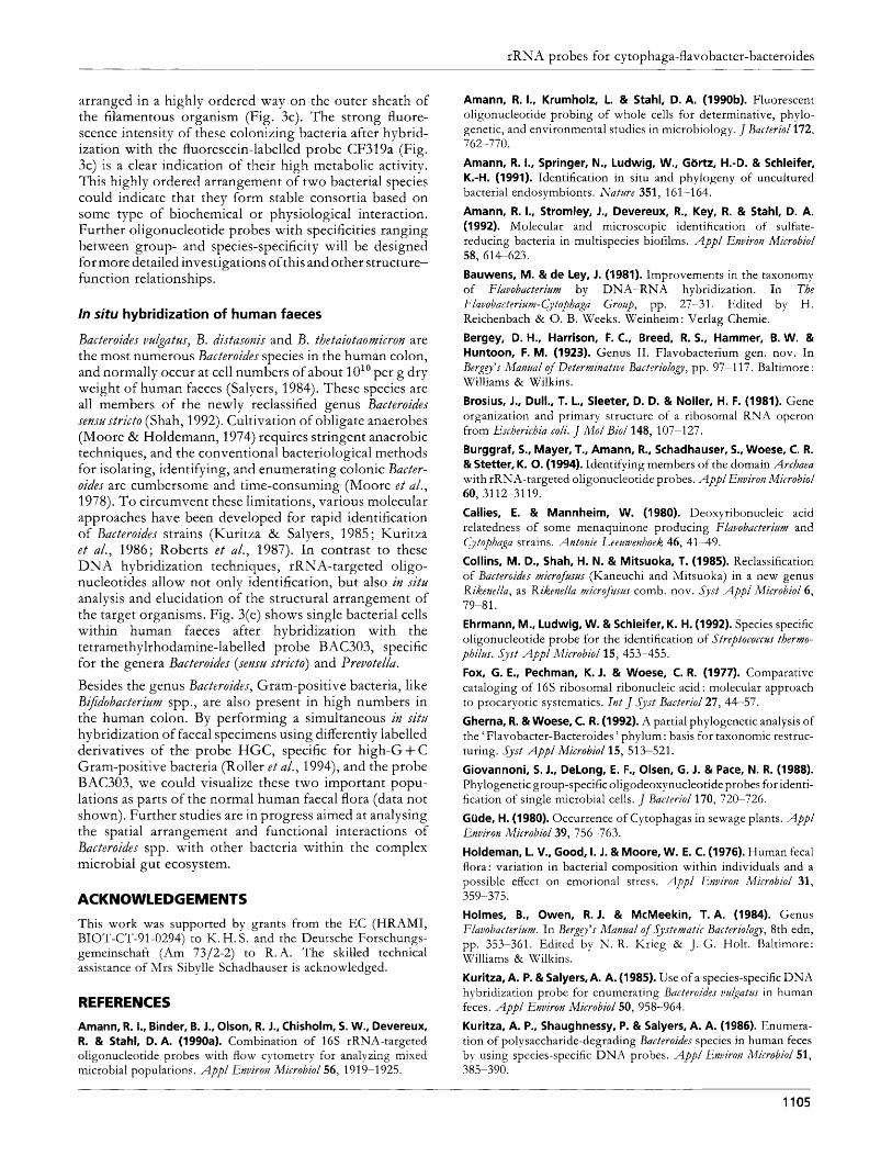
arranged in a highly ordered way on the outer sheath of the filamentous organism (Fig. 3c). The strong fluore- scence intensity of these colonizing bacteria after hybrid- ization with the fluorescein-labelled probe CF319a (Fig. 3c) is a clear indication of their high metabolic activity. This highly ordered arrangement of two bacterial species could indicate that they form stable consortia based on some type of biochemical or physiological interaction. Further oligonucleotide probes with specificities ranging between group- and species-specificity will be designed for more detailed investigations of this and other structure- function relationships.
ln situ hybridization of human faeces
Bacteroides vulgatus, B. distasonis and B. thetaiotaomicron are the most numerous Bacteroides species in the human colon, and normally occur at cell numbers of about lo1* per g dry weight of human faeces (Salyers, 1984). These species are all members of the newly reclassified genus Bacteroides sensu strict0 (Shah, 1992). Cultivation of obligate anaerobes (Moore & Holdemann, 1974) requires stringent anaerobic techniques, and the conventional bacteriological methods for isolating, identifying, and enumerating colonic Bacter- oides are cumbersome and time-consuming (Moore e t a/., 1978). To circumvent these limitations, various molecular approaches have been developed for rapid identification of Bacteroides strains (Kuritza & Salyers, 1985 ; Kuritza e t al., 1986; Roberts e t a/., 1987). In contrast to these DNA hybridization techniques, rRNA-targeted oligo- nucleotides allow not only identification, but also in sitn analysis and elucidation of the structural arrangement of the target organisms. Fig. 3(e) shows single bacterial cells within human faeces after hybridization with the tetramethylrhodamine-labelled probe BAC303, specific for the genera Bacteroides (sensu stricto) and Prevotella.
Besides the genus Bacteroides, Gram-positive bacteria, like Bz3dobacteriztm spp., are also present in high numbers in the human colon. By performing a simultaneous in sitzt hybridization of faecal specimens using differently labelled derivatives of the probe HGC, specific for high-G+C Gram-positive bacteria (Roller e t al., 1994), and the probe BAC303, we could visualize these two important popu- lations as parts of the normal human faecal flora (data not shown). Further studies are in progress aimed at analysing the spatial arrangement and functional interactions of Bacteroides spp. with other bacteria within the complex microbial gut ecosystem.
ACKNOWLEDGEMENTS
This work was supported by grants from the EC (HRAMI, BIOT-CT-91-0294) to I<. H. S. and the Deutsche Forschungs- gemeinschaft (Am 73/2-2) to R.A. The skilled technical assistance of Mrs Sibylle Schadhauser is acknowledged.
REFERENCES
Arnann, R. I., Binder, B. J., Olson, R. J., Chisholrn, 5. W., Devereux, R. & Stahl, D. A. (1990a). Combination of 16s rRNA-targeted oligonucleotide probes with flow cytometry for analyzing mixed microbial populations. Appl Environ Microbiol 56, 191 9-1 925.
Arnann, R. I. , Krurnholz, L. & Stahl, D. A. (1990b). Fluorescent oligonucleotide probing of whole cells for determinative, phylo- genetic, and environmental studies in microbiology. J Bacterioll72,
Arnann, R. I., Springer, N., Ludwig, W., G6rtz. H.-D. & Schleifer, K.-H. (1991). Identification in situ and phylogeny of uncultured bacterial endosymbionts. Nature 351, 161-164.
Arnann, R. I., Strornley, J., Devereux, R., Key, R. & Stahl, D. A. (1 992). Molecular and microscopic identification of sulfate- reducing bacteria in multispecies biofilms. Appl Environ Microbiol
Bauwens, M. & de Ley, J. (1981). Improvements in the taxonomy of Flavobacterium by DNA-RNA hybridization. In The Flavobacterium-Cytopbaga Group, pp. 27-31. Edited by H. Reichenbach & 0. B. Weeks. Weinheim: Verlag Chemie.
Bergey, D. H., Harrison, F. C., Breed, R. S., Hammer, B. W. & Huntoon, F. M. (1923). Genus 11. Flavobacterium gen. nov. In Bergey’s Manual of Determinative Bacteriology, pp. 97-1 17. Baltimore : Williams & Wilkins.
Brosius, J., Dull., T. L., Sleeter, D. D. & Noller, H. F. (1981). Gene organization and primary structure of a ribosomal RNA operon from Escherichia coli. J Mol Bioll48, 107-127.
Burggraf, S., Mayer, T., Arnann, R., Schadhauser, S., Woese, C. R. & Stetter, K. 0. (1994). Identifying members of the domain Archaea with rRNA-targeted oligonucleotide probes. Appl Environ Microbiol
Callies, E. & Mannheirn, W. (1980). Deoxyribonucleic acid relatedness of some menaquinone producing Flavobacterium and Cytophaga strains. Antonie Leeuwenhoek 46, 41-49.
Collins, M. D., Shah, H. N. & Mitsuoka, T. (1985). Reclassification of Bacteroides microfusus (Kaneuchi and Mitsuoka) in a new genus Rikenella, as Rikenella microfusus comb. nov. Syst Appl Microbiol 6,
Ehrrnann, M., Ludwig, W. & Schleifer, K. H. (1992). Species specific oligonucleotide probe for the identification of Streptococcus tbermo- philtls. Syst Appl Microbiol 15, 453-455.
Fox, G. E., Pechrnan, K. 1. & Woese, C. R. (1977). Comparative cataloging of 16s ribosomal ribonucleic acid : molecular approach to procaryotic systematics. Int J Jyst Bacteriol27, 44-57.
Gherna, R. & Woese, C. R. (1992). A partial phylogenetic analysis of the ‘ Flavobacter-Bacteroides ’ phylum : basis for taxonomic restruc- turing. Syst Appl Microbiol 15, 513-521.
Giovannoni, S. J., DeLong, E. F., Olsen, G. 1. & Pace, N. R. (1988). Phylogenetic group-specific oligodeoxynucleotide probes for identi- fication of single microbial cells. J Bacterial 170, 720-726.
Gude, H. (1980). Occurrence of Cytophagas in sewage plants. Appl knviron Microbiol 39, 756-763.
Holdernan, L. V., Good, 1.1. & Moore, W. E. C. (1976). Human fecal flora : variation in bacterial composition within individuals and a possible effect on emotional stress. Appl Environ Microbiol 31, 359-375.
Holmes, B., Owen, R. 1. & McMeekin, T. A. (1984). Genus Flavobacterium. In Bergy’s Manual o f Systematic Bacteriology, 8th edn, pp. 353-361. Edited by N. R. I<rieg & J. G . Holt. Baltimore: Williams & Wilkins.
Kuritza, A. P. & Salyers, A. A. (1985). Use of a species-specific DNA hybridization probe for enumerating Bacteroides vukatus in human feces. Appl Environ Microbioi 50, 958-964.
Kuritza, A. P., Shaughnessy, P. & Salyers, A. A. (1986). Enumera- tion of polysaccharide-degrading Bacteroides species in human feces by using species-specific DNA probes. Appl Environ Microbiol51,
762-770.
58, 614-623.
60, 3112-3119.
79-81.
385-390.
1105

Downloaded from www.microbiologyresearch.org by
IP: 54.237.57.119
On: Fri, 20 May 2016 10:29:03
W. M A N Z a n d O T H E R S
Ludwig, W. & Schleifer, K. H. (1994). Bacterial phylogeny based on 16s and 23s rRNA sequence analysis. FEMS Microbiol Rev 15,
Mannheim, W. (1981). Taxonomically useful test procedures pertaining to bacterial lipoquinones and associated functions, with special reference to Flavobacterium and Cytophaga. In The Flavobacterium-Cytopbaga Group, pp. 11 5-124. Edited by H. Reichenbach & 0. B. Weeks. Weinheim: Verlag Chemie.
Manz, W., Amann, R., Ludwig, W., Wagner, M. & Schleifer, K.-H. (1 992). Phylogenetic oligodeoxynucleotide probes for the major subclasses of proteobacteria : problems and solutions. Sjst Appl Microbiol 15, 593-600.
Manz, W., Szewzyk, U., Eriksson, P., Amann, R., Schleifer, K.-H. & Stenstram, T.-A. (1 993). In situ identification of bacteria in drinking water and adjoining biofilms by hybridization with 16s and 23s rRNA-directed fluorescent oligonucleotide probes. Appl Environ Microbiol59, 2293-2298.
Manz, W., Wagner, M., Amann, R. & Schleifer, K.-H. (1994). In situ characterization of the microbial consortia active in two wastewater treatment plants. Water Res 28, 1715-1723.
Moore, W. E. C. & Holdeman, L. V. (1974). Human fecal flora: the normal flora of 20 Japanese-Hawaiians. Appl Microbi0/27,961-979.
Moore, W. E. C., Cato, E. P. & Holdeman, L. V. (1978). Some current concepts in intestinal bacteriology. A m J Clin Nutr 31, S33S42.
Oyaizu, H. & Komagata, K. (1981). Chemotaxonomic and pheno- typic characterization of the strains of species in the Flavobacterium- Cytophaga complex. J Gen Appl Microbiol27, 57-107.
Paster, 6.1.. Ludwig, W., Weisburg, W. G., Stackebrandt, E., Hespell, R. B., Hahn, C. M., Reichenbach, H., Stetter, K. 0. & Woese, C. R. (1985). A phylogenetic grouping of the Bacteroides, Cytophagas, and certain Flavobacteria. Syst Appl Microbiol 6,
Paster, 6. J., Dewhirst, F. E., Olson, 1. & Fraser, G. J. (1994). Phylogeny of Bacteroides, Prevotella, and Porphyromonas spp. and related bacteria. J Bacterioll76, 725-732.
Pike, L. F. (1975). Aerobic bacteria. In Ecological Aspects of Used Water Treatment, vol. 1, pp. 1-63. Edited by C. R. Curds & H. A. Hawkes. London : Academic Press.
Pipes, W. 0. (1978). Microbiology of activated sludge bulking. Adv Appl Microbiol24, 85-127.
Reichenbach, H. (1989). Order 1. Cytophagales. In Bergy’s Manual of Systematic Bacteriology, vol. 3, pp. 2011-2082. Edited by J. T. Staley, M. P. Bryant, N. Pfennig & J. G. Holt. Baltimore: Williams & Wilkins.
Reichenbach, H., Kohl, W. & Achenbach, H. (1981). The flexirubin- type pigments, chemosystematically useful compounds. In The Fluvobacterium-Cytopbuga Grozlp, pp. 101-108. Edited by H. Reichenbach & 0. B. Weeks. Weinheim: Verlag Chemie.
Roberts, M. C., Moncla, 6. & Kenny, G. E. (1987). Chromosomal DNA probes for the identification of Bacteroides species. J Gen Microbiol133, 1423-1430.
Roller, C., Wagner, M., Amann, R., Ludwig, W. & Schleifer, K.-H.
155-173.
34-42.
(1994). In situ probing of Gram-positive bacteria with high DNA G + C content using 23s rRNA-targeted oligonucleotides. Micro- biology 140, 2849-2858.
Salyers, A. A. (1984). Bacteroides of the human lower intestinal tract. Annu Rev Microbiol38, 293-313.
Schleifer, K. H., Ludwig, W. & Amann, R. (1993). Nucleic acid probes. In Handbook of New Bacterial Systematics, pp. 464-499. Edited by M. Goodfellow & A. G. 0. McDonnell. London: Academic Press.
Shah, H. N. (1992). The genus Bacteroides and related taxa. In The Procar_yotes, pp. 3593-3607. Edited by A. Balows, H. G. Truper, M. Dworkin, W. Harder & K.-H. Schleifer. New York: Springer.
Shah, H. N. & Collins, M. D. (1988). Proposal for reclassification of Bacteroides asaccbarohticzls, Bacteroidesgingival, and Bacteroides endodon- talis in a new genus, Porphyromonas. Int J Syst Bacteriol38, 128-131.
Shah, H. N. & Collins, M. D. (1989). Proposal to restrict the genus Bacteroides (Castellani and Chalmers) to Bacteroides fragilis and closely related species. Int J Syst Bacteriol39, 85-87.
Shewan, 1. M. & McMeekin, T. A. (1983). Taxonomy (and ecology) of Flavobacterium and related genera. Annu Rev Microbiol 37,
Spring, S., Amann, R., Ludwig, W., Schleifer, K. H. & Petersen, N. (1 992). Phylogenetic diversity and identification of nonculturable magnetotactic bacteria. Jyst Appl Microbioll5, 116-122.
Stahl, D. A. & Amann, R. 1. (1991). Development and application of nucleic acid probes in bacterial systematics. In Sequencing and Hybridixation Techniques in Bacterial Systematics, pp. 205-248. Edited by E. Stackebrandt & M. Goodfellow. Chichester: John Wiley.
Trebesius, K., Amann, R., Ludwig, W., Milhlegger, K. & Schleifer, K.-H. (1994). Identification of whole fixed bacterial cells with nonradioactive 23s rRNA-targeted polynucleotide probes. Appl Environ Microbiol 60, 3228-3235.
Wagner, M., Amann, R., Lemmer, H. & Schleifer, K.-H. (1993). Probing activated sludge with oligonucleotides specific for proteo- bacteria : inadequacy of culture-dependent methods for describing microbial community structure. Appl Environ Microbiol 59, 1520-1 525.
Winogradsky, 5. (1929). Etudes sur la microbiologie du sol. Sur la digradation de la cellulose dans le sol. Ann Inst Pasteur 43,549-633.
Woese, C. R. (1987). Bacterial evolution. Microbiol Rev 51,221-271.
Woese, C. R., Maloy, S., Mandelco, L. & Raj, H. D. (1990a). Phylogenetic placement of Spirosomaceae. Sy-rt Appl Microbiol 13,
Woese, C. R., Yang, D., Mandelco, L. & Stetter, K. 0. (1990b). The flexibacter-flavobacter connection. Syst Appl Microbioll3,161-165.
Zarda, B., Amann, R., Wallner, G. & Schleifer, K.-H. (1991). Identification of single bacterial cells using digoxigenin-labelled, rRNA-targeted oligonucleotides. J Gen Microbiol137, 2823-2830.
233-252.
19-23.
Received 7 August 1995; revised 27 November 1995; accepted 11 January 1996.
1106