Redefining Puerto Ricanness in Esmeralda Santiago's When I ...

1 23
Parasitology ResearchFounded as Zeitschrift fürParasitenkunde ISSN 0932-0113Volume 111Number 1 Parasitol Res (2012) 111:79-88DOI 10.1007/s00436-011-2803-8
Redefining the genus Spraguea based onultrastructural and phylogenetic data fromSpraguea gastrophysus n. sp. (PhylumMicrosporidia), a parasite found inLophius gastrophysus (Teleostei) fromBrazilGraça Casal, Sérgio C. S. Clemente,Patrícia Matos, Marcelo Knoff, EdilsonMatos, et al.

1 23
Your article is protected by copyright and
all rights are held exclusively by Springer-
Verlag. This e-offprint is for personal use only
and shall not be self-archived in electronic
repositories. If you wish to self-archive your
work, please use the accepted author’s
version for posting to your own website or
your institution’s repository. You may further
deposit the accepted author’s version on a
funder’s repository at a funder’s request,
provided it is not made publicly available until
12 months after publication.

ORIGINAL PAPER
Redefining the genus Spraguea based on ultrastructuraland phylogenetic data from Spraguea gastrophysus n. sp.(Phylum Microsporidia), a parasite found in Lophiusgastrophysus (Teleostei) from Brazil
Graça Casal & Sérgio C. S. Clemente & Patrícia Matos &
Marcelo Knoff & Edilson Matos & Abdel A. Abdel-Baki &Carlos Azevedo
Received: 10 February 2011 /Accepted: 21 December 2011 /Published online: 6 January 2012# Springer-Verlag 2012
Abstract The ultrastructure of the fish-infecting micro-sporidium Spraguea gastrophysus found in the dorsal gan-glia and kidney of the anglerfish, Lophius gastrophysus(family Lophiidae) collected on the Brazilian Atlantic coastis described. Each whitish xenoma (up to 3.1×1.8 mm)contains several groups of parasites. The host cells arehypertrophied and contain various parasite life stages in-cluding mature spores and several developmental stageswith unpaired nuclei. Monomorphic spores are ellipsoidal,lightly curved and measure about 3.35×1.71 μm. The sporecontains a gradually tapering isofilar polar filament withfive to six coils arranged in a single row. The nucleus
occupies a central zone of the sporoplasm where also severalpolyribosomes are presented. The posterior vacuole containsa voluminous spherical and granular posterosome measur-ing up to ~0.65 μm in diameter. The partial small subunit,intergenic spacer and partial large subunit rRNA gene weresequenced and the phylogenetic analysis places the micro-sporidian described here in the clade that includes allsequences of the Spraguea genus. The ultrastructural mor-phology of the xenoma and the spores of this microsporidianparasite, as well as the molecular and phylogenetic analysis,suggest the description of a new species. A redefining of thegenus Spraguea is also done.
G. Casal : C. Azevedo (*)Department of Cell Biology, Institute of Biomedical Sciences,University of Porto (ICBAS/UP),Lg. A. Salazar no. 2,4099-003 Porto, Portugale-mail: [email protected]
G. Casal : C. AzevedoLaboratory of Pathology, Interdisciplinary Centre of MarineEnvironmental Research, University of Porto (CIIMAR/UP),Gandra, Portugal
G. CasalDepartamento de Ciências, Instituto Superiorde Ciências da Saúde - Norte, CESPU,Gandra, Portugal
S. C. S. ClementeLaboratory of Inspection and Technology of Food,Faculty of Veterinary, Federal Fluminense University,Niterói, Brazil
P. MatosLaboratory of Histology of Aquatic Animals,Federal University of Pará,Belém, Brazil
M. KnoffLaboratory of Helminth Parasites of Vertebrates,Oswaldo Cruz Institute (IOC/FIOCRUZ),Rio de Janeiro, Brazil
E. MatosCarlos Azevedo Research Laboratory,Federal Rural University of Amazonia,Belém, Brazil
A. A. Abdel-Baki : C. AzevedoZoology Department, College of Science, King Saud University,Riyadh, Saudi Arabia
A. A. Abdel-BakiZoology Department, Faculty of Science, Beni-Suef University,Beni-Suef, Egypt
Parasitol Res (2012) 111:79–88DOI 10.1007/s00436-011-2803-8
Author's personal copy

Introduction
The anglerfishes of the genus Lophius (Family Lophii-dae) have a worldwide distribution and are representedby seven species (L. americanus, L. budegassa, L. gas-trophysus, L. litulon, L. piscatorius, L. vaillanti and L.vomerinus). Except for L. vaillanti and L. vomerinus,probably due to lack of documentation, all were foundto harbour microsporidians in the nervous tissues. Theseparasites were first reported from the spinal ganglia ofL. piscatorius Linnaeus, 1758 previously classified asgenus Glugea and later identified as belonging to G.lophii (Doflein 1898). More detailed morphologicalstudies developed by Mrázek (1899) reinforced that thisparasite belongs to Glugea. However, this parasite wassubsequently transferred to the genus Nosema, as N.lophii (Pace 1908), a change that was also corroboratedby Weissenberg (1909, 1911a, b, c). Later, in a footnoteof Weissenberg’s (1976) report, it was referred to forthe first time by the name “Spraguea n. gen.” andsimultaneously Glugea lophii was transferred to Spra-guea lophii (Doflein 1898; Weissenberg 1976) as the typespecies.
The first ultrastructural data of S. lophii, parasite of theEuropean anglerfish L. budegassa and L. piscatorius, bothwith dimorphic spores, were obtained by Loubès et al.(1979). On the other hand, the spinal and cranial gangliaof American anglerfish, L. americanus, caught off the north-east Atlantic coast of the USA, also indicated the presenceof microsporidian spores (Takvorian and Cali 1986). Con-sidering some ultrastructural differences in L. americanus,mainly having monomorphic spore type, relative to previ-ously described dimorphic spore occurring in L. budegassaand L. piscatorius from Europe, the microsporidian wasincluded in the genus Glugea, as G. americanus (Takvorianand Cali 1986). However, molecular results based on the SSUrRNA gene sequences suggested that this species should betransferred to the genus Spraguea, as S. americana (Lom andNilsen 2003; Nilsen 2000; Pomport-Castillon et al. 2000).More recently, on the basis of ultrastructural and molecularstudies, the xenomas found in the nervous tissues of theJapanese anglerfish Lophius litulon containing monomorphicspores were identified as belonging to Spraguea americana(Freeman et al. 2004).
There are also references to the presence of a similarmicrosporidian from the South American anglerfish Lophiusgastrophysus, collected in the Brazilian and Venezuelancoasts (Jakowska 1964, 1966; Jakowska and Nigrelli 1958,1959) but with no microscopical images or schematic draw-ings. This species has a high commercial value and it hasbeen caught by the local fleets for decades. For example,landings all along the upper slope of southern Brazil (Perezet al. 2005) peaked at 7,094 tons in 2001 and 5,129 ton in
2002; however, presently only 2,500 tons are landed(Haimovici et al. 2009). Relative to fish-infecting micro-sporidia in Brazilian fauna, two new genera were identified:Amazonspora (Azevedo and Matos 2003) and Potaspora(Casal et al. 2008). There is also information for another fourparasites: Loma myrophis (Azevedo and Matos 2002), Lomapsittaca (Casal et al. 2009), Kabatana rondoni (Casal et al.2010) and Microsporidium brevirostris (Matos and Azevedo2004), all found in Amazonian fishes.
In the present work, we redefined the diagnosis ofthe genus Spraguea and described a new microsporidianspecies on the basis of the ultrastructural morphology ofthe spores, with special emphasis on host and tissuespecificity and molecular characterization of the SSUrRNA gene.
Materials and methods
Fish, location of infection and prevalence
Thirty-six adult specimens of the marine anglerfish, L. gas-trophysus Miranda-Ribeiro, 1915 (Teleostei, Lophiidae)(Brazilian common name “peixe-sapo pescador”) (27–68 cm long; 0.550–5,600 kg weight) were collected fromthe Atlantic coast of “Cabo Frio” (22°50′S/42°03′W), Stateof Rio de Janeiro, Brazil. The fish were lightly anesthetisedwith MS 222 (Sandoz Laboratories), transported to thelaboratory (UFF–Niterói), dissected and the infected tissuescontaining several whitish cysts (xenomas) were removedfrom the peripheral muscles of the internal abdominal cavityin contact with the dorsal nerves and kidney. The prevalenceof infection was 55.5% (20 fishes in 36 examined) (11/20females, 9/16 males) with similar rates in both sexes.
Light and electron microscopy
For light microscopy, smears of whitish xenoma and freespores were examined by a light microscope equipped withNomarski interference-contrast (DIC) optics.
For ultrastructural studies, small fragments of the para-sitized tissues containing xenomas were excised and fixed in3% glutaraldehyde in 0.2 M sodium cacodylate buffer(pH 7.2) at 4°C for 12 h. After being rinsed overnight inthe same buffer at 4°C and post-fixed in 2.0% osmiumtetroxide in the same buffer for 3 h at 4°C, the fragmentswere dehydrated through an ascending ethanol series, fol-lowed by propylene oxide and embedded in Epon. Semithinsections were stained with methylene blue-Azur II and ob-served by DIC optics. Ultrathin sections were double stainedwith aqueous uranyl acetate and lead citrate and observedunder a transmission electron microscope (TEM) JEOL100CXII operated at 60 kV.
80 Parasitol Res (2012) 111:79–88
Author's personal copy

DNA isolation, PCR amplification and DNA sequencing
Several cysts were dissected from fishes, homogenized toisolate the spores and stored in 80% ethanol at 4°C. Thegenomic DNA of about 6×10⁶ spores was extracted using aGenEluteTM Mammalian Genomic DNA Miniprep Kit(Sigma) following the manufacturer’s instructions for ani-mal tissue, except for the incubation time. The DNA wasstored in 50 μl of TE buffer at −20°C. The majority of theregion coding for the small subunit (SSU) rRNA gene wasamplified by PCR using the primers V1f (5′-CACCAGGTTGATTCTGCC-3′) and 1492r (5′-GGTTACCTTGTTACGACTT-3′) (Nilsen 2000; Vossbrinck et al. 1993). Toamplify the 3′ end of the SSU, internal transcribed spacer(ITS) and 5′ end of the large subunit (LSU) rRNA gene,HG4F (5′-GCGGCTTAATTTGACTCAAC-3′) and HG4R(5′-TCTCCTTGGTCCGTGTTTCAA-3′) primers wereused (Gatehouse and Malone 1998). PCR was carried outin 50 μl reactions using 10 pmol of each primer, 10 nmol ofeach dNTP, 2 mM of MgCl2, 5 μl 10×Taq polymerasebuffer, 1.25 units Taq DNA polymerase (Invitrogen prod-ucts), and 3 μl of the genomic DNA. The reactions were runon a Hybaid PxE Thermocycler (Thermo Electron Corpora-tion, Milford, MA). The amplification program consisted of94°C denaturation for 5 min, followed by 35 cycles of 94°Cfor 1 min, 50°C for 1 min and 72°C for 2 min. A finalelongation step was performed at 72°C for 10 min. PCRproducts were visualized in 5-μl aliquots with ethidiumbromide staining after running on a 1% agarose gel. PCRproducts for the SSU gene and ITS region have approximatesizes of 1,400 and 1,100 bp, respectively. These werecleaned using the NucleoSpin Extract II (Macherey-Nagel),and then three purified PCR products were sequenced inboth directions. The sequencing reactions were done usingBigDye Terminator v1.1 kit (Applied Biosystems) and wererun on an ABI3700 DNA analyzer (Perkin-Elmer, AppliedBiosystems, Stabvida, Co., Oeiras, Portugal).
Distance and phylogenetic analysis
The various forward and reverse sequence segments werealigned manually with ClustalW (Thompson et al. 1994) inMEGA 4 software and ambiguous bases were clarifiedusing corresponding ABI chromatograms. To evaluate therelationship of our parasite to other microsporidians, we firstselected all rDNA sequences that have a fish as host (resultsnot shown) and then we used 18 rDNA sequences, allbelonging to the group 4 of the classification by Lom andNilsen (2003). The rDNA sequence of Nucleospora salmo-nis (U781176) and Vittaforma corneae (L39112) were usedas the outgroup. Sequences were aligned as described byCasal et al. (2008). The alignment was performed throughthe use of Clustal W (Thompson et al. 1994) in MEGA 4
software (Tamura et al. 2007), with an opening gap penaltyof 10 and a gap extension penalty of 4 for both pairwise andmultiple alignments. Subsequent phylogenetic and molecu-lar evolutionary analyses were conducted using MEGA 4,with the sequences for microsporidian species and the out-group species selected. Distance estimation was carried outusing the Kimura-2 parameters model distance matrix fortransitions and transversions. For the phylogenetic treereconstructions, the maximum parsimony analysis was per-formed using the close neighbour interchange (CNI) heuris-tic option with a search factor of 2 and random initial treesaddition of 2,000 replicates. Bootstrap values were calculatedover 100 replicates.
Results
Large whitish cysts (up to 3.1×1.8 mm long) formed byseveral small cysts (xenomas) were macroscopically ob-served in the abdominal cavity in close contact with theinternal abdominal muscle near the dorsal ganglia of theanglerfish, L. gastrophysus (Fig. 1). Similar groups ofsmaller cysts were observed in the kidney. After dissectionand rupture of both types of xenomas, it was observed thatthey had numerous ellipsoidal spores (several thousand)identified as belonging to the phylum Microsporidia(Fig. 2, inset). The xenomas seen in semithin sections wereirregularly shaped and contained several groups of juxta-posed cysts (Fig. 3). At high magnification, it was observedthat the xenomas were formed by a wall encircling a hyper-trophic cell with a central hypertrophic nucleus surroundedby numerous spores in contact with the cytoplasm of thehypertrophic host cell. Different life cycle stages of themicrosporidian were observed intermingled among thespores in the matrix of the xenoma (Figs. 3 and 4).
Systematic position
Phylum Microsporidia Balbiani, 1882Class Marinosporidia Vossbrinck and Debrunner-
Vossbrinck, 2005Family Spraguidae Weissenberg, 1976Genus Spraguea Weissenberg, 1976Species Spraguea gastrophysus n. sp.
Description of the species
Name: Spraguea gastrophysus n. sp.Type host: Lophius gastrophysus Miranda-Ribeiro, 1915
(Teleostei, Lophiidae).Type locality: Atlantic coast of Cabo Frio (22°50′S/42°03′
W), State of Rio de Janeiro, Brazil.
Parasitol Res (2012) 111:79–88 81
Author's personal copy

Location in the host: Xenoma in the dorsal muscle of theinternal abdominal cavity and kidney.
Prevalence of infection: Twenty of 36 examined (55.5%)(11/20 females, 9/16 males) with similar rates in both sexes.
Figs. 1–7 Light and electron micrographs of the xenomas, develop-mental stages and spores of Spraguea gastrophysus n. sp., a parasite ofthe peripheral muscle of the internal abdominal cavity of the teleostLophius gastrophysus (Scale bars in μm). 1. Some grouped xenomas(arrowheads) were observed in DIC. 2. Fresh spores observed in DIC.3. Semithin section of the periphery of a xenoma showing numerousspores (S) and other developmental stages (arrowheads). 4. A group ofsporoblasts (*) showing an electron dense wall (arrowheads) and
spores (S). 5. A mature spore longitudinally sectioned showing thewall (Wa), anchoring disc (AD), polaroplast (Pp), polar filament (PF),nucleus (Nu), posterosome (Ps) and posterior vacuole (Va). 6. Ultra-structural details of the anterior portion of a mature spore showing thepolaroplast organization (Pp), spore wall (Wa), anchoring disc (AD),polar filament (manubrium) (PF) and numerous ribosomes (*). 7.Tangential section of the spore surface showing the external ornamen-tation seeming a fingerprint-like structure (arrowheads)
82 Parasitol Res (2012) 111:79–88
Author's personal copy
Type specimens: One glass slide with a semithin section ofa xenoma containing different developmental stages, mainlymature spores of hapantotype were deposited in the Micro-sporidia Type Slide Collection at the “Instituto Nacional dePesquisa da Amazônia” (INPA), Manaus, Brazil, under acqui-sition number (001/11).
Etymology: The specific epithet “gastrophysus” is derivedfrom the specific epithet of the host species.
Description of the spores
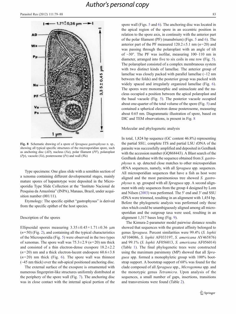
Ellipsoidal spores measuring 3.35±0.45×1.71±0.36 μm(n050) (Fig. 2), and containing all the typical characteristicsof the Microsporidia (Fig. 5) were observed in the two typesof xenomas. The spore wall was 75.3±2.9 (n020) nm thickand consisted of a thin electron-dense exospore 18.2±2.2(n020) nm and a thick electron-lucent endospore 60.6±3.8(n020) nm thick (Fig. 6). The spore wall was thinnest(~45 nm thick) over the sub-apical positioned anchoring disc.
The external surface of the exospore is ornamented withnumerous fingerprint-like structures uniformly distributed atthe periphery of the spore wall (Fig. 7). The anchoring discwas in close contact with the internal apical portion of the
spore wall (Figs. 5 and 6). The anchoring disc was located inthe apical region of the spore in an eccentric position inrelation to the spore axis, in continuity with the anterior partof the polar filament (PF) (manubrium) (Figs. 5 and 6). Theanterior part of the PF measured 120.2±5.1 nm (n020) andwas passing through the polaroplast with an angle of tiltof ~30°. The PF was isofilar, measuring 100–110 nm indiameter, arranged into five to six coils in one row (Fig. 5).The polaroplast consisted of a complex membranous systemwith two distinct kinds of lamellae. The anterior group oflamellae was closely packed with parallel lamellae (~12 nmbetween the folds) and the posterior group was packed withwidely spaced and irregularly organized lamellae (Fig. 6).The spores were monomorphic and uninucleate and the nu-cleus occupied a position between the apical polaroplast andthe basal vacuole (Fig. 5). The posterior vacuole occupiedabout one-quarter of the total volume of the spore (Fig. 5) andcontained a spherical electron dense posterosome, measuringabout 0.65 nm. Diagrammatic illustration of spore, based onDIC and TEM observations, is present in Fig. 8
Molecular and phylogenetic analysis
In total, 1,824 bp sequence (GC content 46.8%) representingthe partial SSU, complete ITS and partial LSU rDNA of theparasite was successfully amplified and deposited in GenBankwith the accession number (GQ868443). A Blast search of theGenBank database with the sequence obtained from S. gastro-physus n. sp. detected close matches to other microsporidianrRNA sequences, namely, with all Spraguea spp. sequences.All microsporidian sequences that have a fish as host werealigned and the most parsimonious tree showed S. gastro-physus n. sp. grouped with all Spraguea spp. A second align-ment with only sequences from the group 4 designed by Lomand Nilsen (2003) was performed. The 5′ end and 3′ end SSUrDNAwere trimmed, resulting in an alignment with 1,454 bp.Before the phylogenetic analysis was performed only thosesites which could be unambiguously aligned among all micro-sporidian and the outgroup taxa were used, resulting in analignment 1,317 bases long (Fig. 9).
The Kimura-2-parameter model pairwise distance resultsshowed that sequences with the greatest affinity belonged togenus Spraguea. Percent similarities were 99.4% (S. lophiiAF104086, S. lophii AF033197, S. americana AY465876)and 99.1% (S. lophii AF056013, S. americana AF056014)(Table 1). The final phylogenetic trees were constructedusing the maximum parsimony (MP) showed that all Spra-guea spp. formed a monophyletic group with 100% boot-strap support. A bootstrap support of 68% was found for theclade composed of all Spraguea spp.,Microgemma spp. andthe monotypic genus Tetramicra. Upon analysis of thesequences, a small number of gaps, insertions, transitionsand transversions were found (Table 2).
Fig. 8 Schematic drawing of a spore of Spraguea gastrophysus n. sp.,showing all typical specific structures of the microsporidian spore, suchas anchoring disc (AD), nucleus (Nu), polar filament (PF), polaroplast(Pp), vacuole (Va), posterosome (Ps) and wall (Wa)
Parasitol Res (2012) 111:79–88 83
Author's personal copy

Discussion
The parasite described in this paper presents the typical mor-phology and characteristics of the phylumMicrosporidia (Lomand Dyková 1992; Cali and Takvorian 1999; Larsson 1999).Among the 18 genera infecting fish, 12 of these producexenomas: Amazonspora Azevedo and Matos 2003; GlugeaThélohan, 1891; Ichthyosporidium Caullery and Mesnil,1905; Loma Morrison and Sprague, 1981; Microfilum Faye,Toguebaye and Bouix, 1991; Microgemma Ralphs andMatthews, 1986; Myosporidium Baquero, Rubio, Moura,Pieniazek and Jordana, 2005;Neonosemoides Faye, Toguebayeand Bouix, 1996; Pseudoloma Matthews, Brown, Larison,Bishop-Stewart, Rogers and Kent, 2001; Potaspora Casal,Matos, Teles-Grilo and Azevedo 2008; SpragueaWeissenberg1976 and Tetramicra Matthews and Matthews 1980.
Among these microsporidians, the genus Spraguea is atypical case of close relationship between the kind of para-site, host and the place of infection. Presently, it is knownthat five (L. piscatorius, L. budegassa, L. americanus, L.
litulon and L. gastrophysus) of seven lophiid species areparasitized in the nervous tissues (spinal nerves of the ver-tebral column, trigeminal nerves, vagal nerves or in themedulla oblongata region of the hind brain) by microspor-idia belonging to the genus Spraguea (Jakowska 1964;Takvorian and Cali 1986; Weissenberg 1911c, 1976). Oneexception for this was observed in the anglerfish Lophiusbudegassa in Spain for the reason that the microsporidiansTetramicra brevifilum and Spraguea lophii were simulta-neously found in hepatocytes and musculature, respectively(Maíllo et al. 1998). The species Tetramicra brevifilum hasno ornamentation on the exospore, is phylogenetically closeto the Spraguea spp. and frequently parasitizes the connec-tive tissues of the musculature of Scophtalmus maximus(Matthews and Matthews 1980).
Irrefutably, the parasite described in this study is mor-phologically and phylogenetically close to two Spragueaspecies previously reported in different anglerfish species.Spraguea lophii was described in Lophius piscatorius and L.budegassa on the European Atlantic and Mediterranean
Fig. 9 The maximum parsimony tree of SSU rDNA sequences ofSpraguea gastrophysus n. sp. and the microsporidian species clusteredin group 4. The numbers on the branches are bootstrap confidence
levels on 100 replicates. GeneBank accession numbers are in paren-theses after the species names and the scale is given under the tree
84 Parasitol Res (2012) 111:79–88
Author's personal copy

coasts (Loubès et al. 1979). Spraguea americana, first de-scribed as Glugea americanus, was described on the USAtlantic coast (in L. americanus) (Takvorian and Cali 1986)and later in L. litulon from the Japanese coast (Freeman et al.2004). The molecular data has shown that the Glugea amer-icanus sequences are close to all other Spraguea lophiisequences and consequently it was transferred to the genusSpraguea and renamed S. americana (Nilsen 2000; Pomport-Castillon et al. 2000; Lom and Nilsen 2003).
The morphology of the spores shows several similaritieswhen compared with other uninuclear spores of the Spra-guea genus, except for the thickness of the two layers of thewall (exospore and endospore). The spore wall found in L.gastrophysus is thicker than that of other species (Table 3).
For some genera, such as Amazonspora (Azevedo andMatos 2003), Kabatana (Lom et al. 1999, 2001; McGourtyet al. 2007) and Spraguea (Loubès et al. 1979; Takvorianand Cali 1986; Freeman et al. 2004) the fingerprint-likestructures of the external surface of the exospore wall are amorphological characteristic common at all species. Usual-ly, the external ornamentation is regularly distributed andthe elevations on the surface of the mature spores, whenthey are observed in tangential section, present a hexagonalfingerprint-like shape (genus Amazonspora) or a tubularshape (genera Kabatana and Spraguea). As in S. lophiiand S. americana this ultrastructural detail was also
observed on the surface of S. gastrophysus n. sp. Insidethe posterior vacuole, there is a spherical electron densestructure, similar to that reported in S. americana caught inthe American North Atlantic coast (Takvorian and Cali1986) and from Japanese lophiid species (Freeman et al.2004).
The most parsimonious phylogenetic tree (Fig. 9) clus-tered all Spraguea sequences in the same clade (bootstrap100%) and this cladogram had a similar topology to previ-ous described trees (Lom and Nilsen 2003; Freeman et al.2004; Casal et al. 2008). Comparison of the SSU rDNAsequence of S. gastrophysus n. sp. with all the other knownsequences from Spraguea infections from Europe, Japanand America showed that the genetic distances range from0.6% to 0.9% (Table 1). The genetic distance between allpreviously reported Spraguea infections is less than or equalto 0.3%, a result which is in accordance with that obtainedby Freeman et al. (2004). It suggests that the microsporidiafound in the anglerfish L. gastrophysus from South Americais the most phylogenetically distanced of the three Spragueaspecies. Small genetic distances using the sequence of theSSU rDNA have been reported in other microsporidian spe-cies. Interestingly, in our analysis, the pairwise sequencevariation between Microgemma tincae and M. vivaresi(0.3% instead 1%) was lower than that observed by Mansouret al. (2005). The similar situation occurs between two
Table 1 Comparison of some SSU rDNA sequences: percentage of identity (top diagonal) and pairwise distance (bottom diagonal) obtained byKimura-2 parameter analysis
1 2 3 4 5 6 7 8 9 10
(1) Spraguea gastrophysus n. sp. 99.4 99.4 99.4 99.1 99.1 95.8 95.5 93.6 93.6
(2) Spraguea lophii AF104086 0.006 100 100 99.7 99.7 96.4 96.1 94.2 94.2
(3) Spraguea lophii AF033197 0.006 0.000 100 99.7 99.7 96.4 96.1 94.2 94.2
(4) Spraguea americana AY465876 0.006 0.000 0.000 99.7 99.7 96.4 96.1 94.2 94.2
(5) Spraguea lophii AF056013 0.009 0.003 0.003 0.003 100 96.1 95.8 93.9 93.9
(6) Spraguea americana AF056014 0.009 0.003 0.003 0.003 0.000 96.1 95.8 93.9 93.9
(7) Microgemma tincae AY651319 0.042 0.036 0.036 0.036 0.039 0.039 99.7 96.1 96.1
(8) Microgemma vivaresi AJ252952 0.045 0.039 0.039 0.039 0.042 0.042 0.003 95.8 95.8
(9) Microgemma caulleryi AY033054 0.064 0.058 0.058 0.058 0.061 0.061 0.039 0.042 100
(10) Tetramicra brevifilum AF364303 0.064 0.058 0.058 0.058 0.061 0.061 0.039 0.042 0.000
Table 2 Comparative analysisof all Spraguea spp. sequenceswith the obtained in this studyfrom of Lophius gastrophysus
The SSU rDNA length, thenumber the gaps, insertions,transitions and transversions areincluded
Spraguea spp. Length of SSU usedin the analysis
Gaps Insertions Transitions Transversions
S. lophii (1) AF104086 1287 7 5 9 4
S. lophii (2) AF033197 1319 0 12 7 7
S. lophii (3) AF056013 1175 0 9 7 6
S. americana (1) AF056014 1174 2 10 5 6
S. americana (2) AY465876 1206 0 4 2 1
Parasitol Res (2012) 111:79–88 85
Author's personal copy

species, Tetramicra brevifilum and M. vivaresi, belonging todifferent genera. According to Cheney et al. (2001), the ge-netic distance between Pleistophora hippoglossoideos fromthe flatfish Hippoglossoides platessoides and Pleistophoratypicalis from the sea scorpion fish Myoxocephalus scorpiusis only 0.4%. Presently, it is known that the SSU rDNAsequence for fish-infecting microsporidia does not providesufficient variation to fully resolve the phylogeny betweenclosely related species, even including the ITS and a part ofthe LSU rDNA (Lom and Nilsen 2003). Unfortunately for thepreviously described Spraguea species there is no geneticinformation for these regions of the rDNA.
According to the original description by Loubès et al.(1979), the genus Spraguea is a dimorphic microsporidianthat produces two types of spores in two distinct develop-mental sequences. One sequence is characterized by allstages having unpaired nuclei and polysporoblastic sporog-ony that produce uninucleate spores whereas the other se-quence is characterized by diplokarya and disporoblasticsporogonic stages that give rise to slender curved diplokary-otic spores. Curiously, the spore dimorphism of the genusand species type is in contradiction with all other ultrastruc-tural descriptions. Apparently, this is an exclusive charac-teristic of the Spraguea infections from Lophius piscatoriusand L. budegassa from European species because the infec-tions from the American and Japanese species only produceuninucleate spores. Definitely, as recommended by Lom(2002), Lom and Nilsen (2003) and Freeman et al. (2004),the genus diagnosis needs to be redefined since the excep-tions must not be general characteristics of the genus.
Diagnosis of genus Spraguea
Monophyletic group of parasite that infect ganglion cells ofthe Lophiid species. Xenoma bounded by a single cellmembrane and with a single hypertrophic nucleus. Mono-morphic or dimorphic development sequences. Alwayspresent is one sequence characterized by isolated nucleusin all stages and polysporoblastic sporogony where uninu-cleate spores form by radial cleaving of the sporogonialplasmodium. Sometimes another sequence also developswhere all stages present diplokarya and by disporoblasticsporogony produces curved spores.
Considering the morphological and molecular data, weredefined the genus Spraguea and designated a new speciesthat should be included in this genus with the name S.gastrophysus n. sp.
Acknowledgements This work was partially supported by Eng. A.Almeida Foundation, Porto, Portugal, “CNPq” and “CAPES”, Brazil.We appreciate the technical assistance of J. Carvalheiro (ICBAS/UP)and Nilza Felizardo (UFF). This work is original and complies with thecurrent laws of the countries in which it has been performed.T
able
3Com
parativ
emeasurementsfrom
Spragu
easpp.
Parasite
Hostspecies
Cou
ntry
(region)
Spo
redimorph
ism
Spo
redimension
s(inμm
)SSO
Wallthick(innm
)PFC
References
Spragu
easpp.
Exo
spore/End
ospo
re
S.loph
iiL.piscatorius
France(A
tlantic
coast)
+3.5×1.5(*)
+?
5–6(*)
Lou
bèset
al.19
794.0×1.25
(**)
3–4(**)
S.loph
iiL.bu
dega
ssa
France(M
editerranean)
+3.5×1.5(*)
−?
5–6(*)
Lou
bèset
al.19
794.0×1.25
(**)
3–4(**)
S.am
erican
aL.am
erican
usUSA
(Atlantic
coast)
−2.8×1.5
+12
.5/70
6–9
Takvo
rian
andCali19
86
S.am
erican
aL.litulon
Japan
−3.4×1.8
+~3
0/~6
55–8
Freem
anet
al.20
04
S.ga
stroph
ysus
n.sp.
L.ga
stroph
ysus
Brasil(A
tlantic
coast)
−3.35
×1.71
+~1
8/~6
05–6
Present
stud
y
Allspeciesparasite
ateleostfish
from
genu
sLop
hius
SSO
sporesurfaceornamentatio
n,PFCpo
larfilamentcoils;sporeof
mon
okaryo
ntype
(*),sporeof
diplok
aryo
ntype
(**),po
sitiv
e(+),negativ
e(−),no
data
(?)
86 Parasitol Res (2012) 111:79–88
Author's personal copy

References
Azevedo C, Matos E (2002) Fine structure of a new species, Lomamyrophis (Phylum Microsporidia), parasite of the Amazonian fishMyrophis phatyrhynchus (Teleostei, Ophichithidae). Eur J Protistol37:445–452
Azevedo C, Matos E (2003) Amazonspora hassar n. gen. and n. sp.(Phylum Microsporidia, fam. Glugeidae), a parasite of the Ama-zonian teleost Hassar orestis (fam. Doradidae). J Parasitol89:336–341
Cali A, Takvorian PU (1999) Developmental morphology and lifecycles of the Microsporidia. In: Wittner M, Weiss L (eds) TheMicrosporidia and Microsporidiosis. American Society of Micro-biology, Washington, DC, pp 85–128
Casal G, Matos E, Teles-Grilo ML, Azevedo C (2008) A new micro-sporidian parasite, Potaspora morhaphis n. gen., n. sp. (Micro-sporidia) infecting the teleostean fish, Potamorhaphis guianensisfrom the River Amazon. Morphological, ultrastructural and molec-ular characterization. Parasitology 135:1053–1064
Casal G, Matos E, Teles-Grilo ML, Azevedo C (2009) Morphologicaland genetical description of Loma psittaca sp. n. isolated from theAmazonian fish species Colomesus psittacus. Parasitol Res105:1261–1271
Casal G, Matos E, Teles-Grilo L, Azevedo C (2010) Ultrastructural andmolecular characterization of a new microsporidium parasite from theAmazonian fish,Gymnorhamphichthys rondoni (Rhamphichthyidae).J Parasitol 96:1155–1163
Cheney SA, Lafranchi-Tristem NJ, Bourges D, Canning EU(2001) Relationships of microsporidian genera, with emphasison the polysporous genera, revealed by sequences of the largestsubunit of RNA polymerase II (RPB1). J Eukaryot Microbiol48:111–117
Doflein F (1898) Studien zur Naturgeschichte der Protozoen: III. UeberMyxosporidien. Zool Jahrb Anat 11:281–350
Freeman MA, Yokoyama H, Ogawa K (2004) A microsporidian parasiteof the genus Spraguea in the nervous tissues of the Japaneseanglerfish Lophius litulon. Folia Parasitol 51:167–176
Gatehouse HS, Malone LA (1998) The ribosomal RNA gene region ofNosema apis (Microspora): DNA sequence for small and largesubunit rRNA genes and evidence of a large tandem repeat unitsize. J Invertebr Pathol 71:97–105
Haimovici M, Fischer LC, Rossi-Wongtschowski CLDBS, BernardesRA, Santos RA (2009) Biomass and fishing potential yield ofdemersal resources from the outer shelf and upper slope of southernBrazil. Lat Am J Aquat Res 37:395–408
Jakowska S (1964) Infecção microsporídea das células nervosas numapopulação de peixes marinhos, Lophius americanus. Annual 2ndCongress Latin-American Zoology (S. Paulo, Brazil, 1962) 1:265–273 (In Portuguese)
Jakowska S (1966) Infection with neurotropic microsporidiansin South American Lophius. Trans Am Microsc Soc 85:161–162
Jakowska S, Nigrelli RF (1958) Preliminary biochemical studies onneurotropic microsporidil Glugea-cyst in the American angler-fish. J Protozool 5(Suppl):16
Jakowska S, Nigrelli RF (1959) Nosematiasis in the American angler-fish. J Protozool 6(Suppl):7
Larsson JIR (1999) Identification of Microsporidia. Acta Protozool38:161–197
Lom J (2002) A catalogue of described genera and species of micro-sporidians parasitic in fish. Syst Parasitol 53:81–99
Lom J, Dyková I (1992) Microsporidia (Phylum Microspora Sprague,1977). In: Lom J, Dyková I (eds) Protozoan parasites of fishes.Developments in aquaculture and fisheries sciences, vol 26.Elsevier, Amsterdam, pp 125–157
Lom J, Dyková I, Tonguthai K (1999) Kabataia gen. n., new genusproposed for Microsporidium spp. infecting trunk muscles offishes. Dis Aquat Org 38:39–46
Lom J, Nilsen F (2003) Fish microsporidia: fine structural diversity andphylogeny. Int J Parasitol 33:107–127
Lom J, Nilsen F, Urawa S (2001) Redescription of Microsporidiumtakedai (Awakura, 1974) as Kabatana takedai (Awakura, 1974)comb. n. Dis Aquat Org 44:223–230
Loubès C, Maurand J, Ormières R (1979) Étude ultrastructurale deSpraguea lophii (Doflein, 1898), microsporidie parasite de la Bau-droie: essai d’interprétation du dimorphisme sporal. Protistologica15:43–54
Maíllo PA, Amigó JM, Baena R, Salvadó H, Gracia MP (1998) Tetrami-cra brevifilum (Matthews & Matthews, 1980) (Microsporida: Tetra-micriidae) in a new fish host, Lophius budegassa (Spinola, 1807) inSpain. Parasitol Res 84:208–212
Mansour L, Prensier G, Jemaa SB, Hassine OKB, Metenier G, VivarèsCP, Cornillot E (2005) Description of a xenoma-inducing micro-sporidian, Microgemma tincae n. sp., a parasite of the teleost fishSymphodus tinca from Tunisian coasts. Dis Aquat Org 65:217–226
Matos E, Azevedo C (2004) Ultrastructural description of Microspori-dium brevirostris sp. n., parasite of the teleostean Brachyhypopo-mus brevirostris (Hypopomidae) from the Amazon River. ActaProtozool 43:261–267
Matthews RA, Matthews BF (1980) Cell and tissue reactions of turbotScophthalmus maximus (L.) to Tetramicra brevifilum gen. n., sp.n. (Microspora). J Fish Dis 3:495–515
McGourty KR, Kinzger AP, Hendrickson GL, Goldsmith GL, Casal G,Azevedo C (2007) A new microsporidian infecting the muscula-ture of the endangered tidewater goby (Gobiidae). J Parasitol93:655–660
Mrázek A (1899) Sporozoenstudien II. Glugea lophii Döflein. StizungsberBöhm Ges Wiss Mathnaturwiss Cl Prag 34:1–8
Nilsen F (2000) Small subunit ribosomal DNA phylogeny of micro-sporidia with particular reference to genera that infect fish. JParasitol 86:128–133
Pace D (1908) Parasiten und Pseudoparasiten der Nervenzelle. VorläufigeMitteilungen über vergleichende Parasitologie des Nervensystems.Z Hyg Infekstionskr 60:62–74
Perez JAA, Pezzuto PR, Andrade HA (2005) Bio-mass assessment ofthe monkfish Lophius gastrophysus stock exploited by a newdeep-water fishery in southern Brazil. Fish Res 72:149–162
Pomport-Castillon C, De Jonckheere JF, Romestand B (2000) RibosomalDNA sequences of Glugea anomala, G. stephani, G. americanusand Spraguea lophii (Microsporidia): phylogenetic reconstruction.Dis Aquat Org 40:125–129
Takvorian PM, Cali A (1986) The ultrastructure of spores (Protozoa:Microsporidia) from Lophius americanus, the angler fish. J Protozool33:570–575
Tamura K, Dudley J, Nei M, Kumar S (2007) MEGA4: Molecularevolutionary genetics analysis (MEGA) software version 4.0. MolBiol Evol 24:1596–1599
Thompson JD, Higgins DG, Gilson TJ (1994) Clustal W: improvingthe sensitivity of progressive multiple sequence alignmentthrough sequence weighting, position-specific gap penalties andweight matrix choice. Nucleic Acids Res 22:4673–4680
Vossbrinck CR, Baker MD, Didier ES, Debrunner-Vossbrinck BA,Shadduck JA (1993) Ribosomal DNA sequences of Encephalito-zoon hellem and Encephalitozoon cuniculi: species identificationand phylogenetic construction. J Eukaryot Microbiol 40:354–362
Weissenberg R (1909) Beiträge zur Kenntnis vonGlugea lophiiDoflein.I. Üeber den Sitz und die Verbreitung der Mikrosporidien-Zysten am Nervensystem von Lophius piscatorius und L.budegassa. Sitzungsber Ges Naturforsch Freunde Berlin 9:557–565
Parasitol Res (2012) 111:79–88 87
Author's personal copy

Weissenberg R (1911a) Über einigeMikrosporidien aus Fischen (Nosemalophii Doflein, Glugea anomala Moniez, Glugea hertwigii nov.spec.). Sitzungsber Ges Naturforsch Freunde Berlin 8:344–357
Weissenberg R (1911b) Beiträge zur Kenntnis vonGlugea lophiiDoflein:II. Über den Bau der Zysten und die Beziehungen zwischen Parasitund Wirtsgewebe. Sitzungsber Ges Naturforsch Freunde Berlin3:149–157
Weissenberg R (1911c) Über Mikrosporidien aus dem Nervensys-tem von Fischen (Glugea lophii Doflein) und die Hypertro-phie der befallenen Ganglienzellen. Arch Mikrosk Anat78:383–421
Weissenberg R (1976) Microsporidian interactions with the host cell.In: Bulla LA, Cheng TC (eds) Comparative pathobiology, vol 1.Plenum, New York, pp 203–237
88 Parasitol Res (2012) 111:79–88
Author's personal copy
Copyright © 2022 FDOKUMEN