Actinoporin-like Proteins Are Widely Distributed in the Phylum ...
20
Citation: Sandoval, K.; McCormack, G.P. Actinoporin-like Proteins Are Widely Distributed in the Phylum Porifera. Mar. Drugs 2022, 20, 74. https://doi.org/10.3390/ md20010074 Academic Editors: Micha Ilan, Shmuel Carmeli, Michelle Kelly and Mark T. Hamann Received: 15 December 2021 Accepted: 10 January 2022 Published: 15 January 2022 Publisher’s Note: MDPI stays neutral with regard to jurisdictional claims in published maps and institutional affil- iations. Copyright: © 2022 by the authors. Licensee MDPI, Basel, Switzerland. This article is an open access article distributed under the terms and conditions of the Creative Commons Attribution (CC BY) license (https:// creativecommons.org/licenses/by/ 4.0/). marine drugs Article Actinoporin-like Proteins Are Widely Distributed in the Phylum Porifera Kenneth Sandoval and Grace P. McCormack * Molecular Evolution and Systematics Laboratory, Zoology, Ryan Institute & School of Natural Sciences, National University of Ireland Galway, 23 University Rd., H91 R8EC Galway, Ireland; [email protected] * Correspondence: [email protected] Abstract: Actinoporins are proteinaceous toxins known for their ability to bind to and create pores in cellular membranes. This quality has generated interest in their potential use as new tools, such as therapeutic immunotoxins. Isolated historically from sea anemones, genes encoding for similar actinoporin-like proteins have since been found in a small number of other animal phyla. Sequencing and de novo assembly of Irish Haliclona transcriptomes indicated that sponges also possess similar genes. An exhaustive analysis of publicly available sequencing data from other sponges showed that this is a potentially widespread feature of the Porifera. While many sponge proteins possess a sequence similarity of 27.70–59.06% to actinoporins, they show consistency in predicted structure. One gene copy from H. indistincta has significant sequence similarity to sea anemone actinoporins and possesses conserved residues associated with the fundamental roles of sphingomyelin recognition, membrane attachment, oligomerization, and pore formation, indicating that it may be an actinoporin. Phylogenetic analyses indicate frequent gene duplication, no distinct clade for sponge-derived proteins, and a stronger signal towards actinoporins than similar proteins from other phyla. Overall, this study provides evidence that a diverse array of Porifera represents a novel source of actinoporin- like proteins which may have biotechnological and pharmaceutical applications. Keywords: Porifera; marine sponge; Haliclona; transcriptomics; actinoporins; pore-forming toxins 1. Introduction Actinoporins (APs) are proteinaceous α-pore-forming toxins originally isolated from and named after sea anemones [1]. This group of toxins typically exhibit several common characteristics, such as a common absence of cysteine residues, a high isoelectric point (>8.8), and a small size (~20 kDa) [2]. Furthermore, they comprise a compact β-sandwich flanked on each side by an α-helix, as indicated by the crystal structures of the well-studied equinatoxin II (EqT-II), stichyolysin II (Stn-II), and fragaceatoxin C (Fra-C) [3–5]. The molecular mechanism of cytolytic pore formation by APs has been extensively researched and appears to involve several steps, which are briefly summarized. First, lipid recognition and membrane binding are accomplished via the interfacial binding site (IBS), which fea- tures a cluster of prominent aromatic residues that bind to phosphocholine (POC) [4,6]. In particular, APs have an affinity for the POC group of sphingomyelin (SM) and are capable of discriminating between this target and other membrane lipids, such as phosphatidyl- choline [6,7]. After binding to a target membrane, APs then undergo a conformational change in which the N-terminal region, containing one of the α-helices, is translocated to lie flat upon the membrane surface [8,9]. This N-terminal region is then inserted into the target membrane and undergoes further conformational change to increase the overall length of the amphipathic α-helix relative to its unbound state [10,11]. The pore is finally formed when oligomerization occurs via the recruitment of additional AP monomers, which undergo the same process in the same region of the membrane to bring about the death of targeted cells by osmotic shock [12,13]. For a more in-depth explanation on the Mar. Drugs 2022, 20, 74. https://doi.org/10.3390/md20010074 https://www.mdpi.com/journal/marinedrugs
-
Upload
khangminh22 -
Category
Documents
-
view
5 -
download
0
Transcript of Actinoporin-like Proteins Are Widely Distributed in the Phylum ...

�����������������
Citation: Sandoval, K.; McCormack,
G.P. Actinoporin-like Proteins Are
Widely Distributed in the Phylum
Porifera. Mar. Drugs 2022, 20, 74.
https://doi.org/10.3390/
md20010074
Academic Editors: Micha Ilan,
Shmuel Carmeli, Michelle Kelly and
Mark T. Hamann
Received: 15 December 2021
Accepted: 10 January 2022
Published: 15 January 2022
Publisher’s Note: MDPI stays neutral
with regard to jurisdictional claims in
published maps and institutional affil-
iations.
Copyright: © 2022 by the authors.
Licensee MDPI, Basel, Switzerland.
This article is an open access article
distributed under the terms and
conditions of the Creative Commons
Attribution (CC BY) license (https://
creativecommons.org/licenses/by/
4.0/).
marine drugs
Article
Actinoporin-like Proteins Are Widely Distributed in thePhylum PoriferaKenneth Sandoval and Grace P. McCormack *
Molecular Evolution and Systematics Laboratory, Zoology, Ryan Institute & School of Natural Sciences, NationalUniversity of Ireland Galway, 23 University Rd., H91 R8EC Galway, Ireland; [email protected]* Correspondence: [email protected]
Abstract: Actinoporins are proteinaceous toxins known for their ability to bind to and create poresin cellular membranes. This quality has generated interest in their potential use as new tools, suchas therapeutic immunotoxins. Isolated historically from sea anemones, genes encoding for similaractinoporin-like proteins have since been found in a small number of other animal phyla. Sequencingand de novo assembly of Irish Haliclona transcriptomes indicated that sponges also possess similargenes. An exhaustive analysis of publicly available sequencing data from other sponges showedthat this is a potentially widespread feature of the Porifera. While many sponge proteins possess asequence similarity of 27.70–59.06% to actinoporins, they show consistency in predicted structure.One gene copy from H. indistincta has significant sequence similarity to sea anemone actinoporins andpossesses conserved residues associated with the fundamental roles of sphingomyelin recognition,membrane attachment, oligomerization, and pore formation, indicating that it may be an actinoporin.Phylogenetic analyses indicate frequent gene duplication, no distinct clade for sponge-derivedproteins, and a stronger signal towards actinoporins than similar proteins from other phyla. Overall,this study provides evidence that a diverse array of Porifera represents a novel source of actinoporin-like proteins which may have biotechnological and pharmaceutical applications.
Keywords: Porifera; marine sponge; Haliclona; transcriptomics; actinoporins; pore-forming toxins
1. Introduction
Actinoporins (APs) are proteinaceous α-pore-forming toxins originally isolated fromand named after sea anemones [1]. This group of toxins typically exhibit several commoncharacteristics, such as a common absence of cysteine residues, a high isoelectric point(>8.8), and a small size (~20 kDa) [2]. Furthermore, they comprise a compact β-sandwichflanked on each side by an α-helix, as indicated by the crystal structures of the well-studiedequinatoxin II (EqT-II), stichyolysin II (Stn-II), and fragaceatoxin C (Fra-C) [3–5]. Themolecular mechanism of cytolytic pore formation by APs has been extensively researchedand appears to involve several steps, which are briefly summarized. First, lipid recognitionand membrane binding are accomplished via the interfacial binding site (IBS), which fea-tures a cluster of prominent aromatic residues that bind to phosphocholine (POC) [4,6]. Inparticular, APs have an affinity for the POC group of sphingomyelin (SM) and are capableof discriminating between this target and other membrane lipids, such as phosphatidyl-choline [6,7]. After binding to a target membrane, APs then undergo a conformationalchange in which the N-terminal region, containing one of the α-helices, is translocatedto lie flat upon the membrane surface [8,9]. This N-terminal region is then inserted intothe target membrane and undergoes further conformational change to increase the overalllength of the amphipathic α-helix relative to its unbound state [10,11]. The pore is finallyformed when oligomerization occurs via the recruitment of additional AP monomers,which undergo the same process in the same region of the membrane to bring about thedeath of targeted cells by osmotic shock [12,13]. For a more in-depth explanation on the
Mar. Drugs 2022, 20, 74. https://doi.org/10.3390/md20010074 https://www.mdpi.com/journal/marinedrugs

Mar. Drugs 2022, 20, 74 2 of 20
molecular mechanisms of pore formation by APs, the reader is referred to reviews whichfocus on this topic [13–15]. The qualities of APs which allow for their membrane-bindingand pore-forming activity have attracted attention regarding potential biotechnologicaland therapeutic applications, such as the design of immunotoxins, nanopores, adjuvants,and SM-specific probes [16–19].
Historically, sea anemones have been the primary source of APs, although similarcytolytic proteins can be found in other anthozoans [20,21]. Indeed, an exhaustive bioin-formatic analysis indicated that actinoporin-like proteins (ALPs) are distributed acrossmultiple phyla with high structural similarity despite low sequence similarity [22]. Inparticular, APs and ALPs have been detected in chordates (primarily teleost fish), cnidar-ians, molluscs, mosses, and ferns. Furthermore, a structural similarity of APs and ALPsto fungal-fruit body lectins has also been determined. A phylogenetic analysis of theseidentified proteins revealed four distinct groups comprising ALPs primarily found in verte-brates, hydrozoan ALPs, APs from cnidarians and plants, and fungal fruit-body lectins, allof which were proposed to comprise the actinoporin-like proteins and fungal fruit-bodylectins superfamily (AF). The presence of ALP genes in non-vertebrate bilaterians has beenfurther illuminated in studies focused on polychaetes of the genus Glycera, the crustaceanXibalbanus tulumensis, the brachiopod Lingula anatina, and many molluscs of the classesGastropoda and Bivalvia [23–25]. Several ALPs have been functionally characterized, in-dicating that they can possess similar qualities to APs regarding membrane binding andcytolytic activity.
The first published example of an ALP was echotoxin II, isolated from the salivaryglands of the predatory mollusc Monoplex echo, which also has an amphipathic N-terminalα-helix, a patch of aromatic residues, and hemolytic activity, but a specificity to gangliosidesrather than SM [26]. In further contrast to APs, an ALP from the zebrafish, Danio rerio,possessed no cytolytic activity and its membrane-binding activity was not specific toSM [22]. Yet bryoporin, from the moss Physcomitrella patens, showed consistency withits close phylogenetic grouping to APs in that it also exhibited specificity for SM as wellas hemolytic activity, although its biological role appears to be related to dehydrationstress [27]. Similarly, clamlysin B from the bivalve Corbicula japonica also exhibits SM-binding and cytolytic activity [28]. Finally, the ALP HALT-1 from the cnidarian Hydramagnipapillata, which is phylogenetically distinct from anthozoan APs, exhibits lowerhemolytic activity, the creation of larger pores, and a lower affinity to SM in comparisonto EqT-II [29]. Altogether, the observation that ALPs from non-anthozoans can possesssimilar biochemical properties to APs supports the notion that they may also be potentialtargets for the aforementioned biotechnological and therapeutic applications which havebeen investigated for EqT-II, Stn-II, and Fra-C.
Sea sponges of the phylum Porifera are benthic, filter-feeding animals which can befound in marine and freshwater environments throughout the world. Given the niche theyfill, challenges faced by these organisms include contact with pathogenic microorganisms,spatial competition with other benthic life, and predation [30–32]. In order to deal withthese challenges, many sponges utilize complex chemical armaments which display anarray of bioactivities towards targets, such as pathogens, fouling organisms and cancer-ous cells [33–35]. While a majority of these bioactivities have been attributed to smallmolecules, some larger proteinaceous toxins have also been identified, such as suberitinefrom Suberities domuncula, halilectin-3 from Haliclona caerulea, and chondrosin from Chon-drosia reniformis [36–38]. In addition, sea sponges have also been shown to be a sourceof cytolytic pore-forming proteins, one of which is an antibacterial, perforin-like proteinfrom S. domuncula, which was found to be upregulated upon exposure to lipopolysac-charide [39,40]. No ALPs have been isolated and characterized from this phylum, but arecent phylogenetic study has indicated that genes encoding for these proteins are presentin the genome of the species Oscarella pearsei (then O. carmela, when its genome was se-quenced) [21,41,42]. Little was reported on this ALP other than it being phylogeneticallydistant from both anthozoan APs, hydrozoan ALPs and mollusc ALPs. Similarly, while

Mar. Drugs 2022, 20, 74 3 of 20
analyzing our transcriptome assemblies of native Irish Haliclona species, we noticed thepresence of numerous genes encoding for proteins with the Pfam domain PF06369, rep-resenting sea anemone cytotoxic proteins. The presence of ALPs in sponges of both theclasses Demospongiae and Homoscleromorpha prompted the questions of whether theseproteins are widely distributed throughout the phylum and how similar they are to knownAPs. As discussed previously, such a quality would expand the possible biotechnologicaland therapeutic applications of sponges. To date, these organisms have been the subject ofnumerous transcriptomic and genomic studies, resulting in a wealth of public data withwhich to carry out such an inquiry [43]. To address these aforementioned questions andexpand the knowledge of APs and ALPs, we herein present an exploration of the diversity,distribution, and predictive function of these proteins in the phylum Porifera.
2. Results2.1. Transcriptome Sequencing and ALP Identification
After processing with fastp, approximately 472.68, 69.80, 257.97, 207.46, and 94.64 Mbpof data were acquired for H. cinerea, H. indistincta, H. oculata, H. simulans, and H. viscosa,respectively. Five separate transcriptomes were then assembled using the Trinity RNA-Seqassembler (Table 1). More data were available for H. cinerea, H. oculata, and H. simulans,which appeared to be reflected in the generally larger assembly size and higher number oftrue genes when compared to H. indistincta and H. viscosa (the latter two species belong to aseparate species group that is phylogenetically distinct from the first three). Furthermore,this division was also apparent regarding GC content, in which the first three exhibiteda value around 39%, while the latter exhibited a value of 44.5%. The total amount oftranslated open reading frames was largely reflected by the size of the assemblies, withH. cinerea yielding the most protein sequences, while H. viscosa yielded the least (Table 2).All five Haliclona transcriptomes exhibited very high completeness when assessed with theBUSCO eukaryote dataset (Table 3).
Table 1. Trinity assembly statistics for the five Haliclona transcriptomes.
Species Total (Mbp) Number of Contigs Number of Trinity ‘Genes’Excluding Isoforms Contig N50 (Kbp) GC (%)
H. cinerea 156.12 123,111 64,261 2.81 39.59H. indistincta 106.87 101,413 48,788 2.03 44.10
H. oculata 142.18 122,855 70,008 2.46 38.56H. simulans 104.12 106,366 55,501 1.89 39.87H. viscosa 94.09 105,831 59,949 1.73 44.50
Table 2. TransDecoder open reading frame statistics for the five Haliclona transcriptomes.
Species Total (aa) Number of Complete ORFs Number of 5′ Partial ORFs Number of 3′ Partial ORFs
H. cinerea 34,243,765 58,606 14,476 6129H. indistincta 26,553,534 29,232 15,794 7290
H. oculata 30,530,592 49,528 16,191 5687H. simulans 24,643,377 31,081 16,112 7561H. viscosa 22,299,218 24,435 12,776 7166
Mar. Drugs 2022, 20, 74 4 of 20
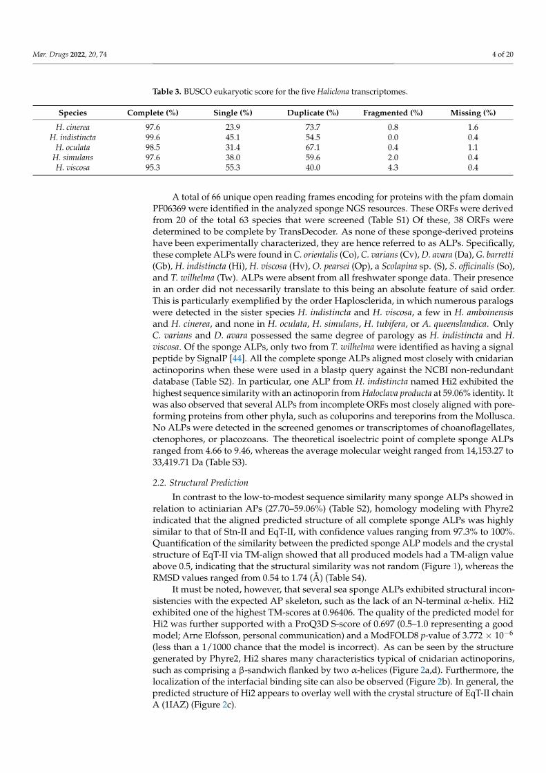
Table 3. BUSCO eukaryotic score for the five Haliclona transcriptomes.
Species Complete (%) Single (%) Duplicate (%) Fragmented (%) Missing (%)
H. cinerea 97.6 23.9 73.7 0.8 1.6H. indistincta 99.6 45.1 54.5 0.0 0.4
H. oculata 98.5 31.4 67.1 0.4 1.1H. simulans 97.6 38.0 59.6 2.0 0.4H. viscosa 95.3 55.3 40.0 4.3 0.4
A total of 66 unique open reading frames encoding for proteins with the pfam domainPF06369 were identified in the analyzed sponge NGS resources. These ORFs were derivedfrom 20 of the total 63 species that were screened (Table S1) Of these, 38 ORFs weredetermined to be complete by TransDecoder. As none of these sponge-derived proteinshave been experimentally characterized, they are hence referred to as ALPs. Specifically,these complete ALPs were found in C. orientalis (Co), C. varians (Cv), D. avara (Da), G. barretti(Gb), H. indistincta (Hi), H. viscosa (Hv), O. pearsei (Op), a Scolapina sp. (S), S. officinalis (So),and T. wilhelma (Tw). ALPs were absent from all freshwater sponge data. Their presencein an order did not necessarily translate to this being an absolute feature of said order.This is particularly exemplified by the order Haplosclerida, in which numerous paralogswere detected in the sister species H. indistincta and H. viscosa, a few in H. amboinensisand H. cinerea, and none in H. oculata, H. simulans, H. tubifera, or A. queenslandica. OnlyC. varians and D. avara possessed the same degree of parology as H. indistincta and H.viscosa. Of the sponge ALPs, only two from T. wilhelma were identified as having a signalpeptide by SignalP [44]. All the complete sponge ALPs aligned most closely with cnidarianactinoporins when these were used in a blastp query against the NCBI non-redundantdatabase (Table S2). In particular, one ALP from H. indistincta named Hi2 exhibited thehighest sequence similarity with an actinoporin from Haloclava producta at 59.06% identity. Itwas also observed that several ALPs from incomplete ORFs most closely aligned with pore-forming proteins from other phyla, such as coluporins and tereporins from the Mollusca.No ALPs were detected in the screened genomes or transcriptomes of choanoflagellates,ctenophores, or placozoans. The theoretical isoelectric point of complete sponge ALPsranged from 4.66 to 9.46, whereas the average molecular weight ranged from 14,153.27 to33,419.71 Da (Table S3).
2.2. Structural Prediction
In contrast to the low-to-modest sequence similarity many sponge ALPs showed inrelation to actiniarian APs (27.70–59.06%) (Table S2), homology modeling with Phyre2indicated that the aligned predicted structure of all complete sponge ALPs was highlysimilar to that of Stn-II and EqT-II, with confidence values ranging from 97.3% to 100%.Quantification of the similarity between the predicted sponge ALP models and the crystalstructure of EqT-II via TM-align showed that all produced models had a TM-align valueabove 0.5, indicating that the structural similarity was not random (Figure 1), whereas theRMSD values ranged from 0.54 to 1.74 (Å) (Table S4).
It must be noted, however, that several sea sponge ALPs exhibited structural incon-sistencies with the expected AP skeleton, such as the lack of an N-terminal α-helix. Hi2exhibited one of the highest TM-scores at 0.96406. The quality of the predicted model forHi2 was further supported with a ProQ3D S-score of 0.697 (0.5–1.0 representing a goodmodel; Arne Elofsson, personal communication) and a ModFOLD8 p-value of 3.772 × 10⁻⁶
(less than a 1/1000 chance that the model is incorrect). As can be seen by the structuregenerated by Phyre2, Hi2 shares many characteristics typical of cnidarian actinoporins,such as comprising a β-sandwich flanked by two α-helices (Figure 2a,d). Furthermore, thelocalization of the interfacial binding site can also be observed (Figure 2b). In general, thepredicted structure of Hi2 appears to overlay well with the crystal structure of EqT-II chainA (1IAZ) (Figure 2c).

Mar. Drugs 2022, 20, 74 5 of 20
Mar. Drugs 2022, 20, 74 4 of 20
A total of 66 unique open reading frames encoding for proteins with the pfam do-main PF06369 were identified in the analyzed sponge NGS resources. These ORFs were derived from 20 of the total 63 species that were screened (Table S1) Of these, 38 ORFs were determined to be complete by TransDecoder. As none of these sponge-derived pro-teins have been experimentally characterized, they are hence referred to as ALPs. Specif-ically, these complete ALPs were found in C. orientalis (Co), C. varians (Cv), D. avara (Da), G. barretti (Gb), H. indistincta (Hi), H. viscosa (Hv), O. pearsei (Op), a Scolapina sp. (S), S. officinalis (So), and T. wilhelma (Tw). ALPs were absent from all freshwater sponge data. Their presence in an order did not necessarily translate to this being an absolute feature of said order. This is particularly exemplified by the order Haplosclerida, in which nu-merous paralogs were detected in the sister species H. indistincta and H. viscosa, a few in H. amboinensis and H. cinerea, and none in H. oculata, H. simulans, H. tubifera, or A. queens-landica. Only C. varians and D. avara possessed the same degree of parology as H. indis-tincta and H. viscosa. Of the sponge ALPs, only two from T. wilhelma were identified as having a signal peptide by SignalP [44]. All the complete sponge ALPs aligned most closely with cnidarian actinoporins when these were used in a blastp query against the NCBI non-redundant database (Table S2). In particular, one ALP from H. indistincta named Hi2 exhibited the highest sequence similarity with an actinoporin from Haloclava producta at 59.06% identity. It was also observed that several ALPs from incomplete ORFs most closely aligned with pore-forming proteins from other phyla, such as coluporins and tereporins from the Mollusca. No ALPs were detected in the screened genomes or tran-scriptomes of choanoflagellates, ctenophores, or placozoans. The theoretical isoelectric point of complete sponge ALPs ranged from 4.66 to 9.46, whereas the average molecular weight ranged from 14,153.27 to 33,419.71 Da (Table S3).
2.2. Structural Prediction In contrast to the low-to-modest sequence similarity many sponge ALPs showed in
relation to actiniarian APs (27.70–59.06%) (Table S2), homology modeling with Phyre2 in-dicated that the aligned predicted structure of all complete sponge ALPs was highly sim-ilar to that of Stn-II and EqT-II, with confidence values ranging from 97.3% to 100%. Quan-tification of the similarity between the predicted sponge ALP models and the crystal struc-ture of EqT-II via TM-align showed that all produced models had a TM-align value above 0.5, indicating that the structural similarity was not random (Figure 1), whereas the RMSD values ranged from 0.54 to 1.74 (Å) (Table S4).
Figure 1. Comparison of complete sponge ALPs with EqT-II. Blue represents the TM-score of all predicted sponge ALP structures via Phyre2 with the crystal structure of EqT-II (1IAZ). Orange
Figure 1. Comparison of complete sponge ALPs with EqT-II. Blue represents the TM-score of allpredicted sponge ALP structures via Phyre2 with the crystal structure of EqT-II (1IAZ). Orangerepresents the sequence identity of the aligned region of the two structures. The orange bar rep-resenting percent identity is overlaid upon the blue bar representing TM-score. Abbreviations areas follows: Co, C. orientalis; Cv, C. varians; Da, Dysidea avara; Hi, H. indistincta; Hv, H. viscosa; Op,O. pearsei; S, Scopalina sp.; So, S. officinalis; Tw, Tethya wilhelma. The protein Sc1 from S. carteri isexcluded due to having unknown residues. Sequence identity is expressed on a scale of 0–1 ratherthan as a percentage.
2.3. Multiple Sequence Alignment and Residue Analysis
Due to its high sequence similarity with cnidarian actinoporins, Hi2 was chosenfor further in-depth analyses to determine whether it exhibited the same membrane-binding and pore-forming activities. A multiple sequence alignment of Hi2 with the finalmature peptides of the well-studied equinatoxin II (EqT-II; P61914), fragaceatoxin C (Fra-C; B9W5G6), and stichyolysin II (Stn-II; P07845) indicated a percent identity of 50.56%,50.00%, and 51.41%, respectively (Figure 3). Despite these modest values, the alignmentillustrated a high degree of conservation regarding residues and motifs critical for thefunctional activities of actinoporins [15]. For example, a majority of the residues associatedwith the interfacial binding site in Hi2 are consistent with those of EqT-II, Fra-C, andStn-II [6]. In addition, Hi2 possesses the conserved residue Tyr112, which is critical forSM recognition; however, a substitution of Leu for Trp at residue 111 is also observed.Furthermore, the presence of Ser53, Val86, Ser104, Pro106, Trp115, Tyre132, Tyr136, andTyr137 are consistent with the POC binding site found in cnidarian APs [4,6,15], and theconserved P-[WYF]-D binding motif found in this region of APs is also present in Hi2 atresidues 106–108 [22]. Oligomerization of actinoporin monomers upon the cell membrane isanother crucial step towards pore formation and is known to be influenced by an Arg–Gly–Asp motif. Hi2 shows inconsistency with this motif, as it instead possesses Lys142, Gly143,and Glu144. However, Hi2 possesses the residue Lys76, which is consistent with similarresidues associated with oligomerization in other APs [45]. The presence of Ile59 andTrp147 are also partially consistent with residues of Fra-C, associated with oligomerizationand protein–protein interaction between protomers; the observed substitution of Ile forVal at this site can be seen in Stn-II [13,46]. Unlike most cnidarian APs, Hi2 exhibits thepresence of cysteine at residue 141, but such a characteristic is not unheard of in theseproteins [21].

Mar. Drugs 2022, 20, 74 6 of 20
Mar. Drugs 2022, 20, 74 5 of 20
represents the sequence identity of the aligned region of the two structures. The orange bar repre-senting percent identity is overlaid upon the blue bar representing TM-score. Abbreviations are as follows: Co, C. orientalis; Cv, C. varians; Da, Dysidea avara; Hi, H. indistincta; Hv, H. viscosa; Op, O. pearsei; S, Scopalina sp.; So, S. officinalis; Tw, Tethya wilhelma. The protein Sc1 from S. carteri is ex-cluded due to having unknown residues. Sequence identity is expressed on a scale of 0–1 rather than as a percentage.
It must be noted, however, that several sea sponge ALPs exhibited structural incon-sistencies with the expected AP skeleton, such as the lack of an N-terminal α-helix. Hi2 exhibited one of the highest TM-scores at 0.96406. The quality of the predicted model for Hi2 was further supported with a ProQ3D S-score of 0.697 (0.5–1.0 representing a good model; Arne Elofsson, personal communication) and a ModFOLD8 p-value of 3.772E-6 (less than a 1/1000 chance that the model is incorrect). As can be seen by the structure generated by Phyre2, Hi2 shares many characteristics typical of cnidarian actinoporins, such as comprising a β-sandwich flanked by two α-helices (Figure 2a,d). Furthermore, the localization of the interfacial binding site can also be observed (Figure 2b). In general, the predicted structure of Hi2 appears to overlay well with the crystal structure of EqT-II chain A (1IAZ) (Figure 2c).
Figure 2. (a) Predicted structure of Hi2 by Phyre2. Red represents α-helices. Green represents β-sheets. (b) Significant functional residues of Hi2. Blue represents the N-terminal α-helix associated with pore formation. Red represents the residues of the interfacial binding site. Green represents the residues associated with oligomerization. (c) Structural alignment of Hi2 in blue upon EqT-II chain A (1IAZ) in orange. For all predicted structures blue and red highlights represent the N- and C-terminus, respectively. (d) Protein topology plot of Hi2 with the same color scheme as (a). The size of the topology plot was manually reduced to be more compact.
2.3. Multiple Sequence Alignment and Residue Analysis Due to its high sequence similarity with cnidarian actinoporins, Hi2 was chosen for
further in-depth analysis to determine whether it exhibited the same membrane-binding
Figure 2. (a) Predicted structure of Hi2 by Phyre2. Red represents α-helices. Green representsβ-sheets. (b) Significant functional residues of Hi2. Blue represents the N-terminal α-helix associatedwith pore formation. Red represents the residues of the interfacial binding site. Green representsthe residues associated with oligomerization. (c) Structural alignment of Hi2 in blue upon EqT-IIchain A (1IAZ) in orange. For all predicted structures blue and red highlights represent the N- andC-terminus, respectively. (d) Protein topology plot of Hi2 with the same color scheme as (a). The sizeof the topology plot was manually reduced to be more compact.
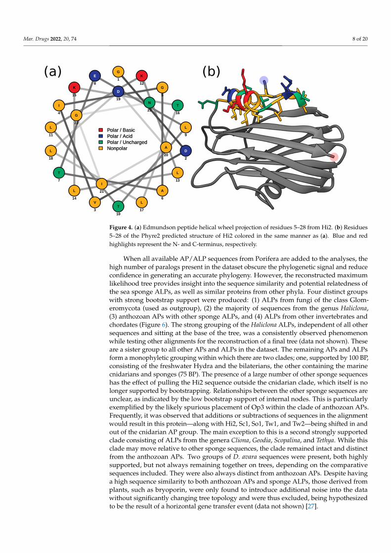
Membrane penetration and pore formation by oligomerized APs is achieved by theirrespective amphipathic N-terminal α-helices. In EqT-II, the N-terminal region undergoes aconformational change, producing an α-helix comprising residues 6–28 which is capable ofspanning a target membrane [10]. These residues correspond to a conserved N-terminalglycine and C-terminal asparagine of the α-helix, which are also present in Fra-C and Hi2.Within this region, Hi2 also showed consistency with several previously determined highlyconserved hydrophobic residues (Val7, Ile8, Leu13, Leu18, Leu22, and Ile25), as well asArg30, which is associated with the insertion of the α-helix into the target membrane [20].Using these sequence boundaries allowed for the construction of an Edmundson peptidehelical wheel of the predicted Hi2 N-terminal α-helix after a hypothetical conformationalchange (Figure 4a). Consistent with the amphipathic nature of the N-terminal α-helix ofEqT-II, Fra-C, and Stn-II, a side comprising a majority of polar amino acids opposite anothercomprising a majority of nonpolar amino acids can be seen in that of Hi2. Furthermore,the hydrophobic moment of Hi2, a measure of helix amphipathicity, was calculated to be
Mar. Drugs 2022, 20, 74 7 of 20
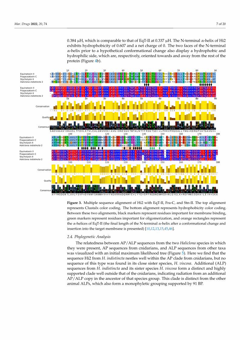
0.384 µH, which is comparable to that of EqT-II at 0.337 µH. The N-terminal α-helix of Hi2exhibits hydrophobicity of 0.607 and a net charge of 0. The two faces of the N-terminalα-helix prior to a hypothetical conformational change also display a hydrophobic andhydrophilic side, which are, respectively, oriented towards and away from the rest of theprotein (Figure 4b).
Mar. Drugs 2022, 20, 74 6 of 20
and pore-forming activities. A multiple sequence alignment of Hi2 with the final mature peptides of the well-studied equinatoxin II (EqT-II; P61914), fragaceatoxin C (Fra-C; B9W5G6), and stichyolysin II (Stn-II; P07845) indicated a percent identity of 50.56%, 50.00%, and 51.41%, respectively (Figure 3). Despite these modest values, the alignment illustrated a high degree of conservation regarding residues and motifs critical for the functional activities of actinoporins [15]. For example, a majority of the residues associated with the interfacial binding site in Hi2 are consistent with those of EqT-II, Fra-C, and Stn-II [6]. In addition, Hi2 possesses the conserved residue Tyr112, which is critical for sphin-gomyelin recognition; however, a substitution of Leu for Trp at residue 111 is also ob-served. Furthermore, the presence of Ser53, Val86, Ser104, Pro106, Trp115, Tyre132, Tyr136, and Tyr137 are consistent with the phosphocholine binding site found in cnidar-ian APs [4,6,15], and the conserved P-[WYF]-D binding motif found in this region of APs is also present in Hi2 at residues 106–108 [22]. Oligomerization of actinoporin monomers upon the cell membrane is another crucial step towards pore formation and is known to be influenced by an Arg–Gly–Asp motif. Hi2 shows inconsistency with this motif, as it instead possesses Lys142, Gly143, and Glu144. However, Hi2 possesses the residue Lys76, which is consistent with similar residues associated with oligomerization in other APs [45]. The presence of Ile59 and Trp147 are also partially consistent with residues of Fra-C, associated with oligomerization and protein–protein interaction between protomers; the observed substitution of Ile for Val at this site can be seen in Stn-II [13,46]. Unlike most cnidarian APs, Hi2 exhibits the presence of cysteine at residue 141, but such a character-istic is not unheard of in these proteins [21].
Figure 3. Multiple sequence alignment of Hi2 with EqT-II, Fra-C, and Stn-II. The top alignment rep-resents Clustalx color coding. The bottom alignment represents hydrophobicity color coding. Be-tween these two alignments, black markers represent residues important for membrane binding, green markers represent residues important for oligomerization, and orange rectangles represent the α-helices of EqT-II (the final length of the N-terminal α-helix after a conformational change and insertion into the target membrane is presented) [10,12,13,15,45,46].
Figure 3. Multiple sequence alignment of Hi2 with EqT-II, Fra-C, and Stn-II. The top alignmentrepresents Clustalx color coding. The bottom alignment represents hydrophobicity color coding.Between these two alignments, black markers represent residues important for membrane binding,green markers represent residues important for oligomerization, and orange rectangles representthe α-helices of EqT-II (the final length of the N-terminal α-helix after a conformational change andinsertion into the target membrane is presented) [10,12,13,15,45,46].
2.4. Phylogenetic Analysis
The relatedness between AP/ALP sequences from the two Haliclona species in whichthey were present, AP sequences from cnidarians, and ALP sequences from other taxawas visualized with an initial maximum likelihood tree (Figure 5). Here we find that thesequence Hi2 from H. indistincta nestles well within the AP clade from cnidarians, but nosequence of this type was found in its close sister species, H. viscosa. Additional (ALP)sequences from H. indistincta and its sister species H. viscosa form a distinct and highlysupported clade well outside that of the cnidarians, indicating radiation from an additionalAP/ALP copy in the ancestor of that species group. This clade is distinct from the otheranimal ALPs, which also form a monophyletic grouping supported by 91 BP.
Mar. Drugs 2022, 20, 74 8 of 20
Mar. Drugs 2022, 20, 74 7 of 20
Membrane penetration and pore formation by oligomerized APs is achieved by their respective amphipathic N-terminal α-helices. In EqT-II, the N-terminal region undergoes a conformational change, producing an α-helix comprising residues 6–28 which is capable of spanning a target membrane [10]. These residues correspond to a conserved N-terminal glycine and C-terminal asparagine of the α-helix, which are also present in Fra-C and Hi2. Within this region, Hi2 also showed consistency with several previously determined highly conserved hydrophobic residues (Val7, Ile8, Leu13, Leu18, Leu22, and Ile25), as well as Arg30, which is associated with the insertion of the α-helix into the target mem-brane [20]. Using these sequence boundaries allowed for the construction of an Edmund-son peptide helical wheel of the predicted Hi2 N-terminal α-helix after a hypothetical con-formational change (Figure 4a). Consistent with the amphipathic nature of the N-terminal α-helix of EqT-II, Fra-C, and Stn-II, a side comprising a majority of polar amino acids op-posite another comprising a majority of nonpolar amino acids can be seen in that of Hi2. Furthermore, the hydrophobic moment of Hi2, a measure of helix amphipathicity, was calculated to be 0.384 µH, which is comparable to that of EqT-II at 0.337 µH. The N-termi-nal α-helix of Hi2 exhibits hydrophobicity of 0.607 and a net charge of 0. The two faces of the N-terminal α-helix prior to a hypothetical conformational change also display a hy-drophobic and hydrophilic side, which are, respectively, oriented towards and away from the rest of the protein (Figure 4b).
Figure 4. (a) Edmundson peptide helical wheel projection of residues 5–28 from Hi2. (b) Residues 5–28 of the Phyre2 predicted structure of Hi2 colored in the same manner as (a). Blue and red high-lights represent the N- and C-terminus, respectively.
2.4. Phylogenetic Analysis The relatedness between AP/ALP sequences from the two Haliclona species in which
they were present, AP sequences from cnidarians, and ALP sequences from other taxa was visualized with an initial maximum likelihood tree (Figure 5). Here we find that the sequence Hi2 from H. indistincta nestles well within the AP clade from cnidarians, but no sequence of this type was found in its close sister species, H. viscosa. Additional (ALP) sequences from H. indistincta and its sister species H. viscosa form a distinct and highly supported clade well outside that of the cnidarians, indicating radiation from an addi-tional AP/ALP copy in the ancestor of that species group. This clade is distinct from the other animal ALPs, which also form a monophyletic grouping supported by 91 BP.
Figure 4. (a) Edmundson peptide helical wheel projection of residues 5–28 from Hi2. (b) Residues5–28 of the Phyre2 predicted structure of Hi2 colored in the same manner as (a). Blue and redhighlights represent the N- and C-terminus, respectively.
When all available AP/ALP sequences from Porifera are added to the analyses, thehigh number of paralogs present in the dataset obscure the phylogenetic signal and reduceconfidence in generating an accurate phylogeny. However, the reconstructed maximumlikelihood tree provides insight into the sequence similarity and potential relatedness ofthe sea sponge ALPs, as well as similar proteins from other phyla. Four distinct groupswith strong bootstrap support were produced: (1) ALPs from fungi of the class Glom-eromycota (used as outgroup), (2) the majority of sequences from the genus Haliclona,(3) anthozoan APs with other sponge ALPs, and (4) ALPs from other invertebrates andchordates (Figure 6). The strong grouping of the Haliclona ALPs, independent of all othersequences and sitting at the base of the tree, was a consistently observed phenomenonwhile testing other alignments for the reconstruction of a final tree (data not shown). Theseare a sister group to all other APs and ALPs in the dataset. The remaining APs and ALPsform a monophyletic grouping within which there are two clades; one, supported by 100 BP,consisting of the freshwater Hydra and the bilaterians, the other containing the marinecnidarians and sponges (75 BP). The presence of a large number of other sponge sequenceshas the effect of pulling the Hi2 sequence outside the cnidarian clade, which itself is nolonger supported by bootstrapping. Relationships between the other sponge sequences areunclear, as indicated by the low bootstrap support of internal nodes. This is particularlyexemplified by the likely spurious placement of Op3 within the clade of anthozoan APs.Frequently, it was observed that additions or subtractions of sequences in the alignmentwould result in this protein—along with Hi2, Sc1, So1, Tw1, and Tw2—being shifted in andout of the cnidarian AP group. The main exception to this is a second strongly supportedclade consisting of ALPs from the genera Cliona, Geodia, Scopalina, and Tethya. While thisclade may move relative to other sponge sequences, the clade remained intact and distinctfrom the anthozoan APs. Two groups of D. avara sequences were present, both highlysupported, but not always remaining together on trees, depending on the comparativesequences included. They were also always distinct from anthozoan APs. Despite havinga high sequence similarity to both anthozoan APs and sponge ALPs, those derived fromplants, such as bryoporin, were only found to introduce additional noise into the datawithout significantly changing tree topology and were thus excluded, being hypothesizedto be the result of a horizontal gene transfer event (data not shown) [27].

Mar. Drugs 2022, 20, 74 9 of 20Mar. Drugs 2022, 20, 74 8 of 20
Figure 5. Phylogenetic tree of ALPs from the genus Haliclona and similar proteins. Numbers on nodes represent maximum likelihood bootstrap values above 75%. The color scheme is as follows: green represents ALPs from fungi of the class Glomeromycota; blue represents ALPs from Haliclona sp.; red represents APs from cnidarians of the class Anthozoa; reddish-purple represents ALPs from cnidarians of the class Hydrozoa; purple represents ALPs from the phylum Mollusca; orange rep-resents ALPs from fish of the class Actinopterygii; black represents ALPs from miscellaneous phyla. Abbreviations are as follows: EqT, equinatoxin; Fra, fragaceatoxin; St, sticholysin; BdP, bandaporin; GgT, gigantoxin; HALT, hydra actinoporin-like toxin; EcT, echotoxin; Clp, coluporin. The glomero-mycete vector image is a reproduced copy of the high-resolution image 4341-2_P1 of Rhizophagus irregularis (Błaszk., Wubet, Renker, and Buscot) C. Walker and A. Schüßler derived from AAFC/CCAMF (https://agriculture.canada.ca/en/agricultural-science-and-innovation/agriculture-and-agri-food-research-centres-and-collections/glomeromycota-vitro-collection-ginco/catalogue-arbuscular-mycorrhizal-fungi-strains-available-glomeromycetes-vitro-collection) (accessed on 25 December 2021). The sea sponge, sea anemone, seashell, and carp vector images were derived from those of Pearson Scott Foresman under a CC0 1.0 license (https://commons.wiki-media.org/wiki/File:Sponge_(PSF).png; https://commons.wikimedia.org/wiki/File:Anem-one_2_(PSF).png; https://commons.wikimedia.org/wiki/File:Seashell_3_(PSF).png; https://com-mons.wikimedia.org/wiki/File:Carp_(PSF).jpg) (accessed on 26 November 2021). The Hydra vulgaris vector image was derived from the Freshwater and Marine Image Bank at the University of Wash-ington under a CC0 1.0 license (https://commons.wikimedia.org/wiki/File:FMIB_50097_Hydra_vul-garis.jpeg) (accessed on 26 November 2021).
Figure 5. Phylogenetic tree of ALPs from the genus Haliclona and similar proteins. Numbers onnodes represent maximum likelihood bootstrap values above 75%. The color scheme is as follows:green represents ALPs from fungi of the class Glomeromycota; blue represents ALPs from Haliclonasp.; red represents APs from cnidarians of the class Anthozoa; reddish-purple represents ALPsfrom cnidarians of the class Hydrozoa; purple represents ALPs from the phylum Mollusca; orangerepresents ALPs from fish of the class Actinopterygii; black represents ALPs from miscellaneousphyla. Abbreviations are as follows: EqT, equinatoxin; Fra, fragaceatoxin; St, sticholysin; BdP,bandaporin; GgT, gigantoxin; HALT, hydra actinoporin-like toxin; EcT, echotoxin; Clp, coluporin.The glomeromycete vector image is a reproduced copy of the high-resolution image 4341-2_P1 ofRhizophagus irregularis (Błaszk., Wubet, Renker, and Buscot) C. Walker and A. Schüßler derivedfrom AAFC/CCAMF (https://agriculture.canada.ca/en/agricultural-science-and-innovation/agriculture-and-agri-food-research-centres-and-collections/glomeromycota-vitro-collection-ginco/catalogue-arbuscular-mycorrhizal-fungi-strains-available-glomeromycetes-vitro-collection)(accessed on 25 November 2021). The sea sponge, sea anemone, seashell, and carp vec-tor images were derived from those of Pearson Scott Foresman under a CC0 1.0 license(https://commons.wikimedia.org/wiki/File:Sponge_(PSF).png; https://commons.wikimedia.org/wiki/File:Anemone_2_(PSF).png; https://commons.wikimedia.org/wiki/File:Seashell_3_(PSF).png;https://commons.wikimedia.org/wiki/File:Carp_(PSF).jpg) (accessed on 26 November 2021).The Hydra vulgaris vector image was derived from the Freshwater and Marine Image Bank at theUniversity of Washington under a CC0 1.0 license (https://commons.wikimedia.org/wiki/File:FMIB_50097_Hydra_vulgaris.jpeg) (accessed on 26 November 2021).

Mar. Drugs 2022, 20, 74 10 of 20
Mar. Drugs 2022, 20, 74 10 of 20
Figure 6. Phylogenetic tree of ALPs from sea sponges and similar proteins. Numbers on nodes rep-resent maximum likelihood bootstrap values above 75%. Blue represents sea sponge AP/ALPs. All other color schemes as well as image acknowledgements are identical to those of Figure 5.
Figure 6. Phylogenetic tree of ALPs from sea sponges and similar proteins. Numbers on nodesrepresent maximum likelihood bootstrap values above 75%. Blue represents sea sponge AP/ALPs.All other color schemes as well as image acknowledgements are identical to those of Figure 5.

Mar. Drugs 2022, 20, 74 11 of 20
3. Discussion
Cytolytic pore-forming toxins are widely distributed throughout prokaryotic andeukaryotic life and often function as immunological defenses in the latter [47,48]. Theproposed AF superfamily includes α-pore-forming toxins derived from diverse eukaryoticlineages with similar predicted protein structure despite low sequence similarity [22]. Mem-bers of this family include anthozoan APs, plant APs, hydrozoan ALPs, ALPs from otheranimals, and fungal fruit-body lectins. Herein, new additions from the phylum Poriferaare proposed to belong to the AF superfamily, as members of the classes Demospongiae,Homoscleromorpha, and Calcarea have been shown to possess genes encoding for ALPs.Many functionally characterized AF proteins appear to serve a role in envenomation as theyhave been localized in the nematocysts of cnidarians and the salivary glands of predatorymolluscs [25,26,29,49]. However, AF proteins appear to also have functions other thanenvenomation, as indicated by their presence in the mesenteric filaments of cnidarians,as well as organisms which do not perform this process, such as bivalves [28,49]. Withthis in consideration, it is not entirely unusual to observe the presence of these proteins innon-venomous animals, such as sponges, in which they may serve a different ecologicalfunction via a similar molecular mechanism.
When considering the taxonomic distribution of ALPs in sponges, no clear pattern canbe discerned. The majority of the identified ALPs were derived from the Demospongiaeand more specifically from the orders Axinellida, Bubarida, Clionaida, Dendroceratida,Dictyoceratida, Haplosclerida, Poecilosclerida, Suberitida, Tethyida, and Tetractinellida,but not Chondrillida, Spongillida, or Verongiida. However, further analysis at the genuslevel indicated that these proteins appear to have been lost in species closely related to thosewhich possess ALPs. This is particularly exemplified by the genera Haliclona and Geodia, inwhich the transcriptomes of numerous species have been reported [43,50]. Furthermore,ALPs appear to be distributed throughout the phylum Porifera, as several were identifiedin the classes Homoscleromorpha and Calcarea, but not in Hexactinellida. However,two caveats should be considered regarding these observations. The first is that the highprevalence of these proteins in the Demospongiae is most likely due to sampling bias, astranscriptomes of this class have been disproportionately sequenced compared to the otherthree. Second, as most of the data analyzed in this study are derived from transcriptomes,a lack of gene expression or sequencing depth cannot be ruled out as an explanation forthese genes not having been identified in a species. That said, the absence of ALPs in theAmphimedon queenslandica genome does appear to indicate that the loss of ALP genes hasoccurred in the order Haplosclerida, which may be an explanation for their absence in otherspecies [51].
The predicted structure of most identified ALPs from sponges exhibited a high degreeof aligned structural similarity to anthozoan APs, despite a low to moderate sequencesimilarity. Such an observation is common in studies of AF proteins from other organ-isms [22,25]. That said, the observed high aligned structural similarity does not necessarilyequate to these sea sponge ALPs having the same membrane-binding and pore-formingcapabilities. This is particularly exemplified by the observation that the N-terminal regionof sea sponge ALPs appears to vary greatly. For example, this region is fully presentin Hi2, truncated in Hi3, and completely absent in Hi4. Such a quality is not unique tosea sponge ALPs, as can be seen by analyzing those of hydrozoans [29]. Similarly, anincomplete N-terminal α-helix was also observed on the ALP Dr1 from D. rerio and washypothesized to influence the lack of pore-forming capabilities and specificity towards SMexhibited by the protein [22]. With this in mind, many of the identified ALPs from seasponges may serve functions other than those associated with well-characterized APs, suchas EqT-II. In contrast, the higher similarity of Hi2 to anthozoan APs at both a sequenceand structural level could indicate that it is capable of SM recognition and pore formation,which prompted a further analysis on this concept.
A multiple sequence alignment of Hi2 with the model APs EqT-II, Fra-C, and Stn-IIindicated that this predicted protein shares numerous conserved residues associated with

Mar. Drugs 2022, 20, 74 12 of 20
the fundamental processes of lipid recognition and membrane binding, insertion of the N-terminal α-helix into the target membrane, and oligomerization allowing for pore formation.The presence of a patch of aromatic residues (Tyr114, Trp117, Tyr134, Tyr138, and Tyr139) inHi2 is consistent with the IBS observed in the model APs EqT-II, Fra-C, and Stn-II. The majorobserved deviation is that a substitution of leucine for tryptophan occurs at residue 113of Hi2. The importance of the equivalent residue in EqT-II, Trp112, in membrane bindingand SM recognition has been exemplified by studies in which this residue was mutated tophenylalanine or subject to 19F NMR studies [52,53]. This substitution also appears to beprevalent in nature, as it exists in numerous anemone APs, as well as the ALP Dr1 fromD. rerio [20,22]. Furthermore, a mutant of EqT-II containing this substitution exhibitedsimilar SM specificity to the wild-type protein [6]. For these reasons, this substitutionin Hi2 is not expected to significantly inhibit any possible membrane-binding and SM-recognition capabilities of the protein. While at a lower level of conservation the N-terminalregion of Hi2 also appears to form an α-helix of similar length to EqT-II, Fra-C, and Stn-II. The notion that this α-helix is capable of inserting itself into a target membrane issupported by the amphipathic nature of the predicted helical wheel and its hydrophobicmoment comparable to previously analyzed AP N-terminal α-helices [20]. Finally, residuesassociated with oligomerization were somewhat consistent between Hi2 and the modelactinoporins. While Hi2 showed conservation at residues Lys76, Ile59, and Trp147, whoseequivalents in anthozoan APs are associated with oligomerization, its RGD motif wasanother site of substitution [12,13,45,46]. Hi2 instead shows a substitution of Lys for Argand Glu for Asp. That said, Lys and Glu are of a similar charge and hydrophobicity to Argand Asp, respectively, and may allow for the retention of the oligomerization function [12].Furthermore, this Lys substitution has been observed in natural actinoporins [20]. Hi2and many other sponge ALPs also exhibited an acidic pI (Supplementary Table), which isuncharacteristic of the typically basic anthozoan APs [15,54]. This observation is not entirelyunusual, however, as there is a precedent for acidic APs derived from anthozoans [20,54,55].
The identification of ALPs from glomeromycete fungi with high sequence similar-ity to known APs supports the previous notion of a pre-metazoan origin of these pro-teins [22]. The presence of these genes in fungi, sponges, and cnidarians, and their absencein choanoflagellates, ctenophores, and placozoans, could possibly be explained by a seriesof gene losses occurring throughout their history. However, additional assemblies fromthese organisms should be assessed prior to making this conclusion. Furthermore, theobservation that many species of sea sponge were the source of numerous ALP isoformsmay be an indication that duplication of this gene is a common event in this phylum,similar to the situation in cnidarians and molluscs [25,54]. This observed diversificationpaired with the fact that many sponge ALPs were derived from transcriptomes indicatesthat these are not simply genomic relics but do play some sort of functional role in sponges.In the phylogenetic analysis, the sea sponge ALPs were not found to cluster together in aclear monophyletic group, which appears to further exemplify the notion of a high degreeof divergence of these proteins in the porifera. However, based on the strong signal thatnumerous sponge ALPs share with anthozoan APs, to the point of being grouped together,and considering the higher sequence similarity many sponge ALPs have with cnidarianAPs, several of these proteins from sponges may instead be classifiable as APs.
4. Materials and Methods4.1. Sample Collection
Haliclona indistincta (MIIG1388; Appendix A Figure A1a) was collected at Corranrooon 17 May 2019 and H. viscosa (MIIG1389 and MIIG1390; Figure A1b) was collected atBridges of Ross on 1 August 2019. The following sample processing protocol was applied toboth species. Visible epibionts were removed. The sponges were rinsed in sterile artificialseawater. The sponges were then dissected into ~1 cm3 pieces and flash-frozen with liquidnitrogen. Samples were stored at −70 ◦C until further use. Voucher specimens were storedin ethanol.

Mar. Drugs 2022, 20, 74 13 of 20
4.2. RNA Extraction
A ~1 cm3 piece of flash-frozen sponge tissue was submerged in 500 µL of Trizol ina 2 mL microcentrifuge tube. The tissue was semi-homogenized by hand with a plasticpestle. An additional 500 µL of Trizol was then added to the sample. The sample wasmixed by gently inverting five times and allowed to incubate at room temperature for5 min. The sample was then inverted and vortexed with a VWR Analogue mini vortexmixer at maximum speed for 2 min. A volume of 100 µL BCP was added to the sample.The sample was mixed by hand for 20 s and then vortexed for 10 s with a VWR Analoguemini vortex mixer at maximum speed. The sample was incubated at room temperaturefor 5–10 min. The sample was then centrifuged at 16,000 g for 15 min at 6 ◦C. The clear,aqueous layer at the top was transferred to a fresh microcentrifuge tube. RNA was purifiedby first adding 500 µL of 100% isopropanol to the aqueous phase sample. The sample wasthen inverted and vortexed with a VWR Analogue mini vortex mixer at maximum speedfor 2 min. The sample was left to incubate for 10 min at room temperature. The sample wasthen centrifuged at 16,000 g for 15 min at 4 ◦C. The supernatant was discarded and 1 mL of75% EtOH was added to the RNA pellet. The pellet was disrupted by vortexing. The RNAsample was then centrifuged for 5 min at 4 ◦C at 7500 g. The EtOH was carefully removedwithout disturbing the pellet. The washing with 75% EtOH was repeated once. The RNAsample was allowed to air dry until the edges of the pellet were visible. Finally, the pelletwas resuspended in 100 µL molecular-grade water. The RNA sample was kept frozen at−70 ◦C until further use. A subsample of each RNA extraction was used for quality andquantity assessment on a 2100 Bioanalyzer RNA Eukaryotic Chip.
4.3. Transcriptome Sequencing
Samples were sent to Macrogen, Inc. for the preparation of Illumina TruSeq StrandedmRNA libraries from poly-A selection, with insert sizes of 150 bp. The libraries weresequenced on a Novaseq, with a targeted 40 million reads per sample.
4.4. Transcriptome Assembly
Previously sequenced raw cDNA Illumina reads of H. cinerea (culture held at CarnaMarine Research Station), H. oculata (MIIG1250 and MIIG1251), H. indistincta (MIIG1093,MIIG1094, MIIG1095), and H. simulans (MIIG1248 and MIIG1249) were acquired from Prof.Grace P. McCormack and Dr. Jose Maria Aguilar-Camacho for use in this study (personalcommunication) [43].
Raw cDNA reads of H. cinerea, H. indistincta, H. oculata, H. simulans, and H. viscosawere processed with fastp version 0.2 using default settings to remove adapters and low-quality regions [56]. The processed reads were then assembled with Trinity version 2.8.5using default settings [57]. Reads were pooled so that one transcriptome per species wasassembled. Isoforms and low-expressed transcripts were retained in the final assembly. Thelongest translated open reading frames per transcript were extracted using TransDecoderversion 5.5.0 [58]. Homology searches using these open reading frames as a query againstthe SwissProt database (accessed on 14 January 2020) with BLASTp version 2.9.0 [59,60], aswell as the Pfam database (accessed on 14 January 2020) with HMMER version 3.2.1 [61,62],were performed. Significant homologous alignments were used to guide TransDecoderin identifying additional open reading frames. The completeness of the transcriptomeswas then assessed using BUSCO version 5.1.2; specifically, the assemblies were queriedagainst the latest version of the eukaryota_odb10 dataset (downloaded 13 April 2021) [63].Transcriptome assembly, open reading frame extraction, BUSCO analysis, and the gen-eration of the maximum likelihood trees were performed with an account at the LeibnizSupercomputing Centre.
4.5. Identification of Novel Actinoporin-like Proteins from Sea Sponges
While analyzing the output of the homology search against the Pfam database, itwas noticed that numerous translated protein sequences possessed the Pfam domain
Mar. Drugs 2022, 20, 74 14 of 20
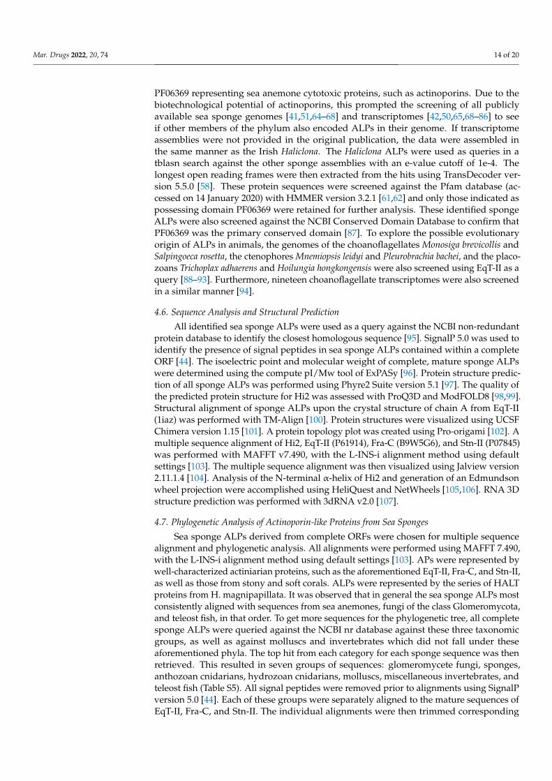
PF06369 representing sea anemone cytotoxic proteins, such as actinoporins. Due to thebiotechnological potential of actinoporins, this prompted the screening of all publiclyavailable sea sponge genomes [41,51,64–68] and transcriptomes [42,50,65,68–86] to seeif other members of the phylum also encoded ALPs in their genome. If transcriptomeassemblies were not provided in the original publication, the data were assembled inthe same manner as the Irish Haliclona. The Haliclona ALPs were used as queries in atblasn search against the other sponge assemblies with an e-value cutoff of 1e-4. Thelongest open reading frames were then extracted from the hits using TransDecoder ver-sion 5.5.0 [58]. These protein sequences were screened against the Pfam database (ac-cessed on 14 January 2020) with HMMER version 3.2.1 [61,62] and only those indicated aspossessing domain PF06369 were retained for further analysis. These identified spongeALPs were also screened against the NCBI Conserved Domain Database to confirm thatPF06369 was the primary conserved domain [87]. To explore the possible evolutionaryorigin of ALPs in animals, the genomes of the choanoflagellates Monosiga brevicollis andSalpingoeca rosetta, the ctenophores Mnemiopsis leidyi and Pleurobrachia bachei, and the placo-zoans Trichoplax adhaerens and Hoilungia hongkongensis were also screened using EqT-II as aquery [88–93]. Furthermore, nineteen choanoflagellate transcriptomes were also screenedin a similar manner [94].
4.6. Sequence Analysis and Structural Prediction
All identified sea sponge ALPs were used as a query against the NCBI non-redundantprotein database to identify the closest homologous sequence [95]. SignalP 5.0 was used toidentify the presence of signal peptides in sea sponge ALPs contained within a completeORF [44]. The isoelectric point and molecular weight of complete, mature sponge ALPswere determined using the compute pI/Mw tool of ExPASy [96]. Protein structure predic-tion of all sponge ALPs was performed using Phyre2 Suite version 5.1 [97]. The quality ofthe predicted protein structure for Hi2 was assessed with ProQ3D and ModFOLD8 [98,99].Structural alignment of sponge ALPs upon the crystal structure of chain A from EqT-II(1iaz) was performed with TM-Align [100]. Protein structures were visualized using UCSFChimera version 1.15 [101]. A protein topology plot was created using Pro-origami [102]. Amultiple sequence alignment of Hi2, EqT-II (P61914), Fra-C (B9W5G6), and Stn-II (P07845)was performed with MAFFT v7.490, with the L-INS-i alignment method using defaultsettings [103]. The multiple sequence alignment was then visualized using Jalview version2.11.1.4 [104]. Analysis of the N-terminal α-helix of Hi2 and generation of an Edmundsonwheel projection were accomplished using HeliQuest and NetWheels [105,106]. RNA 3Dstructure prediction was performed with 3dRNA v2.0 [107].
4.7. Phylogenetic Analysis of Actinoporin-like Proteins from Sea Sponges
Sea sponge ALPs derived from complete ORFs were chosen for multiple sequencealignment and phylogenetic analysis. All alignments were performed using MAFFT 7.490,with the L-INS-i alignment method using default settings [103]. APs were represented bywell-characterized actiniarian proteins, such as the aforementioned EqT-II, Fra-C, and Stn-II,as well as those from stony and soft corals. ALPs were represented by the series of HALTproteins from H. magnipapillata. It was observed that in general the sea sponge ALPs mostconsistently aligned with sequences from sea anemones, fungi of the class Glomeromycota,and teleost fish, in that order. To get more sequences for the phylogenetic tree, all completesponge ALPs were queried against the NCBI nr database against these three taxonomicgroups, as well as against molluscs and invertebrates which did not fall under theseaforementioned phyla. The top hit from each category for each sponge sequence was thenretrieved. This resulted in seven groups of sequences: glomeromycete fungi, sponges,anthozoan cnidarians, hydrozoan cnidarians, molluscs, miscellaneous invertebrates, andteleost fish (Table S5). All signal peptides were removed prior to alignments using SignalPversion 5.0 [44]. Each of these groups were separately aligned to the mature sequences ofEqT-II, Fra-C, and Stn-II. The individual alignments were then trimmed corresponding

Mar. Drugs 2022, 20, 74 15 of 20
to the boundaries of EqT-II, Fra-C, and Stn-II using using Jalview version 2.11.1.4 [104].Several sponge ALPs, while complete, where excluded due to being excessively truncatedcompared to EqT-II or introducing significant gaps. All trimmed sequences were thenpooled together and once more aligned. Maximum likelihood trees were constructed in IQ-TREE version 2.1.4 with 1000 bootstrap pseudoreplicates with the intention of visualizingthe degree of similarity the sponge ALPs had to APs and ALPs from other phyla [108].The resulting phylogenetic trees were modified using the Interactive Tree of Life (iTOL)v6 [109].
4.8. Generation of Figures
Several figures were further modified using the GNU Image Manipulation Program2.10.28 [110] and Inkscape 0.92 [111].
5. Conclusions
Sea sponges, like some other invertebrates, are a source of ALPs. These proteinsexhibit a high degree of predicted structural similarity as well as a phylogenetic signalto APs from cnidarians. One AP, Hi2, also possesses a majority of conserved residuesassociated with essential functions, including the recognition of SM, binding to membranes,oligomerization, and the formation of pores. While their ecological role in sponges remainsto be determined, the aforementioned qualities encourage the exploration of these proteinsfor the biotechnological applications which have been proposed for anthozoan APs.
Supplementary Materials: The following are available online at https://www.mdpi.com/article/10.3390/md20010074/s1, Table S1. Presence; Table S2. Blastp nr; Table S3. pI-Mw; Table S4. TM-align; TableS5. Trees.
Author Contributions: K.S. conceived the experiments, aided in the collection of samples, carriedout all other methodological tasks, prepared figures, and wrote the paper. G.P.M. supervised theresearch and assisted in interpreting the data and in writing the paper. All authors have read andagreed to the published version of the manuscript.
Funding: This project has received funding from the European Union’s Horizon 2020 researchand innovation programme under the Marie Skłodowska-Curie grant agreement No. 764840. TheDepartment of Further and Higher Education, Research, Innovation and Science, through the HigherEducation Authority (HEA), provided funding to higher education institutions to cover costedextensions for research activities that are at risk because of interruptions caused by the COVID-19 pandemic.
Institutional Review Board Statement: Not applicable.
Informed Consent Statement: Not applicable.
Data Availability Statement: The raw RNA-seq reads are available at the NCBI BioProject PR-JNA795170. Transcriptome assemblies, sea sponge ALP sequences, predicted protein structures,multiple sequence alignments, and maximum likelihood trees are available at Mendeley (https://data.mendeley.com/datasets/w9t6zsjjb7/1, accessed on 14 December 2021).
Acknowledgments: The authors thank the technicians of the NUIG Zoology Department for theirhelp in collecting H. indistincta. Olivier P. Thomas and Daniel Rodrigues are thanked for the collectionof H. viscosa. Jose Maria Aguillar-Camacho provided raw data of H. cinerea. Members of the MarieSkłodowska-Curie Actions innovative training network IGNITE—Comparative Genomics of Non-Model Invertebrates are thanked for their advice on methodology. The Irish marine biorepository isthanked for providing a high-quality image of Haliclona viscosa for the graphical abstract.
Conflicts of Interest: The authors declare no conflict of interest.

Mar. Drugs 2022, 20, 74 16 of 20
Appendix A
Mar. Drugs 2022, 20, 74 16 of 20
Conflicts of Interest: The authors declare no conflict of interest.
Appendix A
Figure A1. (a) Haliclona indistincta (MIIG1388). (b) Haliclona viscosa (MIIG1389).
References 1. Kem, W.R. Sea Anemone Toxins: Structure and Action. Biol. Nematocysts 1988, 375–405. 2. Anderluh, G.; Maček, P. Cytolytic Peptide and Protein Toxins from Sea Anemones (Anthozoa: Actiniaria). Toxicon 2002, 40, 111–
124. https://doi.org/10.1016/S0041-0101(01)00191-X. 3. Athanasiadis, A.; Anderluh, G.; Maček, P.; Turk, D. Crystal Structure of the Soluble Form of Equinatoxin II, a Pore-Forming
Toxin from the Sea Anemone Actinia Equina. Structure 2001, 9, 341–346. https://doi.org/10.1016/S0969-2126(01)00592-5. 4. Mancheño, J.M.; Martı n-Benito, J.; Martı nez-Ripoll, M.; Gavilanes, J.G.; Hermoso, J.A. Crystal and Electron Microscopy Struc-
tures of Sticholysin II Actinoporin Reveal Insights into the Mechanism of Membrane Pore Formation. Structure 2003, 11, 1319–1328. https://doi.org/10.1016/j.str.2003.09.019.
5. Mechaly, A.E.; Bellomio, A.; Gil-Cartón, D.; Morante, K.; Valle, M.; González-Mañas, J.M.; Guérin, D.M.A. Structural Insights into the Oligomerization and Architecture of Eukaryotic Membrane Pore-Forming Toxins. Structure 2011, 19, 181–191. https://doi.org/10.1016/j.str.2010.11.013.
6. Bakrač, B.; Gutiérrez-Aguirre, I.; Podlesek, Z.; Sonnen, A.F.-P.; Gilbert, R.J.C.; Maček, P.; Lakey, J.H.; Anderluh, G. Molecular Determinants of Sphingomyelin Specificity of a Eukaryotic Pore-Forming Toxin*. J. Biol. Chem. 2008, 283, 18665–18677. https://doi.org/10.1074/jbc.M708747200.
7. Bakrač, B.; Kladnik, A.; Maček, P.; McHaffie, G.; Werner, A.; Lakey, J.H.; Anderluh, G. A Toxin-Based Probe Reveals Cytoplas-mic Exposure of Golgi Sphingomyelin*. J. Biol. Chem. 2010, 285, 22186–22195. https://doi.org/10.1074/jbc.M110.105122.
8. Malovrh, P.; Viero, G.; Serra, M.D.; Podlesek, Z.; Lakey, J.H.; Maček, P.; Menestrina, G.; Anderluh, G. A Novel Mechanism of Pore Formation: Membrane Penetration by the N-Terminal Amphipathic Region of Equinatoxin. J. Biol. Chem. 2003, 278, 22678–22685. https://doi.org/10.1074/jbc.M300622200.
9. Rojko, N.; Kristan, K.Č.; Viero, G.; Žerovnik, E.; Maček, P.; Serra, M.D.; Anderluh, G. Membrane Damage by an α-Helical Pore-Forming Protein, Equinatoxin II, Proceeds through a Succession of Ordered Step*. J. Biol. Chem. 2013, 288, 23704–23715. https://doi.org/10.1074/jbc.M113.481572.
10. Drechsler, A.; Potrich, C.; Sabo, J.K.; Frisanco, M.; Guella, G.; Dalla Serra, M.; Anderluh, G.; Separovic, F.; Norton, R.S. Structure and Activity of the N-Terminal Region of the Eukaryotic Cytolysin Equinatoxin II. Biochemistry 2006, 45, 1818–1828. https://doi.org/10.1021/bi052166o.
11. Lam, Y.H.; Hung, A.; Norton, R.S.; Separovic, F.; Watts, A. Solid-State NMR and Simulation Studies of Equinatoxin II N-Termi-nus Interaction with Lipid Bilayers. Proteins Struct. Funct. Bioinform. 2010, 78, 858–872. https://doi.org/10.1002/prot.22612.
12. García-Linares, S.; Richmond, R.; García-Mayoral, M.F.; Bustamante, N.; Bruix, M.; Gavilanes, J.G.; Martínez-del-Pozo, Á. The Sea Anemone Actinoporin (Arg-Gly-Asp) Conserved Motif Is Involved in Maintaining the Competent Oligomerization State of These Pore-Forming Toxins. FEBS J. 2014, 281, 1465–1478. https://doi.org/10.1111/febs.12717.
13. Tanaka, K.; Caaveiro, J.M.M.; Morante, K.; González-Mañas, J.M.; Tsumoto, K. Structural Basis for Self-Assembly of a Cytolytic Pore Lined by Protein and Lipid. Nat. Commun. 2015, 6, 6337. https://doi.org/10.1038/ncomms7337.
14. Črnigoj Kristan, K.; Viero, G.; Dalla Serra, M.; Maček, P.; Anderluh, G. Molecular Mechanism of Pore Formation by Actino-porins. Toxicon 2009, 54, 1125–1134. https://doi.org/10.1016/j.toxicon.2009.02.026.
15. Rojko, N.; Dalla Serra, M.; Maček, P.; Anderluh, G. Pore Formation by Actinoporins, Cytolysins from Sea Anemones. Biochim. Et Biophys. Acta (BBA)-Biomembr. 2016, 1858, 446–456. https://doi.org/10.1016/j.bbamem.2015.09.007.
16. Mutter, N.L.; Soskine, M.; Huang, G.; Albuquerque, I.S.; Bernardes, G.J.L.; Maglia, G. Modular Pore-Forming Immunotoxins with Caged Cytotoxicity Tailored by Directed Evolution. ACS Chem. Biol. 2018, 13, 3153–3160. https://doi.org/10.1021/acschem-bio.8b00720.
17. Wloka, C.; Mutter, N.L.; Soskine, M.; Maglia, G. Alpha-Helical Fragaceatoxin C Nanopore Engineered for Double-Stranded and Single-Stranded Nucleic Acid Analysis. Angew. Chem. Int. Ed. 2016, 55, 12494–12498. https://doi.org/10.1002/anie.201606742.
Figure A1. (a) Haliclona indistincta (MIIG1388). (b) Haliclona viscosa (MIIG1389).
References1. Kem, W.R. Sea Anemone Toxins: Structure and Action. Biol. Nematocysts 1988, 375–405.2. Anderluh, G.; Macek, P. Cytolytic Peptide and Protein Toxins from Sea Anemones (Anthozoa: Actiniaria). Toxicon 2002, 40,
111–124. [CrossRef]3. Athanasiadis, A.; Anderluh, G.; Macek, P.; Turk, D. Crystal Structure of the Soluble Form of Equinatoxin II, a Pore-Forming Toxin
from the Sea Anemone Actinia Equina. Structure 2001, 9, 341–346. [CrossRef]4. Mancheño, J.M.; Martín-Benito, J.; Martínez-Ripoll, M.; Gavilanes, J.G.; Hermoso, J.A. Crystal and Electron Microscopy Structures
of Sticholysin II Actinoporin Reveal Insights into the Mechanism of Membrane Pore Formation. Structure 2003, 11, 1319–1328.[CrossRef]
5. Mechaly, A.E.; Bellomio, A.; Gil-Cartón, D.; Morante, K.; Valle, M.; González-Mañas, J.M.; Guérin, D.M.A. Structural Insights intothe Oligomerization and Architecture of Eukaryotic Membrane Pore-Forming Toxins. Structure 2011, 19, 181–191. [CrossRef][PubMed]
6. Bakrac, B.; Gutiérrez-Aguirre, I.; Podlesek, Z.; Sonnen, A.F.-P.; Gilbert, R.J.C.; Macek, P.; Lakey, J.H.; Anderluh, G. MolecularDeterminants of Sphingomyelin Specificity of a Eukaryotic Pore-Forming Toxin. J. Biol. Chem. 2008, 283, 18665–18677. [CrossRef]
7. Bakrac, B.; Kladnik, A.; Macek, P.; McHaffie, G.; Werner, A.; Lakey, J.H.; Anderluh, G. A Toxin-Based Probe Reveals CytoplasmicExposure of Golgi Sphingomyelin. J. Biol. Chem. 2010, 285, 22186–22195. [CrossRef]
8. Malovrh, P.; Viero, G.; Serra, M.D.; Podlesek, Z.; Lakey, J.H.; Macek, P.; Menestrina, G.; Anderluh, G. A Novel Mechanism of PoreFormation: Membrane Penetration by the N-Terminal Amphipathic Region of Equinatoxin. J. Biol. Chem. 2003, 278, 22678–22685.[CrossRef] [PubMed]
9. Rojko, N.; Kristan, K.C.; Viero, G.; Žerovnik, E.; Macek, P.; Serra, M.D.; Anderluh, G. Membrane Damage by an α-HelicalPore-Forming Protein, Equinatoxin II, Proceeds through a Succession of Ordered Step. J. Biol. Chem. 2013, 288, 23704–23715.[CrossRef]
10. Drechsler, A.; Potrich, C.; Sabo, J.K.; Frisanco, M.; Guella, G.; Dalla Serra, M.; Anderluh, G.; Separovic, F.; Norton, R.S. Structureand Activity of the N-Terminal Region of the Eukaryotic Cytolysin Equinatoxin II. Biochemistry 2006, 45, 1818–1828. [CrossRef][PubMed]
11. Lam, Y.H.; Hung, A.; Norton, R.S.; Separovic, F.; Watts, A. Solid-State NMR and Simulation Studies of Equinatoxin II N-TerminusInteraction with Lipid Bilayers. Proteins Struct. Funct. Bioinform. 2010, 78, 858–872. [CrossRef]
12. García-Linares, S.; Richmond, R.; García-Mayoral, M.F.; Bustamante, N.; Bruix, M.; Gavilanes, J.G.; Martínez-del-Pozo, Á. The SeaAnemone Actinoporin (Arg-Gly-Asp) Conserved Motif Is Involved in Maintaining the Competent Oligomerization State of ThesePore-Forming Toxins. FEBS J. 2014, 281, 1465–1478. [CrossRef] [PubMed]
13. Tanaka, K.; Caaveiro, J.M.M.; Morante, K.; González-Mañas, J.M.; Tsumoto, K. Structural Basis for Self-Assembly of a CytolyticPore Lined by Protein and Lipid. Nat. Commun. 2015, 6, 6337. [CrossRef]
14. Crnigoj Kristan, K.; Viero, G.; Dalla Serra, M.; Macek, P.; Anderluh, G. Molecular Mechanism of Pore Formation by Actinoporins.Toxicon 2009, 54, 1125–1134. [CrossRef] [PubMed]
15. Rojko, N.; Dalla Serra, M.; Macek, P.; Anderluh, G. Pore Formation by Actinoporins, Cytolysins from Sea Anemones. Biochim.Biophys. Acta (BBA)-Biomembr. 2016, 1858, 446–456. [CrossRef] [PubMed]
16. Mutter, N.L.; Soskine, M.; Huang, G.; Albuquerque, I.S.; Bernardes, G.J.L.; Maglia, G. Modular Pore-Forming Immunotoxins withCaged Cytotoxicity Tailored by Directed Evolution. ACS Chem. Biol. 2018, 13, 3153–3160. [CrossRef]
17. Wloka, C.; Mutter, N.L.; Soskine, M.; Maglia, G. Alpha-Helical Fragaceatoxin C Nanopore Engineered for Double-Stranded andSingle-Stranded Nucleic Acid Analysis. Angew. Chem. Int. Ed. 2016, 55, 12494–12498. [CrossRef]

Mar. Drugs 2022, 20, 74 17 of 20
18. Makino, A.; Abe, M.; Murate, M.; Inaba, T.; Yilmaz, N.; Hullin-Matsuda, F.; Kishimoto, T.; Schieber, N.L.; Taguchi, T.; Arai, H.;et al. Visualization of the Heterogeneous Membrane Distribution of Sphingomyelin Associated with Cytokinesis, Cell Polarity,and Sphingolipidosis. FASEB J. 2015, 29, 477–493. [CrossRef]
19. Laborde, R.J.; Sanchez-Ferras, O.; Luzardo, M.C.; Cruz-Leal, Y.; Fernández, A.; Mesa, C.; Oliver, L.; Canet, L.; Abreu-Butin, L.;Nogueira, C.V.; et al. Novel Adjuvant Based on the Pore-Forming Protein Sticholysin II Encapsulated into Liposomes EffectivelyEnhances the Antigen-Specific CTL-Mediated Immune Response. J. Immunol. 2017, 198, 2772–2784. [CrossRef]
20. Macrander, J.; Daly, M. Evolution of the Cytolytic Pore-Forming Proteins (Actinoporins) in Sea Anemones. Toxins 2016, 8, 368.[CrossRef]
21. Ben-Ari, H.; Paz, M.; Sher, D. The Chemical Armament of Reef-Building Corals: Inter- and Intra-Specific Variation and theIdentification of an Unusual Actinoporin in Stylophora Pistilata. Sci. Rep. 2018, 8, 251. [CrossRef] [PubMed]
22. Gutiérrez-Aguirre, I.; Trontelj, P.; Macek, P.; Lakey, J.H.; Anderluh, G. Membrane Binding of Zebrafish Actinoporin-like Protein:AF Domains, a Novel Superfamily of Cell Membrane Binding Domains. Biochem. J. 2006, 398, 381–392. [CrossRef]
23. von Reumont, B.M.; Campbell, L.I.; Richter, S.; Hering, L.; Sykes, D.; Hetmank, J.; Jenner, R.A.; Bleidorn, C. A Polychaete’sPowerful Punch: Venom Gland Transcriptomics of Glycera Reveals a Complex Cocktail of Toxin Homologs. Genome Biol. Evol.2014, 6, 2406–2423. [CrossRef] [PubMed]
24. Gerdol, M.; Luo, Y.-J.; Satoh, N.; Pallavicini, A. Genetic and Molecular Basis of the Immune System in the Brachiopod LingulaAnatina. Dev. Comp. Immunol. 2018, 82, 7–30. [CrossRef]
25. Gerdol, M.; Cervelli, M.; Oliverio, M.; Modica, M.V. Piercing Fishes: Porin Expansion and Adaptation to Hematophagy in theVampire Snail Cumia Reticulata. Mol. Biol. Evol. 2018, 35, 2654–2668. [CrossRef]
26. Kawashima, Y.; Nagai, H.; Ishida, M.; Nagashima, Y.; Shiomi, K. Primary Structure of Echotoxin 2, an Actinoporin-like HemolyticToxin from the Salivary Gland of the Marine Gastropod Monoplex Echo. Toxicon 2003, 42, 491–497. [CrossRef]
27. Hoang, Q.T.; Cho, S.H.; McDaniel, S.F.; Ok, S.H.; Quatrano, R.S.; Shin, J.S. An Actinoporin Plays a Key Role in Water Stress in theMoss Physcomitrella Patens. New Phytol. 2009, 184, 502–510. [CrossRef]
28. Takara, T.; Nakagawa, T.; Isobe, M.; Okino, N.; Ichinose, S.; Omori, A.; Ito, M. Purification, Molecular Cloning, and Application ofa Novel Sphingomyelin-Binding Protein (Clamlysin) from the Brackishwater Clam, Corbicula Japonica. Biochim. Biophys. Acta(BBA)-Mol. Cell Biol. Lipids 2011, 1811, 323–332. [CrossRef] [PubMed]
29. Glasser, E.; Rachamim, T.; Aharonovich, D.; Sher, D. Hydra Actinoporin-like Toxin-1, an Unusual Hemolysin from the NematocystVenom of Hydra Magnipapillata Which Belongs to an Extended Gene Family. Toxicon 2014, 91, 103–113. [CrossRef]
30. Luter, H.M.; Bannister, R.J.; Whalan, S.; Kutti, T.; Pineda, M.-C.; Webster, N.S. Microbiome Analysis of a Disease Affecting theDeep-Sea Sponge Geodia Barretti. FEMS Microbiol. Ecol. 2017, 93, fix074. [CrossRef] [PubMed]
31. Syue, S.-T.; Hsu, C.-H.; Soong, K. Testing of How and Why the Terpios Hoshinota Sponge Kills Stony Corals. Sci. Rep. 2021, 11,7661. [CrossRef]
32. Padilla Verdín, C.J.; Carballo, J.L.; Camacho, M.L. Qualitative Assessment of Sponge-Feeding Organisms from the Mexican PacificCoast. Open Mar. Biol. J. 2010, 4. [CrossRef]
33. Laport, M.S.; Santos, O.C.S.; Muricy, G. Marine Sponges: Potential Sources of New Antimicrobial Drugs. Curr. Pharm. Biotechnol.2009, 10, 86–105. [CrossRef]
34. Qi, S.-H.; Ma, X. Antifouling Compounds from Marine Invertebrates. Mar. Drugs 2017, 15, 263. [CrossRef]35. Calcabrini, C.; Catanzaro, E.; Bishayee, A.; Turrini, E.; Fimognari, C. Marine Sponge Natural Products with Anticancer Potential:
An Updated Review. Mar. Drugs 2017, 15, 310. [CrossRef]36. Müller, W.E.G.; Wang, X.; Binder, M.; von Lintig, J.; Wiens, M.; Schröder, H.C. Differential Expression of the Demosponge
(Suberites Domuncula) Carotenoid Oxygenases in Response to Light: Protection Mechanism Against the Self-Produced ToxicProtein (Suberitine). Mar. Drugs 2012, 10, 177–199. [CrossRef]
37. do Nascimento-Neto, L.G.; Cabral, M.G.; Carneiro, R.F.; Silva, Z.; Arruda, F.V.S.; Nagano, C.S.; Fernandes, A.R.; Sampaio, A.H.;Teixeira, E.H.; Videira, P.A. Halilectin-3, a Lectin from the Marine Sponge Haliclona Caerulea, Induces Apoptosis and Autophagyin Human Breast Cancer MCF7 Cells Through Caspase-9 Pathway and LC3-II Protein Expression. Anti-Cancer Agents Med. Chem.(Former. Curr. Med. Chem.—Anti-Cancer Agents) 2018, 18, 521–528. [CrossRef] [PubMed]
38. Scarfì, S.; Pozzolini, M.; Oliveri, C.; Mirata, S.; Salis, A.; Damonte, G.; Fenoglio, D.; Altosole, T.; Ilan, M.; Bertolino, M.; et al.Identification, Purification and Molecular Characterization of Chondrosin, a New Protein with Anti-Tumoral Activity from theMarine Sponge Chondrosia Reniformis Nardo 1847. Mar. Drugs 2020, 18, 409. [CrossRef] [PubMed]
39. Mangel, A.; Leitão, J.M.; Batel, R.; Zimmermann, H.; Müller, W.E.G.; Schröder, H.C. Purification and Characterization of aPore-Forming Protein from the Marine Sponge Tethya Lyncurium. Eur. J. Biochem. 1992, 210, 499–507. [CrossRef] [PubMed]
40. Wiens, M.; Korzhev, M.; Krasko, A.; Thakur, N.L.; Perovic-Ottstadt, S.; Breter, H.J.; Ushijima, H.; Diehl-Seifert, B.; Müller, I.M.;Müller, W.E.G. Innate Immune Defense of the Sponge Suberites Domuncula against Bacteria Involves a MyD88-DependentSignaling Pathway: Induction of a Perforin-like Molecule. J. Biol. Chem. 2005, 280, 27949–27959. [CrossRef] [PubMed]
41. Nichols, S.A.; Roberts, B.W.; Richter, D.J.; Fairclough, S.R.; King, N. Origin of Metazoan Cadherin Diversity and the Antiquity ofthe Classical Cadherin/β-Catenin Complex. Proc. Natl. Acad. Sci. USA 2012, 109, 13046–13051. [CrossRef] [PubMed]
42. Ereskovsky, A.V.; Richter, D.J.; Lavrov, D.V.; Schippers, K.J.; Nichols, S.A. Transcriptome Sequencing and Delimitation ofSympatric Oscarella Species (O. carmela and O. pearsei sp. Nov) from California, USA. PLoS ONE 2017, 12, e0183002. [CrossRef][PubMed]

Mar. Drugs 2022, 20, 74 18 of 20
43. Aguilar-Camacho, J.M.; Doonan, L.; McCormack, G.P. Evolution of the Main Skeleton-Forming Genes in Sponges (PhylumPorifera) with Special Focus on the Marine Haplosclerida (Class Demospongiae). Mol. Phylogenet. Evol. 2019, 131, 245–253.[CrossRef]
44. Almagro Armenteros, J.J.; Tsirigos, K.D.; Sønderby, C.K.; Petersen, T.N.; Winther, O.; Brunak, S.; von Heijne, G.; Nielsen, H.SignalP 5.0 Improves Signal Peptide Predictions Using Deep Neural Networks. Nat. Biotechnol. 2019, 37, 420–423. [CrossRef]
45. Anderluh, G.; Barlic, A.; Potrich, C.; Macek, P.; Menestrina, G. Lysine 77 Is a Key Residue in Aggregation of Equinatoxin II, aPore-Forming Toxin from Sea Anemone Actinia Equina. J. Membr. Biol. 2000, 173, 47–55. [CrossRef]
46. Morante, K.; Caaveiro, J.M.M.; Viguera, A.R.; Tsumoto, K.; González-Mañas, J.M. Functional Characterization of Val60, a KeyResidue Involved in the Membrane-Oligomerization of Fragaceatoxin C, an Actinoporin from Actinia Fragacea. FEBS Lett. 2015,589, 1840–1846. [CrossRef]
47. Peraro, M.D.; van der Goot, F.G. Pore-Forming Toxins: Ancient, but Never Really out of Fashion. Nat. Rev. Microbiol. 2016, 14,77–92. [CrossRef] [PubMed]
48. Spicer, B.A.; Conroy, P.J.; Law, R.H.P.; Voskoboinik, I.; Whisstock, J.C. Perforin—A Key (Shaped) Weapon in the ImmunologicalArsenal. Semin. Cell Dev. Biol. 2017, 72, 117–123. [CrossRef] [PubMed]
49. Basulto, A.; Pérez, V.M.; Noa, Y.; Varela, C.; Otero, A.J.; Pico, M.C. Immunohistochemical Targeting of Sea Anemone Cytolysinson Tentacles, Mesenteric Filaments and Isolated Nematocysts of Stichodactyla Helianthus. J. Exp. Zool. Part A Comp. Exp. Biol.2006, 305A, 253–258. [CrossRef]
50. Koutsouveli, V.; Cárdenas, P.; Santodomingo, N.; Marina, A.; Morato, E.; Rapp, H.T.; Riesgo, A. The Molecular Machinery ofGametogenesis in Geodia Demosponges (Porifera): Evolutionary Origins of a Conserved Toolkit across Animals. Mol. Biol. Evol.2020, 37, 3485–3506. [CrossRef]
51. Srivastava, M.; Simakov, O.; Chapman, J.; Fahey, B.; Gauthier, M.E.A.; Mitros, T.; Richards, G.S.; Conaco, C.; Dacre, M.; Hellsten,U.; et al. The Amphimedon Queenslandica Genome and the Evolution of Animal Complexity. Nature 2010, 466, 720–726.[CrossRef]
52. Hong, Q.; Gutiérrez-Aguirre, I.; Barlic, A.; Malovrh, P.; Kristan, K.; Podlesek, Z.; Macek, P.; Turk, D.; González-Mañas, J.M.; Lakey,J.H.; et al. Two-Step Membrane Binding by Equinatoxin II, a Pore-Forming Toxin from the Sea Anemone, Involves an ExposedAromatic Cluster and a Flexible Helix. J. Biol. Chem. 2002, 277, 41916–41924. [CrossRef]
53. Anderluh, G.; Razpotnik, A.; Podlesek, Z.; Macek, P.; Separovic, F.; Norton, R.S. Interaction of the Eukaryotic Pore-FormingCytolysin Equinatoxin II with Model Membranes: 19F NMR Studies. J. Mol. Biol. 2005, 347, 27–39. [CrossRef] [PubMed]
54. Valle, A.; Alvarado-Mesén, J.; Lanio, M.E.; Álvarez, C.; Barbosa, J.A.R.G.; Pazos, I.F. The Multigene Families of Actinoporins (PartI): Isoforms and Genetic Structure. Toxicon 2015, 103, 176–187. [CrossRef]
55. Jiang, X.; Chen, H.; Yang, W.; Liu, Y.; Liu, W.; Wei, J.; Tu, H.; Xie, X.; Wang, L.; Xu, A. Functional Expression and Characterizationof an Acidic Actinoporin from Sea Anemone Sagartia Rosea. Biochem. Biophys. Res. Commun. 2003, 312, 562–570. [CrossRef]
56. Chen, S.; Zhou, Y.; Chen, Y.; Gu, J. Fastp: An Ultra-Fast All-in-One FASTQ Preprocessor. Bioinformatics 2018, 34, i884–i890.[CrossRef]
57. Grabherr, M.G.; Haas, B.J.; Yassour, M.; Levin, J.Z.; Thompson, D.A.; Amit, I.; Adiconis, X.; Fan, L.; Raychowdhury, R.; Zeng,Q.; et al. Trinity: Reconstructing a Full-Length Transcriptome without a Genome from RNA-Seq Data. Nat. Biotechnol. 2011, 29,644–652. [CrossRef] [PubMed]
58. Haas, B.J.; Papanicolaou, A.; Yassour, M.; Grabherr, M.; Blood, P.D.; Bowden, J.; Couger, M.B.; Eccles, D.; Li, B.; Lieber, M.; et al.De Novo Transcript Sequence Reconstruction from RNA-Seq Using the Trinity Platform for Reference Generation and Analysis.Nat. Protoc. 2013, 8, 1494–1512. [CrossRef]
59. Altschul, S.F.; Gish, W.; Miller, W.; Myers, E.W.; Lipman, D.J. Basic Local Alignment Search Tool. J. Mol. Biol. 1990, 215, 403–410.[CrossRef]
60. O’Donovan, C.; Martin, M.J.; Gattiker, A.; Gasteiger, E.; Bairoch, A.; Apweiler, R. High-Quality Protein Knowledge Resource:SWISS-PROT and TrEMBL. Brief. Bioinform. 2002, 3, 275–284. [CrossRef] [PubMed]
61. Finn, R.D.; Clements, J.; Eddy, S.R. HMMER Web Server: Interactive Sequence Similarity Searching. Nucleic Acids Res. 2011, 39,W29–W37. [CrossRef]
62. Finn, R.D.; Bateman, A.; Clements, J.; Coggill, P.; Eberhardt, R.Y.; Eddy, S.R.; Heger, A.; Hetherington, K.; Holm, L.; Mistry, J.; et al.Pfam: The Protein Families Database. Nucleic Acids Res. 2014, 42, D222–D230. [CrossRef]
63. Simão, F.A.; Waterhouse, R.M.; Ioannidis, P.; Kriventseva, E.V.; Zdobnov, E.M. BUSCO: Assessing Genome Assembly andAnnotation Completeness with Single-Copy Orthologs. Bioinformatics 2015, 31, 3210–3212. [CrossRef]
64. Kenny, N.J.; Francis, W.R.; Rivera-Vicéns, R.E.; Juravel, K.; de Mendoza, A.; Díez-Vives, C.; Lister, R.; Bezares-Calderón, L.A.;Grombacher, L.; Roller, M.; et al. Tracing Animal Genomic Evolution with the Chromosomal-Level Assembly of the FreshwaterSponge Ephydatia Muelleri. Nat. Commun. 2020, 11, 3676. [CrossRef]
65. Kenny, N.J.; Plese, B.; Riesgo, A.; Itskovich, V.B. Symbiosis, Selection, and Novelty: Freshwater Adaptation in the Unique Spongesof Lake Baikal. Mol. Biol. Evol. 2019, 36, 2462–2480. [CrossRef] [PubMed]
66. Francis, W.R.; Eitel, M.; Vargas, S.; Adamski, M.; Haddock, S.H.D.; Krebs, S.; Blum, H.; Erpenbeck, D.; Wörheide, G. The Genomeof the Contractile Demosponge Tethya Wilhelma and the Evolution of Metazoan Neural Signalling Pathways. bioRxiv 2017,120998. [CrossRef]
Mar. Drugs 2022, 20, 74 19 of 20
67. Ryu, T.; Seridi, L.; Moitinho-Silva, L.; Oates, M.; Liew, Y.J.; Mavromatis, C.; Wang, X.; Haywood, A.; Lafi, F.F.; Kupresanin, M.;et al. Hologenome Analysis of Two Marine Sponges with Different Microbiomes. BMC Genom. 2016, 17, 158. [CrossRef]
68. Fortunato, S.A.V.; Adamski, M.; Ramos, O.M.; Leininger, S.; Liu, J.; Ferrier, D.E.K.; Adamska, M. Calcisponges Have a ParaHoxGene and Dynamic Expression of Dispersed NK Homeobox Genes. Nature 2014, 514, 620–623. [CrossRef] [PubMed]
69. Finoshin, A.D.; Adameyko, K.I.; Mikhailov, K.V.; Kravchuk, O.I.; Georgiev, A.A.; Gornostaev, N.G.; Kosevich, I.A.; Mikhailov, V.S.;Gazizova, G.R.; Shagimardanova, E.I.; et al. Iron Metabolic Pathways in the Processes of Sponge Plasticity. PLoS ONE 2020, 15,e0228722. [CrossRef]
70. Manousaki, T.; Koutsouveli, V.; Lagnel, J.; Kollias, S.; Tsigenopoulos, C.S.; Arvanitidis, C.; Magoulas, A.; Dounas, C.; Dailianis, T.A de Novo Transcriptome Assembly for the Bath Sponge Spongia Officinalis, Adjusting for Microsymbionts. BMC Res. Notes2019, 12, 813. [CrossRef]
71. González-Aravena, M.; Kenny, N.J.; Osorio, M.; Font, A.; Riesgo, A.; Cárdenas, C.A. Warm Temperatures, Cool Sponges: TheEffect of Increased Temperatures on the Antarctic Sponge Isodictya sp. PeerJ 2019, 7, e8088. [CrossRef]
72. Leiva, C.; Taboada, S.; Kenny, N.J.; Combosch, D.; Giribet, G.; Jombart, T.; Riesgo, A. Population Substructure and Signals ofDivergent Adaptive Selection despite Admixture in the Sponge Dendrilla Antarctica from Shallow Waters Surrounding theAntarctic Peninsula. Mol. Ecol. 2019, 28, 3151–3170. [CrossRef]
73. Pita, L.; Hoeppner, M.P.; Ribes, M.; Hentschel, U. Differential Expression of Immune Receptors in Two Marine Sponges uponExposure to Microbial-Associated Molecular Patterns. Sci. Rep. 2018, 8, 16081. [CrossRef] [PubMed]
74. Revilla-i-Domingo, R.; Schmidt, C.; Zifko, C.; Raible, F. Establishment of Transgenesis in the Demosponge Suberites Domuncula.Genetics 2018, 210, 435–443. [CrossRef]
75. Kenny, N.J.; de Goeij, J.M.; de Bakker, D.M.; Whalen, C.G.; Berezikov, E.; Riesgo, A. Towards the Identification of AncestrallyShared Regenerative Mechanisms across the Metazoa: A Transcriptomic Case Study in the Demosponge Halisarca Caerulea. Mar.Genom. 2018, 37, 135–147. [CrossRef]
76. Leys, S. Ephydatia Muelleri Trinity Transcriptome. Available online: https://era.library.ualberta.ca/items/d643c77d-aefd-45f3-8ddb-985189eebcf2 (accessed on 2 April 2021).
77. Leys, S. Eunapius Fragilis Trinity Transcriptome. Available online: https://era.library.ualberta.ca/items/6139a88f-895d-44a7-bd0d-e22d455d2785 (accessed on 2 April 2021).
78. Simion, P.; Philippe, H.; Baurain, D.; Jager, M.; Richter, D.J.; Di Franco, A.; Roure, B.; Satoh, N.; Quéinnec, É.; Ereskovsky, A.; et al.A Large and Consistent Phylogenomic Dataset Supports Sponges as the Sister Group to All Other Animals. Curr. Biol. 2017, 27,958–967. [CrossRef]
79. Díez-Vives, C.; Moitinho-Silva, L.; Nielsen, S.; Reynolds, D.; Thomas, T. Expression of Eukaryotic-like Protein in the Microbiomeof Sponges. Mol. Ecol. 2017, 26, 1432–1451. [CrossRef]
80. Borisenko, I.; Adamski, M.; Ereskovsky, A.; Adamska, M. Surprisingly Rich Repertoire of Wnt Genes in the DemospongeHalisarca Dujardini. BMC Evol. Biol. 2016, 16, 123. [CrossRef]
81. Guzman, C.; Conaco, C. Comparative Transcriptome Analysis Reveals Insights into the Streamlined Genomes of HaploscleridDemosponges. Sci. Rep. 2016, 6, 18774. [CrossRef] [PubMed]
82. Alié, A.; Hayashi, T.; Sugimura, I.; Manuel, M.; Sugano, W.; Mano, A.; Satoh, N.; Agata, K.; Funayama, N. The Ancestral GeneRepertoire of Animal Stem Cells. Proc. Natl. Acad. Sci. USA 2015, 112, E7093–E7100. [CrossRef] [PubMed]
83. Qiu, F.; Ding, S.; Ou, H.; Wang, D.; Chen, J.; Miyamoto, M.M. Transcriptome Changes during the Life Cycle of the Red Sponge,Mycale Phyllophila (Porifera, Demospongiae, Poecilosclerida). Genes 2015, 6, 1023–1052. [CrossRef]
84. Whelan, N.V.; Kocot, K.M.; Moroz, L.L.; Halanych, K.M. Error, Signal, and the Placement of Ctenophora Sister to All OtherAnimals. Proc. Natl. Acad. Sci. USA 2015, 112, 5773–5778. [CrossRef] [PubMed]
85. Riesgo, A.; Peterson, K.; Richardson, C.; Heist, T.; Strehlow, B.; McCauley, M.; Cotman, C.; Hill, M.; Hill, A. TranscriptomicAnalysis of Differential Host Gene Expression upon Uptake of Symbionts: A Case Study with Symbiodinium and the MajorBioeroding Sponge Cliona Varians. BMC Genom. 2014, 15, 376. [CrossRef] [PubMed]
86. Riesgo, A.; Farrar, N.; Windsor, P.J.; Giribet, G.; Leys, S.P. The Analysis of Eight Transcriptomes from All Poriferan Classes RevealsSurprising Genetic Complexity in Sponges. Mol. Biol. Evol. 2014, 31, 1102–1120. [CrossRef]
87. Lu, S.; Wang, J.; Chitsaz, F.; Derbyshire, M.K.; Geer, R.C.; Gonzales, N.R.; Gwadz, M.; Hurwitz, D.I.; Marchler, G.H.; Song, J.S.;et al. CDD/SPARCLE: The Conserved Domain Database in 2020. Nucleic Acids Res. 2020, 48, D265–D268. [CrossRef] [PubMed]
88. King, N.; Westbrook, M.J.; Young, S.L.; Kuo, A.; Abedin, M.; Chapman, J.; Fairclough, S.; Hellsten, U.; Isogai, Y.; Letunic, I.; et al.The Genome of the Choanoflagellate Monosiga Brevicollis and the Origin of Metazoans. Nature 2008, 451, 783–788. [CrossRef]
89. Fairclough, S.R.; Chen, Z.; Kramer, E.; Zeng, Q.; Young, S.; Robertson, H.M.; Begovic, E.; Richter, D.J.; Russ, C.; Westbrook, M.J.;et al. Premetazoan Genome Evolution and the Regulation of Cell Differentiation in the Choanoflagellate Salpingoeca Rosetta.Genome Biol. 2013, 14, R15. [CrossRef]
90. Ryan, J.F.; Pang, K.; Schnitzler, C.E.; Nguyen, A.-D.; Moreland, R.T.; Simmons, D.K.; Koch, B.J.; Francis, W.R.; Havlak, P.; Smith,S.A.; et al. The Genome of the Ctenophore Mnemiopsis Leidyi and Its Implications for Cell Type Evolution. Science 2013, 342,1242592. [CrossRef]
91. Moroz, L.L.; Kocot, K.M.; Citarella, M.R.; Dosung, S.; Norekian, T.P.; Povolotskaya, I.S.; Grigorenko, A.P.; Dailey, C.; Berezikov,E.; Buckley, K.M.; et al. The Ctenophore Genome and the Evolutionary Origins of Neural Systems. Nature 2014, 510, 109–114.[CrossRef] [PubMed]

Mar. Drugs 2022, 20, 74 20 of 20
92. Srivastava, M.; Begovic, E.; Chapman, J.; Putnam, N.H.; Hellsten, U.; Kawashima, T.; Kuo, A.; Mitros, T.; Salamov, A.; Carpenter,M.L.; et al. The Trichoplax Genome and the Nature of Placozoans. Nature 2008, 454, 955–960. [CrossRef]
93. Eitel, M.; Francis, W.R.; Varoqueaux, F.; Daraspe, J.; Osigus, H.-J.; Krebs, S.; Vargas, S.; Blum, H.; Williams, G.A.; Schierwater, B.;et al. Comparative Genomics and the Nature of Placozoan Species. PLoS Biol. 2018, 16, e2005359. [CrossRef]
94. Richter, D.J.; Fozouni, P.; Eisen, M.B.; King, N. Gene Family Innovation, Conservation and Loss on the Animal Stem Lineage. eLife2018, 7, e34226. [CrossRef]
95. Pruitt, K.D.; Tatusova, T.; Maglott, D.R. NCBI Reference Sequence (RefSeq): A Curated Non-Redundant Sequence Database ofGenomes, Transcripts and Proteins. Nucleic Acids Res. 2005, 33, D501–D504. [CrossRef]
96. Artimo, P.; Jonnalagedda, M.; Arnold, K.; Baratin, D.; Csardi, G.; de Castro, E.; Duvaud, S.; Flegel, V.; Fortier, A.; Gasteiger, E.;et al. ExPASy: SIB Bioinformatics Resource Portal. Nucleic Acids Res. 2012, 40, W597–W603. [CrossRef]
97. Kelley, L.A.; Mezulis, S.; Yates, C.M.; Wass, M.N.; Sternberg, M.J.E. The Phyre2 Web Portal for Protein Modeling, Prediction andAnalysis. Nat. Protoc. 2015, 10, 845–858. [CrossRef] [PubMed]
98. Uziela, K.; Menéndez Hurtado, D.; Shu, N.; Wallner, B.; Elofsson, A. ProQ3D: Improved Model Quality Assessments Using DeepLearning. Bioinformatics 2017, 33, 1578–1580. [CrossRef]
99. McGuffin, L.J.; Aldowsari, F.M.F.; Alharbi, S.M.A.; Adiyaman, R. ModFOLD8: Accurate Global and Local Quality Estimates for3D Protein Models. Nucleic Acids Res. 2021, 49, W425–W430. [CrossRef] [PubMed]
100. Zhang, Y.; Skolnick, J. TM-Align: A Protein Structure Alignment Algorithm Based on the TM-Score. Nucleic Acids Res. 2005, 33,2302–2309. [CrossRef]
101. Pettersen, E.F.; Goddard, T.D.; Huang, C.C.; Couch, G.S.; Greenblatt, D.M.; Meng, E.C.; Ferrin, T.E. UCSF Chimera—A Visualiza-tion System for Exploratory Research and Analysis. J. Comput. Chem. 2004, 25, 1605–1612. [CrossRef]
102. Stivala, A.; Wybrow, M.; Wirth, A.; Whisstock, J.C.; Stuckey, P.J. Automatic Generation of Protein Structure Cartoons withPro-Origami. Bioinformatics 2011, 27, 3315–3316. [CrossRef] [PubMed]
103. Katoh, K.; Standley, D.M. MAFFT Multiple Sequence Alignment Software Version 7: Improvements in Performance and Usability.Mol. Biol. Evol. 2013, 30, 772–780. [CrossRef] [PubMed]
104. Waterhouse, A.M.; Procter, J.B.; Martin, D.M.A.; Clamp, M.; Barton, G.J. Jalview Version 2—a Multiple Sequence AlignmentEditor and Analysis Workbench. Bioinformatics 2009, 25, 1189–1191. [CrossRef] [PubMed]
105. Gautier, R.; Douguet, D.; Antonny, B.; Drin, G. HELIQUEST: A Web Server to Screen Sequences with Specific α-Helical Properties.Bioinformatics 2008, 24, 2101–2102. [CrossRef]
106. Mól, A.R.; Castro, M.S.; Fontes, W. NetWheels: A Web Application to Create High Quality Peptide Helical Wheel and NetProjections. bioRxiv 2018, 416347. [CrossRef]
107. Wang, J.; Wang, J.; Huang, Y.; Xiao, Y. 3dRNA v2.0: An Updated Web Server for RNA 3D Structure Prediction. Int. J. Mol. Sci.2019, 20, 4116. [CrossRef]
108. Minh, B.Q.; Schmidt, H.A.; Chernomor, O.; Schrempf, D.; Woodhams, M.D.; von Haeseler, A.; Lanfear, R. IQ-TREE 2: New Modelsand Efficient Methods for Phylogenetic Inference in the Genomic Era. Mol. Biol. Evol. 2020, 37, 1530–1534. [CrossRef]
109. Letunic, I.; Bork, P. Interactive Tree of Life (ITOL) v5: An Online Tool for Phylogenetic Tree Display and Annotation. Nucleic AcidsRes. 2021, 49, W293–W296. [CrossRef] [PubMed]
110. GNU Image Manipulation Program 2.10.28. Available online: https://www.gimp.org/ (accessed on 30 December 2020).111. Inkscape 0.92. Available online: https://inkscape.org/ (accessed on 24 January 2020).