Novel Stages in the Life Cycles of Digeneans (Platyhelminthes: Trematoda)
Upload
khangminh22Category
view
0download
0
Parasitology Dr. Maysaloon
Helminths ( Platyhelminthes)
Helminthology:
Medical helminthology: study the worm that parasitized man. The helminthic parasites are multicellular belong to the Kingdom Metazoa
helminth (Greek helmins –worm ) originally referred to
intestinal worms, but now comprises many other worms.
Helminthes, which occur as parasite in humans belong to phylum: 1.Platyhelminthes 2. Nemathelminthes
Phylum Platyhelminthes
The Platyhelminthes are tape-like dorsoventrally flattened worms. They either lack alimentary canal (as in estodes) or their alimentary canal is incomplete ,lacking an anus (as inas in Trematodes
Body cavity is absent. They are mostly hermaphrodites (monoecious
The phylum Platyhelminthes includes 2 classes: Class- Cestoda
2) lass- Trematoda
3) Class - Nematoda
(cestoda)-Tapeworm:
Cestodes have tape-like,dorsoventrally flattened, segmented bodies.
1 of 31
Medical helminthology
The term helminth
as in Cestodes
rematodes).
monoecious).
1) Class
Class
Class (cestoda)
Parasitology Dr. Maysaloon
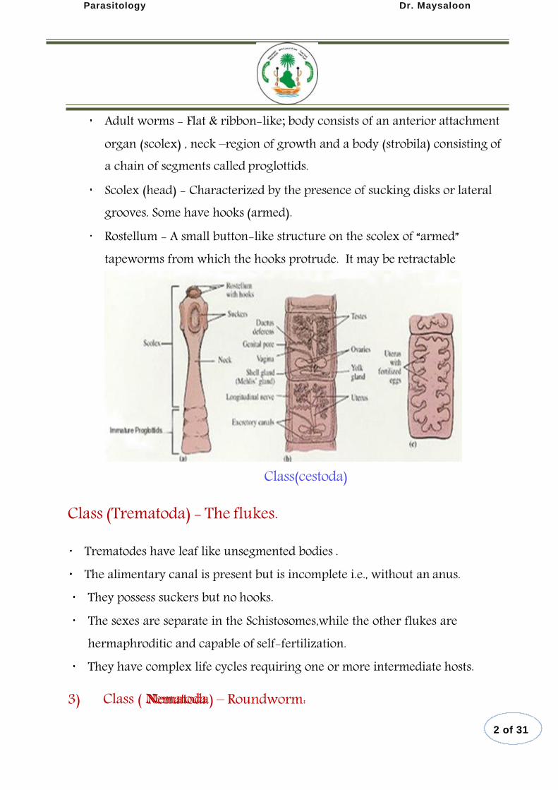
Adult worms - Flat & ribbon-like; body consists of an anterior attachment organ (scolex) , neck –region of growth and a body (strobila) consisting of a chain of segments called proglottids.
Scolex (head) - Characterized by the presence of sucking disks or lateral grooves. Some have hooks (armed).
Rostellum - A small button-like structure on the scolex of “armed” tapeworms from which the hooks protrude. It may be retractable
Class(cestoda)
Class (Trematoda) - The flukes.
Trematodes have leaf like unsegmented bodies . The alimentary canal is present but is incomplete i.e., without an anus. They possess suckers but no hooks. The sexes are separate in the Schistosomes,while the other flukes are hermaphroditic and capable of self-fertilization.
They have complex life cycles requiring one or more intermediate hosts.
3) Nematoda) – Roundworm:
2 of 31
Class ( Nematoda
Parasitology Dr. Maysaloon
Elongated worm, cylindrical, unsegmented and tapering at both ends. Sex separate and male is smaller than female. Head no hooks no suckers. well-developed alimentary canal (complete, anus present). Present of body cavity. Has direct life cycle (only one host). Has digestive system ,no circulatory &respiratory system.
Class-Cestoda
Taxonomic position Phylum platyhelminthes
Class Cestoda
Order Cyclophyllidae Order Pseudophyllidae
3 of 31
Parasitology Dr. Maysaloon
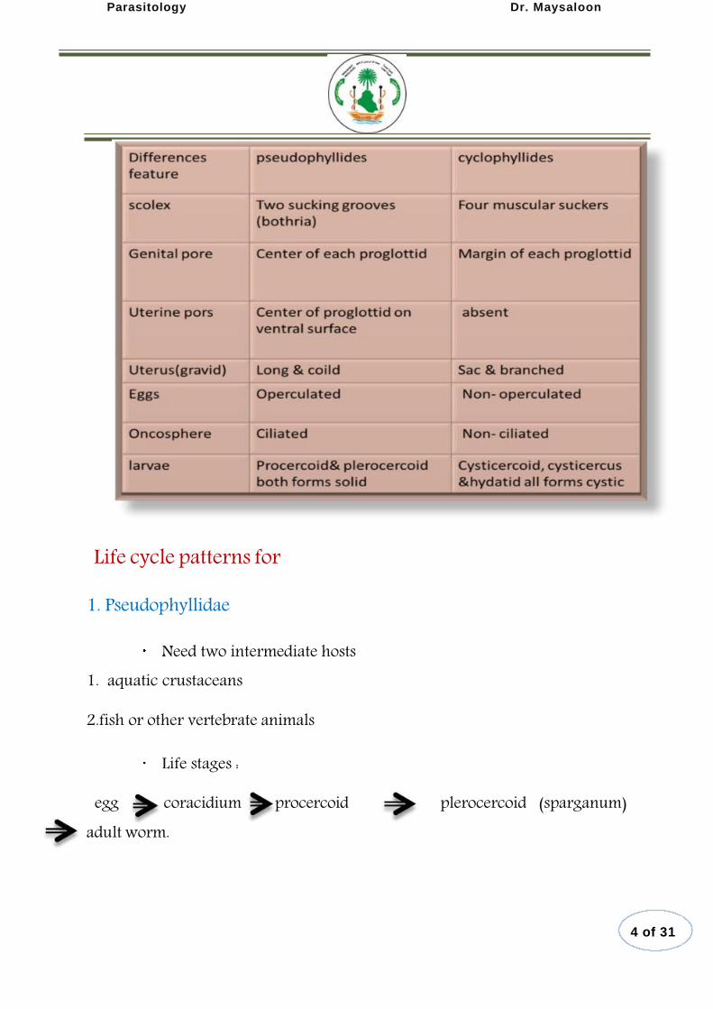
Life cycle patterns for
1. Pseudophyllidae
Need two intermediate hosts 1. aquatic crustaceans
2.fish or other vertebrate animals
Life stages :
egg coracidium procercoid plerocercoid (sparganum)
adult worm.
4 of 31

Parasitology Dr. Maysaloon
2. Cyclophyllidae
Need one intermediate host only -- usually mammals
Life stages :
Egg hexacanth metacestode stage adult worm.
Diphilobothriumlatum– accidental infection in humans.
Life cycle patterns of tape worm that infect human
5 of 31
e.g Diphilobothrium latum

Parasitology Dr. Maysaloon
Larval stage of cestodae:
Metacestode stage: mean larval stage of a cestode that develop in the intermediate host
Cysticercus: Resting stage of larva in intermediate host – develops into ‘bladder worm’ Cysticercoid: A small bladder containing invaginated head proximally and a solid elongated portion
Coenurus: Larval stage in form of Bladder worm contain many invaginating. Hydatid cyst: Larval stage of Echinococcus
Procercoid: First larval stage of Diphyllobothriumfound in cyclops, contain embryonal hooklets. Plerocercoid: soild elongated worm like larva with head invaginated onto the neck . e.g :D.latum.
Man may be infected with the adult tapeworms (Definitive host) or
their larval stages (Intermediate host):
Human infection with adult cestodes (Intestinal cestodes): Taeniasaginata
Taeniasoliun Diphyllobothrium
Hymenolepisnana Hymenolepisdiminuta Dipylidiumcaninum
6 of 31
Cysticercus
Cysticercoid
Coenurus
Hydatid cyst Procercoid
Plerocercoid
iuml atum
Parasitology Dr. Maysaloon
Human infection with larval cestodes (Extraintestinal cestodes):
ydatid cyst of Echinococcusgranulosus.
Alveolar hydatid cyst of E.multilocularis.
Cysticercus cellulosae of Taeniasolium.
Sparganum (plerocercoid ) of Spirometraspp.
Coenurus of Multicepstapeworm.
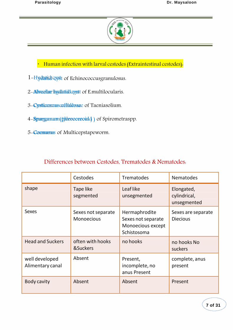
Differences between Cestodes, Trematodes & Nematodes:
Cestodes
Trematodes
Nematodes
shape
Tape like segmented
Leaf like unsegmented
Elongated, cylindrical, unsegmented
Sexes
Sexes not separate Monoecious
Hermaphrodite Sexes not separate Monoecious except Schistosoma
Sexes are separate Diecious
Head and Suckers
often with hooks
&Suckers
no hooks
no hooks No suckers
well developed Alimentary canal
Absent
Present, incomplete, no anus Present
complete, anus present
Body cavity
Absent
Absent
Present
7 of 31
1-Hydatid cyst
2-Alveolar hydatid cyst
3-Cysticercus cellulosae
4-Sparganum (plerocercoid )
5-Coenurus
Parasitology Dr. Maysaloon
Order Cyclophyllidae Taeniasaginata(Beef tapeworm)
Man is the only definitive host, cause disease called taeniasis.
Habitat:
Adult worms live in human small intestine causing taeniasis.
Larval stage (Cysticercus bovis) lives in cattle tissues.
Geographic Distribution:
T.saginatais worldwide in distribution, but the infection is not found in vegetarians and those who do not eat beef.
Morphology:
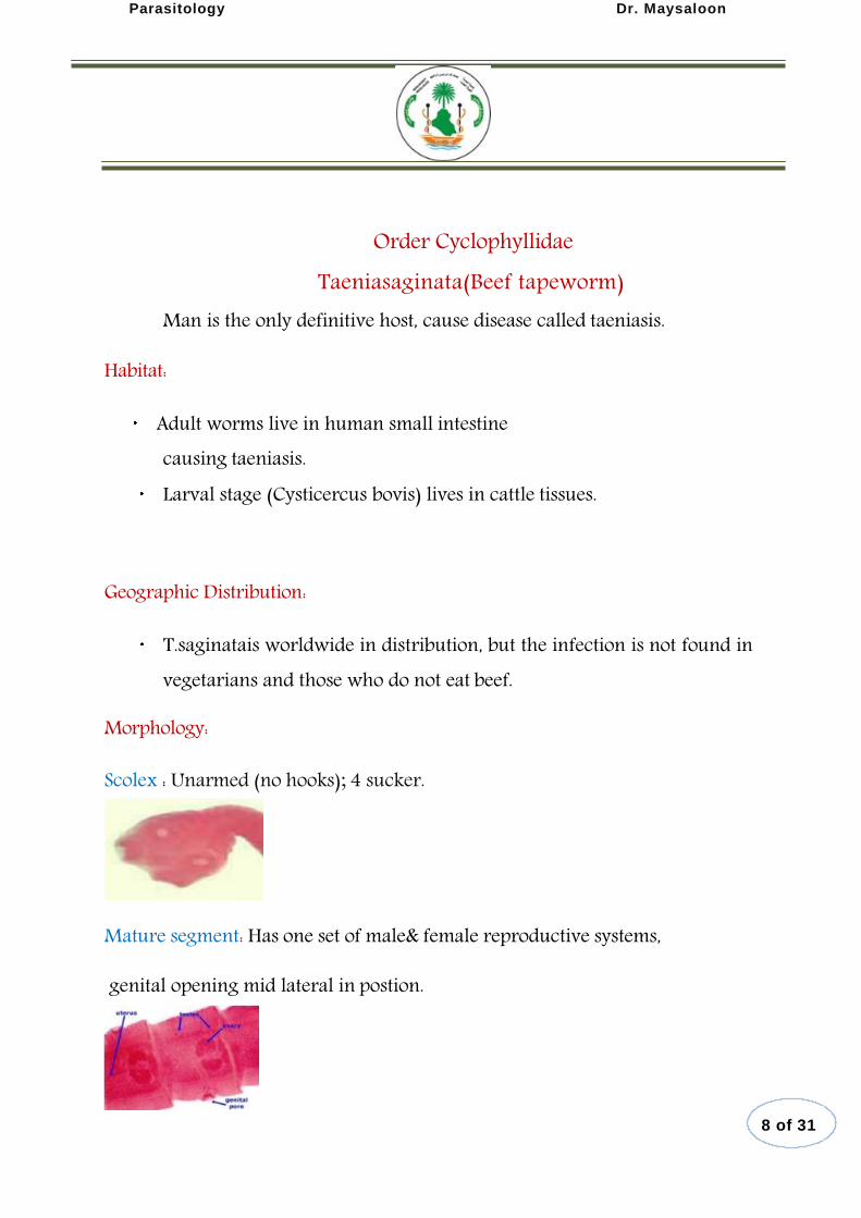
Scolex : Unarmed (no hooks); 4 sucker.
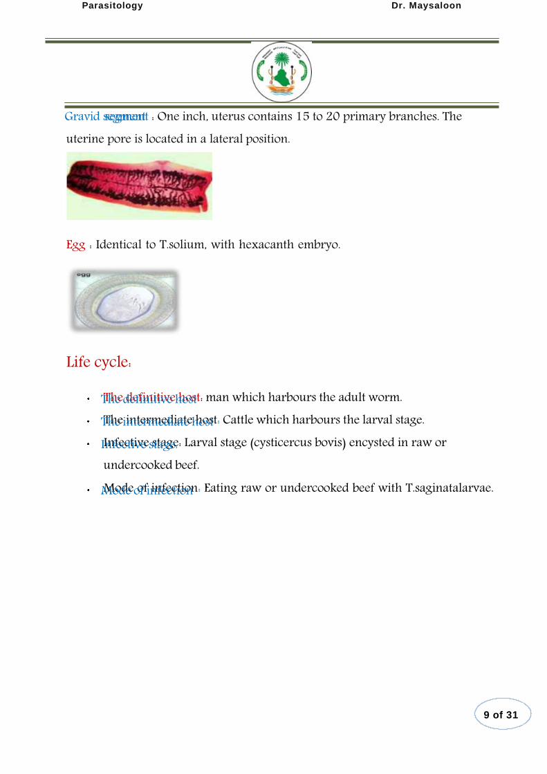
Mature segment: Has one set of male& female reproductive systems,
genital opening mid lateral in postion.
8 of 31
Parasitology Dr. Maysaloon
segment : One inch, uterus contains 15 to 20 primary branches. The uterine pore is located in a lateral position.
Egg : Identical to T.solium, with hexacanth embryo.
Life cycle:
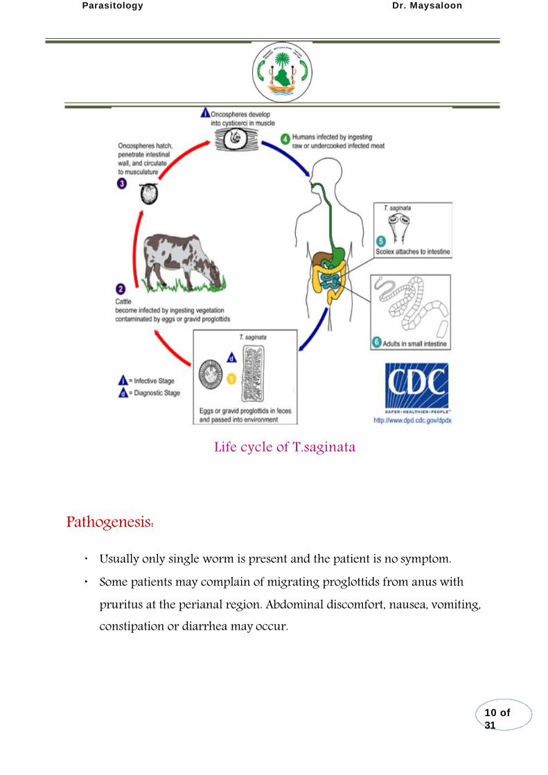
The definitive host: man which harbours the adult worm. The intermediate host: Cattle which harbours the larval stage. Infective stage: Larval stage (cysticercus bovis) encysted in raw or undercooked beef. Mode of infection: Eating raw or undercooked beef with T.saginatalarvae.
9 of 31
Gravid segment
The definitive host The intermediate host Infective stage:
Mode of infection
Parasitology Dr. Maysaloon
Life cycle of T.saginata
Pathogenesis:
Usually only single worm is present and the patient is no symptom. Some patients may complain of migrating proglottids from anus with pruritus at the perianal region. Abdominal discomfort, nausea, vomiting, constipation or diarrhea may occur.
10 of
31
Parasitology Dr. Maysaloon
Diagnosis:
Finding of gravid proglottids or eggs at the perianal region by cellophane tape method.
Treatment:
Praziquantel
Order Cyclophyllidae
Taeniasolium(pork tapeworm)
Man is the only definitive host, but he can also be the intermediate host for
T.soliumcause disease called Taeniasis or Cysticercosis.
Habitat:
Adult worms live in human small intestine . Larval stage ( cysticercus cellulosae ) in pigs muscles or in human tissues.
Geographic Distribution:
Worldwide in distribution & infection is cosmopolitan in countries where pork is eaten raw or undercooked.
11 of 31
(pork tapeworm):

Parasitology Dr. Maysaloon
Morphology:
Scolex: globular with 4 circular suckers; armed with a double row of alternating large and small hooklets.
Gravid segments: The number of main branches on each side the uterus stem is
7-13.
Ova :Same as those of T.saginata
Life cycle:
The definitive host: man which harbours the adult worm. The intermediate host: Pig (accidentally Man, cysticercosis). Infective stage: both egg/gravid proglottid and cysticercus for T.solium.
Mode of infection
1-Eating undercooked pork meat containing larvae.
2- Ingestion of T.soliumegg either in contaminated food or by autoinfection.
12 of
31
The definitive host The intermediate host Infective stage
Mode of infection:
Parasitology Dr. Maysaloon
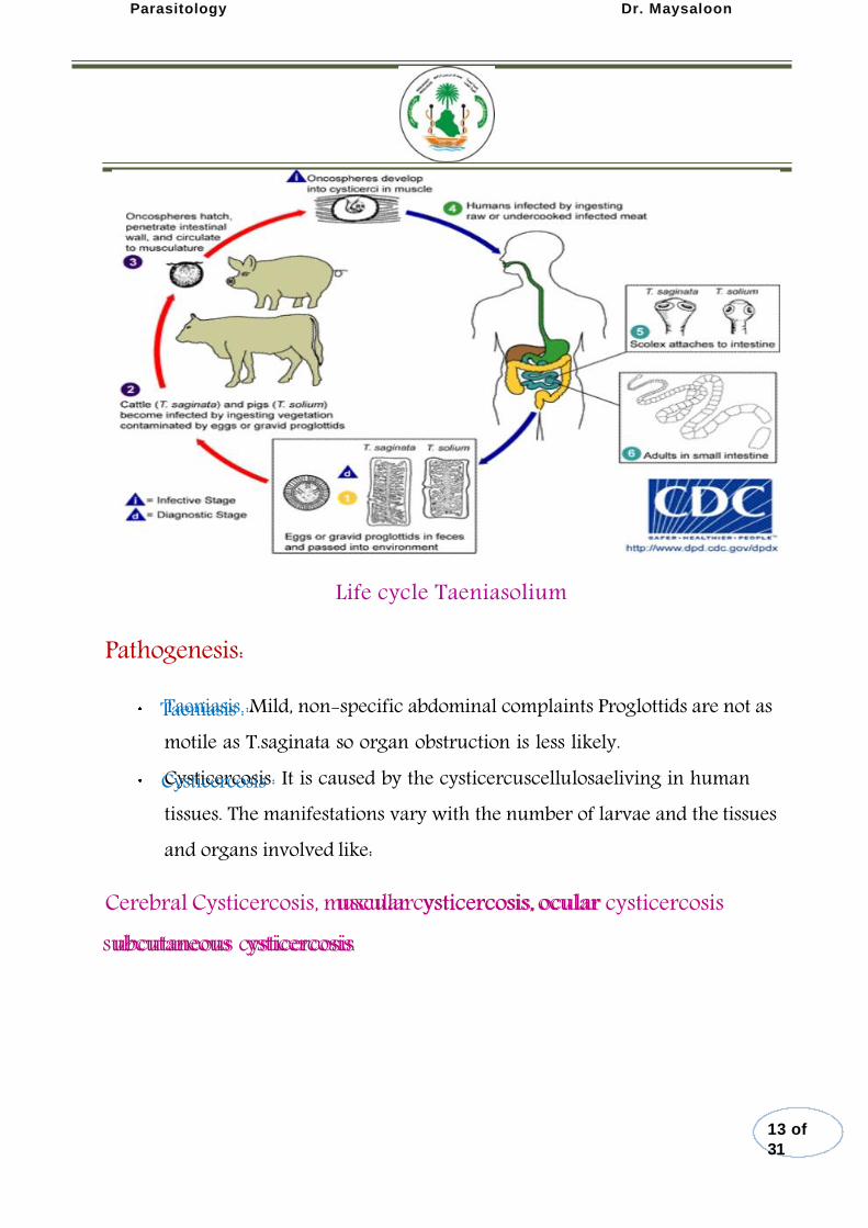
Life cycle Taeniasolium
Pathogenesis:
Taeniasis :Mild, non-specific abdominal complaints Proglottids are not as motile as T.saginata so organ obstruction is less likely. Cysticercosis: It is caused by the cysticercuscellulosaeliving in human tissues. The manifestations vary with the number of larvae and the tissues and organs involved like:
Cerebral Cysticercosis, muscular ysticercosis, ocular cysticercosis ubcutaneous cysticercosis
13 of
31
Taeniasis :
Cysticercosis
uscular cysticercosis , ocular
subcutaneous ysticercosis.

Parasitology Dr. Maysaloon
Diagnosis:
1.Taeniasis: Confirmative diagnosis of taeniasis is made by
finding gravid proglottides or egg in stool or by cellophane-tape technique.
2. Cysticercosis: Biopsy of subcutaneous nodules, X-ray ,CT
0r MR are used for the diagnosis of brain type.
Treatment:
Praziquantel Niclosamide
Differences between T.solium&T.saginata
Order Cyclophyllidae
14 of
31
Parasitology Dr. Maysaloon
Hymenolepisnana(Dwarf tapeworm)
Smallest tapeworm infecting man. Only human tapeworm that can complete its life cycle in a single host Man can harbour both the adult and larval stages of the parasite
Exception to the general rule that: “Helminthes do not multiply inside the body of the definitive host”
Geographic Distribution:
Most common tapeworm throughout the world Mainly among children.
Habitat: Adult found in the ileum
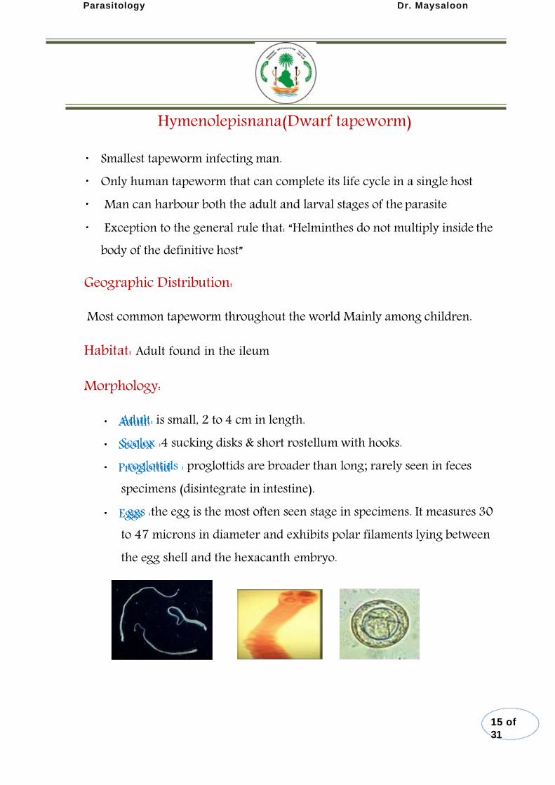
Morphology:
Adult: is small, 2 to 4 cm in length. Scolex :4 sucking disks & short rostellum with hooks. roglottids : proglottids are broader than long; rarely seen in feces
specimens (disintegrate in intestine). ggs :the egg is the most often seen stage in specimens. It measures 30
to 47 microns in diameter and exhibits polar filaments lying between the egg shell and the hexacanth embryo.
15 of
31
Adult: Scolex
Proglottid
Eggs
Parasitology Dr. Maysaloon
Life cycle:
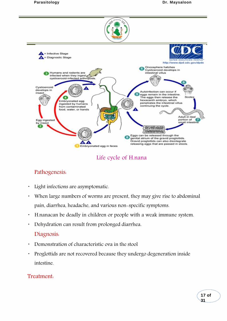
Definitive hosts : Man, rodents. The intermediate hosthost: Insect ,grain beetle and larva of flea. Diagnostic stage: Embryonated egg in feces
Infective stage : Embryonated egg.
Mode of Infection: 1- Direct
i. Ingests eggs by contaminated food &water . ii. Self infection via hand (external autoinfection).
iii. Internal autoinfection (egg hatch in small intestine).
2- Indirect pathway
Accidental ingestion of infected arthropod intermediate host like rice and flour beetles in which cysticercoid larvae are released and develop into adult worms in the small intestine of the host.
16 of
31
Definitive hosts
The intermediate
Diagnostic stage: Infective stage :
Direct:
Indirect pathway:
Parasitology Dr. Maysaloon
Life cycle of H.nana
Pathogenesis:
Light infections are asymptomatic. When large numbers of worms are present, they may give rise to abdominal pain, diarrhea, headache, and various non-specific symptoms.
H.nanacan be deadly in children or people with a weak immune system. Dehydration can result from prolonged diarrhea. Diagnosis:
Demonstration of characteristic ova in the stool Proglottids are not recovered because they undergo degeneration inside intestine.
Treatment:
17 of
31
Parasitology Dr. Maysaloon
Praziquantel is the drug of choice.
Order Cyclophyllidae
Non-human cestodes infection
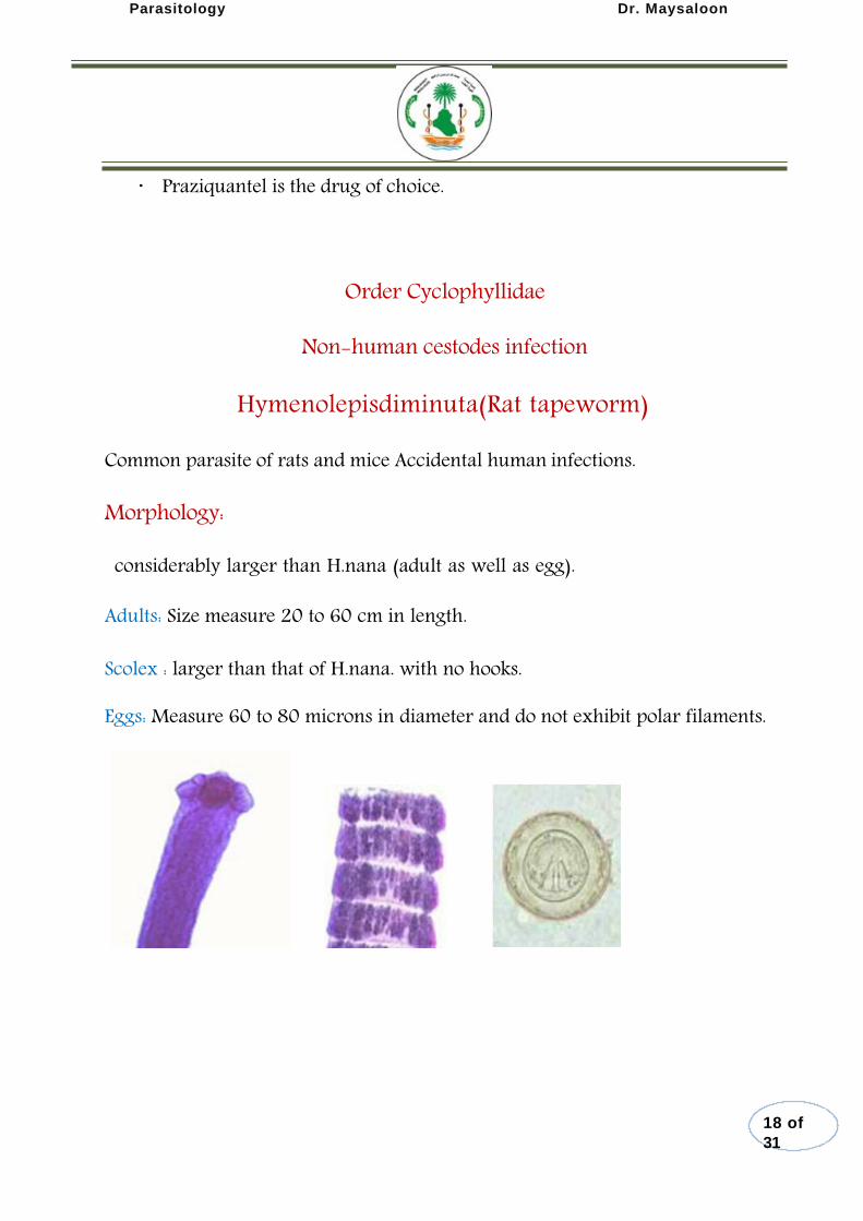
Hymenolepisdiminuta(Rat tapeworm)
Common parasite of rats and mice Accidental human infections.
Morphology:
considerably larger than H.nana (adult as well as egg).
Adults: Size measure 20 to 60 cm in length.
Scolex : larger than that of H.nana. with no hooks.
Eggs: Measure 60 to 80 microns in diameter and do not exhibit polar filaments.
18 of
31

Parasitology Dr. Maysaloon
Life cycle
Life cycle of H.diminuta
Order Cyclophyllidae
Non-human cestodes infection
Dipylidiumcaninum(dog tapeworm)
Dipylidiumcaninum(the double-pored dog tapeworm) mainly infects dogs and cats, but is occasionally found in humans, especially children.
Morphology : Scolex :conical-shaped and has four suckers. There is rostellum armed with several rings of small hooks. Eggs: round to oval and contain an oncosphere that has 6 hooklets. And present as egg capsule contain 5 to 30 or more eggs .
19 of
31
Scolex
Eggs:

Parasitology Dr. Maysaloon
Segments: have two genital pores, one in the middle of each lateral margin. They are pumpkin seed-shaped. gravid segments :contain egg capsule, The number of eggs can range from 5 to 30.
Life cycle: Final Hosts: Dogs, Cats, Humans Intermediate host .: larva of dog fleas.
tive stage for man: cysticercoid.
Life cycle of D.caninum
Pseudophyllidea
20 of
31
Segments:
gravid segments
Final Hosts
Intermediate host Infec Infective stage for man
Parasitology Dr. Maysaloon
Diphyllobothriumlatum(fish tapeworm)
The cestode Diphyllobothriumlatum(the fish or broad tapeworm), the largest human tapeworm.
Geographic Distribution:
Diphyllobothriasis occurs in the Northern Hemisphere and in South America.
Habitat:
Adult worms in small Intestine
Morphology:
Scolex: spoon-shaped or spatulate. Scolex bears 2 slit-like grooves called bothria. Scolex has no rostellum and no hooklets.
Ova: unembryonated ovum with operculum at one end and a small knob at the other end.
Larvae:
Procercoid: Spindle-like solid body with cephalic invagination Found inside the Cyclops. Plerocercoid: Head is invaginated in the neck Found in the fresh water fish
.
Life cycle:
Definitive host: Man, dog, cat .
21 of
31
i. Procercoid
ii. Plerocercoid
Definitive host
Parasitology Dr. Maysaloon
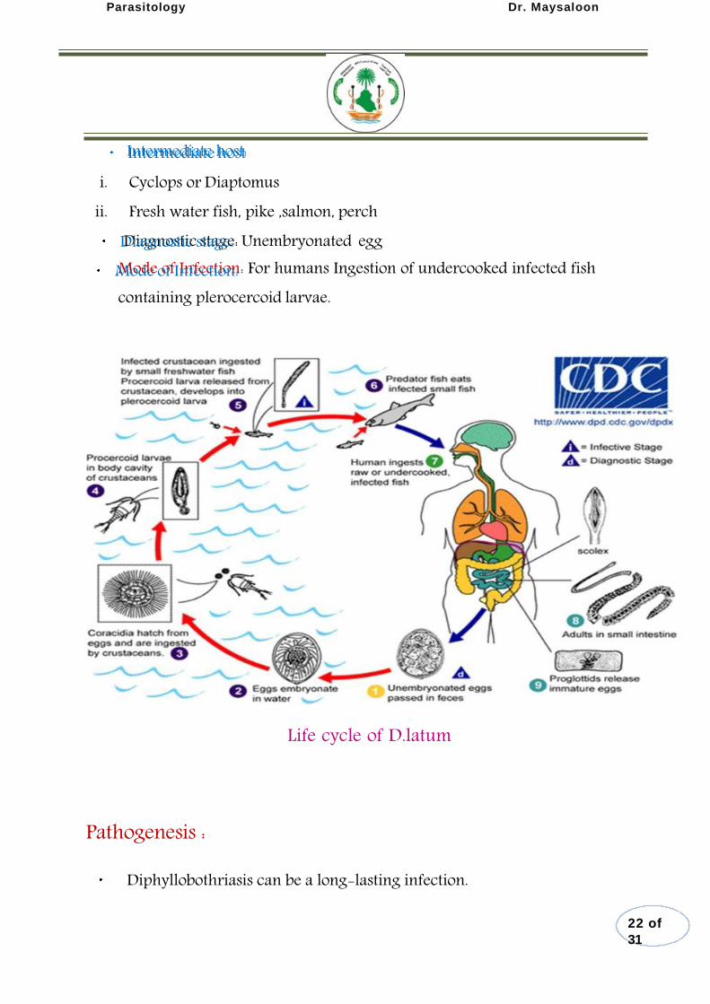
Intermediate host i. Cyclops or Diaptomus
ii. Fresh water fish, pike ,salmon, perch
Diagnostic stage: Unembryonated egg
Mode of Infection: For humans Ingestion of undercooked infected fish containing plerocercoid larvae.
Life cycle of D.latum
Pathogenesis :
Diphyllobothriasis can be a long-lasting infection.
22 of
31
Intermediate host:
Diagnostic stage: Mode of Infection:
Parasitology Dr. Maysaloon
Most infections are asymptomatic. Manifestations may include abdominal discomfort, diarrhea, vomiting, and weight loss.
Vitamin B12 deficiency with pernicious anemia may occur. Massive infections may result in intestinal obstruction. Migration of proglottids can cause cholecystitis or cholangitis.
Diagnosis:
Identification of eggs in the stool is the basis of specific diagnosis. Examination of proglottids passed in the stool is also diagnostic .
Treatment:
Praziquantel.
Human infection with larval cestodes (Extraintestinal cestodes):
Hydatid cyst of Echinococcusgranulosus.
Alveolar hydatid cyst of E.multilocularis.
Cysticercus cellulosae of Taeniasolium.
Sparganum (plerocercoid ) of Spirometraspp.
Coenurus of Multicepstapeworm
Order Cyclophyllidae
granulosis–The Hydatid Tapeworm
23 of
31
1-Hydatid cyst
2-Alveolar hydatid cyst
3-Cysticercus cellulosae
4-Sparganum (plerocercoid )
5-Coenurus
1-Echinococcus Echinococcus granulosis
Parasitology Dr. Maysaloon
Echinococcosis or Hydatid disease in man is caused by the larval stage of the dog tapeworm. Disease
Ecchinococcosis (serious disease).
Geographic Distribution:
Worldwide. most extensively found in Africa, the Middle East and parts of South America and Australia.
Habitat:
Adult: lives in intestine of dogs but never in human intestine. Larvae: Sites of larvae in man liver, lungs, abdominal cavity, spleen, kidneys, heart, bones, central nervous system etc.
Morphology:
Adult worm: approximately 3 – 8.5 mm long. Scolex : has 4 suckers and a rostellum with hooks. Segments: has only 3 – 4 proglottids, immature, mature and the final one is gravid. Egg: The eggs of E.granulosusand Teaniaspp. are indistinguishable
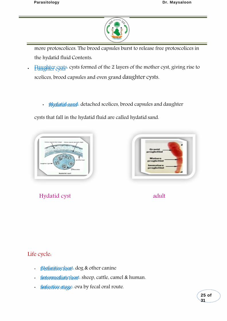
Hydatid cyst: It is a fluid filled cyst The wall of the cyst is made up of an inner cellular germinative layer and an outer acellular laminated layer. The host connective tissue covers the outer layer. Brood capsules develop inside the cyst on the germinative layer 5-6 months later. Each brood capsule contain 20 or
24 of
31
Adult Larvae
Adult worm
Scolex
Segments:
Egg
Hydatid cyst
Parasitology Dr. Maysaloon
more protoscolices. The brood capsules burst to release free protoscolices in the hydatid fluid Contents: Daughter cysts: cysts formed of the 2 layers of the mother cyst, giving rise to scolices, brood capsules and even grand daughter cysts.
Hydatid sand: detached scolices, brood capsules and daughter
cysts that fall in the hydatid fluid are called hydatid sand.
Hydatid cyst adult
Life cycle:
Definitive host: dog & other canine
Intermediate host: sheep, cattle, camel & human. Infective stage: ova by fecal oral route.
25 of
31
Daughter cysts
Hydatid sand
Definitive host Intermediate host Infective stage
Parasitology Dr. Maysaloon
Mode of infection
1-Ingestion of water or vegetable contaminated by infected dog faeces. 2-Handling or caressing infected dogs where the hairs are usually polluted with eggs.
Life cycle:
Adults are only in canines. Eggs are shed in the feces of infected animals. Humans accidentally ingest eggs from an infected animal or from canine feces.
Hexacanth embryo penetrates the intestinal mucosa and migrates to tissues& develops into the larval stage (hydatid cyst) in the tissue (liver, lung or brain, most often). Human is dead end. Viscera containing hydatid cysts are eaten by canines. Adult worms develop in small intestine.
26 of
31
Mode of infection:
Human is dead end

Parasitology Dr. Maysaloon
.
Life cycle of E.granulosis
Pathogenesis:
Depends on the size, the location and the number of cyst.
Unilocular cysts. There is usually surrounding inflammatory reaction and fibrosis. After years, the cyst may die, shrink and calcify. There is general allergic reaction with eosinophilia. Pressure effects can cause local tissue damage and obstruction. Rupture or leakage of the cyst lead to allergic reaction. There can be anaphylactic shock and secondary implantation in the surrounding tissues like the peritoneum. There can be secondary infection with formation of abscesses.
Diagnosis:
27 of
31
Parasitology Dr. Maysaloon
History of contacting with sheep & dogs
Clinical symptoms of a slow-growing tumor accompanied by eosinophilia. Parasitological examination for finding scolexes, brood capsules & daughter cysts
Cysts in organs or calcified cysts can be visualized using x-rays, CT & ultrasound examination
Biopsy are forbidden unless during operation
Serological examination for specific Ab . Intradermal (Casoni) test with hydatid fluid is useful. Antibodies against hydatid fluid antigens have been detected in a sizable population of infected individuals by ELISA or indirect hemagglutination test.
Treatment:
Surgery: with the goal of leaving the cyst intact so new cysts do not form
Mebendazole can be taken over a long period of time at low dosages
Praziquantel
Echinococcusmultilocularis(alveolar hydatid disease)
28 of
31
2-Echinococcus multilocularis
Parasitology Dr. Maysaloon
The larvae of Echinococcusmultilocularis is a dangerous species causing (alveolar hydatid disease) in man and animals. The most common definitive hosts are foxes and wolves in addition to domestic cats and dogs.
Rodents are the intermediate hosts. Man is an accidental host. Man get infection by ingestion of eggs. Unlike E.granulosus,cysts of E.multilocularis in man do not contain daughter cysts with scolices.
Cysts are formed primarily in the liver.
3-Cysticercosis:
Infection of human tissues by Cysticercus cellulosae, the larval stage
of Taeniasolium
Mode of infection: man acquires infection on
Ingestion of the egg of Taeniasolium(the infective stage) by contaminated food or water
External autoinfection: hand to mouth infection. Internal autoinfection: eggs hatch in small intestine in patient infected with T.soliumadult.
Pathogenesis:
The cyst produces a foreign body inflammatory reaction which usually ends in fibrosis and calcification.
Manifestations depend upon the tissue invaded and the number of cysticerci.
29 of
31
athogenesis::
Parasitology Dr. Maysaloon
The commonest sites are subcutaneous tissues, muscles, viscera, brain and orbit.
There may be muscle pains, mild fever and eosinophilia.
Treatment:
Surgical removal, Praziquantel with corticosteroids. Vitamin D and calcium to help calcification.
4-Coenurosis
Human infection with Coenuruscerebralis, the larval stage of
Multicepsmulticeps.
The adults worm , Multicepsmulticeps, lives in the small intestine
of dogs and wild canines. Eggs pass in the faeces.
When the egg is swallowed by the intermediate host (herbivores specially sheep and rarely man) it develops into the larval stage.
Diagnosis: difficult, diagnosed as brain tumor (X-ray and CT).
5-Sparganosis
30 of
31
Diagnosis:
Parasitology Dr. Maysaloon
Infection of human tissues by the plerocercoid larva of Diphyllobothrium
mansoni(Sparganum mansoni) .
The adult lives in the small intestine of dogs and cats.(definitive host). The first intermediate host is Cyclops containing the procercoid larva. The second intermediate host is a frog, snake, bird or mammal containing the plerocercoid larva (Sparganum).
Human is intermediate host. Mode of infection by ingestion procercoid or plerocercoid & the larvae cannot develop to adult in man.
31 of
31
Copyright © 2022 FDOKUMEN