Visualization and 3D Reconstruction of Flame Cells of Taenia solium (Cestoda
11
Visualization and 3D Reconstruction of Flame Cells of Taenia solium (Cestoda) Laura E. Valverde-Islas 1 , Esteban Arrangoiz 4 , Elio Vega 3 , Lilia Robert 1 , Rafael Villanueva 1 , Olivia Reynoso-Ducoing 1 , Kaethe Willms 1 , Armando Zepeda-Rodrı´guez 2 , Teresa I. Fortoul 2 , Javier R. Ambrosio 1 * 1 Departamento de Microbiologı ´a y Parasitologı ´a, Facultad de Medicina, Universidad Nacional Auto ´noma de Me ´xico (UNAM), Me ´xico City, Distrito Federal, Me ´ xico, 2 Departamento de Biologı ´a Celular y Tisular, Facultad de Medicina, Universidad Nacional Auto ´noma de Me ´xico (UNAM), Me ´xico City, Distrito Federal, Me ´ xico, 3 Departamento de Visualizacio ´ n, Direccio ´ n General de Co ´ mputo y de Tecnologı ´as de Informacio ´ n y Comunicacio ´ n (DGTIC), Universidad Nacional Auto ´noma de Me ´xico (UNAM), Me ´xico City, Distrito Federal, Me ´ xico, 4 Laboratorio de 3D, Facultad de Medicina, Universidad Nacional Auto ´noma de Me ´xico (UNAM), Me ´xico City, Distrito Federal, Me ´xico Abstract Background: Flame cells are the terminal cells of protonephridial systems, which are part of the excretory systems of invertebrates. Although the knowledge of their biological role is incomplete, there is a consensus that these cells perform excretion/secretion activities. It has been suggested that the flame cells participate in the maintenance of the osmotic environment that the cestodes require to live inside their hosts. In live Platyhelminthes, by light microscopy, the cells appear beating their flames rapidly and, at the ultrastructural, the cells have a large body enclosing a tuft of cilia. Few studies have been performed to define the localization of the cytoskeletal proteins of these cells, and it is unclear how these proteins are involved in cell function. Methodology/Principal Findings: Parasites of two different developmental stages of T. solium were used: cysticerci recovered from naturally infected pigs and intestinal adults obtained from immunosuppressed and experimentally infected golden hamsters. Hamsters were fed viable cysticerci to recover adult parasites after one month of infection. In the present studies focusing on flame cells of cysticerci tissues was performed. Using several methods such as video, confocal and electron microscopy, in addition to computational analysis for reconstruction and modeling, we have provided a 3D visual rendition of the cytoskeletal architecture of Taenia solium flame cells. Conclusions/Significance: We consider that visual representations of cells open a new way for understanding the role of these cells in the excretory systems of Platyhelminths. After reconstruction, the observation of high resolution 3D images allowed for virtual observation of the interior composition of cells. A combination of microscopic images, computational reconstructions and 3D modeling of cells appears to be useful for inferring the cellular dynamics of the flame cell cytoskeleton. Citation: Valverde-Islas LE, Arrangoiz E, Vega E, Robert L, Villanueva R, et al. (2011) Visualization and 3D Reconstruction of Flame Cells of Taenia solium (Cestoda). PLoS ONE 6(3): e14754. doi:10.1371/journal.pone.0014754 Editor: David Joseph Diemert, The George Washington University Medical Center, United States of America Received July 5, 2010; Accepted February 6, 2011; Published March 11, 2011 Copyright: ß 2011 Valverde-Islas et al. This is an open-access article distributed under the terms of the Creative Commons Attribution License, which permits unrestricted use, distribution, and reproduction in any medium, provided the original author and source are credited. Funding: This work was supported by grants from the Direccion General de Asuntos del Personal Academico (DGAPA)-UNAM (IN201003, IN216107 and IN200610), DGAPA-IXTLI (0709008 and IX200610) and from Consejo Nacional de Ciencia y Tecnologia (CONACYT) V43629-M and 080093. LEVI was supported by a doctoral fellowship from CONACYT Mexico and her Doctoral degree was obtained from Posgrado-UNAM, Doctorado en Ciencias Biomedicas, Mexico. The funders had no role in study design, data collection and analysis, decision to publish, or preparation of the manuscript. Competing Interests: The authors have declared that no competing interests exist. * E-mail: [email protected] Introduction Flame cells (FC) are ciliated cells located within the basal matrix, at the neodermal tissue of cestoda. They are considered as terminal cells in flame or bulbs [1] and as the basic units of the protonephridial system (PS) of invertebrates. In cestodes, like in all parasitic platyhelminthes, the PS are the excretory systems with an important role that allow the parasites to conserve water and eliminate salts and survive in the intestine or body cavities of their hosts and because of that, they act like osmoconformers [2,3]. Parasites need to maintain, within physiological limits, the osmotic pressure of their tissues against that of the host environment [2]. FC have a typical morphology: by light microscopy they look like comets in which the anterior end of the cell corresponds to the cell body while the distal end contains the tuft of cilia known also as the flames [1]. By transmission electron microscopy (TEM), FC of several helminthes exhibits a similar morphology [1,3]: a single nucleus and classical cilia with 9+2 axonemes. The plasma membrane surrounds the cell body that extends through the tip of the tuft of cilia and is apparently in close interdigitation with the membrane of adjoining cells [4]. While the bodies of FC are immersed in the parenchymal tissue, the cilia tufts are extended inside the PS tubules [3,5]. In tapeworms, FC appear to be beating continuously, their cilia producing the impression of flickering flames inside the cell [4], and due to this behavior, FC have also PLoS ONE | www.plosone.org 1 March 2011 | Volume 6 | Issue 3 | e14754
-
Upload
independent -
Category
Documents
-
view
0 -
download
0
Transcript of Visualization and 3D Reconstruction of Flame Cells of Taenia solium (Cestoda

Visualization and 3D Reconstruction of Flame Cells ofTaenia solium (Cestoda)Laura E. Valverde-Islas1, Esteban Arrangoiz4, Elio Vega3, Lilia Robert1, Rafael Villanueva1, Olivia
Reynoso-Ducoing1, Kaethe Willms1, Armando Zepeda-Rodrıguez2, Teresa I. Fortoul2, Javier R.
Ambrosio1*
1 Departamento de Microbiologıa y Parasitologıa, Facultad de Medicina, Universidad Nacional Autonoma de Mexico (UNAM), Mexico City, Distrito Federal, Mexico,
2 Departamento de Biologıa Celular y Tisular, Facultad de Medicina, Universidad Nacional Autonoma de Mexico (UNAM), Mexico City, Distrito Federal, Mexico,
3 Departamento de Visualizacion, Direccion General de Computo y de Tecnologıas de Informacion y Comunicacion (DGTIC), Universidad Nacional Autonoma de Mexico
(UNAM), Mexico City, Distrito Federal, Mexico, 4 Laboratorio de 3D, Facultad de Medicina, Universidad Nacional Autonoma de Mexico (UNAM), Mexico City, Distrito
Federal, Mexico
Abstract
Background: Flame cells are the terminal cells of protonephridial systems, which are part of the excretory systems ofinvertebrates. Although the knowledge of their biological role is incomplete, there is a consensus that these cells performexcretion/secretion activities. It has been suggested that the flame cells participate in the maintenance of the osmoticenvironment that the cestodes require to live inside their hosts. In live Platyhelminthes, by light microscopy, the cells appearbeating their flames rapidly and, at the ultrastructural, the cells have a large body enclosing a tuft of cilia. Few studies havebeen performed to define the localization of the cytoskeletal proteins of these cells, and it is unclear how these proteins areinvolved in cell function.
Methodology/Principal Findings: Parasites of two different developmental stages of T. solium were used: cysticercirecovered from naturally infected pigs and intestinal adults obtained from immunosuppressed and experimentally infectedgolden hamsters. Hamsters were fed viable cysticerci to recover adult parasites after one month of infection. In the presentstudies focusing on flame cells of cysticerci tissues was performed. Using several methods such as video, confocal andelectron microscopy, in addition to computational analysis for reconstruction and modeling, we have provided a 3D visualrendition of the cytoskeletal architecture of Taenia solium flame cells.
Conclusions/Significance: We consider that visual representations of cells open a new way for understanding the role ofthese cells in the excretory systems of Platyhelminths. After reconstruction, the observation of high resolution 3D imagesallowed for virtual observation of the interior composition of cells. A combination of microscopic images, computationalreconstructions and 3D modeling of cells appears to be useful for inferring the cellular dynamics of the flame cellcytoskeleton.
Citation: Valverde-Islas LE, Arrangoiz E, Vega E, Robert L, Villanueva R, et al. (2011) Visualization and 3D Reconstruction of Flame Cells of Taenia solium(Cestoda). PLoS ONE 6(3): e14754. doi:10.1371/journal.pone.0014754
Editor: David Joseph Diemert, The George Washington University Medical Center, United States of America
Received July 5, 2010; Accepted February 6, 2011; Published March 11, 2011
Copyright: � 2011 Valverde-Islas et al. This is an open-access article distributed under the terms of the Creative Commons Attribution License, which permitsunrestricted use, distribution, and reproduction in any medium, provided the original author and source are credited.
Funding: This work was supported by grants from the Direccion General de Asuntos del Personal Academico (DGAPA)-UNAM (IN201003, IN216107 andIN200610), DGAPA-IXTLI (0709008 and IX200610) and from Consejo Nacional de Ciencia y Tecnologia (CONACYT) V43629-M and 080093. LEVI was supported by adoctoral fellowship from CONACYT Mexico and her Doctoral degree was obtained from Posgrado-UNAM, Doctorado en Ciencias Biomedicas, Mexico. The fundershad no role in study design, data collection and analysis, decision to publish, or preparation of the manuscript.
Competing Interests: The authors have declared that no competing interests exist.
* E-mail: [email protected]
Introduction
Flame cells (FC) are ciliated cells located within the basal
matrix, at the neodermal tissue of cestoda. They are considered
as terminal cells in flame or bulbs [1] and as the basic units of the
protonephridial system (PS) of invertebrates. In cestodes, like in
all parasitic platyhelminthes, the PS are the excretory systems
with an important role that allow the parasites to conserve water
and eliminate salts and survive in the intestine or body cavities of
their hosts and because of that, they act like osmoconformers
[2,3]. Parasites need to maintain, within physiological limits, the
osmotic pressure of their tissues against that of the host
environment [2].
FC have a typical morphology: by light microscopy they look
like comets in which the anterior end of the cell corresponds to the
cell body while the distal end contains the tuft of cilia known also
as the flames [1]. By transmission electron microscopy (TEM), FC
of several helminthes exhibits a similar morphology [1,3]: a single
nucleus and classical cilia with 9+2 axonemes. The plasma
membrane surrounds the cell body that extends through the tip of
the tuft of cilia and is apparently in close interdigitation with the
membrane of adjoining cells [4]. While the bodies of FC are
immersed in the parenchymal tissue, the cilia tufts are extended
inside the PS tubules [3,5]. In tapeworms, FC appear to be beating
continuously, their cilia producing the impression of flickering
flames inside the cell [4], and due to this behavior, FC have also
PLoS ONE | www.plosone.org 1 March 2011 | Volume 6 | Issue 3 | e14754

received names like cap cells and bulb cells [5]. Knowledge of
cytoskeletal proteins of FC have shown that polymerized actin was
found by fluorescent phalloidin in FC of the cestode Diphylloboth-
rium dendriticum [6] and in the monogenean Gyrodactylus rysavyi [7]
where it was associated to longitudinal fibers that are in close
contact with the PS excretory system. Tubulin was demonstrated
in FC cilia of the cestode Gymnorinchus [8] and in the trematode
Schistosoma mansoni [9]. However, the knowledge of the FC
morphology, their role in the parasite physiology is scarce and
there is no clear evidence of their functional activities [1]. It is
mainly assumed to be related to excretory activities trough the PS.
In tapeworm infections, as those produced by the metazoan T.
solium, the FC of the parasites appear to be important for the
survival within hosts because they can perform specific functions
such as detoxification or transformation of substances harmful to
the parasites [2,3]. It is well known that parasitic stages of the
medically important of T. solium (the larval stage or cysticercus and
the adult stage or taenia) have many FC in their tissues and the
morphology of these cells has only been demonstrated in
invaginated cysticerci of T. solium stained by Bodians protargol
method [10,11]. The importance of study of these cells is that they
could be excellent targets of the antihelminthic treatments based
on their capacity to inhibit the polymerization of microtubules
using benzimidazole drugs such as albendazole, as shown after the
in vitro treatment of cysticerci of the murine model of T. crassiceps
[12].
The aim of the present work is to define the cytoarchitecture of
T. solium FC with the help of microscopic observations, digital
reconstructions and 3D visualization of the cytoskeleton of these
cells that could be useful as an alternative way of deducing the
functions of microscopic structures of ciliated cells. In addition, we
consider that increasing knowledge of the function of the T. solium
FC could extend our understanding of the cellular biology and
physiology of these cestodes, which are still endemic in many
countries [13].
Materials and Methods
ParasitesLarvae were dissected from muscles of naturally infected pig
meat, extensively washed with cold phosphate buffered saline
(PBS) adjusted to pH 7.2 and divided in groups either for
microscopic observation or for inducing experimental infections in
golden hamsters. Intestinal adult parasites were obtained from
immunosuppressed hamsters 30 days after oral administration of
cysticerci as reported previously [14].
This study was approved by the local ethics and research
committees of the Facultad de Medicina, UNAM (FMED/CI/
RGG/022/08/2006, FMED/CI/RGG/055/08/2007 and 144-
2009.)
MicroscopyLive-Cell Microscopy. Live cysticerci were maintained for one
or two days in vitro in RPMI 1640 medium supplemented with
25 mM HEPES buffer adjusted to pH 7.2 and 30 mM carbonate
salts. Parasites were maintained in a humidified incubator at
37 uC in a 5% CO2 environment.
For filming motion of FC in live parasites, cysticerci were
punctured with a needle in order to eliminate the vesicular fluid
and observations were performed on the internal side of the
bladder walls, by spreading the tissue on a microscope slide and
observing it directly using Nomarsky differential-interference-
contrast microscopy (DIC). Time-lapse recordings of FC dynam-
ics, at room temperature (RT), were carried out as follows: Each
frame was captured at the rate of 3.2 s/frame producing a total of
34 frames using a LSCM Leica.
Processing of parasites for fluorescent assaysParasites were processed by standard procedures for immuno-
fluorescence assays. Briefly, parasites were washed in phosphate-
buffered-saline (PBS), embedded in Tissue-Tek (Sakura, USA),
sectioned (10 mm) in a cryostat (CM Leica 1100) and sections
adhered on poly-L-Lys treated glass slides. Tissues were fixed with
cold acetone and washed with PBS and PBS/0.3% Tween-20.
Blocking of sections was done with PBS/0.3% Tween-20/2% BSA
and finally incubated for 1 h in the presence of anti-a-tubulin
DM1-A (Sigma, Missouri, USA) (1:100), as a primary antibody
and with anti-mouse IgG conjugated to FITC (ZYMED,
California, USA) as secondary antibody (1:30). F-actin was located
using Alexa fluor 594 Phalloidin (Invitrogen, Molecular Probes,
Oregon, USA) (1:40) diluted in the solution that contained the
secondary antibody. Myosin II was revealed by an indirect
immunoassay; as a primary antibody, a polyclonal anti-T. solium
myosin II antibody was prepared as previously published [14,15].
As secondary antibody, an anti-rabbit antibody coupled to biotin
and finally, for developing of the fluorescent red color, a
rhodamine coupled to avidine was used (Vector Laboratories
(Burlingame, CA, USA). Nuclear DNA staining was performed by
adding Propidium Iodide (Sigma, Toluca, Mexico) (1:1000) or
DAPI (Sigma, Toluca, Mexico) (1 mg/ml) 5 min before the slides
were examined. For observation, slides were washed with PBS and
mounted in a commercial mounting solution for preserving
fluorescence (Dako, CA, USA). Control observations were done
on parasite cryosections only incubated with secondary antibodies.
Unless described, all reagents were from Sigma.
Microscopic fluorescence observationsIn order to obtain a photographic composition of sections from
a whole cysticercus, immunofluorescent images were taken and
processed as indicated by us during the evaluation of the tisular
distribution of actin in T. solium [16].
Confocal microscopyTime-lapse recordings were performed using a LSCM Leica
(TCS-SP5) equipped with PlanFluor DIC objective lens (4060.75
NA). The hardware was driven by Leica Microsystems 1.8.0.
Hardware and compilation of image sequences are described
below in conjunction with confocal microscopy observations.
Co-localization of fluorescence was performed using three
different LSCM equipments:
1. LSM 5 PASCAL (Zeiss) equipped with Argon-Krypton and
Helium-Neon lasers using filters BP 450–490 and BP 546/12 at
magnifications of 206 (0.5 NA), 406 (1.3 NA oil-D16) and
1006 (1.3 NA oil pol) using PLAN NEO FLUOR objective
lenses.
2. FV1000 (Olympus) at magnifications of 206 (0.75 NA), 406(1.25–0.75NA), 606 (1.35 NA) and 1006 (1.40 NA) using
UPLSAPO objective lenses.
3. Fluorescence microscope AXIOVERT (Axioplan 2 Imaging
Zeiss) adapted with an Apotome system. For recording,
analyzing and converting images to TIFF formats, images
were processed using specific computational packages of each
equipment: LSM 5 PASCAL Version 2.8, FV10-ASW 1.4 and
Axiovision V4.1. In LSCM equipments, Z sections were
obtained after adjusting the beams for recovering 15–20
sections of ,0.5 mm thickness.
Flame Cells of T. solium
PLoS ONE | www.plosone.org 2 March 2011 | Volume 6 | Issue 3 | e14754

For LSCM equipments, laser beams were adjusted to wave-
lengths for emission and excitation of fluorescein and rhodamine
fluorophores at 488, 633 and 520, 590 nm, respectively.
Electron MicroscopyScanning electron microscopy (SEM) of adult parasites was
performed as described for Fasciola hepatica [17] using a DSM-950
Zeiss equipment adjusted to 25 kV.
Transmission Electron Microscopy (TEM) of cysticerci and
adult parasites was performed using samples embedded in
Lowicryl [18] and in Spurr resins [19], where thin sections (40–
80 nm) were obtained in a microtome (Leica), mounted on 300
mesh formvar covered nickel grids and examined in a JEOL (JEM-
1200 EXII) at 60–70 kv.
Images from SEM and TEM were recorded on Kodabrome II
RC films (Kodak F2 and F5, Rochester, New York) and were
scanned and processed as indicated below.
Immuno-electron TEM microscopy was followed as described
in [18]; a-tubulin was detected using DM1-A antibody as a
primary antibody (1:100) and commercial anti-mouse IgG coupled
to 20 nm colloidal gold (BBInternational, UK) as a secondary
antibody (1:100).
Processing of imagesFor TEM and SEM microscopical observations, micrographs
were scanned in a HP LaserJet 3050 Scanner 3060 and contrast
adjustment was conducted using Adobe Photoshop V.7.0.
Compilation of images from LSCM was processed as described
below.
Computational processing of fluorescent imagesTridimensional reconstruction, 3D visualization and virtual
imaging. After recovering stack Z plain sections, the images were
stored in different formats depending on the microscope used:
TIFF for Axiovert, Leica and Olympus and MLS for Zeiss. Images
were then processed in a SGI Onyx 350 computer localized at the
IXTLI Visualization Observatory (DGTIC, UNAM). Reconstruc-
tion of stack images was performed using AMIRA V3.1 software
where 3D images were produced after creating polygonal surface
models with different colors. 3D images were stored using TIFF
and MPEG formats.
3D dynamic animations. After FC reconstructing by AMIRA
software, the interactive images of virtual morphology, structure
and functionality of FC were produced at the 3D Laboratory
(Facultad de Medicina, UNAM) using a 3DS MAX software V8
(AUTOCAD).
Results
Live-Cell MicroscopyAs seen in the supplemental movie S1, FC exhibit continuous
movements, they are embedded in the interstitial matrix (IM). At
406 magnification with Nomarsky optics, in one single selected
cell, the continuous ciliary tuft flickering was observed inside of an
excretory duct.
Fluorescence MicroscopyBy epifluorescence, recognition of DM1-A antibody was
found to have a wide distribution in T. solium cysticerci tissues,
where it was located in the tegument and less in the
subtegumental layer. At the invaginated scolex, with low
magnification (206), the DM1-A antibody was found to react
with many fluorescent dots scattered mainly in the tissues
surrounding the spiral canal (Figure 1). With increased
magnification, (Figure 2) several fluorescent dots, surrounding
muscular fibers or interstitial matrix were revealed (Figure 2, top
and middle panels). The dots were seen to be elongated
(Figure 2A) and in order to see if they corresponded to FC it was
decided to stain all nuclei with PI and to co-localize them with
fluorescent secondary antibodies against DM1-A as shown in
the spiral canal (Figures 2B and 2C, top panel). In the middle
panel of the figure, DM1-A and phalloidin were found
(Figures 2D–2F in green and red, respectively) each with a
distinct distribution inside of FC. F-actin and a-tubulin, the
recognized proteins, were mainly expressed in specific cellular
structures of the cells as presented in the bottom panel of the
Figure 2: F-actin was found with a distribution that resembled a
belt clasp in the FC, in addition to parenchymal muscular fibers
situated in close vicinity to these cells (Figure 2E and 2F, middle
panel), while a-tubulin was found in the tufts of cilia (indicated
by arrowheads in the corresponding images). Apparently, belt
clasps of FC are surrounding the a-tubulin of the ciliary tufts
(Figure 2G and 2H, bottom panel).
Fluorescent staining for F-actin and myosin II (Figure 3 where
F-actin is in green due to green phalloidin and myosin II in red
due to rhodamine-avidine) revealed that both cytoskeletal proteins
were co-localized at many high fluorescent yellow dots that are
near to circular structures and embedded in the interstitial matrix.
Localization of fluorescent markers was different in myofibers and
rhodamine-avidine was found to be concentrated at circular
structures of similar size as those described as unstained circular
holes at Figure 2B.
3D reconstruction and visualization of flame cellsHaving established that fluorescent probes were found in
specific structures of the cytoskeleton of FC, we processed the Z
image stacks in order to obtain a 3D reconstruction as shown in
movies S3 and S4. In these movies, we have centered our attention
in the reconstruction of the actin clasp and according with the
observations, the structure could be hollow inside (Movie S3) and
its reconstruction presents the complete spatial volume of this
intracellular structure (Movie S4) as seen at the DIC Figure 2I of
Figure 1. T. solium larval stage a-tubulin distribution. Aphotographic composite of images from cysticercus cryosectionsreveals the localization of a-tubulin using DMI-A mAb as a primaryantibody. The fluorescein coupled to the secondary antibody wasdistributed along the tegumentary wall (T), the subtegumentary layer(SL) and in dots corresponding to flame cells (FC) that were mainlyscattered in the folds of the invaginated scolex (SC). Scale bar = 1 mm.doi:10.1371/journal.pone.0014754.g001
Flame Cells of T. solium
PLoS ONE | www.plosone.org 3 March 2011 | Volume 6 | Issue 3 | e14754

the button panel (indicated as CB). In Figure 4 there is a
comparison between a selected image acquired trough LSCM
(Figure 4A) and how it appears after a 3D reconstruction
(Figure 4B) and visualization (Figure 4C–E); In the 3D image,
the original fluorescent colors (Figure 4A) were preserved: nuclei in
blue, a-tubulin in green and F-actin in red while in the 3D
visualization it was possible to include the reconstructed IM in
yellow color (Figure 4B). Four FC were observed, one of them is
displayed in a different orientation; the flame is projected to the
observer. Other FC are in a longitudinal plane and exhibit all the
cellular structures described in Figures 2G and 2H. In the
Figure 4C–E, two FC are seen, however one of them is apparently
in a different plane, projecting the ciliary tuft toward the observer
(Indicated by an arrowhead in the Figure). Movie S5 corresponds
Figure 2. a-tubulin and F-actin in flame cells. a-tubulin (green) and F-actin (red) were revealed in flame cells (FC) of the invaginated scolex (SC).Upper panel: a-tubulin in flames (A–C) and nuclei of the cells stained red with propidium iodide (B and C). In B, under Nomarsky illumination, severalFC are seen surrounding protonephridial ducts (*). Middle panel: a-tubulin in flames (D, F) and F-actin in FC and myofibers (MF) (E, F). Button panel: Ahigher magnification (606) of a cell (G, H) shows the different distribution of a-tubulin and F-actin: the former is in the flame and the later issurrounding an anterior portion of the flame like a belt clasp (CB). Nuclei were stained with DAPI (blue). As seen under Nomarsky illumination (H, I),the cell is embedded in the interstitial matrix (IM) and several cilia can be distinguished (arrowhead) in its ciliary tuft. Scale Bars A and B = 50 mm,C = 10 mm, D–F = 40 mm and G–I = 2.5 mm.doi:10.1371/journal.pone.0014754.g002
Flame Cells of T. solium
PLoS ONE | www.plosone.org 4 March 2011 | Volume 6 | Issue 3 | e14754

to the reconstruction of FC seen in Figure 4B where the ciliary
tufts are apparently oriented to the lumen of an unstained PD. As
the reconstruction of the FC is shown in the movie S5; the actin
clasp is clearly surrounding the flame of the cell in a transverse
section as also seen in Figures 4C–4E.
In order to obtain digital cross sections of the cells focusing from
the nuclei to the tip of the cilia, a rotation of the 3D visualization
image was performed as shown in the sequential images of Figure 4
(4C–E). At the level of the actin clasp, digital sequences indicate
that this structure is embracing the FC as previously described and
was shown in the movie S4. Inside the clasp was found a hollow
place which presumably corresponds to the space shared by the
cytoplasm and the beginning of the ciliary tuft (in the Figure 4C, in
the FC on the right, the green color inside and behind the clasp is
associated to the tip of the tuft) as seen in the movie S3. In digital
sections of images 4C to 4E, the nucleus of one FC is diminishing
in size until the remaining blue color appears as a small blue dot.
IM in yellow and the FC on the left, loses the body to leave only
the blue nucleus.
Ultrastructural ObservationsSeveral FC were observed by TEM as shown in Figures 5 and 6.
In longitudinal sections, cells exhibit the ciliary tuft and a cell body
containing the nucleus and microvesicles; Cytoplasmic extensions
or pseudopods were observed (Figure 5A–C); At the level of the
ciliary tuft, extensions of the cytoplasm, coming from the cross-
striated rootlets, were extended along the cilia until they interact
with the plasma membrane of the protonephridial duct cells and
some internal leptotrichs were seen. Inside of the ciliary tuft a
bunch of long cilia (Figure 5C) were found to be connected to
collecting ducts of cells of the protonephridial system as seen in the
magnification showed in Figure 6A. a-tubulin was detected by
immunogold decoration in ciliary microtubules (MT) (Figure 5D).
The region of the FC that it is not associated with the PD could
Figure 3. F-actin and myosin II in the invaginated scolex. F-actin(green) is shown in green as done in figure 2. Myosin II was revealed inred. A selected field of the invaginated scolex there is a network ofmyofibers (MF) that contain F-actin or myosin II. Myosin was also foundsurrounding several cross-sectioned protonephridial ducts (PD) that, inturn, are surrounded by clusters of bright fluorescent green dots, thatcorrespond to the co-localization of F-actin and myosin II on the beltclasps of flame cells (FC). Some FC appear encrusted in myofiberbundles and in the interstitial matrix (IM). Scale bar = 24 mm.doi:10.1371/journal.pone.0014754.g003
Figure 4. Reconstruction and 3D visualization of flame cells. A) Fluorescent staining of F-actin, a-tubulin and nuclei as indicated in Figure 2F.B) A reconstruction and visualization of the image in A after rendering and reconstructing by AMIRA software a stack of ,20 images. False colors,similar to those seen by fluorescence, were associated to cytoskeletal proteins and nuclei of flame cells (FC) and, in order to reconstruct the interstitialmatrix (IM), a yellow color was selected. Rotation (90u) of the focal plane of FC (C–E), after digital cross sectioning of the image in B, is showing thatinside of the clasp the F-actin is forming a cage. 3D visualization of the button panel shows that FC are embedded in tunnels formed in the matrix(see Movie S5). Scale bar = 10 mm.doi:10.1371/journal.pone.0014754.g004
Flame Cells of T. solium
PLoS ONE | www.plosone.org 5 March 2011 | Volume 6 | Issue 3 | e14754
correspond to the actin clasp structure seen by fluorescence
microscopy (Figure 2).
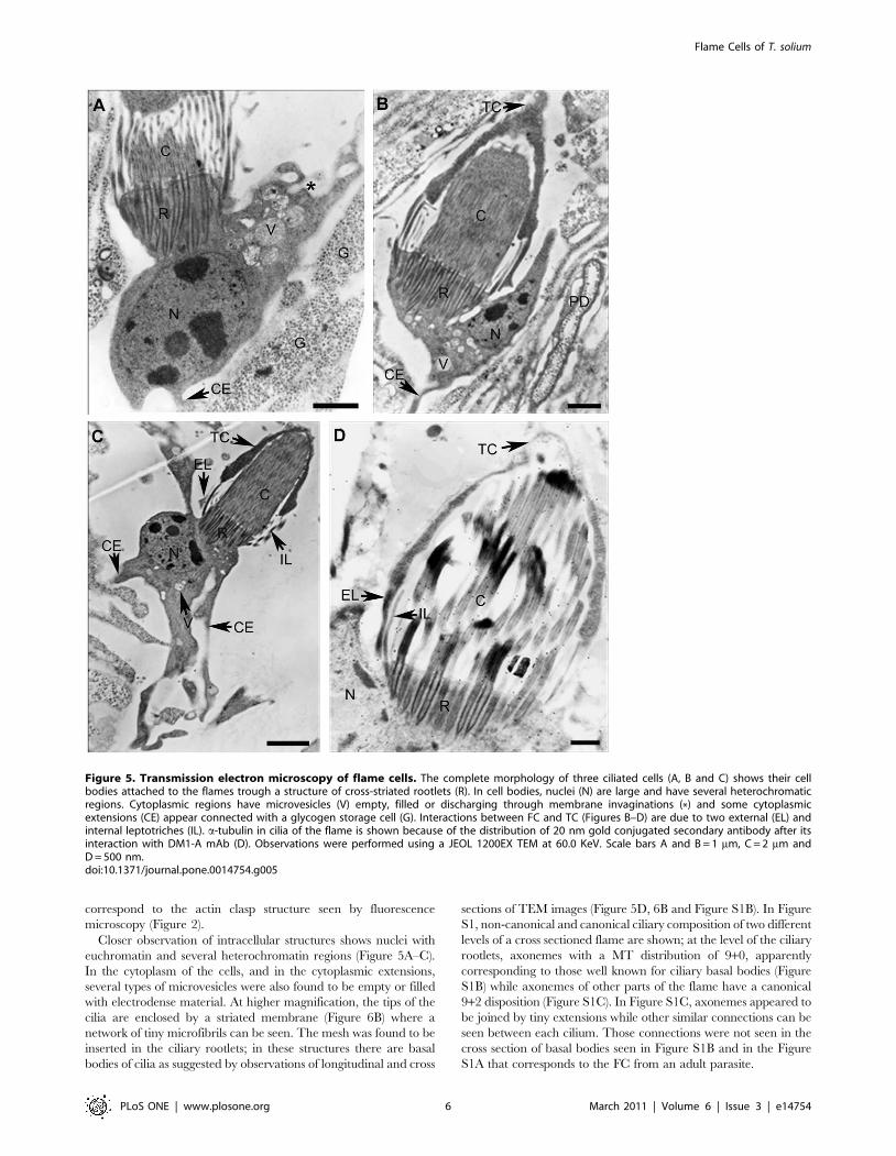
Closer observation of intracellular structures shows nuclei with
euchromatin and several heterochromatin regions (Figure 5A–C).
In the cytoplasm of the cells, and in the cytoplasmic extensions,
several types of microvesicles were also found to be empty or filled
with electrodense material. At higher magnification, the tips of the
cilia are enclosed by a striated membrane (Figure 6B) where a
network of tiny microfibrils can be seen. The mesh was found to be
inserted in the ciliary rootlets; in these structures there are basal
bodies of cilia as suggested by observations of longitudinal and cross
sections of TEM images (Figure 5D, 6B and Figure S1B). In Figure
S1, non-canonical and canonical ciliary composition of two different
levels of a cross sectioned flame are shown; at the level of the ciliary
rootlets, axonemes with a MT distribution of 9+0, apparently
corresponding to those well known for ciliary basal bodies (Figure
S1B) while axonemes of other parts of the flame have a canonical
9+2 disposition (Figure S1C). In Figure S1C, axonemes appeared to
be joined by tiny extensions while other similar connections can be
seen between each cilium. Those connections were not seen in the
cross section of basal bodies seen in Figure S1B and in the Figure
S1A that corresponds to the FC from an adult parasite.
Figure 5. Transmission electron microscopy of flame cells. The complete morphology of three ciliated cells (A, B and C) shows their cellbodies attached to the flames trough a structure of cross-striated rootlets (R). In cell bodies, nuclei (N) are large and have several heterochromaticregions. Cytoplasmic regions have microvesicles (V) empty, filled or discharging through membrane invaginations (*) and some cytoplasmicextensions (CE) appear connected with a glycogen storage cell (G). Interactions between FC and TC (Figures B–D) are due to two external (EL) andinternal leptotriches (IL). a-tubulin in cilia of the flame is shown because of the distribution of 20 nm gold conjugated secondary antibody after itsinteraction with DM1-A mAb (D). Observations were performed using a JEOL 1200EX TEM at 60.0 KeV. Scale bars A and B = 1 mm, C = 2 mm andD = 500 nm.doi:10.1371/journal.pone.0014754.g005
Flame Cells of T. solium
PLoS ONE | www.plosone.org 6 March 2011 | Volume 6 | Issue 3 | e14754

In observations by SEM of parenchymal tissues of one adult
tapeworm, a FC was found (Figure 7): The parenchyma appears
spongy and, inside of a parenchymal crater, a cell body is
protruding (Figure 7A) while the ciliary tuft is inside of the
excretory duct (Figure 7B). These images could correspond to the
flickering cell beating seen at the movie S1. Near to the crater,
there is an empty collecting duct and virtual immersions in these
craters produced the impression they were connected with other
big collecting ducts as it can be seen in the movie S6.
Micrographs of cilia cross-sections, taken at different levels of
the flame and the cell body, depict similarities of ciliary patterns to
those described in textbooks [20,21,22,23]. Association of these
images with those recovered by LSCM and TEM observations
(Figure 2–5 and Figure S1) were used for designing an animation
of a single T. solium FC as shown in movie S6. Similar drawings
were published for other Platyhelminthes [1,4], however in the
present movie were added colors to indicate fluorescence markers
observed in Figures 2 and 4. Using the animation, in order to
illustrate the motions of the cytoskeletal proteins of a T. solium FC
seen by TEM (Figure 5C) we are showing two possible ways for
delivering substances to the cell body; one, inside of vesicles
produced by invagination of the plasmatic membrane and two, for
vesicular trafficking from extended processes of cells in commu-
nication. In the same animation, after the substances were
delivered to the striated rootlet region (as suggested by TEM
images in Figure 5A–C), their contents were discharged to the
flame. After that, due the compression of the F-actin belt-clasp and
the up and down vertical movements of the cilia, the substances
can be throwing delivered to the collecting tubules and finally,
they can be expelled trough the excretory system of the parasites.
In the animated cartoon, the internalization of macromolecules
could be related to the images of the Figure 5A and B as described
earlier about the numerous microvesicles that are in apparent
process of invagination or evagination; in the movie, a macro-
molecule (proposed as a globular protein) is taken by a membrane
invagination, introduced in a microvesicle and transported
through the cytoplasm until the fusion of the microvesicle with
the striated rootlet region. In this region, the liberated macromol-
ecule is diffusing trough the striated rootlet region to the lumen of
the FC where the squeezing and the movement of the cilia redirect
it to the protonephridial duct. Other solutes, considered of low
molecular weight like electrolytes or organic compounds, could be
taken by from the extended processes coming from other cells as
suggested from Figures 5A, B and C and, because of the squeezing
and the ciliar movements, these solutes can be redirected to the PS
ducts for their elimination/reabsortion. Reabsortion could be
Figure 6. Ultrastructure of the protonephridial duct and across-striated rootlet of a flame cell. In A, the lumen of aprotonephridial duct (PD) contains small rounded electrodense vesiclespresumably coming from cilia (C). In B, at the cross-striated rootlet (R),cilia are bound by a fine regular network of microfibrils (small arrows)that cross the region from one side of the FC to the other. The insert isshowing a digital magnification of a selected adjacent region of thecorrespondent basal bodies. Scale bars A = 500 nm and B = 200 nm.doi:10.1371/journal.pone.0014754.g006
Figure 7. Scanning electron microscopy of a flame cell. The micrograph was taken from a cross-sectioned body of the proglottid of an adulttapeworm. In A, a cell body (CB) is proximal to the empty lumen of a protonephridial duct (PD). In B, the cilia of a flame cell (C) are close to extensionsfrom the wall of the PD (arrow). In both images, flame cells (FC) are surrounded by a spongy interstitial matrix (IM). Scale bars A = 1 mm and B = 2 mm.doi:10.1371/journal.pone.0014754.g007
Flame Cells of T. solium
PLoS ONE | www.plosone.org 7 March 2011 | Volume 6 | Issue 3 | e14754

executed by ducts trough the extensions seen at TEM in Figures 6A
and by SEM in Figure 7B.
In order to combine all LCSM and TEM images and
computational animation in order to present the animation in a
3D format, we decided to apply the 3D Max software on a T.
solium FC image obtained by TEM (Movie S7). For this purpose,
we took into account the observations of nuclei stained with DAPI
(Figure 2), the co-expression of F-actin and myosin II (Figure 3),
the 3D reconstruction of the F-actin clasp (Figure 4), the a-tubulin
fluorescence staining (Figure 2) and images obtained by TEM and
SEM (Figure 5, 6 and 7).
Discussion
Results of morphology, dynamics and computationalvisualization of flame cells of tapeworms of medicalimportance
As the present studies illustrate, a complete microscopic analysis
and morphological characterization of T. solium cysticerci flame
cells are described for the first time. The importance and
implications of these studies can be extended to the cestoidea
class and also, they can be applied to the Platyhelminth phylum.
Better knowledge of the composition and the dynamics of FC
could give a better understanding about the terminal cells of the
complex excretory systems of these organisms [1]. In addition, for
medically important helminths, this knowledge could contribute to
better design of antihelminthic drugs since MT are the main
cytoskeletal proteins for the functioning of the FC cilia and any
alteration of them will produce malfunctioning of the excretory
systems as was shown after in vitro treatment of T. crassiceps [12]. In
order to survive inside the host, cysticerci need to carry out a
continuous turnover of substances, for which the maintenance of
an intact excretory system appears to be crucial [3].
Since the flame cells cannot be seen by direct observation of live
intact cysticerci because the parasite tissue layers (brush border,
syncytial tegument and myocyte fibers) impede their visualization.
Punctured parasites were used to observe the flickering FC from
the inner bladder wall where they were easily identified by the
typical movements of beating cilia (Movie S1). The behavior of the
ciliary tufts in FC was found to be similar to that described
previously for T. solium [11] and other cestodes [4] and further
indicates that these ciliary cells are highly dynamic. It is possible
that the dynamics of the ciliary beating could be associated with
the influence of the high content of calcium [22] that is stored in
the calcareous corpuscles, mineral concretions found in the lumen
of the channels of the cysticerci PS [19]. As suggested, by the
presence of the flames in the collecting ducts (Figures 2C, 6A,
Movie S1), the flickering movements could determine the flow of
fluids inside of PS. Since FC are terminal cells of PS in
invertebrates [1] their function is probably important for
delivering substances either for elimination or reabsorption and,
in tapeworms, they are thought to maintain the osmoregulation,
helping to preserve the equilibrium between the hosts and
parasites microenvironments [2]. The dynamics and the biological
roles of FC require that they be interconnected with other types of
cells that belong to the PS as found in the present TEM
observations (Figure 6A) and the computational reconstruction
(Movie S5). According with this, the FC need a specific size and
morphology supported by their cytoskeleton [20,21,22,23] that
can be evaluated with the effects of specific substances that act on
cytoskeletal proteins.
Previous descriptions of the cysticerci FC by light microscopy
offered a static description of these cells [10,11]. In the present
studies, the use of videomicroscopy offers the possibility of linking
the morphology with their dynamics in live parasites and their
interactions with the excreting ducts (Movie S1). Integration of the
real time visualization with fluorescence observations of the
distribution of their cytoskeletal proteins (Figure 2) permitted
reconstruction (Movies S3 and S4) of the internal morphology of
these cells as seen in Figure 4. With the resolution using the
Nomarsky optics it was possible to see the cilia of the flames
(Figure 2I, Movie S1). By the use of electron microscopy combined
with computational reconstructions new details of the previously
described morphological analysis and, as seen in movies S6 and
S7, produced animations of the structure and the cellular
mechanics of a FC at the ultrastructural level. The combination
of several microscopy methods is considered an excellent tool for
presenting visual data [24,25] in addition to the performance of
the computer-based 3D reconstructions using AMIRA software
[26]. More ultrastructural visualizations, like those produced by
electron tomography [27,28], could reveal more details of FC of
these parasites.
Cytoarchitectural distribution of cytoskeletal proteins incilium-based cells
Detection of a-tubulin in cysticerci tissues indicates that this
cytoskeletal protein is highly expressed in the syncytial tegument
(Figure 1) and in many FC; the majority of these ciliated cells
(approximately 80–90%) were found in tissues of the spiral canal of
the invaginated scolex. Cestodes do not have a digestive system, so
that the presence of tubulin is important for their survival and
adaptation inside hosts, because the absorption-excretion-secretion
system permits the continuous absorption of nutrients as well as an
effective waste elimination [2]. The presence of a-tubulin in the
syncytial tegument can be associated with requirements of MT
that are necessary for maintaining the highly dynamic vesicular
traffic required for uptake and secretion of macromolecules as
demonstrated for T. crassiceps cysticerci [29] and are well known in
eukaryotic cells [20]. However, for FC, the MT is necessary for the
function of these ciliated cells [22] as described previously.
However, since these cells are not easy to find in live parasites
due their deeper tisular localization and the constant movements
of the tissues; it could be useful to transfect the parasites with a
GFP-tag for visualization of tubulin and to observe directly the
dynamics of the FC as established for the study of the dynamic
processes in ciliary movements [30].
As in the case of a-tubulin, F-actin is another cytoskeletal
protein highly expressed in FC of T. solium cysticerci. The
expression of the protein in similar cells was observed in other
cestodes such as D. dendriticum [6], in the monogenean G. rysavyi [7]
and the trematode S. mansoni [9]. In the former, it was found as
part of a ring situated in the middle portion of their ciliated cells
while in the later, with 3D reconstructions, it was found to be
concentrated in a hollow barrel. It was interesting that phalloidin
was found in in the belt clasp of T. solium FC (Figure 2E) and
similar fluorescent patterns were observed for D. dendriticum [6] and
S. mansoni [9]; as proposed for S. mansoni [9], the higher
concentration of F-actin in this region could be related with the
contractile motions of these cells. However, there may be an
association with a-tubulin (see later) in order to maintain the
cellular structure and the dynamics of the ciliary tuft.
Only co-localization of F-actin and myosin II were detected
with high yellow fluorescent intensity in dots scattered in the IM as
seen in Figure 3. Due the cluster of fluorescent dots contiguous to
circular structures, presumably corresponding to cross-sectioned
PD as described for flame cells after using the Bodians protargol
method [10], the observation could suggest the presence of an
actomyosin contractile system as found in contractile rings in cell
Flame Cells of T. solium
PLoS ONE | www.plosone.org 8 March 2011 | Volume 6 | Issue 3 | e14754

divisions [31]. If the contractile system is located at the the actin
clasp of T. solium FC (Figure 2), it suggests that myosin II is a linear
molecular motor required for any constriction of the clasp as the
proposed driving force in contractile rings [32]. Localization of F-
actin with a-tubulin to find that the actin clasp is apparently
embracing the ciliary tufts of the FC (Figure 2F–2H) and the
distribution of these cytoskeletal proteins indicate that the actin
clasp could be also associated with the MT of the flames. If this is a
real situation, the role of MT at this site is for inducing the
necessary movements of the suggested contractile system in order
to favor propulsion of substances to the PD as inferred from the
animated movie S7 and movie S1 where a FC is in direct contact
with a PD. There is a microtubule-dependent regulation of the
actomyosin system during the contractile ring assembly [31],
however, this proposal needs to be characterized for the contractile
machinery in T. solium FC.
In ultrastructural observations, at the level of the ciliary tuft and
the cell body (Figures 5 and 6B), it was observed that these cellular
structures were apparently fastened by a striated rootlet region in a
similar fashion as described for ciliated cells [33]; the presence of a
microfibrillar mesh distributed in an horizontal disposition appears
to firmly surround the cilia tips (Figure 6B). One support for the
biological role of this mesh could be the detection of c-tubulin in
this region because this is one of the most important proteins
found inside of basal bodies and in the site of nucleation of ciliary
MT [20,21,34]. Additionally, the structuring of the rootlet region
that involves tiny microfibrillar strands and the ciliary rootlets
(Figures 5D and 6B) could be important for anchoring the flame
during the flickering motion as seen in the movies S1 and S2. It
will be of interest to define the nature of these tiny microfibrils that
could be coordinated with other cytoskeletal proteins that have a
supportive function.
At the level of the longitudinal and cross sections of cilia of the
Figures 5D and 6B, it was found, that 80 to 90 cross-sectioned
axonemes were seen within cysticerci FC (Figure S1B and S1C)
and this number could be useful for phylogenetic considerations
as proposed for the phylum of Platyhelminthes [1]; in the
cestode Trilocularia acanthiaevulgaris there are 50–70 axonemes
[35] while 40–50 axonemes were found in A cirrusspiralis [1].
Apparently, as seen in more detail in the Figure S1B, there are
tiny extensions that are required for joining each cilium of the T.
solium FC cilia. Possibly, any disruption of these tiny structures
could produce a failure of the FC functions and they need to be
characterized.
The ultrastructure of T. solium flame cells appears similar to that
of A. cirrusspiralis [1] (Figure 5 and 6). TEM images present the cells
interacting with PS duct cells trough leptotrichs and ribs and, in
SEM images, T. solium FC (Figure 7B) appeared to interact
through their leptotrich structures with extensions of PS duct cells.
In the SEM images of Figure 7, FC are embedded in the IM, near
to excretory ducts or inside of them and apparently the tuft of cilia
of the image on Figure 7B corresponds to that seen on Figure 6A
by TEM. Both images could suggest how the fluids and their
materials are flowing through the ducts as a result of the ciliary
movements. Ultrastructural observations, suggest also that FC
have the capacity to interact with other cells trough cytoplasmic
extensions that are seen (Figure 5A–5D); apparently there are
intercellular pseudopods that hold FC and PS duct cells together.
Intercommunications between FC and other cells could be part of
the tubular network system and the synctitial structure of the
collecting channels and tubules described for cestodes [36] and it is
a visual example of how they can be present at deeper
parenchymal tissues as seen in the final part of the movie S6.
Continuous synctitial layers in cestodes facilitate their exchange of
nutrients or their capacity to respond immediately to changes in
their microenviroment [2].
3D Reconstruction of the cytoskeletal proteindistribution in flame cells
In order to understand the localization of the cytoskeletal
proteins in the flame cells we used the integration of light,
fluorescence and electron microscopy observations, together with
a previous report that these cells have intense flickering
movements of their ciliary tufts [5], the recording of them (Movie
S1) and their computational visualization could be used as a
approach for studying the functionality and the dynamics of these
cells. Due the computational capability of reconstruction and
resolution produced by the AMIRA software, we were able to
produce a reconstruction of T. solium FC that allowed observation
with an approximation to the ultrastructural level using a LSCM
resolution. The 3D reconstructions of T. solium FC were achieved
and visualizations were obtained that illustrate morphological
details difficult to obtain by conventional TEM or SEM. For
example, the F-actin in T. solium FC was found to be a cellular
structure that looks like an belt clasp that is apparently embracing
the ciliary tufts. The distribution of this actin clasp surrounds and
encloses the ciliary tufts and apparently fastens the soma body.
When the actin clasp is sectioned by computer, as shown in
Figures 4C–E, and the 3D images are rotated in order to observe
the center of the clasp, it was possible to see that there is a hole
(See the animation presented in the movie S5). Co-localization of
DAPI stained nuclei shows they appear to be positioned according
to the plane of orientation of the actin clasp and the ciliary tuft.
The meaning of these observations could be that the actin
structure is necessary for holding the cell body to cilia. Also, it is
possible that the morphological protrusion of the actin clasp, as
seen in the reconstruction of the movie S5, could support the
septate junction found between the extensions of the tubular cell
interconnected cytoplasmic network as described for A. cirrusspiralis
[1]. The AMIRA 3D volume reconstruction produced a solid actin
clasp structure at the resolution of the LSCM; it will be necessary
to know how the disposition of actin inside of the clasp is by
evaluating its expression by immunogold assays for TEM or by the
use of ET.
Hypothetical visualization of the dynamics ofcilium-based cells of the excretory system of tapeworms
It is clear that the results of this analysis show that FC contain at
least three cytoskeletal proteins: F-actin, a-tubulin and myosin II,
all proteins associated with contractile movements in cells. These
cytoskeletal proteins are clearly located in the different structures
of the FC. Flame cells are found in close contact or within the
excretory system, suggesting that they are an integral part of the
protonephridial system. However, whether these associations are
involved in the flow of fluids in the protonephridial system, and
their importance for survival in the host, remains to be established.
However, due in the present studies we were able to combine
experimentally produced images from microscopy with computa-
tional imaging strategies; we consider that they can be integrated
for producing visual dynamic images as presented in the movie S7
according with theorical knowledge of the studied cytoskeletal
proteins. The animated model is an attempt for illustrating the
functional activity of T. solium FC were the ciliated cells have
intense movements of the ciliary tuft as seen in videomicroscopy
(Movie S1). Entrance of the fluids in cells could be done trough the
empty space in the clasp, where there is a connecting tubule of the
tubular network, supported by the digital reconstructions of cross-
Flame Cells of T. solium
PLoS ONE | www.plosone.org 9 March 2011 | Volume 6 | Issue 3 | e14754

sections images presented at Figures 4C–D. Inside of the cells, the
fluids are pushed to the ciliary tuft due the contractibility/
expansion of the actin clasp due to an acto-myosin system that
produce a continuous squeezing of the ciliated cells (Movie S7) as
produced during constriction of cells during the cellular division
[20] and as the distribution of the cytoskeletal proteins was seen
according with the 3D-volume reconstruction seen at Figures 2G,
2H and 4A–E. For macromolecules, their entrance to the cells
could be performed by means of vesicular traffic; as suggested by
electrodense vesicles seen at the soma bodies at TEM images
(Figures 5A, 5B and 5C) and as presented in the animation of the
movie S7; the macromolecules are uptaked into microvesicles seen
in an apparent formation from plasmatic membrane invagina-
tions, these microvesicles apparently travel inside the cytoplasm of
the cell body and finally they are discharged trough the cross-
striated rootlets region to the cilia. Later, because of the
movements of the ciliary tuft, these movements drive out the
contents into the lumen of the PS ducts. In this animated cartoon,
we speculate on how solutes enter the cells, they pass through the
FC and they are discarded to PS ducts. Similar experiments to
those carried out with T. crassiceps cysticerci and schistosomula of S.
mansoni for the uptake of macromolecules [29,37] and the blocking
of the internalization could offer support to the presented
proposal. Resultant of combination of experimental and compu-
tational strategies, as performed in present studies, could be a
valuable approach for trying to explain dynamic aspects and to
think about the mechanical properties of ciliated cells as flame cells
of the cestode T. solium.
Supporting Information
Figure S1 Transmission electron microscopy of flame cell
axonemes. Cross sections of the cilia of three flames are shown
(A, B and C). Axonemes of FC from the invaginated scolex of a
cysticercus that were cross-sectioned in two different regions are in
A and B, while in C, there is an axoneme of a FC from an adult
parasite. In A, axonemes in a distribution of 9+0 are presented and
they were obtained near to the cross-striated rootlets region shown
in figure 6B. In B and C, axonemes (white arrow) show a canonical
distribution of 9+2 and they were obtained from regions localized
at the level of the flames. Axonemes of cilia of cysticerci FC (B)
appear to be more separated and connected by tiny prolongations
(black arrowheads) that emerge from each cilium. In comparison,
axonemes of adult parasite FC (C) appear to be with a closer
interaction and delimited by hexagonal junctions. Observations
were performed using a JEOL TEM at 60.0 KeV. Scale
bars = 500 nm.
Found at: doi:10.1371/journal.pone.0014754.s001 (0.91 MB TIF)
Movie S1 Flickering of a flame cell in a live cysticercus.
Three presentations of flame cells are presented from time-
elapsed sequences, under low magnification (406) of live
parasites seen under Nomarsky illumination conditions. First,
inside of the parasite tissue, embbebed in the interstitial
matrix, several FC could be seen moving. One flickering cell,
surrounded by a red circle, shows their soma body oriented to
the protonephridial duct. Later, in the optical zooming (14.86)
of the flickering cell, the flame shows some cilia signaled by
white arrows. Finally, in the red circle, it is showing the beating
of cilia of a FC inside of a protonephridial duct. The plane
focus of the tissue is continuously moving due its contractibility
(MPEG 4.14 MB).
Found at: doi:10.1371/journal.pone.0014754.s002 (4.34 MB
MP4)
Movie S2 Computational reconstruction of the F-actin in a
flame cell. Z sequences of LCSM images were processed for their
direct volume renderization with AMIRA software. In the movie,
only F-actin (red) was reconstructed and its digital rotation show it
looks like a clasp of a belt that fasten the ciliary tuft (green) (MPEG
8.48 MB).
Found at: doi:10.1371/journal.pone.0014754.s003 (8.90 MB
MPG)
Movie S3 Spatial projection of computational reconstructed F-
actin clasps of several flame cells. After processing the images as
indicated for the movie S2, the F-actin clasps of several flame cells
are manipulated in order to observe how is their spatial volume
and how this is associated with the embracement of the ciliary tufts
(green) (MPEG 8.95 MB).
Found at: doi:10.1371/journal.pone.0014754.s004 (9.39 MB
MPG)
Movie S4 3D visualization and virtual immersion inside of the
invaginated scolex. Z sequences of LCSM images as presented in
figures 2G and 2H were computationally processed as indicated
for figure 4 and movies S2 and S3. During the trip of the virtual
immersion, from low to high magnifications and after performing
approximations to FC, it is possible to distinguish how the ciliated
cells are immersed in the interstitial matrix and how is visualized
the topography of the reconstructed cytoskeletal components of
the FC and the matrix (MPEG 6.67 MB).
Found at: doi:10.1371/journal.pone.0014754.s005 (7.00 MB
MPG)
Movie S5 Visualization of the co-localization of myosin II and
a2tubulin in the invaginated scolex. After computational
reconstructions, Z digital sections of the images permitted to
observe the tisular distribution of muscle fibers (orange), the
interstitial matrix (green), ciliary tufts emerging from the matrix,
clasps of cells and several cytoplasmic extensions that interconnect
cells. In the middle of the clasps, it was found that they have
central empty holes (MPEG 1.21 MB).
Found at: doi:10.1371/journal.pone.0014754.s006 (1.28 MB
MPG)
Movie S6 Reconstruction and animation of cytoskeletal proteins
of a flame cell. Using the TEM image of the figure 5C, according
with the flickering of the FC shown in the movie 1, it was obtained
a 3D Max reconstruction of the cell. For this, a combination of the
figure 4 and movie S3 was done and as a resultant 3D
reconstruction was obtained. In the movie, the hypothetical
animation of the dynamics between the F-actin clasp (red) and the
tubulin of the ciliary tuft (green) is showing the function of these
cytoskeletal proteins of the ciliated cell (MPEG 5.76 MB).
Found at: doi:10.1371/journal.pone.0014754.s007 (6.04 MB
MPG)
Movie S7 Animation of the hypothetical function of flame cells.
Using as a template the images from the figures 2, 4–7, animations
of movies S3 and S4 and videomicroscopy of the movie S1, they
were combined for performing a 3D animation by 3D Max
software. In the animation, the contraction and expansion of the
F-actin clasp and the flickering of the ciliary tuft is responsible for
routing the contents of the vesicles (an orange globular particle) to
the lumen of the protonephridial ducts. These contents could be
come from invaginations at the membranes of the soma bodies or
from the interconnections of other cells, the vesicles go through the
cytoplasm of the cell bodies and they deliver their contents to the
level of the actin clasp (MPEG 3.87 MB).
Found at: doi:10.1371/journal.pone.0014754.s008 (4.07 MB
MPG)
Flame Cells of T. solium
PLoS ONE | www.plosone.org 10 March 2011 | Volume 6 | Issue 3 | e14754

Acknowledgments
Lic. A. Gonzalez for helping with 3D animations. DCV. J. Villamar for
helping in the edition of movies. MVZ S. Reyes Maya, Biol. G. Orozco,
Dra. A. Patron, Dr. R. Coral, Dr. H. Merchant, Dra N. Moreno for
helping with confocal microscopies and Dra. Bertha Espinoza for her
advice during the edition of the manuscript.
Author Contributions
Conceived and designed the experiments: LEVI JRA. Performed the
experiments: LEVI EA EV LR RV ORD AZR JRA. Analyzed the data:
LEVI KW TIF JRA. Contributed reagents/materials/analysis tools: LEVI
EA EV LR KW AZR TIF JRA. Wrote the paper: LEVI JRA.
References
1. Rohde K, Watson NA, Roubal FR (1992) Ultrastructure of protonephridial
system of Anoplodiscus cirrusspiralis (Monogenea Monopisthocotylea). Int J Parasitol22: 443–457.
2. Smyth JD, McManus DP (1989) The adult cestode: special structural featuresrelevant to is physiology. In: Smyth JD, McManus DP, eds. The physiology and
biochemistry of cestodes. New York: Cambridge University Press. pp 5–34.
3. Barnes DR (1987) Platelmintos. In: Barnes DR, ed. Zoologıa de losInvertebrados. Mexico: Interamericana, S.A. de C.V. pp 179–223.
4. Coil HW (1991) Platyhelminthes: Cestoidea. In: Harrison FW, Bogitsh BJ, eds.Microscopic Anatomy of Invertebrates. New York: Wiley-Liss, Inc. pp 211–283.
5. Brusca CR, Brusca JG (2003) Phylum Platyhelminthes. In: Brusca CR,
Brusca JG, eds. Invertebrates. Second ed. Massachusetts: Sinauer Associates,Inc., Publishers. pp 285–318.
6. Wahlberg MH (1998) The distribution of F-actin during the development ofDiphyllobothrium dendriticum (Cestoda). Cell Tissue Res 291: 561–570.
7. Arafa ZS, El-Naggar MM, El-Abbassy SA, Steweart MT, Halton DW (2007)
Neuromusculature of Gyrodactylus rysavyi a monogenean gill and skin parasite ofthe catfish Clarias gariepinus. Parasitol Int 56: 297–307.
8. Moreno MJ, Casado N, Urrea-Parıs MA, Rodriguea-Caabeiro F (2001)Evidence of tubulin in the scolex gland ducts of Gymnorhynchus gigas plerocercoid
(Cestoda: Trypanorhyncha). Folia Parasitol (Praha) 48: 163–164.9. Bahia D, Avelar LG, Vigorosi F, Cioli D, Oliveira GC, et al. (2006) The
distribution of motor proteins in the muscles and flame cells of the Schistosoma
mansoni miracidium and primary sporocyst. Parasitology 133: 321–329.10. Cardenas-Ramırez L, Zaragoza A, Gonzalez-Del Pliego M (1982) Neural and
excretory structures of cysticercus cellulosae. In: Flisser A, Willms K, eds.Cysticercosis: Present State of Knowledge and Perspectives. New York:
Academic Press. pp 281–305.
11. Voge M (1963) Observations on the structure of cysticerci of Taenia solium andTaenia saginata (Cestoda:Taeniidae). J Parasitol 49: 85–90.
12. Palomares F, Palencia G, Ambrosio JR, Ortiz A, Jung-Cook H (2006)Evaluation of the efficacy of the albendazole sulphoxide and praziquantel
combination on Taenia crassiceps: in vitro studies. J Antimicrob Chemother 57:482–488.
13. Roman G, Sotelo J, Del Brutto O, Flisser A, Dumas M, et al. (2000) A proposal
to declarate neurocysticercosis an internacional reportable disease. Bull WorldHealth Organ 78: 399–406.
14. Ambrosio J, Cruz-Rivera M, Allan J, Moran E, Ersfeld K, et al. (1997)Identification and partial characterization of a myosin-Like protein from
cysticerci and adults of Taenia solium using a monoclonal antibody. Parasitology
114: 545–53.15. Cruz-Rivera M, Reyes TA, Reynoso DO, Flisser A, Ambrosio J (2006)
Comparison of biochemical and immunochemical properties of myosin II intaeniid parasites. Cell Biol Int 30: 598–602.
16. Ambrosio JR, Reynoso-Ducoing O, Hernandez SH, Correa PD, Gonzalez ML,et al. (2003) Actin expression in Taenia solium cysticerci (cestoda): tisular
distribution and detection of isoforms. Cell Biol Int 27: 727–733.
17. Rivera N, Ibarra F, Zepeda A, Fortoul T, Hernandez A, et al. (2004)Tegumental surface changes in adult Fasciola hepatica following treatment in vitro
and in vivo with an experimental fasciolicide. Parasitol Res 93: 283–286.
18. Willms K, Presas AM, Jimenez JA, Landa A, Zurabian R, et al. (2005) Taeniid
tapeworm responses to in vitro glucose. Parasitol Res 96(5): 296–301.19. Vargas-Parada L, Merchant MT, Willms K, Laclette JP (1999) Formation of
calcareous corpuscles in the lumen of excretory canals of Taenia solium cysticerci.Parasitol Res 85(2): 88–92.
20. Alberts B, Johnson A, Lewis J, Raff M, Roberts K, et al. (2002) The
Cytoskeleton. In: Alberts B, Johnson A, eds. Molecular Biology of the Cell. NewYork: Garland Science. pp 907–982.
21. Karp G (2005) Citoesqueleto y Movilidad Celular. In: Karp G, ed. BiologıaCelular y Molecular. Bogota: McGraw-Hill Interamericana Editors S.A. pp
357–407.
22. Bray D (2001) Cell Movements from Molecules to Motility. Second ed. NewYork: Garland Publishing. 372 p.
23. Lodish H, Berk A, Zipursky SL, Matsudaira P, Baltimore D, et al. (2000) CellMotility and shape II: Microtubules and Intermediated Filaments. In: Lodish H,
Berk A, eds. Molecular Cell Biology. Fourth ed. USA: W. H. Freeman and
company. pp 796–843.24. McGill G (2008) Molecular Movies… coming to a lecture near you. Cell 133:
1127–11320.25. Iwasa JH (2010) Animating the model figure. Trends Cell Biol. 20: 699–704.
26. Neusser TP, Hess M, Haszprunar G, Schrodl M (2006) Computer-Based Three-Dimensional Reconstruction of the Anatomy of Microhedyle remanei (Marcus,
1953), an Interstitial Acochlidian Gastropod From Bermuda. J Morphol 267:
231–247.27. Nicastro D, Schwartz C, Pierson J, Gaudette R, Porter M, et al. (2006) The
Molecular Architecture of Axonemes Revealed by Cryoelectron Tomography.Science 313: 944–948.
28. McIntosh R, Nicastro D, Mastronarde D (2005) New views of cells in 3D: an
introduction to electron tomography. Trends Cell Biol 15: 43–5l.29. Ambrosio J, Landa A, Merchant MT, Laclette JP (1994) Protein uptake by
cysticerci of Taenia crassiceps. Arch Med Res 25: 325–330.30. McGrath J, Solmo S, Makova S, Tian X, Brueckner M (2003) Two populations
of node monocilia initiate left-right asymmetry in the mouse. Cell 114: 61–73.31. Boal D (2002) Mechanical designs. In: Boal D, ed. Mechanics of the Cell.
Cambridge: Cambridge University Press. pp 323–341.
32. Darenfed H, Mandato AC (2005) Wound-induced contractile ring: a model forcytokinesis. Biochem Cell Biol 83: 711–720.
33. Lemolluis M, Marty M (1990) Immunocytochemical study of the formation ofstriated rootlets during ciliogenesis in quail oviduct. J Cell Science 95: 423–432.
34. Keller LC, Romijn EP, Zamora I, Yates JR, 3rd, Marshall WF (2005) Proteomic
analysis of isolated Chlamydomonas centrioles reveals orthologs of ciliary-diseasegenes. Curr Biol 15: 1090–1098.
35. McCullough JS, Fairweather I (1991) Ultrastructure of the excretory system ofTrilocularia acanthiaevulgaris (Cestoda, Tetraphyllidea). Parasitol Res 77: 157–160.
36. Lumsden DR, Hildreth BM (1983) The Fine Structure of Adult Tapeworms. In:Arme C, Pappas WP, editors Biology of the Eucestoda. London: Academic Press
Inc. pp 177–227.
37. Tan HH, Thornhill JA, Al-Adhami BH, Akhkha A, Kusel JR (2003) A study ofthe effect of surface damage on the uptake of Texas Red-BSA by schistosomula
of Schistosoma mansoni. Parasitology 126: 235–240.
Flame Cells of T. solium
PLoS ONE | www.plosone.org 11 March 2011 | Volume 6 | Issue 3 | e14754