Taenia solium disease in humans and pigs: an ancient parasitosis disease rooted in developing...
16
Review Taenia solium disease in humans and pigs: an ancient parasitosis disease rooted in developing countries and emerging as a major health problem of global dimensions Edda Sciutto a *, Gladis Fragoso a , Agnes Fleury b,c , Juan Pedro Laclette a , Julio Sotelo a,b , Aline Aluja d , Laura Vargas a , Carlos Larralde a a Departamento de Inmunología, Instituto de Investigaciones Biomédicas, Universidad Nacional Autónoma de México, UNAM, AP70228, Mexico D.F. 04510, Mexico b Instituto Nacional de Neurologia y Neurocirugia, Mexico D.F., Insurgentes Sur 3877, Mexico D.F. 14269, Mexico c Institut d’epidémiologie neurologique et de neurologie tropicale, Université de Limoges, 2, rue du Dr. Marcland, Limoges 87025, France d Facultad de Medicina Veterinaria y Zootecnia, Universidad Nacional Autónoma de México, UNAM, AP70228, Mexico D.F. 04510, Mexico ABSTRACT – This article reviews current knowledge on human and porcine cysticercosis caused by Taenia solium. It highlights the conditions favorable for its prevalence and transmission, as well as current trends in research on its natural history, epidemiology, immunopathology, diagnosis, treatment and prevention. Our opinions on the most urgent needs for further research are also presented. © 2000 Éditions scientifiques et médicales Elsevier SAS Taenia solium / cysticercosis-taeniasis / diagnosis / epidemiology / vaccination / pathogenesis 1. Introduction Taenia solium is a parasite whose larva (cysticercus) may localize in the central nervous system of humans, causing neurocysticercosis (NCC) – prevalent in develop- ing countries and re-emerging in affluent societies. NCC may adopt different forms: the clinically mild forms caus- ing no or few and endurable symptoms, and the clinically severe forms causing a life-threatening, often fatal and frequently disabling form of disease. Cysticerci may also develop in other organs (eyes, skeletal muscle, heart, subcutaneous tissue) where they cause comparatively benign pathology (cysticercosis, CC). Cysticerci also develop in the skeletal muscle of the pig (the intermediary host) as a necessary step in their life cycle. In order to attain the adult tapeworm stage cysticerci must be ingested by a human (the only definitive host), in which it dwells as an intestinal parasite causing minor gastrointestinal distur- bances in its host. However, the tapeworm is of major importance for transmission, as it produces tens of thou- sands of eggs on a daily basis, for years, which are shed into the environment by defecation. Each egg has the potential of becoming a cysticercus upon its ingestion by pigs or humans. T. solium infection of humans is a disease known since antiquity [1]. Western European countries progressively controlled and finally eradicated it in the early 1900s, mainly through social development as well as meat inspec- tion and confiscation programs [2, 3]. T. solium disease is still deeply rooted in Latin America, Africa and Asia, where it develops its complete cycle and displays its multiple interactive facets, with considerable effects on both public health and the economy. From its enclave among the poor, T. solium threatens the more privileged social sectors of endemic areas and the people of devel- oped countries by way of tapeworm carriers among migrant workers [4–6]. Biomedical scientific communities in endemic areas are well aware of T. solium disease and have contributed significantly to its description and man- agement as well as to scientific research [7–16]. The recent but highly noticeable emergence of human NCC in the USA [5, 17–19] and to a lesser extent in some Euro- pean countries [6, 20, 21], mostly related to imported cases in migratory workers, has helped in the growing medical concern and contributed greatly to modern research [5, 18]. World awareness of the threat T. solium poses to human health and the economies of endemic countries (presumed to be enormous) is thus progressively increasing. * Correspondence and reprints. E-mail address: [email protected] (E. Sciutto). Microbes and Infection, 2, 2000, 1875-1890 © 2000 Éditions scientifiques et médicales Elsevier SAS. All rights reserved S1286457900013368/REV Microbes and Infection 2000, 1875-1890 1875
Transcript of Taenia solium disease in humans and pigs: an ancient parasitosis disease rooted in developing...

Review
Taenia solium disease in humans and pigs:an ancient parasitosis disease rooted in developingcountries and emerging as a major health problem
of global dimensionsEdda Sciuttoa*, Gladis Fragosoa, Agnes Fleuryb,c, Juan Pedro Laclettea, Julio Soteloa,b, Aline Alujad, Laura Vargasa,
Carlos Larraldea
aDepartamento de Inmunología, Instituto de Investigaciones Biomédicas, Universidad Nacional Autónoma de México,UNAM, AP70228, Mexico D.F. 04510, Mexico
bInstituto Nacional de Neurologia y Neurocirugia, Mexico D.F., Insurgentes Sur 3877, Mexico D.F. 14269, MexicocInstitut d’epidémiologie neurologique et de neurologie tropicale, Université de Limoges, 2, rue du Dr. Marcland, Limoges 87025, France
dFacultad de Medicina Veterinaria y Zootecnia, Universidad Nacional Autónoma de México, UNAM, AP70228, Mexico D.F. 04510, Mexico
ABSTRACT – This article reviews current knowledge on human and porcine cysticercosis causedby Taenia solium. It highlights the conditions favorable for its prevalence and transmission, as well ascurrent trends in research on its natural history, epidemiology, immunopathology, diagnosis, treatmentand prevention. Our opinions on the most urgent needs for further research are also presented.© 2000 Éditions scientifiques et médicales Elsevier SAS
Taenia solium / cysticercosis-taeniasis / diagnosis / epidemiology / vaccination / pathogenesis
1. IntroductionTaenia solium is a parasite whose larva (cysticercus)
may localize in the central nervous system of humans,causing neurocysticercosis (NCC) – prevalent in develop-ing countries and re-emerging in affluent societies. NCCmay adopt different forms: the clinically mild forms caus-ing no or few and endurable symptoms, and the clinicallysevere forms causing a life-threatening, often fatal andfrequently disabling form of disease. Cysticerci may alsodevelop in other organs (eyes, skeletal muscle, heart,subcutaneous tissue) where they cause comparativelybenign pathology (cysticercosis, CC). Cysticerci alsodevelop in the skeletal muscle of the pig (the intermediaryhost) as a necessary step in their life cycle. In order toattain the adult tapeworm stage cysticerci must be ingestedby a human (the only definitive host), in which it dwells asan intestinal parasite causing minor gastrointestinal distur-bances in its host. However, the tapeworm is of majorimportance for transmission, as it produces tens of thou-sands of eggs on a daily basis, for years, which are shedinto the environment by defecation. Each egg has thepotential of becoming a cysticercus upon its ingestion bypigs or humans.
T. solium infection of humans is a disease known sinceantiquity [1]. Western European countries progressivelycontrolled and finally eradicated it in the early 1900s,mainly through social development as well as meat inspec-tion and confiscation programs [2, 3]. T. solium disease isstill deeply rooted in Latin America, Africa and Asia,where it develops its complete cycle and displays itsmultiple interactive facets, with considerable effects onboth public health and the economy. From its enclaveamong the poor, T. solium threatens the more privilegedsocial sectors of endemic areas and the people of devel-oped countries by way of tapeworm carriers amongmigrant workers [4–6]. Biomedical scientific communitiesin endemic areas are well aware of T. solium disease andhave contributed significantly to its description and man-agement as well as to scientific research [7–16]. Therecent but highly noticeable emergence of human NCC inthe USA [5, 17–19] and to a lesser extent in some Euro-pean countries [6, 20, 21], mostly related to importedcases in migratory workers, has helped in the growingmedical concern and contributed greatly to modernresearch [5, 18]. World awareness of the threat T. soliumposes to human health and the economies of endemiccountries (presumed to be enormous) is thus progressivelyincreasing.
* Correspondence and reprints.E-mail address: [email protected] (E. Sciutto).
Microbes and Infection, 2, 2000, 1875−1890© 2000 Éditions scientifiques et médicales Elsevier SAS. All rights reserved
S1286457900013368/REV
Microbes and Infection2000, 1875-1890
1875

Recent scientific efforts have concentrated on theimprovement of diagnosis and therapeutics, as well as ona better understanding of the role of geographic andsocioeconomic variables in transmission [22, 23]. Immu-nological and genetic factors of both host and parasite,presumably related to the natural history, pathology andprevention of the disease, are also coming into focus.Indeed, some advances have been made in several ofthese areas, but the available information is not completeand some important points are still debatable.
The clinical and pathological descriptions of the humandisease by conventional methods, as well as the histologi-cal structure of the parasite, are the most thoroughlydocumented topics [18, 24–26] and they are merely brieflysketched here. Topics that are medically important orbiologically intriguing and that need further research con-stitute the main scope of this review. Some of these includethe role of immunity and genetic background in pathogen-esis, the usefulness of immunodiagnostic tools in seroepi-demiology and medical diagnosis, the ways of detectingthe tapeworm carrier, the role of vaccination to prevent pigCC and consequently decrease infection pressure uponhumans, the long-term benefit of the chemically treatedhuman NCC case, the role of gender and age in suscepti-bility, and the parasite’s antigenic, pathogenic and geneticdiversity. Other topics about which we know nothing butwhich are of paramount importance are only mentionedhere, such as the nature of molecules and events that leadand determine the establishment of cysticerci in certaintarget organs and the mechanisms underlying high host–parasite specificity.
Detailed and systematic studies are necessary to solvemany of the puzzles posed by T. solium, and even moreso, to deal effectively with the medical aspects of humanand porcine T. solium disease in endemic countries withthe transmission conditions that prevail.
2. The T. solium parasite
A brief summary meant as a reminder of the parasite’sprincipal biological features (i.e. morphology, life cycle,histology, metabolism, fertilization and diversity) is con-densed in the legends of figures 1–8, alongside a gallery ofclassic illustrations taken from different sources [27–36].Perhaps one should insist here on a few biological featuresin dire need of research. There is not the slightest hintconcerning the mechanisms underlying the parasite’s highspecificity for pigs and humans, whilst other carnivoresand coprophagic mammals are only occasionally afflictedby CC [37, 38]. Hamsters and chinchillas may becometapeworm carriers especially if they are forcefully treatedwith corticosteroids [39–41]. Likewise, T. solium’s tissuetropism towards the central nervous system (CNS) ofhumans seems to be more frequent in Latin America,whilst it is said that in India the subcutaneous cysticercusis more frequent [42]. Perhaps some of the answers lie inits almost unexplored metabolism and in the as yet barelyapproached crucial aspects of its genetic, antigenic andpathogenic diversity. Surely, the issue of antigenic diver-sity of T. solium is of great practical importance for the
development of immunodiagnostic technology and for thedesign of vaccines. In the only formal immunotaxonomicstudy reported, it was found that all the 15 different col-lections of cysticerci shared but 30% of their antigens inimmunoelectrophoresis [43]. Routine Western blot tech-nology, which can differentiate as many as 50–60 differentbands in whole antigen extracts [44], would surely pro-vide very useful information for local and global serologi-cal and immunological studies. Also, because of its as yetuntapped potential use in the diagnosis and perhaps treat-ment of human and porcine disease, we should mentionhere the parasite’s unusually high concentrations of freeand zinc-bound porphyrins in its vesicular fluid (figure 9).This accumulation of porphyrins makes the cysticercistrongly fluorescent and perhaps sensitive to ultravioletlight [45].
3. The human diseaseThe most frequent and severe human disease caused by
T. solium in Latin America is the neurological form ofcysticercosis, NCC. CC located elsewhere rarely leads toconsultation, except that in the eyes [25], and is thereforeessentially uncharacterized medically and epidemiologi-cally.
3.1. The clinical picture
The clinical picture of NCC is heterogeneous and non-specific [46]. Approximately 30–40% of the cases are
Figure 1. Scanning electron micrograph of the scolex of T. so-lium. Adult T. solium is a tapeworm that inhabits the intestine ofman. The worm shows a globular structure at one end, called ascolex, which tapers on one side forming a region of proliferativetissues from which proglottides are produced in a chain, formingthe strobila. Note the suckers (four in total) and the large rostel-lum with two rows of 11–14 hooklets 100–160 µm long. Bothsuckers and rostellum allow attachment of the worm to theintestinal wall. R, rostellum; S, sucker; N, neck.
Review Sciutto et al.
1876 Microbes and Infection2000, 1875-1890

subclinical. In the symptomatic cases, almost any neuro-logical symptomatology may be presented: from mildsymptoms such as headache, dizziness or occasional sei-zures to a very severe neurological picture with intracra-nial hypertension or dementia. NCC’s clinical pleomor-phism is thought to result from parasite factors (location,size, number) and host factors (degree of immune andinflammatory reactions developed) and makes the diagno-sis on clinical grounds alone impossible. The most com-mon symptom is epilepsy, usually in the form of partialseizures with or without secondary generalization, whichoccurs principally in parenchymal brain CC [47]. Intrac-ranial hypertension, related to perturbation of the normalcirculation of the cerebrospinal fluid (CSF) and/or theobstruction of its absorption, is also frequent, mainly incase of subarachnoid CC. In this case, two different patho-physiological mechanisms are responsible for clinicalmanifestations: the cyst’s mass compressing vicinal struc-tures and the inflammation of the subarachnoid space(arachnoiditis). Many other symptoms have also beendescribed: focal neurological signs (pyramidal tract alter-ations, sensory deficits, involuntary movements, brain-stem dysfunction, etc.), psychiatric manifestations (mild tosevere cognitive impairment, depression, psychotic epi-sodes), alterations of cranial nerves, symptomatic vasculi-tis and progressive decrease of visual acuity [13]. Somedifferences in NCC presentation relate to age and genderof the host. In children, subarachnoidal, ventricular andmuscular locations are rare, whilst ocular location is morefrequent [13]. With respect to gender preferences, whilstmost studies – serological [23, 48] and necropsy series[49] – have found that the disease and positive serologyare almost equally prevalent in men as in women, womenhave considerably higher titers of anticysticercal antibod-
ies than men [48], and the pathological studies did notsearch for finer differences (i.e. numbers of cysts, loca-tions, inflammatory reactions, etc). Also, diffuse cysticer-cal infection (figure 10), which provokes an acute inflam-matory response, quite visible in magnetic resonanceimagery (MRI), is particularly severe in women (encepha-litis syndrome) [50]. Thus, it would appear that there mightexist a significant sexual dimorphism in exposure or sus-ceptibility or in pathogenesis in human NCC, as has beenclearly shown in murine experimental CC caused byT. crassiceps [51–54].
3.2. Anatomic pathology
There are many thorough descriptions of the pathologi-cal changes occurring in cysticercotic human NCC, mostfrom conventional histological studies of necropsy cases[31, 49]. A most recent report includes 481 cases of NCCfound in 20 206 complete autopsies performed in theHospital General of Mexico City from 1954 to 1989 [49].This perhaps unique study confirms NCC’s asymptomaticor clinically mild profile in approximately half of the casescorrelating with low inflammatory responses, in contrastwith the full-blown arachnoidal and ependymal inflam-mation found in severe neurological disease. This obser-vation is perhaps the strongest existing suggestion thatthere are potent immunological, inflammatory and geneticfactors involved in the severity of human NCC. The studyclearly documents the rather fast evolution of NCC in the1950s and 1960s from overt clinical onset to death: in85% of the cases where NCC was the principal disease,the patient died within 5 years of diagnosis. A strongassociation of NCC with malignant hematologic neoplasiawas also found in that study and later it was found that itcorrelated with chromosomal abnormalities [14, 55, 56].
Figure 2. Schematic illustration of a mature proglottide. Proglottids near the neck are immature and undifferentiated, whilst the mostdistant are progressively mature, containing a large number of eggs (27). The strobila length is usually between 2 and 3 m with 800–1 000segments, although strobila as long as 7 m have been reported. A) Reproductive system. The male system consists of ejaculatory duct (Ey),numerous testicles (Ts) connected to a sperm duct. One end of the sperm duct forms a copulatory structure termed cirrus (Ci), the other endtogether with the vagina connect to the genital pore (Gp). The vagina ends in the seminal receptacle (Sr), which in turn is connected to theoviduct (Ovi). The mature oocysts leave the ovary (Ova) through a single oviduct, where fertilization takes place. Cells from the vitellinegland (Vg) migrate through the vitelline duct (Vd) to associate with the zygote. Unicellular glands, called Mehlis glands (Mg) surroundthe ovotype (Ovo), covering the zygote with a mucous and serous secretion that presumably serve as a substrate for the embryophoreformation. In the uterus (Ut), the developing larva matures to complete embryonation. Infective eggs are kept stored until the proglottidesare liberated into the environment. Excretory canal (Ex) and nervous branch (Ns) are also shown. B) Gravid proglottide of T. solium. Thecharacteristic branching of the uterus in the mature segments serves to distinguish between Taeniia species. Gravid proglottides measure12 × 6 mm and posses a uterus with seven to ten lateral branches (Ub) containing both testes and ovary.
Taenia solium disease Review
Microbes and Infection2000, 1875-1890
1877

In other studies inflammation was found to vary from caseto case and even between cysticerci of the same case [24].In general it is agreed that there is focal and surprisinglyscant lymphocytic infiltrate in most seemingly viable cysts.This is another finding that supports the notion that thehost–parasite relationship may be quite convivial at times,and that it is an exaggerated inflammatory response thatcauses most damage to the host. However, in clinicallysevere NCC, collapsed and necrotic cysticerci are found,surrounded by a prominent cellular infiltrate. Some lesionsshow numerous lymphocytes and plasma cells, macro-phages, giant multinucleated cells and few eosinophils,and there is also significant edema, areas of tissue necro-sis, gliosis and connective tissue proliferation. Other NCClesions display prominent infiltration by polymorpho-nuclear leukocytes, in what would appear a massive andsudden inflammatory attack by the host on the parasitethat damages innocent bystanding host components. Instill other NCC cases, usually clinically benign, cysticerciare reduced to a mere scar with few parasite structures leftrecognizable and sometimes substituted by caseous necro-sis with deposits of calcium salts. Typically, in these ‘cal-cified’ cysticercal lesions, the inflammatory infiltrate is
scant, mainly mononuclear and lymphocytic in nature,irregularly distributed among tenuous strands of fibroustissue around the cyst. In meningeal and ependymal NCCcases, usually associated with severe endocranial hyper-tensive disease, chronic inflammation, extensive fibrosisand obstructive endarteritis are quite prominent, constitut-ing a strong argument for inflammation as an importantpathogenic mechanism in severe NCC. Most intriguing isthat in some of the NCC cases with multiple cysticercithere is variation in the integrity and in the degree ofinflammation around each cysticercus within the samehost: some appearing as non-inflammatogenic (thoseviable or calcified) whilst others are necrotic and sur-rounded by prominent inflammation. Barring the possibil-ity of repeated infections at different times – and maybeeven if such is the case – the fact is that each cysticercuswithin the same host may have its own possibly indepen-dent interaction with the host. Heterogeneity in the set ofall host–parasite relationships within each host would beexpected to be of consequence for the evolution, symp-tomatology and response to treatment of NCC patients.There is amazingly little information as to what functionaltype of immune cells and molecules of parasite and hostorigin (i.e. antigen, immunoglobulins, complement andlymphokines) are present in the host–parasite interphase.It would appear worthwhile to investigate these matterswith modern immunohistochemical and genetical in situmethods in a sufficiently rich collection of necropsy casesso as to critically explore the notion that inflammationplays a major role in the pathogenesis of NCC.
Figure 3. T. solium eggs. A) Light micrograph of isolated eggs.The embryophore gives the characteristic striated appearanceunder the light microscope. Eggs measure 30–45 µm and containan oncosphere, also known as hexacanth larvae because of its sixhooks, surrounded by several protective envelopes including theprominent embryophore, the most protective envelope, made upof protein blocks joined by a cementing substance. Reprintedwith permission from [28], copyright 1982. B) Transmissionelectron micrograph of an immature egg. The capsule is indicatedby an arrow; Y, yolk; E, embryophore; EC, embryophore cell;OM, oncospheral membrane; On, oncosphere; H, hook. C) Scan-ning electron micrograph of eggs of 35–45 µm × 30–40 µm. D)Scanning electron micrograph of a broken egg. Arrows indicatethe position of a pair of oncospheral hooks. Digestion of thecementing substance by proteolytic enzymes disgregates theembryophoric blocks. The signal that promotes activation of theoncosphere apparently consists of an increase in the permeabilityof the oncospheral membrane. Once activated, the oncospheretears its membrane and penetrates the intestinal wall [29].
Figure 4. T. solium metacestodes (cysticerci) consist of a translu-cent vesicle filled with fluid (0.5–2 cm in diameter) containing asmall invaginated scolex. The scolex has four suckers and anarmed rostellum like that of the adult. A) Cysticercus dissectedfrom skeletal muscle of pigs. B) Schematic illustration of acysticerci. Sc, spiral canal; S, scolex; V, vestibule; VW, vesicularwall. Modified picture from [30].
Review Sciutto et al.
1878 Microbes and Infection2000, 1875-1890

3.3. Immune response and host–parasite relationship
Only fragmentary knowledge exists of the immuneresponse of humans to the parasite. Most NCC patients(80–90%) develop a strong antibody response demon-strable in serum or CSF to a wide variety of the parasite’santigens: at least 50 different electrophoretic bands have
Figure 5. Racemose form of T. solium cysticercus. Another form,often associated with serious cases of NCC, is known as racemosecysticercus; it can reach up to 15 cm and be formed as a largebladder with few lobulations, or as a complex bladder arranged asa cluster of grapes. Reprinted with permission from [31], copy-right 1982.
Figure 7. Cestode tissues are mostly syncytial, showing numer-ous connections between different specialized structures. Theparasite surface in contact with the tissues of the intermediatehost is the tegument. A) Micrography of the tegument of T. so-lium (reprinted with permission from [32], copyright 1982). B)Schematic illustration of the tegument of T. solium (modifiedpicture from [33]). CL, cytoplasmic layer; M, mitochondria; Mt,microtriches; Tc, tegumental cytons; Ip, internuncial processes;Ms, muscular tissue; N, nucleus; L, lipid deposit; Gl, glycogendeposit; Pm, plasmatic membrane. The tegument consists of acytoplasmic layer filled with microvesicles and mitochondriawith an outer membrane enlarged by well-organized foldingsknown as microtriches [34]. The surface of the microtriches iscovered by the glycocalix. The inner membrane of the tegumentrests on a fibrous basement membrane. Underneath the basementmembrane are the tegumental cytons that connect with thetegument through cytoplamic extensions or internuncial pro-cesses. The tegumental cytons synthesize proteins and othersurface components that are exported to the tegument in the formof vesicles reaching the distal cytoplasm via the internuncialprocesses [35].
Figure 6. Life cycle of T. solium. The T. solium life cycle mainlyinvolves the pig as the intermediate host (bearing cysticerci) andhumans as the only definitive host (bearing the tapeworm).Humans can also be infected by cysticerci but unless cannibalis-tic, are not intermediary hosts for transmission. Dogs, sheep andwild boar have occasionally been found to naturally harbor T. so-lium cysticerci. Gravid proglottides or eggs are passed out in thefeces of humans infected with the adult worm. Each gravidsegment contains 30 000–50 000 eggs [27]; about 50% containinvasive onchospheres. Fully developed T. solium adult wormsspontaneously release four to five proglottides per day, deliveringeggs to the environment. The cycle is completed when under-cooked pork containing viable cysticerci is ingested by the defini-tive host. The scolex and neck within the bladder wall of a viablecysticercus evaginate in response to bile salts and trypsin [8]. Theworm attaches to the surface of the small intestinal wall andbegins development to reach the form of an adult tapeworm in5–12 weeks.
Taenia solium disease Review
Microbes and Infection2000, 1875-1890
1879

been detected in Western blots using complete antigenextracts, some of which appear to be specific for T. solium[44, 57, 58]. However, there are always some 10% ofconfirmed NCC patients that do not have antibodies at thetime of sampling [48]. Serologically negative NCC patientsare usually cases with single, few or calcified cysticercallesions, generally referred to as the inactive form of NCC[44, 59]. Whilst the prominent humoral immune responsehas some diagnostic value, its protective role against themetacestode has not been established nor is there a clearcorrelation between the severity of disease and the overalltiters, Ig class or antigen specificity of such antibodies[60]. To judge from the cysticidal role of antibodies inother cestode diseases, it would appear that they arecapable of destroying only the early larvae but not the fullydeveloped cysticerci [61], although it has recently beenreported that some of these antibodies, while not destruc-tive to the cysticercus, may severely impair its transforma-tion into a tapeworm [62]. The parasite’s paramyosin hasbeen shown to inhibit the complement cascade reactions,thus providing the cysticerci with a mechanism to resistantibody-dependent complement damage [58]. It has beenclaimed that human NCC is associated with immuno-depression of the host cellular immune response [63] butsince these claims were made from studies including veryfew and terminal cases of NCC, questions may be raised asto the general validity of this notion. Very recently, reportsfrom the USA’s imported cases reveal immune responsesin brain autopsies characterized by antibody response(IgM), NK response, infiltrate with abundant macro-phages, granulocytes and T cells. In addition Th-1-type 1cytokines (IL-2, IL-12 and IFN-γ) are detectable in theinflammatory response [18, 64]. From studies performedon other cestode diseases, the role of Th-1-mediatedimmune response is associated with protection, whereasTh-2 activity is either irrelevant or frankly permissive of
parasite growth and reproduction [65–67]. This is inapproximate accordance with prevalent claims in humandisease but, obviously, more carefully designed studies inhuman T. solium NCC disease are necessary before estab-lishing such important issues. On equally tenuous groundsare the claims that the heterogeneity in the immuneresponse of humans is associated with differences in immu-nogenetic factors such as HLA [68]. More recent studieson necropsy series significantly relate NNC with malig-
Figure 9. Lower view of the brain of a cysticercotic pig showinga cluster of subarachnoidal cysticerci glowing their characteristicred fluorescence emission when illuminated with long-wave (405nm) UV light due to the abnormally high concentration ofporphyrins in T. solium cysticerci (5–10 µg/mL). It would appearfrom enzymatic studies that the T. solium cysticercus accumulatesporphyrins due to deficiency in coproporphirinogen oxidase activ-ity [45].
Figure 10. Brain section showing multiple parenchymal sub-arachnoidal cysticerci in vesicular stage (encephalitic form). Pho-tograph taken from the brain of a human NCC case by JuanOlvera, MD. Reprinted with permission from [49], copyright1988.
Figure 8. The excretory system (protonephridia) consists of acomplex network of tubular canals, some of them ending in flamecells. T. solium has mineral concretions termed calcareous cor-puscles that are associated with the excretory duct. Reprintedwith permission from [36], Copyright Springer Verlag, 1999. Cc,calcareous corpuscles in the T. solium protonephridial canal; D,protonephridial duct; DC, duct cell. The function of the proto-nephridial system in cestodes has not been precisely defined,although it is accepted that it carries out removal of metabolicwaste products [8].
Review Sciutto et al.
1880 Microbes and Infection2000, 1875-1890

nant hematologic neoplasias [55, 56], thus strengtheningthe notion that NCC relates with concomitant immuno-logical impairment.
3.4. Diagnosis
Neuroimages by computed tomography (CT) and mag-netic resonance imaging (MRI) are the ‘gold standard’ forNCC in clinical diagnosis (figure 11). The use of CT andMRI is complementary; whenever possible both should beused in most patients. CT is very helpful for the study ofsupratentorial parenchymal lesions: it permits the visual-ization of active and inactive forms of NCC, and providesorientation on its localization. MRI permits a better visu-alization of the lesions at the base of the brain, in the brainstem, in the ventricles, in the spinal cord and posteriorfossa, and gives a precise evaluation of the inflammatoryreaction around the parasite. Its high-contrast resolutionpermits the recognition of cysts not visualized by CT. Itsmajor limitation is its failure to detect small granulomasand calcifications, the most common CT finding [69].Although these tools generally permit a precise diagnosis,there is some confusion with tuberculoma, bacterial orfungal abscesses, primary cystic astrocytoma or congeni-tal arachnoid cysts. In some of these cases, a therapeutictrial with cysticidal drugs seems to be a useful diagnostictool [25].
Immunodiagnosis of NCC would be a great advantageto minimize costs of medical diagnosis and for the inter-
pretation of seroepidemiological surveys, but is a persist-ing challenge. Almost all techniques have been tried andalmost all have claimed success but none has stood up tothe harsh problems posed by antigen cross-reactivity ordubious representativity of the tested antigens, discrimina-tion with CC elsewhere or past and resolved infection,inactive forms of NCC usually immunologically non-responsive, as well as technical difficulties prevalent inendemic countries. The most widely used in actuality arethe enzyme-linked immunosorbent assay (ELISA) and theenzyme-linked immunoelectrotransfer blot, using total orpartially purified antigen preparations. The different pub-lications show great variability in sensitivity and specific-ity of these methods, perhaps because of differences in thestudied population (figure 12), in the antigens used, in thedifferent developmental stage of the cysticerci inside eachhost and in the sample (serum or CSF) tested. This meth-odological and biological diversity prevents a fair com-parison of the data. We maintain, however, that, despitethe claims of optimal success that usually accompany theapplication of a new technology, the general experience ofmany centers is that serology in search of antibodies lackssensitivity (approximately 70%), missing cases of inactiveNCC or single cysticercotic lesions [59, 70, 71]. Further-more, serology of NCC may be plagued with apparentlyfalse positive results for several reasons: some healthysubjects from endemic countries may have serum antibod-ies induced by previous infections that did not progress tothe establishment of cysticerci, or because they bear cys-ticerci that are localized in clinically inconspicuous ana-tomic sites. Also, a major cause of truly false positiveserology in methods using whole or partially purifiedantigen preparations is the extensive sharing of antigenepitopes of many cestodes and helminths [44]. Figure 13more clearly depicts the relationship between positive andnegative serology in all possible states of the population atrisk of infection. Recently, in studying positive serology toT. solium antigens as a predictor of NCC diagnosed bytomography in 480 inhabitants of a village in Honduras, itwas shown that only 23% of the positive serological caseshad NCC, and only 4 % of the NCC cases were serologi-cally positive [22]. These results demonstrate the limita-tions of serology in detecting NCC in epidemiologicalstudies, and stress the need to develop more sensitive andspecific serological methods than those existing. The dif-ferentiation of T. solium contact cases (previous resolvedinfections and other forms of CC) from NCC cases andfrom other infections with cross-reacting organisms bymore purified reagents and markers of ongoing neurologi-cal disease, would perhaps increase specificity and sensi-tivity for true ongoing NCC cases. A new possible hope forspecific immunotests is the recently developed recombi-nant 10-kDa protein of T. solium that scored 98% speci-ficity when tested with 180 sera from patients with para-sitic infections and from normal controls [71]. The use ofstatistically discriminating western blot image analysis inblots with as many as 30 or so clearly distinguishablebands (ongoing research) is also promising. In contrast tothe usually disappointing diagnostic performance of sero-logical methods, the detection of antibodies and antigensin CSF is claimed to fare better, especially when inflam-
Figure 11. Images of NCC. A) MRI showing two vesicularcysticerci. There is no abnormal enhancement nor perilesionaledema. B) Constrast-enhanced CT shows large subarachnoidcysts. Arrows in A and B show cysticercus lesions. C) Contrast-enhanced CT showing colloidal cysts (1) surrounded by markedperilesional edema and contrast enhancement, vesicular cysts (2)and brain calcifications (3). D) CT showing multiple parenchy-mal brain calcifications corresponding to dead parasites.
Taenia solium disease Review
Microbes and Infection2000, 1875-1890
1881

matory changes exist in the CSF [70]. Clearly, this notori-ously different performance of immunological tests in CSFsamples is of great practical importance in the diagnosis ofNCC in hospital situations where image analysis is impos-sible or costly, but it is not applicable to seroepidemiologi-cal studies or medical diagnosis in non-institutional situa-tions. In short, the design of a sensitive (approximately85%) serological method, capable of discerning CC fromNCC and from a simple past exposure with residual immu-nological scars and from unexposed controls, is badlyneeded. Perhaps careful selection of purified natural orrecombinant protein antigens or peptides, with or withoutglycosidic motifs, would minimize cross-reactions andimprove specificity. Technical variants to maximize sensi-tivity by detecting very few antibody or antigen moleculesand yet be cost-effective are more difficult to envisage.
3.5. Treatment
In the Mexican experience the short-term prognosis ofmost patients with NCC has improved radically since theutilization of the cysticidal drugs (praziquantel andalbendazol) [13]. Indications for surgical managementhave decreased since the advent of chemical treatment.The longitudinal studies performed in the USA are lessoptimistic about drug therapy [18]. Perhaps significantdifferences in response to treatment arise from Mexico,where we deal with a more heterogeneous group of NCCpatients, with more chronic and clinically severe cases(and thus with greater room for improvement), incapableof migrating, whilst the USA immigrant cases may have ahigher proportion of early and benign cases (where totalcure is sought). In Mexico, albendazol is the drug ofchoice to begin therapy: approximately 85% of parenchy-
mal cysts are destroyed by a single course, approximately75% by a single course of praziquantel, and more than95% by the sequential use of one drug and then the other[72]. Also, it has been demonstrated that ventricular andarachnoid cysts can be eliminated, in most cases, by theuse of albendazol alone [46]. The main problem withchemical treatment of NCC is an increase in the CNSinflammatory reaction around the cysticercus concomi-tant to its sudden destruction. This inflammatory flare-upevent is frequently associated with transient clinical dete-rioration and can be the cause of fibrosis of the leptom-eninges and subsequent obstructive hydrocephalus [73].The control of this inflammatory reaction is thus primor-dial. For this reason, the administration of anti-inflammatory drugs (corticosteroids) is recommended. Thetreatment protocol (drug used, schedule, dose and dura-tion) of both cysticidal drugs and corticosteroid will dependon the parasite location and number, and on the inflam-matory response to cysticidal treatment of each individual.Immunosuppressive drugs are being considered asadjuncts of treatment in cases where corticosteroid therapydoes not palliate inflammation [13]. Thus, it is essential tosearch for new alternative therapeutical management thatwould destroy the parasite without greatly increasing theinflammatory reaction in the host’s CNS.
4. Porcine CC in MexicoCC in pigs is still a frequent condition in the developing
world, especially in countries where rustic methods of pig
Figure 12. Western blot images of 28 sera from neurocysticer-cotic patients reacting with all antigens present in the vesicularfluid of T. solium cysticercus. Note extreme image variation rang-ing from no positive bands to more than 20 bands distributedfrom 230 to 15 kDa. Clearly, the antibody response of neurocys-ticercotic patients varies considerably from case to case. Also,efficient diagnosis by Western blot images will require computer-assisted image analysis and perhaps some simplification of theantigen mixture.
Figure 13. Distribution of serological results among all classesof individuals with respect to CC. Although most seropositives(shaded areas) fall into cysticercotic subclasses, it has been shownthat these include a number of cases not having NCC (that couldcorrespond to either inactive NCC (iNCC), CC elsewhere (EWC),past CC (PC), or technical error (TE). It has also been shown thata few aNCC (asymptomatic) cases and most of the iNCC cases areserologically negative. Truly false positive results are those of TE,probably due to cross-reactivity, and that is technically solvableby antigen purification. More problematic is the design of a testcapable of discriminating among all the forms of CC (NCC, EWCand PC). The few false negatives among the aNCCs may reflectsome sort of immune impairment (some of these patients areunder corticosteroid therapy), whilst the seronegative resultsamong most of the iNCCs can be attributed to insufficientantigenic stimulation at the time of sampling (i.e. long-pastresolved disease, short immunological memory).
Review Sciutto et al.
1882 Microbes and Infection2000, 1875-1890
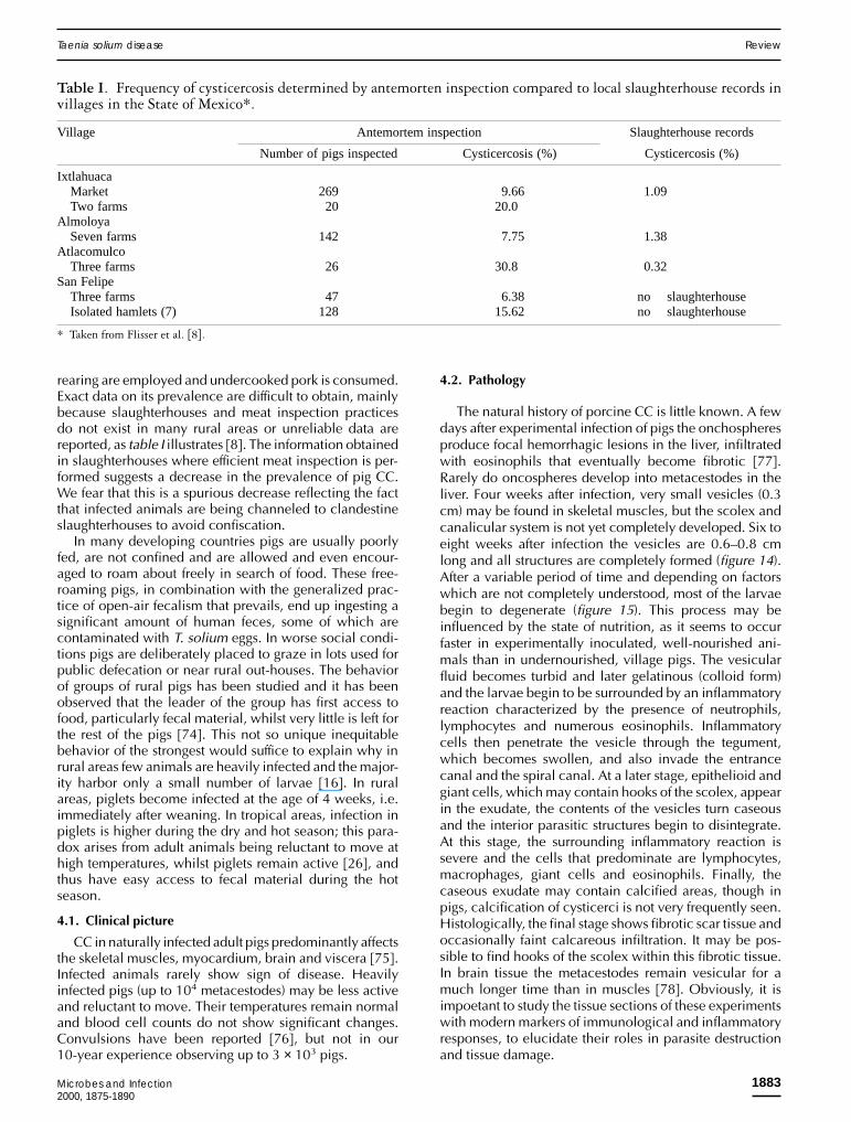
rearing are employed and undercooked pork is consumed.Exact data on its prevalence are difficult to obtain, mainlybecause slaughterhouses and meat inspection practicesdo not exist in many rural areas or unreliable data arereported, as table I illustrates [8]. The information obtainedin slaughterhouses where efficient meat inspection is per-formed suggests a decrease in the prevalence of pig CC.We fear that this is a spurious decrease reflecting the factthat infected animals are being channeled to clandestineslaughterhouses to avoid confiscation.
In many developing countries pigs are usually poorlyfed, are not confined and are allowed and even encour-aged to roam about freely in search of food. These free-roaming pigs, in combination with the generalized prac-tice of open-air fecalism that prevails, end up ingesting asignificant amount of human feces, some of which arecontaminated with T. solium eggs. In worse social condi-tions pigs are deliberately placed to graze in lots used forpublic defecation or near rural out-houses. The behaviorof groups of rural pigs has been studied and it has beenobserved that the leader of the group has first access tofood, particularly fecal material, whilst very little is left forthe rest of the pigs [74]. This not so unique inequitablebehavior of the strongest would suffice to explain why inrural areas few animals are heavily infected and the major-ity harbor only a small number of larvae [16]. In ruralareas, piglets become infected at the age of 4 weeks, i.e.immediately after weaning. In tropical areas, infection inpiglets is higher during the dry and hot season; this para-dox arises from adult animals being reluctant to move athigh temperatures, whilst piglets remain active [26], andthus have easy access to fecal material during the hotseason.
4.1. Clinical picture
CC in naturally infected adult pigs predominantly affectsthe skeletal muscles, myocardium, brain and viscera [75].Infected animals rarely show sign of disease. Heavilyinfected pigs (up to 104 metacestodes) may be less activeand reluctant to move. Their temperatures remain normaland blood cell counts do not show significant changes.Convulsions have been reported [76], but not in our10-year experience observing up to 3 × 10³ pigs.
4.2. Pathology
The natural history of porcine CC is little known. A fewdays after experimental infection of pigs the onchospheresproduce focal hemorrhagic lesions in the liver, infiltratedwith eosinophils that eventually become fibrotic [77].Rarely do oncospheres develop into metacestodes in theliver. Four weeks after infection, very small vesicles (0.3cm) may be found in skeletal muscles, but the scolex andcanalicular system is not yet completely developed. Six toeight weeks after infection the vesicles are 0.6–0.8 cmlong and all structures are completely formed (figure 14).After a variable period of time and depending on factorswhich are not completely understood, most of the larvaebegin to degenerate (figure 15). This process may beinfluenced by the state of nutrition, as it seems to occurfaster in experimentally inoculated, well-nourished ani-mals than in undernourished, village pigs. The vesicularfluid becomes turbid and later gelatinous (colloid form)and the larvae begin to be surrounded by an inflammatoryreaction characterized by the presence of neutrophils,lymphocytes and numerous eosinophils. Inflammatorycells then penetrate the vesicle through the tegument,which becomes swollen, and also invade the entrancecanal and the spiral canal. At a later stage, epithelioid andgiant cells, which may contain hooks of the scolex, appearin the exudate, the contents of the vesicles turn caseousand the interior parasitic structures begin to disintegrate.At this stage, the surrounding inflammatory reaction issevere and the cells that predominate are lymphocytes,macrophages, giant cells and eosinophils. Finally, thecaseous exudate may contain calcified areas, though inpigs, calcification of cysticerci is not very frequently seen.Histologically, the final stage shows fibrotic scar tissue andoccasionally faint calcareous infiltration. It may be pos-sible to find hooks of the scolex within this fibrotic tissue.In brain tissue the metacestodes remain vesicular for amuch longer time than in muscles [78]. Obviously, it isimpoetant to study the tissue sections of these experimentswith modern markers of immunological and inflammatoryresponses, to elucidate their roles in parasite destructionand tissue damage.
Table I. Frequency of cysticercosis determined by antemorten inspection compared to local slaughterhouse records invillages in the State of Mexico*.
Village Antemortem inspection Slaughterhouse records
Number of pigs inspected Cysticercosis (%) Cysticercosis (%)
IxtlahuacaMarket 269 9.66 1.09Two farms 20 20.0
AlmoloyaSeven farms 142 7.75 1.38
AtlacomulcoThree farms 26 30.8 0.32
San FelipeThree farms 47 6.38 no slaughterhouseIsolated hamlets (7) 128 15.62 no slaughterhouse
* Taken from Flisser et al. [8].
Taenia solium disease Review
Microbes and Infection2000, 1875-1890
1883

The ratio of vesicular/caseous metacestodes found atnecropsy after experimental infections varies with the totalnumber of metacestodes. At approximately 4 months post-infection the majority remain vesicular in those animalswith a great number of them. In those that harbor fewparasites, the majority are caseous. The presence of manycaseous larvae in a carcass has important implications forhuman health, in view of the fact that they are not infec-tious, i.e. they will not develop into tapeworms.
The immune response of pigs and its relevance todisease is being explored in experimental infections withT. solium eggs [79]. From initial results it would appearthat most infected pigs produce antibodies to a number ofthe parasite antigens, albeit in variable quantities. Theantibody response usually lasts for a few weeks. Antibod-ies do not seem to destroy a developed cysticercus butsome are capable of functionally damaging the cysticerci,especially in their capacity to transform into tapeworms[62]. The infected pig lymphocyte’s cellular response to invitro stimulation with specific antigens and conventionalmitogens is apparently depressed (ongoing research). Hereagain more detailed experimentation is required in earlyand late infections to assess the role of immunity in thenatural history of porcine cysticercotic disease.
4.3. Diagnosis
The most common and reliable method of diagnosingthe condition in vivo at the village and slaughterhouselevel is tongue inspection. The vesicular metacestodes canbe palpated and easily seen. Fibrous or calcified larvae aremore difficult to detect, as they may be quite small. In ourexperience, it is estimated that more than 50% of pigs thatharbor metacestodes show them in the tongue [80]. Theroutine postmortem inspection in slaughterhouses is adeep cut in the shoulder muscles. It has been shown thatthe masseter muscles are more frequently parasitized thanother skeletal muscles [75], but it would be impractical torecommend cutting these during routine meat inspection,as the position of the head of the carcass makes it difficultfor the inspector to reach and also because the musclemass is small and contains a considerable amount offibrous tissue.
Among serological methods, ELISA and immunoelec-trotransference are the most frequently used, but havesimilar limitations as those reported for human patients[70]. We have found that western blot gives positiveresults as long as the metacestodes are in the vesicularstage, but when they become caseous the result tends tobe negative. Results using ELISA show the same tendency[79].
4.4. Treatment
Praziquantel and albendazol are quite effective againstpig CC. However, the high costs of these chemicals pre-vents the treatment of infected pigs as a means of reducingtransmission.
5. EpidemiologyPrevalence data clearly point to endemics being located
in the developing countries of Latin America, Asia andAfrica, whilst Europe, Australia, the USA and Canada arerarely afflicted and mostly by imported cases (reviewed in[13]). Prevalence rates vary in the endemic countries butare usually less than 1:1 000 for human taeniasis (tape-worm carriers), 1–10% for human CC (undifferentiated CCand NCC cases), and up to 20–40% for pig CC, withvariations depending on sample design (i.e. rural versusurban), form of detection (i.e. image versus serologic) andrisk factors (i.e. family history of taeniasis). In other studies,human NCC was found in 1–5 % of a large autopsy seriesin Mexico; it accounted for about 25% of craniotomiesand was the most frequent cause of epilepsy in neurologi-cal patients [46]. Children are less frequently afflicted byNCC than adults, presumably because of their shorter timeof exposure and/or different immune responsiveness. Nosignificant differences in prevalence of CC due to genderor genetic background have been solidly established inhumans. There are some indications of women most fre-quently presenting an encephalitic form of disease [46]and some sort of preference of human NCC to HLA A28types [68]. Recent findings of female gender preferenceand the identification of a resistance gene in T. crassicepsmurine CC [51–54, 81] hold promise for the elucidation ofthe role of these major biological factors in the suscepti-
Figure 14. Parenchymatous portion of a T. solium cysticercus inpig muscle. The parasite is viable, with no signs of inflammatoryreaction in the surrounding tissue. Long arrow, rostellum (R)with hooks (H); short arrow, sucker (S); thick arrow, spiral canal(SC). HE × 200.
Figure 15. Caseous/calcified cysticerci. The cavity is filled withnecrotic material with moderate calcareous infiltrate. HE × 200.
Review Sciutto et al.
1884 Microbes and Infection2000, 1875-1890

bility of humans to CC. The disproportionate prevalencerates of taeniasis and CC are interpreted as resulting fromthe strategy of reproduction of the tapeworm, which pro-duces millions of eggs in its life span, each one capable ofbecoming a single cysticercus. Additionally, it would seemthat very few of the many cysticerci ever become a tape-worm, presumably because most are destroyed by cook-ing and/or by intermediate host factors, such as immunity[8, 62]. Differences in sensitivity of diagnostic procedurescould also be involved. A combination of these factorscould explain the notoriety of cysticercotic disease and thelow epidemiological profile of tapeworm disease: a mostdisturbing idea, because especially in some rural areas,pig CC may be as high as 20% and yet none or only anoccasional sampled inhabitant carries a tapeworm [16].Human T. solium disease is prevalent in countries wherethere are sectors of the population in extreme poverty (lowsanitary conditions at home, untidy personal health prac-tices, lack of education, very low income, social margin-ality from national public health programs), and whererustic pig-rearing and the use of improper cooking meth-ods are widespread. In non-endemic countries, most casesseem to be imported or acquired through contact with animmigrant human tapeworm carrier [82]. A notoriousepisode of imported disease is that of the originally non-endemic Irian Jaya in 1971, where the introduction ofcysticercotic pigs from Bali caused an epidemic outburstof taeniasis followed by one of human NCC [83]. Riskfactors in endemic areas are all those that favor the estab-lishment and progression of the parasite’s life-cycle or thatimply some likelihood of encountering an infecting para-site. These include residence in an endemic area, lowsanitary household conditions, poor personal hygiene,rustic pig-rearing practices, domestic slaughtering andconsumption of infected and improperly cooked pork,open-air fecalism which favors feces consumption byroaming pigs, low educational levels, history of personalor family taeniasis, and deliberate feeding of pigs withhuman feces. The personal risk of sharing living quarterswith a tapeworm carrier has been greatly emphasized inrecent times, since it involves exposure to greater eggdoses and continuous infection opportunities. Besides itssound logic, the proximity issue is epidemiologicallystrengthened by the finding that seropositive cases aremore prevalent in the household of taeniasic patients thanelsewhere [84]. This is most certainly so, especially inrural areas and in the miserable quarters of the urban poorwhere people live in quite crowded conditions. Yet, prox-imity to tapeworm carriers does not preclude the signifi-cance of extensive contamination of the environment withT. solium eggs and the possibility of a widespread form ofinfection, especially in large cities, where deficient sani-tary installations, open-air fecalism, and extremely dryand windy seasons would help in the wide distribution ofeggs, and where improper cooking methods, low personalhygiene of food handlers and extensive and intensiveout-of-home eating habits are prevalent. Together, thesecombined risk factors make a dreary combination thatmay account for most (90%) seropositive cases beingdistributed singly, not clustered, in households, and for thesurprisingly (albeit statistically significant) small differ-
ences in seroprevalence among the socioeconomic strataof the population, as found in a national serological sur-vey, comprising 70 000 sera, performed in Mexico [48]. Innon-endemic countries the risk of CC is associated withclose contact between residents and immigrants fromendemic countries, employed as domestic workers andcarrying the intestinal tapeworm [82]. Also, tourism toendemic countries is involved in isolated cases but, con-sidering the millions of tourists who travel to these places,the number of imported tourist cases are strikingly low andsuggest the importance of continuous exposure in themaking of new cases. As for the role of meat inspection inreducing transmission of taeniasis CC in endemic coun-tries – a practice credited for its eradication in Europeancountries [85] – it is presently questioned in underdevel-oped countries because of there being very few properslaughterhouses and because meat inspection is not per-formed with the necessary zeal in all of them. In fact, it hasbeen argued that in these countries meat inspection actu-ally encourages domestic pig slaughtering and illegal meatmarketing as ways of avoiding confiscation of the infectedcarcasses. Thus, taeniasis CC is a major cause of humanmisery and economic loss in Latin American countries. Itstotal impact upon the rest of the globe is also presumed tobe huge, judging from the initial statistics so far availablefrom Asia and Africa. It is not unreasonable to fear itsspread to more privileged countries or social sectors withinendemic countries by way of the parasite’s tremendousbiotic potential coupled with extensive immigration andemployment practices that mix in rather close quarterscultures differing in sanitary standards.
6. Prevention
Teniasis CC prevention should be a primary publichealth objective in developing countries. Strategies thatmay assist prevention are based on social developmentand/or on biological intervention. Considering that this isa disease of poverty, the improvement of socioeconomicconditions implies the definitive solution of this disease[86]. Unfortunately, social development is a slow processand if it does not include the low social classes, T. soliumdisease will subsist. In fact, it has been reported that 2years after the initiation of one focal health educationprogram in a Mexican village, only 2% of adults correctlyanswered questions about the life cycle of the parasite[86].
With respect to biotechnological interventions to curbtransmission, these are oriented towards treating oftapeworm-carrying individuals by massive or selectivetapeworm treatment or decreasing prevalence of pig CC.As for chemotherapy of taeniasis, massive treatment ofhuman populations to eliminate tapeworms was exten-sively implemented in the Soviet republics [85] andclaimed to have induced important reductions in pig CC.However, if not really massive and sustained, this strategycannot but slowly eliminate the source of tapeworms (pigCC), as transmission would continue due to new tape-worm cases and to immigration of tapeworm carriers fromuntreated sectors. Also, massive treatment programs should
Taenia solium disease Review
Microbes and Infection2000, 1875-1890
1885

take special precautions not to increase the risk of focalcontamination of the environment with an avalanche ofeggs following mass chemotherapy, an event that couldgive results quite contrary to the original intentions [86,87]. The treatment of confirmed or suspected humantapeworm carriers is a focused, inexpensive alternative[88] that could be a realistic tool in controlling transmis-sion. Its epidemiological impact will depend on improvingthe still-deficient methods for detecting tapeworm carrierswho would escape treatment. Recently, successful effortshave been made to develop specific and sensitive diag-nostic procedures to detect T. solium tapeworm carriers byELISA and PCR in feces; these procedures are yet to beevaluated in the field [89]. It is also necessary to considerthat medical supervision of such treatment interventions isrequired because non-symptomatic cysticercotic infec-tions may be exacerbated by drug treatment [90]. Inaddition, the induction of drug resistance must be consid-ered [91], as well as the potential side effects in treatedchildren [92].
Biotechnological strategies aimed at reduction of pigCC allow for some optimism concerning effective controlin the short term. Vaccination of pigs is an amenablestrategy of intervention because of the pigs’ usual shortlives before slaughter (approximately 1 year), not demand-ing long-lasting immune protection. Also, the vaccine ispresumed to be better accepted by the usually impover-ished pig owners, since the animals are not confiscated.Vaccination is also an economy-conscious method, aspigs will resist infection even if rustically bred with gar-bage and yet will sell at higher prices in slaughterhouses ifthe meat is clean of cysticerci. Thus, over the past 20 yearsthere have been some efforts to study the pigs’ immuneresponse to T. solium with the aim of producing a vaccineagainst this disease. Optimism prevails because of theeffective immunity induced against different cestodes andbecause of the extensive similarities in natural history,pathology and antigenic compositions among cestodeinfections, pointing to the possible usefulness of heterolo-gous or synthetic antigens [93]. Two main strategies havebeen used to identify antigens of interest to be employedas a source of vaccine against T. solium: that considering
their expression in onchospheres and/or early developedcysticerci (based on the argument that these are the mostvulnerable stages of the parasite to immune attack), andthat based on the identification of protective antigens,whatever their location, based on experimental murinemodels. Table II shows that antigens of both T. soliumoncospheres and metacestodes can stimulate strong pro-tection against both homologous and heterologous taeniidspecies. These two strategies have been used with encour-aging results in experimental conditions: not so much inthe number of pigs with sterile immunity (40–50%), butbetter yet in the 97% reduction of parasite loads. Reducingthe number of infected pigs is not the most relevant effectin curbing transmission, but rather it is the number ofviable cysticerci in the immunized pigs that really matters.Although not much emphasis has been placed on theadditional therapeutic effect of vaccines [16, 100, 101],recent reports suggest that it could critically affect thedynamics of transmission of the disease by hindering thedevelopment of cysticerci into tapeworms [62]. Due totheir intrinsic difficulties and high costs, there are fewvaccine field trials [16, 100], but the existing reports havegiven promising results. Experimental challenge of pigsmay be a convenient method of selecting candidate anti-gens for a vaccine, but field evaluation of the candidatevaccine is absolutely essential to determine its protectivecapacity in real conditions (i.e. in genetically heteroge-neous and undernourished pigs, possibly with a low immu-nological status, reared in rural circumstances and thusexposed to multiple natural challenges of unknown doseand frequency at various ages). These studies are extremelyhard to perform properly. Initial results of the only onesuch controlled field study performed in Mexico are quitepositive, using for immunogen synthetic peptide epitopes[102].
Another point that merits comment in discussing bio-logical approaches of controlling transmission is thatgenetically heterogeneous pigs are quite different in theirsusceptibility to CC when experimentally challenged [16].This finding, together with the recent identification of agene related to resistance in an experimental model of
Table II. Host protective immunity against cysticercosis induced by vaccination with different sources of T. soliumantigens.
Source of antigens Experimental trial Ref. Field trial Ref.
Total extract ND, 82.12, 33.33b [94] ND, ND, 48.93b, 813c [100]From cysticerci 58.2, 53.1, 03a [95]
50, 28.63a [96]From eggs 91.7, 96.7, 203b [97]
Purified ND, 94.7, 03a [98]From cysticerci ND, 64.5, 803c [10]
ND, 96.6, 83.33 ND, 97.8, [96]503b [99]
Recombinant and synthetic 31–84 [96]* 98.71, 97.92, 503b [102]91.7, 92.1, 03b [95]0–94 [12]*
* Antigens shared by T. solium tested in an experimental murine model. 1 Protective efficacy (expected – obtained/expected) considering the establishmentof viable cysticerci, 2 the total number of cysticerci, 3 the percent of pigs, completely protected after one3a, two3b or three3c vaccine doses.
Review Sciutto et al.
1886 Microbes and Infection2000, 1875-1890

crassiceps CC [81], opens the possibility of increasingresistance of hosts against CC by transgenesis.
In concluding this section, it is of special interest tostress the high vulnerability of T. solium cysticerci to bio-logical factors. In fact, it is estimated that cysticerci arecalcified without deliberate medical intervention inapproximately 30–40% of human cases, and are sponta-neously destroyed and almost disappear after 1 year ofexperimental infections of well-nourished pigs. In addi-tion, most vaccination experiments report promising resultspointing to immunological curtailment of the number andthe viability of the parasite. Based on this information it isreasonable to postulate that measures taking advantage ofthe parasites’ vulnerability will contribute to the success ofcontrol programs until the slow process of social develop-ment in endemic areas reaches the high levels needed fordefinitive control.
Thus, it would seem that with the knowledge gained ofthe factors involved in transmission, the careful design of ataeniasis treatment program and the upcoming availabilityof effective and inexpensive vaccines against pig CC, aneffective T. solium eradication program could be a realityin the near future.
7. Conclusions
T. solium taeniasis CC is a major parasitic disease com-plex of global proportions and poses very serious threats tohuman health and economy. It also poses very interestingscientific questions related to the host–parasite relation-ship, inflammation, immunity and genetics; a fertile fieldfor useful and basic research. In endemic countries riskfactors involve rustic pig-rearing and inadequate meatinspection, poor household sanitary conditions, low per-sonal health practices, open-air fecalism and proximity totapeworm carriers. In non-endemic countries, most casesare imported from immigrants from endemic areas, whilstthe few autochthonous cases are associated with immi-grant tapeworm carriers, some employed as domesticworkers. Pharmacological treatment of human NCC withcystocidal drugs greatly reduces symptomatology andextends life expectancy in the Mexican experience, but itslong-term benefits in the immigrant US cases are underquestion. Imaging techniques (MRI and CT) are the diag-nostic tools of choice for human neurocysticercotic dis-ease, whilst immunological methods are still in need ofimprovement for medical diagnosis, although they may beuseful as methods of massive serological surveillance.Although the available knowledge of T. solium taeniasisCC seems to be sufficient for control, its persistence andextension clearly demands systematic eradication pro-grams possibly combining risk control, treatment of tape-worm carriers and pig vaccination.
Acknowledgments
This work was supported by grants from CONACyTG25955; DGAPA, UNAM, IN 212798; Fundación Miguel
Alemán; ANUIES, Ecos program, The British Council andthe French embassy.
References
[1] Griesinger W., Cysticerken und ihe Diagnose, Archive fürHeilkunde, Gesammelte Abhandlungen, Berlin, 1862,p. 3999.
[2] Hitchcock E.R., Cysticercosis in the UK, J. Neurol. Neu-rosurg. Psychiatry 50 (1987) 1080–1081.
[3] Sotelo J., Del Brutto O., Roman G., in: Remington J.,Swartz M. (Eds.), Cysticercosis: Current Clinical Topics inInfectious Diseases, Blackwell Science, 1996,pp. 240–259.
[4] Rosenfeld E.A., Byrd S.E., Shulman S.T., Neurocysticer-cosis among children in Chicago, Clin. Infect. Dis. 23(1996) 262–268.
[5] Schantz P.M., Wilkins P.P., Tsang V.C.W. (Eds.), Immi-grants, Imaging, and Immunoblots: The Emergence ofNeurocysticercosis as a Major Public Health Problem,Am. Soc. Microbiol., Washington DC, USA, 1998,pp. 213–242.
[6] Font Puig C., Ruiz Postigo J.A., Munoz Batet C., PardosArnal F., Corachan-Cuyas M., Neurocysticercosis in Spain.Apropos 4 cases seen in immigrant patients from endemiccountries, Ann. Med. Intern. 16 (1999) 89–91.
[7] Flisser A., Woodhouse E., Larralde C., Human cysticerco-sis: antigens, antibodies and non-responders, Clin. Exp.Immunol. 39 (1980) 27–37.
[8] Flisser A., Willms K., Laclette J.P., Larralde C.,Ridaura C., Beltran F. (Eds.), Cysticercosis: Present Stateof Knowledge and Perspectives, Academic Press, NewYork, 1982.
[9] Flisser A., Taeniosis and cysticercosis due to Taenia solium,Prog. Clin. Parasitol. 4 (1994) 77–116.
[10] Nascimento E., Costa J.O., Guimaraes M.P., Tavares C.A.,Effective immune protection of pigs against cysticercosis,Vet. Immunol. Immunopathol. 45 (1995) 127–137.
[11] Garcia H.H., Harrison L.J., Parkhouse R.M., Montene-gro T., Martinez S.M., Tsang V.C., Gilman R.H., A spe-cific antigen-detection ELISA for the diagnosis of humanneurocysticercosis. The Cysticercosis Working Group inPeru, Trans. R. Soc. Trop. Med. Hyg. 92 (1998) 411–414.
[12] Toledo A., Larralde C., Fragoso G., Gevorkian G., Man-outcharian K., Hernandez M., Acero G., Rosas G., Lopez-Casillas F., Garfias C.K., Vazquez R., Terrazas I.,Sciutto E., Towards a Taenia solium cysticercosis vaccine:an epitope shared by Taenia crassiceps and Taenia soliumprotects mice against experimental cysticercosis, Infect.Immun. 67 (1999) 2522–2530.
[13] Sotelo J., Del Brutto O.H., Brain cysticercosis, Arch.Med. Res. 31 (2000) 3–14.
[14] Herrera L.A., Ramirez T., Rodriguez U., Corona T.,Sotelo J., Lorenzo M., Ramos F., Verdorfer I., Gebhart E.,Ostrosky-Wegman P., Possible association between Taeniasolium cysticercosis and cancer: increased frequency ofDNA damage in peripheral lymphocytes from neurocys-ticercosis patients, Trans. R. Soc. Trop. Med. Hyg. 94(2000) 61–65.
Taenia solium disease Review
Microbes and Infection2000, 1875-1890
1887

[15] Hernandez M., Beltran C., Garcia E., Fragoso G., Gev-orkian G., Fleury A., Parkhouse M., Harrison L., Sotelo J.,Sciutto E., Cysticercosis: towards the design of a diagnos-tic kit based on synthetic peptides, Immunol. Lett. 71(2000) 13–17.
[16] Huerta M., Sciutto E., García G., Villalobos N., Hernán-dez M., Fragoso G., Díaz J., Díaz A., Ramírez R., Luna S.,García J., Aguilar E., Espinoza S., Castilla G., Boba-dilla J.R., José M.V., Larralde C., de Aluja A.S., Vaccina-tion against Taenia solium cysticercosis in underfed rusticpigs of Mexico: role of age, genetic background and anti-body response, Vet. Parasitol. 90 (2000) 203–219.
[17] Shandera W.X., White A.C. Jr, Chen J.C., Diaz P., Arm-strong R., Neurocysticercosis in Houston, Texas. A reportof 112 cases, Medicine 73 (1994) 37–52.
[18] White A.C. Jr, Neurocysticercosis: updates on epidemiol-ogy, pathogenesis, diagnosis, and management, Annu.Rev. Med. 51 (2000) 187–206.
[19] Zee C.S., Go J.L., Kim P.E., DiGiorgio C.M., Imaging ofneurocysticercosis, Neuroimag. Clin. N. Am. 10 (2000)391–407.
[20] Chatel G., Gulletta M., Scolari C., Bombana E.,El-Hamad I., Matteelli A., Carosi G., Neurocysticercosisin an Italian traveler to Latin America, Am. J. Trop. Med.Hyg. 60 (1999) 255–256.
[21] Hansen N.J., Hagelskjaer L.H., Christensen T., Neuro-cysticercosis: a short review and presentation of a Scandi-navian case, Scand. J. Infect. Dis. 24 (1992) 255–262.
[22] Sanchez A.L., Lindback J., Schantz P.M., Sone M.,Sakai H., Medina M.T., Ljungstrom I., A population-based, case-control study of Taenia solium taeniosis andcysticercosis, Ann. Trop. Med. Parasitol. 93 (1999)247–258.
[23] Rodriguez-Canul R., Fraser A., Allan J.C., Dominguez-Alpizar J.L., Argaez-Rodriguez F., Craig P.S., Epidemio-logical study of Taenia solium taeniosis/cysticercosis in arural village in Yucatan state, Mexico, Ann. Trop. Med.Parasitol. 93 (1999) 57–67.
[24] Escobar A., The pathology of neurocysticercosis, in: Pala-cios E., Rodríguez-Carbajal J., Taveras J.M. (Eds.), Cys-ticercosis of the Central Nervous System, Thomas Spring-field, USA, 1983, pp. 27–54.
[25] Sotelo J., in: Roos K.L. (Ed.), Central Nervous SystemInfectious Diseases and Therapy, Marcel Dekker, Inc.,New York, 1997, pp. 545–571.
[26] de Aluja A.S., Martínez M.J.J., Villalobos A.N.M., Taeniasolium-cisticercosis in young pigs: age of first infection andhistological characteristics of the infection and antibodyresponse, Vet. Parasitol. 76 (1998) 71–79.
[27] Manson-Bahr P.E.C., Bell D.R. (Eds.), Manson’s TropicalDiseases, Bailliere Tindall, UK, 1987.
[28] Laclette J.P., Ornelas Y., Merchant M.T., Willms K., in:Flisser A., Willms K., Laclette J.P., Larralde C.,Ridaura C., Beltran F. (Eds.), Cysticercosis: Present Stateof Knowledge and Perspectives, Academic Press, NewYork, 1982, pp. 375–387.
[29] Yoshino K., Studies on the post-embryonal developmentof Taenia solium. Part I. On the hatching of the eggs ofTaenia solium, J. Med. Assoc. Formosa 32 (1933) 139–141.
[30] Slais J., The Morphology and Pathogenicity of the BladderWorms, Czechoslavak Academy of Science, Czechoslova-kia, 1970, pp. 13–50.
[31] Rabiela-Cervantes M.T., Rivas A., Rodríguez J.,Castillo S., Cancino F., in: Flisser A., Willms K.,Laclette J.P., Larralde C., Ridaura C., Beltran F. (Eds.),Cysticercosis: Present State of Knowledge and Perspec-tives, Academic Press, New York, 1982, pp. 179–200.
[32] Ramírez-Bon E., Merchant M.T., González-del Pliego M.,Cañedo L., in: Flisser A., Willms K., Laclette J.P., Lar-ralde C., Ridaura C., Beltran F. (Eds.), Cysticercosis:Present State of Knowledge and Perspectives, AcademicPress, New York, 1982, pp. 261–280.
[33] Smyth J.D., The Physiology of Cestodes. UniversityReview in Biology, Oliver and Boyd, 1969, pp. 1–225.
[34] Rothman A.H., Electron microscope studies of tapeworms:The surface structures of Hymenolepis diminuta (Rudolphi,1819; Blanchard, 1891), Trans. Am. Microsc. Soc. 82(1963) 22–30.
[35] Lumsden R.D., Hildreth M.B., in: Arme C., Pappas P.W.(Eds.), Biology of Eucestoda, Vol. 1, Academic Press, NewYork, 1983, pp. 177–233.
[36] Vargas-Parada L., Merchant M.T., Willms K.,Laclette J.P., Formation of calcareous corpuscles in thelumen of excretory canals of Taenia solium cysticerci, Para-sitol. Res. 85 (1999) 88–92.
[37] Theis J.H., Cleary M., Syvanen M., Gilson A., Swift P.,Banks J., Johnson E., DNA-confirmed Taenia solium cys-ticercosis in black bears (Ursus americanus) from California,Am. J. Trop. Med. Hyg. 4 (1996) 456–458.
[38] Buback J.L., Schulz K.S., Walker M.A., Snowden K.F.,Magnetic resonance imaging of the brain for diagnosis ofneurocysticercosis in a dog, J. Am. Vet. Med. Assoc. 208(1996) 1846–1848.
[39] Schmidt R.E., Selected topics in laboratory animal medi-cine volume XXIV: the hamster, Aeromed. Rev. 5 (1975)5–49.
[40] Monroy-Ostria A., Monroy-Ostria T.J., Gomez G.J., Her-nandez M.O., Some studies on the experimental infectionof golden hamsters with Taenia solium, Rev. Latinoam.Microbiol. 35 (1993) 91–98.
[41] Maravilla P., Avila G., Cabrera V., Aguilar L., Flisser A.,Comparative development of Taenia solium in experimen-tal models, J. Parasitol. 5 (1998) 882–886.
[42] Singh G., Neurocysticercosos in South-Central Americaand the Indian subcontinent. A comparative evaluation,Arq. Neuropsiquiatr. 55 (1997) 349–356.
[43] Yakoleff-Greenhouse V., Flisser A., Sierra A., Larralde C.,Analysis of antigenic variation in cysticerci of Taeniasolium, J. Parasitol. 68 (1982) 39–47.
[44] Larralde C., Montoya R.M., Sciutto E., Diaz M.L.,Govezensky T., Coltorti E., Deciphering western blots oftapeworm antigens (Taenia solium, Echinococcus granulosus,and Taenia crassiceps) reacting with sera from neurocys-ticercosis and hydatid disease patients, Am. J. Trop. Med.Hyg. 40 (1989) 282–290.
[45] Larralde C., Zedillo G.M., Lagunoff D., Ludowyke R.,Montoya R.M., Goodsaid F., Dreyfus G., Sciutto E.,Govezensky T., Diaz M.L., Porphyrin content of the cys-ticercus of Taenia solium, J. Parasitol. 72 (1986) 569–577.
Review Sciutto et al.
1888 Microbes and Infection2000, 1875-1890

[46] Del Brutto O.H., Sotelo J., Roman G.C. (Eds.), Neuro-cysticercosis. A Clinical Handbook, 1998.
[47] Sotelo J., Guerrero V., Rubio F., Neurocysticercosis: a newclassification based on active and inactive forms. A studyof 753 cases, Arch. Intern. Med. 145 (1985) 442–445.
[48] Larralde C., Padilla A., Hernández M., Govezensky T.,Sciutto E., Gutiérrez G., Tapia-Conyer R., Salvatierra B.,Sepúlveda J., Seroepidemiology of cysticercosis in Mexico,Salud Pública Méx. 34 (1992) 197–210.
[49] Villagran J., Olvera J.E., Cisticercosis Humana: Estudioclínico y Patológico de 481 casos de autopsia, Patología 26(1988) 149–156.
[50] Del Brutto O.H., Garcia E., Talamás O., Sotelo J., Sex-related severity of inflammation in parenchymal braincysticercosis, Arch. Intern. Med. 148 (1988) 544–546.
[51] Sciutto E., Fragoso G., Diaz M.L., Valdez F., Mon-toya R.M., Govezensky T., Lomeli C., Larralde C., MurineTaenia crassiceps cysticercosis: H-2 complex and sex influ-ence on susceptibility, Parasitol. Res. 77 (1991) 243–246.
[52] Huerta L., Terrazas L.I., Govezensky T., Larralde C.,Immunologic mediation of gonadal effects on experimen-tal murine cysticercosis caused by Taenia crassiceps, J. Para-sitol. 87 (1992) 471–476.
[53] Bojalil R., Terrazas L.I., Govezensky T., Sciutto E., Lar-ralde C., Thymus-related cellular immune mechanisms insex associated resistance to experimental murine cysticer-cosis (Taenia crassiceps), J. Parasitol. 79 (1993) 384–389.
[54] Larralde C., Morales J., Terrazas I., Govezensky T.,Romano M.C., Sex hormone changes induced by the para-site lead to feminization of the male host in murine Taeniacrassiceps cysticercosis, J. Steroid Biochem. Mol. Biol. 52(1995) 575–580.
[55] Del Brutto O.H., Castillo P.R., Mena I.X., Freire A.X.,Neurocysticercosis among patients with cerebral gliomas,Arch. Neurol. 54 (1997) 1125–1128.
[56] Herrera L.A., Benita-Bordes A., Sotelo J., Chavez L.,Olvera J., Rascon A., Lopez M., Ostrosky-Wegman P.,Possible relationship between neurocysticercosis andhematological malignancies, Arch. Med. Res. 30 (1999)154–158.
[57] Tsang V.C., Brand J.A., Boyer A.E., An enzyme-linkedimmunoelectrotransfer blot assay and glycoprotein anti-gens for diagnosing human cysticercosis (Taenia solium),J. Infect. Dis. 59 (1989) 50–59.
[58] Garcia E., Ordonez G., Sotelo J., Antigens from Taeniacrassiceps cysticerci used in complement fixation, enzyme-linked immunosorbent assay, and western blot (immuno-blot) for diagnosis of neurocysticercosis, J. Clin. Micro-biol. 33 (1995) 3324–3325.
[59] White A.C., Robinson P., Kuhn R., Taenia solium cysticer-cosis: Host-parasite interactions and the immune response,Chem. Immunol. 66 (1997) 209–230.
[60] White A.C., Neurocysticercosis: a major cause of neuro-logical disease worldwide, Clin. Infect. Dis. 24 (1997)101–113.
[61] Rickard M.D., Immunization against infection with lar-val taeniid cestodes using oncospheral antigens, in: Flis-ser A., Willms K., Laclette J.P., Larralde C., Ridaura C.,Beltran F. (Eds.), Cysticercosis: Present State of Knowl-edge and Perspectives, Academic press, New York, 1982,pp. 633–646.
[62] Garcia G., Sciutto E., Fragoso G., Cruz Revilla C.,Toledo A., Villalobos N., Flores I., Aluja A., Jose M.,Larralde C., Taeniosis-cysticercosis: GK1 antibodiesinhibit development of Taenia solium and Taenia crassicepstowards reproductive and pathogenic stages, J. Parasitol.(2000) in press.
[63] Sealey M., Ortiz-Ortiz L., Cellular immunity and cysticer-cosis. A review, in: Flisser A., Willms K., Laclette J.P.,Larralde C., Ridaura C., Beltran F. (Eds.), Cysticercosis:Present State of Knowledge and Perspectives, Academicpress, New York, 1982, pp. 565–574.
[64] Restrepo B.I., Llaguno P., Sandoval M.A., Enciso J.A.,Teale J.M., Analysis of immune lesions in neurocysticer-cosis patients: central nervous system response to helm-inth appears Th1-like insteat of Th2, J. Neuroimmunol.89 (1998) 64–72.
[65] Villa O.F., Kuhn R.E., Mice infected with the larvae ofTaenia crassiceps exhibit a Th2-like immune response withconcomitant anergy and downregulation of Th1-associatedphenomena, Parasitology 112 (1996) 561–570.
[66] Terrazas L.I., Bojalil R., Govezensky T., Larralde C., Shiftfrom an early protective Th1-type immune response to alate permissive Th2-type response in murine cysticercosis(Taenia crassiceps), J. Parasitol. 84 (1998) 74–81.
[67] Toenjes S.A., Spolski R.J., Mooney K.A., Kuhn R.E., Thesystemic immune response of BALB/c mice infected withlarval Taenia crassiceps is a mixed Th1/Th2-type response,Parasitology 118 (1999) 623–633.
[68] Del Brutto O.H., Granados G., Talamas O., Sotelo J.,Gorodezky C., Genetic pattern of the HLA system: HLAA, B, C, DR, and DQ antigens in Mexican patients withparenchymal brain cysticercosis, Human Biol. 63 (1991)85–93.
[69] Martinez H.R., Rangel-Guerra R., Elizondo G., MR imag-ing in neurocysticercosis: a study of 56 cases, AJNR 10(1989) 1011–1019.
[70] Ramos-Kuri M., Montoya R.M., Padilla A., Govenzen-sky T., Díaz M.L., Sciutto E., Sotelo J., Larralde C.,Immunodiagnosis of neurocysticercosis: disappointingperformance of serology (enzyme-linked immunosorbentassay) in an unbiased sample of neurological patients,Arch. Neurol. 49 (1992) 633–636.
[71] Chung J.Y., Bahk Y.Y., Huh S., Kang S.Y., Kong Y.,Cho S.Y., A recombinant 10-kDa protein of Taenia soliummetacestodes specific to active neurocysticercosis,J. Infect. Dis. 180 (1999) 1307–1315.
[72] Del Brutto O.H., Sotelo J., Roman G.C., Therapy forneurocysticercosis: a reappraisal, Clin. Infect. Dis. 17(1993) 730–735.
[73] Sotelo J., Update: the new ventriculoperitoneal shunt atthe Institute of Neurology of Mexico, Surg. Neurol. 46(1996) 19–20.
[74] Copado F., Aluja A., Mayagoitia L., Galindo F., Social andindividual behaviors of free ranging pigs in the Mexicantropics, Proceeding of the 31st International Congress ofthe International Society for Applied Ethology, Prague,Czech Republic, 1997.
Taenia solium disease Review
Microbes and Infection2000, 1875-1890
1889

[75] Vargas M., Saldierna V., Navarro F.R., Acevedo H.A.,Flisser A., de Aluja A.S., Localización del cisticerco de laTaenia solium en diferentes regiones musculares del cerdo ysu importancia para la inspección sanitaria, Vet. Méx. 17(1986) 275–279.
[76] Zürn F.A., Die tierischen Parasiten auf und in dern Köperunserer Hanssäugetiere, Verlag B. F. Voigt, Wéimar, 1882.
[77] Aluja A.S., Manchas de leche (Milk spots) por metacésto-dos de Taenia solium en hígados de cerdo, Vet. Méx. 25(1994) 155.
[78] Serrano-Palapa D., Aluja A., Lecumberri I., Villalobos A.,Comparación de la reacción inflamatoria causada por elmetacéstodo de Taenia solium en músculos y encéfalo decerdos, Vet. Méx. 28 (1997) 1–5.
[79] Aluja A.S., Villalobos A.N.M., Plancarte A., Rodarte L.F.,Hernández M., Zamora C., Sciutto E., Taenia solium cys-ticercosis: Immunity in pigs induced by primary infec-tion, Vet. Parasitol. 81 (1999) 129–135.
[80] Sciutto E., Martinez J.J., Villalobos N.M., Hernandez M.,Jose M.V., Beltran C., Rodarte F., Flores I., Bobadilla J.R.,Fragoso G., Parkhouse M.E., Harrison L.J., de Aluja A.S.,Limitations of current diagnostic procedures for diagnosisof Taenia solium cysticercosis in rural pigs, Vet. Parasitol.27 (1998) 299–313.
[81] Fragoso G., Lamoyi E., Mellor A., Lomeli C., Hernan-dez M., Sciutto E., Increased resistance to Taenia crassicepsmurine cysticercosis in Qa-2 transgenic mice, Infect.Immun. 66 (1998) 760–764.
[82] Schantz P.M., Moore A.C., Munoz J.L., Hartman B.J.,Schaefer J.A., Aron A.M., Persaud D., Sarti E., Wilson M.,Flisser A., Neurocysticercosis in an Orthodox Jewish com-munity in New York City, N. Engl. J. Med. 327 (1992)692–695.
[83] MacManus D.P., Improved diagnosis as an aid to bettersurveillance of Taenia solium cysticercosis, a potential pub-lic health threat to Papua New Guinea, PNG Med. J. 38(1995) 287–294.
[84] Diaz Camacho S.P., Candil Ruiz A., Suate Peraza V.,Zazueta Ramos M.L., Felix Medina M., Lozano R.,Willms K., Epidemiologic study and control of Taeniasolium infections with praziquantel in a rural village ofMexico, Am. J. Trop. Med. Hyg. 45 (1991) 522–531.
[85] Gemmell M., Matyas Z., Pawlowski Z., Soulsby E., Lar-ralde C., Nelson G.S., Rosicky B., Guidelines for surveil-lance prevention and control of Taeniasis/cysticercosis,VPH/83-49, World Health Organization, Geneva, 1985.
[86] Keilbach N.M., de Aluja A.S., Sarti-Gutierrez E., A pro-gramme to control taeniosis-cysticercosis (Taenia solium):experiences in a Mexican village, Acta Leidensia 57 (1989)181–189.
[87] Pawlowski Z.S., Efficiency of low doses of praziquantel intaeniosis, Acta Trop. 48 (1989) 83–89.
[88] Roman G., Sotelo J., Del Brutto O., Flisser A., Dumas M.,Wadia N., Botero D., Cruz M., Garcia H., de Bitten-court P.R., Trelles L., Arriagada C., Lorenzana P.,Nash T.E., Spina-Francia A., A proposal to declare neuro-cysticercosis an international reportable disease, Bull.World Health Org. 78 (2000) 399–406.
[89] Gonzalez L.M., Montero E., Harrison L., Parkhouse M.,Garate T., Differential diagnosis of Taenia saginata andTaenia solium by PCR, J. Clin. Microbiol. 38 (2000)737–744.
[90] Flisser A., Madrazo I., Plancarte A., Schantz P., Allan J.,Craig P., Sarti E., Neurological symptoms in occult neu-rocysticercosis after single taeniacidal dose of praziquan-tel, Lancet 342 (1993) 748.
[91] Jackson F., Anthelmintic resistance-the state of play, Br.Vet. J. 149 (1993) 123–138.
[92] Forrester J.E., Bailar J.C. 3rd, Esrey S.A., Jose M.V.,Castillejos B.T., Ocampo G., Randomised trial of albenda-zole and pyrantel in symptomless trichuriasis in children,Lancet 352 (1998) 1103–1108.
[93] Lightowlers M.W., Eradication of Taenia solium cysticer-cosis: a role for vaccination of pigs, Int. J. Parasitol. 29(1999) 811–817.
[94] Molinari J.L., Soto R., Tato P., Rodriguez D., Retana A.,Sepulveda J., Palet A., Immunization against porcinecysticercosis in an endemic area in Mexico: a field andlaboratory study, Am. J. Trop. Med. Hyg. 49 (1993)502–512.
[95] Sciutto E., Aluja A., Fragoso G., Rodarte L.F., Hernan-dez M., Villalobos M.N., Padilla A., Keilbach N.,Baca M., Govezensky T., Immunization of pigs againstTaenia solium cysticercosis: factors related to effective pro-tection, Vet. Parasitol. 60 (1995) 53–67.
[96] Manoutcharian K., Rosas G., Hernandez M., Fragoso G.,Aluja A., Villalobos N., Rodarte L.F., Sciutto E., Cysticer-cosis: identification and cloning of protective recombinantantigens, J. Parasitol. 82 (1996) 250–254.
[97] Plancarte A., Flisser A., Gauci C.G., Lightowlers M.W.,Vaccination against Taenia solium cysticercosis in pigsusing native and recombinant oncosphere antigens, Int.J. Parasitol. 29 (1999) 643–647.
[98] Pathak K.M., Gaur S.N., Immunization of pigs withculture antigens of Taenia solium, Vet. Parasitol. 34 (1990)353–356.
[99] Kumar D., Gaur S.N.S., Pathak K.M.L., Immunization ofpigs against the cysticercus of Taenia solium using fraction-ated first and second peaks of cysticercus cellulosae scolexantigen, Indian J. Anim. Sci. 57 (1987) 932–935.
[100] Molinari J.L., Rodriguez D., Tato P., Soto R.,Arechavaleta F., Solano S., Field trial for reducing porcineTaenia solium cysticercosis in Mexico by systematic vacci-nation of pigs, Vet. Parasitol. 69 (1997) 55–63.
[101] Evans C.A., Gonzalez A.E., Gilman R.H., Verastegui M.,Garcia H.H., Chavera A., Pilcher J.B., Tsang V.C., Immu-notherapy for porcine cysticercosis: implications for pre-vention of human disease. Cysticercosis Working Groupin Peru, Am. J. Trop. Med. Hyg. 56 (1997) 33–37.
[102] Huerta M., Aluja A., Fragoso G., Jose M., Larralde C.,Hernandez M., Toledo A., Villalobos M., Diaz A., Diaz J.,Alvarez I., Avila B., Huerta Ma., Gevorkian G., BeltranC., Sciutto E., Resultados de la vacunación en campo conuna vacuna sintética en contra de la cisticercosis porcinapor Taenia solium, IV Congreso Latinoamericano de Para-sitología, Acapulco, Guerrero, Mexico, 1999.
Review Sciutto et al.
1890 Microbes and Infection2000, 1875-1890