Differentiating Taenia eggs found in human stools: does Ziehl-Neelsen staining help?:...
10
Differentiating Taenia eggs found in human stools - Does Ziehl Neelsen staining help? Juan A. Jimenez 1 , Silvia Rodriguez 1,2 , Luz M. Moyano 1 , Yesenia Castillo 1 , and Héctor H. García 1,2,3 for the Cysticercosis Working Group in Peru 1 Department of Microbiology, School of Sciences, (JJ, SR, YC, HG) and Center for Global Health - Tumbes (LM, HG), Universidad Peruana Cayetano Heredia, Av. H. Delgado 430, SMP, Lima 31, Lima, Perú 2 Cysticercosis Unit, Instituto Nacional de Ciencias Neurologicas, Jr. Ancash 1271, Lima 1, Lima, Perú 3 Instituto Peruano de Parasitología Clínica y Experimental (INPPACE), Lima, Perú SUMMARY Introduction—Unlike other tapeworms, T. solium infections carry risk for neurocysticercosis. Differential diagnosis of human tapeworm infections relies on morphology of the scolex or proglottids, frequently unavailable. DNA-based assays are poorly available in endemic areas. Ziehl Neelsen staining has been suggested but not tested in controlled designs. We validated whether Ziehl Neelsen staining could differentiate T. solium and T. saginata eggs. Methods—Tapeworm proglottids (33 specimens, 23 T. solium and 10 T. saginata) and eggs (31 specimens, 13 T. solium and 10 T. saginata) were stained. Four eggs from each sample were measured and average diameters were recorded. Results—T. saginata eggs stained entirely magenta in seven of 13 cases. T. solium eggs stained entirely blue/purple in 4/18 cases and entirely magenta in one. Eggs of T. saginata were slightly larger and always ovoid, while T. solium eggs were smaller and were mostly spheric. Conclusions—Ziehl Neelsen staining can occasionally distinguish fully mature T. solium from T. saginata eggs. This distinction is poorly sensitive and not completely specific. Differential staining suggest differences in embryophore components between species, evident along egg maturation. In this small series, egg morphology (shape, maximal diameter) provided appropriate differentiation between T. solium and T. saginata eggs. Keywords Taenia; Taenia solium; Taenia saginata; Ziehl Neelsen; cestodes; Perú INTRODUCTION Three big tapeworms lodge in the human intestine: Diphyllobothrium sp, Taenia solium, and Taenia saginata, with a fourth, Taenia asiatica, still in debate on whether it is a new species or a T. saginata subspecies.(Flisser et al., 2004; Garcia et al., 2007) From these, only Taenia solium can lead to severe disease because of its capacity to infect the human brain with its larval form causing neurocysticercosis, the major cause of acquired epilepsy in most of the * Corresponding author: Hector H. Garcia, Department of Microbiology, Universidad Peruana Cayetano Heredia, Avenida H. Delgado 430, SMP, Lima, 31, Perú. Telephone: +511 328 7360 Fax: +51 1 3284038 [email protected]. Europe PMC Funders Group Author Manuscript Trop Med Int Health. Author manuscript; available in PMC 2012 August 28. Published in final edited form as: Trop Med Int Health. 2010 September ; 15(9): 1077–1081. doi:10.1111/j.1365-3156.2010.02579.x. Europe PMC Funders Author Manuscripts Europe PMC Funders Author Manuscripts
-
Upload
independent -
Category
Documents
-
view
1 -
download
0
Transcript of Differentiating Taenia eggs found in human stools: does Ziehl-Neelsen staining help?:...
Differentiating Taenia eggs found in human stools - Does ZiehlNeelsen staining help?
Juan A. Jimenez1, Silvia Rodriguez1,2, Luz M. Moyano1, Yesenia Castillo1, and Héctor H.García1,2,3 for the Cysticercosis Working Group in Peru1Department of Microbiology, School of Sciences, (JJ, SR, YC, HG) and Center for Global Health- Tumbes (LM, HG), Universidad Peruana Cayetano Heredia, Av. H. Delgado 430, SMP, Lima 31,Lima, Perú2Cysticercosis Unit, Instituto Nacional de Ciencias Neurologicas, Jr. Ancash 1271, Lima 1, Lima,Perú3Instituto Peruano de Parasitología Clínica y Experimental (INPPACE), Lima, Perú
SUMMARYIntroduction—Unlike other tapeworms, T. solium infections carry risk for neurocysticercosis.Differential diagnosis of human tapeworm infections relies on morphology of the scolex orproglottids, frequently unavailable. DNA-based assays are poorly available in endemic areas.Ziehl Neelsen staining has been suggested but not tested in controlled designs. We validatedwhether Ziehl Neelsen staining could differentiate T. solium and T. saginata eggs.
Methods—Tapeworm proglottids (33 specimens, 23 T. solium and 10 T. saginata) and eggs (31specimens, 13 T. solium and 10 T. saginata) were stained. Four eggs from each sample weremeasured and average diameters were recorded.
Results—T. saginata eggs stained entirely magenta in seven of 13 cases. T. solium eggs stainedentirely blue/purple in 4/18 cases and entirely magenta in one. Eggs of T. saginata were slightlylarger and always ovoid, while T. solium eggs were smaller and were mostly spheric.
Conclusions—Ziehl Neelsen staining can occasionally distinguish fully mature T. solium fromT. saginata eggs. This distinction is poorly sensitive and not completely specific. Differentialstaining suggest differences in embryophore components between species, evident along eggmaturation. In this small series, egg morphology (shape, maximal diameter) provided appropriatedifferentiation between T. solium and T. saginata eggs.
KeywordsTaenia; Taenia solium; Taenia saginata; Ziehl Neelsen; cestodes; Perú
INTRODUCTIONThree big tapeworms lodge in the human intestine: Diphyllobothrium sp, Taenia solium, andTaenia saginata, with a fourth, Taenia asiatica, still in debate on whether it is a new speciesor a T. saginata subspecies.(Flisser et al., 2004; Garcia et al., 2007) From these, only Taeniasolium can lead to severe disease because of its capacity to infect the human brain with itslarval form causing neurocysticercosis, the major cause of acquired epilepsy in most of the
*Corresponding author: Hector H. Garcia, Department of Microbiology, Universidad Peruana Cayetano Heredia, Avenida H.Delgado 430, SMP, Lima, 31, Perú. Telephone: +511 328 7360 Fax: +51 1 3284038 [email protected].
Europe PMC Funders GroupAuthor ManuscriptTrop Med Int Health. Author manuscript; available in PMC 2012 August 28.
Published in final edited form as:Trop Med Int Health. 2010 September ; 15(9): 1077–1081. doi:10.1111/j.1365-3156.2010.02579.x.
Europe PM
C Funders A
uthor Manuscripts
Europe PM
C Funders A
uthor Manuscripts

world.(Garcia & Del Brutto, 2005) The differential diagnosis between these tapeworms isbased on morphology of the adult tapeworm scolex or proglottids. In most cases, however,only tapeworm eggs are found in stool samples, and no parasite tissue is available. AlthoughDiphyllobothrium eggs are easily distinguishable, eggs from T. solium and T. saginata cannot be differentiated by microscopical examination. Only DNA-based probes have obtainedspecific identification between these eggs; this type of assays are hardly available inendemic areas.(Flisser et al., 2004; Garcia et al., 2007; Gonzalez et al., 2000; Mayta et al.,2000)
A few decades ago, Capron, A. and Rose, F. (1962) described the use of the acid-fast (ZiehlNeelsen) staining to distinguish T. solium from T. saginata.(Capron & Brygoo, 1959;Capron & Rose, 1962) While their work is occasionally quoted, it has neither beenreplicated nor refuted. We examined fresh and preserved material from both parasite speciesto validate whether this method can differentiate between these two tapeworm species.
MATERIAL AND METHODSSpecies diagnosis of tapeworm material
Expelled parasite material from human origin was used for this study. Cases were defined asT. solium or T. saginata on the basis of carmine staining and counting of main uterinebranches (Flisser et al., 2004; Garcia et al., 2007; Mayta et al., 2000) as well as PCR intapeworm material.(Mayta et al., 2000) There were no discordances between results witheither method.
Tapeworm proglottidsPre-existing archive parasite material was used for this part of the study, comprising matureand gravid proglottids obtained from 33 patients (23 T. solium carriers, 10 T. saginatacarriers) after anti-parasitic treatment.(Jeri et al., 2004) In some cases multiple gravid andpre-gravid proglottids were available (14 and 9 proglottids from two T. solium tapeworms,and 7 and 4 proglottids from two T. saginata tapeworms) and were processed to assesschanges in staining according to proglottid maturation. As per our standard routine,proglottids were washed with distilled water, fixed in 10% formalin-phosphate bufferedsaline (PBS) and stored at room temperature to be later processed by histology.
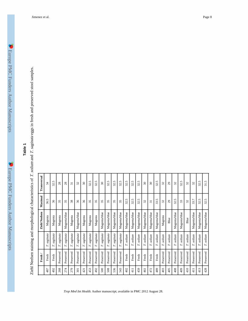
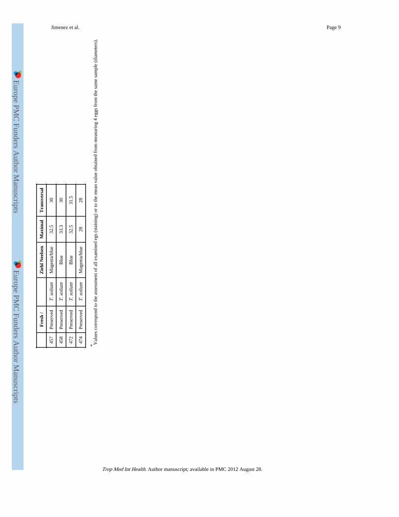
Defined fresh and archive stool samplesEggs from 8 pre- or post- treatment stool samples (2 T. saginata and 6 T. solium) wereexamined and stained in fresh, before any fixation. Also archive stool samples from 23patients, preserved in 5% formalin-PBS, were used for staining (11 T. saginata, 12 T.solium). Stool samples were concentrated by tube sedimentation. Sediments were placed inmicroscopy slides with polilysine and left to dry for staining. Four eggs from each samplewere measured and the average maximal and transversal diameters were recorded (Table 1).
HistologyProglottids were washed to eliminate the excess of formalin and then passed throughincreasing ethanol concentrations (70°, 80°, 90°, and 100°) and then three times in xilol.Proglottid samples were then placed in paraffin blocks, sliced in 6 um sections, de-paraffined, and placed on microscopy slides with polilysine for staining (Luna, 1968).
Ziehl-Neelsen stainingSamples were stained with carbol-fuchsin 3% for 15′, washed with tap water, and thendecolored with 70% ethanol 1% HCl for 2′. After a second washing the slide was contrasted
Jimenez et al. Page 2
Trop Med Int Health. Author manuscript; available in PMC 2012 August 28.
Europe PM
C Funders A
uthor Manuscripts
Europe PM
C Funders A
uthor Manuscripts
with 3% methylene blue for 5′, washed again, and left to dry at room temperature.(Chapin& Lauderdale, 2007; Clavel et al., 1999).
RESULTSStaining of Taenia eggs in proglottid material
Staining of more proximal and more distal gravid T. solium and T. saginata proglottidsshowed that the oncospheres always stain blue in both species, with magenta hooks. As theeggs mature, a blue oncospheral membrane is clearly defined, around which magenta blocksbegin to form the embryophore. A substance apparently secreted from the oncosphere thenbegins to fill the space between blocks. This substance is initially blue in both species. Asthe embryophore matures and becomes thicker, coloration gets more intense, departing fromblue to gradually acquire some mixed magenta tones in T. solium, and a more markedmagenta color in material from T. saginata (Figure 1).
Staining of Taenia eggs from stool samplesThere was no difference in staining of eggs from fresh versus preserved stool samples. In T.saginata eggs the external cover or embryophore was colored entirely magenta on ZiehlNeelsen in 7/13 cases (Figure 2a), and magenta with dark blue (some close to dark purple)areas in the remaining six. In T. solium eggs the embryophore stained usually in a mix ofmagenta and blue being entirely blue in four of the 18 cases (Figure 2b) and entirelymagenta in one case (Table 1).
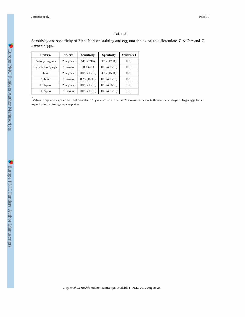
Some apparent morphological differences could also be observed. The eggs of T. saginatawere slightly larger, with a maximal diameter of 35.58 +/− 0.91 μm compared with 32.08 +/− 1.45 μm for T. solium eggs (n=13 for T. saginata, n=18 for T. solium; mean +/− SD;p<0.001, Mann Whitney test). T. saginata eggs were always ovoid (ratio between largerdiameter and its transverse diameter was 1.14+/− 0.07), while most T. solium eggs werespheric (ratio was 1.03+/− 0.03; p<0.001 compared to T. saginata, Mann Whitney test). In 3out of 18 cases, however, T. solium eggs looked ovoid in shape (ratios 1.11, 1.08, and 1.07)(Table 1).
In direct comparison of sensitivity and specificity with T. saginata and T. solium eggs, eggsize and form were better predictors for species differentiation than Ziehl Neelsen staining(Table 2).
DISCUSSIONTaenia spp. eggs are covered by a thick embryophore composed by prismatic keratin blocks(which give it its typical radial appearance), kept together by a cement substance. By thetime they reach the environment the embryophore is still surrounded by a colloid vitellumlayer. It has been previously described that the vitelogen glands in the oncosphere produce aacid-fast resistant substance which takes the spaces between embryophoric blocks, likelyresponsible by the changes in coloration along egg maturation.(Capron & Brygoo, 1959;Capron & Rose, 1962)
This series showed that Ziehl Neelsen staining can occasionally distinguish fully mature T.solium from T. saginata eggs, in cases where the cover is entirely magenta (7/13 T.saginata), or entirely blue/purple (4/18 T. solium). Staining was mixed in 19/31 cases, andequivocal (magenta) in one case of T. solium. While this distinction provided a speciesdiagnosis in 35% of cases, it is by no means absolute and seems poorly useful in practice. Itsapplication assumes eggs are fully mature (which is not possible to determine in aproglottid), and carries some degree of subjectivity because color differences may be subtle,
Jimenez et al. Page 3
Trop Med Int Health. Author manuscript; available in PMC 2012 August 28.
Europe PM
C Funders A
uthor Manuscripts
Europe PM
C Funders A
uthor Manuscripts

adding to the requirement of highly trained personal. In most centers, the numbers ofspecimens to be tested for species differentiation will be small, as will be the experience ofthe operators. Oncospheres stained blue in both species, not helping in differentiation.Morphological criteria (diameter and shape) seemed more consistent in this series. In thissmall series, an arbitrary cut off of 35 μm had 100% predictive value for differentiation.This had been reported before (Verster, 1969) but not really replicated. Morphologicaldifferences are however minor and would need to be replicated with T. saginata and T.solium material from other parts of the world.
Of interest, our data suggest differences in specific components of the embryophore betweenthese two species, evident along egg maturation, compatible with previous data on differentchemical composition of embryophoric blocks.(Morseth, 1966) Given that the ability of theoncosphere of T. solium to infect the human host and cause cysticercosis is not shared by T.saginata, further understanding and characterization of the enzymes and other activemolecules present in one species but absent in the other may provide species-specificdiagnostics and potential vaccine targets.
AcknowledgmentsFINANCIAL SUPPORT
This work was partially supported by the Fogarty International Center, National Institutes of Health, Bethesda,USA (H.G., training grant number D43 TW001140); and the Bill and Melinda Gates Foundation (H.G., grantsnumber 23981 and 33848).
REFERENCESCapron A, Brygoo ER. [Constitution of helminth eggs. I. Presence and formation of an acid-alcohol
resistant substance in the shell]. Bull Soc Pathol Exot Filiales. 1959; 52:574–577. [PubMed:13807576]
Capron A, Rose F. [On the composition of helminth eggs. II. Alcohol-acid-resistance in cestodes.Difference of Ziehl stainability of embryophores of Taenia saginata and Taenia solium]. Bull SocPathol Exot Filiales. 1962; 55:765–767. [PubMed: 14018471]
Chapin, K.; Lauderdale, TL. Reagents, stains, and media: bacteriology. In: Murray, PR.; Baron, EJ.;Jorgensen, JH.; Landr, ML.; Pfaller, MA., editors. Manual of Clinical Microbiology. 9th ed.American Society for Microbiology; Washington, D.C: 2007. p. 344
Clavel A, Varea M, Doiz O, et al. Visualization of hydatid elements: comparison of several techniques.Journal of Clinical Microbiology. 1999; 37:1561–1563. [PubMed: 10203521]
Flisser A, Viniegra AE, Aguilar-Vega L, Garza-Rodriguez A, Maravilla P, Avila G. Portrait of humantapeworms. Journal of Parasitology. 2004; 90:914–916. [PubMed: 15357104]
Garcia HH, Del Brutto OH. Neurocysticercosis: updated concepts about an old disease. LancetNeurology. 2005; 4:653–661. [PubMed: 16168934]
Garcia, HH.; Jimenez, JA.; Escalante, H. Cestodes. In: Murray, PR.; Baron, EJ., editors. Manual ofClinical Microbiology. ASM Press; Washington DC: 2007. p. 2166-2174.
Gonzalez LM, Montero E, Harrison LJ, Parkhouse RM, Garate T. Differential diagnosis of Taeniasaginata and Taenia solium infection by PCR. Journal of Clinical Microbiology. 2000; 38:737–744.[PubMed: 10655377]
Jeri C, Gilman RH, Lescano AG, et al. Species identification after treatment for human taeniasis.Lancet. 2004; 363:949–950. [PubMed: 15043964]
Luna, LG., editor. Manual of Histologic Staining Methods of the Armed Forces Institute of Pathology.3rd Ed. McGraw-Hill; New York: 1968. p. 28
Mayta H, Talley A, Gilman RH, et al. Differentiating Taenia solium and Taenia saginata infections bysimple hematoxylin-eosin staining and PCR-restriction enzyme analysis. Journal of ClinicalMicrobiology. 2000; 38:133–137. [PubMed: 10618076]
Jimenez et al. Page 4
Trop Med Int Health. Author manuscript; available in PMC 2012 August 28.
Europe PM
C Funders A
uthor Manuscripts
Europe PM
C Funders A
uthor Manuscripts

Morseth DJ. Chemical composition of embryophoric blocks of Taenia hydatigena, Taenia ovis, andTaenia pisiformis eggs. Experimental Parasitology. 1966; 18:347–354. [PubMed: 5949306]
Verster A. A taxonomic revision of the genus Taenia Linnaeus, 1758 s. str. Onderstepoort Journal ofVeterinary Research. 1969; 36:3–58. [PubMed: 5407584]
Jimenez et al. Page 5
Trop Med Int Health. Author manuscript; available in PMC 2012 August 28.
Europe PM
C Funders A
uthor Manuscripts
Europe PM
C Funders A
uthor Manuscripts

Figure 1.Histological sections showing stages of maturation of eggs in proglottids of Taenia saginata(left) and Taenia solium (right) showing oncospheres surrounded by an oncospheralmembrane, small, magenta embryophoric blocks, and interstitial substance.
Jimenez et al. Page 6
Trop Med Int Health. Author manuscript; available in PMC 2012 August 28.
Europe PM
C Funders A
uthor Manuscripts
Europe PM
C Funders A
uthor Manuscripts

Figure 2.Mature eggs of Taenia solium (left) and Taenia saginata (right) as seen in stool samples,showing differences in staining tonalities.
Jimenez et al. Page 7
Trop Med Int Health. Author manuscript; available in PMC 2012 August 28.
Europe PM
C Funders A
uthor Manuscripts
Europe PM
C Funders A
uthor Manuscripts
Europe PM
C Funders A
uthor Manuscripts
Europe PM
C Funders A
uthor Manuscripts
Jimenez et al. Page 8
Tabl
e 1
Zie
hl N
eels
en s
tain
ing
and
mor
phol
ogic
al c
hara
cter
istic
s of
T. s
oliu
m a
nd T
. sag
inat
a eg
gs in
fre
sh a
nd p
rese
rved
sto
ol s
ampl
es.
Fre
sh /
Zie
hl N
eels
enM
axim
alT
rans
vers
al
487
Fres
hT
. sag
inat
aM
agen
ta36
.534
492
Fres
hT
. sag
inat
aM
agen
ta36
32.5
268
Pres
erve
dT
. sag
inat
aM
agen
ta35
28
274
Pres
erve
dT
. sag
inat
aM
agen
ta/b
lue
3528
279
Pres
erve
dT
. sag
inat
aM
agen
ta38
31
301
Pres
erve
dT
. sag
inat
aM
agen
ta/b
lue
3632
421
Pres
erve
dT
. sag
inat
aM
agen
ta36
30
453
Pres
erve
dT
. sag
inat
aM
agen
ta35
32.5
492
Pres
erve
dT
. sag
inat
aM
agen
ta35
32.5
500
Pres
erve
dT
. sag
inat
aM
agen
ta/b
lue
3530
508
Pres
erve
dT
. sag
inat
aM
agen
ta/b
lue
3532
.5
536
Pres
erve
dT
. sag
inat
aM
agen
ta/b
lue
3532
.5
543
Pres
erve
dT
. sag
inat
aM
agen
ta/b
lue
3532
.5
402
Fres
hT
. sol
ium
Mag
enta
/blu
e32
.532
.5
411
Fres
hT
. sol
ium
Mag
enta
/blu
e32
.532
.5
459
Fres
hT
. sol
ium
Mag
enta
/blu
e32
.532
.5
461
Fres
hT
. sol
ium
Mag
enta
/blu
e32
30
472
Fres
hT
. sol
ium
Mag
enta
/blu
e31
30
489
Fres
hT
. sol
ium
Mag
enta
/blu
e33
.532
.5
403
Pres
erve
dT
. sol
ium
Mag
enta
3232
405
Pres
erve
dT
. sol
ium
Blu
e29
29
408
Pres
erve
dT
. sol
ium
Mag
enta
/blu
e32
.532
409
Pres
erve
dT
. sol
ium
Mag
enta
/blu
e33
32.5
410
Pres
erve
dT
. sol
ium
Blu
e32
32
411
Pres
erve
dT
. sol
ium
Mag
enta
/blu
e33
.732
413
Pres
erve
dT
. sol
ium
Mag
enta
/blu
e32
.532
.5
428
Pres
erve
dT
. sol
ium
Mag
enta
/blu
e32
.531
.5
Trop Med Int Health. Author manuscript; available in PMC 2012 August 28.
Europe PM
C Funders A
uthor Manuscripts
Europe PM
C Funders A
uthor Manuscripts
Jimenez et al. Page 9
Fre
sh /
Zie
hl N
eels
enM
axim
alT
rans
vers
al
457
Pres
erve
dT
. sol
ium
Mag
enta
/blu
e32
.530
458
Pres
erve
dT
. sol
ium
Blu
e33
.330
472
Pres
erve
dT
. sol
ium
Blu
e32
.531
.5
474
Pres
erve
dT
. sol
ium
Mag
enta
/blu
e28
28
* Val
ues
corr
espo
nd to
the
asse
ssm
ent o
f al
l exa
min
ed e
gs (
stai
ning
) or
to th
e m
ean
valu
e ob
tain
ed f
rom
mea
suri
ng 4
egg
s fr
om th
e sa
me
sam
ple
(dia
met
ers)
.
Trop Med Int Health. Author manuscript; available in PMC 2012 August 28.
Europe PM
C Funders A
uthor Manuscripts
Europe PM
C Funders A
uthor Manuscripts
Jimenez et al. Page 10
Table 2
Sensitivity and specificity of Ziehl Neelsen staining and egg morphological to differentiate T. solium and T.saginata eggs.
Criteria Species Sensitivity Specificity Youden’s J
Entirely magenta T. saginata 54% (7/13) 96% (17/18) 0.50
Entirely blue/purple T. solium 50% (4/8) 100% (13/13) 0.50
Ovoid T. saginata 100% (13/13) 83% (15/18) 0.83
Spheric T. solium 83% (15/18) 100% (13/13) 0.83
> 35 μm T. saginata 100% (13/13) 100% (18/18) 1.00
< 35 μm T. solium 100% (18/18) 100% (13/13) 1.00
*Values for spheric shape or maximal diameter < 35 μm as criteria to define T. solium are inverse to those of ovoid shape or larger eggs for T.
saginata, due to direct group comparison
Trop Med Int Health. Author manuscript; available in PMC 2012 August 28.