Early intrauterine embryonic development in Khawia sinensis Hsü, 1935 (Cestoda, Caryophyllidea,...
9
ORIGINAL PAPER Early intrauterine embryonic development in Khawia sinensis Hsü, 1935 (Cestoda, Caryophyllidea, Lytocestidae), an invasive tapeworm of carp (Cyprinus carpio): an ultrastructural study Magdaléna Bruňanská & John S. Mackiewicz & Daniel Młocicki & Zdzisław Świderski & Jana Nebesářová Received: 7 June 2011 /Accepted: 1 August 2011 /Published online: 6 September 2011 # Springer-Verlag 2011 Abstract Intrauterine embryonic development in the caryo- phyllidean tapeworm Khawia sinensis has been investigated using transmission electron microscopy and cytochemical staining with periodic acid-thiosemicarbazide-silver protei- nate for glycogen. Contrary to previous light microscopy findings that reported the release of non-embryonated eggs of K. sinenesis to the external environment, the present study documents various stages of embryonation (ovoviviparity) within the intrauterine eggs of this cestode. At the initial stage of embryonic development, each fertilised oocyte is accompanied by several vitellocytes that become enclosed within the operculate, electrondense shell. Cleavage divi- sions result in formation of blastomeres (up to about 24 cells) of various sizes. Mitotic divisions and apparent rosette arrangment of the blastomeres, the latter atypical within the Eucestoda, are observed for the first time in the intrauterine eggs of K. sinenesis. The early embryo enclosed within the electrondense shell is surrounded by a thin membraneous layer which in some enlarged regions shows presence of nuclei. Simultaneously to multiplication and differentiation, some of the blastomeres undergo deterioration. A progressive degeneration of the vitellocytes within eggs provides nutritive reserves, including lipids, for the developing embryo. The possible significance of this atypical timing of the intrauterine embryonic development to (1) the ecology of K. sinensis and that of a recent introduction of another invasive tapeworm, the caryophyllidean Atractolytocestus huronensis Anthony, 1958 to Europe; and (2) the affiliation of caryophyllideans with other lower cestodes, are discussed. Introduction The order Caryophyllidea occupies a key position among the Eucestoda (Mackiewicz 2003). Caryophyllidean tapeworms are characterised by having a monopleuroid body, i.e. lacking proglottisation and with a single set of reproductive organs. While eggs are generally unembryonated when expelled (Mackiewicz 1968, 1972), some exceptions have been reported, e.g. progenetic Archigetes in which embryonation is usually completed in utero, or at least begins in utero and terminates ex utero (Wiśniewski 1930; Kennedy 1965; Calentine 1984), or Djombangia, Wenyonia spp., Hunturella, or Biacetabulum sp. (Mackiewicz 1972). Detailed observa- M. Bruňanská (*) Parasitological Institute, Slovak Academy of Sciences, Hlinkova 3, 040 01 Košice, Slovak Republic e-mail: [email protected] J. S. Mackiewicz Department of Biological Sciences, University at Albany, State University of New York, Albany, NY, USA D. Młocicki : Z. Świderski W. Stefanski Institute of Parasitology, Polish Academy of Sciences, Twarda 51/55, 00-818 Warsaw, Poland D. Młocicki : Z. Świderski Department of General Biology and Parasitology, Medical University of Warsaw, Chałubińskiego 5, 02-004 Warsaw, Poland J. Nebesářová Institute of Parasitology, Biology Centre of the Academy of Sciences of the Czech Republic, Branišovská 31, 37005 České Budějovice, Czech Republic Parasitol Res (2012) 110:1009–1017 DOI 10.1007/s00436-011-2590-2
Transcript of Early intrauterine embryonic development in Khawia sinensis Hsü, 1935 (Cestoda, Caryophyllidea,...
ORIGINAL PAPER
Early intrauterine embryonic development in Khawiasinensis Hsü, 1935 (Cestoda, Caryophyllidea, Lytocestidae),an invasive tapeworm of carp (Cyprinus carpio):an ultrastructural study
Magdaléna Bruňanská & John S. Mackiewicz &
Daniel Młocicki & Zdzisław Świderski & Jana Nebesářová
Received: 7 June 2011 /Accepted: 1 August 2011 /Published online: 6 September 2011# Springer-Verlag 2011
Abstract Intrauterine embryonic development in the caryo-phyllidean tapeworm Khawia sinensis has been investigatedusing transmission electron microscopy and cytochemicalstaining with periodic acid-thiosemicarbazide-silver protei-nate for glycogen. Contrary to previous light microscopyfindings that reported the release of non-embryonated eggsof K. sinenesis to the external environment, the present studydocuments various stages of embryonation (ovoviviparity)within the intrauterine eggs of this cestode. At the initialstage of embryonic development, each fertilised oocyte is
accompanied by several vitellocytes that become enclosedwithin the operculate, electrondense shell. Cleavage divi-sions result in formation of blastomeres (up to about 24 cells)of various sizes. Mitotic divisions and apparent rosettearrangment of the blastomeres, the latter atypical within theEucestoda, are observed for the first time in the intrauterineeggs of K. sinenesis. The early embryo enclosed within theelectrondense shell is surrounded by a thin membraneouslayer which in some enlarged regions shows presence ofnuclei. Simultaneously to multiplication and differentiation,some of the blastomeres undergo deterioration. A progressivedegeneration of the vitellocytes within eggs provides nutritivereserves, including lipids, for the developing embryo. Thepossible significance of this atypical timing of the intrauterineembryonic development to (1) the ecology of K. sinensis andthat of a recent introduction of another invasive tapeworm,the caryophyllidean Atractolytocestus huronensis Anthony,1958 to Europe; and (2) the affiliation of caryophyllideanswith other lower cestodes, are discussed.
Introduction
The order Caryophyllidea occupies a key position among theEucestoda (Mackiewicz 2003). Caryophyllidean tapewormsare characterised by having a monopleuroid body, i.e. lackingproglottisation and with a single set of reproductive organs.While eggs are generally unembryonated when expelled(Mackiewicz 1968, 1972), some exceptions have beenreported, e.g. progenetic Archigetes in which embryonationis usually completed in utero, or at least begins in utero andterminates ex utero (Wiśniewski 1930; Kennedy 1965;Calentine 1984), or Djombangia, Wenyonia spp., Hunturella,or Biacetabulum sp. (Mackiewicz 1972). Detailed observa-
M. Bruňanská (*)Parasitological Institute, Slovak Academy of Sciences,Hlinkova 3,040 01 Košice, Slovak Republice-mail: [email protected]
J. S. MackiewiczDepartment of Biological Sciences, University at Albany,State University of New York,Albany, NY, USA
D. Młocicki : Z. ŚwiderskiW. Stefanski Institute of Parasitology,Polish Academy of Sciences,Twarda 51/55,00-818 Warsaw, Poland
D. Młocicki : Z. ŚwiderskiDepartment of General Biology and Parasitology,Medical University of Warsaw,Chałubińskiego 5,02-004 Warsaw, Poland
J. NebesářováInstitute of Parasitology, Biology Centre of the Academyof Sciences of the Czech Republic,Branišovská 31,37005 České Budějovice, Czech Republic
Parasitol Res (2012) 110:1009–1017DOI 10.1007/s00436-011-2590-2
tions on the early intrauterine development are limited toArchigetes appendiculatus, a light microscopy study byMotomura (1929) and Wenyonia virilis, an ultrastructuralstudy by Młocicki et al. (2010b).
In the course of recent ultrastructural studies on the malereproductive system of Khawia sinenesis by Bruňanská(2009, 2010), new observations were made on the finestructure of female reproductive organs. Surprisingly, thepreliminary findings indicated signs of ongoing embryonicdevelopment within intrauterine eggs. On the other hand,earlier reports by Demshin and Dvoryadkin (1980),Protasova et al. (1990) and Scholz (1991) described freshlyreleased eggs of K. sinenesis as being unembryonated, andcomposed of a zygote and several vitellocytes. Unfortu-nately, there are no studies of the embryonic developmentof K. sinensis that describe cell types, cell number orassociate structures as envelopes.
The aim of the present study is two-fold: (a) to clarifythe state of intrauterine egg development in K. sinensis and(b) to relate new observations to the present knowledge onembryonic development in caryophyllidean cestodes andconsider the functional significance of intrauterine embryo-genesis in K. sinensis.
Materials and methods
Specimens of K. sinensis were removed from the intestineof Cyprinus carpio from the fish pond Bošilec (nearTřeboň), South Bohemia, Czech Republic in November2003. Living worms were cooled in 0.9% NaCl solutionand then fixed in 2.5% glutaraldehyde in 0.1 M cacodylatebuffer, pH 7.2, for 3 h at 4°C. The worms were cut intosmall pieces, rinsed in the same buffer and post-fixed in 1%OsO4 at 4°C for 2 h, followed by dehydration in gradedalcohol series and embedding in Spurr's epoxy resin. Aseries of ultrathin sections were cut using a Leica UltracutUCT ultramicrotome, placed on copper grids and double-stained with uranyl acetate and lead citrate. The grids wereexamined in a JEOL 1010 transmission electron micro-scope operated at 80 kV.
The periodic acid-thiosemicarbazide-silver proteinatetechnique of Thiéry (1967) was applied in order todetermine specific cytochemical localization of glycogenat the ultrastructural level.
Results
Intrauterine eggs of K. sinensis are already thick-shelledand operculate (Figs. 1, 2 and 3). The operculate pole iselectrondense and often slightly more pitted than the rest ofthe shell (Fig. 2). A conspicuous constriction with a suture
indicates the junction separating the operculum from theremaining electrondense shell (Fig. 3). This ultrastructuralfeature indicates that the operculum is fully formed andable to be opened. A great variety of developmental stages,from 2 to about 24 blastomeres, was observed within theintrauterine eggs.
The earliest developmental stage is represented by afertilised oocyte which is accompanied by several (up toseven) vitellocytes (Figs. 1 and 4). The cytoplasm of thevitellocytes within the egg still contains prominent electro-ndense aggregations of glycogen, together with shell globules(Figs. 1 and 4). However, rapid degeneration of vitellocytes isin progress, including sporadical occurrence of lipid droplets(Fig. 5). In addition, GER-bodies (=structures originating ofgranular endoplasmic reticulum) are detected in the cyto-plasm of degenerating vitellocytes (Figs. 2; 5 inset).
The fertilised oocyte contains a large nucleus with anelectrondense very small nucleolus and a few minuteheterochromatin islands scattered in the nucleoplasm(Fig. 6). Numerous small mitochondria are present in theoocyte cytoplasm. The zygote stage of the embryonicdevelopment is followed by cleavage divisions, giving riseto macromeres, mesomeres and micromeres (Fig. 7).
The macromeres are the largest blastomeres with largelobed nuclei and spherical nucleoli. The moderately electro-ndense nucleoplasm includes numerous small heterochromatinislands.Within the granular cytoplasm of the macromeres are afew mitochondria. Mesomeres and micromeres differ frommacromeres by their size and the fine structure of their nuclei(Fig. 7). Medium-sized mesomeres have large nuclei whosenucleoplasm is equipped with more numerous and largeislands of condensed heterochromatin, when compared withmacromeres. Micromeres are characterised by a spherical,highly condensed nucleus surrounded by a very thin layer ofgranular cytoplasm.
A rosette-like arrangement of the blastomeres (Figs. 8and 9) was observed occasionaly. The cells of these rosettesare apparently connected to the central cytoplasmic regionby narrow cytoplasmic processes. The central cytoplasm isabout the same electrondensity as the adjacent blastomeres.At this stage, large osmiophilic dense bodies can beidentified in the cytoplasm of degenerating vitellocyteswithin the egg (Fig. 9).
Typical mitotic divisions of the blastomeres are readilyapparent in the intrauterine eggs of K. sinensis (Figs. 10, 11,12, 13 and 14). Dividing cells in pro- and anaphase aresituatedmostly at the periphery of the early embryos. Dividingblastomeres in later stages of development of the embryo aresituated in close vicinity of shell or degenerating vitellocytes(Fig. 11). In addition, the shape of the nucleus of severalblastomeres becomes lobed.
Blastomere multiplication and differentiation takes placesimultaneously with a degeneration of some of them (Figs. 10,
1010 Parasitol Res (2012) 110:1009–1017

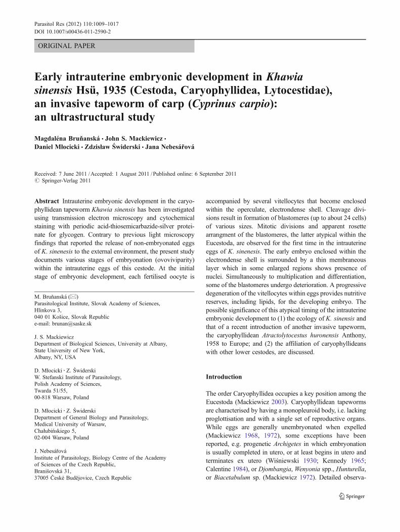
Fig. 1–5 1 Intrauterine eggs of K. sinensis. Fertilised oocyte (O) andvitellocytes (VC) enclosed in a thick electrondense shell (ES) with anoperculum (Op). Note numerous shell globule clusters (SGC) withinthe cytoplasm of vitellocytes. g glycogen. Bar=10 μm. 2 A detail ofthe operculate region (Op) of the two intrauterine eggs. Both operculahave a pitted appearance. VC vitellocyte, GB GER body. Bar=5 μm. 3A conspicuous constriction with a suture (arrow) indicates thejunction that separates the operculum (Op) from the remaining
electrondense shell (ES). VC vitellocyte. Bar=5 μm. 4 Vitellocytes(VC) within intrauterine eggs are rich in glycogen (g). Thiéry method.O fertilised oocyte, N nucleus. Bar=10 μm. 5 Lipid droplets (L) canbe found occasionally in the degrading vitellocytes (VC) within theintrauterine eggs. Thiéry method. g glycogen, ES eggshell. Bar=2.5 μm.Inset A detail of GER-bodies (GB) within the cytoplasm of the degradingvitellocytes from intrauterine eggs. Bar=0.65 μm
Parasitol Res (2012) 110:1009–1017 1011

12 and 13). Degenerating blastomeres resembling areas offocal cytoplasmic degeneration increase in number. They arelocalised either at the periphery or in the centre of theembryo. Lobed nuclei of degenerating blastomeres increasein number (Fig. 12). The lobed nuclei and degeneration ofblastomeres may be directly associated with each other(Fig. 13). The remnants of degenerating vitellocytes havenever been observed to fuse with the cytoplasm ofblastomeres. The late cleavage embryo is surrounded by a
thin membraneous layer, which may contain flattened nuclei(Figs. 12 and 14).
Discussion
Results of the present study provide direct evidence for thefirst time that embryonic development of K. sinensisalready starts in utero. Contrary to previous light microscopy
Fig. 6–9 6 A fertilised oocyte has a large nucleus (N) with prominentnucleolus (Nu) and a minute heterochromatin islands (Hc). Thecytoplasm is rich in mitochondria (M). Bar=2 μm. 7 Early cleavagedivision results in formation of three types of blastomeres: macromeres(Ma), mesomeres (Me) and micromeres (Mi). Hc heterochromatin, Mmitochondrion, N nucleus, Nu nucleolus, VC vitellocyte. Bar=5 μm.8 A portion of a rosette like arrangement of several blastomeres (B)
linked by cytoplasmic bridges to a central cytoplasm mass (CP). Notethe lobed nuclei (N) of some of the blastomeres. DBo dense bodies ofdegenerating vitellocytes. Bar=5 μm. 9 Rosette arrangement ofblastomeres (B) at more advanced stage of embryo development. CPcentral cytoplasm mass, ES electrondense shell, Op operculum, VCvitelline cell, DBo dense bodies of degenerating vitellocytes. Bar=10 μm
1012 Parasitol Res (2012) 110:1009–1017

observations that reported a zygote and several (five to eight)vitellocytes within freshly released eggs of caryophyllideans(Demshin and Dvoryadkin 1980; Protasova et al. 1990;Scholz 1991, 1993), our observations revealed early stagesof embryonic development within the intrauterine eggs of K.sinensis. Until now, typical mitotic divisions had never beendetected at the ultrastructural level in caryophyllidean eggs.The only evidence for cleavage divisions was the increasingnumber of blastomeres in advanced stages of embryonicdevelopment of W. virilis (Młocicki et al. 2010b). Earlier,Motomura (1929) had pictured mitotic divisions and
cleavage in Archigetes; however, his observations were atthe light microscope level.
K. sinensis has a type of egg, characteristic of theCaryophyllidea, that also occurs in the monozoic Gyroco-tylidea and Amphilinidea as well as in some lower polyzoicgroups, e.g. Spathebothriidea, Diphyllobothriidea or Trypa-norhyncha (Świderski and Xylander 2000; Conn andŚwiderski 2008). Their egg is polylecithal and has a thick,hardened shell, and in several orders, there may be anoperculum and a small, sometimes inconspicuous, terminalknob or boss at the opposite end. The occurrence of an
Fig. 10–13 10 Early embryo is composed of several blastomeres.Note nuclei of dividing cells in metaphase (me) and anaphase (an).DM degenerating micromere. Bar=5 μm. 11 More advanced embryocontains dividing blastomeres in anaphase (an), which are close eitherto the remnants of vitellocytes (VC) or the shell (ES). Note lobednucleus (N) of blastomere. Bar=5 μm. 12 Degeneration of some
micromeres (DM). Note that peripheral blastomeres (DB) seem to becontinuous with a thin membraneous layer (ML) which includes alsonucleus of a blastomere (NML). Bar=5 μm. 13 Part of the embryoshowing four degenerating micromeres (DM) and cell division ofblastomere in anaphase (an). Note lobed nucleus (N) and its closeassociation with DM. ML a thin membraneous layer. Bar=2 μm
Parasitol Res (2012) 110:1009–1017 1013

operculum in caryophyllideans has been a subject of muchconfusion (see Mackiewicz 1972), largely because it is smalland not readily seen when eggs are viewed in utero at thelight microscope level. As shown in the present study,intrauterine eggs of K. sinensis have an electrondense shelland an operculum which may be pitted. As described andpictured by Scholz (1991), a small abopercular knob ispresent at the wider pole of the egg. It is best observed onwhole eggs. Apart from the Caryophyllidea, operculate eggsoccur also in the Gyrocotylidea, Spathebothriidea andHaplobothriidea; however, the Pseudophyllidea (= Bothrio-cephalidea and Diphyllobothriidea according to Kuchta et al.2008) and Trypanorhyncha are polymorphic in this character(Hoberg et al. 1997).
There is little information on the ultrastructure of theembryo and associated envelopes of caryophyllid cestodesand no studies that illustrate the embryo, its envelopes andshell in a single TEM micrograph. The structure of apreoncosphere embryo and a fully formed oncosphere of acestode with polylecithal eggs, but lacking a ciliatedembryophore (e.g. Caryophyllidea, Spathebothriidea, someBothriocephalidea), has been illustrated by Conn andŚwiderski (2008) who recognized that an embryo at theend of the cleavage divisions is surrounded by outer andinner embryonic envelopes and a vitelline capsule. Thehexacanth is surrounded by an oncospheral membrane,situated under an inner envelope, and shell originating fromthe vitelline capsule. It was not until recently that the latterpattern was described in ultrastructural studies of theintrauterine eggs of the caryophyllidean tapeworm W. virilisfamily Caryophyllaeidae; see Młocicki et al. (2010b). Onthe other hand, vitelline capsule or typical embryonicenvelopes were not detected in the intrauterine eggs of K.
sinensis (family Lytocestidae) in the present study, althoughvarious blastomere stages ranging from 2 to about 24 cellswere observed. An embryo in early cleavage is surroundedby degenerating vitellocytes and an electrondense shell.Vitellocytes were never observed to take part in theformation of the outer embryonic envelope. Typically, theouter envelope of cestode eggs is formed by a fusion ofmacromeres with the remnants of degenerating vitellocytes(Świderski 1994a, b; Świderski et al. 2005; Młocicki et al.2010a, b). The late cleavage embryo of K. sinensis issurrounded by a thin membraneous layer and degeneratingvitellocytes, both compenents being enclosed within anelectrondense shell. Nuclei and degenerating blastomeresoccurring within a thin membraneous layer indicate that thissurrounding structure may be related to the embryonicenvelope (Świderski 1994b). In contrast, the thin membra-neous layer of K. sinensis is substantially reduced comparedwith embryonic envelopes of bothriocephalideans (Świderski1994b; Świderski et al. 2005). Its ultrastructure mayresemble an oncospheral membrane, described between thehexacanth and the inner envelope of fully formed oncospheres(Conn and Świderski 2008). However, its origin and stage offormation is completely different. A typical oncospheralmembrane is an anucleated structure and appears at the finalstage of embryonic development and originates fromdelamination of the innermost layer of the inner envelope(Tkach and Świderski 1998; Świderski et al. 2001; Młocickiet al. 2005). Embryonic envelopes were not detected neitherduring the early development of the caryophyllideanArchigetes appendiculatus by Motomura (1929) and in twospecies of Khawia by Scholz (1991, 1993); however, theseobservations were at the light microscope level. The presenceof outer and inner embryonic envelopes in the Caryophyllideawas reported only in the intrauterine eggs of W. virilis byMłocicki et al. (2010b). It is of interest that the intrauterineembryonated eggs of another basal tapeworm, spathebothrii-dean Didymobothrium rudolphii, also do not includeembryonic envelopes (Świderski et al. 2010). In contrast,the operculate eggs of another spathebothriidean, Cyathoce-phalus truncatus, are considered to be fully embryonatedalready ex utero after releasing of unembryonated eggs intowater (Wiśniewski 1932).
Glycogen remains as the predominant nutrient withinvitellocytes of all caryophyllideans so far examined(Mackiewicz 1968; Świderski and Mackiewicz 1976;Świderski and Xylander 2000; Świderski et al. 2004a, b,2009). Lipid droplets in degenerating vitellocytes observedin the intrauterine eggs of K. sinensis (present study) werereported recently by Młocicki et al. (2010b) in anothercaryophyllidean, W. virilis. Both findings are at leastunusual as lipid droplets have not been reported previouslyin mature vitellocytes of caryophyllideans (Mackiewicz1968; Świderski and Mackiewicz 1976; Świderski et al.
Fig. 14 14 A portion of the embryo surrounded by a thin membraneouslayer (ML) with a nucleus (NML). Note nucleus of blastomereundergoing mitotic division in anaphase (an). ES electrondense shell,VC vitelline cell. Bar=5 μm
1014 Parasitol Res (2012) 110:1009–1017

2004a, b; Bruňanská et al. 2009a) including vitellocytes ofK. sinensis (Bruňanská et al. 2009b). The only exception isAtractolytocestus huronensis, that has individual lipiddroplets observed occasionaly in the cytoplasm of someof the mature vitellocytes (Bruňanská et al. 2007). Thepossible explanation supports a hypothesis that degenerat-ing vitellocytes are sites of autolysis and may progressivelyaccumulate waste metabolic products of the developingembryo (Młocicki et al. 2010a; b). However, this presump-tion needs futher affirmation.
As we have found, there are clear signs of degenerationin some blastomeres during early and more advanced stagesof the embryonic development in K. sinensis. The degen-eration process or apoptosis, results in a great reduction inthe number of oncospheral cells, a common characteristicfor cestode embryos (Rybicka 1961; Świderski 1968).
Degenerating vitellocytes in the intrauterine eggs of K.sinensis contain GER-bodies which were described previ-ously in parasitic flatworms using various terminologies, e.g. lamellar granules, dark concentric bodies, yolk bodies,ribosomal complexes, glycan vesicles (see Młocicki et al.2011). Such structures have been reported in the maturevitellocytes of other caryophyllideans (Bruňanská et al.2009a) or spathebothriideans (Bruňanská et al. 2005;Poddubnaya et al. 2006). In W. virilis (Młocicki et al.2011), GER-bodies were described only in the vitellocytesof newly formed eggs, but not in mature vitellocytes of thevitelline system. In K. sinensis, however, they occur at firstin the mature vitellocytes (Bruňanská, unpublished data).GER bodies are supposed to participate in synthesis ofglycoproteins or may represent remnants of GER undergo-ing degeneration and transformation into areas of focalcytoplasmic degradation (Młocicki et al. 2011).
The origin of large osmiophilic dense bodies in thecytoplasm of degenerating vitellocytes is unknown and amatter of speculation. They may represent remnants of shellmaterial, highly condensed vitellocyte nuclei or some kindof lysosomal structure.
The functional significance of intrauterine embryogenesisin K. sinesis remains unknown. It is of interest that theoccurrence of K. sinensis in carp has decreased dramaticallysince the early 2000s, after A. huronensis appeared in Europe(Majoros et al. 2003; Oros et al. 2004; Kappe et al. 2006;Oros et al. 2009). During the late 1980s and early 1990s, K.sinensis, also an invasive species but from Asia, was the onlycaryophyllidean parasite found in carp from fishponds inSouth Bohemia (Scholz 1991). At that time, there was noevidence of intrauterine embryogenesis or ovovivipary in K.sinensis (Demshin and Dvoryadkin 1980; Protasova et al.1990; Scholz 1991). However, embryonation within theintrauterine eggs has now been found in specimens of K.sinensis collected in 2003 from South Bohemia. Suchdevelopment may be classified as precocious when com-
pared with earlier data. This atypical early development mayreflect a response to interspecific competition between twoinvasive cestode species in carp. Indeed, A. huronensis, withits parthenogenetic reproduction (Jones and Mackiewicz1969; Bruňanská et al. 2011), has been quite successful incolonizing new regions (Oros et al. 2009). It should benoted, however, that intrauterine embryonation was observedin specimens collected in November 2003, when the coldertemperatures would be expected to retard rather than accelerateembryo development. It is well-known that oncospheredevelopment is accelerated with increasing temperature in K.sinensis, Diphyllobothrium or Triaenophorus (Kuhlow 1953;Kuperman 1973; Scholz 1991).
On the other hand, given that there are no experimentalor field data on interspecific competition between K.sinensis and A. huronensis in a stable environment and thatmultiple cestode infections of carp are common (Oros et al.2009), further research is required to ascertain if intrauterineembryonation in K. sinensis is related to interspeciescompetition or other environmental factors. However, itcannot be excluded that differences in the resolution of lightand electron microscope and techniques used by previousauthors (Demshin and Dvoryadkin 1980; Protasova et al.1990; Scholz 1991) resulted in misinterpretation of thedevelopmental stages within eggs of Khawia.
In utero embryonic development of the caryophyllideanW. virilis (Młocicki et al. 2010b), K. sinensis (present study)and spathebothriidean D. rudolphii (Świderski et al. 2010)differs significantly from that in the bothriocephalideanswhich release largely either unembryonated operculate eggs(Świderski 1994a, b) or embryonated anoperculate eggs(Świderski et al. 2005). This pattern clearly indicates thatembryonic development in the basal orders Caryophyllideaand Spathebothriidea is not identical with that of theBothriocephalidea, although a great variation exists inembryonation stages of intrauterine eggs in different cestodes,even within the same order as examplified by Bothriocepha-lidea. Given that evolutionary relationships can often beascertained from a study of conservative and fundamentalstructures or developmental stages, comparative embryogen-esis may offer insights into the evolutionary relationshipsamong the various cestode groups. Further ultrastructuralinvestigations of early intrauterine development of lowercestodes are needed for a better understanding of reproductivebiology and its role in evolution within the Cestoda.
Acknowledgements We are grateful to Blanka Škoríková, Dr.Borislav Kostič and the staff of the Laboratory of Electron Micrscopyof the Institute of Parasitology AS CR, České Budějovice, CzechRepublic for excellent technical assistance. This research was madewithin the framework of the joint research project supported by abilateral agreements on scientific cooperation signed by the the Polishand Slovak Academies of Sciences. The study was supported by the theGrant Agency of the Slovak Republic VEGA (projects no. 2/0018/08 and
Parasitol Res (2012) 110:1009–1017 1015

2/0047/11). The work was realizedwithin a frame of Centre of Excellencefor Parasitology (Code ITMS: 26220120022) based on the support of theOperational Programme “Research & Development” funded from theEuropean regional Development Fund (rate 0.2).
References
Bruňanská M (2009) Spermatological characters of the caryophyllideancestode Khawia sinensis Hsü, 1935, a carp parasite. Parasitol Res105:1603–1610
Bruňanská M (2010) Recent insights into spermatozoa developmentand ultrastructure in the Eucestoda. In: Lejeune T, Delvaux P (eds)Human spermatozoa: maturation, capacitation and abnormalities.Nova Science Publishers, Inc., New York, pp 327–354
Bruňanská M, Poddubnaya LG, Dezfuli BS (2005) Vitellogenesis intwo spathebothriidean cestodes. Parasitol Res 96:390–397
Bruňanská M, Drobníková P, Oros M (2007) Vitellocytes of thecaryophyllidean cestode Atractolytocestus huronensis. Proceed XInt Helminth Symposium. Stará Lesná, Slovak Republic, 9–14September 2007, p. 14
Bruňanská M, Drobníková P, Oros M (2009a) Vitellogenesis in thecestode Atractolytocestus huronensis Anthony, 1958 (Caryophyl-lidea: Lytocestidae). Parasitol Res 105:647–654
Bruňanská M, Drobníková P, Oros M (2009b) Vitellocytes of thecaryophyllidean cestode Khawia sinensis Hsü, 1935, a carpparasite. Mikroskopia 2009. Stará Lesná, Slovak Republic, 25–26 March, 2009, p. 17
Bruňanská M, Nebesářová J, Oros M (2011) Ultrastructural aspects ofspermatogenesis, testes and vas deferens in the parthenogenetictapeworm Atractolytocestus huronensis Anthony, 1958 (Cestoda:Caryophyllidea), a carp parasite from Slovakia. Parasitol Res108:61–68
Calentine RL (1984) Biology of Archigetes (Caryophyllaeidae) inLimnodrilus hoffmeisteri (Tubificidae). Proc Helminthol SocWash 51:109–112
Conn DB, Świderski Z (2008) A standardized terminology of theembryonic envelopes and associated developmental stages oftapeworms (Platehelminthes: Cestoda). Folia Parasitol 55:42–52
Demshin NI, Dvoryadkin VA (1980) Biology of Khawia sinensis Hsü,1935 (Caryophyllidea, Cestoda) a parasite of Cyprinus carpiohaematopterus. Hydrobiologia Zh 16:77–82 (In Russian)
Hoberg EP, Mariaux J, Justine J-L, Brooks DR, Weekes PJ (1997)Phylogeny of the orders of the Eucestoda (Cercomeromorphae)based on comparative morphology: historical perspectives and anew working hypothesis. J Parasitol 83:1128–1147
Jones AW, Mackiewicz JS (1969) Naturally occurring triploidy andparthenogenesis inAtractolytocestus huronensis Anthony (Cestoidea:Caryophyllidea) from Cyprinus carpio L. in North America. JParasitol 55:1105–1118
Kappe A, Seifert T, El-Nobi G, Bräuer G (2006) Occurrence ofAtractolytocestus huronensis (Cestoda: Caryophyllaeidae), inGerman pond-farmed common carp Cyprinus carpio. Dis AquatOrg 70:255–259
Kennedy CR (1965) Taxonomic studies on Archigetes Leuckart, 1878(Cestoda, Caryophyllidea). Parasitology 55:439–451
Kuchta R, Scholz T, Brabec J, Bray RA (2008) Suppression of thetapeworm order Pseudophyllidea (Platyhelminthes:Eucestoda)and the proposal of two new orders, Bothriocephalidea andDiphyllobothriidea. Int J Parasitol 38:49–55
Kuhlow F (1953) Űber die Entwicklung und Anatomie vonDiphyllobothrium dendriticum Nitzsch 1824. Z Parasitenkd16:1–35
Kuperman BI (1973) Tapeworms of the genus Triaenophorus, parasitesof fishes. Leningrad, Izdateľstvo Nauka, 208 pp (In Russian,
English translation (1981)). Amerind Publishing Co. Pvt. Ltd, NewDelhi, p 222
Mackiewicz JS (1968) Vitellogenesis and egg-shell formation inCaryophyllaeus laticeps (Pallas) and Caryophyllaeoides fennica(Schneider) (Cestoidea: Caryophyllidea). Z Parasitenkd 30:18–32
Mackiewicz JS (1972) Caryophyllidea (Cestoidea): a review. ExpParasitol 31:417–512
Mackiewicz JS (2003) Caryophyllidea (Cestoidea): molecules,morphology and evolution. Acta Parasitol 48:143–154
Majoros G, Gy C, Molnár K (2003) Occurrence of Atractolytocestushuronensis Anthony, 1958 (Cestoda: Caryophyllaeidae), inHungarian pond-farmed common carp. Bull Eur Ass Fish Pathol23:167–175
Młocicki D, Świderski Z, Eira C, Miquel J (2005) An ultrastructuralstudy of embryonic envelope formation in the anoplocephalidcestode Mosgovoyia ctenoides (Railliet, 1890) Beveridge, 1978.Parasitol Res 95:243–251
Młocicki D, Świderski Z, Conn DB (2010a) Ultrastructure of the earlyembryonic development of Corallobothrium fimbriatum (Cestoda:Proteocephalidea). J Parasitol 96:839–846
Młocicki D, Świderski Z, Mackiewicz JS, Ibraheem MH (2010b)Ultrastructure of intrauterine eggs: evidence of early ovoviviparityin the caryophyllidean cestode Wenyonia virilis Woodland, 1923.Acta Parasitol 55:349–358
Młocicki D, Świderski Z, Mackiewicz JS, Ibraheem MH (2011)Ultrastructural and cytochemical studies of GER-bodies in theintrauterine eggs of Wenyonia virilis Woodland, 1923 (Cestoda,Caryophyllidea). Acta Parasitol 56:40–47
Motomura I (1929) On the early development of monozoic cestode,Archigetes appendiculatus, including the oogenesis and fertilization.Annot Zool Jap 12:109–129
Oros M, Hanzelová V, Scholz T (2004) The cestode Atractolytocestushuronensis (Caryophyllidea) continues to spread in Europe: newdata on the helminth parasite of the common carp. Dis Aquat Org62:115–119
Oros M, Hanzelová V, Scholz T (2009) Tapeworm Khawia sinensis:review of the introduction and subsequent decline of a pathogenof carp, Cyprinus carpio. Vet Parasitol 164:217–222
Poddubnaya LG, Gibson DI, Świderski Z, Olson PD (2006)Vitellocyte ultrastructure in the cestode Didymobothrium rudolphi(Monticelli, 1890): possible evidence for the recognition ofdivergent taxa within the Spathebothriidea. Acta Parasitol51:255–263
Protasova EN, Kuperman BI, Roytman VA, Poddubnaya LG (1990)Caryophyllids of the fauna in SSSR. Nauka, Moscow, p 238, InRussian
Rybicka K (1961) Cell reduction in the embryonic development of thecestode Diorchis ransomi Schultz, 1940. Nature 192:771–772
Scholz T (1991) Early development of Khawia sinensis Hsü, 1935(Cestoda: Caryophyllidea), a carp parasite. Folia Parasitol38:133–142
Scholz T (1993) On the development of Khawia baltica Szidat, 1942(Cestoda: Lytocestidae), a parasite of tench, Tinca tinca (L.).Folia Parasitol 40:99–103
Świderski Z (1968) An electron microscopic evidence of thedegeneration of some micromeres during embryonic developmentof the cestode Catenotaenia pusilla (Goeze, 1782) (Cyclophyllidea,Catenotaeniidae). Zool Polon 18:469–474
Świderski Z (1994a) Origin, differentiation and ultrastructure ofegg envelopes surrounding the coracidia of Bothriocephalusclavibothrium. Acta Parasitol 39:73–81
Świderski Z (1994b) Homology and analogy of egg enevelopessurrounding the coracidia of Bothriocephalus clavibothrium andmiracidia of Schistosoma mansoni. Acta Parasitol 39:123–130
Świderski Z, Bruňanská M, Poddubnaya LG (2004a) Ultrastructuraland cytochemical studies on vitellogenesis in the caryophyllidean
1016 Parasitol Res (2012) 110:1009–1017

cestode Caryophyllaeus laticeps. Proceed. IX Eur. Multicolloq.Parasitol. Valencia, Spain, 18–23 July 2004, p. 602
Świderski Z, Mackiewicz JS (1976) Electron microscope study ofvitellogenesis in Glaridacris catostomi (Cestoidea: Caryophyllidea).Int J Parasitol 6:61–73
Świderski Z, Xylander WER (2000) Vitellocytes and vitellogenesis incestodes in relation to embryonic development, egg productionand life cycle. Int J Parasitol 30:805–817
Świderski Z, Ndiaye PI, Tkach V, Miquel J, Marchand B, Chomicz L,Sereda MJ (2001) Ultrastructural study of the embryonicdevelopment of the anoplocaphalid cestode Anoplocephaloidesdentata, an intestinal parasite of Arvicolidae rodents. I. Eggenvelope formation. Acta Parasitol 46:171–185
Świderski Z, Bruňanská M, Poddubnaya LG, Mackiewicz JS (2004)Cytochemical and ultrastructural study on vitellogenesis incaryophyllidean cestode Khawia armeniaca (Cholodkovski,1915). Acta Parasitol 49:16–24
Świderski Z, Bruňanská M, Młocicki D, Conn DB (2005) Ultrastructureof the oncospheral envelopes in the pseudophyllidean Eubothriumsalvelini (Schrank, 1790). Acta Parasitol 50:312–318
Świderski Z, Młocicki D, Mackiewicz JS, Miquel J, Ibraheem MH,Bruňanská M (2009) Ultrastructure and cytochemistry ofvitellogenesis in Wenyonia virilis Woodland, 1923 (Cestoda,Caryophyllidea). Acta Parasitol 54:131–142
Świderski Z, Gibson DI, Santos MJ, Poddubnaya LG (2010)Ultrastructure of the intrauterine eggs of Didymobothrium rudolphi(Monticelli, 1890) (Cestoda, Spathebothriidea, Acrobothriidae) andits phylogenetic implications. Acta Parasitol 55:254–269
Thiéry J-P (1967) Mise en évidence des polysaccharides sur coupesfines en microscopie électronique. J Microsc 6:987–1018
Tkach VV, Świderski Z (1998) Differentiation and ultrastructure of theoncospheral envelopes in the hymenolepidid cestode Staph-ylocystoides stefanskii (Żarnowski, 1954). Acta Parasitol 43:222–231
Wiśniewski LW (1930) Das Genus Archigetes R. Leuck. Eine Studiezur Anatomie, Histogenese, Systematik und Biologie. Mém AcadPol Sci Lett Class Sci Mathém Natur Ser B Sci Nat 2:160
Wiśniewski LW (1932)Cyathocephalus truncatus Pallas. II. AllgemeineMorphologie. Bull Int Acad Pol Sci. Class Sci Mathem Natur, SerB: Sciences Nat (II):311–327
Parasitol Res (2012) 110:1009–1017 1017