The highly antigenic 53/25 kDa Taenia solium protein fraction with cathepsin-L like activity is...
14
The highly antigenic 53/25 kDa Taenia solium protein fraction with cathepsin-L like activity is present in the oncosphere/ cysticercus and induces non-protective IgG antibodies in pigs Mirko Zimic a,* , Mónica Pajuelo a , Robert H. Gilman a,b , Andrés H. Gutiérrez a , Luis D. Rueda a , Myra Flores a , Nancy Chile a , Manuela Verástegui a , Armando Gonzalez c , Héctor H. García a,d , and Patricia Sheen a for the Cysticercosis Working Group in Peru a Laboratorios de Investigación y Desarrollo. Facultad de Ciencias y Filosofía, Universidad Peruana Cayetano Heredia, Av. Honorio Delgado 430 Urb. Ingeniería SMP-Lima 31, Lima, Perú b Department of International Health, Johns Hopkins Bloomberg School of Public Health, Baltimore, 615 N. Wolfe St. RM W5515 Baltimore, MD 21205 – Maryland, USA c School of Veterinary Medicine, Universidad Nacional Mayor de San Marcos, Av. Circunvalación Cdra. 28 s/n, San Borja-Lima 41 Lima, Perú d Cysticercosis Unit, Instituto de Ciencias Neurológicas, Jr. Ancash 1271 Lima 1, Perú Abstract Cathepsin L-like proteases are secreted by several parasites including Taenia solium. The mechanism used by T. solium oncospheres to degrade and penetrate the intestine and infect the host is incompletely understood. It is assumed that intestinal degradation is driven by the proteolytic activity of enzymes secreted by the oncosphere. Blocking the proteolytic activity by an antibody response would prevent the oncosphere penetration and further infection. Serine and cysteine proteases including chymotrypsin, trypsin, elastase, and cathepsin L, are secreted by T. solium and Taenia saginata oncospheres when cultured in vitro, being potential vaccine candidates. However, the purification of a sufficient quantity of proteases secreted by oncospheres to conduct a vaccine trial is costly and lengthy. A 53/25 kDa cathepsin L-like fraction partially purified from T. solium cyst fluid was described previously as an important antigen for immunodiagnostics. In this study we found that this antigen is present in the T. solium oncosphere and is also secreted by the cysticercus. This protein fraction was tested for its ability to protect pigs against an oral challenge with T. solium oncospheres in a vaccine trial. IgG antibodies against the 53/25 kDa cathepsin L-like protein fraction were elicited in the vaccinated animals but did not confer protection. © 2011 Elsevier B.V. All rights reserved. * Corresponding author. (511) 3190000 ext 2604. [email protected]. Universidad Peruana Cayetano Heredia. Facultad de Ciencias y Filosofía. Laboratorios de Investigación y Desarrollo. Av. Honorio Delgado 430, SMP. Lima, Peru. Conflict of interest statement All authors disclose any conflict of interest Publisher's Disclaimer: This is a PDF file of an unedited manuscript that has been accepted for publication. As a service to our customers we are providing this early version of the manuscript. The manuscript will undergo copyediting, typesetting, and review of the resulting proof before it is published in its final citable form. Please note that during the production process errors may be discovered which could affect the content, and all legal disclaimers that apply to the journal pertain. NIH Public Access Author Manuscript Vet Immunol Immunopathol. Author manuscript; available in PMC 2013 January 15. Published in final edited form as: Vet Immunol Immunopathol. 2012 January 15; 145(1-2): 171–178. doi:10.1016/j.vetimm.2011.11.002. NIH-PA Author Manuscript NIH-PA Author Manuscript NIH-PA Author Manuscript
-
Upload
independent -
Category
Documents
-
view
1 -
download
0
Transcript of The highly antigenic 53/25 kDa Taenia solium protein fraction with cathepsin-L like activity is...

The highly antigenic 53/25 kDa Taenia solium protein fractionwith cathepsin-L like activity is present in the oncosphere/cysticercus and induces non-protective IgG antibodies in pigs
Mirko Zimica,*, Mónica Pajueloa, Robert H. Gilmana,b, Andrés H. Gutiérreza, Luis D. Ruedaa,Myra Floresa, Nancy Chilea, Manuela Verásteguia, Armando Gonzalezc, Héctor H. Garcíaa,d,and Patricia Sheena for the Cysticercosis Working Group in PeruaLaboratorios de Investigación y Desarrollo. Facultad de Ciencias y Filosofía, UniversidadPeruana Cayetano Heredia, Av. Honorio Delgado 430 Urb. Ingeniería SMP-Lima 31, Lima, PerúbDepartment of International Health, Johns Hopkins Bloomberg School of Public Health,Baltimore, 615 N. Wolfe St. RM W5515 Baltimore, MD 21205 – Maryland, USAcSchool of Veterinary Medicine, Universidad Nacional Mayor de San Marcos, Av. CircunvalaciónCdra. 28 s/n, San Borja-Lima 41 Lima, PerúdCysticercosis Unit, Instituto de Ciencias Neurológicas, Jr. Ancash 1271 Lima 1, Perú
AbstractCathepsin L-like proteases are secreted by several parasites including Taenia solium. Themechanism used by T. solium oncospheres to degrade and penetrate the intestine and infect thehost is incompletely understood. It is assumed that intestinal degradation is driven by theproteolytic activity of enzymes secreted by the oncosphere. Blocking the proteolytic activity by anantibody response would prevent the oncosphere penetration and further infection. Serine andcysteine proteases including chymotrypsin, trypsin, elastase, and cathepsin L, are secreted by T.solium and Taenia saginata oncospheres when cultured in vitro, being potential vaccinecandidates. However, the purification of a sufficient quantity of proteases secreted by oncospheresto conduct a vaccine trial is costly and lengthy.
A 53/25 kDa cathepsin L-like fraction partially purified from T. solium cyst fluid was describedpreviously as an important antigen for immunodiagnostics. In this study we found that this antigenis present in the T. solium oncosphere and is also secreted by the cysticercus. This protein fractionwas tested for its ability to protect pigs against an oral challenge with T. solium oncospheres in avaccine trial. IgG antibodies against the 53/25 kDa cathepsin L-like protein fraction were elicitedin the vaccinated animals but did not confer protection.
© 2011 Elsevier B.V. All rights reserved.*Corresponding author. (511) 3190000 ext 2604. [email protected]. Universidad Peruana Cayetano Heredia. Facultad de Ciencias yFilosofía. Laboratorios de Investigación y Desarrollo. Av. Honorio Delgado 430, SMP. Lima, Peru.Conflict of interest statementAll authors disclose any conflict of interestPublisher's Disclaimer: This is a PDF file of an unedited manuscript that has been accepted for publication. As a service to ourcustomers we are providing this early version of the manuscript. The manuscript will undergo copyediting, typesetting, and review ofthe resulting proof before it is published in its final citable form. Please note that during the production process errors may bediscovered which could affect the content, and all legal disclaimers that apply to the journal pertain.
NIH Public AccessAuthor ManuscriptVet Immunol Immunopathol. Author manuscript; available in PMC 2013 January 15.
Published in final edited form as:Vet Immunol Immunopathol. 2012 January 15; 145(1-2): 171–178. doi:10.1016/j.vetimm.2011.11.002.
NIH
-PA Author Manuscript
NIH
-PA Author Manuscript
NIH
-PA Author Manuscript

Keywordscathepsin L; cysticercosis; oncosphere; protease; Taenia solium; vaccine
IntroductionTaeniasis and cysticercosis are parasitic diseases caused by different life stages of thetapeworm T. solium. Taeniasis (infection with the adult tapeworm) is exclusive to humansand results from the ingestion of larval cysts. Cysticercosis (infection with the larval stage ofthe parasite) resulting from the ingestion of eggs usually occurs in pigs, the intermediatehost. Humans may also act as an intermediate host and develop cysticercosis after theaccidental ingestion of eggs through contaminated food or fecal oral transmission. Cysticlarvae often locate in the central nervous system, causing the clinical disorder known asneurocysticercosis (NCC), a major global cause of acquired epilepsy (Carpio and Hauser,2002; Garcia and Del Brutto, 2000; Nicoletti et al., 2002) that is associated with significantneurological morbidity in developing countries (White and Garcia, 1999). Documentation ofTaenia solium in the US and other developed countries, at least in modern times, has almostentirely involved “imported cases” from foreign endemic regions (Marconi et al., 2006;Schantz et al., 1992; Tsang V and M, 1995; White, 1997). Despite several recentinvestigations using mass chemotherapy alone to treat populations for this infection, nosustainable eradication has yet been achieved (Garcia et al., 2007; Garcia et al., 2006;Gonzalez et al., 1998). Porcine vaccines are promising tools given the essential role of thepig as obligatory intermediate host. Such a tool is indispensable to achieving sustainedcontrol of this disease. Porcine vaccination strategies to control this disease have beenconducted in multiple studies by several groups (Gauci et al., 1998; Lightowlers, 2003;Lightowlers et al., 2003; Nascimento et al., 1995; Pathak and Gaur, 1990).
Although not demonstrated yet, it is assumed that the infection starts with the adherence ofthe oncosphere to the intestine followed by a proteolytic attack to degrade the intestinal walland start the penetration. Blocking either the adherence or the proteolytic attack of theoncosphere would prevent infection. Antibodies raised to the specific proteins involved inthese processes, as a result of immunization, would confer a desired protection.
Porcine vaccines are promising tools in the control of taeniasis/cysticercosis, given theessential role of the pig as the obligate intermediate host. Vaccines are crucial to achievesustained control and possible eradication of this disease. Oncosphere antigens are currentlythe most effective vaccine candidates in preventing porcine cysticercosis (Assana et al.;Flisser et al., 2004; Gonzalez et al., 2005; Lightowlers et al., 2003; Pathak and Gaur, 1990;Plancarte et al., 1999; Verastegui et al., 2002). T. solium oncospheral protein TSOL18 iscurrently the best antigen able to induce near total protection against a T. solium proglottidoral challenge in pigs (Lightowlers, 2003). TSOL18 is an oncosphere protein homologue toT. saginata HP6. HP6 has been characterized as an adherence protein with a fibronectin typeIII domain, able to interact with integrins from epithelial cells (Bonay et al., 2002; Ferrer etal., 2007; Parkhouse et al., 2008).
Several proteases including mainly serine proteases (chymotrypsin-like and trypsin-likeproteases), but also cysteine proteases (cathepsin L-like proteases) were found to be secretedby T. solium oncospheres (Zimic et al., 2007), eventually to be used to degrade the intestinalwall and let the oncosphere penetrate into the circulatory system. Therefore oncosphereproteases are potential vaccine candidates to prevent the intestinal penetration of theoncosphere. The purification of a large amount of oncosphere proteases for a vaccine trial isextremely expensive and lengthy.
Zimic et al. Page 2
Vet Immunol Immunopathol. Author manuscript; available in PMC 2013 January 15.
NIH
-PA Author Manuscript
NIH
-PA Author Manuscript
NIH
-PA Author Manuscript

In a previous study, we described a 53/25 kDa protein fraction with cathepsin L-like activity(from now on TsCLf), purified from the cyst fluid. TsCLf showed a high sensitivity andspecificity for the diagnostics of NCC, in both a Western immunoblot and an ELISA assay(Zimic et al., 2009).
In the present study we tested the presence of the TsCLf in the oncosphere and in theexcretory/secretory antigens of the cyst, and tested the TsCLf as a vaccine candidate forporcine cysticercosis.
Materials and methodsCysticercus fluid and TsCLf purification
Eight pigs naturally infected with porcine cysticercosis, confirmed by tongue test, wereselected from an endemic area in the central highlands of Peru (Gonzalez et al., 1990). Theanimals were sedated with a combination of xylazine and ketamine, and euthanasia wasperformed using a pentobarbital overdose. The carcasses were dissected and T. solium cystsidentified. Cyst fluid was recovered by aspiration with a syringe. Approximately 400 mL ofcyst fluid was recovered from 4 000 viable cysts and stored at −70 °C. To obtain the TsCLfpartially purified antigen, the cyst fluid was processed following the procedures previouslydescribed (Zimic et al., 2009).
Detection of TsCLf in the excretory/secretory antigens of the cysticercusMetacestodes of T. solium were individually collected from muscle tissue of naturallyinfected pigs. Cysts were cultivated for 72 hours at 37°C in 5% CO2 in 12-well plates withcyst medium (buffered RPMI 1640 with antibiotics) as reported (Mahanty et al., 2011).After each 24 hour period, the supernatant of each well containing excretory/secretory (ES)antigens was harvested, collected and replaced. The collected ES antigens were resolved in aSDS-PAGE and revealed by Western immunoblot using pre-immune and post-immune antiTsCLf rabbit sera as previously described (Zimic et al., 2009). Briefly, after SDS-PAGE, theantigen was transferred to nitrocellulose membrane (BIO RAD). Membranes were incubatedovernight at 4°C with pre-immune and post-immune rabbit anti-TsCLf sera (1:200).Membranes were washed three times with PBS-0.3% Tween20, incubated with peroxidase-conjugated goat anti-Rabbit IgG (H+L) antibodies diluted 1: 400 (KPL) for 1h at roomtemperature and developed with 3,3-diaminobenzidine (Sigma) as the substrate.
Additionally, the excretory/secretory antigens from the cysticercus were testedfluorometrically for cysteine protease activity with the substrate Z-Phe-Arg-AMC andinhibition with E64 as previously described (Zimic et al., 2007).
Detection of TsCLf in the oncosphereThe total antigens from activated oncospheres were obtained following the procedurepreviously reported (Verastegui et al., 2003). Adult T. solium tapeworms were collectedfrom newly diagnosed patients. The eggs were obtained from gravid proglottids of adulttapeworms by gentle homogenization in 2.5 % potassium dichromate solution (Sigma, St.Louis, Missouri). With a solution of 0.75% sodium hypochlorite (Mallinckrodt Baker, Inc,Phillipsburg, NJ) in water for 10 min at 4°C, the eggs were hatched and oncospheres werereleased. Oncospheres were then washed three times in RPMI medium (Sigma). Onosphereswere activated by incubation at 37°C for 45 min with artificial intestinal fluid (1 gpancreatin [Sigma Chemical Co., St. Louis, MO], 200 mg Na2CO3, and 1 mL pig bile, withthe volume adjusted to 100 mL with RPMI 1640 medium [pH 8.04]) followed by anotherincubation for 1 hour at 37°C RPMI medium (Verastegui et al., 2007). After activation, theoncospheres were washed three times in phosphate-buffered saline (PBS), pH 7.5. Then the
Zimic et al. Page 3
Vet Immunol Immunopathol. Author manuscript; available in PMC 2013 January 15.
NIH
-PA Author Manuscript
NIH
-PA Author Manuscript
NIH
-PA Author Manuscript

activated oncospheres were sonicated at 4°C at 70 Hz (Sonic-Dismembrato model 300;Fisher Scientific, Pittsburgh, PA) using three one-minute periods separated by one-minuterest intervals, and centrifuged at 10 000 × g for 30 minute at 4°C and the supernatant wereused as total antigens.
The oncosphere total antigens were resolved in a SDS-PAGE and revealed by Westernimmunoblot using a pre-immune and post-immune rabbit anti-TsCLf sera as describedabove.
Additionally, TsCLf was resolved in a SDS-PAGE and revealed by Western immunoblotusing pre-immune and post-immune rabbit anti-oncosphere sera, as described before (Zimicet al., 2009).
Rabbit anti-TsCLf seraThree two-month old New Zealand white rabbits, each weighting approximately 2 kg, wereimmunized with the TsCLf fraction in four doses given at intervals of four days. The firstdose was 5 μg of antigen with 50 μg of saponin as adjuvant. The second, third, and fourthdoses included 10, 20, and 40 μg of antigen respectively with 50 μg of saponin as adjuvantwith each dose. 0.5 mL of blood was sampled on the day prior to each immunization. Twoweeks after the last dose, the animals were sacrificed and almost 100 mL of blood wasrecovered. Pre-immune and post-immune anti-TsCLf sera corresponding to the second andfourth dose were tested in a Western immunoblot using the TsCLf as the antigen accordingto the conditions described previously (Zimic et al., 2009).
Additionally, two similar rabbits were immunized with total T. solium oncosphere antigensprepared as described above. The immunization protocol was similar to the described above.Pre-immune and post-immune anti-oncosphere sera was obtained.
Porcine vaccine trialSix two-month-old pigs (Pietrain/Landrace mixed breed) were included in this vaccine trial.This study was authorized by the Animals Ethics Committee of the School of VeterinaryMedicine, Universidad Nacional Mayor de San Marcos. Each animal was immunized withtwo doses of purified TsCLf (0.2 mg) given with 1 mg saponin as adjuvant given two weeksapart. Because this study was nested in a larger vaccine trial that tested a second antigen thatwas fused to Glutathione S-transferase (GST), obtained from the TOPO vector (Invitrogen).The control/placebo group included six similar pigs that received two doses of recombinantGST (0.5 mg, expressed from the TOPO vector), with 1 mg Quil A as adjuvant (SuperflosBiosector, Vedbaek, Denmark) given two weeks apart. It has been proved that GST does notprotect pigs against cysticercosis infection (Flisser et al., 2004), therefore its administrationto the placebo group is not critical. Blood samples were obtained before immunization andone week after the first and second doses. Sera were tested for anti-TsCLf IgG antibodies ina Western immunoblot. Other study testing Tsol18 showed that at 2 weeks after the seconddose, antibodies are able to elicit strong immunity or confer protection against cysticercosisin pigs (Assana et al.; Jeri et al., 2004; Verastegui et al., 2003). Flisser et al. tested Tsol18and Tsol45 with an infection at 3 weeks after the last dose (Flisser et al., 2004). A similarstudy confirmed protection of this vaccine with an infection at 10 days after the last dose(Gonzalez et al., 2005). After confirming that anti-TsCLf IgG antibodies were elicited andstrongly reacted in a Western immunoblot, animals were challenged with one gravidproglottid given orally (Jeri et al., 2004; Verastegui et al., 2003) 15 days after the last dose,to ensure an appropriate immune response. Five different tapeworms were used for thechallenge.
Zimic et al. Page 4
Vet Immunol Immunopathol. Author manuscript; available in PMC 2013 January 15.
NIH
-PA Author Manuscript
NIH
-PA Author Manuscript
NIH
-PA Author Manuscript

Twelve weeks after challenge, all animals were humanely euthanized and the entire carcassand brain were assessed for the presence of cysticerci (Gonzalez et al., 2005; Verastegui etal., 2002). Cysts were classified as either viable, if a defined cystic structure with a liquidcontent was still present, or degenerated, if this had been replaced by semisolid contents orinflammatory scar tissue (Gonzalez et al., 2005).
Statistical analysisThe total number of cysts as well as the number of cysts of each category (i.e. viable anddegenerated) was compared between the control and the vaccinated groups. To adjust forpotential confounding, the comparison was performed using a multiple Poisson regressionafter adjusting for the sex and age of the pig, as well as the tapeworm identity becauseproglottids from different tapeworms were used for the challenge (proglottids from differenttapeworms may have different infectivity). An indicator variable referring to the vaccinegroup was used to test the vaccine efficacy.
ResultsDetection of TsCLf in T. solium oncosphere and cysticercus excretory/secretory antigens
The oncosphere total antigens, the cysticercus ES antigens, and the purified TsCLf fractionreacted positively in the Western immunoblot against rabbit anti-TsCLf sera. Reactive bandsat 53 kDa and 25 kDa were detected. No reaction was observed against the pre-immunerabbit sera (figure 1). The ES antigens from the cysticercus showed a positive cathepsin L-like activity by being able to hydrolyze the Z-Phe-Arg-AMC substrate, and was inhibited byE64. A weak band appeared between 25 and 53kDa. This was previously described as acontamination of the partial purification (Zimic et al., 2009). This band is not associated to aproteolytic activity and is not sensible/specific for cysticercosis.
Porcine vaccine trialIgG antibodies against the TsCLf were induced in pigs after the second dose ofimmunization. The pre-immune sera of the six pigs were not reactive against the TsCLf inthe Western immunoblot (Figure 3). All vaccinated animals harbored cysts, therefore noevidence of complete protection was observed in any animal. A trend towards reducednumbers of total cysticerci in the vaccinated group (mean= 718 cysts/pig) compared to thecontrol group (mean= 1 025 cysts/pig) was observed (Table 1), although this difference wasnot statistically significant (P= 0.3 for a Poisson regression adjusted for the tapewormidentity). Furthermore, no significant difference was observed in the number of viable anddegenerated cysts between the vaccinated and control groups. One vaccinated pig showedonly four degenerated cysts and no viable cysts. One taenia-proglottid was not infective,causing one and no cysts in two control animals respectively. The data corresponding to thistaenia was not considered in the analysis.
DiscussionThe TsCLf was confirmed to be present in the T. solium oncosphere and to be secreted bythe cysticercus. In a previous study, we demonstrated that the oncosphere antigens were ableto hydrolyze the Z-Phe-Arg-AMC substrate, and were inhibited by E64 (Zimic et al., 2007).These results support the current finding of TsCLf in the oncosphere. However the TsCLfdid not confer a complete protection in vaccinated pigs against an oral challenge with T.solium proglottids. This negative result could be taken as evidence to contribute towards theunderstanding of the molecular mechanism of T. solium oncosphere infection.
Zimic et al. Page 5
Vet Immunol Immunopathol. Author manuscript; available in PMC 2013 January 15.
NIH
-PA Author Manuscript
NIH
-PA Author Manuscript
NIH
-PA Author Manuscript

In both humans and pigs, oncospheres cross the intestinal lumen and are transportedthroughout the body in the blood stream depositing most often in muscle, eyes, and braintissue. The mechanism used by the oncospheres to penetrate the intestine is still unknown;however, it may involve a two-stage process starting with adherence to the intestinalepithelium followed by degradation of the intestinal wall driven by oncosphere-secretedproteolytic enzymes. The interruption of any of these two events should cause a level ofprotection against cysticercosis infection. Therefore, proteins involved in either of these twosteps may be used as vaccine antigens.
TSOL18 is a T. solium oncosphere protein that has been shown repeatedly to be highlyprotective when used as a vaccine against porcine cysticercosis (Kyngdon et al., 2006;Lightowlers, 2003). The recombinant form of this protein is currently the most importantvaccine which confers almost complete protection in pigs (Assana et al., 2010). Proteolyticactivity is widely used by many parasites to facilitate tissue penetration and infection.Serine-, cysteine-, metallo- and amino-peptidases are secreted for this purpose (Fukase et al.,1984, 1985; Halawa and Jakacka, 1977; Kong et al., 1994; Marco and Nieto, 1991;McKerrow, 1989; Song and Chappell, 1993; White et al., 1992). In a previous study wefound that T. solium and T. saginata oncospheres secrete several proteases includingchymotrypsin-like, trypsin-like, elastase-like and cathepsin L-like proteases (Zimic et al.,2007). In the case of Fasciola hepatica, a secreted cathepsin L facilitates the penetrationthrough the tissue of its host and is also an important immunodiagnostic and protectiveantigen (Collins et al., 2004; Cordova et al., 1997; Cordova et al., 1999; Cornelissen et al.,1999; Harmsen et al., 2004; Tantrawatpan et al., 2005). A study on Taenia spp.demonstrated that metacestodes secrete a number of proteolytic enzymes including metallo-aminopeptidases and cysteine proteases that degraded human IgG (Baig et al., 2005). As inseveral parasites, Taenia metacestodes may be using proteases to modulate and assist inevasion of the host immune response. Cathepsin L-like proteases are involved in thepathogenesis of several helminths (Trap and Boireau, 2000). They contribute to the tissueinvasion by degradation of the extracellular matrix. Several studies have demonstrated thatcathepsin L-like proteases are important and required for tissue invasion, as occurs with thetrematode F. hepatica (Collins et al., 2004). A F. hepatica cathepsin L-like protease hasbeen shown to be an important immunogen to prevent infection in cattle. A probableprotective mechanism of cathepsin L-like protease activity in this parasite assumes theimmune response can be directed against the pro-enzyme, preventing its maturation(Harmsen et al., 2004).
Previous studies demonstrated the production of IgG antibodies after immunization with thehighly protective TSOL18 and it was believed that IgG antibodies are responsible forprotection (Assana et al.; Flisser et al., 2004). Since our vaccine trial involved intradermalimmunization, a systemic IgG response was expected. However, it is possible that an IgAimmune response at the intestinal mucosal level was also elicited. Using proglottids asexperimental source of infection is problematic due to the great variability in resultingnumbers of cysts (de Aluja et al., 1996; Sciutto et al., 1995). Even proglottids from a singletapeworm differ significantly in numbers, maturity, and viability of the ova. This is clearlyillustrated in the data here in which controls varied from 0 and 1 to over 4 000 parasitescounted. While this variability precludes accurate assessment of protective efficacy, therewere established infections in most controls and cases and thus our main conclusion ofabsence of complete protection is well supported by the data. The variability of the yieldhowever, precludes us from ruling out the existence of partial protection.
The vaccine trial was nested in a larger study, and the controls received rGST and a differentadjuvant. This suboptimal design was chosen given the opportunity raised by a concurrenttrial with consequent cost and logistical advantages. Although imperfect, the choice of
Zimic et al. Page 6
Vet Immunol Immunopathol. Author manuscript; available in PMC 2013 January 15.
NIH
-PA Author Manuscript
NIH
-PA Author Manuscript
NIH
-PA Author Manuscript

controls did not affect the interpretation of the negative result given the lack of totalprotection observed in the vaccine group. Regardless the outcome in the control group, thefinding of infected animals in the vaccinated group determines a lack of total protection ofthe vaccine. The presence of viable cysts in the vaccinated animals suggests that circulatinganti-TsCLf antibodies do not result harmful to the cysticercus after their establishment in thedifferent organs of the pig. This result is consistent with our recent finding, which showedthat TsCLf is secreted by the cysticercus during infection in pig brain, and it is located onthe internal surface of the cysticercus membrane (to be published elsewhere).
The lack of protection of TsCLf suggests that this protease is not likely to be involved in theintestinal wall degradation. Given the basic pH of the intestine environment and given thatoncosphere secretes majorly serine proteases (Zimic et al., 2007), it is likely that thepenetration of the intestine should be driven by a serine protease rather than a cysteineprotease such as the TsCLf tested in this study.
In conclusion, TsCLf was confirmed to be present in the oncosphere and secreted by thecysticercus and was not able to confer protection for porcine cysticercosis.
AcknowledgmentsWe are grateful to Dr. Eduardo Barrón, Dr. Lelia Sánchez, Daphne Ramos and the veterinary team for their work inthe animal management, and very especially to Dr. Daniela Kirwan for revising and editing the manuscript.
Financial support
This work was partially funded by research grants numbers CRP/PER08-02 from the ICGEB, TDR-WHO2009/53930-0, P01 AI51976 TMRC and D43 TW007120 from the National Institutes of Health, USA. Thesponsors had no role in the design or writing of this manuscript.
ReferencesAssana E, Gauci CG, Kyngdon CT, Zoli AP, Dorny P, Geerts S, Lightowlers MW. Antibody responses
to the host-protective Taenia solium oncosphere protein TSOL18 in pigs are directed againstconformational epitopes. Parasite Immunol. 32:399–405. [PubMed: 20500670]
Baig S, Damian RT, Morales-Montor J, Olecki P, Talhouk J, Hashmey R, White AC Jr.Characterization of excretory/secretory endopeptidase and metallo-aminopeptidases from Taeniacrassiceps metacestodes. J Parasitol. 2005; 91:983–987. [PubMed: 16419737]
Bonay P, Gonzalez LM, Benitez L, Foster M, Harrison LJ, Parkhouse RM, Garate T. Genomic andfunctional characterisation of a secreted antigen of Taenia saginata oncospheres. Mol BiochemParasitol. 2002; 121:269–273. [PubMed: 12034461]
Carpio A, Hauser WA. Prognosis for seizure recurrence in patients with newly diagnosedneurocysticercosis. Neurology. 2002; 59:1730–1734. [PubMed: 12473760]
Collins PR, Stack CM, O’Neill SM, Doyle S, Ryan T, Brennan GP, Mousley A, Stewart M, MauleAG, Dalton JP, Donnelly S. Cathepsin L1, the major protease involved in liver fluke (Fasciolahepatica) virulence: propetide cleavage sites and autoactivation of the zymogen secreted fromgastrodermal cells. J Biol Chem. 2004; 279:17038–17046. [PubMed: 14754899]
Cordova M, Herrera P, Nopo L, Bellatin J, Naquira C, Guerra H, Espinoza JR. Fasciola hepaticacysteine proteinases: immunodominant antigens in human fascioliasis. Am J Trop Med Hyg. 1997;57:660–666. [PubMed: 9430523]
Cordova M, Reategui L, Espinoza JR. Immunodiagnosis of human fascioliasis with Fasciola hepaticacysteine proteinases. Trans R Soc Trop Med Hyg. 1999; 93:54–57. [PubMed: 10492791]
Cornelissen JB, Gaasenbeek CP, Boersma W, Borgsteede FH, van Milligen FJ. Use of a pre-selectedepitope of cathepsin-L1 in a highly specific peptide-based immunoassay for the diagnosis ofFasciola hepatica infections in cattle. Int J Parasitol. 1999; 29:685–696. [PubMed: 10404262]
Zimic et al. Page 7
Vet Immunol Immunopathol. Author manuscript; available in PMC 2013 January 15.
NIH
-PA Author Manuscript
NIH
-PA Author Manuscript
NIH
-PA Author Manuscript

de Aluja AS, Villalobos AN, Plancarte A, Rodarte LF, Hernandez M, Sciutto E. Experimental Taeniasolium cysticercosis in pigs: characteristics of the infection and antibody response. Vet Parasitol.1996; 61:49–59. [PubMed: 8750683]
Ferrer E, Gonzalez LM, Martinez-Escribano JA, Gonzalez-Barderas ME, Cortez MM, Davila I,Harrison LJ, Parkhouse RM, Garate T. Evaluation of recombinant HP6-Tsag, an 18 kDa Taeniasaginata oncospheral adhesion protein, for the diagnosis of cysticercosis. Parasitol Res. 2007;101:517–525. [PubMed: 17351832]
Flisser A, Gauci CG, Zoli A, Martinez-Ocana J, Garza-Rodriguez A, Dominguez-Alpizar JL,Maravilla P, Rodriguez-Canul R, Avila G, Aguilar-Vega L, Kyngdon C, Geerts S, LightowlersMW. Induction of protection against porcine cysticercosis by vaccination with recombinantoncosphere antigens. Infect Immun. 2004; 72:5292–5297. [PubMed: 15322025]
Fukase T, Matsuda Y, Akihama S, Itagaki H. Some hydrolyzing enzymes, especially arginine amidase,in plerocercoids of Spirometra erinacei (Cestoda; diphyllobothriidase). Japanese Journal ofParasitology. 1984; 33:283–290.
Fukase T, Matsuda Y, Akihama S, Itagaki H. Purification and some properties of cysteine protease ofSpirometra erinacei plerocercoid (Cestoda; diphyllobothriidase). Japanese Journal of Parasitology.1985:34.
Garcia HH, Del Brutto OH. Taenia solium cysticercosis. Infect Dis Clin North Am. 2000; 14(97):119–ix.
Garcia HH, Gonzalez AE, Del Brutto OH, Tsang VC, Llanos-Zavalaga F, Gonzalvez G, Romero J,Gilman RH. Strategies for the elimination of taeniasis/cysticercosis. J Neurol Sci. 2007
Garcia HH, Gonzalez AE, Gilman RH, Moulton LH, Verastegui M, Rodriguez S, Gavidia C, TsangVC. Combined human and porcine mass chemotherapy for the control of T. solium. Am J TropMed Hyg. 2006; 74:850–855. [PubMed: 16687692]
Gauci CG, Flisser A, Lightowlers MW. A Taenia solium oncosphere protein homologous to host-protective Taenia ovis and Taenia saginata 18 kDa antigens. Int J Parasitol. 1998; 28:757–760.[PubMed: 9650055]
Gonzalez AE, Cama V, Gilman RH, Tsang VC, Pilcher JB, Chavera A, Castro M, Montenegro T,Verastegui M, Miranda E, et al. Prevalence and comparison of serologic assays, necropsy, andtongue examination for the diagnosis of porcine cysticercosis in Peru. Am J Trop Med Hyg. 1990;43:194–199. [PubMed: 2389823]
Gonzalez AE, Falcon N, Gavidia C, Garcia HH, Tsang VC, Bernal T, Romero M, Gilman RH. Time-response curve of oxfendazole in the treatment of swine cysticercosis. Am J Trop Med Hyg. 1998;59:832–836. [PubMed: 9840607]
Gonzalez AE, Gauci CG, Barber D, Gilman RH, Tsang VC, Garcia HH, Verastegui M, LightowlersMW. Vaccination of pigs to control human neurocysticercosis. Am J Trop Med Hyg. 2005;72:837–839. [PubMed: 15964973]
Halawa B, Jakacka B. Proteolytic activity of Taenia saginata oncospheres examined by means of theisotopic method. Wiad Parazytol. 1977; 23:363–367. [PubMed: 930087]
Harmsen MM, Cornelissen JB, Buijs HE, Boersma WJ, Jeurissen SH, van Milligen FJ. Identificationof a novel Fasciola hepatica cathepsin L protease containing protective epitopes within thepropeptide. Int J Parasitol. 2004; 34:675–682. [PubMed: 15111089]
Jeri C, Gilman RH, Lescano AG, Mayta H, Ramirez ME, Gonzalez AE, Nazerali R, Garcia HH.Species identification after treatment for human taeniasis. Lancet. 2004; 363:949–950. [PubMed:15043964]
Kong Y, Chung YB, Cho SY, Kang SY. Cleavage of immunoglobulin G by excretory-secretorycathepsin S-like protease of Spirometra mansoni plerocercoid. Parasitology. 1994; 109 (Pt 5):611–621. [PubMed: 7831096]
Kyngdon CT, Gauci CG, Gonzalez AE, Flisser A, Zoli A, Read AJ, Martinez-Ocana J, Strugnell RA,Lightowlers MW. Antibody responses and epitope specificities to the Taenia solium cysticercosisvaccines TSOL18 and TSOL45–1A. Parasite Immunol. 2006; 28:191–199. [PubMed: 16629704]
Lightowlers MW. Vaccines for prevention of cysticercosis. Acta Trop. 2003; 87:129–135. [PubMed:12781387]
Zimic et al. Page 8
Vet Immunol Immunopathol. Author manuscript; available in PMC 2013 January 15.
NIH
-PA Author Manuscript
NIH
-PA Author Manuscript
NIH
-PA Author Manuscript

Lightowlers MW, Gauci CG, Chow C, Drew DR, Gauci SM, Heath DD, Jackson DC, Dadley-MooreDL, Read AJ. Molecular and genetic characterisation of the host-protective oncosphere antigens oftaeniid cestode parasites. Int J Parasitol. 2003; 33:1207–1217. [PubMed: 13678636]
Mahanty S, Paredes A, Marzal M, Gonzalez E, Rodriguez S, Dorny P, Guerra-Giraldez C, Garcia HH,Nash T. Sensitive in vitro system to assess morphological and biochemical effects of praziquanteland albendazole on Taenia solium cysts. Antimicrob Agents Chemother. 2011; 55:211–217.[PubMed: 21041508]
Marco M, Nieto A. Metalloproteinases in the larvae of Echinococcus granulosus. Int J Parasitol. 1991;21:743–746. [PubMed: 1757206]
Marconi VC, Garcia HH, Katz JT. Neurocysticercosis. Curr Infect Dis Rep. 2006; 8:293–300.[PubMed: 16822373]
McKerrow JH. Parasite proteases. Exp Parasitol. 1989; 68:111–115. [PubMed: 2645160]Nascimento E, Costa JO, Guimaraes MP, Tavares CA. Effective immune protection of pigs against
cysticercosis. Vet Immunol Immunopathol. 1995; 45:127–137. [PubMed: 7604530]Nicoletti A, Bartoloni A, Reggio A, Bartalesi F, Roselli M, Sofia V, Rosado Chavez J, Gamboa
Barahona H, Paradisi F, Cancrini G, Tsang VC, Hall AJ. Epilepsy, cysticercosis, and toxocariasis:a population-based case-control study in rural Bolivia. Neurology. 2002; 58:1256–1261. [PubMed:11971095]
Parkhouse RM, Bonay P, Gonzalez LM, Ferrer E, Garate T, Aguilar CM, Cortez AM, Harrison LJ.TSOL18/HP6-Tsol, an immunogenic Taenia solium oncospheral adhesion protein and potentialprotective antigen. Parasitol Res. 2008; 102:921–926. [PubMed: 18214543]
Pathak KM, Gaur SN. Immunization of pigs with culture antigens of Taenia solium. Vet Parasitol.1990; 34:353–356. [PubMed: 2316180]
Plancarte A, Flisser A, Gauci CG, Lightowlers MW. Vaccination against Taenia solium cysticercosisin pigs using native and recombinant oncosphere antigens. Int J Parasitol. 1999; 29:643–647.[PubMed: 10428641]
Schantz PM, Moore AC, Munoz JL, Hartman BJ, Schaefer JA, Aron AM, Persaud D, Sarti E, WilsonM, Flisser A. Neurocysticercosis in an Orthodox Jewish community in New York City. N Engl JMed. 1992; 327:692–695. [PubMed: 1495521]
Sciutto E, Aluja A, Fragoso G, Rodarte LF, Hernandez M, Villalobos MN, Padilla A, Keilbach N,Baca M, Govezensky T, et al. Immunization of pigs against Taenia solium cysticercosis: factorsrelated to effective protection. Vet Parasitol. 1995; 60:53–67. [PubMed: 8644459]
Song CY, Chappell CL. Purification and partial characterization of cysteine proteinase fromSpirometra mansoni plerocercoids. J Parasitol. 1993; 79:517–524. [PubMed: 8331472]
Tantrawatpan C, Maleewong W, Wongkham C, Wongkham S, Intapan PM, Nakashima K.Serodiagnosis of human fascioliasis by a cystatin capture enzyme-linked immunosorbent assaywith recombinant Fasciola gigantica cathepsin L antigen. Am J Trop Med Hyg. 2005; 72:82–86.[PubMed: 15728871]
Trap C, Boireau P. Proteases in helminthic parasites. Vet Res. 2000; 31:461–471. [PubMed:11050741]
Tsang VMW. Taenia solium cysticercosis, an under-recognized but serious public health problem.Parasitol Today. 1995; 11:124–126.
Verastegui M, Gilman RH, Arana Y, Barber D, Velasquez J, Farfan M, Chile N, Kosek JC, Kosek M,Garcia HH, Gonzalez A. Cysticercosis Working Group in, P. Taenia solium oncosphere adhesionto intestinal epithelial and Chinese hamster ovary cells in vitro. Infect Immun. 2007; 75:5158–5166. [PubMed: 17698575]
Verastegui M, Gilman RH, Garcia HH, Gonzalez AE, Arana Y, Jeri C, Tuero I, Gavidia CM, LevineM, Tsang VC. Cysticercosis Working Group in, P. Prevalence of antibodies to unique Taeniasolium oncosphere antigens in taeniasis and human and porcine cysticercosis. Am J Trop MedHyg. 2003; 69:438–444. [PubMed: 14640505]
Verastegui M, Gilman RH, Gonzales A, Garcia HH, Gavidia C, Falcon N, Bernal T, Arana Y, TsangVC. Taenia solium oncosphere antigens induce immunity in pigs against experimentalcysticercosis. Vet Parasitol. 2002; 108:49–62. [PubMed: 12191899]
Zimic et al. Page 9
Vet Immunol Immunopathol. Author manuscript; available in PMC 2013 January 15.
NIH
-PA Author Manuscript
NIH
-PA Author Manuscript
NIH
-PA Author Manuscript

White AC, Garcia HH. Recent Developments in the Epidemiology, Diagnosis, Treatment, andPrevention of Neurocysticercosis. Curr Infect Dis Rep. 1999; 1:434–440. [PubMed: 11095820]
White AC Jr. Neurocysticercosis: a major cause of neurological disease worldwide. Clin Infect Dis.1997; 24:101–113. quiz 114–105. [PubMed: 9114131]
White AC Jr, Molinari JL, Pillai AV, Rege AA. Detection and preliminary characterization of Taeniasolium metacestode proteases. J Parasitol. 1992; 78:281–287. [PubMed: 1556644]
Zimic M, Pajuelo M, Rueda D, Lopez C, Arana Y, Castillo Y, Calderon M, Rodriguez S, Sheen P,Vinetz JM, Gonzales A, Garcia HH, Gilman RH. Cysticercosis Working Group in, P. 2009; Utilityof a protein fraction with cathepsin L-Like activity purified from cysticercus fluid of Taeniasolium in the diagnosis of human cysticercosis. Am J Trop Med Hyg. 80:964–970. [PubMed:19478259]
Zimic MJ, Infantes J, Lopez C, Velasquez J, Farfan M, Pajuelo M, Sheen P, Verastegui M, GonzalezA, Garcia HH, Gilman RH. Comparison of the peptidase activity in the oncosphere excretory/secretory products of Taenia solium and Taenia saginata. J Parasitol. 2007; 93:727–734. [PubMed:17918349]
Zimic et al. Page 10
Vet Immunol Immunopathol. Author manuscript; available in PMC 2013 January 15.
NIH
-PA Author Manuscript
NIH
-PA Author Manuscript
NIH
-PA Author Manuscript

Figure 1. Presence of the 53/25 kDa cathepsin L-like purified fraction in the oncosphere and theexcretory/secretory antigens of the cyst(A) Western immunoblot of the oncosphere total antigens revealed with anti-TsCLf rabbitsera, (a) pre-immune sera, (b) post-immune sera; (B) Western immunoblot of the excretory/secretory cysticercus antigens revealed with anti-TsCLf rabbit sera, (a) pre-immune sera, (b)post-immune sera; (C) Western immunoblot of the purified TsCLf revealed with anti-oncosphere rabbit sera, (a) pre-immune sera, (b) post-immune sera.
Zimic et al. Page 11
Vet Immunol Immunopathol. Author manuscript; available in PMC 2013 January 15.
NIH
-PA Author Manuscript
NIH
-PA Author Manuscript
NIH
-PA Author Manuscript

Figure 2. Antibody response elicited in a rabbit immunized with the 53/25 kDa cathepsin L-likepurified fraction in a Western immunoblot assayPattern of the Western immunoblot of the purified 53/25 kDa cathepsin L-like fractionrevealed against the pre-immune and the post-immune (fourth dose) anti-TsCLf rabbit sera.(+) Positive control and (-) negative control, revealed against a positive and negativecysticercosis human sera. The reactive bands at 53, 25, 10 and 7.5 kDa match with what waspreviously reported (Zimic et al., 2009).
Zimic et al. Page 12
Vet Immunol Immunopathol. Author manuscript; available in PMC 2013 January 15.
NIH
-PA Author Manuscript
NIH
-PA Author Manuscript
NIH
-PA Author Manuscript
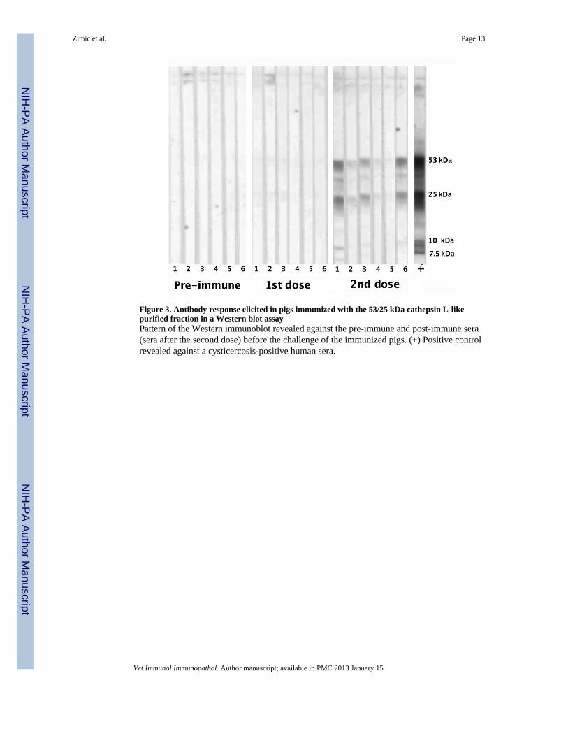
Figure 3. Antibody response elicited in pigs immunized with the 53/25 kDa cathepsin L-likepurified fraction in a Western blot assayPattern of the Western immunoblot revealed against the pre-immune and post-immune sera(sera after the second dose) before the challenge of the immunized pigs. (+) Positive controlrevealed against a cysticercosis-positive human sera.
Zimic et al. Page 13
Vet Immunol Immunopathol. Author manuscript; available in PMC 2013 January 15.
NIH
-PA Author Manuscript
NIH
-PA Author Manuscript
NIH
-PA Author Manuscript

NIH
-PA Author Manuscript
NIH
-PA Author Manuscript
NIH
-PA Author Manuscript
Zimic et al. Page 14
Tabl
e 1
Num
bers
of T
aeni
a so
lium
cys
ticer
ci in
con
trol a
nd v
acci
nate
d pi
gs
Gro
upT
apew
orm
ID u
sed
for
chal
leng
eC
yst C
ount
Mea
n of
tota
l cys
ts [9
5% C
onf.
Inte
rval
]V
iabl
eD
egen
erat
edT
otal
Con
trol
rGST
40
00
rGST
14
092
364
128
rGST
532
878
910
732.
7(1
025)
*
[0.2
151
]([0
.3 2
30])
*
rGST
40
11
rGST
30
3333
rGST
10
2222
rGST
22
3335
Vac
cina
ted
Cat
heps
in L
217
7693
Cat
heps
in L
50
44
599(
718)
*
Cat
heps
in L
181
2510
6[0
.156
1]([
0.19
30])
*
Cat
heps
in L
597
316
31
136
Cat
heps
in L
40
11
Cat
heps
in L
32
196
582
254
* stat
istic
s cal
cula
ted
afte
r rem
ovin
g th
e da
ta o
f the
thre
e pi
gs c
halle
nged
with
tape
wor
m #
4.
Vet Immunol Immunopathol. Author manuscript; available in PMC 2013 January 15.