Revision of Trigonostomum Schmidt, 1852 (Platyhelminthes, Typhloplanoida, Trigonostomidae) with the...
26
Zoological Journal of the Linnean Society, 2004, 141, 271–296. With 11 figures © 2004 The Linnean Society of London, Zoological Journal of the Linnean Society, 2004, 141, 271–296 271 Blackwell Science, LtdOxford, UKZOJZoological Journal of the Linnean Society0024-4082The Lin- nean Society of London, 2004? 2004 141? 271296 Original Article W. R. WILLEMS ET AL.REVISION OF TRIGONOSTOMUM *Corresponding author. E-mail: [email protected] Revision of Trigonostomum Schmidt, 1852 (Platyhelminthes, Typhloplanoida, Trigonostomidae) with the description of seven new species WIM R. WILLEMS*, TOM J. ARTOIS, WOUTER A. VERMIN and ERNEST R. SCHOCKAERT Limburgs Universitair Centrum, Department SBG, Centre for Environmental Sciences, Research Group Biodiversity, Phylogeny and Population Studies, Universitaire Campus, B-3590 Diepenbeek, Belgium Received March 2003; accepted for publication February 2004 A morphological and taxonomic account of the genus Trigonostomum is provided. All known species are discussed and briefly re-described where necessary. Seven new species are described: T. franki from Curaçao, Florida (USA), the East African Coast and New Caledonia, T. nataschae from the French sub-Antarctic island Kerguelen, T. spinigerum from New Caledonia and T. watsoni from the Australian East Coast and New Caledonia. T. tori and T. galapagoensis, both formerly enclosed in T. setigerum, are considered new species, while T. australis also belongs to the same species group. Proxenetes denhartogi is transferred to Trigonostomum. Based on a comparison of old and new material, T. marki is synonymized with T. penicillatum, while T. prytherchi and T. divae are regarded as junior synonyms of T. lilliei; T. intermedium and T. quadrifolium are considered synonyms of T. coronatum. Three species are considered species inquirendae: T. brunchorsti, T. piriforme and Marinellia lingulifera. Similarities and differences of the 17 valid species are discussed and summarized in an identification key. © 2004 The Linnean Soci- ety of London, Zoological Journal of the Linnean Society, 2004, 141, 271–296. ADDITIONAL KEYWORDS: identification key – morphology – phylogenetic nomenclature – taxonomy – Trigonostominae. INTRODUCTION The genus Trigonostomum was introduced by Schmidt (1852) for a single species, T. setigerum. Thirty years later, Graff (1882) changed the name to Hyporhynchus and added a new species, H. coronatus. He also brought three previously described species - Kylosphaera armata Jensen, 1878, Orcus venenosus Uljanin, 1870 and Vortex penicillatus Schmidt, 1857 - into the genus and considered Spiroclytus nisus Schmidt, 1857 and S. euryalus Schmidt, 1857 to be synonyms of Hyporhynchus setigerus. The number of species grew rapidly in the late 19th and early 20th centuries (Uljanin, 1870; Pereyaslawzewa, 1893; Attems, 1897; Graff, 1905, 1911a, b, 1913) most of them described as belonging to Hyporhynchus, although Graff (1905) changed Hyporhynchus veneno- sus (Uljanin, 1870) into Hyporcus venenosus. Eventu- ally, Meixner (1924b) re-introduced the first name Trigonostomum Schmidt, 1852, and also considered Woodsholia lilliei Graff, 1911, to be a species of Trigonostomum. When changing the name to Hyporhynchus in 1882, Graff placed the genus within the subfamily Pseudorhynchina of the family Proboscida Carus, 1863. All members of this ‘family’ were characterized by the presence of a ‘proboscis’, a muscular frontal organ for capturing prey. In 1905, Graff changed the name Proboscida to Kalyptorhynchia, enclosing four families: Trigonostomidae, Schizorhynchidae, Poly- cystididae and Gyratricidae. However, in a detailed study of the proboscis of several Kalyptorhynchia, Meixner (1924b, 1926) showed that the ‘proboscis’ of Trigonostomum is by no means the same structure as that found in the other Kalyptorhynchia. He there- fore transferred the Trigonostomidae from the Kalyp- torhynchia to the Euliporhynchia Graff, 1905, which also included the families Proxenetidae Graff 1908, Byrsophlebidae Graff, 1905 and Typhloplanidae
Transcript of Revision of Trigonostomum Schmidt, 1852 (Platyhelminthes, Typhloplanoida, Trigonostomidae) with the...
Zoological Journal of the Linnean Society, 2004, 141, 271–296. With 11 figures
© 2004 The Linnean Society of London, Zoological Journal of the Linnean Society, 2004, 141, 271–296 271
Blackwell Science, LtdOxford, UKZOJZoological Journal of the Linnean Society0024-4082The Lin-nean Society of London, 2004? 2004141?271296Original Article
W. R. WILLEMS ET AL.REVISION OF TRIGONOSTOMUM
*Corresponding author. E-mail: [email protected]
Revision of Trigonostomum Schmidt, 1852 (Platyhelminthes, Typhloplanoida, Trigonostomidae) with the description of seven new species
WIM R. WILLEMS*, TOM J. ARTOIS, WOUTER A. VERMIN and ERNEST R. SCHOCKAERT
Limburgs Universitair Centrum, Department SBG, Centre for Environmental Sciences, Research Group Biodiversity, Phylogeny and Population Studies, Universitaire Campus, B-3590 Diepenbeek, Belgium
Received March 2003; accepted for publication February 2004
A morphological and taxonomic account of the genus Trigonostomum is provided. All known species are discussedand briefly re-described where necessary. Seven new species are described: T. franki from Curaçao, Florida (USA),the East African Coast and New Caledonia, T. nataschae from the French sub-Antarctic island Kerguelen,T. spinigerum from New Caledonia and T. watsoni from the Australian East Coast and New Caledonia. T. tori andT. galapagoensis, both formerly enclosed in T. setigerum, are considered new species, while T. australis alsobelongs to the same species group. Proxenetes denhartogi is transferred to Trigonostomum. Based on a comparisonof old and new material, T. marki is synonymized with T. penicillatum, while T. prytherchi and T. divae are regardedas junior synonyms of T. lilliei; T. intermedium and T. quadrifolium are considered synonyms of T. coronatum. Threespecies are considered species inquirendae: T. brunchorsti, T. piriforme and Marinellia lingulifera. Similarities anddifferences of the 17 valid species are discussed and summarized in an identification key. © 2004 The Linnean Soci-ety of London, Zoological Journal of the Linnean Society, 2004, 141, 271–296.
ADDITIONAL KEYWORDS: identification key – morphology – phylogenetic nomenclature – taxonomy –Trigonostominae.
INTRODUCTION
The genus Trigonostomum was introduced by Schmidt(1852) for a single species, T. setigerum. Thirty yearslater, Graff (1882) changed the name to Hyporhynchusand added a new species, H. coronatus. He alsobrought three previously described species -Kylosphaera armata Jensen, 1878, Orcus venenosusUljanin, 1870 and Vortex penicillatus Schmidt, 1857 -into the genus and considered Spiroclytus nisusSchmidt, 1857 and S. euryalus Schmidt, 1857 to besynonyms of Hyporhynchus setigerus. The number ofspecies grew rapidly in the late 19th and early 20thcenturies (Uljanin, 1870; Pereyaslawzewa, 1893;Attems, 1897; Graff, 1905, 1911a, b, 1913) most ofthem described as belonging to Hyporhynchus,although Graff (1905) changed Hyporhynchus veneno-sus (Uljanin, 1870) into Hyporcus venenosus. Eventu-
ally, Meixner (1924b) re-introduced the first nameTrigonostomum Schmidt, 1852, and also consideredWoodsholia lilliei Graff, 1911, to be a species ofTrigonostomum.
When changing the name to Hyporhynchus in1882, Graff placed the genus within the subfamilyPseudorhynchina of the family Proboscida Carus,1863. All members of this ‘family’ were characterizedby the presence of a ‘proboscis’, a muscular frontalorgan for capturing prey. In 1905, Graff changed thename Proboscida to Kalyptorhynchia, enclosing fourfamilies: Trigonostomidae, Schizorhynchidae, Poly-cystididae and Gyratricidae. However, in a detailedstudy of the proboscis of several Kalyptorhynchia,Meixner (1924b, 1926) showed that the ‘proboscis’ ofTrigonostomum is by no means the same structure asthat found in the other Kalyptorhynchia. He there-fore transferred the Trigonostomidae from the Kalyp-torhynchia to the Euliporhynchia Graff, 1905, whichalso included the families Proxenetidae Graff 1908,Byrsophlebidae Graff, 1905 and Typhloplanidae

272 W. R. WILLEMS ET AL.
© 2004 The Linnean Society of London, Zoological Journal of the Linnean Society, 2004, 141, 271–296
Luther, 1904. Two decades later, Luther (1948)grouped the Proxenetidae within the Trigonostomi-dae. Den Hartog (1964) split this taxon again, thistime into Promesostomidae and Trigonostomidae,based on the structure of the female genital system:the Trigonostomidae are characterized by femalegonads connected with the genital atrium by twoducts. Within the Trigonostomidae Den Hartog (1964)recognized two taxa, Trigonostominae and Parame-sostominae, while a third, Mariplanellinae, wasadded by Ax & Heller (1970). The Trigonostominaecomprises 11 genera, the phylogenetic relationshipsof which were described by Ax (1971), who consideredMarinellia lingulifera Riedl, 1954 to be the sistertaxon of Trigonostomum.
Due to the detailed studies by Graff (1913) andMeixner (1924b), the general anatomy of Trigonosto-mum is well known. The anatomy of a number of otherrepresentatives of the family, in particular that of thegenital system, has been thoroughly described byLuther (1943). In the morphological section we sum-marize the essentials of the anatomy of representa-tives of Trigonostomum.
Not all described species are easy to recognize, oftendue to inaccurate descriptions and poor figures, espe-cially in the older literature. The main purpose of thisrevision is to elucidate the species’ identities and syn-onyms. A neotype is designated for each of the follow-ing species - T. armatum, T. breitfussi, T. coronatum,T. lilliei, T. mirabile, T. penicillatum, T. setigerum andT. venenosum - as a name-bearing type was never des-ignated for any of them. Data for these species can befound in the taxonomy section, and all conditions forconsidering the designation of a neotype are fulfilled(ICZN, 1999: Art. 75). One new combination(T. denhartogi) and seven new species are added, threeof them resulting from the analysis of the polymorphic‘T. setigerum’. Since their anatomy is uniform, thedescriptions of the separate species focus on the hardparts of the copulatory organ and the bursal append-age, with additional remarks where needed. A sepa-rate section provides a comparison of the species andthe taxonomic conclusions resulting from this analy-sis, and includes an identification key, replacing thedifferential diagnoses.
Some preliminary indications of relationshipswithin Trigonostomum are mentioned, but we refrainfrom discussing in depth possible phylogenetic rela-tionships both within the taxon and with other taxawithin the Trigonostomidae. A cladistic analysis of theTyphloplanoida, based on morphological and molecu-lar characters, is currently in progress. Consequently,each diagnosis given now should not be seen as a phy-logenetic definition (De Queiroz & Gauthier, 1990,1992, 1994). However, as, in our view, species are fun-damentally different from higher taxa because they
cannot be monophyletic in a Hennigian sense (seeNixon & Wheeler, 1990), we propose for every speciesan alternative to the Linnean binomen, which can beused within the framework of phylogenetic nomencla-ture. This alternative species name consists of apraenomen and epithet, connected with a hyphen, andis unchangeable (see Cantino et al., 1999; Artois,2001). In the taxonomy section, the species are listedin alphabetical order.
MATERIAL AND METHODS
The zoological collection of Limburgs UniversitairCentrum (LUC), Diepenbeek, Belgium, contains veryrich material of both new and known Trigonostomumspecies from many parts of the world, collected by theauthors (Australia, New Caledonia, Curaçao, Florida,East Africa, Zanzibar, French Mediterranean coast,Greece, Sardinia, Sweden, Kerguelen, and the Wed-dell Sea) and by some earlier collaborators of theDiepenbeek group: Corsica (Dr P. Martens), Kenya (DrP. Jouk and Mr G. De Clerck). Material from the Swed-ish Museum of Natural History (SMNH) and of theSmithsonian Institution (SI-NMNH) was also at ourdisposal. Of the following species no material is avail-able: T. brunchorsti Graff, 1905, T. intermedium(Attems, 1897) Graff, 1913, T. marki Graff, 1911,T. piriforme (Pereyaslawzewa, 1893) Graff, 1905 andMarinellia lingulifera Riedl, 1954.
Animals were extracted from sediment or fromalgae using the MgCl2 decantation method (see Schoc-kaert, 1996), studied while live and mounted in lac-tophenol. If sufficient specimens were available, somewere fixed in hot Bouin’s solution, embedded in paraf-fin and serially sectioned. The 4 mm-thick sectionswere stained with Heidenhain’s iron haematoxylinusing eosin as a counterstain. Camera lucida draw-ings of the hard parts were made with Nomarskiphase contrast microscopy on a Reichert Polyvarmicroscope. Drawings without a scale are freehand.Measurements of the (inner) stylet and the bursalappendage are taken along the axis, unless indicatedotherwise in the text. The length of the whole copula-tory organ is given as a top-bottom measurement; it isnot measured for species where the organ is coiled asthe spires can spread, causing a great degree of vari-ability in such a measurement. The positions of thegonopore and organs are expressed in percentages ofthe total body length (distance from the anterior tip ofthe body).
All material (including type material), except thatfrom Australia and that belonging to other institu-tions, is deposited in LUC. The type material of theAustralian species (T. australis and T. watsoni spp.nov.) is deposited in the collection of the QueenslandMuseum, Brisbane, Australia.
REVISION OF TRIGONOSTOMUM 273
© 2004 The Linnean Society of London, Zoological Journal of the Linnean Society, 2004, 141, 271–296
GENERAL MORPHOLOGY
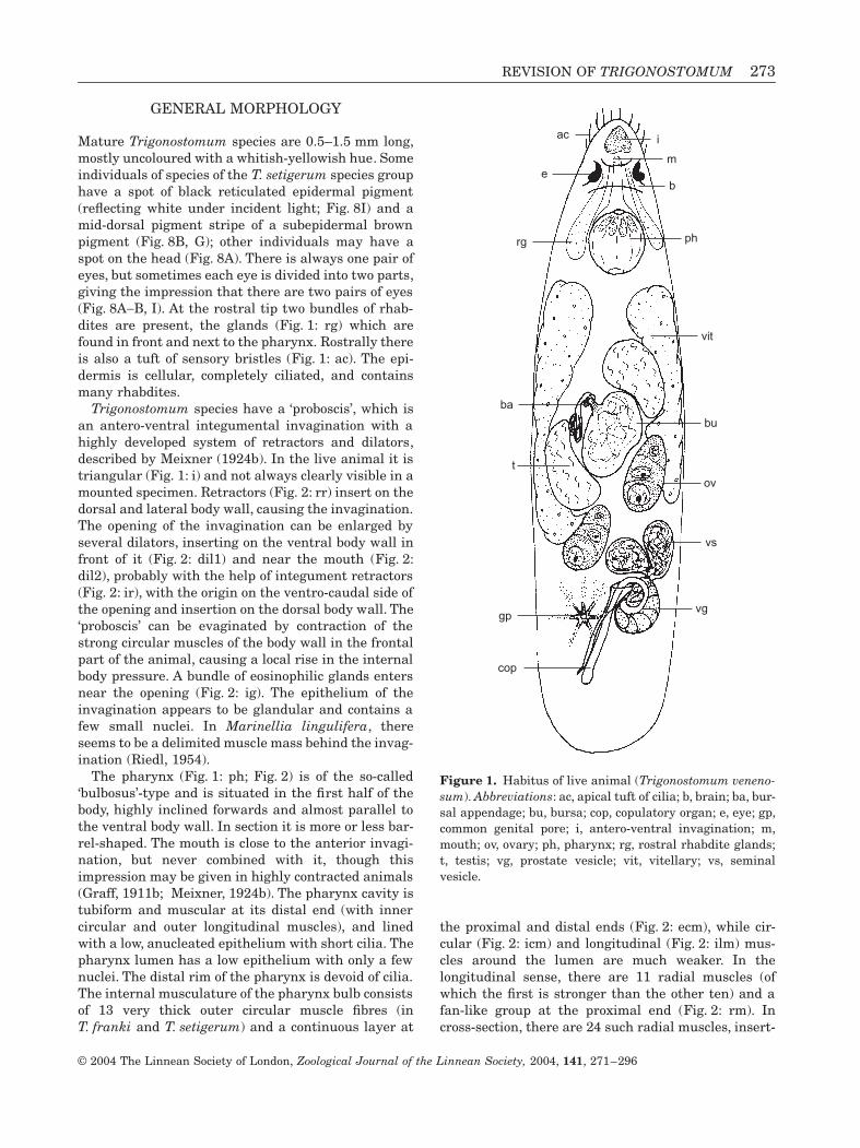
Mature Trigonostomum species are 0.5–1.5 mm long,mostly uncoloured with a whitish-yellowish hue. Someindividuals of species of the T. setigerum species grouphave a spot of black reticulated epidermal pigment(reflecting white under incident light; Fig. 8I) and amid-dorsal pigment stripe of a subepidermal brownpigment (Fig. 8B, G); other individuals may have aspot on the head (Fig. 8A). There is always one pair ofeyes, but sometimes each eye is divided into two parts,giving the impression that there are two pairs of eyes(Fig. 8A-B, I). At the rostral tip two bundles of rhab-dites are present, the glands (Fig. 1: rg) which arefound in front and next to the pharynx. Rostrally thereis also a tuft of sensory bristles (Fig. 1: ac). The epi-dermis is cellular, completely ciliated, and containsmany rhabdites.
Trigonostomum species have a ‘proboscis’, which isan antero-ventral integumental invagination with ahighly developed system of retractors and dilators,described by Meixner (1924b). In the live animal it istriangular (Fig. 1: i) and not always clearly visible in amounted specimen. Retractors (Fig. 2: rr) insert on thedorsal and lateral body wall, causing the invagination.The opening of the invagination can be enlarged byseveral dilators, inserting on the ventral body wall infront of it (Fig. 2: dil1) and near the mouth (Fig. 2:dil2), probably with the help of integument retractors(Fig. 2: ir), with the origin on the ventro-caudal side ofthe opening and insertion on the dorsal body wall. The‘proboscis’ can be evaginated by contraction of thestrong circular muscles of the body wall in the frontalpart of the animal, causing a local rise in the internalbody pressure. A bundle of eosinophilic glands entersnear the opening (Fig. 2: ig). The epithelium of theinvagination appears to be glandular and contains afew small nuclei. In Marinellia lingulifera, thereseems to be a delimited muscle mass behind the invag-ination (Riedl, 1954).
The pharynx (Fig. 1: ph; Fig. 2) is of the so-called‘bulbosus’-type and is situated in the first half of thebody, highly inclined forwards and almost parallel tothe ventral body wall. In section it is more or less bar-rel-shaped. The mouth is close to the anterior invagi-nation, but never combined with it, though thisimpression may be given in highly contracted animals(Graff, 1911b; Meixner, 1924b). The pharynx cavity istubiform and muscular at its distal end (with innercircular and outer longitudinal muscles), and linedwith a low, anucleated epithelium with short cilia. Thepharynx lumen has a low epithelium with only a fewnuclei. The distal rim of the pharynx is devoid of cilia.The internal musculature of the pharynx bulb consistsof 13 very thick outer circular muscle fibres (inT. franki and T. setigerum) and a continuous layer at
the proximal and distal ends (Fig. 2: ecm), while cir-cular (Fig. 2: icm) and longitudinal (Fig. 2: ilm) mus-cles around the lumen are much weaker. In thelongitudinal sense, there are 11 radial muscles (ofwhich the first is stronger than the other ten) and afan-like group at the proximal end (Fig. 2: rm). Incross-section, there are 24 such radial muscles, insert-
Figure 1. Habitus of live animal (Trigonostomum veneno-sum). Abbreviations: ac, apical tuft of cilia; b, brain; ba, bur-sal appendage; bu, bursa; cop, copulatory organ; e, eye; gp,common genital pore; i, antero-ventral invagination; m,mouth; ov, ovary; ph, pharynx; rg, rostral rhabdite glands;t, testis; vg, prostate vesicle; vit, vitellary; vs, seminalvesicle.
ph
bu
ov
cop
vs
ba
t
vg
ac
e
i
rg
m
gp
vit
b

274 W. R. WILLEMS ET AL.
© 2004 The Linnean Society of London, Zoological Journal of the Linnean Society, 2004, 141, 271–296
ing on the pharynx lumen between 24 longitudinalmuscles. Fine-grained eosinophilic glands enter thepharyngeal lumen at its distal end and coarse-grainedbasophilic ones more proximally. There are 16 of each.
The gonads are paired. The testes are positionedmid-body, just behind the pharynx. Ovaries and vitel-laria are not separated, forming a pair of so-called‘ovovitellaria’. The vitellarian parts extend at bothsides of the body from the level of the pharynx, and theovaries are positioned slightly in front of the gonopore,situated at about 80%. The common genital atrium(Fig. 3: cga) is lined with a high, nucleated epitheliumand surrounded by outer longitudinal and inner circu-lar muscles, forming a sphincter at the porus.
The seminal vesicles (Fig. 3: vs) are paired, linedwith a low epithelium without nuclei, and surroundedby spirally running muscles. Both vesicles narrow andjoin to form the short seminal duct (Fig. 3: sd), which
enters the prostate vesicle (Fig. 3: vg) and runs axiallythrough it, surrounded by mainly eosinophilic andsome basophilic prostate glands, all parts of which areinside the vesicle. In some live individuals extracap-sular parts of these glands can be seen. Relativelystrong circular muscles surround the prostate vesicle.The male atrium, where the hard parts of the copula-tory organ lie, is surrounded by an inner circular andan outer longitudinal muscle sheath, and enters thecommon genital atrium dorsally.
The copulatory organ proper consists of a stylet, sur-rounded by a second, very thin-walled stylet, calledthe ‘mantle’ by Ax (1971). The organ can thus be con-sidered as double-walled with an inner ‘stylet’ (Fig. 4:s) and an outer ‘mantle’ (Fig. 4: mt), as shown in thetheoretical diagram in Figure 4A.
In the species of the first group (see comparison ofspecies, below) the mantle is attached to one side of
Figure 2. Reconstruction on sagittal sections of the rostral body end from the left hand side (T. franki sp. nov.). Abbre-viations: cbm, circular body muscles; dil1–2, dilators of opening of antero-ventral invagination; e, eye; ecm, external circularpharyngeal muscles; elm, external longitudinal pharyngeal muscles; i, antero-ventral invagination; icm, internal circularpharyngeal muscles; ig, invagination glands; ilm, internal longitudinal pharyngeal muscles; ir, integument retractors; lbm,longitudinal body muscles; m, mouth; pc, prepharyngeal cavity; pg, pharynx glands; ppt, pharynx protractors; prt, pharynxretractors; ps, pigment stripe; rm, radial pharyngeal muscles; rr, retractors of antero-ventral invagination.
elm
cbmrr
eps
ig
m
ecm
dil1
pc
ilm
lbm
dil2
icm
prt
pptrm pg
i
ir

REVISION OF TRIGONOSTOMUM 275
© 2004 The Linnean Society of London, Zoological Journal of the Linnean Society, 2004, 141, 271–296
the proximal rim of the stylet and distally divided intospiny plates (Fig. 4B). The stylet is a narrow tube,proximally curved over 90-270∞, with a relativelythick wall and a broad proximal opening. In the spe-
cies of the second group, the mantle follows the coils ofthe stylet over almost its entire length, forming twospiny plates at the very end (Fig. 4C). In sectionedmaterial of T. setigerum, the way the mantle sur-
Figure 3. Reconstruction on sagittal sections of the genital system from the left hand side (T. franki sp. nov.). Abbrevi-ations: ba, bursal appendage; bs, bursal stalk; bu, bursa; cga, common genital atrium; cop, copulatory organ; ds, spermaticduct; fd, female duct; fg, female glands; gp, common genital pore; n, nucleus; od, oviduct; ov, ovary; rh, rhabdite; sd, seminalduct; vg, prostate vesicle; vit, vitellary; vs, seminal vesicle.
ov
od vs
vg
gp
cop
fg
fd
bu ds
ba
vit
cga
rhn
bs
sd
Figure 4. Schematic representation of the copulatory organ. A, theoretical diagram of a double-walled copulatory organ. B,diagram of the copulatory organ of the representatives of group 1. C, diagram of the stylet of the representatives of group2. Abbreviations: mt, mantle; s, stylet.
C
s
mt
s
mt
mt
s
A B

276 W. R. WILLEMS ET AL.
© 2004 The Linnean Society of London, Zoological Journal of the Linnean Society, 2004, 141, 271–296
rounds the stylet can be clearly seen. In T. denhartogiand in T. spinigerum the copulatory organ is far morecomplicated than in the other species, and the struc-ture of the mantle is not yet well understood.
According to Meixner (1924b), the prostate secretionis discharged in the mantle, while sperm is released inthe stylet. Observations on sectioned material ofT. franki, T. armatum and T. setigerum revealed thatboth sperm and secretion are released through the(inner) stylet.
The ovaries are connected to the genital atrium byan efferent duct and an afferent system (see Luther,1943). The oviducts are very short and lined with anucleated epithelium. They join at the ‘fecundato-rium’ (Luther’s terminology) from where the (effer-ent) female duct (Fig. 3: fd) runs towards the atrium.This duct is long and narrow, lined with a low,nucleated epithelium and muscles are lacking. Itenters the common genital atrium anteriorly, ven-trally of the male system and dorsally of the affer-ent system. Eosinophilic glands (Fig. 3: fg) open intothe female duct near the atrium. The afferent sys-tem consists of a bursal stalk (Fig. 3: bs), the bursa(Fig. 3: bu) and the sclerotic appendage (Fig. 3: ba)that leads into the spermatic duct (or inseminationduct; Fig. 3: ds). This duct ends near the ovaries inthe ‘fecundatorium’. The bursal stalk is long andnarrow (although short in T. penicillatum accordingto Meixner, 1924b). The whole afferent system islined with a low nucleated epithelium. Only the bur-sal stalk is surrounded by (circular) muscles. Thereis no uterus.
Meixner (1924b) recognized two types of bursalappendage: (1) the setigerum type consists of two moreor less coiled narrow tubes, attached to a ring- or fun-nel-like part at the bursa wall; (2) the penicillatumtype consists of a bundle of many closely adheringtubules, which protrude into the bursa for some dis-tance, where they may be held together by a casing.There are further variations, discussed below.
All Trigonostomum species are marine and arefound all over the world, in areas ranging from tropi-cal to polar. Most have been found in the intertidalzone, mainly on algae or seagrasses. Some species alsooccur in sand. Only two species, T. messoplanoidesArtois, Vermin and Schockaert, 2000 and T. nataschaesp. nov., have not so far been found on algae (see fur-ther), while T. messoplanoides and T. setigerum arethe only species collected at greater depths: 499–515 m and 137–150 m respectively (Westblad, 1952;Artois et al., 2000).
COMPARISON OF THE SPECIES
The main differences between the species of Trigono-stomum are to be found in the construction of the hard
parts of the copulatory organ, the bursal appendageand, to a lesser extent, in the pigmentation.
As mentioned above and diagrammatically repre-sented in Figure 4B, C, the copulatory organ consistsof a proximally curved stylet surrounded by a thin-walled mantle, divided into spiniform plates. In thefirst group of species, the proximal rim of the mantledoes not follow the curvature of the stylet, and thusonly surrounds the distal (straight) part of the stylet(Fig. 4B). In two of these species (group 1A,T. messoplanoides and T. venenosum, see Fig. 6) thestylet is long and narrow and makes a proximal turnof 270∞, while the mantle forms a narrow ringand bears one long, slender spine (spoon-like inT. venenosum). This kind of organ resembles that ofspecies of the genus Messoplana Den Hartog, 1966.
The bursal appendage of T. messoplanoides andT. venenosum consists of two tubes attached to thebursa by a more or less conspicuous ring, and may befused proximally in some individuals of T. venenosum(Fig. 6A4). In the construction of both the copulatoryorgan and the bursal appendage, these species dem-onstrate plesiomorphy, as found in several other taxaof the Trigonostominae (see Ax, 1971).
In the other species of this first group (group 1B; seeFig. 7), the stylet is relatively wider (and shorter) andmakes a turn of about 90∞ in T. coronatum,T. penicillatum and T. watsoni to about 180∞ in theother species (T. breitfussi, T. lilliei, T. mirabile andT. nataschae). The mantle is divided into three pointedplates in the first three species, into two plates inT. mirabile and T. nataschae, and forms only one platein T. breitfussi and T. lilliei. Some other, more subtledifferences are seen: the plate is rather blunt inT. breitfussi and shorter than the stylet, which has acrested proximal rim, while in T. lilliei the plate endsin a little hook and is as long as the stylet, which inthis case lacks the crested rim. In T. nataschae, thetwo plates are blunt and shorter than the stylet, whilethey are pointed in T. mirabile and at least as long asthe stylet. In both these species the stylet has a widespoon-like terminal opening, but in T. nataschae it hasa proximal crested rim, not present in T. mirabile.Differences between the copulatory organs ofT. coronatum and T. penicillatum are difficult to dis-cern, while the plates in T. watsoni are very thin andeasily folded. Moreover, in this last species, the styletis inconspicuous and often impossible to find in someindividuals. However, the bursal appendage in thesethree species is very different, as is the overall size ofthe copulatory organ, thus enabling clear distinctionbetween the species.
The bursal appendage in a number of species ofgroup 1B is clearly of Meixner’s penicillatum type. Itconsists of many closely adhering parallel tubules,which protrude into the bursa for some distance,
REVISION OF TRIGONOSTOMUM 277
© 2004 The Linnean Society of London, Zoological Journal of the Linnean Society, 2004, 141, 271–296
where they may be held together by a casing resem-bling a little barrel, connected to the bursa wall by aring. This kind of appendage is found in T. breitfussi,T. mirabile and in T. penicillatum, where the tubulesdiverge at the very end, giving the appendage a brush-like appearance, but probably also in T. mirabile (seefig. 27 in Pereyaslawzewa, 1893). This brush-likeappearance may be an artefact caused by squeezingthe animals, especially when the tubules are long, asis the case in these two species.
In T. nataschae very narrow tubules protrude intothe bursa beyond the proximal ‘barrel’ that has arather thick wall. Just outside the bursa the tubulesare still visible, but more distally they are very faintand separated into two coiled bundles, giving theimpression that the bursal appendage ends in twobroad tubes. The bursal appendage of T. lilliei has ahighly coiled single part that ends in six tubules; theproximal ‘barrel’ seems to be missing, but there is aproximal ring at the inside of the bursa wall, beyondwhich the tubules protrude into the bursa (see alsofig. 18 in Marcus, 1948 of T. divae = T. lilliei). Theappendage of T. coronatum, on the other hand, has aproximal part that looks like a clover-leaf, and one,sometimes two (perhaps three: see fig. 23 in Attems,1897) broad terminal ‘tubes’ with a fine striation. InT. watsoni the bursal appendage starts at the bursawith a broad funnel that splits into two highly coiledbroad tubes, also with a fine striation, suggesting thatthey may consist of subtubules.
Though the bursal appendage of the species in thisgroup exhibits variation (which needs further investi-gation), it clearly deviates in structure from that of allother members of the taxon Trigonostomum and evenof all other taxa of the Trigonostominae. Perhaps thisis a synapomorphy.
In a second group of species (group 2; see Fig. 10),the proximal mantle rim closely adheres to the proxi-mal rim of the stylet, envelopes the stylet over its
entire length, and diverges at the very end into twospiny plates with a terminal hook (see Fig. 4C). Thenarrow stylet is spirally coiled (except in T. franki, butsee below) and the mantle follows the coils of thestylet. The stylet is at the periphery of the spire, whilethe mantle forms a double plate towards the centre,where it can be slightly thickened (wrongly calledSekretrorh by Graff, 1913) and shows radial wrinkles.At the very end, where the two plates are situated,there may be a straighter part of the stylet. The hooksof the plates are invariably directed towards the con-vex side of the curve. This combination of characters,especially the fact that the mantle entirely envelopesthe stylet, is unique within the Trigonostominae andis without any doubt a synapomorphy for this group ofspecies. On the other hand, the setigerum type ofappendage (Fig. 8C) has the same construction as thatof the majority of the other Trigonostominae, consist-ing of two tubules attached by a ring to the bursa, withboth tubes coiled over 360∞.
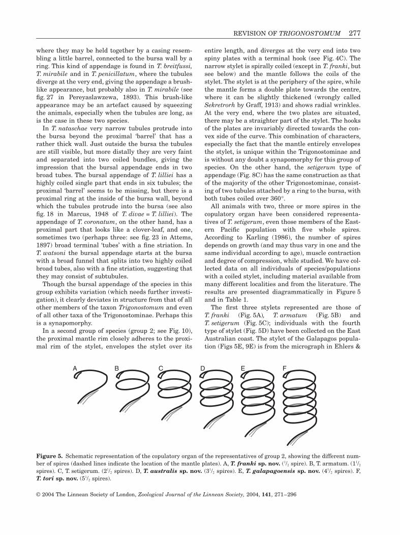
All animals with two, three or more spires in thecopulatory organ have been considered representa-tives of T. setigerum, even those members of the East-ern Pacific population with five whole spires.According to Karling (1986), the number of spiresdepends on growth (and may thus vary in one and thesame individual according to age), muscle contractionand degree of compression, while studied. We have col-lected data on all individuals of species/populationswith a coiled stylet, including material available frommany different localities and from the literature. Theresults are presented diagrammatically in Figure 5and in Table 1.
The first three stylets represented are those ofT. franki (Fig. 5A), T. armatum (Fig. 5B) andT. setigerum (Fig. 5C); individuals with the fourthtype of stylet (Fig. 5D) have been collected on the EastAustralian coast. The stylet of the Galapagos popula-tion (Figs 5E, 9E) is from the micrograph in Ehlers &
Figure 5. Schematic representation of the copulatory organ of the representatives of group 2, showing the different num-ber of spires (dashed lines indicate the location of the mantle plates). A, T. franki sp. nov. (1/2 spire). B, T. armatum. (11/2
spires). C, T. setigerum. (21/2 spires). D, T. australis sp. nov. (31/2 spires). E, T. galapagoensis sp. nov. (41/2 spires). F,T. tori sp. nov. (51/2 spires).
A B C D E F

278 W. R. WILLEMS ET AL.
© 2004 The Linnean Society of London, Zoological Journal of the Linnean Society, 2004, 141, 271–296
Table 1. Morphological differences between different populations of Trigonostomum franki sp. nov., T. armatum,T. setigerum, T. australis sp. nov., T. galapagoensis sp. nov. and T. tori sp. nov. (species group 2). Abbreviations: SL,stylet length (in mm); BAL, bursal appendage length (in mm); BAT, bursal appendage tubules; MSM, minimum no. of spec-imens measured; NM, not measurable; ? not known
Species Distribution Pigmentation Coils SL BAL BAT MSM
T. franki Caribbean, Florida longitudinal stripe 1/2 86–101 84–100 normal 8East Africa longitudinal stripe 1/2 90–101 120 normal 1New Caledonia longitudinal stripe 1/2 95–111 62–166 normal
constricted4
T. armatum northern Atlantic none 1 158–196 NM ? 2southern Atlantic rostral spot 1 434 149 normal 1Caribbean rostral spot 1 274–378 100–108 normal 9eastern Australia none 1 183 NM ? 1New Caledonia none 1 386 127 normal 1
T. setigerum western Mediterranean rostral spot 2 413–492 87–113 normal 4eastern Mediterranean none
longitudinal striperostral spot 2 284–462 64–113 normal 21
southern Atlantic rostral spot 2 482 141 constricted 1East Africa none 2 403 93 normal 1English Channel rostral spot 2 477 NM ? 1Bermuda none 2 434 NM constricted 1
T. australis eastern Australia longitudinal stripe 3 414–445 34 normal 3T. galapagoensis Galapagos rostral spot 4 ? ? ? 0T. tori California none 5 683–853 80–106 normal 3
Figure 6. Copulatory organs and bursal appendages of the representatives of group 1A. A, T. venenosum: A1, 3 copulatoryorgan; A2, 4 bursal appendage. (A1, 2 specimen from Norway, neotype; A3,4 specimen from Sardinia). B, T. messoplanoides: B1
copulatory organ; B2 bursal appendage. (from the holotype; after Artois et al., 2000).
A1 B1
B2
A2
A3
A4
20 µm
A-B

REVISION OF TRIGONOSTOMUM 279
© 2004 The Linnean Society of London, Zoological Journal of the Linnean Society, 2004, 141, 271–296
Figure 7. Copulatory organs and bursal appendages of the representatives of group 1B, A, T. coronatum: A1 copulatoryorgan; A2 bursal appendage. (from the neotype). B, T. watsoni sp. nov. B1, 2 copulatory organ; B3 bursal appendage (B1 fromthe holotype; B2, 3 from the paratype). C, T. penicillatum: C1 copulatory organ; C2 bursal appendage (from the neotype). D,T. lilliei: D1, 3 copulatory organ; D2, 4 bursal appendage. [D1, 2 specimen from Australia; D3, 4 specimen from the USA (neotype= holotype of T. prytherchi)]. E, T. breitfussi: E1 copulatory organ; E2 bursal appendage; E3 section through the bursa and thebursal appendage (different specimens from Sweden; E2, 3 freehand drawings). F, T. mirabile: F1, 2 copulatory organ; F3 bur-sal appendage (F1, 3 from the neotype; F2 from another specimen from the Black Sea). G, T. nataschae sp. nov. G1 Cop-ulatory organ; G2 Bursal appendage (from the holotype).
A1
B1
B2
A2
B3
C1
C2
20 µm
abba
F1
A-E
; F-G
1
D1
D3
D2
D4
E1 E
2
E3
F2 F
3G
1
G2

280 W. R. WILLEMS ET AL.
© 2004 The Linnean Society of London, Zoological Journal of the Linnean Society, 2004, 141, 271–296
Ax (1974), while the last type of stylet (Fig. 5F) isfound in the population from California, as describedby Karling (1986).
In T. franki, the stylet makes a turn of about 180∞(although it shows all other characteristics of thestylet of this group of species) with an additional, moreor less straightened part where the two spines of themantle are (dashed lines in Fig. 5). This stylet thushas a half spire. In T. armatum there is one completespire over 360∞ and an additional part (11/2 spires), inT. setigerum 21/2 spires (see Figs 5C, 9C, 10C1), and so
on. Muscle contraction and a degree of squeezing theanimal causes the spires to stay close to each other orgo apart (in some of our whole mounts of individuals ofthe Greek population, the spires had virtually disap-peared). The number of coils in the spiral is constantin all studied populations, though one may count a dif-ferent number of coils, depending on the side fromwhich the stylet is viewed. In T. setigerum, forinstance, with 21/2 spires, one counts three spires whenlooking at the stylet from the side where the styletbegins (including the ‘half ’ spire), and only two when
Figure 8. A, rostral spot in T. armatum (whole mounted specimen from Curaçao). B, dorsal stripe in T. setigerum (wholemounted specimen from Greece). C, bursal appendage of T. tori sp. nov. (from the paratype). D, constricted distal tubes ofbursal appendage (arrow indicates constriction) in T. setigerum (specimen from Bermuda). E, copulatory organ ofT. denhartogi comb. nov. (specimen from New Caledonia). F, copulatory organ of T. spinigerum sp. nov. (from the holo-type). G, habitus of T. franki sp. nov. (live specimen from New Caledonia). H, bursal appendage of T. spinigerum sp. nov.(from the holotype). I, head region of T. franki sp. nov. showing both epidermal and subepidermal pigment (live specimenfrom New Caledonia). Scale bars: A, B = 200 mm. C-F = 20 mm. H = 10 mm.
A B C
DD
F G H
I
E

REVISION OF TRIGONOSTOMUM 281
© 2004 The Linnean Society of London, Zoological Journal of the Linnean Society, 2004, 141, 271–296
looking from the opposite side. The spires turn clock-wise (seen from the proximal end of the stylet) in allindividuals; in those with a very long stylet (e.g. theCalifornian populations and some individuals in theGreek population), one of the spires may swing in theother direction, as often happens in spirals made offlexible material.
We believe that these stylets indicate that we aredealing with individuals from different species. Hencewe give species status to the populations from Austra-
lia, the Galapagos and California, respectively, asT. australis sp. nov., T. galapagoensis sp. nov. (thoughno material is available, except the micrograph inEhlers & Ax, 1974), and T. tori sp. nov., described asT. setigerum by Tor Karling in 1986. There may besome minor differences in some populations: Karling(1986) pointed to the constriction at the end of thetubes of the bursal appendage in the populations ofT. setigerum in Bermuda, the Falkland Islands, SouthGeorgia and in the English Channel (Fig. 8D; Table 1;
Figure 9. Copulatory organ of the representatives of group 2. A, T. franki sp. nov. (specimen from Curaçao) B,T. armatum (specimen from Curaçao). C, T. setigerum (from the neotype). D, T. australis sp. nov. (from the holotype). E,T. galapagoensis sp. nov. (from the holotype; after Ehlers and Ax, 1974: fig. 13C). F, T. tori sp. nov. (from the holotype).Scale bars: A-D, F = 20 mm.
C
ED F
A B

282 W. R. WILLEMS ET AL.
© 2004 The Linnean Society of London, Zoological Journal of the Linnean Society, 2004, 141, 271–296
Karling, 1978: fig. 28; Karling, 1986: figs 40, 48). Inone specimen of T. franki from New Caledonia thetubes are also slightly constricted.
On the other hand, considering the distribution ofthese species, some doubts may arise. T. franki and
T. armatum have been found together on Curaçao,T. australis and T. armatum occur sympatrically onthe Australian East coast, while T. armatum andT. setigerum can be found together in both NorthAtlantic (Westblad’s material from Plymouth; Gamble,
Figure 10. Copulatory organs and bursal appendages of the representatives of group 2. A, T. franki sp. nov. A1, 3 copu-latory organ; A2, 4 bursal appendage (A1, 2 from the holotype; A3, 4 from a specimen from Kenya). B, T. armatum sp. nov. B1,
3 copulatory organ; B2, 4 bursal appendage (B1, 2 specimen from France; B3, 4 specimen from Curaçao). C, T. setigerum: C1 cop-ulatory organ; C2 bursal appendage (specimen from Kenya). D, T. australis sp. nov. D1 copulatory organ; D2 bursalappendage (from the holotype). E, T. tori sp. nov. E1 copulatory organ; E2 bursal appendage (from the holotype).
A1 B1
B2
A2
A 3
A4
B3
4B
C1
C2
D1
D2
E2
E1
20 µmA-E

REVISION OF TRIGONOSTOMUM 283
© 2004 The Linnean Society of London, Zoological Journal of the Linnean Society, 2004, 141, 271–296
1893; Southern, 1912, 1936; Graff, 1913) and SouthAtlantic populations (Westblad’s material from SouthGeorgia, all considered T. setigerum by Karling, 1986).T. setigerum is the only species that has, so far, beenfound in the Mediterranean Sea, while those specieswith more than 21/2 spires are confined to the Pacific(the further east, the more spires). We began to won-der whether these are all populations of a single ‘spe-cies complex’ similar to that of the kalyptorhynchGyratrix hermaphroditus Ehrenberg, 1831 (Curini-Galletti & Puccinelli, 1989, 1990, 1994, 1998, Artoiset al., 2000; Artois & Schockaert, 2001), albeit of ahighly polymorphic species. Are we dealing with acline in the Indo-Pacific? We prefer, with the data nowavailable, to consider these populations as differentspecies, until proven otherwise.
It is also in this group that a variation in pigmen-tation occurs (see Table 1): a mid-dorsal stripe(Fig. 8B, G), a rostral spot (Fig. 8A) or no pigmenta-tion. Graff (1905) considered that, based on pigmen-tation, there were three subspecies of T. setigerum.
However, Southern (1912) rejected this finding; we fol-low the latter view as these three pigmented forms areknown to occur sympatrically (see Table 1 andremarks on T. setigerum).
Finally, two more species, T. denhartogi andT. spinigerum, deserve comment regarding the con-struction of the copulatory organ and the bursalappendage. With the material now available ofT. denhartogi a first analysis of the copulatory organcan be given (Fig. 11A3). The stylet is rather broadwith a wide proximal funnel to which the proximal rimof the mantel is attached. On the concave side of thestylet the mantle forms 4–5 plates or spines, while onthe convex side it bears a bundle of numerous finefolds or rods, one of which is very long and flagelliform.The bursal appendage has a straight initial part,thickened at one side, and two extremely long, highlycoiled distal tubes. Both characters are clear autapo-morphies for the species, and have no equivalentwithin the Trigonostominae. With only one individualavailable, the precise structure of the copulatory
Figure 11. A, T. denhartogi comb. nov. A1, 3 copulatory organ; A2 bursal appendage (A1, 2 specimen from Curaçao; A3 spec-imen from New Caledonia). B, T. spinigerum sp. nov. B1 copulatory organ; B2 bursal appendage (from the holotype).Abbreviations: a-e, explained in text.
e
a
e1
b
a1
c
d
A1
A3
A2
B1
B2
20 µm
A1-3
20 µm
B1-2

284 W. R. WILLEMS ET AL.
© 2004 The Linnean Society of London, Zoological Journal of the Linnean Society, 2004, 141, 271–296
organ of T. spinigerum (Fig. 11B1) cannot yet be wellunderstood. The mantle seems to have its rim proxi-mal to the beginning of the stylet, and envelopes thelatter completely. On the convex side and in the prox-imal half, the mantle bears 8–10 spines (or folds?); onthe concave side and in the distal half, the mantleforms plates and spines similar to those in species ofgroup 1B (e.g. T. coronatum or T. watsoni). The bursalappendage on the other hand is very similar to that ofT. watsoni, constituting a proximal funnel with twobroad tubes coiled over more than 360∞.
To conclude, within Trigonostomum two commontypes of copulatory organ occur: (1) a proximallycurved stylet surrounded by the mantle in its distal
part, the latter divided into spiny plates; (2) a coiledstylet completely surrounded by a mantle that ends intwo spiny plates. The first condition is plesiomorphic,the second synapomorphic. The mantle exhibits amuch more complicated structure in T. denhartogi andT. spinigerum. Three types of bursal appendage occur:(1) two tubes attached to the bursa with a ring or fun-nel; (2) a funnel with two highly coiled tubes; (3) manytubules held together in one way or another. The firstcondition is plesiomorphic, the second a possible auta-pomorphy, and the third synapomorphic, though vari-ations may occur. The differences discussed here,together with some other characters, are reflected inthe following key.
IDENTIFICATION KEY
1. Copulatory organ 106–121 mm long and very complex, consisting of a gutter-shaped plate,enveloping a bundle of rods, of which one has a long thread-like distal tip; styletfunnel-shaped, ± 65 mm long; bursal appendage with initial straight part(22–44 mm long) and two heavily coiled distal tubes. (Figs 8E, 11A) .................................... T. denhartogi comb. nov.
- Copulatory organ ± 60 mm long; stylet bent over 90∞, surrounded by the mantlethat has 8–10 small spines at the convex side and with broad distal plates andspines distally (Fig. 11B4); bursal appendage with two broad, highly coiled tubes(Figs 8F, H, 11B) ........................................................................................................................... T. spinigerum sp. nov.
- Copulatory organ otherwise ................................................................................................................................................22. Proximal part of the stylet curved over 90-270∞ and surrounded by the mantle only
in its distal part. (Figs 4B, 6, 7)..........................................................................................................................................3- Copulatory organ spirally coiled over 360∞ or more (except in T. franki, where it is coiled
over 180∞); stylet surrounded over its whole length by the mantle, latter mostlywrinkled and split distally into two narrow, spiny plates with a terminal hook;bursal appendage consisting of two tubes (coiled over 270-360∞), with a straightdistal part; animals with dorsal stripe or rostral spot or without pigmentation(Figs 4C, 5, 8A, B, G, I, 9, 10)..............................................................................................................................................7
3. Stylet long and narrow with a proximal turn of 270∞; mantle forming a narrow ringand carrying a long and slender spine; bursal appendage with two tubes, proximallywith a ring (Fig. 6)...............................................................................................................................................................4
- Stylet broad, proximally curved over 90∞ or 180∞; mantle split into one, two or threeplates (Fig. 7)........................................................................................................................................................................5
4. Copulatory organ consisting of a ±225 mm long slender stylet; mantle with flagelliformspine (± 75 mm); bursal appendage ± 22 mm long (Fig. 6B)...................................................................T. messoplanoides
- Copulatory organ 74–130 mm long; stylet 117–194 mm long; mantle with a straightspine of 70–78 mm; bursal appendage ± 54 mm long (Fig. 6A)..................................................................... T. venenosum
5. Stylet bent over 90∞, mantle split into three pointed plates:* Copulatory organ 30–45 mm long; stylet 44–53 mm long; bursal appendage 60–70 mm
long, with a proximal barrel-like casing and ± 12 tubules, diverging distally (Fig. 7C)........................ T. penicillatum* Copulatory organ 28–36 mm long; stylet 29–41 mm; bursal appendage 62–78mm long,
consisting of a short funnel-shaped proximal part and two coiled (>360∞) tubes,all with a faint striation (Fig. 7B) ......................................................................................................T. watsoni sp. nov.
* Copulatory organ ± 44 mm long; stylet ± 70 mm long; bursal appendage ± 78 mm long,with a proximal part, shaped like a clover leaf, and one to three distal bent tubeswith a fine striation (Fig. 7A) .........................................................................................................................T. coronatum
- Stylet bent over 180∞; mantle with one or two plates.......................................................................................................66. Mantle split into two plates, stylet with spoon-like terminal opening (Fig. 7F1, 2, G1)
(note that the copulatory organ of T. franki may easily be confused with this type):* Plates of the mantle blunt and shorter than the stylet; copulatory organ ± 78 mm
long; stylet ± 104 long, proximally with a crest; bursal appendage ± 65 mm long,curved proximally over 270∞, with proximal barrel-like part and a bundle of verynarrow, faint tubules, which form two bundles distally (Fig. 7G) .............................................. T. nataschae sp. nov.

REVISION OF TRIGONOSTOMUM 285
© 2004 The Linnean Society of London, Zoological Journal of the Linnean Society, 2004, 141, 271–296
* Plates of the mantle pointed and at least as long as the stylet; copulatory organslender, 72–74 mm long; stylet 83–84 mm long; bursal appendage ±66 mm long,with a proximal barrel-like part and ±10 distal tubules (Fig. 7F) ................................................................. T. mirabile
- Mantle with only one plate (see Fig. 7D1, 3, E1):* Plate of the mantle as long as the stylet and ending in a little hook; copulatory
organ 42–46 mm long; stylet 40–57 mm long; bursal appendage 95–98 mm longconsisting of a coiled tube, which distally splits into five to six tubules(Fig. 7D) .................................................................................................................................................................... T. lilliei
* Plate of the mantle blunt and shorter than the stylet; copulatory organ ± 62 mmlong; stylet ± 64 mm long, proximally with a crest; bursal appendage20–24 mm long with proximal barrel-like part and ±10 distal tubules(Fig. 7E) ..............................................................................................................................................................T. breitfussi
7. Copulatory organ 86–111 mm long, proximally bent over 180∞ and continuing in astraighter part, where the two spiny plates are; bursal appendage 62–166 mm long,coiled over 270∞; animals mostly with dorsal pigment stripe (Figs 5A, 9A, 10A) .............................T. franki sp. nov.
- Copulatory organ with 1-5 complete spire(s) of 360∞, continuing in a straightersection, where the two spiny plates are situated (Fig. 5B-F):
* Copulatory organ with one spire, 158–434 mm long; bursal appendage50–149 mm long, coiled over 270∞ (Figs 5B, 9B, 10B) ..................................................................................... T. armatum
* Copulatory organ with two spires, 284–498 mm long; bursal appendage 64–161 mmlong, coiled over 360∞ (Figs 5C, 9C, 10C)......................................................................................................... T. setigerum
* Copulatory organ with three spires, 414–445 mm long; bursal appendage ± 34 mmlong, coiled over 270∞ (Figs 5D, 9D, 10D) ........................................................................................ T. australis sp. nov.
* Copulatory organ with four spires (Figs 5E, 9E).................................................................. T. galapagoensis sp. nov.* Copulatory organ with five spires, 683–853 mm long; bursal appendage 80–106 mm
long, coiled over 270∞ (Figs 5F, 9F, 10E).................................................................................................... T. tori sp. nov.
TAXONOMY
TRIGONOSTOMUM SCHMIDT, 1852
Trigonostomum Schmidt, 1852: 500; Graff, 1905:73, 113; 1908: 2542; 1913: 302–303; Meixner,1924b: 91–92, 96, 103; Luther, 1948: 36, 38;Den Hartog, 1964: 373, t. 1; Ax, 1971: 146–150, fig. 1.
Vortex Schmidt, 1857: 352, 356.Spiroclytus Schmidt, 1857: 352, 356; Claparède, 1863:
15.Orcus Uljanin, 1870: 19.Kylosphaera Jensen, 1878: 16, 36, 44–45.Hyporhynchus Graff, 1882: 336.Hyporcus Graff, 1905: 73, 1910; 1908: 2542; 1913: 299.Woodshollia n.n. Graff, 1910: 947.Woodsholia Graff, 1911a: 198; 1911b: 61; 1913: 312.Woodsholia Graff, 1911b: 65.
Diagnosis: Trigonostominae with anterior integumen-tal invagination, connected with the body wall by sev-eral muscles. Pharynx situated anteriorly, stronglyinclined forwards, with 11 radial muscles lengthwiseand 24 in cross section. Paired testes at 50%, caudal tothe pharynx.
Type species: Trigonostomum setigerum Schmidt,1852
TRIGONOSTOMUM ARMATUM (JENSEN, 1878) GAMBLE, 1900
(FIGS 5B, 8A, 9B, 10B; TABLE 1)
Alternative species name: trigonostomum-armatumKylosphaera armata Jensen, 1878: 7, 12, 14, 17, 45–
47, t. 3, figs 14–22.Hyporhynchus armatus Graff, 1882: 337; Gamble,
1893: 466–467; Attems, 1897: 228, t. 2, fig. 26.Trigonostomum armatum Gamble, 1900: 813; South-
ern, 1912: 3, 9; Graff, 1913: 305–307, fig. 265; South-ern, 1915: 34; Meixner, 1924b: 89, 94, 96, 99–100,102; 1925: 256; Steinböck, 1931: 13, 23; Southern,1936: 45, 57; Steinböck, 1938: 13, 22; Ax, 1952: 90–91, fig. 1; Westblad, 1954: 9.
Known distribution: Norway (Jensen, 1878; Graff,1882; Westblad, 1954), English Channel (Gamble,1893), North Sea (Attems, 1897; Meixner, 1924b,1925), Ireland (Gamble, 1893, 1900; Southern, 1912,1915, 1936), Faeroe Islands (Steinböck, 1931), Iceland(Steinböck, 1938), Baltic Sea (Ax, 1952).
New localities: Norway, Bergen, Karlsög, on algae,9 July 1953, Westblad (coll. SMNH). Norway,Trondheim, Munkholmen, in sand, 45–50 m deep,22 July 1955. Westblad (coll. SMNH). Norway,Leröy-Buröy, in fine-grained sand, 5–7 m deep, 1August 1968, Karling (coll. SMNH; type locality).

286 W. R. WILLEMS ET AL.
© 2004 The Linnean Society of London, Zoological Journal of the Linnean Society, 2004, 141, 271–296
United Kingdom, Plymouth, Salcombe, Saltstone,on algae, 11 July 1949, Westblad (coll. SMNH).Sweden, Gullmaren, Gåsövik, sheltered bay onbrown algae, 19 August 2001. France, Wimereux,Langue du Chien, on algae, 20 October 1999.Curaçao (Dutch Antilles), Playa Canoa, on greenalgae, 10 December 1998. Curaçao (Dutch Anti-lles), Dam di Cabicuchi, ‘Spaanse water’, on Turbi-naria-like algae, 14 and 30 December 1998 and 5January 1999. Australia, New South Wales,Arrawarra, south of the marine station, on Sargas-sum sp., 1 November 1997. New Caledonia,Nouméa, Nouville, on algae in a lagoon, 10 August2003. South Georgia, Cumberland Bay, May Creek,on seaweed, 9 May 1902 (coll. SMNH).
Material examined: Several individuals studied alive.Neotype (SMNH, no. 46427) from Norway. Wholemounts from France (1), Australia (1), New Caledonia(1), Curaçao (10) and South Georgia (SMNH, no.46448). Serially sectioned specimens from Sweden (3),Curaçao (1), Norway (SMNH, no. 46425–6) and Ply-mouth (SMNH, nos. 46419–20).
Diagnosis: Trigonostomum species with coiled copula-tory organ, with one whole spire. Stylet 158–434 mmlong, enveloped by the mantle over its entire length.Mantle distally split into two spiny plates with termi-nal hook. Bursal appendage 50–149 mm long, with twotubules, proximally curved over 270∞ and withstraight distal part.
TRIGONOSTOMUM AUSTRALIS SP. NOV.(FIGS 5D, 9D, 10D; TABLE 1)
Alternative species name: trigonostomum-australis sp.nov.
Holotype: Whole mount, Australia, Queensland,North Stradbroke Island, Point Lookout, algae in tide-pool, 12 August 1996.
Other material: Observations on live material. Twowhole mounts from Australia, New South Wales,Arrawarra, on Sargassum sp., 28 August 1996 and 1November 1997.
Etymology: Reflects the species’ occurrence in thesouthern hemisphere.
Diagnosis: Trigonostomum species with coiled copula-tory organ, with three whole spires. Stylet 414–445 mm long, enveloped by the mantle over its entirelength. Mantle distally split into two spiny plates withterminal hook. Bursal appendage 34 mm long, proxi-mally curved over 270∞ and with straight distalsection.
TRIGONOSTOMUM BREITFUSSI (GRAFF, 1905) MEIXNER 1924
(FIG. 7E)
Alternative species name: trigonostomum-breitfussi.Hyporcus breitfussi Graff, 1905: 112, t. 3, figs 12–16;
1913: 301–302, figs 261, 262.Hyporhynchus breitfussi Meixner, 1925: 256.Trigonostomum breitfussi Meixner, 1924b: 89, 91–94,
96–99, 105, figs 3, 4; Steinböck, 1932: 309; Ax, 1952:91–92, fig. 2; Westblad, 1954: 9; Armonies & Hell-wig-Armonies, 1987: 104, table 5; Joffe & Kotikova,1989: 70–72, 74–77, 79–82, figs 2, 3, 6 and 7.
Known distribution: Barents Sea (Graff, 1905), Bal-tic Sea (Ax, 1952), Greenland (Steinböck, 1932),North Sea (Meixner, 1924b, 1925; Ax, 1952; Armo-nies & Hellwig-Armonies, 1987), Norway (Westblad,1954).
New locality: Sweden, Gullmaren, Kristineberg, 23,26 and 27 July 1932, Westblad (coll. SMNH; type local-ity). Sweden, Gullmaren, on algae, 6 August 1945,Westblad (coll. SMNH). Sweden, Gullmaren, HarpoBedar, on red algae, 20 m deep, 7 August 2001.
Material examined: Observations on live materialfrom Sweden. Neotype (SMNH, no. 47461). Two wholemounts (SMNH, nos. 47462–3) and two seriallysectioned specimens (SMNH, nos. 47469–70) fromSweden.
Diagnosis: Trigonostomum species with copulatoryorgan ± 62 mm long. Stylet ± 64 mm long, proximallybent over, 180∞ and with a crest. Mantle with oneblunt plate (shorter than the stylet), surrounds onlythe distal part of the stylet. Bursal appendage 20–24 mm long, proximally with a barrel-like casing andnine or ten distal tubes.
Remarks: The observation of Ax (1952) that there isonly one plate-like structure instead of two (Graff,1905), surrounding the tubiform stylet, can be con-firmed. This plate has a spine-like projection at its dis-tal end, which lies close to the stylet but is displaced inhighly squeezed animals (Fig. 7E1). The bursalappendage is short (Graff, 1913 24 mm; Ax, 1952: 20–21 mm), barrel-shaped and consists of nine, maybe ten,short tubes (Fig. 7E3).
TRIGONOSTOMUM CORONATUM (GRAFF, 1882) GRAFF, 1913
(FIG. 7A)
Alternative species name: trigonostomum-coronatumHyporhynchus coronatus Graff, 1882: 340, t. 9, fig. 21.Hyporhynchus intermedius Attems, 1897: 228, t. 2,
figs 22, 23.

REVISION OF TRIGONOSTOMUM 287
© 2004 The Linnean Society of London, Zoological Journal of the Linnean Society, 2004, 141, 271–296
Trigonostomum intermedium Graff, 1913: 308,fig. 267; Meixner, 1924b: 96, 98; Southern, 1936: 45,57.
Trigonostomum coronatum Graff, 1913: 307–308,fig. 266; Meixner, 1924b: 96; Steinböck, 1933: 29.
Trigonostomum quadrifolium Riedl, 1954: 220–223,figs 28, 29.
Known distribution: Madeira (Graff, 1882), Irish Sea(Southern, 1936), North Sea (Attems, 1897), Mediter-ranean Sea (Steinböck, 1933; Riedl, 1954).
New locality: France, Banyuls, Ile Gros, on algae leftof the jetty, 23 June 2000 (type locality).
Material examined: One specimen studied alive andmounted (neotype, LUC no. 228).
Diagnosis: Trigonostomum species with copulatoryorgan of ± 44 mm. Stylet ± 70 mm long, proximally bentover 90 ∞. Mantle split into three pointed plates, sur-rounding only the distal part of the stylet. Bursalappendage 78 mm long, with proximal crown-like partand one terminal bent, striated tube.
Remarks: According to Graff (1882, 1913) the copula-tory organ of T. coronatum consists of an inner and anouter tube. The inner tube is proximally bent andtwice as long as the outer one, which forms a broadmantle around the distal part of the inner tube. Theseobservations can more or less be confirmed on thespecimen from Banyuls, although it was squashed andthe copulatory organ was rather damaged. The bursalappendage consists of a crown-like proximal part,enveloped by the bursa, and a tubular distal part. Thebursal appendage of the Banyuls specimen consists ofa plate proximally carrying a crown-like part, which isenveloped by the bursa, probably consisting of fourplates, which are proximally split. Distally, the bursalappendage forms two bent tubes. These tubes show aninconspicuous striation, giving the impression thatthey consist of a number of smaller tubules.
Based on this resemblance, the Banyuls specimen isplaced within T. coronatum. However, two other spe-cies have the same structure of the bursal appendage- T. intermedium Attems, 1897 and T. quadrifoliumRiedl, 1954 - although little is known of the structureof their copulatory organs. In T. quadrifolium theappendage carries only one tube (Riedl, 1954),whereas the tube is apparently split into three tubulesin T. intermedium (Attems, 1897; Graff, 1913), ofwhich no material was available. These species alsodiffer in the number of plates forming the crown of theappendage: four in T. quadrifolium (Riedl, 1954) andfive in T. intermedium (Attems, 1897: fig. 22; Graff,1913) and T. coronatum (Graff, 1882). Based on thedescriptions of T. intermedium and T. quadrifoliumand our experience that the mentioned differences are
often difficult to assess, we synonymise both specieswith T. coronatum.
TRIGONOSTOMUM DENHARTOGI (KARLING, 1978) COMB. NOV.
(FIGS 8E, 11A)
Alternative species name: trigonostomum-denhartogiProxenetes denhartogi Karling, 1978: 233, figs 35, 36.
Known distribution: Bermuda (Karling, 1978).
New localities: Kenya, Mombasa area, McKenziePoint, in shallow pool on seagrass, 30 September 1991.Curaçao (Dutch Antilles), Dam di Cabicuchi (‘Spaansewater’), on Turbinaria-like algae from exposed rocks,14 December 1998. Curaçao (Dutch Antilles) ‘Spaansewater’, mixed sample of algae, 30 December 1998.New Caledonia, Nouméa, Nouville, on algae in alagoon south of the asylum, 3 August 2003. New Cale-donia, Nouméa, Anse Vata, on algae (Ulva sp. andEnteromorpha sp.) from a little estuary, 22 August2003.
Material examined: Holotype (SMNH, no. 2965). Livematerial and five whole mounts, one from each newlocality.
Diagnosis: Trigonostomum species with very complexcopulatory organ, 106–121 mm long. Mantle withnumerous folds, rods and spines, one of which has athread-like distal part. Stylet 61–66 mm long. Bursalappendage with a straight initial part, 22–44 mm long,and two heavily coiled tubes.
Remarks and additional data: Karling (1978) descr-ibed this species from Bermuda based on one wholemounted specimen, but without observations of liveanimals. On the holotype, the anterior invagination(‘proboscis’) is not visible and Karling therefore did notobserve this important feature. Based on the structureof the copulatory organ he reluctantly placed the spe-cies within Proxenetes Jensen, 1878. He explicitlymentioned, however, that the bursal appendage wasvery unlike that of any other species of Proxenetes,where the bursal appendage consists of a split tube,surrounded by a ring. Observations on live materialclearly show that this species indeed belongs toTrigonostomum, as it has the typical anteriorinvagination.
The specimens from Curaçao and New Caledoniaare ± 0.8 mm long. The copulatory organ is of exactlythe same structure as in the specimen from Bermuda(Karling, 1978), consisting of an outer plate-like struc-ture (Fig. 11A1: a) that forms a broad gutter enclosingseveral long rods (Fig. 11A1: e). One of these rods hasa long distal thread-like point (Fig. 11A1: e1). A second,triangular, plate-like part (Fig. 11A1: d) surrounds the

288 W. R. WILLEMS ET AL.
© 2004 The Linnean Society of London, Zoological Journal of the Linnean Society, 2004, 141, 271–296
rods and carries three distal hooks (Fig. 11A1: a1, b andc). The length of the copulatory organ (excluding thethread-like tip) is 107–111 mm (Curaçao) and 109–121 mm (New Caledonia), which is almost identical tothat of the holotype (115 mm: Karling, 1978). The exactnumber of rods could not be determined. The tubularstylet is only clearly visible in the New Caledonianspecimens (Figs 8E, 11A3). It is 61–66 mm long (n = 2)and rather broad, with a wide proximal funnel towhich the mantle is attached. The bursal appendage ofthe specimens from Curaçao and New Caledoniaclearly consists of two heavily coiled tubes and a prox-imal basal piece. This proximal part is 31 mm and44 mm in the two specimens from Curaçao, 28 mm and36 mm in the specimens from New Caledonia, and22 mm in the specimen from Bermuda (Karling, 1978).Karling (1978) could not determine the exact numberof coiled tubes in the Bermuda individuals.
TRIGONOSTOMUM FRANKI SP. NOV.(FIGS 2, 3, 5A, 8G, I, 9A, 10A; TABLE 1)
Alternative species name: trigonostomum-franki sp.nov.
Holotype: Whole mount, Curaçao (Dutch Antilles),Dam di Cabicuchi (‘Spaanse water’), on Turbinaria-like algae from exposed rocks at the side of ‘Caracas-baai’, 14 December 1998 (LUC no. 225).
Paratype: Whole mount, same data as for the holotype(LUC no. 226).
Other material: Observations on live material. Twowhole mounts and one serially sectioned specimenfrom Curaçao (Dutch Antilles), ‘Spaanse water’, mixedsample of algae containing mainly Caulerpa sp. andHalimedia gantia, 30 December 1998. One wholemount from Florida (USA), Fort Pierce, in fine detri-tus-rich sand between rocks, 16 November 1994.Three whole mounts - Kenya, Mombasa area, Mc-Kenzie Point - on algae and seagrasses, 4 and 15 June1987 and 27 September and 1 October 1991. Onewhole mount from Tanzania, Zanzibar, Pete, on sea-grasses, 16 August 1995. Three whole mounts, NewCaledonia, Nouméa, Nouville, on algae in a lagoonsouth of the asylum, 3 and 10 August 2003. One wholemount, New Caledonia, Nouméa, Baie des Citrons, onalgae in a lagoon, 8 August 2003. Live observationsfrom New Caledonia, Nouméa, Nouville, on algae cov-ered with shells and sand on a reef in the western partof the Kuendu Bay, 16 August 2003.
Etymology: Dedicated to Mr Frank Van Belleghem,who helped to collect the material on Curaçao.
Diagnosis: Trigonostomum species with moderatelycoiled copulatory organ, with 1/2 spire. Stylet 86–
111 mm long, enveloped by the mantle over its entirelength. Mantle distally split into two spiny plates withterminal hook. Bursal appendage 61–166 mm long,with two tubules, proximally curved over 270∞ andwith straight distal part.
TRIGONOSTOMUM GALAPAGOENESIS SP. NOV.(FIGS 5E, 9E; TABLE 1)
Alternative species name: trigonostomum-galapagoen-sis sp. nov.
Trigonostomum setigerum Ehlers & Ax, 1974: 664–666, 668, fig. 13A-C.
Holotype: One micrograph (fig. 13C in Ehlers & Ax,1974), Galapagos (Ecuador), Santa Cruz, Bahía Acad-emy (Ehlers & Ax, 1974).
Etymology: The islands where the type material wasfound.
Diagnosis: Trigonostomum species with coiled copu-latory organ, four whole spires. Stylet enveloped bythe mantle over its entire length. Mantle distally splitinto two spiny plates with terminal hook. Bursalappendage coiled, with two tubules.
Remarks: Pigment spot rostrally, between the eyes(see Ehlers & Ax, 1974: fig. 13A). The length of bothhard parts, copulatory organ and bursal appendage,could not be measured, because this species is onlyknown from a micrograph of a live individual (Ehlers& Ax, 1974: fig. 13), without a scale bar.
TRIGONOSTOMUM LILLIEI (GRAFF, 1911A) MEIXNER 1924B
(FIG. 7D)
Alternative species name: trigonostomum-lillieiWoodsholia lilliei Graff, 1911b: 61–65, fig. 3, t. IV,
figs 29–43; 1913: 312–314, figs 277–279; Meixner,1924b: 91, 92.
Trigonostomum prytherchi Kepner, Ferguson & Stire-walt, 1941: 243–252, figs 1–3, pl. 3.
Trigonostomum divae Marcus, 1948: 121–125, 189, t.III, figs 13–18.
Trigonostomum lilliei Meixner, 1924b: 92, 94, 96, 99,102.
Known distribution: North American Atlantic coast(Graff, 1911b, 1913; Kepner et al., 1941), Brazil (Mar-cus, 1948).
New locality: Australia, New South Wales,Arrawarra, rocky tidepool at low tide, on brown algae,29 August 1996; south of the marine station, mid-eulittoral, on Sargassum sp., 1 November 1997.
REVISION OF TRIGONOSTOMUM 289
© 2004 The Linnean Society of London, Zoological Journal of the Linnean Society, 2004, 141, 271–296
Material examined: Observations on two live speci-mens, two whole mounts (all from Australia), the holo-type of T. prytherchi, which is designated neotype(whole mount; SI-NMNH, Cat. no. 20593; Kepneret al., 1941) and Marcus’ (1948) material of T. divae(sections and whole mounts; SMNH, nos. 42204–8).
Diagnosis: Trigonostomum species with copulatoryorgan 42–46 mm long. Stylet 40–57 mm long, proxi-mally bent over, 180∞. Mantle with one hooked plate(as long as the stylet) surrounds only the distal part ofthe stylet. Bursal appendage 95–98 mm long, heavilycoiled, consisting of one tube, which is distally splitinto five or six finer tubes.
Remarks and additional data: The stylet of theexamined specimens is 40–57 mm long (57 mm in theholotype of T. prytherchi; 40–43 mm in Marcus’ mate-rial of T. divae, n = 2; 44–46 mm in the Australianspecimens, n = 2). The bursal appendage is a 95–98 mm long (n = 2). The bursal appendage could notaccurately be measured in the second Australian spec-imen, or in Marcus’ material.
According to the description, including figures, ofKepner et al. (1941), it seems that they observed notone but three bursal appendages (including twosmaller nonfunctioning appendages). These observa-tions could be due to a misinterpretation of the seriallysectioned material, because a thorough examination ofthe type material revealed only one bursal appendage.
According to Marcus’ (1948) drawings and descrip-tion, the difference between T. divae and T. lilliei isthe presence of only five tubes in the bursal appendageinstead of six, a feature, which is often very difficult toassess. The overall construction of the bursal append-age of T. divae (i.e. the number and extent of coils) isidentical to that of the Australian specimens. The bur-sal appendage of T. prytherchi is also identical instructure. The difference between T. prytherchi andT. lilliei would also be the ending of the prepharyngealcavity in the ‘proboscis’ cavity as observed by Graff(1911b: fig. 31). According to Meixner (1924b) this fea-ture is the result of the degree of contraction, and hetherefore rejected the taxon Woodsholia. Because ofthese doubtful differences between these ‘species’ andthe overall similarity in the structure of both the cop-ulatory organ and the bursal appendage, we syn-onymise T. prytherchi and T. divae, with T. lilliei.
TRIGONOSTOMUM MESSOPLANOIDES ARTOIS ET AL., 2000
(FIG. 6B)
Alternative species name: trigonostomum-messoplanoides
Trigonostomum messoplanoides Artois et al., 2000:104–105, fig. 1.
Known distribution: Weddell Sea, Antarctica (Artoiset al., 2000).
Material examined: Holotype (LUC, no. 207).
Diagnosis: Trigonostomum species with stylet225 mm long, proximally bent over 270∞. Mantle withstylet 75 mm long, flagelliform spine surrounding onlythe distal part of the stylet. Bursal appendage 22 mmlong, with a proximal ring and two distal tubes.
Remarks: The copulatory organ of T. mess-oplanoides resembles that of some species of Mes-soplana (Artois et al., 2000), for instance M. elegans(Luther, 1948) Den Hartog, 1966, M. helgolandicaAx, 1971, M. pacifica Karling, 1986 and M. rugataEhlers, 1974. However, the presence of a ventralinvagination clearly confirms that it is a species ofTrigonostomum.
TRIGONOSTOMUM MIRABILE (PEREYASLAWZEWA, 1893) GRAFF, 1913
(FIG. 7F)
Alternative species name: trigonostomum-mirabileHyporhynchus mirabilis Pereyaslawzewa, 1893: 267,
t. 4, fig. 27.Trigonostomum mirabile Graff, 1913 : 310–311,
fig. 273; Meixner, 1924b: 96, 98; Ax, 1959: 98–99,figs 80, 81; Mack-Fira, 1968: 179–180, figs 2–4;1974: 249, 265, 273, 281, 284.
Proxenetes lictor Beklemischev, 1927: 190–191, 203–204, t. I, figs 8, 9.
Known distribution: Black Sea (Pereyaslawzewa,1893; Beklemischev, 1927; Ax, 1959; Mack-Fira, 1968,1974); Sea of Marmara (Ax, 1959).
New locality: Romania, Black Sea, Agigea, on Ceram-ium sp., 26 September 1968, Mack-Fira (coll. SMNH;type locality).
Material examined: Two individuals from Agigea(mounted on the same slide: SMNH, no. 47474). One ofthem designated neotype.
Diagnosis: Trigonostomum species with copulatoryorgan 72–74 mm long. Stylet 83–84 mm long, proxi-mally bent over, 180∞. Mantle with two pointed plates(as long as the stylet) surrounds only the distal partof the stylet. Bursal appendage 66 mm long, proxi-mally with a barrel-like casing and ± ten distaltubules.
Remarks: The synonymization of Proxenetes lictorwith T. mirabile was made by Ax (1959), but itwas already recognized by Beklemischev in a per-sonal note on the offprints of his article (see Ax,1959).

290 W. R. WILLEMS ET AL.
© 2004 The Linnean Society of London, Zoological Journal of the Linnean Society, 2004, 141, 271–296
TRIGONOSTOMUM NATASCHAE SP. NOV.(FIG. 7G)
Alternative species name: trigonostomum-nataschaesp. nov.
Holotype: Whole mount, Kerguelen (France, subant-arctic territory), Port Raymond, tidepool with finesand and silt, mixed with shells, 23 November 1992(LUC no. 227).
Other material: Observations on live material fromKerguelen.
Etymology: Dedicated to Mrs Natascha Steffanie,technical assistant at LUC, Diepenbeek (Belgium).
Diagnosis: Trigonostomum species with copulatoryorgan ± 78 mm long. Stylet ± 104 mm long, proximallybent over, 180∞ and with a crest. Mantle with twoblunt plates (shorter than the stylet) surrounds onlythe distal part of the stylet. Bursal appendage ± 65 mmlong, with proximal barrel-like part and very narrowtubules, forming two curved bundles of tubulesdistally.
TRIGONOSTOMUM PENICILLATUM (SCHMIDT, 1857) MICOLETZKY, 1910
(FIG. 7C)
Alternative species name: trigonostomum-penicillatum
Vortex penicillatus Schmidt, 1857: 352, t. 1, fig. 3.Hyporhynchus penicillatus Diesing, 1862: 227; Graff,
1882 : 341, t. 9, figs 15–20; Gamble, 1893: 467;Fuhrmann, 1898: 459; Gamble, 1900: 813; Meixner,1925: 256; 1926: 577.
Trigonostomum intermedium n.n. Graff 1910: 4.Trigonostomum penicillatum Micoletzky, 1910: 174;
Graff, 1913 : 308–309, figs 268–270; Southern, 1912:3, 9; 1915: 34; Meixner, 1924a: 202–203; 1924b: 89,92, 94, 96, 98, 99, 105; Southern, 1936: 45, 58; West-blad, 1954: 8; Den Hartog, 1964: 378; Ax, 1971: 216–217, fig. 45.
Trigonostomum marki Graff, 1911b: 60, t.4, figs 44, 45;1913: 309–310, fig. 271; Meixner, 1924b: 96, 99.
Known distribution: North American Atlantic coast(Graff, 1911b), French Atlantic coast (Ax, 1971),English Channel (Gamble, 1893), North Sea (Graff,1913; Meixner, 1924b), Ireland (Southern, 1912,1936), Norway (Westblad, 1954), Mediterranean Sea(Schmidt, 1857; Graff, 1913; Meixner, 1925, 1926) andAdriatic Sea (Meixner, 1925, 1926).
New localities: Great Britain, Plymouth, 5 July 1949,Westblad (coll. SMNH). France, Corsica, Ocellutia,large sandflat with coarse sand, 10–12 m deep, 19
October 1982 and 18 September 1983. Italy, Sardinia,Porticciolo, on algae at ± 10 m deep, 14 August 1994(type locality). France, Banyuls, Ile Gros, on greenalgae near the jetty behind the station, 20, 22 and 23June 2000. Yugoslavia, Adriatic Sea, Dubrovnik, 24June 1952, Westblad (coll. SMNH). Bulgaria, Varna(Black Sea), on algae, 21 December 1953, Valkanov(coll. SMNH).
Material examined: Observations on live materialfrom Corsica, Sardinia and Banyuls. Neotype (LUCno. 229) from Sardinia. One whole mount from Bul-garia (SMNH, no. 47485) and serially sectioned spec-imens from Great Britain (SMNH, nos. 47480–3) andYugoslavia (SMNH, nos. 47484). Seven whole mountsfrom Banyuls and three from Corsica.
Diagnosis: Trigonostomum species with copulatoryorgan 30–45 mm long. Stylet 44–53 mm long, proxi-mally bent over 90∞. Mantle with three spiny plates,surrounds only the distal part of the stylet. Bursalappendage 60–70 mm long, with a barrel-like casingand ± 12 distal tubules.
Remarks: According to Meixner (1924b), the bursalappendage of T. penicillatum partially consists of ± 26fine rods. However, our observations on serially sec-tioned material of T. penicillatum (SMNH) revealedonly 12 fine, slightly bent rods in the bursal append-age of a specimen from Plymouth, though more in theindividuals in the whole mounts.
In 1897, T. intermedium was described by Attems,but the same name was used by Graff as a nomennudum in 1910 for a species he later (Graff, 1911b)described as T. marki. The structure of the bursalappendage of T. marki is identical to that ofT. penicillatum (Graff, 1911b, 1913). According toGraff (1913) the species differ in the structure of thecopulatory organ. In T. penicillatum the organ has astylet, enclosed by a mantle, which carries threeplates; the same structure is described and drawn byGraff (1911b: 61, table 4, figs 44, 45; 1913: 309,fig. 271) for T. marki. T. marki is therefore synony-mized with T. penicillatum.
TRIGONOSTOMUM SETIGERUM SCHMIDT, 1852(FIGS 5C, 8B, D, 9C, 10C; TABLE 1)
Alternative species name: trigonostomum-setigerumTrigonostomum setigerum Schmidt, 1852: 500, t. 47,
fig. 13; Diesing, 1862: 229; Graff, 1905: 113–114, t.3, figs 17–21; Micoletzky, 1910: 173; Southern, 1912:3, 9; Graff, 1913: 303–305, figs 263, 264; Southern,1915: 34; Meixner, 1924a: 202–203; 1924b: 89, 90,92, 93–94, 96, 99–101, 102, figs 1, 2, 5; 1925: 256;1926: 577; Steinböck, 1933: 10–11; Southern, 1936:45, 57; Meixner, 1938: 25, 114, fig. 23; Westblad,1952: 30–31; Ax, 1959: 97; Riedl, 1959: 319–322,

REVISION OF TRIGONOSTOMUM 291
© 2004 The Linnean Society of London, Zoological Journal of the Linnean Society, 2004, 141, 271–296
fig. 6; Den Hartog, 1964: 375; Karling, 1978: 231,figs 27, 28; 1986: 209–210, figs 39, 40, 47,48; Ax &Armonies, 1990: 100.
Trigonostomum setigerum setigerum Graff, 1905: 113,t. 3, figs 19–21; 1913: 305.
Trigonostomum setigerum album Graff, 1905: 114, t.3,figs 17, 18; 1913: 305.
Trigonostomum setigerum lunulatum Graff, 1905:114; 1913: 305.
Spiroclytus nisus Schmidt, 1857: 356, 365, t. 3, fig. 8;Diesing, 1862: 225.
Spiroclytus euryalus Schmidt, 1857: 356, 365, t. 3,fig. 8.
Spiroclytus setigerus Claparède, 1863: 15.Vortex ornatus Uljanin, 1870:, 18, t. 4, fig. 15.Hyporhynchus setigerus Graff, 1882: 338–339, t. 9,
figs 6–14, t. 11, fig. 27; Pereyaslawzewa, 1893: 267,t.4, fig. 29, t. 10, figs 60a-e, 63d; Fuhrmann, 1898:459; Sekera, 1901: 81; Sabussow, 1905: 488.
Known distribution: Mediterranean Sea (Schmidt,1852, , 1857; Graff, 1905; Sabussow, 1905; Micoletzky,1910; Meixner, 1925; Steinböck, 1933; Riedl, 1959),Adriatic Sea (Meixner, 1925, 1926), Black Sea (Ulja-nin, 1870; Pereyaslawzewa, 1893; Graff, 1905; Ax,1959), Sea of Marmara (Ax, 1959), northern AtlanticOcean (Fuhrmann, 1898; Graff, 1905; Southern, 1912,1915, 1936; Karling, 1978), North Sea (Meixner,1924b), southern Atlantic Ocean (Westblad, 1952).
New localities: France, Corsica, Port de la stationStareso, on algae, 10 April, 9 May and 19 October1982; Punta Reveletta, on algae, 11 May and 22 Octo-ber 1982. France, Banyuls, Ile Gros, on green algaenear the jetty behind the station, 20–23 June 2000.Greece, Perea, east side of the beach, coarse-graineddetritus-rich sand mixed with shell gravel; on greenand red algae, 22 July 2002; on green algae andseagrasses, ± 2 m deep, 31 July 2002. Greece, NeaMichaniona, flat exposed beach, on Enteromorpha sp.and seagrasses, 22 July 2002. Greece, Aghias Triada,flat beach, on green algae and seagrasses ± 2.5 m deep,22 July 2002 and 6 August 2002 (type locality). Greece,Nea Fokea (Kassandra Peninsula), heavily exposedbeach, on algae, 28 July 2002. United Kingdom, Ply-mouth, Wembury, tidepool, 21 July 1949, Westblad(coll. SMNH). Kenya, Tiwi, on algae, 6 October 1991.
Material examined: Several specimens studied alive.Neotype (LUC no. 230) from Greece. Whole mountsfrom Banyuls (2), Bermuda (SMNH, no. 46460), Cor-sica (5), Falkland (SMNH, nos. 46429, 46430, 46440),Greece (26), Kenya (1), Plymouth (SMNH, no. 46452)and South Georgia (SMNH, no. 46448). Serially sec-tioned specimens from Falkland (SMNH, nos. 46431–9, 46441–4), Plymouth (SMNH, nos. 46449–51), SouthGeorgia (SMNH, nos. 46445–7) and Greece.
Diagnosis: Trigonostomum species with coiled copu-latory organ, with two whole spires. Stylet 284–498 mm long, enveloped by the mantle over its entirelength. Mantle distally split into two spiny plates withterminal hook. Bursal appendage 64–161 mm long,with two tubules, proximally curved over 360∞.
Remarks: Graff (1905, 1913) recognized three sub-species, based on the pattern of pigmentation:T. setigerum setigerum (with dorsal stripe),T. setigerum lunulatum (with a rostral spot betweenthe eyes) and T. setigerum album (without pigmenta-tion). The validity of these subspecies was questionedby Southern (1912). Observations on a large numberof live animals from one population in Greece showedthat the three subspecies occurred sympatrically andthat the difference between these three is not alwaysclear. For example, specimens with a rostral spot(T. s. lunulatum of Graff, 1905) showed a large varia-tion in size of the spot, making the difference with T. s.album very small. There is also variation in the lengthand width of the dorsal stripe: some individuals havea short dorsal stripe, which is broader between theeyes, while in other specimens the dorsal stripe is longand very broad over its whole length, giving it a net-work-like appearance. Based on the occurrence of allforms in one population (Greece, Thessaloniki), thelack of other distinctive features and the large varia-tion of the three ‘forms’, the three subspecies are syn-onymized with T. setigerum.
TRIGONOSTOMUM SPINIGERUM SP. NOV.(FIGS 8F, H, 11B)
Alternative species name: trigonostomum-spinigerumsp. nov.
Holotype: Whole mount, New Caledonia, Nouméa,Nouville, on algae in a lagoon south of the asylum, 3August 2003 (LUC no. 231).
Etymology: Refers to the spines on the mantle sur-rounding the stylet; spinigerum (L.) = prickly, spiny,thorny.
Diagnosis: Trigonostomum species with copulatoryorgan 61 mm long. Stylet 57 mm long, proximally bentover almost 90∞. Mantle surrounds the whole styletand carries 8–10 small spines on the convex side. Bur-sal appendage ± 35 mm long, proximally funnel-shapedwith two heavily coiled distal tubes, very faintlystriated.
Remarks: The bursal appendage is measured (anddrawn) on the whole mount, in which it appearedextremely thin-walled. Therefore, the length (35 mm)has to be interpreted as the minimum length. Further-more, only one distal tube could be observed (two in

292 W. R. WILLEMS ET AL.
© 2004 The Linnean Society of London, Zoological Journal of the Linnean Society, 2004, 141, 271–296
the live individual), probably because both tubes aresituated exactly above each other in the mountedspecimen.
TRIGONOSTOMUM TORI SP. NOV.(FIGS 5F, 9F, 10E; TABLE 1)
Alternative species name: trigonostomum-tori sp. nov.Trigonostomum setigerum Karling, 1986: 209–210,
figs 45, 46.
Holotype: One whole mount, USA, California, PacificGrove (SMNH, no. 46459; Karling, 1986).
Paratypes: Two whole mounts (SMNH, nos. 46457–8;Karling, 1986).
Etymology: Dedicated to Prof. Dr Tor G. Karling, whocollected the material.
Diagnosis: Trigonostomum species with coiled copu-latory organ, with five whole spires. Stylet 683–853 mm long, enveloped by the mantle over its entirelength. Mantle distally split into two spiny plates withterminal hook. Bursal appendage 80–106 mm long,with two tubules, proximally curved over 270∞ andwith straight distal part.
TRIGONOSTOMUM VENENOSUM (ULJANIN, 1870) MEIXNER, 1924B
(FIGS 1, 6A)
Alternative species name: trigonostomum-venenosumOrcus venenosus Uljanin, 1870: 19, t. 2, fig. 5.Hyporhynchus venenosus Graff, 1875: 419; 1882: 341;
Pereyaslawzewa, 1893: 265, 266, t. 4, fig. 28;Attems, 1897: 227, t. 2, figs 24, 25.
Hyporcus venenosus Graff, 1905: 110, t. 3, figs 9–11;Southern, 1912: 3, 8–9; Graff, 1913: 299–301,fig. 260; Southern, 1915: 34; Meixner, 1924b: 91, 92;Southern, 1936: 45, 57; Meixner, 1926: 577.
Trigonostomum venenosum Meixner, 1924b: 89, 92,94, 96, 99, 102; 1925: 256; Steinböck, 1931: 12, 23;1938: 12–13, 22; Ax, 1959: 98, figs 78, 79; Mack-Fira& Cristea-Nastasesco, 1971: 225, 227, figs 5, 6;Mack-Fira, 1974: 249, 265, 273, 281–282, 284.
Known distribution: Iceland (Steinböck, 1938), Ire-land (Southern, 1912, 1936), Faeroe Islands (Stein-böck, 1931), North Sea (Attems, 1897; Meixner, 1924b,1925), Mediterranean Sea (Graff, 1882; Meixner,1926), Adriatic Sea (Meixner, 1926), Black Sea (Ulja-nin, 1870; Pereyaslawzewa, 1893; Graff, 1905; Ax,1959; Mack-Fira & Cristea-Nastasesco, 1971; Mack-Fira, 1974).
New localities: Sweden, Gullmarsfjord, Gåsövik,among algae, August 1945, Westblad (coll. SMNH).
Norway, Bergen, Bay south of Tyssöy, stones, gravel,algae and fine shell-sand, 5–8 m depth, 29 July 1968,Karling (coll. SMNH). Norway, Bergen, sound betweenLeröy and Buröy; sand and mud, 5 m depth; 1 August1968, Karling (coll. SMNH). France, Corsica, Port dela station Stareso, on algae, 6–4 m deep, 9 May 1982and 12 March 1983, Martens; Bay of Calvi, 11 April1984 and 26 March 1985. Italy, Sardinia, Porticello,about 0.2–1.5 m deep, on Vaucheria-like algae onrocks, 14 August 1994. France, Banyuls, Ile Gros, ongreen algae near the jetty behind the station, 22–23June 2000. Kerguelen, Port Couvreux, bay at theright, green algae, 25 November 1992.
Material examined: Several specimens studied alive.Neotype (SMNH, no. 47499). Whole mounts from Sar-dinia (2), Corsica (4), Banyuls (3), Kerguelen (6), Nor-way (SMNH, nos. 47496–8) and Romania (SMNH, no.47500–1). Two serially sectioned specimens (SMNH,nos. 47493–4) from Sweden.
Diagnosis: Trigonostomum species with copulatoryorgan 74–130 mm long. Stylet 117–194 mm long, prox-imally bent over 270∞. Mantle with one pointed plate,surrounds only the distal part of the stylet. Bursalappendage ± 54 mm long, with a proximal ring and twodistal tubes.
Remarks and additional data: The length of thestylet shows large variation between the populationsof Banyuls (117–124 mm; n = 2), Sardinia (148–156 mm; n = 2), Corsica (165–179 mm; n = 2) and Ker-guelen (157–194 mm; n = 3). The stylet resembles thatof some species of Messoplana (e.g. M. elegans Luther,1948; M. helgolandica Ax, 1971; M. pacifica Karling,1986; M. rugata Ehlers, 1974).
TRIGONOSTOMUM WATSONI SP. NOV.(FIG. 7B)
Alternative species name: trigonostomum-watsoni sp.nov.
Holotype: Whole mount, Australia, New South Wales,Arrawarra, on Pavonina-like algae in shallow tidepoolbetween rocks near beach, 27 August 1996.
Paratype: One whole mount.
Other material: Observations on live material. Sixwhole mounts and six serially-sectioned specimens,Australia, New South Wales, Lennox Head, on beachwith coarse sand and on algae in tide pool, 27 Octoberand 1 November 1997. One whole mount, Australia,New South Wales, Arrawarra, on Sargassum sp. inpermanent pool, 27 August 1996. One whole mount,Australia, New South Wales, Arrawarra, Mullawayheadland, on algae in deep rock pools, 24 July 2003.
REVISION OF TRIGONOSTOMUM 293
© 2004 The Linnean Society of London, Zoological Journal of the Linnean Society, 2004, 141, 271–296
Two whole mounts, New Caledonia, Nouméa,Magenta, permanent pool near mangroves, on largealgae covered with epiphytes, 22 August 2003.
Etymology: Dedicated to Dr Nikki Watson, Armidale,Australia, who assisted with the collection of materialat Arrawarra.
Diagnosis: Trigonostomum species with copulatoryorgan 28–36 mm long. Stylet 29–41 mm long, proxi-mally bent over 90∞. Mantle with three spine-likeplates surrounds only the distal part of the stylet. Bur-sal appendage 62–78 mm long, with two coiled (morethan 360∞) striated tubes.
SPECIES INQUIRENDAE
TRIGONOSTOMUM BRUNCHORSTI GRAFF, 1905
Trigonostomum brunchorsti Graff, 1905: 115–116, t.III, figs 24, 25; 1913: 311, figs 274, 275; Meixner,1924b: 96, 98; Steinböck, 1932: 309.
Distribution: Norway (Graff, 1905), Greenland (Stein-böck, 1932).
According to Graff ’s (1905, 1913) description, theoverall morphology of the copulatory organ resemblesthat of T. penicillatum. The two plate-like structuresdiffer somewhat in shape, ending both proximally aswell as distally in a hook-like structure (see Graff,1905: taf. 3, fig. 24). The bursal appendage is describedas resembling that of T. venenosum. The position ofthe pharynx in the middle of the body is exceptionalfor a member of Trigonostomum and new material isneeded to confirm the validity and/or the taxonomy ofthis species.
TRIGONOSTOMUM PIRIFORME (PEREYASLAWZEWA, 1893) GRAFF, 1905
Hyporhynchus piriformis Pereyaslawzewa, 1893: 266,t. 4, fig. 30.
Trigonostomum piriforme Graff, 1905: 115, t. III,figs 22,23; 1913: 310, fig. 272; Meixner, 1924b: 96,99.
Distribution: Black Sea (Pereyaslawzewa, 1893;Graff, 1905)
According to Graff ’s (1905) drawings and descrip-tion, the copulatory organ consists of three parallelplate-like structures, but without a central stylet. Thebursal appendage is described as being identical withthat of T. penicillatum. This species could be identicalwith T. penicillatum, but observations on new mate-rial are needed.
MARINELLIA LINGULIFERA RIEDL, 1954
Marinellia lingulifera Riedl, 1954: 223–231, figs 30–32.
Distribution: Mediterranean Sea (Riedl, 1954).This species was considered by Riedl (1954) to be a
close relative of Trigonostomum, mainly based on thepresence of an antero-ventral invagination forminga kind of ‘proboscis’. Other features in whichM. lingulifera resembles Trigonostomum species arethe pharynx, which is strongly inclined forwards andshows very strong external muscles and weak radialones (Riedl, 1954: 227), the double connection in thefemale system (Riedl, 1954: fig. 31), the presence of abursa (Riedl, 1954: fig. 31: rs) with a long and narrowbursal stalk, paired ovovitellaria and a bursal append-age that consists of two coiled tubes, fused at theirbase (Riedl, 1954: 225). Three other features of M. lin-gulifera are not found in any Trigonostomum species:the presence of a muscular tongue in front of the phar-ynx (Riedl, 1954: 227–228, fig. 31: z), a muscular sep-tum connecting the invagination with the pharynx(Riedl, 1954: fig. 31: ms), and an unpaired seminalvesicle (Riedl, 1954: 226). Ax (1971) considered M. lin-gulifera to be the sister group of Trigonostomum,based only on the presence of the antero-ventralinvagination. There is, however, no material of thisspecies available and some of the observations, such asthe unpaired seminal vesicle, the detailed structure ofthe copulatory organ and the invagination certainlyneed confirmation. In addition, the copulatory organand the bursal appendage are insufficiently describedand not even figured in the original description.
ACKNOWLEDGEMENTS
We wish to thank the following persons and institu-tions: Mr G. Doignon for providing accommodation inBanyuls-sur-Mer; Dr Bakhuis, Dr Debrott and all theother staff members of the Caribbean Marine Biolog-ical Institute for their help during a five-week stay onCuraçao; Prof. Dr M. Curini-Galletti for providingworking space and material in Sardinia; Prof. DrChintiroglou for his hospitality and help during a stayin Thessaloniki; Dr Bertrand Richer de Forges (Direc-teur UR ‘Connaissance des Faunes et Flores MarinesTropicales’ UMR 7138 ‘Systématique, Adaptation,Evolution’) and Prof. Dr J.-L. Justine (Institut deRecherche pour le Developpement in Nouméa, NewCaledonia) for their help and hospitality; Prof. Dr U.Jondelius and Dr S. Boström, for sending materialfrom the Swedish Museum for Natural History; theSmithsonian Institution, for sending the type materialof T. prytherchi; Mrs N. Steffanie, for the preparationof the collected material; Mr F. Van Belleghem, for hishelp with the figures and with collection of material onCuraçao; Dr N. Watson, for her critical reading of themanuscript and correction of the English. The firstauthor is supported by a specialization grant of theFlemish Institute for the Benefit of Scientific and
294 W. R. WILLEMS ET AL.
© 2004 The Linnean Society of London, Zoological Journal of the Linnean Society, 2004, 141, 271–296
Technological Research in the Industry (IWT). Thesampling expeditions to Corsica, Kenya, Kerguelen,the Weddell Sea, Curaçao and Australia were sup-ported by several grants of the Fund for ScientificResearch, Flanders.
REFERENCES
Armonies W, Hellwig-Armonies M. 1987. Synoptic patternsof meiofaunal and macrofaunal abundances and specificcomposition in littoral sediments. Helgoländer Meeresunter-suchungen 41: 82–111.
Artois T. 2001. Phylogenetic nomenclature: the end of binom-inal nomenclature? Belgian Journal of Zoology 131: 87–89.
Artois T, Schockaert E. 2001. Interstitial fauna of theGalapagos: Duplacrorhynchinae, Macrorhynchinae, Poly-cystidinae, Gyratricinae (Platyhelminthes, Polycystididae).Tropical Zoology 14: 63–85.
Artois T, Vermin W, Schockaert E. 2000. Rhabdocoela(Platyhelminthes) from the Weddell Sea (Antarctica) withthe description of eight new species. Belgian Journal of Zool-ogy 130: 103–110.
Attems C. 1897. Beiträge zür Meeresfauna von Helgoland, VI.Beitrag zur Kenntnis der rhabdocoelen Turbellarien Helgo-lands. Wissenschaftliche Meeresüntersüchungen, Neue Folge2: 219–232, Tf. II.
Ax P. 1952. Turbellaria Trigonostominae aus der Kieler Bucht.Kieler Meeresforschungen 9: 90–93.
Ax P. 1959. Zur Systematik, Ökologie und Tiergeographie derTurbellarienfauna in den ponto-kaspischen Brackwasser-meeren. Zoologische Jahrbücher (Abteilung für Systematik,Ökologie und Geographie der Tiere) 87: 43–, 184.
Ax P. 1971. Zur Systematik und Phylogenie der Trigonostom-inae (Turbellaria, Neorhabdocoela). Mikrofauna des Meeres-bodens 4: 141–220.
Ax P, Armonies W. 1990. Brackish water Platyhelminthesfrom Alaska as evidence for the existence of a boreal brack-ish water community with circumpolar distribution. Micro-fauna Marina 6: 7–109.
Ax P, Heller R. 1970. Neue Neorhabdocoela (Turbellaria) vomSandstrand der Nordsee-Insel Sylt. Mikrofauna des Meeres-bodens 2: 55–98.
Beklemischev W. 1927. Über die Turbellarienfauna derBucht von Odessa und der in dieselbe mündenden Quellen.Bulletin of the Institute of Biology (University of Perm) 5:177–207, Tab. I. (In Russian with German summary.)
Cantino PD, Bryant HN, De Queiroz K, Donoghue MJ,Eriksson T, Hillis DM, Lee MSY. 1999. Species names inphylogenetic nomenclature. Systematic Biology 48: 790–807.
Claparède ARE. 1863. Beobachtungen über Anatomie undEntwicklungsgeschichte wirbelloser Thiere an der Küste derNormandie angestellt. Leipzig. Tab. III–V.
Curini-Galletti MC, Puccinelli I. 1989. Karyometric andmorphological analysis of two sympatric marine species ofthe Gyratrix hermaphroditus complex (Platyhelminthes:Kalyptorhynchia) occurring at Roscoff (Brittany, France).Hydrobiologia 173: 63–68.
Curini-Galletti MC, Puccinelli I. 1990. The Gyratrixhermaphroditus species complex (Platyhelminthes:Kalyptorhynchia) in the Darwin Area (Northern Territory,Australia). Transactions of the American Microscopical Soci-ety 109: 368–379.
Curini-Galletti MC, Puccinelli I. 1994. The Gyratrix her-maphroditus species complex (Platyhelminthes: Kalypto-rhynchia) in marine tropical areas: first data from theCaribbean. Belgian Journal of Zoology 124: 157–166.
Curini-Galletti MC, Puccinelli I. 1998. The Gyratrix her-maphroditus species complex (Kalyptorhynchia: Polycystid-idae) in marine habitats of eastern Australia. Hydrobiologia383: 287–298.
De Queiroz K, Gauthier J. 1990. Phylogeny as a centralprinciple in taxonomy: phylogenetic definitions of taxonnames. Systematic Zoology 39: 307–322.
De Queiroz K, Gauthier J. 1992. Phylogenetic taxonomy.Annual Review of Ecology and Systematics 23: 449–480.
De Queiroz K, Gauthier J. 1994. Toward a phylogenetic sys-tem of biological nomenclature. Trends in Ecology and Evo-lution 9: 27–31.
Den Hartog C. 1964. A preliminary revision of the Proxenetesgroup (Trigonostomidae, Turbellaria). Proceedings Konin-klijke Nederlandse Akademie van Wetenschappen (Series C)67: 371–407.
Diesing KM. 1862. Revision der Turbellarien, Rhabdocoelen.Sitzungsberichte der Akademie der Wissenschaften Wien 45:191–3, 18.
Ehlers U, Ax P. 1974. Interstitielle Fauna von GalapagosVIII. Trigonostominae (Turbellaria, Typhloplanoida). Mikro-fauna des Meeresbodens 30: 641–671.
Fuhrmann O. 1898. Nouveaux Rhabdocoelides Marins de labaie de Concarneau. Archives d’Anatomie Microscopique 1:458–480, Pl. XX.
Gamble FW. 1893. Contributions to a knowledge of BritishMarine Turbellaria. Quarterly Journal of Microscopical Sci-ence 34: 433–528. Pl. XXXIX, XL, XLI.
Gamble FW. 1900. Report on the Turbellaria of Valencia Har-bour. Proceedings of the Royal Irish Academy 5: 812.
von Graff L. 1875. Neue Mittheilungen über Turbellarien.Zeitschrift für Wissenschaftliche Zoologie 25: 407–425.
von Graff L. 1882. Monographie der Turbellarien. I, Rhab-docoelida. Leipzig.
von Graff L. 1905. Marine Turbellarien Orotavas und derKüsten Europas. II. Rhabdocoela. Zeitschrift für Wissen-schaftliche Zoologie 83: 68–148, Taf. II–VI.
von Graff L. 1908. I. Acoela und Rhabdocoelida. In: BronnHG, ed. Klassen und Ordnungen des Thier-Reichs, Wissen-schaftlich Dargestellt in Wort und Bild. Band 4. Vermes.Abteilung I. C. Turbellaria. Leipzig, 1733–2599.
von Graff L. 1910. Vergleichung der nordamerikanischenund europäischen Turbellarien-fauna. Proceedings of theSeventh International Congress Boston Meeting, 19–24August 1907.
von Graff L. 1911a. Vorläufiger Bericht über seine mit Unter-stützung der kaiserlichen Akademie ausgeführten Studienüber die nordamerikanischen Turbellarien. II. Rhabdocoelaund III. Alloeocoela. Anzeiger der Kaiserlichen Akademie der

REVISION OF TRIGONOSTOMUM 295
© 2004 The Linnean Society of London, Zoological Journal of the Linnean Society, 2004, 141, 271–296
Wissenschaften (Mathematisch-naturwissenschaftlicheClasse) 48.
von Graff L. 1911b. Acoela, Rhabdocoela und Alloeocoela desOstens der Vereinigten Staaten von Amerika. Zeitschrift fürWissenschaftliche Zoologie 99: 1–108, Taf. I–VI.
von Graff L. 1913. Das Tierreich 35. Turbellaria II. Rhab-docoelida. Berlin.
ICZN. 1999. International code of zoological nomenclature.London: International Trust for Zoological Nomenclature,4th edn.
Jensen O. 1878. Turbellaria ad litora Norvegiae occidentalia.Turbellarier ved Norges vestkyst. Bergen. (In Norwegian.)
Joffe BI, Kotikova EA. 1989. Catecholamines in the nervoussystem of turbellarians from the family Trigonostomidae(Neorhabdocoela, Typhloplanoida). Proceedings of the Zoo-logical Institute, Leningrad 195: 70–83. (In Russian.)
Karling TG. 1978. Anatomy and Systematics of Marine Tur-bellaria from Bermuda. Zoologica Scripta 7: 225–248.
Karling TG. 1986. Free-living marine Rhabdocoela (Platy-helminthes) from the N. American Pacific coast. Withremarks on species from other areas. Zoologica Scripta 15:201–219.
Kepner WA, Ferguson FF, Stirewalt MA. 1941. A new Tur-bellarian (Rhabdocoele) from Beaufort, North Carolina, Trig-onostomum prytherchi n. sp. Journal of the Elisha MitchellScientific Society 57: 243–252, Pl. 3.
Luther A. 1943. Untersuchungen an Rhabdocoelen Turbellar-ien. IV. Über einige Repräsentanten der familie Proxe-netidae. Acta Zoologica Fennica 35: 3–95.
Luther A. 1948. Untersuchungen an Rhabdocoelen Turbellar-ien. VII. Über einige marine Dalyellioida. VIII. Beiträge zurKenntnis der Typhloplanoida. Acta Zoologica Fennica 55: 3–122.
Mack-Fira V. 1968. Turbellariés de la Mer Noire. Rapport dela Commission pour la Mer Méditerranée 19: 179–, 182.
Mack-Fira V. 1974. The Turbellarian fauna of the Romanianlittoral waters of the Black Sea and its annexes. In: RiserNW, Morse MP, eds. Biology of the Turbellaria. Libbie H.Hyman Memorial Volume. New York: McGraw-Hill, 248–290.
Mack-Fira V, Cristea-Nastasesco MC. 1971. Sur la faunelittorale des Turbellariés, côte roumaine de la mer Noire.Rapport de la Commission pour la Mer Méditerranée 20:225–228.
Marcus E. 1948. Turbellaria do Brasil. Boletins da Faculdadede Filosofia, Ciencias e Letras, Universidade de S. Paulo 13:111–243.
Meixner J. 1924a. Über das Ovarium von Microstomum lin-eare (Müll.) und die Abscheidungsfolge des Schalen- undDottermaterials bei rhabdocoelen Turbellarien. ZoologischerAnzeiger 58: 195–213.
Meixner J. 1924b. Studien zu einer Monographie der Kalyp-torhynchia und zum System der Turbellaria-Rhabdocoela.Zoologischer Anzeiger 60: 89–105, 113–125.
Meixner J. 1925. Beitrag zur Morphologie und zum Systemder Turbellaria-Rhabdocoela. I. Die Kalyptorhynchia.Zeitschrift für Morphologie und Ökologie der Tiere 3: 255–343, Taf. II–III.
Meixner J. 1926. Beitrag zur Morphologie und zum Systemder Turbellaria-Rhabdocoela. II. Über Typhlorhynchusnanus Laidlaw und die parasitischen Rhabdocoelen nebstnachträgen zu den Calyptorhynchia. Zeitschrift für Morphol-ogie und Ökologie der Tiere 5: 577–624.
Meixner J. 1938. Turbellaria (Strudelwürmer) I. (Allge-meiner Teil). In: Grimpe G, Wagler E, eds. Die Tierwelt derNordiska- und Ostsee, 33: Ivb. Leipzig: Akademische Verlag-gesellschaft, 1–146.
Micoletzky H. 1910. Die Turbellarienfauna des Golfes vonTriest. Arbeiten an der Zoologischen Instituten Wien und derZoologische Station Triest 18: 167–182.
Nixon KC, Wheeler QD. 1990. An amplification of the phy-logenetic species concept. Cladistics 6: 298–318.
Pereyaslawzewa S. 1893. Monographie des Turbellariés dela Mer Noire. Memoires de la Société des Naturalistes de laNouvelle-Russie 17: 1–303. (In Russian.)
Riedl R. 1954. Neue Turbellarien aus dem mediterranenFelslittoral. – Ergebnisse der ‘Unterwasser ExpeditionAUSTRIA 1948–49’. Zoologische Jahrbücher (Abteilung fürSystematik, Ökologie und Geographie der Tiere) 82: 157–244.
Riedl R. 1959. Turbellarien aus submarinen Höhlen, 3. Seri-ata und Neorhabdocoela. Ergebnisse der ÖsterreichischenTyrrhenia Expedition 1952, Teil IX. Pubblicazioni di Stazi-one Zoologica di Napoli 30: 305–332.
Sabussow H. 1905. Zur Kenntnis der Turbellarienfaunades Golfes von Villefranche s. m. Zoologischer Anzeiger 28:488.
Schmidt O. 1852. Neue Rhabdocoelen aus dem nordischenund dem adriatischen Meere. Sitzungsberichte der Kaiserli-chen Akademie der Wissenschaften zu Wien (Mathematisch-Naturwissenschaftliche Classe) 9: 490–507, Taf. I–IV.
Schmidt O. 1857. Zur Kenntnis der Turbellarien rhabdocoelaund einiger anderer Würmer des Mittelmeeres. Sitzungsber-ichte der Kaiserlichen Akademie der Wissenschaften zu Wien(Mathematisch-Naturwissenschaftliche Classe) 23: 347–368,Taf. I–V.
Schockaert ER. 1996. Turbellarians. In: Hall GS, ed. Meth-ods for the examination of organismal diversity in soils andsediments. Wallingford: CAB International, 211–226.
Sekera E. 1901. Ueber eine marine Art der Gattung GyratorEhrbg. Zoologischer Anzeiger 24: 79–81.
Southern R. 1912. Platyhelmia. Clare Island Survey, Part 56.Proceedings of the Royal Irish Academy 31: 1–, 18.
Southern R. 1915. Turbellaria. In: Results of a biological sur-vey of Blacksod Bay, Co. Mayo. Fisheries, Ireland, ScientificInvestigations, 1914, III.
Southern R. 1936. Turbellaria of Ireland. Proceedings of theRoyal Irish Academy 43B: 43–72.
Steinböck O. 1931. Marine Turbellaria. Zoology of the Faroes8: 1–26.
Steinböck O. 1932. Die Turbellarien des arktischen Gebietes.Fauna Arctica 6: 295–342.
Steinböck O. 1933. Die Turbellarienfauna der Umgebung vonRovigno. Thalassia 1: 1–33.
Steinböck O. 1938. Marine Turbellaria. Zoology of Iceland 2:1–26.

296 W. R. WILLEMS ET AL.
© 2004 The Linnean Society of London, Zoological Journal of the Linnean Society, 2004, 141, 271–296
Uljanin W. 1870. Die Turbellarien der Bucht von Sebastopol.Berichte des Vereins der Freunde der Naturwissenschaften zuMoskau 4: 1–96. Taf. I–VII. (In Russian.)
Westblad E. 1952. Turbellaria (excl. Kalyptorhynchia) of theSwedish South Polar expedition 1901–03. Further Zoologi-
cal Results of the Swedish Antarctic Expedition 1901–03 4:1–55.
Westblad E. 1954. Some Hydroidea and Turbellaria fromWestern Norway with description of three new species of Tur-bellaria. Publications from the Biological Station 11: 1–22.


![\"La Edad Media en el arte postal franquista: de la Guerra Civil a la `Transición´\". Olivar, 15 (22) (2014) [ISSN 1852-4478]](https://static.fdokumen.com/doc/165x107/63443fc703a48733920acd97/la-edad-media-en-el-arte-postal-franquista-de-la-guerra-civil-a-la-transicion.jpg)