The Archetype of Connection: Mycelium Networks, Myth, and the Internet
Upload
independentCategory
view
0download
0
APPLIED AND ENVIRONMENTAL MICROBIOLOGY,0099-2240/00/$04.0010
Nov. 2000, p. 5035–5042 Vol. 66, No. 11
Copyright © 2000, American Society for Microbiology. All Rights Reserved.
Phylogenetic Characterization and In Situ Detection of aCytophaga-Flexibacter-Bacteroides Phylogroup Bacterium
in Tuber borchii Vittad. Ectomycorrhizal MyceliumELENA BARBIERI,1 LUCIA POTENZA,1 ISMAELA ROSSI,1 DAVIDE SISTI,2 GIOVANNA GIOMARO,2
SIMONA ROSSETTI,3 CLAUDIA BEIMFOHR,4 AND VILBERTO STOCCHI1*
“Giorgio Fornaini” Institute of Biochemistry1 and Institute of Botany,2 University of Urbino, 61029 Urbino,and Water Research Institute, CNR, 00198 Rome,3 Italy, and Vermicon AG, 80992 Munich, Germany4
Received 29 September 1999/Accepted 2 August 2000
Mycorrhizal ascomycetous fungi are obligate ectosymbionts that colonize the roots of gymnosperms andangiosperms. In this paper we describe a straightforward approach in which a combination of morphologicaland molecular methods was used to survey the presence of potentially endo- and epiphytic bacteria associatedwith the ascomycetous ectomycorrhizal fungus Tuber borchii Vittad. Universal eubacterial primers specific forthe 5* and 3* ends of the 16S rRNA gene (16S rDNA) were used for PCR amplification, direct sequencing, andphylogenetic analyses. The 16S rDNA was amplified directly from four pure cultures of T. borchii Vittad.mycelium. A nearly full-length sequence of the gene coding for the prokaryotic small-subunit rRNA wasobtained from each T. borchii mycelium studied. The 16S rDNA sequences were almost identical (98 to 99%similarity), and phylogenetic analysis placed them in a single unique rRNA branch belonging to the Cytophaga-Flexibacter-Bacteroides (CFB) phylogroup which had not been described previously. In situ detection of the CFBbacterium in the hyphal tissue of the fungus T. borchii was carried out by using 16S rRNA-targeted oligonu-cleotide probes for the eubacterial domain and the Cytophaga-Flexibacter phylum, as well as a probe specificallydesigned for the detection of this mycelium-associated bacterium. Fluorescent in situ hybridization showedthat all three of the probes used bound to the mycelium tissue. This study provides the first direct visualevidence of a not-yet-cultured CFB bacterium associated with a mycorrhizal fungus of the genus Tuber.
The mycorrhizal ascomycetous fungi belonging to the genusTuber, commonly called truffles, are obligate ectosymbiontsthat colonize the roots of gymnosperms and angiosperms (23,39, 45). Ectomycorrhizal fungi are present in natural and ag-ricultural ecosystems, provide health benefits to plants, andcontribute to soil nutrient cycling. The symbiotic developmentof mycorrhizal fungi on plant roots has been reported to beinfluenced by bacteria present in the mycorrhizosphere (10, 16,35, 47). Although various bacterial populations, such as fluo-rescent Pseudomonas strains and the spore-forming bacteriaMicrococcus spp., Moraxella spp., Corynebacterium spp., andStaphylococcus spp., have been isolated from truffles (3, 14,20), no molecular evidence has been found concerning therelationship between these organisms and their specific loca-tions in the Tuber host tissue. There is also no informationconcerning the endo- and epiphytic bacteria which throughoutlife or during part of the life cycle may invade the tissues of theliving fungus and cause unapparent and asymptomatic infec-tions throughout the fungal life cycle and thus during produc-tion of the mycelium, contact with the host root, and develop-ment of the ectomycorrhizae and fruit bodies.
Since at least 95% of all soil bacteria have not been cultured(2, 25, 36), bacteria isolated from truffles could represent onlya fraction of the entire natural bacterial community associatedwith truffles. For this reason in this study we attempted to usea combination of morphological and molecular 16S rRNA-based approaches to survey the presence of potential bacterialendo- and epiphytes associated with the ectomycorrhizal fun-
gus Tuber borchii Vittad. These tools have been used previ-ously to identify endophytic bacteria such as the Burkholderiaendosymbiont of Gigaspora margarita (7) and members of thealpha and beta subclasses of the Proteobacteria detected asepibionts in ectomycorrhizae of Fagus sylvatica, Lactarius vel-lereus, and Lactarius subdulcis (33).
In order to achieve our goal, we used a model for in vitroectomycorrhizal synthesis developed recently for biotechnolog-ical applications (43).
MATERIALS AND METHODS
Biological materials: mycelia, mycorrhizal roots, and bacteria. Four differentmycelia (1BO [5 ATCC 96540], 10RA, 17BO, and Z43) were isolated from freshT. borchii fruit bodies collected in natural truffle grounds in central Italy. Driedsamples of each specimen are preserved in the herbarium of the MycologyCenter of Bologna (Bologna, Italy). The isolates were grown in the dark at 24°Cwith no agitation in modified Melin-Norkrans nutrient solution (MMN) (pH 6.6)by using the method of Molina (34). Each 100-ml flask contained 70 ml ofmedium inoculated with fungus cultured in potato dextrose agar plugs, as de-scribed by Saltarelli et al. (41). Ectomycorrhizae of T. borchii were obtainedaseptically in vitro in a peat-vermiculite nutrient mixture from infection of Tiliaplatyphyllos Scop. with mycelium strains 1BO, 10RA, 17BO, and Z43 (43). Pseu-domonas fluorescens B20 and Bacillus subtilis C15 were isolated on tryptic soyagar (Oxoid) from a T. borchii fruit body collected in central Italy (20).
Morphological observations. Extreme care was taken to avoid bacterial con-tamination: all solutions used in this study were filter sterilized, and sterileprocedures were used during fixing and/or crushing. Microscopic observationswere carried out with each culture of T. borchii mycelium and with the mediumused. The mycelium strains were grown in parallel in potato dextrose agar plateson which sterile cover slides were placed. The hyphae growing on the cover slideswere directly stained with 49,69-diamidino-2-phenylindole (DAPI) (0.01 mg/ml).Samples were viewed with a Zeiss Axioskope microscope (Carl Zeiss, Milan,Italy) with contrast phase and epifluorescence and equipped with UV, rhoda-mine, or fluorescein excitation filter sets.
DNA extraction. Mycelial genomic DNA was extracted from 1-month-oldcultures of T. borchii 1BO, 10RA, 17BO, and Z43 by using the protocol describedby Erland et al. (17). Ectomycorrhizal and plant DNA were extracted by usingthe method of Henrion et al. (24). Bacterial DNA was extracted directly from
* Corresponding author. Mailing address: “Giorgio Fornaini” Insti-tute of Biochemistry, University of Urbino, Via Saffi, 2, 61029 Urbino(PU), Italy. Phone: 39 0722 305262. Fax: 39 0722 320188. E-mail:[email protected].
5035
on February 12, 2016 by guest
http://aem.asm
.org/D
ownloaded from

colonies by using the standard phenol DNA extraction method (42). In order toeliminate the possible presence of contaminants from our templates, DNA froma different clone of the same mycelium strain, 1BO (5 ATCC 96540), wasextracted in the laboratory of P. Bonfante, University of Turin, Turin, Italy.Furthermore, to confirm the presence of the bacterium in other T. borchiimycelium strains, DNA was extracted from strain B2 (at the University ofUrbino, Urbino, Italy), as well as strains A1 and A2 (at Centre INRA, ClermontFerrand, France), and DNA from a strain of Neurospora crassa was used as anegative control.
PCR conditions. Before starting our study, we confirmed that the myceliumstrains belonged to the species T. borchii Vittad. by using PCR strategies devel-oped in previous work on species-specific identification (5, 6). Once the myceliawere identified as T. borchii, amplification of the 16S rRNA gene (rDNA) ofbacteria potentially associated with the ectomycorrhizal fungus was performed in25-ml (final volume) mixtures by using 100- to 200-ng portions of genomic DNAfrom mycelia, ectomycorrhizae, nonmycorrhizal roots, and bacteria. Universaleubacterial primers (UP-Forward and UP-Reverse) were used to amplify the 16SrDNA from all samples (Table 1). A specific primer was designed based on thesequence data obtained from mycelium strain 17BO (b-17BO-f [Table 1]). Thespecificity of primer b-17BO-f was checked with the Ribosomal Database Project(RDP) (31) sequence database, and no matches with other available sequenceswere found. Primer b-17BO-f was used in combination with UP-Reverse tospecifically detect the endophytic bacterium in other T. borchii mycelium strainsand in other samples. The PCR conditions were as follows: 30 cycles consistingof denaturation at 94°C for 45 s, annealing at 53°C for 45 s, and elongation at72°C for 2 min. Amplified products were purified with a QIAquick PCR purifi-cation kit (Qiagen GmbH, Hilden, Germany) and then digested with TaqI, AluI,and MspI enzymes for restriction fragment length polymorphism analyses. Rep-resentative PCR products with identical restriction fragment length polymor-phism patterns were chosen for direct sequencing with an ABI Prism cyclesequencing kit (dRhodamine terminator cycle sequencing kit with AmpliTaqDNA polymerase FS; Perkin-Elmer).
Phylogenetic analysis. The Check_Chimera program of the RDP was used tosearch for chimeric sequences. Closely related or phylogenetically relevant se-quences were obtained from the RDP database and the DDBJ/EMBL/GenBankdatabases. Sequences were aligned on the basis of secondary structure by usingthe RDP data and the sequence alignment editor with SeqPup, version 0.5 (22).Sequences from the RDP database served as the alignment guidelines. Correctedpairwise distances were computed by using the Jukes-Cantor correction (26). Adistance matrix was inferred with the DNADIST program, and phylogenetictrees were constructed from the evolutionary distance matrix by using FITCHand from the alignment by using the DNAPAR program for parsimony analysisand DNAML for maximum-likelihood analysis, as implemented in the PHYLIPsoftware package, version 3.5c (18). Bootstrap analyses were based on 200 re-samplings of the sequence alignment and were performed with DNABOOT asimplemented in PHYLIP, version 3.5c. The TreeView program was used to plotthe tree files (37).
Fluorescent in situ hybridization (FISH): oligonucleotide probes. The 16SrRNA sequence was added to the VERMICON 16S rRNA database containingabout 12,000 16S rRNA sequences by using the ARB program package (http://www.biol.chemie.tu-muenchen.de/pub/ARB/). The ARB_EDIT tool was usedfor sequence alignment. Probe design was computed by using the appropriatetool in the ARB software package. The nucleotide sequence of the 16S rRNA-targeted oligonucleotide probe STBb-654, which is specific for the Cytophaga-Flexibacter-Bacteroides phylogroup (CFB) bacterium described here, is shownin Table 1. This probe was labeled at the 59 end with Cy3 (MWG BiotechAG, Ebersberg, Germany). Probes specific for the Cytophaga-Flexibacter group(CF319) (32), labeled at the 59 end with fluorescein, and for the bacterial domain(EUB338) (1), labeled at the 59 end with fluorescein, were purchased fromAmersham Pharmacia Biotech, Cologno Monzese MI, Italy. The probe specifi-cally designed for detecting the CFB bacterium and probe CF319 were used withhyphal tissue homogenates fixed in ethanol and paraformaldehyde (2). All theprobes were used with DAPI and/or the EUB338 probe.
In situ hybridization. Mycelia growing in MMN liquid medium were fixed informaldehyde–70% ethanol–acetic acid (5:90:5), dehydrated in a graded aqueousethanol series (70, 80, 95, and 100% ethanol), clarified in xylol, embedded inparaffin wax (56 to 58°C), and cut with a rotary microtome (Top Rotary M, S132Pablish) (thickness, 8 to 10 mm). The sections were mounted on glass slides, andthe paraffin was removed by immersion in xylene for 15 min. Thin sections usedfor in situ hybridization with fluorescent probes were rehydrated with a gradedethanol series by 5-min incubations in 98%, 80%, and 60% ethanol. The samespecimens were homogenized by using a sterile glass pestle and 500 ml of sterileMMN liquid medium, washed with sterile phosphate-buffered saline, and fixed inethanol and paraformaldehyde as described by Amann et al. (2); this was fol-lowed by DAPI staining. Homogenized samples consisted of hyphal fragmentsranging from 7.3 to 33 mm long. Fixed samples of hyphal tissue homogenate wereimmobilized on glass slides by air drying and were dehydrated in 60, 80, and 98%(vol/vol) ethanol (3 min each). In situ hybridizations were performed as previ-ously described by Amann et al. (2). The optimal hybridization stringency forprobe STBb-654 was obtained by adding formamide to a final concentration of35%. For combinations of probes with different optimal hybridization stringen-cies, two hybridizations were done successively; hybridization with the probewhich required the higher formamide concentration (EUB338) was performedfirst, and this was followed by a second hybridization at the lower stringency withthe other specific probes (STBb-654 or CF319). Each hybridization set includedan unstained sample used as a control for autofluorescence.
Nucleotide sequence accession numbers. The 16S rDNA sequences of T. bor-chii mycelium bacterial endophytes b-17BO, b-Z43, b-10RA, and b-1BO havebeen deposited in the DDBJ/EMBL/GenBank databases under accession num-bers AF070444, AF233292, AF233293, and AF233294, respectively.
RESULTS
Morphological observations. DAPI staining revealed sev-eral cytoplasmic structures, as well as nuclei, in the 15-day-oldcultures of T. borchii mycelia analyzed (1BO [5 ATCC 96540],10RA, 17BO, and Z43). Although several cytoplasmic roundedorganelle-like structures and ca. two nuclei for each septumwere fluorescent, no external bacteria or bacterium-like or-ganelles were observed.
PCR assays. Molecular data obtained by PCR, as well asFISH, provided consistent proof of a bacterial presence in theectomycorrhizal mycelium of T. borchii Vittad. High-molec-ular-weight DNA was extracted from the T. borchii Vittad.mycelium strains, and 16S rDNAs were amplified with eubac-terium-specific PCR primers. The resulting products were es-timated to be approximately 1,500 bp (Fig. 1A). Negative con-trols with no template consistently gave no amplificationproducts (Fig. 1). Consensus sequence data encompassing1,457 bp were obtained from the individual strains and weresubmitted to the DDBJ/EMBL/GenBank databases asb-17BO, b-1BO, b-10RA, and b-Z43. These sequences differedon average by about 1%.
A specific primer (b-17BO-f) was designed based on thevariable region (positions 451 to 465) of the b-17BO 16S rDNAsequence, in order to check for the occurrence of this bacte-rium in vitro by using the model recently developed for ecto-mycorrhizal synthesis of T. borchii mycelium and micropropa-gated T. platyphyllos Scop. plantlets. This model was developed
TABLE 1. Primers and probes used in the present study
Primer or probe Type Sequence (59-39) Positiona Reference
Primersb-17BO-f AAC CCT TTC ACG TGT G 451–465 This studyUP-Forward Eubacteria AGA GTT TGAT YM TGG C 8–24 48UP-Reverse Eubacteria GYT ACC TTG TTA CGA CTT 1493–1513 48
ProbesCF319 Cytophaga-Flexibacter cluster TGG TCC GTG TCT CAG TAC 319–336 32EUB338 Eubacteria GCT GCC TCC CGT AGG AGT 338–355 1STBb-654 GCC CAC ATC ATC TGT ACT 654–672 This study
a E. coli numbering (9).
5036 BARBIERI ET AL. APPL. ENVIRON. MICROBIOL.
on February 12, 2016 by guest
http://aem.asm
.org/D
ownloaded from

under controlled sterile conditions (43), in which exposure tobacterial infection or contamination was avoided and influenceof other biotic and abiotic factors on the interactions betweenthe fungus and the host was ruled out. Primer b-17BO-f wasused in combination with the universal reverse primer in PCRassays performed with T. borchii mycelia and roots of T. platy-phyllos Scop. infected with T. borchii. To assess primer speci-ficity and to rule out the possibility that the amplified andsequenced DNA fragments were derived from contaminants,controls were tested with both universal and specific primersets. Primer b-17BO-f gave a product of the expected length
(1,043 bp) for all T. borchii mycelia tested and for roots in-fected with T. borchii (Fig. 1A). No amplification was obtainedfrom nonmycorrhizal T. platyphyllos roots, N. crassa, P. fluore-scens, B. subtilis, and the medium used for mycelium growth.PCR products of the expected length were obtained from thecontrols when the eubacterial primers were used. T. platyphyl-los roots, N. crassa, and the medium gave no amplificationproducts (Fig. 1B). The 1,043-bp band obtained from the fourmycelial ectomycorrhizae and the other mycelial strains ofT. borchii used as controls (B2, A1, and 1BO) was digested withthe TaqI, AluI, and MspI restriction enzymes. Each enzymeprovided the same patterns for mycelia and ectomycorrhizasamples (data not shown), suggesting high sequence similarity.This hypothesis was confirmed by the nearly identical se-quences obtained (ca. 1% difference).
All of the sequences obtained were of bacterial origin due tothe specificity of the UP-Forward primer; however, a searchwas performed by using the RDP mitochondrial database. Thisscreening analysis revealed a range of similarity values between13.5 and 14% for 1,281 nucleotides aligned with the mitochon-drial rRNA gene sequences of some Aspergillus species (27). Apairwise comparison between the b-17BO sequence and asmall-subunit mitochondrial sequence from a fruit body ofT. borchii (44) revealed only about 15% similarity. This resultruled out the possibility of mycelial small-subunit mitochon-drial gene amplification.
Phylogenetic analysis. In order to describe the relationshipbetween the presumptive bacterial endo- or epiphyte and pre-viously recognized bacterial taxa, extensive phylogenetic anal-yses using distance matrix, parsimony, and maximum-likeli-hood criteria were performed with nearly full-length sequences(length, approximately 1,500 bp). Table 2 shows the 16S rRNA
FIG. 1. PCR experiments. PCR assays were performed to check for thepresence of the CFB bacterium in T. borchii Vittad. ectomycorrhizal mycelium.(A) Agarose (1%) gel electrophoresis of PCR products amplified with theb-17BO-f and UP-Reverse primers (lanes 1 to 8) or eubacterial primers UP-Forward and UP-Reverse (lanes 9 to 15). The templates used were T. borchiimycelium strains 1BO (5 ATCC 96540) (lanes 1 and 9), 17BO (lanes 2 and 10),10RA (lanes 3 and 11), Z43 (lanes 4 and 12), B2 (lanes 5 and 13), and A1 (lanes6 and 14) and 1BO template extracted in the laboratory of P. Bonfante, Univer-sity of Turin (lanes 7 and 15); no DNA was included in lanes 8 and 16. Lane Mcontained a fragment size marker (1-kb DNA ladder; GIBCO/BRL). (B) Aga-rose (1%) gel electrophoresis of PCR products amplified with the b-17BO-f andUP-Reverse primers (lanes 1 to 10) or eubacterial primers UP-Forward andUP-Reverse (lanes 11 to 20). The control templates used were ectomycorrhizaeof T. borchii on T. platyphyllos, including ectomycorrhizae of 1BO (lanes 1 and11), 17BO (lanes 2 and 12), 10RA (lanes 3 and 13), and Z43 (lanes 4 and 14).Mycelium strain 1BO was used as a positive control (lanes 5 and 15). Bacterialstrains, including P. fluorescens C5 (lanes 6 and 16) and B. subtilis C15 (lanes 7and 17), N. crassa (lanes 8 and 18), mycelial growth medium (lanes 9 and 19), andno DNA (lanes 10 and 20) were also used. Lanes M contained a fragment sizemarker (1-kb DNA ladder; GIBCO/BRL).
TABLE 2. Eubacterial 16S rDNA sequences from T. borchii Vittad.ectomycorrhizal fungi (mycelium strains 1BO, 17BO,
10RA, and Z43) and their close relatives
Taxon Accession no. Source orreference
b-17BO AF070444 This studyb-10RA AF233293 This studyb-1BO AF233294 This studyb-1BO University of Turin This studyb-Z43 AF233292 This studyBacteroides fragilis M11656 49Burkholderia endosymbiont X89727 7CFB group strain PB90-2 BSA229236 13CFB group strain A103 FSU85887 8CFB group strain DF-3 U41355 46CFB group strain XB45 BSA229237 13Clone S-B5 AF029041 Phelps et al.a
Cytophaga flevensis M58767 21Cytophaga sp. strain BD2-2 AB015532 29Cytophaga sp. uncultured strain Sva1038 UCY240979 40Escherichia coli J01695 9Flavobacterium aquatile M28236 21Flavobacterium balustinum M58771 21Flavobacterium ferrugineum FVBRRDD 21Flexibacter canadensis M62793 52Flexibacter flexilis M28056 52Sphingobacterium heparinum M11657 49Sphingobacterium mizutau M58796 21Sphingobacterium sp. AB020206 Tsukamotob
Sphingobacterium spiritivorum M58778 21Uncultured eubacterium WCHB1-69 AF050545 15Uncultured eubacterium WCHB1-53 AF050539 15
a Phelps et al., DDBJ/EMBL/GenBank database.b T. Tsukamoto, DDBJ/EMBL/GenBank database.
VOL. 66, 2000 CFB BACTERIUM IN TRUFFLE MYCELIUM 5037
on February 12, 2016 by guest
http://aem.asm
.org/D
ownloaded from
TA
BL
E3.
Evo
lutio
nary
dist
ance
sam
ong
smal
l-sub
unit
rRN
As
ofva
riou
sre
pres
enta
tives
ofth
eC
FB
phyl
um
Org
anis
mE
volu
tiona
rydi
stan
ce
12
34
56
78
910
1112
1314
1516
1718
1920
2122
2324
25
1.E
sche
richi
aco
li2.
End
osym
bion
t0.
2059
3.b-
17B
O0.
3093
0.32
024.
b-10
RA
0.31
930.
3316
0.01
295.
b-1B
O0.
3171
0.32
180.
0129
0.02
436.
b-1B
OU
nive
rsity
ofT
urin
0.33
750.
3565
0.03
090.
0293
0.04
277.
b-Z
430.
3209
0.33
570.
0169
0.01
210.
0285
0.02
108.
Stra
inPB
90-2
0.29
370.
3246
0.18
130.
1895
0.18
540.
2043
0.19
189.
Stra
inX
B45
0.29
250.
3209
0.18
030.
1885
0.18
440.
2032
0.19
070.
0080
10.F
lavo
bact
eriu
m0.
3315
0.31
650.
1979
0.20
420.
1979
0.22
370.
2065
0.16
560.
1646
11.C
ytop
haga
stra
inB
D-2
0.29
490.
3183
0.17
520.
1834
0.17
520.
2022
0.18
760.
1062
0.10
620.
1900
12.C
ytop
haga
stra
inSv
a10
390.
2949
0.32
180.
1771
0.18
320.
1781
0.20
200.
1875
0.11
450.
1164
0.19
210.
1099
13.U
ncul
ture
dst
rain
WC
HB
1-53
0.28
920.
3149
0.18
660.
1949
0.18
870.
2119
0.19
710.
1165
0.11
650.
1850
0.11
190.
1372
14.U
ncul
ture
dst
rain
WC
HB
1-69
0.28
920.
3237
0.17
640.
1845
0.17
960.
2024
0.18
680.
1403
0.13
840.
2061
0.13
080.
1509
0.12
0315
.Clo
neSB
50.
2879
0.31
490.
1639
0.17
190.
1649
0.19
160.
1762
0.09
770.
0958
0.17
770.
0895
0.12
180.
1210
0.13
5416
.Sph
ingo
bact
eriu
msp
.0.
3007
0.31
180.
1734
0.17
950.
1716
0.19
810.
1817
0.17
720.
1783
0.20
420.
1732
0.18
640.
1815
0.17
440.
1852
17.S
phin
goba
cter
ium
hepa
rinum
0.29
950.
3155
0.16
640.
1734
0.16
660.
1919
0.17
560.
1844
0.18
540.
2116
0.18
030.
1926
0.17
840.
1754
0.18
620.
0285
18.S
phin
goba
cter
ium
spiri
tivor
um0.
3113
0.32
010.
1736
0.18
380.
1789
0.20
280.
1840
0.18
260.
1805
0.23
750.
1795
0.18
260.
1880
0.17
660.
1814
0.10
000.
1093
19.F
lexi
bact
erca
nade
nsis
0.29
910.
3100
0.16
050.
1696
0.15
960.
1883
0.17
180.
1796
0.18
170.
2126
0.16
320.
1755
0.18
290.
1593
0.16
810.
1025
0.10
150.
1087
20.S
phin
goba
cter
ium
miz
utau
0.30
630.
3268
0.16
750.
1768
0.17
700.
1970
0.17
700.
1687
0.17
490.
2310
0.17
700.
1864
0.18
230.
1688
0.18
000.
1018
0.10
180.
0672
0.12
0021
.Bac
tero
ides
frag
ilis
0.33
070.
3512
0.21
340.
2231
0.21
360.
2396
0.22
330.
1749
0.17
590.
2372
0.17
890.
1933
0.17
400.
1924
0.18
090.
2166
0.19
660.
2315
0.20
690.
2248
22.C
FB
grou
pst
rain
DF
-30.
3381
0.34
440.
2158
0.22
760.
2181
0.24
540.
2289
0.17
200.
1700
0.20
930.
1730
0.18
320.
1834
0.18
230.
1708
0.21
470.
2073
0.21
640.
1932
0.21
100.
1403
23.C
ytop
haga
fleve
nsis
0.31
990.
3138
0.20
080.
2072
0.20
190.
2260
0.20
850.
1579
0.15
890.
0411
0.18
550.
1762
0.17
740.
1901
0.17
930.
2040
0.21
700.
2246
0.20
380.
2212
0.22
530.
2038
24.F
lavo
bact
eriu
maq
uatil
e0.
3236
0.32
320.
2139
0.22
040.
2139
0.23
820.
2227
0.15
850.
1555
0.05
370.
1838
0.17
770.
1748
0.19
980.
1664
0.19
690.
2107
0.23
110.
2182
0.22
280.
2419
0.21
780.
0561
25.F
lavo
bact
eriu
mba
lust
inum
0.33
130.
3167
0.21
770.
2266
0.21
670.
2460
0.22
900.
1818
0.18
180.
1659
0.19
020.
1966
0.17
580.
1864
0.18
220.
2036
0.22
110.
2126
0.20
870.
2162
0.25
440.
2248
0.16
460.
1553
26.F
lavo
bact
eriu
mfe
rrug
ineu
m0.
3119
0.31
940.
2175
0.22
620.
2199
0.23
860.
2275
0.22
620.
2262
0.22
750.
2132
0.22
080.
2342
0.22
540.
2429
0.23
510.
2328
0.26
640.
2281
0.26
680.
2516
0.26
440.
2239
0.23
870.
2438
aA
llpo
sitio
nsus
edto
calc
ulat
eth
edi
stan
ces
mee
tthe
cond
ition
that
akn
own
nucl
eotid
eis
pres
enti
nal
loft
hese
quen
ces
anal
yzed
(51)
.The
alig
nmen
tuse
dw
asba
sed
onse
quen
ces
from
posi
tion
177
topo
sitio
n14
57(E
.col
inum
beri
ng).
5038 BARBIERI ET AL. APPL. ENVIRON. MICROBIOL.
on February 12, 2016 by guest
http://aem.asm
.org/D
ownloaded from
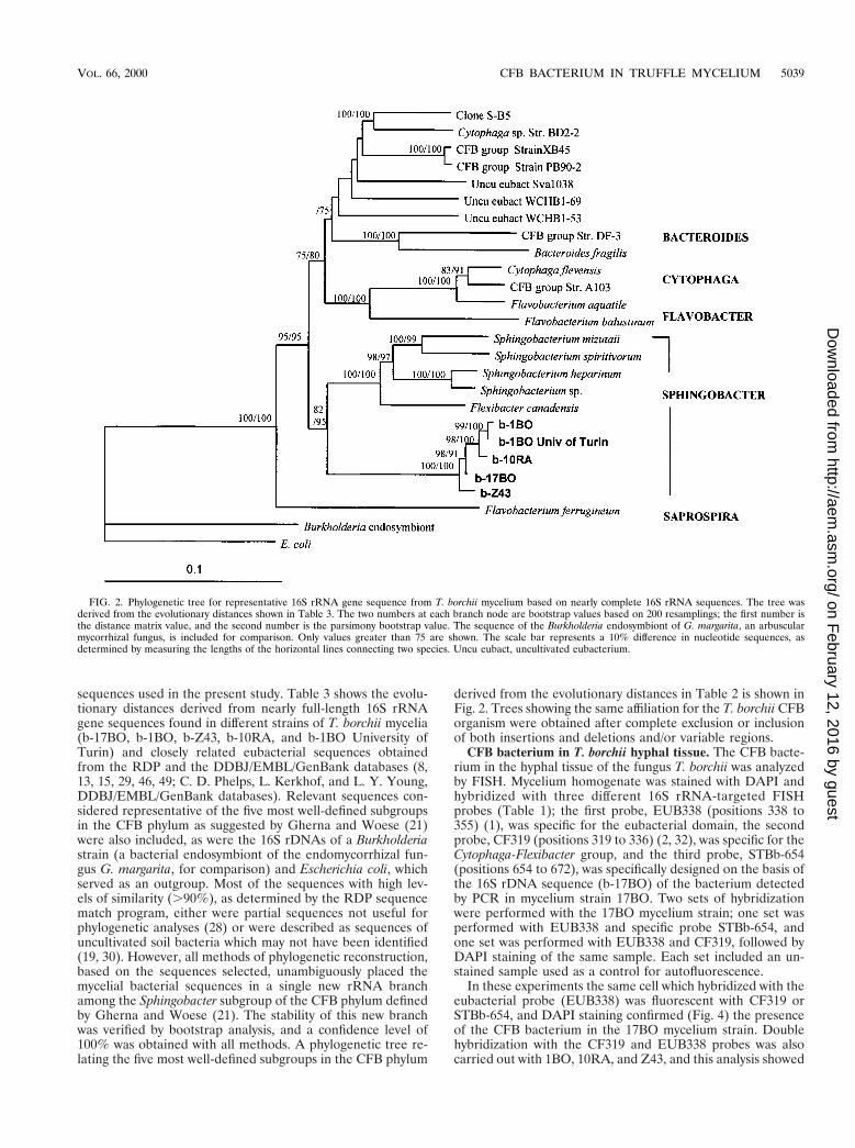
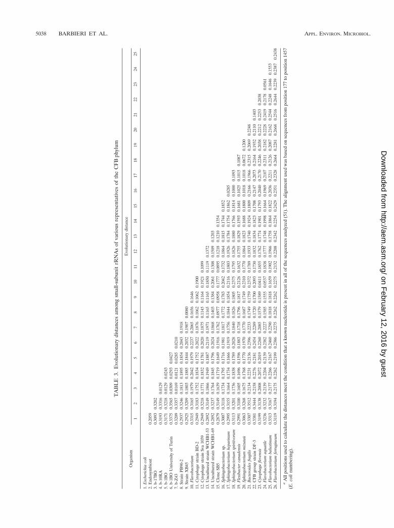
sequences used in the present study. Table 3 shows the evolu-tionary distances derived from nearly full-length 16S rRNAgene sequences found in different strains of T. borchii mycelia(b-17BO, b-1BO, b-Z43, b-10RA, and b-1BO University ofTurin) and closely related eubacterial sequences obtainedfrom the RDP and the DDBJ/EMBL/GenBank databases (8,13, 15, 29, 46, 49; C. D. Phelps, L. Kerkhof, and L. Y. Young,DDBJ/EMBL/GenBank databases). Relevant sequences con-sidered representative of the five most well-defined subgroupsin the CFB phylum as suggested by Gherna and Woese (21)were also included, as were the 16S rDNAs of a Burkholderiastrain (a bacterial endosymbiont of the endomycorrhizal fun-gus G. margarita, for comparison) and Escherichia coli, whichserved as an outgroup. Most of the sequences with high lev-els of similarity (.90%), as determined by the RDP sequencematch program, either were partial sequences not useful forphylogenetic analyses (28) or were described as sequences ofuncultivated soil bacteria which may not have been identified(19, 30). However, all methods of phylogenetic reconstruction,based on the sequences selected, unambiguously placed themycelial bacterial sequences in a single new rRNA branchamong the Sphingobacter subgroup of the CFB phylum definedby Gherna and Woese (21). The stability of this new branchwas verified by bootstrap analysis, and a confidence level of100% was obtained with all methods. A phylogenetic tree re-lating the five most well-defined subgroups in the CFB phylum
derived from the evolutionary distances in Table 2 is shown inFig. 2. Trees showing the same affiliation for the T. borchii CFBorganism were obtained after complete exclusion or inclusionof both insertions and deletions and/or variable regions.
CFB bacterium in T. borchii hyphal tissue. The CFB bacte-rium in the hyphal tissue of the fungus T. borchii was analyzedby FISH. Mycelium homogenate was stained with DAPI andhybridized with three different 16S rRNA-targeted FISHprobes (Table 1); the first probe, EUB338 (positions 338 to355) (1), was specific for the eubacterial domain, the secondprobe, CF319 (positions 319 to 336) (2, 32), was specific for theCytophaga-Flexibacter group, and the third probe, STBb-654(positions 654 to 672), was specifically designed on the basis ofthe 16S rDNA sequence (b-17BO) of the bacterium detectedby PCR in mycelium strain 17BO. Two sets of hybridizationwere performed with the 17BO mycelium strain; one set wasperformed with EUB338 and specific probe STBb-654, andone set was performed with EUB338 and CF319, followed byDAPI staining of the same sample. Each set included an un-stained sample used as a control for autofluorescence.
In these experiments the same cell which hybridized with theeubacterial probe (EUB338) was fluorescent with CF319 orSTBb-654, and DAPI staining confirmed (Fig. 4) the presenceof the CFB bacterium in the 17BO mycelium strain. Doublehybridization with the CF319 and EUB338 probes was alsocarried out with 1BO, 10RA, and Z43, and this analysis showed
FIG. 2. Phylogenetic tree for representative 16S rRNA gene sequence from T. borchii mycelium based on nearly complete 16S rRNA sequences. The tree wasderived from the evolutionary distances shown in Table 3. The two numbers at each branch node are bootstrap values based on 200 resamplings; the first number isthe distance matrix value, and the second number is the parsimony bootstrap value. The sequence of the Burkholderia endosymbiont of G. margarita, an arbuscularmycorrhizal fungus, is included for comparison. Only values greater than 75 are shown. The scale bar represents a 10% difference in nucleotide sequences, asdetermined by measuring the lengths of the horizontal lines connecting two species. Uncu eubact, uncultivated eubacterium.
VOL. 66, 2000 CFB BACTERIUM IN TRUFFLE MYCELIUM 5039
on February 12, 2016 by guest
http://aem.asm
.org/D
ownloaded from

the presence of fluorescent cells in all of the samples examined.In all of the FISH experiments no autofluorescence from thesamples was observed. To clarify the location of the CFBbacterium in the ectomycorrhizal fungus, thin sections of T.borchii mycelium tissue were hybridized with EUB338, thegeneral bacterial probe used for the homogenate. Few cells perseptum hybridized with the general bacterial probe. In con-trast, the difficulty of determining the exact position of theCFB bacterium with respect to the cytoplasm or the hyphalwall was evident, and it was difficult to determine where thebacterium was located since hybridization was successful inhomogenate samples and in sections in which the hyphal wallwas heterogeneously fragmented.
DISCUSSION
This paper describes molecular characterization of a CFBbacterium that is found in the mycorrhizal T. borchii myceliumand has not been cultured yet. PCR assays demonstrated thatthis uncultured CFB bacterium is present in all of the myceliaof T. borchii studied and in in vitro ectomycorrhizae. Simulta-neous hybridization of the general eubacterial and specificprobes with the hyphal tissues revealed rare, small (diameter,0.3 to 0.5 mm) but viable CFB bacteria within the hyphae (Fig.3 and 4).
Although several recent papers have described numerousbacteria, such as members of the genus Pseudomonas, theBacillaceae, and the Actinomycetes, living among the hyphae of
the fruiting bodies of truffles (3, 14, 20), no molecular charac-terizations of these bacteria or the microbe-host associationsare available. In general, few data for uncultured bacteria infungi have been presented (11, 12, 50), and only recently havea few phylogenetic studies identified an endophytic bacteriumthat is the endosymbiont of G. margarita and is a member ofthe genus Burkholderia (7) and members of the alpha and betasubclasses of the Proteobacteria detected in ectomycorrhizae ofF. sylvatica, L. vellereus, and L. subdulcis (33).
To our knowledge, no member of the Cytophagales has beenidentified previously in ectomycorrhizal symbioses. Althoughuncultivated CFB bacteria have been detected in soil environ-ments (30), few of these bacteria have been described as sym-bionts and commensals (25). The discovery of a CFB bacte-rium in the T. borchii ectomycorrhizal mycelium and molecularcharacterization of this organism represent a starting point forsystematic molecular identification and functional studies ofbacterium-fungus-plant symbioses. Concerning phylogeneticposition, we found that the overall tree topology is consistentwith other 16S rDNA phylogenetic analyses (4, 28, 38, 52), andfor all analyses, the bootstrap values supporting the new clusterwere significant for all of the criteria used (distance matrix,parsimony, maximum likelihood). However, since few environ-mental 16S rDNA sequences from soil bacteria are nearly fulllength, it is not possible to know if the CFB bacterium is closelyrelated to other uncultivated soil bacteria. A decision aboutrank and a formal description must await the availability of
FIG. 3. Detection of the CFB bacterium associated with T. borchii Vittad. hyphal tissue (mycelial strain 17BO). (a) Phase-contrast micrograph of T. borchii hyphaltissue homogenate. (b) Same sample after hybridization with fluorescein-labeled eubacterial probe EUB338. Fluorescent CFB cells are visible. Scale bars, 5 mm.
5040 BARBIERI ET AL. APPL. ENVIRON. MICROBIOL.
on February 12, 2016 by guest
http://aem.asm
.org/D
ownloaded from

more nearly complete sequences from environmental samplesand phenotypic data.
The question of whether the new CFB bacterium is involvedin the life cycle of the T. borchii truffle remains to be answered.PCR products obtained by using the specific b-17BO-f primerwith templates from the mycelia and ectomycorrhizae and theprobes used in the FISH experiment showed that this bacte-rium is a stable component of the T. borchii mycelium. Thisstudy provides the first direct evidence that a not-yet-culturedCFB bacterium is detectable in association with a mycorrhizalfungus of the genus Tuber.
ACKNOWLEDGMENTS
This work was supported by “Progetto Strategico CNR-RegioniTuber: Biotecnologia della micorrizazione.”
We thank G. Chevalier (INRA, Clermont Ferrant, France), B. Cit-terio (University of Urbino), G. Macino (University La Sapienza,Rome, Italy), and L. Garnerio (University of Turin) for providingsamples and the Molecular Evolution Workshop ’98 staff at the Marine
Biological Laboratory, Woods Hole, Mass., for the phylogenetic anal-yses.
REFERENCES1. Amann, R. I., L. Krumholz, and D. A. Stahl. 1990. Fluorescent-oligonucle-
otide probing of whole cells for determinative, phylogenetic, and environ-mental studies in microbiology. J. Bacteriol. 172:762–770.
2. Amann, R. I., W. Ludwig, and K. H. Schleifer. 1995. Phylogenetic identifi-cation and in situ detection of individual microbial cells without cultivation.Microbiol. Rev. 59:143–169.
3. Bedini, S., G. Bagnoli, C. Sbrana, C. Leporini, E. Tola, C. Dunne, C. Filippi,F. D’Andrea, F. O’Gara, and M. P. Nuti. 1999. Pseudomonads isolated fromwithin fruit bodies of T. borchii are capable of producing biological controlor phytostimulatory compounds in pure culture. Symbiosis 26:223–236.
4. Bernadet, J. F., P. Segers, M. Vancanneyt, F. Berthe, K. Kesters, and P.Vandamme. 1996. Cutting a Gordian knot: emended classification and de-scription of the genus Flavobacterium, emended description of the familyFlavobacteriaceae, and proposal of Flavobacterium hydatis nom. nov. (ba-sonym, Cytophaga aquatilis Strohl and Tait 1978). Int. J. Syst. Bacteriol.46:128–148.
5. Bertini, L., D. Agostini, L. Potenza, I. Rossi, S. Zeppa, A. Zambonelli, and V.Stocchi. 1998. Molecular markers for the identification of the ectomycorrhi-zal fungus Tuber borchii. New Phytol. 139:565–570.
6. Bertini, L., L. Potenza, A. Zambonelli, A. Amicucci, and V. Stocchi. 1998.
FIG. 4. (a) Phase-contrast micrograph of T. borchii hyphal tissue homogenate. (b) Detail of the same sample after hybridization with fluorescein-labeled eubacterialprobe EUB338. (c) Detail of the same sample after hybridization with CY3-labeled probe specific for b-17BO. The panel on the lower right is an overlap of panelsb and c showing the same cell hybridizing with both EUB338 and STBb-654 specific for the CFB bacterium. Scale bars, 5 mm.
VOL. 66, 2000 CFB BACTERIUM IN TRUFFLE MYCELIUM 5041
on February 12, 2016 by guest
http://aem.asm
.org/D
ownloaded from
RFLP species-specific patterns in the identification of white truffles. FEMSMicrobiol. Lett. 164:397–401.
7. Bianciotto, V., C. Bandi, D. Minerdi, M. Sironi, H. V. Tichy, and P. Bon-fante. 1996. An obligately endosymbiotic mycorrhizal fungus itself harborsobligately intracellular bacteria. Appl. Environ. Microbiol. 62:3005–3010.
8. Bowman, J. P., S. A. McCammon, M. V. Brown, D. S. Nichols, and T. A.McMeekin. 1997. Diversity and association of psychrophilic bacteria in Ant-arctic sea ice. Appl. Environ. Microbiol. 63:3068–3078.
9. Brosius, J., M. L. Palmer, P. J. Kennedy, and H. F. Noller. 1978. Completenucleotide sequence of a 16S ribosomal RNA gene from Escherichia coli.Proc. Natl. Acad. Sci. USA 75:4801–4805.
10. Campbell, R., and M. P. Greaves. 1990. Anatomy and community structureof the rhizosphere, p. 11–34. In J. M. Lynch (ed.), The rhizosphere. Wiley &Sons, Chichester, United Kingdom.
11. Chang, K.-P., G. A. Dasch, and E. Weiss. 1984. Endosymbionts of fungi andinvertebrates other than arthropods, p. 833–836. In N. R. Krieg and J. G.Holt (ed.), Bergey’s manual of systematic bacteriology, vol. 1. Williams &Wilkins, Baltimore, Md.
12. Chanway, C. P. 1996. Endophytes: they’re not just fungi! Can. J. Bot. 74:321–322.
13. Chin, K. J., D. Hahn, U. Hengstmann, W. Liesack, and P. H. Janssen. 1999.Characterization and identification of numerically abundant culturable bac-teria from the anoxic bulk soil of rice paddy microcosms. Appl. Environ.Microbiol. 65:5042–5049.
14. Citterio, B., P. Cardoni, L. Potenza, A. Amicucci, V. Stocchi, G. Gola, andM. P. Nuti. 1995. Isolation of bacteria from sporocarps of Tuber magnatumPico, Tuber borchii Vittad. and Tuber maculatum Vitt.: identification andbiochemical characterization, p. 241–248. In V. Stocchi, P. Bonfante andM. P. Nuti (ed.), Biotechnology of ectomycorrhizae. Molecular approaches.Plenum Publishing Corporation, New York, N.Y.
15. Dojka, M. A., P. Hugenholtz, S. K. Haack, and N. R. Pace. 1998. Microbialdiversity in a hydrocarbon- and chlorinated-solvent-contaminated aquiferundergoing intrinsic bioremediation. Appl. Environ. Microbiol. 64:3869–3877.
16. Duponnois, R., and J. Garbaye. 1992. Some mechanisms involved in growthstimulation of ectomycorrhizal fungi by bacteria. Can. J. Bot. 68:2148–2152.
17. Erland, S., B. Henrion, F. Martin, L. A. Glover, and I. J. Alexander. 1993.Identification of the ectomycorrhizal basidiomycete Tylospora fibrillosa Donkby RFLP analysis of the PCR-amplified ITS and IGS regions of ribosomalDNA. New Phytol. 12:525–532.
18. Felsenstein, J. 1993. PHYLIP (phylogeny inference package) version 3.5c.Department of Genetics, University of Washington, Seattle.
19. Felske, A., B. Englen, U. Nubel, and H. Backhaus. 1996. Direct ribosomalisolation from soil to extract bacterial rRNA for community analysis. Appl.Environ. Microbiol. 62:4162–4167.
20. Gazzanelli, G., M. Malatesta, A. Pianetti, W. Baffone, V. Stocchi, and B.Citterio. 1999. Bacteria associated to fruit bodies of the ecto-mycorrhizalfungus Tuber borchii Vittad. Symbiosis 26:211–222.
21. Gherna, R., and C. R. Woese. 1992. A partial phylogenetic analysis of the“Flavobacter-Bacteroides” phylum: basis for taxonomic restructuring. Syst.Appl. Microbiol. 15:513–521.
22. Gilbert, D. 1996. SeqPup sequence editor version 0.5. Indiana UniversityBiology Department, Bloomington.
23. Harley, J. L., and S. E. Smith. 1993. Mycorrhizal symbiosis. Academic Press,London, United Kingdom.
24. Henrion, B., G. Chevalier, and F. Martin. 1994. Typing truffle species byPCR amplification of the ribosomal DNA spacers. Mycol. Res. 98:37–43.
25. Hugenholtz, P., M. B. Goebel, and N. R. Pace. 1998. Impact of culture-independent studies on the emerging phylogenetic view of bacterial diversity.J. Bacteriol. 180:4765–4774.
26. Jukes, T. H., and C. R. Cantor. 1969. Evolution of protein molecules, p.21–132. In H. N. Munro (ed.), Mammalian protein metabolism, vol. 3.Academic Press, Inc., New York, N.Y.
27. Kochel, H. G., and H. Kuntzel. 1981. Nucleotide sequence of the Aspergillusnidulans mitochondrial gene coding for the small ribosomal subunit RNA:homology to E. coli 16S rRNA. Nucleic Acids Res. 9:5689–5696.
28. Kuske, C. H., S. M. Barns, and J. Bush. 1997. Diverse uncultivated bacterialgroups from soil of the arid southwestern United States that are present inmany geographic regions. Appl. Environ. Microbiol. 63:3614–3621.
29. Li, L., C. Kato, and K. Horikoshi. 1999. Bacterial diversity in deep-seasediments from different depths. Biodivers. Conserv. 8:659–677.
30. Liesack, W., and E. Stackebrandt. 1992. Occurrence of novel groups of thedomain Bacteria as revealed by analysis of genetic material isolated from anAustralian terrestrial environment. J. Bacteriol. 174:5072–5078.
31. Maidak, B. L., G. J. Olsen, N. Larsen, R. Overbeek, M. J. McCaughey, andC. R. Woese. 1997. The RDP (Ribosomal Database Project). Nucleic AcidsRes. 25:109–110.
32. Manz, W., R. I. Amann, W. Ludwig, M. Vancanneyt, and K. H. Schleifer.1996. Application of a suite of 16S rRNA-specific oligonucleotide probesdesigned to investigate bacteria of the phylum Cytophaga-Flavobacter-Bac-teroides in the natural environment. Microbiology 142:1097–1106.
33. Mogge, B., C. Loferer, R. Agerer, P. Hutzler, and A. Hartmann. 2000.Bacterial community structure and colonization patterns of Fagus sylvatica.Ectomycorrhizospheres as determined by fluorescence in situ hybridizationand confocal laser scanning microscopy. Mycorrhiza 5:271–278.
34. Molina, R. 1979. Pure culture synthesis and host specificity of red aldermycorrhizae. Can. J. Bot. 57:1223–1228.
35. Mosse, B. 1970. Honey colored, sessile endogone spores. II. Changes in finestructure during spore development. Arch. Microbiol. 74:129–145.
36. Ovreas, L., and V. Torsvik. 1998. Microbial diversity and community struc-ture in two different agricultural soil communities. Microb. Ecol. 36:303–315.
37. Page, R. D. M. 1996. TREEVIEW: an application to display phylogenetictrees on personal computers. Comput. Applic. Biosci. 12:357–358.
38. Paster, J. B., F. E. Dewhirst, I. Olsen, and G. Fraser. 1994. Phylogeny ofBacteroides, Prevotella, and Porphyromonas spp. and related bacteria. J. Bac-teriol. 176:725–732.
39. Pegler, D. N., B. M. Spooner, and T. W. K. Young. 1993. British truffles. Arevision of British hypogeous fungi. Royal Botanic Garden, Kew, UnitedKingdom.
40. Ravenschlag, K., K. Sahm, J. Pernthaler, and R. Amann. 1999. High bacte-rial diversity in permanently cold marine sediments. Appl. Environ. Micro-biol. 65:3982–3989.
41. Saltarelli, R., P. Ceccaroli, L. Vallorani, A. Zambonelli, B. Citterio, M.Malatesta, and V. Stocchi. 1998. Biochemical and morphological modifica-tions during the growth of Tuber borchii mycelium. Mycol. Res. 102:403–409.
42. Sambrook, J., E. F. Fritsch, and T. Maniatis. 1989. Molecular cloning: alaboratory manual, 2nd ed. Cold Spring Harbor Laboratory Press, ColdSpring Harbor, N.Y.
43. Sisti, D., A. Zambonelli, G. Giomaro, I. Rossi, P. Ceccaroli, B. Citterio, P. A.Benedetti, and V. Stocchi. 1998. In vitro mycorrhizal synthesis of micropropa-gated Tilia platyphyllos Scop. plantlets with Tuber borchii Vittad. mycelium inpure culture. Acta Hortic. (ISHS) 457:379–387.
44. Tavaglini, J., A. Bolchi, R. Percudani, S. Petrucco, G. L. Rossi, and S.Ottonello. 1995. Testing a selected region of Tuber mitochondrial smallsubunit rDNA as molecular marker for evolutionary and bio-diversity stud-ies, p. 205–211. In V. Stocchi, P. Bonfante, and M. P. Nuti (ed.), Biotech-nology of ectomycorrhizae. Molecular approaches. Plenum Publishing Cor-poration, New York, N.Y.
45. Trappe, J. M. 1979. The orders, families, and genera of hypogeus Ascomy-cotina (truffles and their relatives). Mycotaxon 9:297–340.
46. Vandamme, P., M. Vancanneyt, A. van Belkum, P. Segers, W. G. V. Quint, K.Kersters, B. J. Paster, and F. E. Dewhirst. 1996. Polyphasic analysis ofstrains from the genus Capnocytophaga and Centers for Disease Controlgroup DF-3. Int. J. Syst. Bacteriol. 46:782–791.
47. Varese, G. C., S. Portinaro, A. Trotta, S. Scannerini, A. M. Luppi-Mosca,and M. G. Martinotti. 1996. Bacteria associated with Suillus grevillei sporo-carps and ectomycorrhizae and their effects on in vitro growth of the myco-biont. Symbiosis 21:129–147.
48. Weisburg, W. G., M. Barns, D. A. Pelletier, and D. J. Lane. 1991. 16Sribosomal DNA amplification for phylogenetic study. J. Bacteriol. 173:697–703.
49. Weisburg, W. G., Y. Oyaizu, H. Oyaizu, and C. R. Woese. 1985. Naturalrelationship between Bacteroides and flavobacteria. J. Bacteriol. 164:230–236.
50. Wilson, D. 1995. Endophyte: the evolution of a term, and clarification of itsuse and definition. Oikos 73:274–276.
51. Woese, C. R., R. Gutell, R. Gupta, and H. F. Noller. 1983. Detailed analysisof the higher-order structure of the 16S-like ribosomal ribonucleic acids.Microbiol. Rev. 47:621–699.
52. Woese, C. R., D. Yang, L. Mandelco, and K. O. Stetter. 1990. The Flexibacter-Flavobacter connection. Syst. Appl. Microbiol. 13:161–165.
5042 BARBIERI ET AL. APPL. ENVIRON. MICROBIOL.
on February 12, 2016 by guest
http://aem.asm
.org/D
ownloaded from
Copyright © 2022 FDOKUMEN