Cell organisation, sulphur metabolism and ion transport-related genes are differentially expressed...
13
BioMed Central Page 1 of 13 (page number not for citation purposes) BMC Genomics Open Access Research article Cell organisation, sulphur metabolism and ion transport-related genes are differentially expressed in Paracoccidioides brasiliensis mycelium and yeast cells Rosângela V Andrade 1 , Hugo C Paes 1 , André M Nicola 1 , Maria José A de Carvalho 1 , Ana Lúcia Fachin 2 , Renato S Cardoso 2 , Simoneide S Silva 1 , Larissa Fernandes 1 , Silvana P Silva 3 , Eduardo A Donadi 2 , Elza T Sakamoto- Hojo 2 , Geraldo AS Passos 2 , Célia MA Soares 3 , Marcelo M Brígido 1 and Maria Sueli S Felipe* 1 Address: 1 Depto. de Biologia Celular, Universidade de Brasília, 70910–900. Brasília-DF, Brazil, 2 Depto de Genética, Faculdade de Medicina de Ribeirão Preto, Universidade de São Paulo, 14040–900, Ribeirão Preto, SP, Brazil and 3 Depto de Bioquímica e Biologia Molecular, Universidade Federal de Goiás, 74001–970, Goiânia, GO, Brazil Email: Rosângela V Andrade - [email protected]; Hugo C Paes - [email protected]; André M Nicola - [email protected]; Maria José A de Carvalho - [email protected]; Ana Lúcia Fachin - [email protected]; Renato S Cardoso - [email protected]; Simoneide S Silva - [email protected]; Larissa Fernandes - [email protected]; Silvana P Silva - [email protected]; Eduardo A Donadi - [email protected]; Elza T Sakamoto-Hojo - [email protected]; Geraldo AS Passos - [email protected]; Célia MA Soares - [email protected]; Marcelo M Brígido - [email protected]; Maria Sueli S Felipe* - [email protected] * Corresponding author Abstract Background: Mycelium-to-yeast transition in the human host is essential for pathogenicity by the fungus Paracoccidioides brasiliensis and both cell types are therefore critical to the establishment of paracoccidioidomycosis (PCM), a systemic mycosis endemic to Latin America. The infected population is of about 10 million individuals, 2% of whom will eventually develop the disease. Previously, transcriptome analysis of mycelium and yeast cells resulted in the assembly of 6,022 sequence groups. Gene expression analysis, using both in silico EST subtraction and cDNA microarray, revealed genes that were differential to yeast or mycelium, and we discussed those involved in sugar metabolism. To advance our understanding of molecular mechanisms of dimorphic transition, we performed an extended analysis of gene expression profiles using the methods mentioned above. Results: In this work, continuous data mining revealed 66 new differentially expressed sequences that were MIPS(Munich Information Center for Protein Sequences)-categorised according to the cellular process in which they are presumably involved. Two well represented classes were chosen for further analysis: (i) control of cell organisation – cell wall, membrane and cytoskeleton, whose representatives were hex (encoding for a hexagonal peroxisome protein), bgl (encoding for a 1,3-β-glucosidase) in mycelium cells; and ags (an α-1,3-glucan synthase), cda (a chitin deacetylase) and vrp (a verprolin) in yeast cells; (ii) ion metabolism and transport – two genes putatively implicated in ion transport were confirmed to be highly expressed in mycelium cells – isc and ktp, respectively an iron-sulphur cluster-like protein and a cation transporter; and a putative P-type cation pump (pct) in yeast. Also, several enzymes from the cysteine de novo biosynthesis pathway were shown to be up regulated in the yeast form, including ATP sulphurylase, APS kinase and also PAPS reductase. Published: 14 August 2006 BMC Genomics 2006, 7:208 doi:10.1186/1471-2164-7-208 Received: 19 February 2006 Accepted: 14 August 2006 This article is available from: http://www.biomedcentral.com/1471-2164/7/208 © 2006 Andrade et al; licensee BioMed Central Ltd. This is an Open Access article distributed under the terms of the Creative Commons Attribution License (http://creativecommons.org/licenses/by/2.0 ), which permits unrestricted use, distribution, and reproduction in any medium, provided the original work is properly cited.
Transcript of Cell organisation, sulphur metabolism and ion transport-related genes are differentially expressed...
BioMed CentralBMC Genomics
ss
Open AcceResearch articleCell organisation, sulphur metabolism and ion transport-related genes are differentially expressed in Paracoccidioides brasiliensis mycelium and yeast cellsRosângela V Andrade1, Hugo C Paes1, André M Nicola1, Maria José A de Carvalho1, Ana Lúcia Fachin2, Renato S Cardoso2, Simoneide S Silva1, Larissa Fernandes1, Silvana P Silva3, Eduardo A Donadi2, Elza T Sakamoto-Hojo2, Geraldo AS Passos2, Célia MA Soares3, Marcelo M Brígido1 and Maria Sueli S Felipe*1Address: 1Depto. de Biologia Celular, Universidade de Brasília, 70910–900. Brasília-DF, Brazil, 2Depto de Genética, Faculdade de Medicina de Ribeirão Preto, Universidade de São Paulo, 14040–900, Ribeirão Preto, SP, Brazil and 3Depto de Bioquímica e Biologia Molecular, Universidade Federal de Goiás, 74001–970, Goiânia, GO, Brazil
Email: Rosângela V Andrade - [email protected]; Hugo C Paes - [email protected]; André M Nicola - [email protected]; Maria José A de Carvalho - [email protected]; Ana Lúcia Fachin - [email protected]; Renato S Cardoso - [email protected]; Simoneide S Silva - [email protected]; Larissa Fernandes - [email protected]; Silvana P Silva - [email protected]; Eduardo A Donadi - [email protected]; Elza T Sakamoto-Hojo - [email protected]; Geraldo AS Passos - [email protected]; Célia MA Soares - [email protected]; Marcelo M Brígido - [email protected]; Maria Sueli S Felipe* - [email protected]
* Corresponding author
AbstractBackground: Mycelium-to-yeast transition in the human host is essential for pathogenicity by the fungus Paracoccidioidesbrasiliensis and both cell types are therefore critical to the establishment of paracoccidioidomycosis (PCM), a systemicmycosis endemic to Latin America. The infected population is of about 10 million individuals, 2% of whom will eventuallydevelop the disease. Previously, transcriptome analysis of mycelium and yeast cells resulted in the assembly of 6,022sequence groups. Gene expression analysis, using both in silico EST subtraction and cDNA microarray, revealed genesthat were differential to yeast or mycelium, and we discussed those involved in sugar metabolism. To advance ourunderstanding of molecular mechanisms of dimorphic transition, we performed an extended analysis of gene expressionprofiles using the methods mentioned above.
Results: In this work, continuous data mining revealed 66 new differentially expressed sequences that wereMIPS(Munich Information Center for Protein Sequences)-categorised according to the cellular process in which they arepresumably involved. Two well represented classes were chosen for further analysis: (i) control of cell organisation – cellwall, membrane and cytoskeleton, whose representatives were hex (encoding for a hexagonal peroxisome protein), bgl(encoding for a 1,3-β-glucosidase) in mycelium cells; and ags (an α-1,3-glucan synthase), cda (a chitin deacetylase) and vrp(a verprolin) in yeast cells; (ii) ion metabolism and transport – two genes putatively implicated in ion transport wereconfirmed to be highly expressed in mycelium cells – isc and ktp, respectively an iron-sulphur cluster-like protein and acation transporter; and a putative P-type cation pump (pct) in yeast. Also, several enzymes from the cysteine de novobiosynthesis pathway were shown to be up regulated in the yeast form, including ATP sulphurylase, APS kinase and alsoPAPS reductase.
Published: 14 August 2006
BMC Genomics 2006, 7:208 doi:10.1186/1471-2164-7-208
Received: 19 February 2006Accepted: 14 August 2006
This article is available from: http://www.biomedcentral.com/1471-2164/7/208
© 2006 Andrade et al; licensee BioMed Central Ltd.This is an Open Access article distributed under the terms of the Creative Commons Attribution License (http://creativecommons.org/licenses/by/2.0), which permits unrestricted use, distribution, and reproduction in any medium, provided the original work is properly cited.
Page 1 of 13(page number not for citation purposes)
BMC Genomics 2006, 7:208 http://www.biomedcentral.com/1471-2164/7/208
Conclusion: Taken together, these data show that several genes involved in cell organisation and ion metabolism/transport are expressed differentially along dimorphic transition. Hyper expression in yeast of the enzymes of sulphurmetabolism reinforced that this metabolic pathway could be important for this process. Understanding these changes byfunctional analysis of such genes may lead to a better understanding of the infective process, thus providing new targetsand strategies to control PCM.
BackgroundThe availability of great amounts of raw genomic andtranscriptome data collected from several organisms hasprompted the development of large-scale gene expressionanalysis which will ultimately help to unravel the func-tion of many genes in diverse biological contexts. Differ-ent approaches such as cDNA microarrays [1-3], in silicoESTs subtraction [4,5] and serial analysis of gene expres-sion – SAGE [6,7] are widely employed to assess differen-tial gene expression patterns leading to the discovery of agreat number of genes that are over or under expressed ineach physiological context. The successful use of thecDNA microarray approach in fungal pathogens such asCandida albicans [8-13], Histoplasma capsulatum [14] andCryptococcus neoformans [15] has resulted in the identifica-tion of genes involved in cell viability and opened newexperimental perspectives to understand host-parasiteinteractions and thus develop new therapeuticapproaches to systemic mycoses [8,11].
Paracoccidioidomycosis (PCM) is a human illnessendemic to Latin America [16]; its area of incidencespreads non-uniformly from Mexico to Argentina [17],being higher in Brazil, Venezuela, Colombia and Argen-tina [18,19,16]. An estimation for Brazil points to an inci-dence rate between 1 and 3 and a mortality rate of 1.4 permillion [20]. McEwen et al. [21] reported an overallinfected population of 10 million individuals in LatinAmerica, 2% of whom will eventually develop the disease.In nature, another important mammalian host is thearmadillo Dasypus novemcinctus [22]. PCM affects the skin,lymph nodes and various internal organs, including thelungs – where it causes granulomatous processes – and thecentral nervous system [19,23]. Its clinical presentationsrange from a localised and benign disease to a progressiveand potentially lethal systemic infection [24]. The diseaseis more frequent in adult males, who account for up to90% of all cases. Healthy rural workers are the main tar-gets, but PCM affects immunosuppressed individuals aswell [25,26], including as much as 30% of AIDS patients[27]. All patients from whom the fungus is isolated mustbe treated and, in spite of new antifungal drugs, pulmo-nary fibrosis is still the most frequent sequel. The outcomeof infection depends on several factors, including hostresponses and the virulence of the infecting isolate.
The causative agent of PCM, the thermo-regulated dimor-phic fungus P. brasiliensis, is believed to be a free-livingmycelium saprobe that undergoes transition to the yeastpathogenic form upon temperature change from the envi-ronmental 24–26°C to the mammalian body temperatureof 37°C. This switch is necessary and sufficient to triggermorphotype interconversion in vitro, which makes thisfungus an interesting model to study fungal cell differen-tiation at the molecular level. The biochemical events reg-ulating dimorphic transition in P. brasiliensis are yetpoorly defined, although relevant molecular-level infor-mation on this process has been partially described in thetranscriptome analyses of two different P. brasiliensis iso-lates [28-30].
The exact ecological niche of this pathogen is stillunknown [17], but P. brasiliensis can be retrieved from thesoil. The fungus Penicillium marneffei is greatly similar inthat it is a human opportunistic pathogen that also under-goes thermally-controlled dimorphic transition uponinfection, can also infect a wild mammal (the bamboorat) and has an yet unknown natural reservoir. Genomicdata provided evidence that, in the case of P. marneffei, thefungus may have a sexual stage as a free-living organism[31].
Phylogenetic analysis of members of the order Onygen-ales demonstrated a close relationship of P. brasiliensiswith the pathogenic fungi Blastomyces dermatitidis, Emmon-sia parva and Histoplasma capsulatum [32]. P. brasiliensiscan be fitted with B. dermatitidis and E. parva in the familyOnygenacea [33]. Recently it was reported that P. brasilien-sis is in fact a complex of at least three closely correlatedphylogenetic species [34]. So far, the sexual phase of theascomycete P. brasiliensis was not reported limiting ourknowledge about the mechanisms that contribute to itsdimorphism, pathogenicity, and virulence. P. brasiliensisisolates shows chromosomal polymorphism; it contains4–5 chromosomal DNA molecules with molecular sizesranging from 2–10 Mb [35,36]. The genome size was esti-mated to be around 30 Mb [37] and DNA sequencing of~ 50 Kb revealed a density of one gene per 3.5–4.5 Kb,suggesting a total of 7,500–9,000 genes [38].
Recently, our group analysed the transcriptome of thePb01 isolate, represented by a set of 6,022 clusters. The 16genes that were then found to be differentially expressed
Page 2 of 13(page number not for citation purposes)
BMC Genomics 2006, 7:208 http://www.biomedcentral.com/1471-2164/7/208
by both methods used – in silico EST subtraction andcDNA microarray – were categorised by function. Wechose to discuss in that work those that were involved incore metabolic pathways such as sugar metabolism [28].Now, continued overlap analysis from raw data revealed66 new genes that are differentially expressed in one orother morphotype. Upon categorisation by known data-bases we have selected two MIPS [39] classes, which werechosen to be confirmed by northern blotting. Here wepresent the result of this extended analysis, and discussthe putative roles the differential genes – related to cellorganisation and ion metabolism and transport – play inthe corresponding morphotype of this pathogen. One ofthe discussed pathways – de novo cysteine synthesis frominorganic sulphate, a branch of sulphur metabolism – wasalmost entirely up-regulated in the yeast form. The impor-tance of sulphur metabolism to the life cycle of patho-genic fungi has been extensively reviewed elsewhere[40,41] and recently new data from microarray experi-ments have arisen from work in H. capsulatum that sup-port a role of organic sulphate in the maintenance of theyeast phase [14]. In a previous report [42], the importance
of organic sulphates to the growth and differentiation ofP. brasiliensis was assessed. This phenomenon demandedfurther investigation and prompted us to assess up- anddownregulation of sulphur metabolism genes in myc-elium and yeast cells and also dimorphic transition inboth directions without inorganic sulphate as a sulphursource. We have thus found that this compound is unnec-essary for the process.
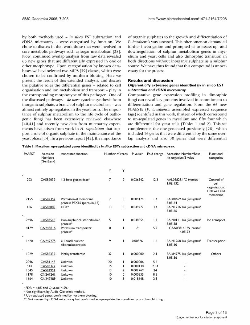
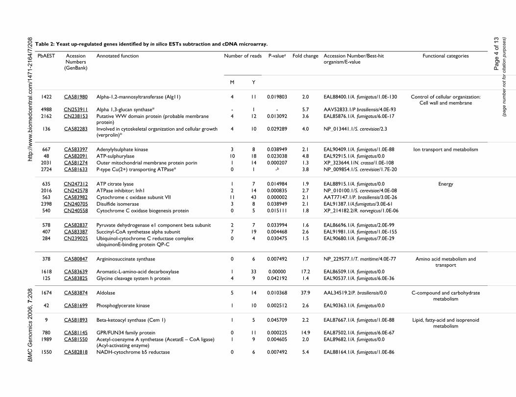
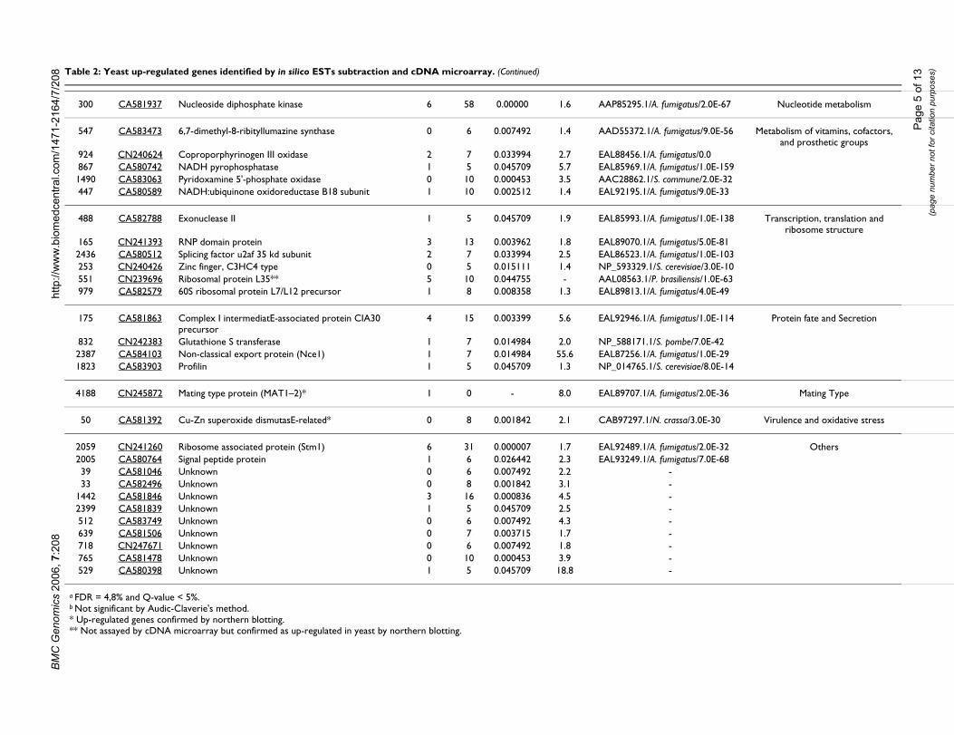
Results and discussionDifferentially expressed genes identified by in silico EST subtraction and cDNA microarrayComparative gene expression profiling in dimorphicfungi can reveal key proteins involved in commitment todifferentiation and gene regulation. From the 66 newPbAESTs (P. brasiliensis assembled expressed sequencetags) identified in this work, thirteen of which correspondto up-regulated genes in mycelium and fifty four whichare differential for yeast cells (Tables 1 and 2). This setcomplements the one generated previously [28], whichincluded 16 genes that were differential by the same over-lap analysis and also 30 genes that were differential
Table 1: Mycelium up-regulated genes identified by in silico ESTs subtraction and cDNA microarray.
PbAEST Acession Numbers (GenBank)
Annotated function Number of reads P-valuea Fold change Accession Number/Best-hit organism/E-value
Functional categories
M Y
202 CA582032 1,3-beta-glucosidase* 7 2 0.036942 12.3 AAL09828.1/C. immitis/1.0E-132
Control of cell
organization: Cell wall and membrane
2155 CA582352 Peroxisomal membrane protein PEX16 (peroxin-16)
7 0 0.004174 1.4 EAL88469.1/A. fumigatus/3.0E-64
186 CA583085 HEX* 13 8 0.049272 3.4 EAL91716.1/A. fumigatus/3.0E-66
2496 CA583518 Iron-sulphur cluster nifU-like protein*
5 1 0.048854 1.7 EAL90111.1/A. fumigatus/8.0E-58
Ion transport
4179 CN245816 Potassium transporter protein*
0 1 -b 5.2 CAA08814.1/N. crassa/4.0E-22
1420 CN247275 U1 small nuclear ribonucleoprotein
9 1 0.00526 1.6 EAL91268.1/A. fumigatus/1.0E-60
Transcription
1029 CA582332 Methyltransferase 32 1 0.000000 2.1 EAL84975.1/A. fumigatus/1.0E-56
Others
2096 CA581148 Unkown 20 1 0.000006 5.6 -514 CA583322 Unkown 15 1 0.000138 23.4 -1045 CA581951 Unkown 13 2 0.001769 24 -1178 CN247241 Unkown 10 0 0.000535 8.5 -1664 CN247289 Unkown 10 3 0.018648 2.5 -
a FDR = 4,8% and Q-value < 5%.b Not significant by Audic-Claverie's method.* Up-regulated genes confirmed by northern blotting.** Not assayed by cDNA microarray but confirmed as up-regulated in mycelium by northern blotting.
Page 3 of 13(page number not for citation purposes)
Page
4 o
f 13
(pag
e nu
mbe
r not
for c
itatio
n pu
rpos
es)
Best-hit Functional categories
atus/1.0E-130 Control of cellular organization: Cell wall and membrane
liensis/4.0E-93atus/6.0E-17
visiae/2.3
atus/1.0E-88 Ion transport and metabolismatus/0.0ssa/1.0E-108visiae/1.7E-20
atus/0.0 Energyvisiae/4.0E-08
iliensis/3.0E-26atus/3.0E-61vegicus/1.0E-06
atus/2.0E-99atus/1.0E-155atus/7.0E-29
ritime/4.0E-77 Amino acid metabolism and transport
atus/0.0atus/6.0E-36
liensis/0.0 C-compound and carbohydrate metabolism
atus/0.0
atus/1.0E-88 Lipid, fatty-acid and isoprenoid metabolism
atus/6.0E-67atus/0.0
atus/1.0E-86
BMC
Gen
omic
s 20
06, 7
:208
http
://w
ww
.bio
med
cent
ral.c
om/1
471-
2164
/7/2
08 Table 2: Yeast up-regulated genes identified by in silico ESTs subtraction and cDNA microarray.
PbAEST Acession Numbers (GenBank)
Annotated function Number of reads P-valuea Fold change Accession Number/organism/E-value
M Y
1422 CA581980 Alpha-1,2-mannosyltransferase (Alg11) 4 11 0.019803 2.0 EAL88400.1/A. fumig
4988 CN253911 Alpha 1,3-glucan synthase* - 1 - 5.7 AAV52833.1/P brasi2162 CN238153 Putative WW domain protein (probable membrane
protein)4 12 0.013092 3.6 EAL85876.1/A. fumig
136 CA582283 Involved in cytoskeletal organization and cellular growth (verprolin)*
4 10 0.029289 4.0 NP_013441.1/S. cere
667 CA583397 Adenylylsulphate kinase 3 8 0.038949 2.1 EAL90409.1/A. fumig48 CA582091 ATP-sulphurylase 10 18 0.023038 4.8 EAL92915.1/A. fumig
2031 CA581274 Outer mitochondrial membrane protein porin 1 14 0.000207 1.3 XP_323644.1/N. cra2724 CA581633 P-type Cu(2+) transporting ATPase* 0 1 -b 3.8 NP_009854.1/S. cere
635 CN247312 ATP citrate lyase 1 7 0.014984 1.9 EAL88915.1/A. fumig2016 CN242578 ATPase inhibitor; Inh1 2 14 0.000835 2.7 NP_010100.1/S. cere563 CA583982 Cytochrome c oxidase subunit VII 11 43 0.000002 2.1 AAT77147.1/P. bras2398 CN240705 Disulfide isomerase 3 8 0.038949 2.1 EAL91387.1/A.fumig540 CN240558 Cytochrome C oxidase biogenesis protein 0 5 0.015111 1.8 XP_214182.2/R. nor
578 CA582837 Pyruvate dehydrogenase e1 component beta subunit 2 7 0.033994 1.6 EAL86696.1/A. fumig407 CA583387 Succinyl-CoA synthetase alpha subunit 7 19 0.004468 2.6 EAL91981.1/A. fumig284 CN239025 Ubiquinol-cytochrome C reductase complex
ubiquinonE-binding protein QP-C0 4 0.030475 1.5 EAL90680.1/A. fumig
378 CA580847 Argininosuccinate synthase 0 6 0.007492 1.7 NP_229577.1/T. ma
1618 CA583639 Aromatic-L-amino-acid decarboxylase 1 33 0.00000 17.2 EAL86509.1/A. fumig125 CA583825 Glycine cleavage system h protein 4 9 0.042192 1.4 EAL90537.1/A. fumig
1674 CA583874 Aldolase 5 14 0.010368 37.9 AAL34519.2/P. brasi
42 CA581699 Phosphoglycerate kinase 1 10 0.002512 2.6 EAL90363.1/A. fumig
9 CA581893 Beta-ketoacyl synthase (Cem 1) 1 5 0.045709 2.2 EAL87667.1/A. fumig
780 CA581145 GPR/FUN34 family protein 0 11 0.000225 14.9 EAL87502.1/A. fumig1989 CA581550 Acetyl-coenzyme A synthetase (AcetatE – CoA ligase)
(Acyl-activating enzyme)1 9 0.004605 2.0 EAL89682.1/A. fumig
1550 CA582818 NADH-cytochrome b5 reductase 0 6 0.007492 5.4 EAL88164.1/A. fumig
Page
5 o
f 13
(pag
e nu
mbe
r not
for c
itatio
n pu
rpos
es)
gatus/2.0E-67 Nucleotide metabolism
igatus/9.0E-56 Metabolism of vitamins, cofactors, and prosthetic groups
atus/0.0atus/1.0E-159mune/2.0E-32atus/9.0E-33
atus/1.0E-138 Transcription, translation and ribosome structure
atus/5.0E-81atus/1.0E-103visiae/3.0E-10liensis/1.0E-63atus/4.0E-49
atus/1.0E-114 Protein fate and Secretion
be/7.0E-42atus/1.0E-29visiae/8.0E-14
atus/2.0E-36 Mating Type
sa/3.0E-30 Virulence and oxidative stress
atus/2.0E-32 Othersatus/7.0E-68
BMC
Gen
omic
s 20
06, 7
:208
http
://w
ww
.bio
med
cent
ral.c
om/1
471-
2164
/7/2
08
300 CA581937 Nucleoside diphosphate kinase 6 58 0.00000 1.6 AAP85295.1/A. fumi
547 CA583473 6,7-dimethyl-8-ribityllumazine synthase 0 6 0.007492 1.4 AAD55372.1/A. fum
924 CN240624 Coproporphyrinogen III oxidase 2 7 0.033994 2.7 EAL88456.1/A. fumig867 CA580742 NADH pyrophosphatase 1 5 0.045709 5.7 EAL85969.1/A. fumig1490 CA583063 Pyridoxamine 5'-phosphate oxidase 0 10 0.000453 3.5 AAC28862.1/S. com447 CA580589 NADH:ubiquinone oxidoreductase B18 subunit 1 10 0.002512 1.4 EAL92195.1/A. fumig
488 CA582788 Exonuclease II 1 5 0.045709 1.9 EAL85993.1/A. fumig
165 CN241393 RNP domain protein 3 13 0.003962 1.8 EAL89070.1/A. fumig2436 CA580512 Splicing factor u2af 35 kd subunit 2 7 0.033994 2.5 EAL86523.1/A. fumig253 CN240426 Zinc finger, C3HC4 type 0 5 0.015111 1.4 NP_593329.1/S. cere551 CN239696 Ribosomal protein L35** 5 10 0.044755 - AAL08563.1/P. brasi979 CA582579 60S ribosomal protein L7/L12 precursor 1 8 0.008358 1.3 EAL89813.1/A. fumig
175 CA581863 Complex I intermediatE-associated protein CIA30 precursor
4 15 0.003399 5.6 EAL92946.1/A. fumig
832 CN242383 Glutathione S transferase 1 7 0.014984 2.0 NP_588171.1/S. pom2387 CA584103 Non-classical export protein (Nce1) 1 7 0.014984 55.6 EAL87256.1/A. fumig1823 CA583903 Profilin 1 5 0.045709 1.3 NP_014765.1/S. cere
4188 CN245872 Mating type protein (MAT1–2)* 1 0 - 8.0 EAL89707.1/A. fumig
50 CA581392 Cu-Zn superoxide dismutasE-related* 0 8 0.001842 2.1 CAB97297.1/N. cras
2059 CN241260 Ribosome associated protein (Stm1) 6 31 0.000007 1.7 EAL92489.1/A. fumig2005 CA580764 Signal peptide protein 1 6 0.026442 2.3 EAL93249.1/A. fumig39 CA581046 Unknown 0 6 0.007492 2.2 -33 CA582496 Unknown 0 8 0.001842 3.1 -
1442 CA581846 Unknown 3 16 0.000836 4.5 -2399 CA581839 Unknown 1 5 0.045709 2.5 -512 CA583749 Unknown 0 6 0.007492 4.3 -639 CA581506 Unknown 0 7 0.003715 1.7 -718 CN247671 Unknown 0 6 0.007492 1.8 -765 CA581478 Unknown 0 10 0.000453 3.9 -529 CA580398 Unknown 1 5 0.045709 18.8 -
a FDR = 4,8% and Q-value < 5%.b Not significant by Audic-Claverie's method.* Up-regulated genes confirmed by northern blotting.** Not assayed by cDNA microarray but confirmed as up-regulated in yeast by northern blotting.
Table 2: Yeast up-regulated genes identified by in silico ESTs subtraction and cDNA microarray. (Continued)
BMC Genomics 2006, 7:208 http://www.biomedcentral.com/1471-2164/7/208
according to in silico EST subtraction alone. MIPS func-tional categories [43] were used to classify the 66 PbAESTsinto 14 major groups (data not shown). Gene categorisa-tion revealed some that are involved in energy production(11%) – this was expected considering the adaptationprocess that is required for the mycelium-to-yeast transi-tion; control of cell wall organisation (10%); ion metabo-lism and transport (8%); transcription, translation andribosome structure (8%); virulence and oxidative stress(4%). Manual annotation under stringent criteria ofsequence alignment with other dimorphic fungi gene setsallowed us to ascribe a putative biological function tomany of those genes. The genes that belonged in two cat-egories – cell wall organisation and ion metabolism andtransport – were selected for confirmation by northernblotting.
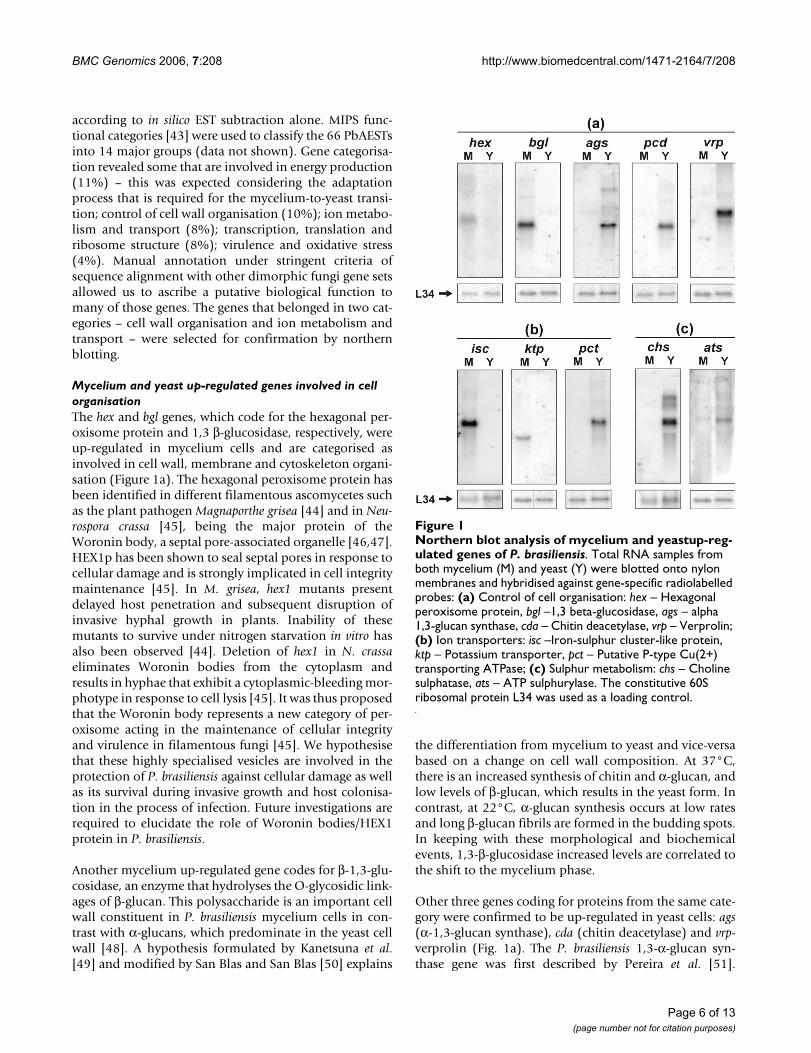
Mycelium and yeast up-regulated genes involved in cell organisationThe hex and bgl genes, which code for the hexagonal per-oxisome protein and 1,3 β-glucosidase, respectively, wereup-regulated in mycelium cells and are categorised asinvolved in cell wall, membrane and cytoskeleton organi-sation (Figure 1a). The hexagonal peroxisome protein hasbeen identified in different filamentous ascomycetes suchas the plant pathogen Magnaporthe grisea [44] and in Neu-rospora crassa [45], being the major protein of theWoronin body, a septal pore-associated organelle [46,47].HEX1p has been shown to seal septal pores in response tocellular damage and is strongly implicated in cell integritymaintenance [45]. In M. grisea, hex1 mutants presentdelayed host penetration and subsequent disruption ofinvasive hyphal growth in plants. Inability of thesemutants to survive under nitrogen starvation in vitro hasalso been observed [44]. Deletion of hex1 in N. crassaeliminates Woronin bodies from the cytoplasm andresults in hyphae that exhibit a cytoplasmic-bleeding mor-photype in response to cell lysis [45]. It was thus proposedthat the Woronin body represents a new category of per-oxisome acting in the maintenance of cellular integrityand virulence in filamentous fungi [45]. We hypothesisethat these highly specialised vesicles are involved in theprotection of P. brasiliensis against cellular damage as wellas its survival during invasive growth and host colonisa-tion in the process of infection. Future investigations arerequired to elucidate the role of Woronin bodies/HEX1protein in P. brasiliensis.
Another mycelium up-regulated gene codes for β-1,3-glu-cosidase, an enzyme that hydrolyses the O-glycosidic link-ages of β-glucan. This polysaccharide is an important cellwall constituent in P. brasiliensis mycelium cells in con-trast with α-glucans, which predominate in the yeast cellwall [48]. A hypothesis formulated by Kanetsuna et al.[49] and modified by San Blas and San Blas [50] explains
the differentiation from mycelium to yeast and vice-versabased on a change on cell wall composition. At 37°C,there is an increased synthesis of chitin and α-glucan, andlow levels of β-glucan, which results in the yeast form. Incontrast, at 22°C, α-glucan synthesis occurs at low ratesand long β-glucan fibrils are formed in the budding spots.In keeping with these morphological and biochemicalevents, 1,3-β-glucosidase increased levels are correlated tothe shift to the mycelium phase.
Other three genes coding for proteins from the same cate-gory were confirmed to be up-regulated in yeast cells: ags(α-1,3-glucan synthase), cda (chitin deacetylase) and vrp-verprolin (Fig. 1a). The P. brasiliensis 1,3-α-glucan syn-thase gene was first described by Pereira et al. [51].
Northern blot analysis of mycelium and yeastup-regulated genes of P. brasiliensisFigure 1Northern blot analysis of mycelium and yeastup-reg-ulated genes of P. brasiliensis. Total RNA samples from both mycelium (M) and yeast (Y) were blotted onto nylon membranes and hybridised against gene-specific radiolabelled probes: (a) Control of cell organisation: hex – Hexagonal peroxisome protein, bgl –1,3 beta-glucosidase, ags – alpha 1,3-glucan synthase, cda – Chitin deacetylase, vrp – Verprolin; (b) Ion transporters: isc –Iron-sulphur cluster-like protein, ktp – Potassium transporter, pct – Putative P-type Cu(2+) transporting ATPase; (c) Sulphur metabolism: chs – Choline sulphatase, ats – ATP sulphurylase. The constitutive 60S ribosomal protein L34 was used as a loading control.
Page 6 of 13(page number not for citation purposes)
BMC Genomics 2006, 7:208 http://www.biomedcentral.com/1471-2164/7/208
Recently, it was demonstrated that it is strongly up-regu-lated in yeast cells [28,52], which was confirmed in thiswork by northern blotting analysis. Rappleye et al. [53]silenced the 1,3-α-glucan synthase gene in H. capsulatumand demonstrated that α-(1,3)-glucan is an important vir-ulence factor and affects the ability of H. capsulatum to killmacrophages and colonise murine lungs. In C. neoform-ans, mutants for 1,3-α-glucan synthase failed to assemblethe capsule, which is an important virulence factor of thispathogen [54]. Morphogenetic transition is the essence ofP. brasiliensis life cycle: for instance, low levels of α-1,3-glucan in the cell wall of the yeast form have been corre-lated with low virulence [55]. Virulent cultures of P. brasil-iensis isolates grown i n vitro for long periods have thinnercell walls, low α-1,3-glucan levels and are consequentlyless virulent [56]. Our results suggest that α-glucan syn-thase is involved in the dimorphic transition of P. brasil-iensis and possibly in its virulence. The cell wall is anessential and dynamic fungal structure that has beenimplicated in several pathogenic processes. Being absentin mammalian cells, it may be a relevant target to drugtherapies. In this context, the gene that encodes α-1,3-glu-can synthase was demonstrated to be a virulence factorusing RNAi approaches in Cryptococcus neoformans [54]and H. capsulatum [53], and seems to be an ideal target fornew antifungal drugs. In P. brasiliensis glucan polymersconstitute 95% of yeast cell wall [49] and thus any inter-ference in cell wall synthesis through glucan synthases islikely to affect virulence directly.
Chitin deacetylase enzyme (CDA) catalyses the conver-sion of chitin to chitosan by deacetylation of N-acetyl-D-glucosamine residues. Chitosan is a flexible, soluble poly-mer that integrates the cell wall of some fungi, such as S.cerevisiae [57] and C. neoformans [58]. In S. cerevisiae, chi-tosan is only found during sporulation [59]. The molecu-lar characterisation of two sporulation-specific chitindeacetylase genes, CDA1 and CDA2, both of which con-tribute to spore wall rigidity, was described previously[59]. In S. cerevisiae, cda1 mutants present a more diffusechitosan layer, while their surface layer remains intact. Incda2 mutant cells, by comparison, the chitosan layer is notdetected at all. In the spore walls of cda1 and cda2 mutantsboth outer layers are missing due to defects on wall matu-ration. However, in C. neoformans, a study reported thatchitin is present in the yeast cell wall and most of it is con-tinually deacetylated to chitosan. Mutants for chitindeacetylase show suppression of growth due to the lack ofchitosan and therefore have a reduced infection capability[58]. The same study hypothesized that this constantremodelling of the cell wall contributed to cellular integ-rity in this fungus. In P. brasiliensis, we identified a highlyexpressed cda gene in yeast cells that presents similarity tothe C. neoformans. If the C. neoformans model is closer towhat is found in P. brasiliensis, then chitin synthase and
chitin deacetylase may be potential targets to antifungaltherapy.
Verprolin is required for a fully polarised distribution ofcortical actin patches and viability at high temperature.This is the first time that verprolin is described in P. brasil-iensis, a pathogen that has as an intrinsic characteristic theability to grow at the human body temperature, 37°C. Theinability of vrp-1 mutants to grow at 37°C was reported byNaqvi et al. [60] in the non-pathogenic yeast S. cerevisiae.Likewise, we hypothesise that verprolin is involved in theability of P. brasiliensis to grow at 37°C and in cellcytoskeleton organisation since this gene is over expressedin yeast cells. Considering that the actin cytoskeletonplays a crucial role on fundamental processes such as cellgrowth, differentiation and migration, localised mem-brane growth, endocytosis, and cell division [61], thisprotein is likely to play a key role in cell maintenance andviability of P. brasiliensis inside the host cell.
Mycelium and yeast up-regulated genes involved in ion metabolism and transportTwo genes putatively implicated in ion transport wereconfirmed to be highly expressed in mycelium cells: iscand ktp, an iron-sulphur cluster protein and a cation trans-porter, respectively. In contrast, a putative P-type cationpump (pct) was up-regulated in the yeast form (Figure1b).
It has been reported that the ISC protein is responsible formitochondrial uptake of iron and seems to monitor thecytoplasmic levels of this ion. In S. cerevisiae, the doubleknock-out of the homologues ISU1 and ISU2 is lethal.Defective mutants are distinguished by iron accumulationin the mitochondrial matrix and its respective decrease inthe cytosol [62]. In C. neoformans, complementation,cloning and sequencing of such genes has recently beenaccomplished [63]. It has long been hypothesised thatiron is a limiting factor for infectivity during cryptococco-sis as well as in other systemic mycoses, in that the hostnormally provides only limited amounts of this com-pound. Arango and Restrepo [64]demonstrated ironavailability to be essential for growth of mycelium andyeast of P. brasiliensis; but especially for mycelium, whosegrowth was totally prevented by the addition of the ironchelator phenanthroline to the medium, an effectobserved only to a lesser extent in yeast. The effect ofphenanthroline was reversed partially in mycelium andtotally in yeast by addition of excess iron. This is in goodagreement with the overexpression of the ISC protein inthe mycelial phase. In P. brasiliensis it could be involved inmonitoring the amount of iron in the environment and inproviding a means of storage of this metal.
Page 7 of 13(page number not for citation purposes)
BMC Genomics 2006, 7:208 http://www.biomedcentral.com/1471-2164/7/208
The ktp sequence from P. brasiliensis aligned best withpotassium transporter proteins of the HAK family, whichare mainly implicated in the resistance to potassium star-vation. In N. crassa, the closest homolog of P. brasiliensis,KTP coexists with another potassium transporter of theTRK family [65]. It has been hypothesised that soil organ-isms are universally equipped with a powerful K+-concen-trating apparatus, as these organisms are faced with a verydiluted and variable environment, thus being forced topump potassium in against a steep gradient [65]. This islikely to be the case of P. brasiliensis, whose ecologicalniche for the mycelium form is thought to be the soil.
Another yeast up-regulated gene is pct, a putative memberof the E1-E2 (P-type) family of ATPases. These are ATP-dependent proteins which regulate transmembrane flowof all relevant cations, including Na+, H+, Mg2+, Ca2+,Cd2+, Cu2+ and K+ [66]. In C. albicans, the E1-E2 ATPasegene, CDR1, confers resistance to both copper and silver,the latter being used as an antimicrobial agent [67]. A sim-ilar function could be attributed to the P. brasiliensis pctgene, although alignment data are insufficient to identifywhich cation this protein transports.
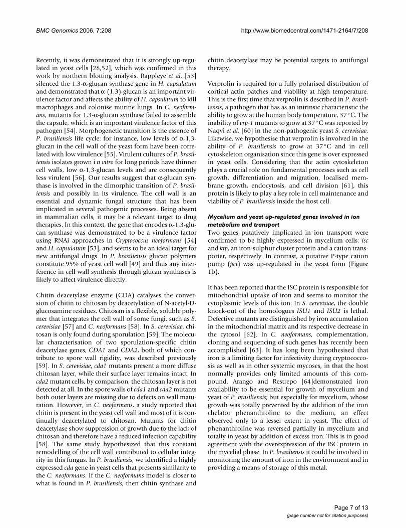
Sulphur metabolismSeveral enzymes from the cysteine de novo biosynthesispathway (Figure 2) were shown to be up-regulated in the
yeast form of P. brasiliensis. Our previous analysis [28] hadalready confirmed over expression of paps reductase (thethird in the pathway). In silico EST subtraction and cDNAmicroarray showed yeast up-regulation for atp sulphury-lase and aps kinase; the former was confirmed by north-ern blotting (Figure 1c). Thus, we can strongly suggest thatthe yeast form synthesises cysteine actively from inorganicsulphate.
In order to reinforce these data, we have evaluated theimportance of inorganic sulphate to growth and differen-tiation. Auxotrophy of P. brasiliensis yeast for severalsources of organic sulphate – including cysteine itself andsulphydrylic compounds – has been reported before [42].It was concluded then that organic sulphate deprivationsuppressed growth in the yeast phase and prevented myc-elium-to-yeast differentiation, whereas the mycelial phaseis able to grow on either inorganic or organic sulphur[68]. Also, the saprophytic, mycelial form of H. capsulatumis prototrophic while the pathogenic yeast form requirescysteine [69]. It has been reported that exogenous cysteineis required for both yeast phase growth and morphologi-cal transition from mycelium-to-yeast of H. capsulatum[41,70]. In this work, both mycelium and yeast cells of P.brasiliensis were incubated in modified MVM mediumwithout inorganic sulphate, apart from the negligibleamounts present in the trace elements solution. Dimor-
Up-regulated genes encoding enzymes from the cysteine de novo biosynthesis pathwayFigure 2Up-regulated genes encoding enzymes from the cysteine de novo biosynthesis pathway. Arrows indicate enzymes identified as up-regulated both by in silico subtraction, cDNA microarray and confirmed by northern blotting experiments. (*) enzyme identified as up-regulated by both in silico subtraction and cDNAs microarray but not assayed by northern blotting. (**) indicates an enzyme not found in the transcriptome of P. brasiliensis.
Page 8 of 13(page number not for citation purposes)
BMC Genomics 2006, 7:208 http://www.biomedcentral.com/1471-2164/7/208
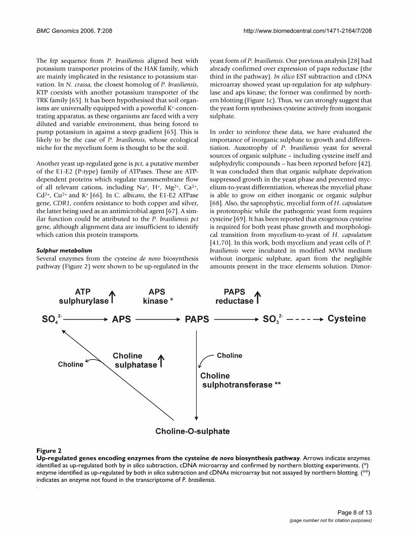
phic transition was assessed in the mycelium to yeastdirection and in the opposite way.Sustained growth wasobserved for both morphotypes (data not shown) and,upon the corresponding temperature shifts, differentia-tion was successfully triggered in both directions (Figure3). Thus, inorganic sulphate seems to be unnecessary forthe transition, quite contrarily to organic sulphate. In thiscontext, it is interesting to consider a branch of thecysteine biosynthetic pathway (Fig. 2). In fungi and plantsa fraction of PAPS, which is toxic to fungi if it reaches highcytosolic levels, is used by choline sulphotransferase toproduce choline-O-sulphate [40], which serves as anosmoprotectant and cytosolic sulphur store in theseorganisms. We have not found a homologue of cholinesulphotranferase in P. brasiliensis to date, but the enzymecholine sulphatase, which degrades its product to cholineand sulphate, is also over expressed in the yeast morpho-type, as confirmed here (Figure 1c) and previouslyreported [52]. The C. neoformans met3 mutant, which lacksATP sulphurylase activity, had a substantial defect in mel-anin formation, significantly reducedgrowth rate, andgreatly increased thermotolerance. In the murine inhala-tion infection model, the met3 mutant was avirulent andwas deficient in its ability to survive in mice [71]. In thiscontext, disrupting the genes encoding choline sulphataseor ATP sulphurylase in P. brasiliensis should reveal its role
in the growth, maintenance of yeast cells and pathogenic-ity of this fungus. It is interesting that another intracellularpathogen of humans, the bacterium Mycobacterium tuber-culosis, depends on sulphur compounds for expression ofits full virulence, drug resistance and overall survivalinside the macrophage. It has developed a very efficientsulphate activation pathway (SAC) that ensures constantsynthesis of PAPS at high rates, from which sulphate maybe distributed to other synthetic pathways [72]. The SACincludes the bacterial counterparts of ATP sulphurylaseand APS kinase, the latter of which performs PAPS synthe-sis by coupling it with GTP hydrolysis by a GTPase that isalso present in SAC. Whether similar mechanisms arepresent in pathogenic fungi such as P. brasiliensis remainsto be investigated.
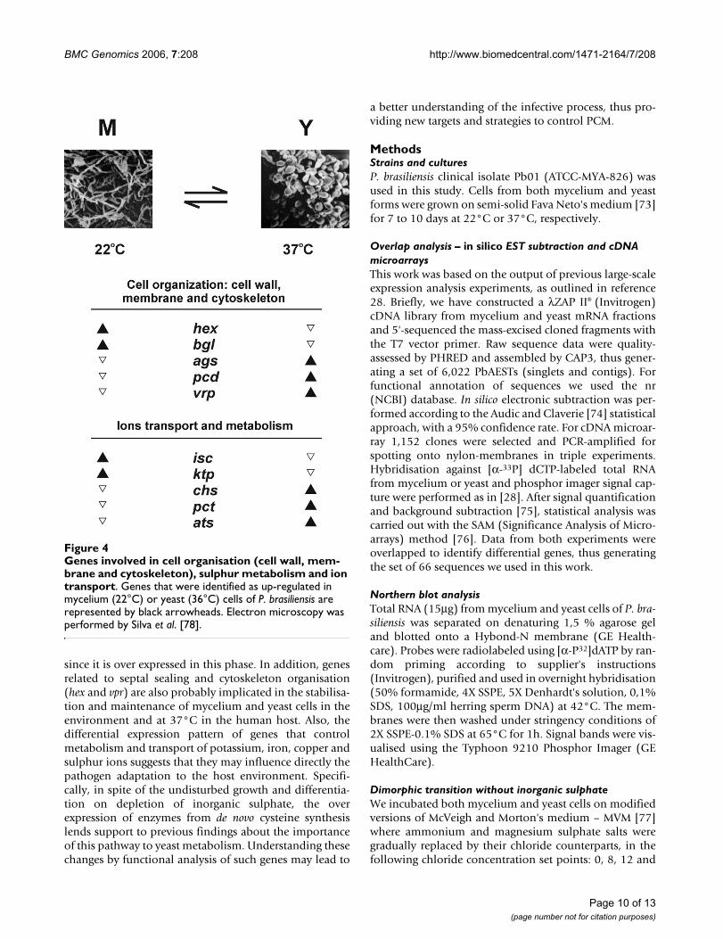
ConclusionTaken together, these data show that several genesinvolved in cell organisation and ion metabolism/trans-port are differential in their expression along dimorphictransition, which is in accordance with the proposedmodel for this process in Figure 4. While α-glucan is syn-thesised during yeast phase due to high expression of 1,3α-glucan synthase, β-glucan is degraded by the action of1,3 β-glucosidase during hyphal growth. The cda gene isprobably involved in the cell wall synthesis of yeast cells,
Cell differentiation of P. brasiliensis in modified MVM medium without inorganic sulphateFigure 3Cell differentiation of P. brasiliensis in modified MVM medium without inorganic sulphate. The fungus was grown in four different concentrations of sulphate salts (0, 8, 12 and 17 mM; the latter is the original concentration of MVM medium). (A) The appearance of yeast cells was verified daily in the transition from mycelium to yeast after temperature shift to 37°C, (B) The disappearance of yeast cells was verified daily in the transition from yeast to mycelium after temperature shift to 22°C. Triple samples were counted for each time point. The coloured boxes indicate the average of the three samples and bars rep-resent the standard deviation of the mean. As observed, the presence or absence of inorganic sulphate did not affect transition in either direction.
Page 9 of 13(page number not for citation purposes)
BMC Genomics 2006, 7:208 http://www.biomedcentral.com/1471-2164/7/208
since it is over expressed in this phase. In addition, genesrelated to septal sealing and cytoskeleton organisation(hex and vpr) are also probably implicated in the stabilisa-tion and maintenance of mycelium and yeast cells in theenvironment and at 37°C in the human host. Also, thedifferential expression pattern of genes that controlmetabolism and transport of potassium, iron, copper andsulphur ions suggests that they may influence directly thepathogen adaptation to the host environment. Specifi-cally, in spite of the undisturbed growth and differentia-tion on depletion of inorganic sulphate, the overexpression of enzymes from de novo cysteine synthesislends support to previous findings about the importanceof this pathway to yeast metabolism. Understanding thesechanges by functional analysis of such genes may lead to
a better understanding of the infective process, thus pro-viding new targets and strategies to control PCM.
MethodsStrains and culturesP. brasiliensis clinical isolate Pb01 (ATCC-MYA-826) wasused in this study. Cells from both mycelium and yeastforms were grown on semi-solid Fava Neto's medium [73]for 7 to 10 days at 22°C or 37°C, respectively.
Overlap analysis – in silico EST subtraction and cDNA microarraysThis work was based on the output of previous large-scaleexpression analysis experiments, as outlined in reference28. Briefly, we have constructed a λZAP II® (Invitrogen)cDNA library from mycelium and yeast mRNA fractionsand 5'-sequenced the mass-excised cloned fragments withthe T7 vector primer. Raw sequence data were quality-assessed by PHRED and assembled by CAP3, thus gener-ating a set of 6,022 PbAESTs (singlets and contigs). Forfunctional annotation of sequences we used the nr(NCBI) database. In silico electronic subtraction was per-formed according to the Audic and Claverie [74] statisticalapproach, with a 95% confidence rate. For cDNA microar-ray 1,152 clones were selected and PCR-amplified forspotting onto nylon-membranes in triple experiments.Hybridisation against [α-33P] dCTP-labeled total RNAfrom mycelium or yeast and phosphor imager signal cap-ture were performed as in [28]. After signal quantificationand background subtraction [75], statistical analysis wascarried out with the SAM (Significance Analysis of Micro-arrays) method [76]. Data from both experiments wereoverlapped to identify differential genes, thus generatingthe set of 66 sequences we used in this work.
Northern blot analysisTotal RNA (15μg) from mycelium and yeast cells of P. bra-siliensis was separated on denaturing 1,5 % agarose geland blotted onto a Hybond-N membrane (GE Health-care). Probes were radiolabeled using [α-P32]dATP by ran-dom priming according to supplier's instructions(Invitrogen), purified and used in overnight hybridisation(50% formamide, 4X SSPE, 5X Denhardt's solution, 0,1%SDS, 100μg/ml herring sperm DNA) at 42°C. The mem-branes were then washed under stringency conditions of2X SSPE-0.1% SDS at 65°C for 1h. Signal bands were vis-ualised using the Typhoon 9210 Phosphor Imager (GEHealthCare).
Dimorphic transition without inorganic sulphateWe incubated both mycelium and yeast cells on modifiedversions of McVeigh and Morton's medium – MVM [77]where ammonium and magnesium sulphate salts weregradually replaced by their chloride counterparts, in thefollowing chloride concentration set points: 0, 8, 12 and
Genes involved in cell organisation (cell wall, membrane and cytoskeleton), sulphur metabolism and ion transportFigure 4Genes involved in cell organisation (cell wall, mem-brane and cytoskeleton), sulphur metabolism and ion transport. Genes that were identified as up-regulated in mycelium (22°C) or yeast (36°C) cells of P. brasiliensis are represented by black arrowheads. Electron microscopy was performed by Silva et al. [78].
Page 10 of 13(page number not for citation purposes)
BMC Genomics 2006, 7:208 http://www.biomedcentral.com/1471-2164/7/208
17 mM, where the first corresponds to the original recipeand the last, to virtual absence of inorganic sulphate, apartfrom negligible amounts in the trace elements solution (~8 μM). Molar concentrations of both magnesium andammonium were thus conserved. We have also evaluatedwhether dimorphic transition occurred normally in themedium without inorganic sulphur. To achieve this, fiveflasks containing 100 ml of modified MVM were inocu-lated with comparable amounts of mycelium (100 mg wetmass) and yeast (2.5 × 10⁷ cells) previously grown onstandard MVM. Samples were incubated in rotating shak-ers (120 rpm) at 36 and 22°C, respectively, thus triggeringdimorphic transition. Fungal viability and progress oftransition were assessed by serial 100 μl sampling every 24hours (three independent samples). Each sample was col-oured with Janus Green and the number of yeast cells wascounted in a light microscope with the aid of a Neubauercounting chamber.
Accession numbersThe accession numbers of the EST sequences analysed inthis work are shown in the Tables 1 and 2.
Abbreviationsags alpha 1,3-glucan synthase
aps adenosine 5'-phosphosulphate
ats ATP sulphurylase
bgl 1,3 beta-glucosidase,
BLAST basic local alignment search tool
cda chitin deacetylase
cDNA complementary DNA
chs choline sulphatase
COG clusters of orthologous groups
e-value extreme value distributionESTs
ESTs expressed sequence tags
GO gene ontology
hex hexagonal peroxisome protein
isc iron-sulphur cluster-like protein
ktp potassium transporter
MIPS Munich information center for proteins sequences
PAPS phosphoadenylyl-sulfate reductase
PbAETs P. brasiliensis assembled EST sequences
PCM paracoccidioidomycosis
pct putative P-type Cu(2+) transporting ATPase
SAGE serial analysis of gene expression
SAM significance analysis of microarrays
vrp verprolin
Authors' contributionsRA and MF planned and designed the study, developedthe experiments and the data analysis, wrote the maindraft of the paper and support the preparation of the fig-ures and tables. HP supports the discussion of the resultsand revised the manuscript. AN participated in the in silicoESTs subtraction analysis of the raw data generated by thetranscriptome project. MC analysed the results of themicroarray experiments, helped in the manuscript edi-tion, and prepared the figures. AL executed the microarrayexperiments. MC, RC and MB participated in the normal-ization process of the microarray raw data and helped tomake the statistical analyses. SS participated of the differ-entiation experiment involved of the inorganic sulphur,and of the preparation of the RNA of P. brasiliensis to makethe microarray experiments. LF participated of the analy-sis of the cell wall organization. SP helped in the ESTsamplification and on the analysis of the sulphur metabo-lism. GP, ES, ED designed the microarray experiments. CSparticipated on the Pb ESTs annotation. All authors readand approved the final manuscript.
AcknowledgementsWe are most indebted to Hugo Costa Paes for text draft revision, Maria Emilia M. T. Walter and Shana Santos for technical bioinformatics support. This work was supported by MCT/CNPq, CNPq, CAPES, FAP-DF, Fapesp, FUB, UFG.
References1. Churchill GA: Fundamentals of experimental design for cDNA
microarrays. Nat Genet 2002, 32(Suppl):490-495.2. Lashkari DA, DeRisi JL, McCusker JH, Namath AF, Gentile C, Hwang
SY, Brown PO, Davis RW: Yeast microarrays for genome wideparallel genetic and gene expression analysis. Proc Natl Acad SciUSA 1997, 94:13057-13062.
3. Pollack JR, Perou CM, Alizadeh AA, Eisen MB, Pergamenschikov A,Williams CF, Jeffrey SS, Botstein D, Brown PO: Genome-wideanalysis of DNA copy-number changes using cDNA microar-rays. Nat Genet 1999, 23:41-46.
4. Paillisson A, Dade S, Callebaut I, Bontoux M, Dalbies-Tran R, VaimanD, Monget P: Identification, characterization and metagen-ome analysis of oocyte-specific genes organized in clusters inthe mouse genome. BMC Genomics 2005, 6:76.
5. Rajkovic A, Yan C, Klysik M, Matzuk MM: Discovery of germ cell-specific transcripts by expressed sequence tag database anal-ysis. Fertil Steril 2001, 76:550-554.
Page 11 of 13(page number not for citation purposes)
http://www.ncbi.nlm.nih.gov/entrez/query.fcgi?cmd=Retrieve&db=PubMed&dopt=Abstract&list_uids=9371799
BMC Genomics 2006, 7:208 http://www.biomedcentral.com/1471-2164/7/208
6. Steen BR, Lian T, Zuyderduyn S, MacDonald WK, Marra M, Jones SJ,Kronstad JW: Temperature-regulated transcription in thepathogenic fungus Cryptococcus neoformans. Genome Res 2002,12:1386-1400.
7. Steen BR, Zuyderduyn S, Toffaletti DL, Marra M, Jones SJM, PerfectJR, Kronstad J: Cryptococcus neoformans gene expression dur-ing experimental cryptococcal meningitis. Eukaryot Cell 2003,2:1336-1349.
8. Backer MD, Ilyina T, Ma X, Vandoninck S, Luyten WHM, Bossche HV:Genomic profiling of the response of Candida albicans to Itra-conazole treatment using a DNA microarray. AntimicrobAgents Chemother 2001, 45:1660-1670.
9. Enjalbert B, Nantel A, Whiteway M: Stress-induced gene expres-sion in Candida albicans : Absence of a general stressresponse. Mol Biol Cell 2003, 14:1460-1467.
10. Fradin C, Kretschmar M, Nichterlein T, Gaillardin C, d'Enfert C, HubeB: Stage-specific gene expression of Candida albicans inhuman blood. Mol Microbiol 2003, 47:1523-1543.
11. Lane S, Birse C, Zhou S, Matson R, Liu H: DNA array studies dem-onstrate convergent regulation of virulence factors by Cph1,Cph2, and Efg1 in Candida albicans. J Biol Chem 2001,276:48988-48996.
12. Lorenz MC, Bender JA, Fink GR: Transcriptional response ofCandida albicans upon internalization by macrophages.Eukaryot Cell 2004, 3:1076-1087.
13. Nantel A, Dignard D, Bachevich C, Harcus D, Marcil A, Bouin AP,Sensen CW, Hogues H, Hoog MVH, Gordon P, Rigby T, Benoit F,Tessier DC, Thomas DY, Whiteway M: Transcription profiling, ofCandida albicans cells undergoing the yeast-to-hyphal transi-tion. Mol Biol Cell 2002, 13:3452-3465.
14. Hwang H, Hocking-Murray D, Bahrami AK, Andersson M, Rine J, SilA: Identifying phase-specific genes in the fungal pathogenHistoplasma capsulatum using a genomic shotgun microar-ray. Mol Biol Cell 2003, 14:2314-2326.
15. Kraus PR, Boily MJ, Giles SS, Stajich JE, Allen A, Cox GM, Dietrich FS,Perfect JR, Heitman J: Identification of Cryptococcus neoformansTemperature-Regulated Genes with a Genomic-DNA. Micro-array Eukaryot Cell 2004, 3:1249-1260.
16. Manns BJ, Baylis BW, Urbanski SJ, Gibb AP, Rabi HR: Paracoccidio-idomycosis: case report and review. Clin Infect Dis 1996,23:1026-1032.
17. Restrepo M: The ecology of Paracoccidioides brasiliensis : a puz-zle still unsolved. Sabouradia 1985, 23:323-334.
18. Blotta MH, Mamoni RL, Oliveira SJ, Nouer SA, Papaiordanou PM,Goveia A, Camargo ZP: Endemic regions of paracoccidioidomy-cosis in Brazil: a clinical and epidemiologic study of 584 casesin the southeast region. Am J Trop Med Hyg 1999, 61:390-394.
19. Brummer E, Castañeda E, Restrepo A: Paracoccidioidomycosis:an update. Clin Microbiol 1993, 6:89-117.
20. Restrepo A, McEwen JG, Castañeda E: The habitat of Paracoccid-ioides brasiliensis : how far from solving the riddle? Med Mycol2001, 39:233-241.
21. McEwen JG, Garcia AM, Ortiz BL, Botero S, Restrepo A: In searchof the natural habitat of Paracoccidioides brasiliensis. Arch MedRes 1995, 26:305-306.
22. Bagagli E, Sano A, Coelho KI, Alquati S, Miyaji M, de Camargo ZP,Gomes GM, Franco M, Montenegro MR: Isolation of Paracoccidi-oides brasiliensis from armadillos (Dasypus noveminctus) cap-tured in an endemic area of paracoccidioidomycosis. Am JTrop Med Hyg 1998, 58:505-512.
23. Tristano AG, Chollet ME, Willson M, Perez J, Troccoli M: Centralnervous system paracoccidioidomycosis: case report andreview. Invest Clin 2004, 45:277-288.
24. De Camargo ZP, Franco MF: Current knowledge on pathogene-sis and immunodiagnosis of paracoccidioidomycosis. RevIberoam Micol 2000, 17:41-48.
25. Restrepo A, Salazar ME, Cano LE, Stover EP, Feldman D, Stevens DA:Estrogens inhibit mycelium-to-yeast transformation in thefungus Paracoccidioides brasiliensis : implications for resist-ance of females to paracoccidioidomycosis. Infect Immun 1984,46:346-353.
26. Restrepo A, McEwen JG, Castaneda E: The habitat of Paracoccid-ioides brasiliensis : how far from solving the riddle? Med Mycol2001, 39:233-241.
27. Marques SA, Robles AM, Tortorano AM, Tuculet MA, Negroni R,Mendes RP: Mycoses associated with AIDS in the ThirdWorld. Med Mycol 2000, 38(Suppl 1):269-279.
28. Felipe MSS, Andrade RV, Arraes FBM, Nicola AM, Maranhão AQ,Torres FAG, Silva-Pereira I, Poças-Fonseca MJ, Campos EG, MoraesLMP, Andrade PA, Tavares AHFP, Silva SS, Kyaw CM, Souza DP, PbG-enome Network, Pereira M, Jesuíno RSA, Andrade EV, Parente JA,Oliveira GS, Barbosa MS, Martins NF, Fachin AL, Cardoso RS, PassosGAS, Almeida NF, Walter MEMT, Soares CMA, Carvalho MJA, Brig-ido MM: Transcriptional profiles of the human pathogenicfungus Paracoccidioides brasiliensis in mycelium and yeastcells. J Biol Chem 2005, 280:24706-24714.
29. Felipe MSS, Andrade RV, Petrofeza SS, Maranhão AQ, Torres FAG,Albuquerque P, Arraes FB, Arruda M, Azevedo MO, Baptista AJ,Bataus LAM, Borges CL, Campos EG, Cruz MR, Daher BS, Dantas A,Ferreira MA, Ghil GV, Jesuíno RS, Kyaw CM, Leitão L, Martins CR,Moraes LM, Neves EO, Nicola AM, Alves ES, Parente JA, Pereira M,Poças-Fonseca MJ, Resende R, Ribeiro BM, Saldanha RR, Santos SC,Silva-Pereira I, Silva MA, Silveira E, Simões IC, Soares RB, Souza DP,De-Souza MT, Andrade EV, Xavier MA, Veiga HP, Venâncio EJ, Car-valho MJ, Oliveira AG, Inoue MK, Almeida NF, Walter ME, SoaresCMA, Brigido MM: Transcriptome characterization of thedimorphic and pathogenic fungus Paracoccidioides brasiliensisby EST analysis. Yeast 2003, 20:263-271.
30. Goldman GH, dos Reis Marques E, Duarte Ribeiro DC, de Souza Ber-nardes LA, Quiapin AC, Vitorelli PM, Savoldi M, Semighini CP, deOliveira RC, Nunes LR, Travassos LR, Puccia R, Batista WL, FerreiraLE, Moreira JC, Bogossian AP, Tekaia F, Nóbrega MP, Nóbrega FG,Goldman MH: Expressed sequence tag analysis of the humanpathogen Paracoccidioides brasiliensis yeast phase: identifica-tion of putative homologues of Candida albicans virulenceand pathogenicity genes. Eukaryot Cell 2003, 2:34-48.
31. Yuen K, Pascal G, Wong SSY, Glaser P, Woo PCY, Kunst F, Cai JJ,Cheung EYL, Médigue C, Danchin A: Exploring the Penicilliummarneffei genome. Arch Microbiol 2003, 179:339-353.
32. Bailek R, Ibricevic A, Fothergill A, Begerow D: Small subunit ribos-omal DNA sequence shows Paracoccidioides brasiliensisclosely related to Blastomyces dermatitidis. J Clin Microbiol 2000,38:3190-3193.
33. San-Blas G, Niño-Vega G, Iturriaga T: Paracoccidioides brasiliensisand paracoccidioidomycosis: molecular approaches to mor-phogenesis, diagnosis, epidemiology, taxonomy and genet-ics. Med Mycol 2002, 40:225-242.
34. Matute DR, McEwen JG, Puccia R, Montes BA, San-Blas G, Bagagli E,Rauscher JT, Restrepo A, Morais F, Niño-Vega G, Taylor JW: CrypticSpeciation and Recombination in the Fungus Paracoccidio-ides brasiliensis as Revealed by Gene Genealogies. Mol Biol Evol2006, 23:65-73.
35. Feitosa LS, Cisalpino PS, dos Santos MR, Mortara RA, Barros TF,Morais FV, Puccia R, da Silveira JF, de Camargo ZP: Chromosomalpolymorphism, syntenic relationships and ploidy in the path-ogenic fungus Paracoccidioides brasiliensis. Fungal Genet 2003,39:60-69.
36. Montoya AE, Moreno MN, Restrepo A, McEwen JG: Electro-phoretic karyotype of clinical isolates of Paracoccidioides bra-siliensis. Fungal Genet 1997, 21:223-227.
37. Cano MIN, Cisalpino PS, Galindo I, Ramírez JL, Mortara RA, da Sil-veira JF: Electrophoretic karyotypes and genome sizing of thepathogenic fungus Paracoccidioides brasiliensis. J Clin Microbiol1998, 36:742-747.
38. Reinoso C, Nino-vega G, San-Blas G, Dominguez A: Randomsequencing of Paracoccidioides brasiliensis genes. Med Mycol2005, 43:681-689.
39. Munich Information Center for Proteins Sequences [http://mips.gsf.de/]
40. Marluf GA: Molecular genetics of sulfur assimilation in fila-mentous fungi and yeast. Annu Rev Microbiol 1997, 51:73-96.
41. Boguslawski G, Akagi JM, Ward LG: Possible role for cysteine bio-synthesis in conversion from mycelial to yeast form of Histo-plasma capsulatum. Nature 1976, 261:336-338.
42. Medoff G, Painter A, Kobayashi GS: Mycelial-to yeast-phase tran-sitions of the dimorphic fungi Blastomyces dermatitidis andParacoccidioides brasiliensis. J Bacteriol 1987, 169:4055-4060.
43. Munich Information Center for Protein Sequences [http://mips.gsf.de/]
Page 12 of 13(page number not for citation purposes)
http://www.ncbi.nlm.nih.gov/entrez/query.fcgi?cmd=Retrieve&db=PubMed&dopt=Abstract&list_uids=8922797
http://www.ncbi.nlm.nih.gov/entrez/query.fcgi?cmd=Retrieve&db=PubMed&dopt=Abstract&list_uids=8922797
http://www.ncbi.nlm.nih.gov/entrez/query.fcgi?cmd=Retrieve&db=PubMed&dopt=Abstract&list_uids=8580685
http://www.ncbi.nlm.nih.gov/entrez/query.fcgi?cmd=Retrieve&db=PubMed&dopt=Abstract&list_uids=9574800
http://www.ncbi.nlm.nih.gov/entrez/query.fcgi?cmd=Retrieve&db=PubMed&dopt=Abstract&list_uids=9574800
http://www.ncbi.nlm.nih.gov/entrez/query.fcgi?cmd=Retrieve&db=PubMed&dopt=Abstract&list_uids=6500694
http://www.ncbi.nlm.nih.gov/entrez/query.fcgi?cmd=Retrieve&db=PubMed&dopt=Abstract&list_uids=6500694
http://www.ncbi.nlm.nih.gov/entrez/query.fcgi?cmd=Retrieve&db=PubMed&dopt=Abstract&list_uids=9508305
http://www.ncbi.nlm.nih.gov/entrez/query.fcgi?cmd=Retrieve&db=PubMed&dopt=Abstract&list_uids=9343344
http://www.ncbi.nlm.nih.gov/entrez/query.fcgi?cmd=Retrieve&db=PubMed&dopt=Abstract&list_uids=9343344
http://www.ncbi.nlm.nih.gov/entrez/query.fcgi?cmd=Retrieve&db=PubMed&dopt=Abstract&list_uids=1272413
BMC Genomics 2006, 7:208 http://www.biomedcentral.com/1471-2164/7/208
Publish with BioMed Central and every scientist can read your work free of charge
"BioMed Central will be the most significant development for disseminating the results of biomedical research in our lifetime."
Sir Paul Nurse, Cancer Research UK
Your research papers will be:
available free of charge to the entire biomedical community
peer reviewed and published immediately upon acceptance
cited in PubMed and archived on PubMed Central
yours — you keep the copyright
Submit your manuscript here:http://www.biomedcentral.com/info/publishing_adv.asp
BioMedcentral
44. Soundararajan S, Jedd G, Li X, Ramos-Pamplona M, Chua NH, NaqviaNI: Woronin body function in Magnaporthe grisea is essentialfor efficient pathogenesis and for survival during nitrogenstarvation stress. Plant Cell 2004, 16:1564-1574.
45. Jedd G, Chua NH: A new self-assembled peroxisomal vesiclerequired for efficient resealing of the plasma membrane. NatCell Biol 2000, 2:226-231.
46. Markham P, Collinge AJ: Woronin bodies in filamentous fungi.FEMS Microbiol Rev 1987, 46:1-11.
47. Trinci AP, Collinge AJ: Occlusion of the septal pores of dam-aged hyphae of Neurospora crassa by hexagonal crystals. Pro-toplasma 1974, 80:57-67.
48. Kanetsuna F, Carbonell LM: Cell wall Glucans of the yeast andmycelial forms of Paracoccidioides brasiliensis. J Bacteriol 1970,101:675-680.
49. Kanetsuna F, Carbonell LM, Azuma I, Yamamura Y: Biochemicalstudies on the thermal dimorphism of Paracoccidioides brasil-iensis. J Bacteriol 1972, 110:208-218.
50. San Blas G: Paracoccidioides brasiliensis: cell wall glucans, path-ogenicity, and dimorphism. Curr Top Med Mycol 1985, 1:235-257.
51. Pereira M, Felipe MSS, Brígido MM, Soares CMA, Azevedo MO:Molecular cloning and characterization of a glucan synthasegene from the human pathogenic fungus Paracoccidioidesbrasiliensis. Yeast 2000, 16:451-462.
52. Marques ER, Ferreira MES, Drummond RD, Felix JM, Menossi M,Savoldi M, Travassos LR, Puccia R, Batista W, Carvalho KC, GoldmanMHS, Goldman GH: Identification of genes preferentiallyexpressed in the pathogenic yeast phase of Paracoccidioidesbrasiliensis, using suppression subtraction hybridization anddifferential macroarray analysis. Mol Genet Genomics 2004,271:667-677.
53. Rappleye CA, Engle JT, Goldman WE: RNA interference in Histo-plasma capsulatum demonstrates a role for α-(1,3)-glucan invirulence. Mol Microbiol 2004, 53:153-165.
54. Reese AJ, Doering TL: Cell wall alpha-1,3-glucan is required toanchor the Cryptococcus neoformans capsule. Mol Microbiol2003, 50:1401-1409.
55. Hallak J, San-Blas F, San-Blas B: Isolation and wall analysis ofdimorphic mutants of Paracoccidioides brasiliensis. Sabouradia1982, 20:51-62.
56. San Blas G, San Blas F: Paracoccidioides brasiliensis : cell wallstructure and virulence. Mycopathologia 1977, 62:77-86.
57. Zakrzewska A, Boorsma A, Brul S, Hellingwerf KJ, Klis FM: Tran-scriptional response of Saccharomyces cerevisiae to theplasma membrane-perturbing compound chitosan. EukaryotCell 2005, 4:703-715.
58. Banks IR, Specht CA, Donlin MJ, Gerik KJ, Levitz SM, Lodge JK: A chi-tin synthase and its regulator protein are critical for chitosanproduction and growth of the fungal pathogen Cryptococcusneoformans. Eukaryot Cell 2005, 4:1902-1912.
59. Christodoulidou A, Bouriotis V, Thireos G: Two sporulation-spe-cific chitin deacetylase-encoding genes are required for theascospore wall rigidity of Saccharomyces cerevisiae. J Biol Chem1996, 271:31420-31425.
60. Naqvi SN, Feng Q, Boulton VJ, Zahn R, Munn AL: Vrp1p functionsin both actomyosin ring-dependent and Hof1p-dependentpathways of cytokinesis. Traffic 2001, 2:189-201.
61. Drubin DG, Nelson WJ: Origins of cell polarity. Cell 1996,84:335-344.
62. Dutkiewicz R, Schilke B, Cheng S, Knieszner H, Craig EA, MarszalekJ: Sequence-specific interaction between mitochondrial Fe-Sscaffold protein Isu and Hsp70 Ssq1 is essential for their invivo function. J Biol Chem 2004, 279:29167-29174.
63. Jacobson ES, Troy AJ, Nyhus KJ: Mitochondrial functioningofconstitutive iron uptake mutations in Cryptococcus neoform-ans. Mycopathologia 2005, 159:1-6.
64. Arango R, Restrepo A: Growth and production of iron chelantsby Paracoccidioides brasiliensis mycelial and yeast forms. JMed Vet Mycol 1988, 26:113-118.
65. Haro R, Sainz L, Rubio F, Rodríguez-Navarro A: Cloning of twogenes encoding potassium transporters in Neurospora crassaand expression of the corresponding cDNAs in Saccharomy-ces cerevisiae. Mol Microbiol 1999, 31:511-520.
66. Smith DL, Tao T, Maguire ME: Membrane topology of a P-typeATPase. J Biol Chem 1993, 268:22469-22479.
67. Riggle PJ, Kumamoto CA: Role of a Candida albicans P1-TypeATPase in resistance to copper and silver ion toxicity. J Bac-teriol 2000, 182:4899-4905.
68. Paris S, Durán-González S, Mariat F: Nutritional studies on Para-coccidioides brasiliensis : the role of organic sulfur in dimor-phism. Sabouraudia 1985, 23:85-92.
69. Boguslawski G, Stetler DA: Aspects of physiology of Histoplasmacapsulatum. (A review). Mycopathologia 1979, 67:17-24.
70. Maresca B, Lambowitz AM, Kumar VB, Grant GA, Kobayashi GS,Medoff G: Role of cysteine in regulating morphogenesis andmitochondrial activity in the dimorphic fungus Histoplasmacapsulatum. Proc Natl Acad Sci USA 1981, 78:4596-600.
71. Yang Z, Pascon RC, Alspaugh A, Cox GM, McCusker JH: Molecularand genetic analysis of the Cryptococcus neoformans MET3gene and a met3 mutant. Microbiology 2002, 148:2617-2625.
72. Sun M, Andreassi II JL, Liu S, Pinto R, Triccas JA, Leyh TS: The Tri-functional Sulfate-activating Complex (SAC) of Mycobacte-rium tuberculosis. J Biol Chem 2005, 280:7861-7866.
73. Fava-Neto C: Estudos quantitativos sobre a fixação do com-plemento na Blastomicose Sul-Americana com antígenospolissacarídicos. Arq Cirurg Clin Exp 1955, 18:97-254.
74. Audic S, Claverie JM: The significance of digital gene expressionprofiles. Genome Res 1997, 7:986-995.
75. Quackenbush J: Microarray data normalization and transfor-mation. Nat Genet 2002, 32(suppl):496-501.
76. Tusher VG, Tisbshirani R, Chu G: Significance analysis of micro-arrays applied to the ionizing radiation response. Proc NatlAcad Sci USA 2001, 98:5116-5121.
77. Restrepo A, Jiménez BE: Growth of Paracoccidioides brasiliensisyeast phase in a chemically defined culture medium. J ClinMicrobiol 1980, 12:279-281.
78. Silva SP, Borges-Walmsley MI, Pereira IS, Soares CM, Walmsley AR,Felipe MSS: Differential expression of an hsp70 gene duringtransition from the mycelial to the infective yeast form ofthe human pathogenic fungus Paracoccidioides brasiliensis.Mol Microbiol 1999, 31:1039-1050.
Page 13 of 13(page number not for citation purposes)
http://www.ncbi.nlm.nih.gov/entrez/query.fcgi?cmd=Retrieve&db=PubMed&dopt=Abstract&list_uids=4275239
http://www.ncbi.nlm.nih.gov/entrez/query.fcgi?cmd=Retrieve&db=PubMed&dopt=Abstract&list_uids=5442818
http://www.ncbi.nlm.nih.gov/entrez/query.fcgi?cmd=Retrieve&db=PubMed&dopt=Abstract&list_uids=5018021
http://www.ncbi.nlm.nih.gov/entrez/query.fcgi?cmd=Retrieve&db=PubMed&dopt=Abstract&list_uids=3916768
http://www.ncbi.nlm.nih.gov/entrez/query.fcgi?cmd=Retrieve&db=PubMed&dopt=Abstract&list_uids=3916768
http://www.ncbi.nlm.nih.gov/entrez/query.fcgi?cmd=Retrieve&db=PubMed&dopt=Abstract&list_uids=8940152
http://www.ncbi.nlm.nih.gov/entrez/query.fcgi?cmd=Retrieve&db=PubMed&dopt=Abstract&list_uids=8608587
http://www.ncbi.nlm.nih.gov/entrez/query.fcgi?cmd=Retrieve&db=PubMed&dopt=Abstract&list_uids=3418467
http://www.ncbi.nlm.nih.gov/entrez/query.fcgi?cmd=Retrieve&db=PubMed&dopt=Abstract&list_uids=8226755
http://www.ncbi.nlm.nih.gov/entrez/query.fcgi?cmd=Retrieve&db=PubMed&dopt=Abstract&list_uids=8226755
http://www.ncbi.nlm.nih.gov/entrez/query.fcgi?cmd=Retrieve&db=PubMed&dopt=Abstract&list_uids=4012515
http://www.ncbi.nlm.nih.gov/entrez/query.fcgi?cmd=Retrieve&db=PubMed&dopt=Abstract&list_uids=4012515
http://www.ncbi.nlm.nih.gov/entrez/query.fcgi?cmd=Retrieve&db=PubMed&dopt=Abstract&list_uids=6945601
http://www.ncbi.nlm.nih.gov/entrez/query.fcgi?cmd=Retrieve&db=PubMed&dopt=Abstract&list_uids=9331369
http://www.ncbi.nlm.nih.gov/entrez/query.fcgi?cmd=Retrieve&db=PubMed&dopt=Abstract&list_uids=9331369
http://www.ncbi.nlm.nih.gov/entrez/query.fcgi?cmd=Retrieve&db=PubMed&dopt=Abstract&list_uids=7229010