Ditosylate Salt of Itraconazole and Dissolution Enhancement Using Cyclodextrins

Neto et al. BMC Genomics 2014, 15:254http://www.biomedcentral.com/1471-2164/15/254
RESEARCH ARTICLE Open Access
Transcriptional profile of Paracoccidioides spp. inresponse to itraconazoleBenedito Rodrigues da Silva Neto1, Patrícia Fernanda Zambuzzi Carvalho1, Alexandre Melo Bailão1,Wellington Santos Martins2, Célia Maria de Almeida Soares1 and Maristela Pereira1*
Abstract
Background: Itraconazole is currently used to treat paracoccidioidomycosis. The mechanism of action of azoles hasbeen elucidated in some fungi, although little is known regarding its mechanism of action in Paracoccidioides spp.The present work focused on identification of regulated transcripts using representational difference analysis ofParacoccidioides spp. yeast cells treated with itraconazole for 1 and 2 h.
Results: Paracoccidioides Pb01 genes up-regulated by itraconazole included genes involved in cellular transport,metabolism/energy, transcription, cell rescue, defense and virulence. ERG11, ERG6, ERG3, ERG5 and ERG25 wereup-regulated at multiple time points. In vivo infection experiments in mice corroborated the in vitro results. Ergosterollevels and distribution were evaluated in Paracoccidioides Pb18 yeast cells, and the results demonstrate that both factorswere changed in the fungus treated with itraconazole.
Conclusion: To our knowledge, this is the first transcriptional analysis of Paracoccidioides spp. exposed to a triazoledrug. Here acetyl seems to be intensively produced from different metabolic pathways to produce ergosterol by theaction of ergosterol synthesis related enzymes, which were also affected in other fungi. Among the genes affected, weidentified genes in common with other fungi, as well as genes unique to Paracoccidioides Pb01. Those genes could beconsidered target to new drugs. Voltage-gated Ca2+ alpha subunit (CAV), Tetracycline resistance protein (TETA) andHemolisyn-iii channel protein (HLYiii) were found only here and a probably involvement with resistence to itraconazolecould be investigated in the future. However our findings do not permit inference to current clinical practice.
Keywords: Paracoccidioides spp., Transcriptional response, Itraconazole, Ergosterol
BackgroundParacoccidioides spp., a complex of several phylogeneticspecies, is the agent of paracoccidioidomycosis (PCM).Paracoccidioides spp. is a thermodimorphic fungus, whichgrows in the soil as saprobic mycelium, resulting in theformation of propagules, which initiate infection inhumans when inhaled into the respiratory tract. Subse-quently, in the lung, the mycelia propagules developinto yeast cells [1]. PCM is endemic in Latin America[2], with 80% of cases reported in Brazil, where it is theeighth-leading cause of mortality among infectious andparasitic diseases, establishing it as a serious publichealth problem [3,4].
* Correspondence: [email protected] de Bioquímica e Biologia Molecular, Laboratório de BiologiaMolecular, Instituto de Ciências Biológicas, ICBII, Campus II, UniversidadeFederal de Goiás, C.P. 131, 74001-970 Goiânia, GO, BrazilFull list of author information is available at the end of the article
© 2014 Neto et al.; licensee BioMed Central LtCommons Attribution License (http://creativecreproduction in any medium, provided the or
Itraconazole is suggested to be the best alternative forfirst-line therapy of PCM and should be administeredover a long period [5]. Itraconazole is a triazole antifun-gal drug, which are multi-ringed synthetic compoundscontaining three nitrogen atoms in the azole ring. Mech-anistically, the triazole drugs inhibit the synthesis of ergos-terol, an essential component of fungal cell membranes,and cause abnormalities in the membrane permeabilityand consequently cell death [6]. Itraconazole and relatedazole derivatives act by blocking the ergosterol biosyn-thesis pathway through the inhibition of the fungal cyto-chrome P450 enzyme lanosterol demethylase (Erg11) [7].The global response to azoles, including itraconazole,
of fungi such as Saccharomyces cerevisiae [8], Trichophy-ton rubrum [9], Aspergillus fumigatus [10] and Candidaalbicans [11,12] has been studied using transcriptionaland proteomic approaches. In general, the findings re-vealed both specific and nonspecific antifungal-induced
d. This is an Open Access article distributed under the terms of the Creativeommons.org/licenses/by/2.0), which permits unrestricted use, distribution, andiginal work is properly credited.

Neto et al. BMC Genomics 2014, 15:254 Page 2 of 15http://www.biomedcentral.com/1471-2164/15/254
changes in gene and protein regulation. There was anincrease in expression of the genes involved in lipid,fatty acid and sterol metabolism, as well as genes in-volved in drug adaptation, including cell stress response,drug efflux and small molecule transport.Despite of the importance of Paracoccidioides spp.,
nothing is known about the mechanism of itraconazoleinhibition in this pathogen. Here, cDNA libraries wereconstructed to obtain expressed sequence tags (ESTs) ofParacoccidioides spp. The representational difference ana-lysis (RDA) technique was used to identify changes in thetranscriptional profile of Paracoccidioides spp. in responseto itraconazole, with the aim of identifying the adaptativeresponse of the fungus to the compound. Transcript levelswere also measured during the infection process. Inaddition, the transcript levels of ERG genes, ergosterollevels and ergosterol localization were evaluated.
ResultsLibraries characteristicsA total of 861 ESTs were successfully sequenced. Fromthese, 224 up- and 208 down-regulated ESTs were obtainedfrom yeast cells after incubation with itraconazole for 1 h,containing 55 singlets and 26 contigs for up-regulatedtranscripts and three singlets and 20 contigs for down-regulated ones. In addition, 230 up- and 199 down-regulated ESTs were obtained from yeast cells afterincubation with itraconazole for 2 h, containing threesinglets and 10 contigs for up-regulated and seven singletsand 12 contigs for down-regulated. The ESTs obtainedwere submitted to the National Center for BiotechnologyInformation (NCBI) database under accession numbers:LIBEST_028165 Paracoccidioides Pb01 itraconazole 1 h upLibrary, LIBEST_028164 Paracoccidioides Pb01 itracona-zole 1 h down Library, LIBEST_028167 ParacoccidioidesPb01 itraconazole 2 h up Library and LIBEST_028166Paracoccidioides Pb01 itraconazole 2 h down Library.The ESTs were processed using the Blast2GO pro-
gram, which allowed us to annotate and identify the dif-ferent functional groups. The functional classificationwas based on the homology of each EST, consideringe-values ≤10−5 significant, using BLASTx against theGenBank non-redundant database and the MIPS func-tional annotation scheme (Table 1). The analyses indicatedthe presence of transcripts from different functionalcategories: metabolism/energy, transcription, cell res-cue, defense and virulence, protein synthesis and bio-genesis, protein fate, cellular transport, biogenesis ofcellular components and cellular communication.
Global gene expression monitoring in ParacoccidioidesPb01 upon itraconazole treatmentA total of 86 genes were differentially expressed uponexposure to itraconazole, of which 55 were up-regulated
and 31 were down-regulated. ESTs obtained from 1 htreatment with itraconazole were clustered into func-tional classes which were defined as metabolism/energy(26.12%); transcription (17.09%); cell rescue, defenseand virulence (10.32%); protein synthesis and biogenesis(2.90%); protein fate (2.90%); cellular transport (23.87%);biogenesis of cellular components (1.61%); cellular com-munication (1.29%); and unclassified proteins (13.87%).ESTs from 2 h samples were clustered into functionalclasses which were defined as: metabolism/energy (11.61%);transcription (19.5%); cell rescue, defense and virulence(20.74%); protein synthesis and biogenesis (0.82%); proteinfate (0.41%); cellular transport (41.90%); and unclassifiedprotein (4.97%) (Figure 1).It were found genes precursors of acety groups, from
different metabolic pathways, such as acyl-CoA dehydro-genase (ADH), isovaleryl-CoA dehydrogenase (IVD),pyruvate kinase (PK) and cysteine desulfurase (CYSD).In addition, genes precursors to the components of mem-
brane and cell walls were found, such as phospholipids andcarbohydrates, as well as genes related to detoxification.These components are diacylglycerol o-acyltransferase(DGAT), chitin synthase regulator 2 (CHSr), hemolysin-iii channel protein (HLYiii), tetracycline resistance pro-tein (TETA), voltage-gated Ca2+ alpha subunit (CAV)and the MFS transporter.
Expression profiles of genes in Paracoccidioides Pb01yeast cellsConfirmation of the expression levels of the ESTs foundin the redundancy analysis was performed by qRT-PCRanalysis, including Paracoccidioides Pb01 glutathioneS-transferase (GST), (CHSr), betaine aldehyde dehydro-genase (BADH), CYSD, ribulose-phosphate 3-epimerase(RP3E), carnitine/acyl-carnitine carrier (CAR), C6 transcrip-tion factor (CTFIB), ADH, heat shock protein (HSP30),GPR1/FUN34/YAAH family protein, PK, DGAT, IVD,ubiquitin-protein ligase (UBI), family integral membraneprotein (IMP), HSP10, HSP70 and ATP synthase f0 subunit9 (ATPS9). These genes were chosen because of their highfrequency or as representatives of different functionalcategories. Differential expression profiles of genescorroborated RDA data (Figure 2A).
Analysis of ERG transcripts by qRT-PCRBecause ERG transcripts and proteins levels were changedin the presence of azoles in fungi such as T. rubrum [9],S. cerevisiae [8], C. albicans [11,12] and A. fumigatus [13]we investigated whether ergosterol synthesis-related tran-scripts such as lanosterol 14 α-demethylase (ERG11), C5,6-desaturase (ERG3), delta-24-sterol C-methyltransferase(ERG6), C-22 sterol desaturase (ERG5) and C-4 methylsterol oxidase (ERG25) were changed in ParacoccidioidesPb01 after 1 h, 2 h and 6 h of exposure to itraconazole
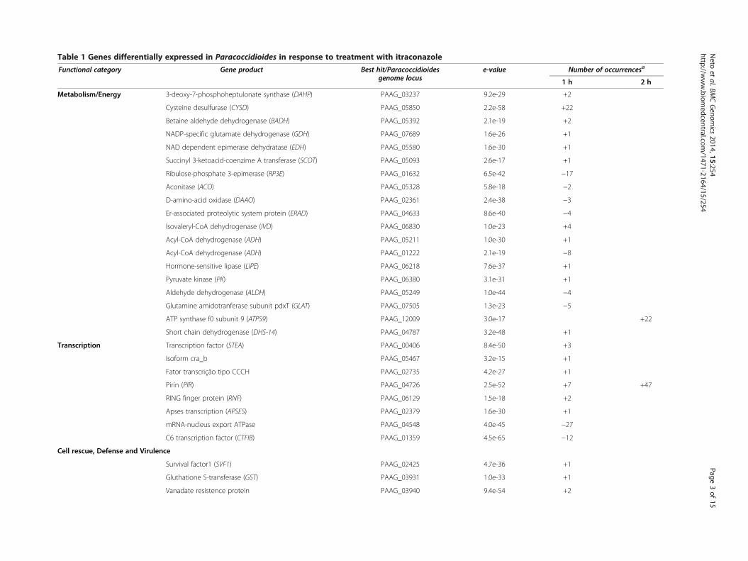
Table 1 Genes differentially expressed in Paracoccidioides in response to treatment with itraconazole
Functional category Gene product Best hit/Paracoccidioidesgenome locus
e-value Number of occurrencesa
1 h 2 h
Metabolism/Energy 3-deoxy-7-phosphoheptulonate synthase (DAHP) PAAG_03237 9.2e-29 +2
Cysteine desulfurase (CYSD) PAAG_05850 2.2e-58 +22
Betaine aldehyde dehydrogenase (BADH) PAAG_05392 2.1e-19 +2
NADP-specific glutamate dehydrogenase (GDH) PAAG_07689 1.6e-26 +1
NAD dependent epimerase dehydratase (EDH) PAAG_05580 1.6e-30 +1
Succinyl 3-ketoacid-coenzime A transferase (SCOT) PAAG_05093 2.6e-17 +1
Ribulose-phosphate 3-epimerase (RP3E) PAAG_01632 6.5e-42 −17
Aconitase (ACO) PAAG_05328 5.8e-18 −2
D-amino-acid oxidase (DAAO) PAAG_02361 2.4e-38 −3
Er-associated proteolytic system protein (ERAD) PAAG_04633 8.6e-40 −4
Isovaleryl-CoA dehydrogenase (IVD) PAAG_06830 1.0e-23 +4
Acyl-CoA dehydrogenase (ADH) PAAG_05211 1.0e-30 +1
Acyl-CoA dehydrogenase (ADH) PAAG_01222 2.1e-19 −8
Hormone-sensitive lipase (LIPE) PAAG_06218 7.6e-37 +1
Pyruvate kinase (PK) PAAG_06380 3.1e-31 +1
Aldehyde dehydrogenase (ALDH) PAAG_05249 1.0e-44 −4
Glutamine amidotranferase subunit pdxT (GLAT) PAAG_07505 1.3e-23 −5
ATP synthase f0 subunit 9 (ATPS9) PAAG_12009 3.0e-17 +22
Short chain dehydrogenase (DHS-14) PAAG_04787 3.2e-48 +1
Transcription Transcription factor (STEA) PAAG_00406 8.4e-50 +3
Isoform cra_b PAAG_05467 3.2e-15 +1
Fator transcrição tipo CCCH PAAG_02735 4.2e-27 +1
Pirin (PIR) PAAG_04726 2.5e-52 +7 +47
RING finger protein (RNF) PAAG_06129 1.5e-18 +2
Apses transcription (APSES) PAAG_02379 1.6e-30 +1
mRNA-nucleus export ATPase PAAG_04548 4.0e-45 −27
C6 transcription factor (CTFIB) PAAG_01359 4.5e-65 −12
Cell rescue, Defense and Virulence
Survival factor1 (SVF1) PAAG_02425 4.7e-36 +1
Gluthatione S-transferase (GST) PAAG_03931 1.0e-33 +1
Vanadate resistence protein PAAG_03940 9.4e-54 +2
Neto
etal.BM
CGenom
ics2014,15:254
Page3of
15http://w
ww.biom
edcentral.com/1471-2164/15/254

Table 1 Genes differentially expressed in Paracoccidioides in response to treatment with itraconazole (Continued)
Heat shock protein (STI1) PAAG_06811 2.2e-24 +2
Heat shock protein (HSP10) PAAG_05142 6.5e-32 +1 +2
Heat shock protein (HSP30) PAAG_00871 5.4e-52 −26 −33
Heat shock protein (HSP70) PAAG_08003 4.4e-40 +3
Heat shock protein (HSP60) PAAG_08059 4.3e-55 −12
Protein synthesis and biogenesis
ATP-dependent RNA helicase (ELF4A) PAAG_00689 1.3e-24 +7
Serine threonine-protein kinase (SRK1) PAAG_06726 7.6e-66 −2
40S ribosomal protein S4 (RPS4) PAAG_03816 7.3e-37 +2
Protein fate (folding, modification, destination)
Ubiquitin-protein ligase (UBI) PAAG_02632 3.7e-11 +2
WD repeat containing protein (WDR) PAAG_00103 1.0e-25 +1
Ubiquitin thioesterase (OTU1) PAAG_08841 1.0e-32 −5
Ubiquitin fusion degradation protein (UFD) PAAG_01475 1.0e-62 +1
Proteasome component (PREP6) PAAG_07802 6.1e-5 +1
Cellular transport, transport facilities and transport routes
Tetracycline resistance protein (TETA) PAAG_01353 1.0e-56 +13
Mfs transporter (MFS) PAAG_02191 7.3e-56 +9
Nucleoporin (SONB) PAAG_02655 1.1e-36 +1
Voltage-gated Ca2+ alpha subunit (CAV) PAAG_01353 9.9e-15 +1
Sodium-dependent phosphate transporter (SPIT) PAAG_03892 1.5e-11 +1
Zinc finger membrane protein (DHHC) PAAG_06616 6.6e-53 −4
autophagy regulatory protein PAAG_04970 2.6e-33 −2
GPR1/FUN34/YAAH family protein PAAG_08587 1.2e-47 −13 −5
Carnitine/acyl carnitine carrier (CAR) PAAG_03452 1.7e-30 −18 −89
General secretion pathway protein. PAAG_05009 2.7e-60 −10
Family integral membrane protein (IMP) PAAG_03183 4.1e-50 +1
Hemolisyn-iii channel protein (HLYiii) PAAG_01871 6.0e-34 +3
Integral membrane MPV17/PMP22 PAAG_02868 6.6e-37 −2
Vesicular fusion protein (SEC17) PAAG_06233 2.8e-77 −1
Biogenesis of Cellular Components (cell wall/membrane)
Chitin synthase regulator 2 (CHSr) PAAG_04860 2.0e-17 +3
Neto
etal.BM
CGenom
ics2014,15:254
Page4of
15http://w
ww.biom
edcentral.com/1471-2164/15/254
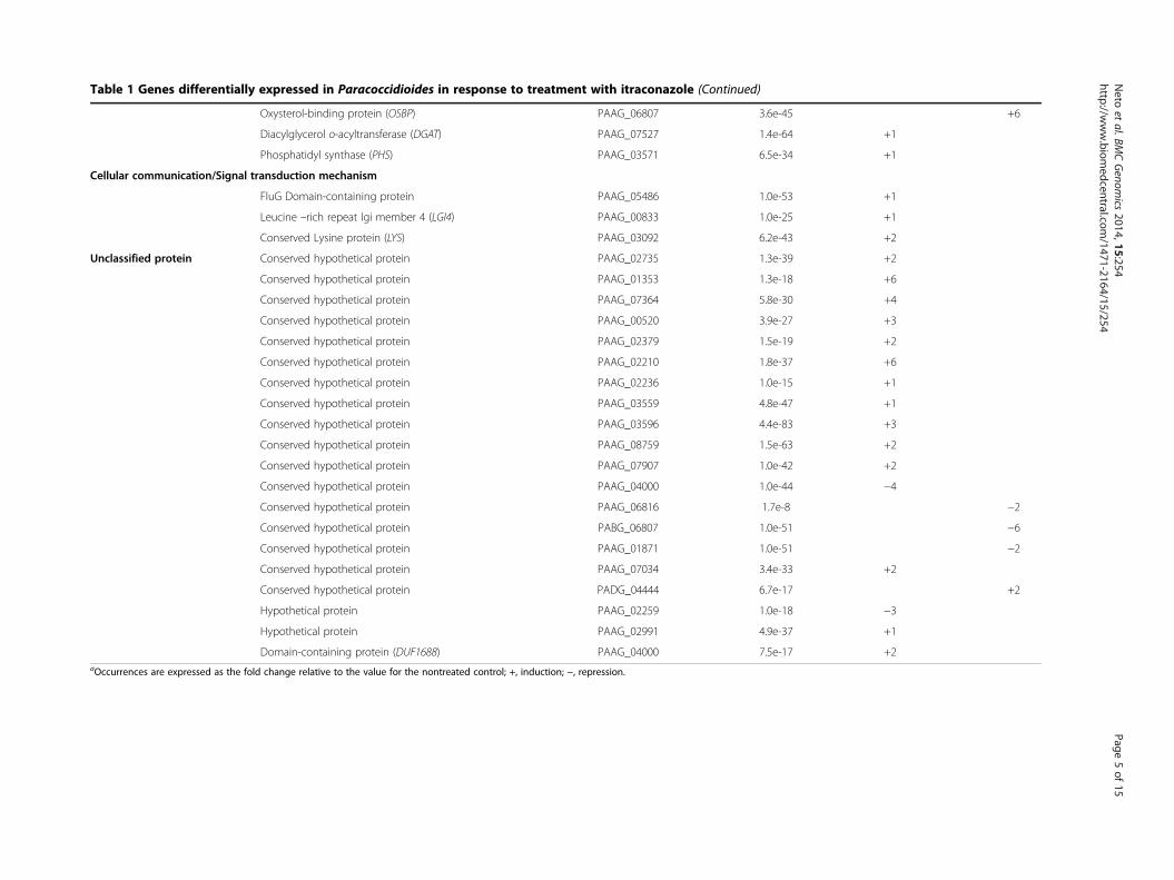
Table 1 Genes differentially expressed in Paracoccidioides in response to treatment with itraconazole (Continued)
Oxysterol-binding protein (OSBP) PAAG_06807 3.6e-45 +6
Diacylglycerol o-acyltransferase (DGAT) PAAG_07527 1.4e-64 +1
Phosphatidyl synthase (PHS) PAAG_03571 6.5e-34 +1
Cellular communication/Signal transduction mechanism
FluG Domain-containing protein PAAG_05486 1.0e-53 +1
Leucine –rich repeat Igi member 4 (LGI4) PAAG_00833 1.0e-25 +1
Conserved Lysine protein (LYS) PAAG_03092 6.2e-43 +2
Unclassified protein Conserved hypothetical protein PAAG_02735 1.3e-39 +2
Conserved hypothetical protein PAAG_01353 1.3e-18 +6
Conserved hypothetical protein PAAG_07364 5.8e-30 +4
Conserved hypothetical protein PAAG_00520 3.9e-27 +3
Conserved hypothetical protein PAAG_02379 1.5e-19 +2
Conserved hypothetical protein PAAG_02210 1.8e-37 +6
Conserved hypothetical protein PAAG_02236 1.0e-15 +1
Conserved hypothetical protein PAAG_03559 4.8e-47 +1
Conserved hypothetical protein PAAG_03596 4.4e-83 +3
Conserved hypothetical protein PAAG_08759 1.5e-63 +2
Conserved hypothetical protein PAAG_07907 1.0e-42 +2
Conserved hypothetical protein PAAG_04000 1.0e-44 −4
Conserved hypothetical protein PAAG_06816 1.7e-8 −2
Conserved hypothetical protein PABG_06807 1.0e-51 −6
Conserved hypothetical protein PAAG_01871 1.0e-51 −2
Conserved hypothetical protein PAAG_07034 3.4e-33 +2
Conserved hypothetical protein PADG_04444 6.7e-17 +2
Hypothetical protein PAAG_02259 1.0e-18 −3
Hypothetical protein PAAG_02991 4.9e-37 +1
Domain-containing protein (DUF1688) PAAG_04000 7.5e-17 +2aOccurrences are expressed as the fold change relative to the value for the nontreated control; +, induction; −, repression.
Neto
etal.BM
CGenom
ics2014,15:254
Page5of
15http://w
ww.biom
edcentral.com/1471-2164/15/254

Figure 1 Functional classification of genes responding to itraconazole in Paracoccidioides. cDNAs obtained from RNAs from yeast cellsafter incubation with itraconazole for 1 h (A) and 2 h (B). The numbers of ESTs are indicated with white bar segments for the up-regulated genesand black bar segments for the down-regulated genes. The annotation of genes was performed using the Blast2GO program with a cut-off forsignificant homology of≤ 1e−5. Sequences were grouped into functional categories according to their classification in the MIPS functional catalog.Additionally, sequences were grouped into functional categories using the PEDANT 3 database. Each functional class is represented as a segmentand expressed as a number of ESTs in each library.
Neto et al. BMC Genomics 2014, 15:254 Page 6 of 15http://www.biomedcentral.com/1471-2164/15/254
using specific oligonucleotides in qRT-PCR. The resultsshowed that all transcripts were increased at all time points(Figure 2B).
Paracoccidioides Pb18 transcripts identified in micetreated with itraconazoleWe investigated whether the regulated transcripts identi-fied by RDA experiments using Pb01 also occurred inanother cryptic species, Pb18, in vivo. Balb/c mice in-fected with Paracoccidioides Pb18 were treated with itra-conazole, and spleens were removed. The treatmentwith itraconazole reduced the fungal burden 42% in thespleens. RNAs extracted from recovered fungus were an-alyzed in qRT-PCR experiments using MFS, GST andCHSr genes. In agreement with the RDA data, all theevaluated genes were up-regulated in spleen fungal sam-ples after treatment with itraconazole (Figure 2C).
GST-specific activity correlates with transcriptional dataBecause GST transcripts were up-regulated in our studyand are described in the literature as important for thedetoxification of many different xenobiotics [14], we eval-uated the GST-specific activity in protein extracts of fun-gus grown in the presence of itraconazole. GST-specificactivity in the presence of itraconazole (0.26 μmol/mg/min) was 6.5 times higher than in the absence of itracona-zole (0.04 μmol/mg/min) (Figure 2D).
Analysis of the ergosterol levelBecause transcript levels of ergosterol pathway compo-nents were changed in the presence of itraconazole, we
evaluated if itraconazole could disturb the total intracellularlevel of ergosterol. The method for quantification of ergos-terol used here takes advantage of the unique four-peakspectral absorption pattern produced by extracted sterolsbetween 240 and 300 nm. Comparing the scans obtainedfrom control (1.0 g of ergosterol/g yeast cells to Pb01; 1.2 gof ergosterol/g yeast cells to Pb18) and the correspondingitraconazole-exposed cultures (0.80 g of ergosterol/g yeastcells to Pb01; 0.62 g of ergosterol/g yeast cells to Pb18), adecrease of 39% and 48.6% was identified in the ergosterolcontent of Paracoccidioides Pb01 and Pb18 yeast cells, re-spectively grown in the presence of itraconazole.
Effect of itraconazole on ergosterol localization inParacoccidioides Pb01 and Pb18 yeast cellsBecause itraconazole induces changes in transcript levelsin the ergosterol pathway and disturbs the total intracel-lular ergosterol content [15], the localization of ergos-terol molecules was assessed in Paracoccidioides Pb01and Pb18 yeast cells. Ergosterol was detected by its abil-ity to bind to the dye filipin. This characteristic has beenused to detect ergosterol in dimorphic fungi [16], yeasts,filamentous fungi [17,18] and mammalian cells [19].The distribution of ergosterol on the surface of
Paracoccidioides Pb01 and Pb18 yeast cells treated withitraconazole was strikingly different from that observedin the control untreated cells. Control cells showeda homogeneous fluorescence distribution (Figure 3Aand C). In contrast, the cells treated with itraconazoledisplayed dark regions without filipin fluorescence(Figure 3B and D).

Figure 2 Relative fold change for genes determined by qRT-PCR. The gene expression profile for (A) twenty genes listed in Table 2, (B) theergosterol pathway genes and (C) in vivo samples of Paracoccidioides recovered directly from systemically infected mouse spleens. Changes ingene expression levels were calculated by relative standard curve method using the control, untreated samples as the calibrator. (D) GST activitywas measured in protein extract from Paracoccidioides Pb18 yeast cells grown in the presence or absence of itraconazole. Each error bar represents thestandard error of the mean (±SE) from three independent experiments performed in triplicate, and significant fold-changes are denoted by asterisks inthe figure (*p≤ 0.05). Data were normalized to the transcript encoding the α-tubulin protein. Student’s t test was used for statistical comparisons.
Neto et al. BMC Genomics 2014, 15:254 Page 7 of 15http://www.biomedcentral.com/1471-2164/15/254
A model for the Paracoccidioides spp. adaptation to theitraconazoleThe most prominent adaptations undergone by Paracoc-cidioides spp. during exposure to itraconazole are sum-marized in Figure 4. See the Discussion for details.
DiscussionAmong the Paracoccidioides Pb01 genes regulated byitraconazole were those involved in cellular transport,metabolism/energy, transcription, cell rescue, defenseand virulence. Similar and different groups were also

Figure 3 Sterol distribution in Paracoccidioides spp.. Yeast cells were fixed, stained with filipin and observed by fluorescence microscopy.Staining in the control Pb01 (A) and Pb18 (C) cells was diffuse with homogeneous labeling. Pb01 (B) and Pb18 (D) cells treated with itraconazoledisplayed heterogeneous fluorescence.
Neto et al. BMC Genomics 2014, 15:254 Page 8 of 15http://www.biomedcentral.com/1471-2164/15/254
observed in other fungi in response to different azoles[8,9,11,12,15] (Figure 5). Among the genes affected, weidentified genes in common with other fungi, as well asgenes unique to Paracoccidioides. In fact few Paracoc-cidioides spp. genes were shared with genes from otherfungi. This could be due to different techniques andclasses of azoles used in the works. The comparisonwith ither fungi show that cell processes related tostress response, xenobiotic efflux are trigered uponitraconazol in different fungi. The genes exclusivelyregulated in Paracoccidioides spp. reveal that fungi re-sponse to drugs can partially involve specific processesthat may be related to different sensibility of differetnfungi to itraconazol treatment. This could be due todifferent techniques and classes of azoles used in theworks.Although ERG genes were not identified in the RDA
experiments, qRT-PCR results showed that ERG11,ERG3, ERG6, ERG5 and ERG25 genes were temporallyregulated, particularly after longer contact with the drug(6 h). Acetyl is a carbon donator in the cell productionof ergosterol [8]. Acetyl CoA seems to be intensivelyproduced due to up-regulation of transcripts from differ-ent metabolic pathways, including lipid degradation byhormone-sensitive lipase (LIPE) and ACAD and aminoacid metabolism by IVD. Acetyl CoA pool increasing inthe cell is optimized with reduction of aconitase trasn-cripts (ACO), once this enzymes participates of theacetyl-CoA oxidation in TCA. In addition, the induction
of BADH and CYSD could lead to production of thiamine,a cofactor to pyruvate dehydrogenase (PDH), which pro-duces acetyl CoA from pyruvate, whose production isincreased by the action of pyruvate kinase (Figure 4).Ergosterol is produced by the action of erg enzymes
[20]. Here, the action of itraconazole on ergosterol bio-synthesis and its distribution on Paracoccidioides Pb01and Pb18 yeast cells surface was documented. Ergosterolis an essential component of fungal plasma membranes;it affects membrane permeability and the activities ofmembrane-bound enzymes. This sterol is a major com-ponent of secretory vesicles and has an important role inmitochondrial respiration and oxidative phosphorylation[21,22]. It can thus be expected that changes in ergos-terol levels and in sterol structure could influence theactivities of several metabolic pathways. The mechanismresponsible for the global up-regulation of ERG genes inresponse to azoles remains unclear. One theory postulatesthat depletion of ergosterol or another sterol formed latein the pathway increases global ERG expression; anotherargues that accumulation of an early substrate or toxicsterol by product induces ERG expression [23].The correlation between cell wall integrity and perturb-
ation of the ergosterol pathway in T. rubrun suggests thatchanges in the cell wall may compensate for stress in theplasma membrane [9]. The phospholipid level in the cellmembrane seems to be affected in Paracoccidioides Pb01,as indicated by up-regulation of DGAT and phosphatidylsynthase (PHS), which produce phospholipids. DGAT has

Figure 4 Hypothetical model for the mode of action of itraconazole against Paracoccidioides. The up-regulation of transcripts such ashormone-sensitive lipase, ADH, IVD and ACO from different metabolic pathways would produce acetyl CoA that would be used for ergosterolsynthesis by ERG enzymes. Acetyl CoA would produce phospholipids for the membrane by the action of DGAT and PHS. The induction of BADHand CYSD would lead to production of thiamine, a cofactor of PDH, which would also produce acetyl CoA. GST would conjugate glutathione toxenobiotics and would remove itraconazole from the cell using transporters, allowing for detoxification.
Neto et al. BMC Genomics 2014, 15:254 Page 9 of 15http://www.biomedcentral.com/1471-2164/15/254
been found in several transcriptomes to date, indicating itmay be important for the fungi response to azoles [15,24].CHS and their regulatory genes are important for the
growth and virulence of human fungal pathogens, includ-ing C. albicans [25,26]. It has been observed that highergosterol levels can inhibit chitin synthases, whereasC. albicans mutants with low ergosterol content showedincreased levels of chitin synthesis [27]. CHSr was up-regulated in Paracoccidioides Pb01 in the presence ofitraconazole.Glutathione S-transferases, which are important for the
detoxification of many xenobiotic compounds, are a familyof multifunctional enzymes that play a role in cellular de-toxification and excretion of a wide variety of xenobioticsubstances [14]. It has been reported that GlutathioneS-transferases correlate with fungi defense in response todamage caused by oxidative stress, xenobiotics and anti-fungal compounds [28]. GST was up-regulated in Paracoc-cidioides in the presence of itraconazole.
In Paracoccidioides Pb01, genes encoding several clas-ses of transporters were up-regulated upon exposure toitraconazole. MFS transporter and TETA, for example,have been implicated in azole resistance [29]. Drugresistance is often associated with the overexpression ofgenes encoding efflux pumps, which is presumed toprevent intracellular accumulation of itraconazole infungus [9,30]. The up-regulation of ParacoccidioidesPb01 MFS, GST and CHSr transcripts also occur in vivo,as demonstrated here by qRT-PCR using RNAs ex-tracted from spleens of mice.It should be noted that a number of genes involved in
small molecule transport, especially in ion transport,were differentially expressed in Paracoccidioides Pb01 inresponse to itraconazole. Up-regulated genes includedCAV, IMP and HLYiii. Down-regulated genes included(CAR) and integral membrane MPV17/PMP22. The in-hibition of ergosterol, which is an essential componentof fungal biological membranes, including the plasma

Figure 5 Distribution of genes responding to itraconazole in P. brasiliensis isolate Pb01. Data are shown for a subset of genes that weresignificantly up or down regulated (e-values ≤10−10). The search for functional categories was performed by using the Blast2GO program that joints inone application GO annotation based on similarity searches with statistical analysis and highlight visualization on directed acyclic graphs. GO termsshown are those that were considered significantly over represented by the analysis. Sequences were grouped in functional categories according tothe classification of the MIPS functional catalog (Munich Center for Protein Sequences; http://mips.gsf.de/). Specific genes for P. brasiliensis isolate Pb01are underlined, genes found in other fungi when exposed to itraconazole and other azoles are represented with * and represented with ◊, respectively.Numbers in parentheses represent changes in gene expression. Positive signal indicate induction, and negative indicate repression.
Neto et al. BMC Genomics 2014, 15:254 Page 10 of 15http://www.biomedcentral.com/1471-2164/15/254
membrane, can lead to destabilization of the membrane,leakage of cellular components and influx of extracellu-lar ingredients. Therefore, the regulation of transportergenes is necessary to maintain ionic homeostasis withinthe fungal cell when membranes are damaged by itraco-nazole [9,31].
ConclusionThis is the first study to analyze the changes in the Para-coccidioides spp. gene expression profile following tri-azole exposure. Among the genes affected, we identifiedgenes unique to Paracoccidioides Pb01, as well as genesin common with other fungi. In vitro results were vali-dated by in vivo experiments. The results obtained hereshould assist in understanding the mode of action ofitraconazole in Paracoccidioides spp.
MethodsCulture and cell viabilityParacoccidioides Pb01 and Pb18 have been studied atour laboratory previously [32,33]. Pb01 and Pb18 yeastphase was maintained in vitro by subculturing at 36°C inFava Netto’s semisolid medium [34] every seven days.Fava Netto’s semisolid medium components were asfollows: 1% (w/v) peptone, 0.5% (w/v) yeast extract, 0.3%(w/v) proteose peptone, 0.5% (w/v) beef extract, 0.5%(w/v) NaCl, 4% (w/v) glucose and 1.2% (w/v) agar, pH 7.2.The determination of IC50 was performed according toSantos et al. [23] and in accordance with the macro dilu-tion method described in the Clinical and LaboratoryStandards Institute (CLSI) M27-A2(2005), with modifica-tions. To determine the IC50, yeast cells in the exponentialgrowth phase were maintained in the chemically defined

Neto et al. BMC Genomics 2014, 15:254 Page 11 of 15http://www.biomedcentral.com/1471-2164/15/254
solid medium McVeigh Morton (MMcM) [35], for sevendays at 36°C and inoculated in liquid MMcM. A stocksolution (1 mg/ml) containing sterile itraconazole (Sigma-Aldrich, St. Louis, MO, USA) was prepared in dimethyl-sulfoxide (DMSO). The final concentration of the solventin the medium never exceeded 2% (v/v) and had no effecton the cell growth. From this stock solution, the drug wasserially diluted in sterile MMcM (pH 7.0), producing afinal concentration of 1.25-320 μg/ml (5–1260 mM). Thedrug concentration range was selected based on previousstudies [36]. The controls without antifungal and DMSOwere included. The concentrations of inoculums weredetermined by spectrophotometer using a yeast cell sus-pension in sterile 0.85% NaCl with 10% transmittance at520 nm. The mixture was stirred to disperse aggregatedcells. Yeast cells were collected from the liquid MMcMand counted in a Neubauer chamber. An initial inoculumcontaining 5 × 10⁶ cells/ml was collected, and 0.1 ml ali-quots were added to 2.4 ml of MMcM containing the drugdilutions. The fungus was grown at 36°C under agitationat 150 rpm for five days. The IC50 was determined usingmeasurements of the turbidity of the medium [37]. Theexperiments were processed in triplicate.For viability experiments, yeast cells were grown in
the presence or absence of 4 μg/ml (IC50) of itraconazoleand were kept in liquid MMcM [35] for 1, 2, 3, 4 and5 h at 36°C before the viability of the cells was deter-mined by Trypan Blue method [38]. In brief, cells fromall incubation times were incubated with a dye solution(0.1% Trypan Blue Stain) for 5 min at room temperature,and viability was assessed by counting viable and un-viable cells in a Neubauer chamber.
RDA: RNA extraction and cDNAs synthesisParacoccidioides Pb01 yeast cells were cultured inMMcM broth medium in the presence or absence of4 μg/ml of itraconazole for 1 h and 2 h, correspondingto a viability of 95% and 85%, respectively. For RNA iso-lation, cells were harvested by centrifugation, washed incold water and the RNA from driver and tester cultureswere extracted with Trizol (Invitrogen, Carlsbad, CA,USA) according to the manufacturer’s instructions. RNAquality was assessed using the A260nm/A280nm ratio. TheRNA was treated with DNAse I RNAse-free (Invitrogen)to remove chromosomal DNA. The concentration andpurity of RNA were determined by spectrophotometer,and RNA integrity was visualized after electrophoresison 1.2% agarose gel. The RNAs were used to constructsubtracted libraries and qRT-PCR experiments.The cDNA fragments used for processing the RDA
were generated according to the protocol previouslydescribed by Hubank and Schatz [39] and modified byPastorian et al. [40]. Briefly, first-strand cDNA synthesiswas performed with 1 μg total RNA, obtained from
driver and tester cultures, using SuperScript III reversetranscriptase (Invitrogen). The first-strand cDNA obtained(3 μl) was used as template to synthesize the second-strandof cDNA. The cDNA was prepared using the SMARTPCR cDNA synthesis kit (Clonetech Laboratories, PaloAlto, CA, USA).
RDA: Subtractive hybridizationThe cDNAs were digested with the restriction enzymeSau3AI. Two successive rounds of subtraction employ-ing different adapters (J-Bam and N-Bam, Table 2) wereperformed to enrich the differentially expressed se-quences. Four cDNA-subtracted libraries were con-structed. The cDNA libraries containing up-regulatedgenes were constructed from driver cDNA obtainedfrom Paracoccidioides Pb01 yeast cells grown for 1 hand 2 h in MMcM medium and from tester cDNA,which was synthesized from RNA extracted from Para-coccidioides Pb01 yeast cells grown for 1 h and 2 h inMMcM medium plus itraconazole. The cDNA librariescontaining down-regulated genes were constructed fromdriver cDNA, obtained from Paracoccidioides Pb01 yeastcells, grown for 1 h and 2 h in MMcM medium plusitraconazole and tester cDNA, which was synthesizedfrom RNA extracted from Paracoccidioides Pb01 yeastcells grown for 1 h and 2 h in MMcM medium. Theresulting products were purified using a GFX kit (GEHealthcare, Uppsala, Sweden). The tester-digestedcDNA, from 1 h and 2 h samples, was linked to adapters(a 24-mer annealed to a 12-mer) and amplified by PCR.For the generation of the differentially up- and down-
regulated products, the tester and driver cDNAs of bothconditions were mixed separately; the hybridization oc-curred at 67°C for 18 h and the amplification occurredby PCR using the oligonucleotide matching the 24-meradaptor [41]. The successive rounds of subtraction andamplification were performed using hybridization tester-driver ratios of 1:10 and 1:100. Adapters (Table 2) werechanged between cross-hybridization, and the differentproducts were purified using the GFX kit. After the sec-ond subtractive reaction, the cDNA was purified andcloned directly into the pGEM-T Easy vector (Promega,Madison, WI, USA). Escherichia coli XL1 Blue compe-tent cells were transformed with the ligation products.The plasmid DNAs were prepared from selected clonesof subtracted libraries and sequenced with the ET DyeTerminator kit Dyenamic (GE Healthcare) in a Mega-BACE 1000 DNA sequencer (GE Healthcare) usingprimers corresponding to the pGEM-T Easy vector.
Processing and annotation of ESTsThe sequences of at least 75 nucleotides, with a PHREDscore ≥ 20 were considered for the assembly and formationof clusters. The assembly of these ESTs was performed

Table 2 Oligonucleotide primers used in RDA assays and qRT-PCR
Sequence name Forward primer (5′-3′) Reverse primer (5′-3′) Amplicon size (bp)
Glutathione S-transferase (GST) GAACCGCAAACCCTAACCCT ACAGCGGCTGAAAAGTCCCA 157
Chitin synthase regulator 2 (CHSr) AGAGCTGCAGAATTAGGCCTT TTTCGCCCGTTCATCTCCGT 140
Betaine aldehyde dehydrogenase (BADH) GTTGAAGAGCCATTTGGTCC CAGATCATTGGACCACACAGA 120
Cysteine desulfurase (CYSD) CAACAGAAGAGATGGAGTATGA AGCGAATGACACGTTGACACA 143
Ribulose-phosphate 3-epimerase (RP3E) CAATGGATCGACCTGATATGG GACCTCCGTCAACTTCGATG 141
Carnitine/acyl-carnitine carrier (CAR) GAAGGCATTGCCAGGGGGT CATTATGAACGGGGACGGTG 139
Glutamine amidotransferase subunit pdxT (GLAT) TGAGAGACTTTGTCAAGAACCA TGCGCGGATAAATACACCCAT 143
Mfs transporter (MFS) CTAATTATGTTCTTTTGGGGTAC GCATCGCCTATACCAACAAGA 136
C6 transcription factor (CTF1B) CAAACCACTCGTCAACACAATC GATTGCCTTGAGTCTGATAGAG 138
Acyl-CoA dehydrogenase (ACAD) GAGAACGAGACGCCCGAAG GTTGTAGTAAGGACTCTTGTAG 108
GPR1/FUN34/YAAH family protein ACTGGCTGGGATGTGGGAG TTCTTCTCCGTCATTTCCTTGA 141
Pyruvate kinase (PK) ATGCGATGATAAATATCTCTACG GACACTTGGCGCGGAGAGA 143
Diacylglycerol o-acyltransferase (DGAT) TATTAGATATACCAAGTGGCCG TACCCTGGGTTTGTATTCAATG 143
Isovaleryl-CoA dehydrogenase (IVD) GATGTGGATTACCAACGGGC TCATGCCAAGCTTGTCGAGTT 152
Ubiquitin-protein ligase (UBI) GGAGGCATGCAGATCTTCGT ACGACCGTCCTCAAGCTGC 168
Family integral membrane protein (IMP) CGCCAGCAATCTGATTATCTC AACCCAGCTGACCTTCATTAC 142
Heat shock protein (HSP10) TCTTCCTCCCAGAGAGCGC CAGGGCTGCCTCCATACTG 143
Heat shock protein (HSP30) GGCCTTGACAGCATTCTGG CTGGCGATAAAGGGCAGAAG 130
Heat shock protein (HSP70) GCAGAAGGAGCTTGAAAGTGT GTCAACCTCCTCGACAGTAG 181
ATP synthase f0 subunit 9 (ATPS9) AAGCAGCGAAAATAATGGGATC GCAAATAATCCTGTAGCTTCTG 181
Lanosterol 14 α-demethylase (ERG11) CTGAGCTGTAGGGAAAAGTAC TCCTCAGCGCAAACGTCCTT 131
C5,6-desaturase (ERG3) GGAGAATATGTATACCAGCCC ATCCAAGTGATGAGATACAGAG 128
Delta-24-sterol C-methyltransferase (ERG6) GCTACTCTTACCCGACATTAC AATGGGCAAGGTAATGTTCATG 142
C-22 sterol desaturase (ERG5) GGTCCCATGTTCAAAATCCCT AAATTTGTGGAAAACCGAGACG 123
C-4 methyl sterol oxidase (ERG25) GGACCATGGCCTACCAAATC GCGGAGTATTGGTGGTGGAT 129
cDNA* AGCAGTGGTATCAACGACAGAGTACGCGGG -
CDS* AAGCAGTGGTATCAACGCAGAGTACT(30)N1N -
PCRII* AAGCAGTGGTATCAACGCAGAGT -
JBam12* GATCCGTTCATG -
JBam24* ACCGACGTCGACTATCCATGAACG -
NBam12* GATCCTCCCTCG -
NBam24* AGGCAACTGTGCTATCCGAGGGAG -
RBam12* GATCCTCGGTGA -
RBam24* AGCACTCTCCAGCCTCTCACCGAG -
T7* GTAATACGACTCACTATAGGGC -
Oligo (dT)15* AAGCAGTGGTATCAACGCAGAGTACT(30)N1N -
*Primers used in RDA experiments.
Neto et al. BMC Genomics 2014, 15:254 Page 12 of 15http://www.biomedcentral.com/1471-2164/15/254
using CAP3 [42] and clustered to generate contigs and sin-glets, which were analyzed. All these tools were integratedin a specific pipeline (http://www.lbm.icb.ufg.br/pipeli-neUFG/). The annotation of genes was performed usingthe program Blast2GO (http://www.blast2go.org/), whichprovides a comparison between clusters of sequences ob-tained from public databases. The BLAST program fromthe National Center for Biotechnology Information (NCBI)
(http://www.ncbi.lm.nih.gov/BLAST), processed with thenon-redundant sequences (nr) GenBank and the nucleotidedatabase generated from Paracoccidioides spp. structuralgenome (http://www.broad.mit.edu/annotation/genome/paracoccidioides_brasiliensis/MultiHome.html), was used forthe annotation. The database sequence matches were con-sidered significant at e-values ≤10−5. The program INTER-PROSCAN (http://www.ebi.ac.uk/interpro/) [43] was used

Neto et al. BMC Genomics 2014, 15:254 Page 13 of 15http://www.biomedcentral.com/1471-2164/15/254
to obtain information about the domains present in clus-ters and the classification of families. The metabolic path-ways were analyzed using maps obtained from the KEGGdatabase (Kyoto Encyclopedia of Genes and Genomes)(http://www.genome.ad.jp/kegg) [44] with annotated ECnumbers, and this information was used to help elucidatethe function of ESTs. The Munich Information Centerfor Protein Sequences (MIPS) (http://mips.gsf.de/) wasused to designate the functional categories. Addition-ally, sequences were grouped into functional categoriesusing the PEDANT 3 database (http://pedant.helm-holtz-muenchen.de/index.jsp).
Analysis of RNA transcripts by qRT-PCRAn aliquot of RNA from treated and untreated sampleswas used to perform reverse transcription qRT-PCR.Total RNAs from Paracoccidioides Pb01 yeast cells cul-tured in the presence or absence of itraconazole wereobtained as previously described, in independent experi-ments from those used in the RDA assays. After treat-ment with DNAse, cDNAs were synthesized from totalRNA using Superscript III reverse transcriptase (Invitro-gen) and oligo (dT)15 primer according to the supplier’sinstructions. Gene-specific primers were designed forthe selected genes and for the control gene, α-tubulin,using Primer Express software (Applied Biosystems, Fos-ter City, CA, USA) (Table 2). qRT-PCRs were performedin triplicate in a StepOnePlus™ real time PCR system(Applied Biosystems). The PCR thermal cycling programconsisted of 40 cycles of 95°C for 15 sec; 60°C for 1 min.The SYBR green PCR master mix (Applied Biosystems)was supplemented with 1 ρmol of each gene-specificoligonucleotide and 40 ng of template cDNA in a finalvolume of 20 μl. A curve melting analysis was performedto confirm the amplification of a single PCR product.The data were normalized to the α-tubulin transcriptamplified in each set of qRT-PCR experiments. A no-template control was included. Samples of each cDNAwere pooled and serially diluted 1:5 to generate a rela-tive standard curve. Relative expression levels of geneswere calculated using the standard curve method forrelative quantification [45]. Statistical comparisonswere performed using Student’s t-test and samples withp-values < 0.05 were considered statistically significant.The specific sense and antisense primers are listed inTable 2. Itraconazole-regulated transcripts were se-lected for qRT-PCR validation assays.
Preparation of protein extracts and validation of dataobtained by specific activity of Glutathione S-Transferase(GST)GST activity was measured with GST assay kit (Sigma-Aldrich). Briefly, the GST Assay Kit employs 1-Chloro-2,4-dinitrobenzene (CDNB) to produce 1-glutathionyl-2,4-
dinitrobenzene (GS-DNB) by conjugation of the thiolgroup of glutathione (GSH). The reaction productGS-DNB absorbs at 340 nm, and the rate of increase inthe absorption is directly proportional to the GSTactivity of the sample.Protein extracts from Paracoccidioides Pb18 yeast cells
were prepared by inoculating 50 ml of Fava Netto’s liquidmedium with 106 cells/ml. Cultures were incubatedovernight at 36°C with gentle shaking for 16 h. Cellswere centrifuged at 5,000 x g for 5 min and transferredinto MMcM media containing itraconazole for 1 h.Control cells were incubated in MMcM without drug.The cells were centrifuged at 10,000 x g for 15 min at4°C, frozen in liquid nitrogen and disrupted by macer-ation [46]. Extraction buffer (20 mM Tris–HCl pH 8.8;2 mM CaCl2) containing a mixture of protease inhibitors(serine, cysteine and calpain inhibitors) (GE Healthcare)was added to the yeast cells. After the addition of glassbeads (0.45 mm), the cells were lysed in a bead-beater,followed by centrifugation at 10,000 x g for 15 min at 4°C.The supernatant was collected, and the protein concentra-tions were determined using Bradford reagent (Sigma-Al-drich). The samples were stored in aliquots at −80°C.The increase in absorbance is directly proportional to
the GST activity. The GST-specific activity is defined asmmol of GS-DNB per mg of total protein per min(mmol/mg/min). The enzymatic activity results repre-sent the mean of three independent determinations, andstatistical comparisons were performed using Student’st test. The samples with p-values ≤0.05 were consideredstatistically significant.
Sterol quantification methodThe quantification of total intracellular ergosterol wasperformed as previously described [47], with slight mod-ifications. Cell extracts from Paracoccidioides Pb01 andPb18 yeast cells were prepared as already describedabove. Five ml of 25% alcoholic potassium hydroxide so-lution (25 g KOH and 35 ml sterile distilled water addedto 100 ml 100% ethanol) was added to each tube, andthe samples were mixed on a vortex for 2 min. The cellsuspensions were incubated in an 85°C water bath for3 h and allowed to cool to room temperature. Sterolswere extracted by addition of 2 ml of sterile distilledwater and 5 ml n-heptane (Sigma-Aldrich), followed byvigorous mixing in a vortex mixer for 5 min. The sam-ples were kept at room temperature for 1 to 2 h to allowthe phases to separate or were stored at 4°C overnight.One ml of the heptane layer (containing ergosterol) wastransferred to a 1.5 ml quartz cuvette and analyzed spec-trophotometrically by scanning at wavelengths between200 and 300 nm. If necessary, the samples were dilutedfive fold with 100% ethanol and reanalyzed. The ergos-terol content as a percentage of the wet cell weight was

Neto et al. BMC Genomics 2014, 15:254 Page 14 of 15http://www.biomedcentral.com/1471-2164/15/254
calculated by the following equations: value 1 = [(A281.5/290) × F]/wet cell weight, value 2 = [(A230/518) × F]/wetcell weight, and percent ergosterol = value 1 - value 2. Fis the factor for dilution in ethanol, and 290 and 518 arefixed values determined for crystalline ergosterol and 24(28) dihydroergosterol, respectively.
Fluorescence microscopyFilipin stained samples were prepared using a previouslydescribed protocol for fixing and staining filamentousfungi [48]. Paracoccidioides Pb01 and Pb18 yeast cellswere prepared by inoculating 50 ml of Fava Netto’s li-quid medium with 108 cells/ml. Cultures were incubatedovernight at 36°C under gentle shaking for 16 h. Cellswere centrifuged at 5,000 × g for 5 min and transferredin MMcM media containing itraconazole. Control cellswere incubated in MMcM without drug. The fungus wasthen removed and fixed for 30 min in 3.7% formalde-hyde and rinsed with ddH2O. A 5 mg/ml stock solutionof filipin (Sigma-Aldrich) dissolved in DMSO was di-luted to 25 μg/ml and used to stain the fixed samples for10 min. Samples were then rinsed with ddH2O, mountedon a microscope slide and sealed with nail varnish.
BALB/c mice infection with Paracoccidioides Pb18The animals were bred at the Universidade Federal deGoiás animal facility under specific-pathogen-free con-ditions. All animal experiments were performed inaccordance with the international rules for animal ex-perimentation. The animal protocol was approved bythe Universidade Federal de Goiás committee of the eth-ical treatment of animals (Number: 008/11 CEUA-UFG).Female BALB/c mice, 8–12 weeks old, were inoculated
with 1 × 10⁷ of Paracoccidioides Pb18 yeast cells growthin liquid MMcM. In brief, yeast cell suspension in the7th day of in vitro growth were washed in PBS 1× andinoculated intraperitoneally in mice. The mice were di-vided into three groups of five animals to be sacrificed21 days post-challenge. Each group was subdivided bytreatment options as follows: five uninfected mice (nega-tive controls), five yeast cell-infected mice (positive con-trols) and five yeast cell-infected mice treated withitraconazole starting at the third week post-infection.The animals were sacrificed in the sixth week after infec-tion. The spleens were removed and homogenized in5 ml of sterile PBS 1X. The homogenized sample wasplated in brain heart infusion agar supplemented with4% (v/v) fetal calf serum and 2% (w/v) glucose. Theplates were incubated at 36°C and colony-forming unitswere determined after 20 days.Gene expression analyses of Paracoccidioides Pb18
from infected mice were performed by isolating yeastcells from spleens as previously described with minormodifications [49]. The spleens of infected mice were
homogenized in 1× PBS using a tissue grinder. Thehomogenate was then filtered using nylon mesh to removelarge pieces of animal tissue. The sample was frozen in li-quid nitrogen and then centrifuged at 500 × g for 5 min toremove any remaining animal tissue. Next, the sample wascentrifuged at 7,000 × g for 15 min to isolate fungal cells.Total RNA was extracted from recovered cells using TRI-ZOL reagent (Invitrogen), according to the manufacturer’sinstructions. RNA was used to perform qRT-PCR as de-scribed above.
Competing interestsThe authors declare that they have no competing interests.
Authors’ contributionsBRSN performed RNA extraction and Construction of cDNA librariessubtractive, annotation of ESTs, Analysis of RNA transcripts by qRT-PCR,Analysis of ERG transcripts by qRT-PCR, Preparation of protein extracts andspecific activity of Glutathione S-Transferase (GST), Analysis of the ergosterollevel, Fluorescence microscopy, and model for the Paracoccidioides spp.adaptation to the itraconazole. PFZC perfomed experiment sub-inhibitoryconcentration. WSM perfomed processing of ESTs. BRSN and AMB madeBALB/c mice infection with Paracoccidioides Pb18. BRSN, AMB, CMAS and MPcontributed to the discussion of the data and preparation of the manuscript.MP conceived, designed and coordinated the study. All authors contributedto the discussion of results. All the authors have read and approved the finalmanuscript.
AcknowledgementsThis work performed at Universidade Federal de Goiás was supported byMinistério da Ciência e Tecnologia/Conselho Nacional de DesenvolvimentoCientífico e Tecnológico (MCTI/CNPq), Fundo Nacional de DesenvolvimentoCientífico e Tecnológico (FNDCT), Fundação de Amparo à Pesquisa doEstado de Goiás (FAPEG), Coordenação de Aperfeiçoamento de Pessoal deNível Superior (CAPES), Financiadora de Estudos e Projetos (FINEP) andINCT_IF (Instituto Nacional de Ciência e Tecnologia para Inovação Farmacêutica).Additionally, B.R.S.N. was supported by a fellowship from CNPq and P.F.Z.C. byCAPES.
Author details1Departamento de Bioquímica e Biologia Molecular, Laboratório de BiologiaMolecular, Instituto de Ciências Biológicas, ICBII, Campus II, UniversidadeFederal de Goiás, C.P. 131, 74001-970 Goiânia, GO, Brazil. 2Instituto deInformática, Universidade Federal de Goiás, Goiânia, Goiás, Brazil.
Received: 19 July 2013 Accepted: 26 March 2014Published: 1 April 2014
References1. Franco M: Host–parasite relationship in paracoccidioidomycosis.
J Clin Microbiol 1987, 25:5–18.2. Restrepo A, McEwen JG, Castañeda E: The habitat of Paracoccidiodes
brasiliensis: how far from solving the ridle? Med Mycol 2001, 39:233–241.3. San-Blas G, Niño-Vega G, Iturriaga T: Paracoccidioides brasiliensis and
paracoccidioidomycosis: molecular approaches to morphogenesis,diagnosis, epidemiology, taxonomy and genetics. Med Mycol 2002,40:225–242.
4. Coutinho Z, Silva D, Lazera M, Oliveira RM, Sabroza PC, Wanke B:Paracoccidioidomycosis mortality in Brasil 1980–1995. Cad Saúde Pública2002, 18:1441–1454.
5. Shikanai-Yasuda MA, Telles FQ, Mendes RP, Colonbo AL, Moretti ML:Guidelines in paracoccidioidomycosis. Rev Soc Bras Med Trop 2006,39:297–310.
6. Burgess DS, Hastings RW: A comparison of dynamic characteristics offluconazole, itraconazole, and amphotericin B against Cryptococcusneoformans using time-kill methodology. Diagnost Microbiol Infect Dis2000, 38:87–93.

Neto et al. BMC Genomics 2014, 15:254 Page 15 of 15http://www.biomedcentral.com/1471-2164/15/254
7. Lamb DC, Maspahy S, Kelly DE, Manning NJ, Geber A, Bennett JE, Kelly SL:Purification, reconstitution, and inhibition ofcytochrome P-450 sterol,D22-desaturase from the pathogenic fungus Candida glabrata.Antimicrob Agents Chemother 1999, 43:1725–1728.
8. Bammert GF, Fostel JM: Genome-wide expression patterns inSaccharomyces cerevisiae: comparison of drug treatments and geneticalterations affecting biosynthesis of ergosterol. Antimicrob AgentChemother 2000, 44:1255–1265.
9. Diao Y, Zhao R, Deng X, Leng W, Peng J, JIN Q: Transcriptional profiles ofTrichophyton rubrum in response to itraconazole. Med Mycol 2009,47:237–247.
10. Georgiadou SP, Kontoyiannis DP: The impact of azole resistance onaspergillosis guidelines. Ann N Y Acad Sci 2012, 12721:5–22.
11. Liu TT, Lee REB, Barker KS, Lee RE, Wei L, Homayouni R, Rogers PD:Genome-wide expression profiling of the response to azole, polyene,echinocandin, and pyrimidine antifungal agents in Candida albicans.Antimicrob Agents Chemother 2005, 49:2226–2236.
12. Hoehamer CF, Cummings ED, Hilliard GM, Rogers PD: Changes in theproteome of Candida albicans in response to azole, polyene, andechinocandin antifungal agents. Antimicrob Agents Chemother 2010,54:1655–1664.
13. Georgiadou SP, Kontoyiannis DP: The impact of azole resistance onaspergillosis guidelines. Ann N Y Acad Sci 2012, 1272:15–22.
14. Hayes JD, Flanagan JU, Jowsey IR: Glutathione transferases. Annu RevPharmacol Toxicol 2005, 45:51–88.
15. De Backer MD, Ilyina T, Ma XJ, Vandoninck S, Luyten WH, Vanden BosscheH: Genomic profiling of the response of Candida albicans to itraconazoletreatment using a DNA microarray. Antimicrob Agents Chemot 2001,45:1660–1670.
16. Munkacsi AB, Pentchev PG, Sturley SL: Spreading the wealth: Niemann-Picktype C proteins bind and transport cholesterol. Cell Metab 2009, 10:3–4.
17. Breakspear A, Pasquali M, Brozc K, Donga Y, Kistler HC: Npc1 is involved insterol trafficking in the filamentous fungus Fusarium graminearum.Fungal Genet Biol 2011, 48:725–730.
18. Van Leeuwen MR, Smant W, de Boer W, Dijksterhuis J: Filipin is a reliableinsitu marker of ergosterol in the plasma membrane of germinating conidia(spores) of Penicillium discolor and stains intensively at the site of germtubeformation. J Microbiol Method 2008, 74:64–73.
19. Beh CT, Rine J: A role for yeast oxysterol-binding protein homologs inendocytosis and in the maintenance of intracellular sterol-lipid distribu-tion. J Cell Sci 2004, 117:2983–2996.
20. Akins RA: An update on antifungal targets and mechanisms of resistancein Candida albicans. Med Mycol 2005, 43:285–318.
21. Vanden Bossche H, Marichal P, Gorrens J, Bellens D, Moereels H, JanssenPAJ: Mutation in cytochrome P450-dependent 14ademethylase results indecreased affinity for azole antifungals. Biochemic Soc Transact 1990,18:56–59.
22. Daum G, Lees ND, Bard M, Dickson R: Biochemistry, cell biology andmolecular biology of lipids of Saccharomyces cerevisiae. Yeast 1998,14:1471–1510.
23. Santos GD, Ferri PH, Santos SC, Bao SN, Soares CMA, Pereira M: OenotheinB inhibits the expression of PbFKS1 transcript and induces morphologicalchanges in Paracoccidioides brasiliensis. Med Mycol 2007, 45:609–618.
24. Liu Q, Siloto RMP, Lehner R, Stone SJ, Weselake RJ: Acyl-CoA: diacylglycerolacyltransferase: molecular biology, biochemistry and biotechnology.Progr Lipid Resear 2012, 51:350–377.
25. Munro CA, Winter K, Buchan A, Henry K, Becker JM, Brown AJ, Bulawa CE,Gow NA: Chs1 of Candida albicans is an essentialchitin synthase requiredfor synthesis of the septum and for cell integrity. Mol Microbiol 2001,39:1414–1426.
26. Banks IR, Specht CA, Donlin MJ, Gerik KJ, Levitz SM, Lodge JK: A chitinsynthase and its regulator protein are critical for Chitosan productionand growth of the fungal pathogen Cryptococcus neoformans. EukaryotCell 2005, 4:1902–1912.
27. Vanden Bossche H: Biochemical targets for antifungal azole derivativeshypothesis on the mode of action. Curr Top Med Mycol 1985, 1:313–351.
28. Wang W, Ballatori N: Endogenous glutathione conjugates: occurrence andbiological functions. Pharmacol Rev 1998, 50:335–356.
29. Ghannoum MA, Rice LB: Antifungal agents: mode of action, mechanismsof resistance, and correlation of these mechanisms with bacterialresistance. Clin Microbiol Rev 1999, 12:501–517.
30. Lupetti A, Danesi R, Campa M, Del Tacca M, Kelly S: Molecular basis ofresistance to azole antifungals. Trend Mol Med 2002, 8:76–81.
31. Edlind T, Smith L, Henry K, Katiyar S, Nickels J: Antifungal activity inSaccharomyces cerevisiae is modulated by calcium signaling. Mol Microbiol2002, 46:257–268.
32. Da Silva Cruz AH, Brock M, Zambuzzi-Carvalho PF, Santos-Silva LK, Troian RF,Goes AM, Soares CMA, Pereira M: Phosphorylation is the major mechanismregulating isocitrate lyase activity in Paracoccidioides brasiliensis yeast cells.FEBS J 2011, 278:2318–2332.
33. De Oliveira KM, Neto BRS, Parente JA, Da Silva RA, Quintino GO, Voltan AR,Mendes-Giannini MJ, Soares CMA, Pereira M: Intermolecular interactions ofthe malate synthase of Paracoccidioides spp. BMC Microbiol 2013, 13:107.
34. Fava Netto C, Vegas VS, Sciannamea JJ, Guarnieri DB: Antígenopolissacarídeo do Paracoccidioides brasiliensis. Estudo do tempo decultura do P.brasiliensis necessário ao preparo do antigen. Rev Inst MedTrop 1969, 11:177–181.
35. Restrepo A, Jimenez BE: Growth of Paracoccidioides brasiliensis yeast phase ina chemically defined culture medium. J Clin Microbiol 1980, 12:279–281.
36. Hahn RC, Conceição YTM, Santos NL, Ferreira JF, Hamdan JS: Disseminatedparacoccidioidomycosis: correlation between clinical and in vitroresistance to ketoconazole and trimethoprim sulphamethoxazole.Mycoses 2003, 46:342–347.
37. Hahn RC, Hamdan JS: In vitro susceptibilities of Paracoccidioidesbrasiliensis yeast form to antifungal drugs. Mycoses 2000, 43:403–407.
38. Freshney R: Culture of Animal Cells: A Manual of Basic Technique. New York:Alan R Liss; 1987. Inc.
39. Hubank M, Schatz AG: Identifying differences in mRNA expression byrepresentational difference analysis of cDNA. Nucleis Acids Res 1994,22:5640.
40. Pastorian K, Havell L 3rd, Byus CV: Optimization of cDNA representationaldifference analysis for the identification of differentially expressedmRNAs. Anal Biochem 2000, 283:89–98.
41. Dutra V, Nakazato L, Broetto IS, Vainstein MH, Schrank A: Application ofrepresentational difference analysis to identify sequence tags expressedby Metarhizium anisopliae during the infection process of the Boophilusmicroplus cuticle. Res Microbiol 2004, 155:245–251.
42. Huang X, Madan A: CAP3: a DNA sequence assembly program. Genom Res1999, 9:868–877.
43. Apweiler R, Bairoch A, Wu CH: Protein sequence databases. Curr OpinChem Biol 2004, 8:76–80.
44. Kanehisa M, Goto S: KEGG: Kyoto encyclopedia of genes and genomes.Nucleic Acids Res 2000, 28:27–30.
45. Kramer MF: High-throughput real-time quantitative reverse transcriptionPCR. In Current Protocols in Molecular Biology. Edited by Ausubel FM, BrentR, Kingston RE, Moore DD, Seidman JG, Smith JA, Struhl K. Hoboken NJ:John Wiley and Sons; 2006:1581e1628.
46. Fonseca CA, Jesuino RS, Felipe MS, Cunha DA, Brito WA, Soares CMA:Two-dimensional electrophoresis andcharacterization of antigens fromParacoccidioides brasiliensis. Microb Infect 2001, 3:535–542.
47. Arthington-Skaggs BA, Jradi H, Desai T, Morrison CJ: Quantitation ofergosterol content: novel method for determination of fluconazolesusceptibility of Candida albicans. J Clin Microbiol 1999, 37:3332–3337.
48. Momany M: Using microscopy to explore the duplication cycle. In TheMolecular and Cellular Biology of Filamentous Fungi: A Practical Approach.Edited by Talbot N. Oxford: University Press; 2001. Oxford.
49. Andes D, Lepak A, Pitula A, Marchillo K, Clark J: A simple approachforestimating gene expression in Candida albicans directly from asystemic infectionsite. J Infect Dis 2005, 192:893–900.
doi:10.1186/1471-2164-15-254Cite this article as: Neto et al.: Transcriptional profile of Paracoccidioidesspp. in response to itraconazole. BMC Genomics 2014 15:254.
Copyright © 2022 FDOKUMEN