Overview and perspectives on the transcriptome of Paracoccidioides brasiliensis
Hydroxamate Production as a High Affinity IronAcquisition Mechanism in Paracoccidioides Spp.Mirelle Garcia Silva-Bailao1,2, Elisa Flavia Luiz Cardoso Bailao1,3, Beatrix Elisabeth Lechner4,
Gregory M. Gauthier6, Herbert Lindner5, Alexandre Melo Bailao1, Hubertus Haas4,
Celia Maria de Almeida Soares1*
1 Laboratorio de Biologia Molecular, Instituto de Ciencias Biologicas, Universidade Federal de Goias, Goiania, Goias, Brazil, 2 Programa de Pos-Graduacao em Patologia
Molecular, Universidade de Brasılia, Brasılia, Brazil, 3 Unidade Universitaria de Ipora, Universidade Estadual de Goias, Ipora, Goias, Brazil, 4 Division of Molecular Biology/
Biocenter, Innsbruck Medical University, Innsbruck, Austria, 5 Division of Clinical Biochemistry/Biocenter, Innsbruck Medical University, Innsbruck, Austria, 6 Department of
Medicine, Section of Infectious Diseases, University of Wisconsin, Madison, Wisconsin, United States of America
Abstract
Iron is a micronutrient required by almost all living organisms, including fungi. Although this metal is abundant, itsbioavailability is low either in aerobic environments or within mammalian hosts. As a consequence, pathogenicmicroorganisms evolved high affinity iron acquisition mechanisms which include the production and uptake ofsiderophores. Here we investigated the utilization of these molecules by species of the Paracoccidioides genus, the causativeagents of a systemic mycosis. It was demonstrated that iron starvation induces the expression of Paracoccidioides orthologgenes for siderophore biosynthesis and transport. Reversed-phase HPLC analysis revealed that the fungus produces andsecretes coprogen B, which generates dimerumic acid as a breakdown product. Ferricrocin and ferrichrome C were detectedin Paracoccidioides as the intracellular produced siderophores. Moreover, the fungus is also able to grow in presence ofsiderophores as the only iron sources, demonstrating that beyond producing, Paracoccidioides is also able to utilizesiderophores for growth, including the xenosiderophore ferrioxamine. Exposure to exogenous ferrioxamine and dimerumicacid increased fungus survival during co-cultivation with macrophages indicating that these molecules play a role duringhost-pathogen interaction. Furthermore, cross-feeding experiments revealed that Paracoccidioides siderophores promotesgrowth of Aspergillus nidulans strain unable to produce these iron chelators. Together, these data denote that synthesis andutilization of siderophores is a mechanism used by Paracoccidioides to surpass iron limitation. As iron paucity is found withinthe host, siderophore production may be related to fungus pathogenicity.
Citation: Silva-Bailao MG, Bailao EFLC, Lechner BE, Gauthier GM, Lindner H, et al. (2014) Hydroxamate Production as a High Affinity Iron Acquisition Mechanism inParacoccidioides Spp.. PLoS ONE 9(8): e105805. doi:10.1371/journal.pone.0105805
Editor: Roy Martin RoopII, East Carolina University School of Medicine, United States of America
Received February 17, 2014; Accepted July 25, 2014; Published August 26, 2014
Copyright: � 2014 Silva-Bailao et al. This is an open-access article distributed under the terms of the Creative Commons Attribution License, which permitsunrestricted use, distribution, and reproduction in any medium, provided the original author and source are credited.
Funding: Work at Universidade Federal de Goias was supported by grants from Financiadora de Estudos e Projetos (FINEP), Conselho Nacional deDesenvolvimento Cientıfico e Tecnologico (CNPq), Fundacao de Amparo a Pesquisa do Estado de Goias (FAPEG), Coordenacao de Pessoal do Ensino Superior(CAPES) and Pronex. Work at Innsbruck Medical University was supported by the Austrian Science Foundation (FWF P21643-B11 to HH). MGSB was supported by afellowship from CAPES. The funders had no role in study design, data collection and analysis, decision to publish, or preparation of the manuscript.
Competing Interests: The authors have declared that no competing interests exist.
* Email: [email protected]
Introduction
The requirement of iron for growth and proliferation is a
feature of virtually all organisms, with the exception of a few
bacteria [1,2]. The biological significance of iron lies on its ability
to cycle between two oxidation states: the reduced ferrous (Fe2+)
and oxidized ferric (Fe3+). The capacity to accept and donate
electrons gives iron a redox versatility to function as a cofactor for
various cellular enzymes involved in several essential biological
processes including respiration, the tricarboxylic acid cycle,
synthesis of amino acids, deoxyribonucleotides, lipids and sterols
as well as oxidative stress detoxification [1]. Although essential,
iron can also be toxic in high concentrations since Fe2+ has the
potential to generate cell damaging reactive oxygen species (ROS)
via the Fenton/Haber Weiss reaction [3,4]. Thereby, cellular iron
homeostasis depends on the precise regulation of iron acquisition,
utilization and storage.
Under aerobic conditions, iron is oxidized and Fe3+ is essentially
insoluble in water at neutral pH [5]. Beyond the environmental
low iron availability, pathogenic microorganisms are also con-
fronted by iron scarcity during interaction with the host. In
mammalian hosts, the assimilated iron is bound to proteins, such
as hemoglobin, transferrin, ferritin and lactoferrin [6]. Following
infection, iron concentrations in extracellular fluid and plasma
decrease [7]. Macrophages play an important role in the iron
withholding. These defense cells limit the release of iron obtained
from damaged and senescent erythrocytes and, under the
influence of cytokines, inhibit multiplication of phagocytosed
microorganisms by moving iron from the phagosome to cytoplas-
mic ferritin [8,9]. Since both host and pathogen require iron for
metabolism, the control over access to this nutrient can dictate the
fate of an infection.
Microorganisms, including fungi, have evolved high affinity
uptake strategies for iron acquisition in order to overcome the low
bioavailability of this ion in aqueous environments (concentration
of free Fe3+ approximately 10218 M at pH 7) and within
mammalian hosts (concentration of free iron in serum in the
PLOS ONE | www.plosone.org 1 August 2014 | Volume 9 | Issue 8 | e105805
order of 10224 M) [10]. One of these strategies consists of the
synthesis and secretion of siderophores, defined as low molecular
weight organic chelators with high affinity for Fe3+ [11]. Such
molecules are produced under iron limiting conditions and make
insoluble Fe3+ available for consumption [11]. The high affinity for
iron allows siderophores to compete with host proteins transferrin
and lactoferrin. Indeed, the pathogen Aspergillus fumigatusovercomes the iron limitation of serum by secreting siderophores
which remove iron from serum transferrin [12,13].
With the exception of carboxylates produced by zygomycetes
[14], virtually all fungal siderophores are hydroxamates, derived
from the non proteinogenic amino acid ornithine [15] (FigureS1). In the proposed biosynthetic pathway for fungal hydro-
xamates, ornithine-N5-monooxygenase (SidA) catalyzes N5-hy-
droxylation of ornithine [13,15,16]. The hydroxamate group is
formed next by N5-acylation of N5-hydroxyornithine catalyzed by
N5-transacylases [15]. In A. fumigatus two transacylases, which
add different acyl groups to hydroxyornithine, were identified thus
far: SidF [17], which adds anhydromevalonyl-CoA, and SidL [18],
which catalyzes the addition of acetyl-CoA. In this step, the
pathway for distinct siderophores splits for the first time since the
choice of the acyl group defines the nature of the molecules.
Ferricrocin and ferrichrome, for example, are linked to acetyl
whilst fusarinines and coprogens possess anhydromevalonyl [19].
The latter moiety is derived of mevalonate, from the ergosterol
biosynthetic pathway, by the subsequent action of the acyl-CoA
ligase SidI and the enoyl-CoA hydratase SidH [20]. The following
step is the covalent linkage of hydroxamates via peptide
(ferrichromes, coprogens) or ester bonds (fusarinines, coprogens),
performed by non-ribosomal peptide synthetases (NRPSs). The
NRPSs SidD and SidC are involved, respectively, in the synthesis
of extra- and intracellular siderophores in A. fumigatus [17].
Given the role of siderophores as iron scavengers, the ability to
produce these molecules is advantageous for pathogenic microor-
ganisms and has been considered a virulence attribute for either
human or plant fungal pathogens [17,21,22,23,24,25].
The dimorphic fungal pathogens of Paracoccidioides genus
belong to two species, Paracoccidioides brasiliensis and Paracoc-cidioides lutzii. These fungi are the causative agents of paracoc-
cidioidomycosis, a systemic mycosis endemic in Latin America. P.brasiliensis includes four cryptic species, S1, PS2, PS3 [26,27] and
PS4 [28,29], each containing a different number of isolates. P.lutzii comprises the previously described ‘‘Pb01-like’’ group
[30,31]. Once inhaled by the host, fungal propagules are
converted into yeasts in the lungs, from where they can
disseminate throughout the body. It was already demonstrated
that both mycelial and yeasts forms of P. brasiliensis have a
metabolic requirement for iron [32] and that iron availability
increases the susceptibility of mice to fungus infection [33]. A
former report described that P. brasiliensis infected patients who
have restricted pulmonary disease exhibit no alterations in
transferrin saturation or in levels of serum iron. On the other
hand, low iron concentrations and reduced saturation of
transferrin were found in patients with disseminated disease
[34,35]. Differential gene expression analysis revealed that genes
involved in high-affinity iron uptake were induced in P. lutzii upon
infection of mice and during the incubation with human blood and
plasma [36,37]. It was also demonstrated recently that the human
plasma protein hemopexin, which tightly binds to heme group,
associates with P.brasiliensis cell wall [38].
Taken together, these data demonstrate that the fungus faces
iron deprivation within the host and have to overcome the scarcity
of this micronutrient. Even though the production of iron chelants
by Paracoccidioides was already reported, the details about this
iron acquisition pathway as well as the nature of the produced
molecules were unknown [32]. In a previous study, we demon-
strated that P. brasiliensis and P. lutzii genomes encode orthologs
for siderophore biosynthesis (sidA, sidF, sidC, sidD) as well as
siderophore uptake genes (sit1, mirB, mirC) [39]. In the current
study we show that iron limiting conditions trigger synthesis and
secretion of hydroxamates coprogen B and dimerumic acid by
Paracoccidioides genus. The fungus also produces ferricrocin and
ferrichrome C as intracellular siderophores. Additionally, both P.brasiliensis and P. lutzii are able to grow in presence of
siderophores as irons sources, including the xenosiderophore
ferrioxamine. Siderophore utilization is also important during
fungal infection as demonstrated by interaction with macrophages.
The findings point to a possible role of siderophores in fungal
pathogenicity.
Materials and Methods
Strains and growth conditionsFungal strains used in this study are listed in Table 1.
Paracoccidioides yeasts cells were maintained in brain heart
infusion (BHI) medium supplemented with 4% glucose at 36uC.
Except for expression analysis under infectious conditions, all the
experiments were performed with strains cultivated in chemically
defined medium MMcM [40] after growth to exponential phase in
liquid BHI and two washes with phosphate buffered saline solution
1X (PBS 1X; 1.4 mM KH2PO4, 8 mM Na2HPO4, 140 mM
NaCl, 2.7 mM KCl; pH 7.4). For growth on iron sources, Pb01
and Pb18 were incubated in MMcM with no iron addition and
containing 50 mM of bathophenanthroline-disulfonic acid (BPS; B-
1375 Sigma-Aldrich, St. Louis, MO), a ferrous iron-specific
chelator, for 24 h under rotation. Cells were collected by
centrifugation and washed twice with PBS 1X. Serial 10-fold
dilutions of cellular suspensions were then spotted on MMcM agar
plates containing 50 mM BPS supplemented or not with 10 mM of
the siderophores dimerumic acid (DA), ferricrocin (FC) and
ferrioxamine (FO). Agar plates supplemented with 10 mM
ammonium ferrous sulfate and ammonium ferric citrate, in the
absence of BPS, were used as controls. DA and FC were
purchased from EMC Microcollections, Tuebingen, Germany.
FO was prepared by incubating equal molar amounts of FeCl3and deferoxamine mesylate (D9533 Sigma-Aldrich, St. Louis,
MO) together in 1 M Tris pH 7.4 for 30 min at room
temperature.
A. nidulans DsidA strain [16] was grown at 37uC in Aspergillusminimal medium (AMM), as described [41], containing 1%
glucose as carbon source, 20 mM glutamine as the nitrogen
source, 10 mM FeSO4, 20 mg l21 biotin and 10 mM triacetylfusar-
inine C (TAFC).
RNA isolation and quantitative real time PCR (qRT-PCR)Pb01yeast cells were incubated in MMcM supplemented with
50 mM BPS or in MMcM containing 3.5 mM ammonium ferrous
sulfate. Cells were collected after 24 h and total RNA was isolated
using trizol (TRI Reagent, Sigma-Aldrich, St. Louis, MO) and
mechanical cell rupture (Mini-Beadbeater - Biospec Products Inc.,
Bartlesville, OK). RNAs were reverse-transcribed using Super-
Script III First-Strand Synthesis SuperMix (Invitrogen, Life
Technologies) and cDNAs were submitted to qRT-PCR in the
StepOnePlus real-time PCR system (Applied Biosystems Inc.).
SYBR green PCR master mix (Applied Biosystems, Foster City,
CA) was used in the reaction mixture and the PCR thermal
cycling was 40 cycles of 95uC for 15 s and 60uC for 1 min. The
sequences of forward and reverse oligonucleotides used are listed
Siderophore Utilization by Paracoccidioides Spp.
PLOS ONE | www.plosone.org 2 August 2014 | Volume 9 | Issue 8 | e105805
in Table S1. One primer in each pair spanned an intron,
preventing amplification from genomic DNA. The qRT-PCR
reaction was performed in triplicate for each cDNA sample and a
melting curve analysis was accomplished to confirm a single PCR
product. The data were normalized with the transcript for a-
tubulin (GenBank accession number XM_002796593) amplified
in each set of qRT-PCR experiments. A non-template control was
included. A relative standard curve was generated by pooling an
aliquot from each cDNA sample which was serially diluted 1:5 to
1:125. Relative expression levels of transcripts of interest were
calculated using the standard curve method for relative quantifi-
cation [42]. Student’s t-test was applied in the statistical analyses
and P values of 0.05 or less were considered statistically significant.
Upstream sequence analysisUpstream regions of siderophore biosynthesis and mirB genes of
Pb01 were inspected for the presence of conserved sequences
related to iron regulated transcription of siderophore genes. For all
genes (sidD, sidF, sidA, sidI and mirB) the upstream region
comprehends the entire intergenic region from the 59open reading
frame (Figure S2).
In silico analysis of putative Paracoccidioides sidH and sidIorthologs
The amino acid sequences of putative Paracoccidioides sidH and
sidI orthologs were obtained at the Dimorphic Fungal Database of
the Broad Institute site (http://www.broadinstitute.org/annotation/
genome/dimorph_collab//MultiHome.html) based on homology
search. The sequences have been submitted to GenBank with the
following accession numbers SidH: Pb01 (XP_002791730), Pb18
(EEH45393) and Pb03 (EEH20785); SidI: Pb01 (XP_002796673),
Pb18 (EEH43810) and Pb03 (EEH21513). The amino acid
sequences of Paracoccidioides and A. fumigatus (SidH XP_748661;
SidI XP_753087) orthologs were aligned using CLUSTALX2 [43].
The peroxisomal targeting signal 1 (PTS1) scores of proteins were
obtained using the PTS1-predictor program http://mendel.imp.
ac.at/mendeljsp/sat/pts1/PTS1predictor.jsp [44]. Positive scores
indicate high probability of peroxisomal targeting. Peroxisomal
targeting signal 2 (PTS2) motifs were identified using the PTS2 finder
http://www.peroxisomedb.org/diy_PTS2.html.
Chrome azurol S (CAS) assaysSiderophore production by P. lutzii and P. brasiliensis was
qualitatively analyzed with an overlay-CAS (O-CAS) as described
[45]. Pb01, Pb18, Pb02 and PbEpm83 yeasts were grown for 13
days at 36uC on MMcM agar plates, without iron addition. For
iron sufficiency (control), ammonium ferrous sulfate was used in a
final concentration of 30 mM. CAS medium was prepared
according to [46] with minor modifications. Briefly, 100 ml of
O-CAS was prepared with 6.05 mg CAS dissolved in 5 ml water
and mixed with 83.2 ml of ferric chloride solution (30 mM FeCl3. 6
H2O in HCl 10 mM). Under stirring this solution was slowly
added to 7.29 mg hexadecyltrimetyl ammonium bromide
(HDTMA) dissolved in 4 ml water. The resultant dark blue liquid
was autoclaved at 121uC for 15 min. A mixture of 3.024 g
piperazine-1,4-bis(2-ethanesulfonic acid) (PIPES) dissolved in
75 ml water (pH 6.8) was also autoclaved with agarose (0.9%,
w/v) as the gelling agent. After cooling to 50uC, both PIPES and
dye solutions were mixed with enough care to avoid foaming. After
that, 15 ml of O-CAS were applied over the plates in order to
detect secreted siderophores. The ternary complex chrome azurol
S/Fe3+/HDTMA serves as an indicator. When a strong chelator,
such as siderophores, removes the iron from the dye its color turns
from blue to orange, which indicates the presence of hydro-
xamates according to Perez-Miranda and co-workers [45].
The percent siderophore activity in Pb01 and Pb18 superna-
tants was determined as described [47]. Yeast cells were cultured
at 36uC in MMcM liquid medium with no iron addition and
MMcM containing 30 mM ammonium ferrous sulfate. Superna-
tants were collected after 6, 10 and 15 days of incubation. After
sterile filtration with 0.22 mM pore filter, 500 ml of supernatants as
well as a reference prepared with non-inoculated MMcM were
added to 500 ml of CAS liquid medium also prepared according to
Schwyn and Neilands [46]. Briefly, 6 ml of 10 mM HDTMA
solution was placed in 100 ml volumetric flask. A mixture of
1.5 ml of ferric chloride solution (3 mM FeCl3. 6 H2O in HCl
10 mM) and 2 mM aqueous CAS solution was slowly added to the
HDTMA flask under stirring. An aqueous solution containing
4.307 g PIPES (pH 5.6) was added to the volumetric flask which
was then filled with water to afford 100 ml of CAS assay solution.
The mixture of CAS-supernatants (s) and CAS-reference (r) was
incubated at room temperature and absorbance at 630 nm was
measured after 1 h (Ultraspec 2000 UV/Visible Spectrophotom-
eter, Pharmacia Biotech). The percent siderophore activity was
calculated by subtracting the sample absorbance values from the
reference according to the formula [(Ar–As/Ar)] 6100.
Glassware was acid treated to remove residual traces of iron
[48]. All the reagents used for CAS medium preparation were
purchased from Sigma-Aldrich, St. Louis, MO.
Ferric perchlorate assayThe presence of hydroxamates in the Pb01 and Pb18
supernatants was checked with the colorimetric ferric perchlorate
assay [49]. Yeast cells were cultivated for 10 days at 36uC in
MMcM liquid medium with no iron addition and MMcM
containing 30 mM ammonium ferrous sulfate. After sterile
filtration with 0.22 mM pore filter, an aliquot of each supernatant
was lyophilized. Samples were then dissolved and concentrated to
one tenth of the original volume with MilliQ-water. The volume of
1.25 ml of 5 mM Fe(ClO4)3 in 0.1 M HClO4 solution was added
to 250 ml of concentrated supernatants, as well as to a reference
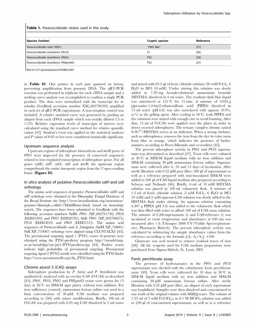
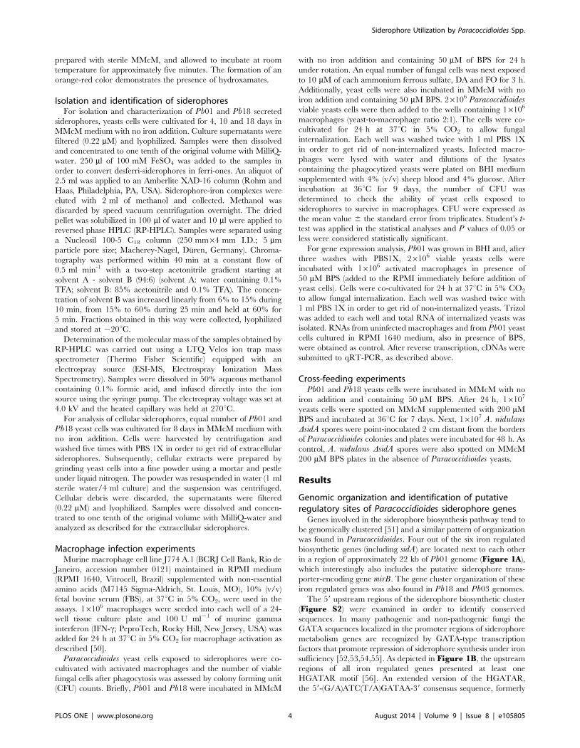
Table 1. Paracoccidioides strains used in this study.
Species (isolate) Cryptic species Reference
Paracoccidioides lutzii (Pb01) ‘‘Pb01-like’’ [31]
Paracoccidioides brasiliensis (Pb18) S1 [26]
Paracoccidioides brasiliensis (Pb02) PS2 [26]
Paracoccidioides brasiliensis (PbEpm83) PS3 [27]
doi:10.1371/journal.pone.0105805.t001
Siderophore Utilization by Paracoccidioides Spp.
PLOS ONE | www.plosone.org 3 August 2014 | Volume 9 | Issue 8 | e105805
prepared with sterile MMcM, and allowed to incubate at room
temperature for approximately five minutes. The formation of an
orange-red color demonstrates the presence of hydroxamates.
Isolation and identification of siderophoresFor isolation and characterization of Pb01 and Pb18 secreted
siderophores, yeasts cells were cultivated for 4, 10 and 18 days in
MMcM medium with no iron addition. Culture supernatants were
filtered (0.22 mM) and lyophilized. Samples were then dissolved
and concentrated to one tenth of the original volume with MilliQ-
water. 250 ml of 100 mM FeSO4 was added to the samples in
order to convert desferri-siderophores in ferri-ones. An aliquot of
2.5 ml was applied to an Amberlite XAD-16 column (Rohm and
Haas, Philadelphia, PA, USA). Siderophore-iron complexes were
eluted with 2 ml of methanol and collected. Methanol was
discarded by speed vacuum centrifugation overnight. The dried
pellet was solubilized in 100 ml of water and 10 ml were applied to
reversed phase HPLC (RP-HPLC). Samples were separated using
a Nucleosil 100-5 C18 column (250 mm64 mm I.D.; 5 mm
particle pore size; Macherey-Nagel, Duren, Germany). Chroma-
tography was performed within 40 min at a constant flow of
0.5 ml min-1 with a two-step acetonitrile gradient starting at
solvent A - solvent B (94:6) (solvent A: water containing 0.1%
TFA; solvent B: 85% acetonitrile and 0.1% TFA). The concen-
tration of solvent B was increased linearly from 6% to 15% during
10 min, from 15% to 60% during 25 min and held at 60% for
5 min. Fractions obtained in this way were collected, lyophilized
and stored at 220uC.
Determination of the molecular mass of the samples obtained by
RP-HPLC was carried out using a LTQ Velos ion trap mass
spectrometer (Thermo Fisher Scientific) equipped with an
electrospray source (ESI-MS, Electrospray Ionization Mass
Spectrometry). Samples were dissolved in 50% aqueous methanol
containing 0.1% formic acid, and infused directly into the ion
source using the syringe pump. The electrospray voltage was set at
4.0 kV and the heated capillary was held at 270uC.
For analysis of cellular siderophores, equal number of Pb01 and
Pb18 yeast cells was cultivated for 8 days in MMcM medium with
no iron addition. Cells were harvested by centrifugation and
washed five times with PBS 1X in order to get rid of extracellular
siderophores. Subsequently, cellular extracts were prepared by
grinding yeast cells into a fine powder using a mortar and pestle
under liquid nitrogen. The powder was resuspended in water (1 ml
sterile water/4 ml culture) and the suspension was centrifuged.
Cellular debris were discarded, the supernatants were filtered
(0.22 mM) and lyophilized. Samples were dissolved and concen-
trated to one tenth of the original volume with MilliQ-water and
analyzed as described for the extracellular siderophores.
Macrophage infection experimentsMurine macrophage cell line J774 A.1 (BCRJ Cell Bank, Rio de
Janeiro, accession number 0121) maintained in RPMI medium
(RPMI 1640, Vitrocell, Brazil) supplemented with non-essential
amino acids (M7145 Sigma-Aldrich, St. Louis, MO), 10% (v/v)
fetal bovine serum (FBS), at 37uC in 5% CO2, were used in the
assays. 16106 macrophages were seeded into each well of a 24-
well tissue culture plate and 100 U ml21 of murine gamma
interferon (IFN-c; PeproTech, Rocky Hill, New Jersey, USA) was
added for 24 h at 37uC in 5% CO2 for macrophage activation as
described [50].
Paracoccidioides yeast cells exposed to siderophores were co-
cultivated with activated macrophages and the number of viable
fungal cells after phagocytosis was assessed by colony forming unit
(CFU) counts. Briefly, Pb01 and Pb18 were incubated in MMcM
with no iron addition and containing 50 mM of BPS for 24 h
under rotation. An equal number of fungal cells was next exposed
to 10 mM of each ammonium ferrous sulfate, DA and FO for 3 h.
Additionally, yeast cells were also incubated in MMcM with no
iron addition and containing 50 mM BPS. 26106 Paracoccidioidesviable yeasts cells were then added to the wells containing 16106
macrophages (yeast-to-macrophage ratio 2:1). The cells were co-
cultivated for 24 h at 37uC in 5% CO2 to allow fungal
internalization. Each well was washed twice with 1 ml PBS 1X
in order to get rid of non-internalized yeasts. Infected macro-
phages were lysed with water and dilutions of the lysates
containing the phagocytized yeasts were plated on BHI medium
supplemented with 4% (v/v) sheep blood and 4% glucose. After
incubation at 36uC for 9 days, the number of CFU was
determined to check the ability of yeast cells exposed to
siderophores to survive in macrophages. CFU were expressed as
the mean value 6 the standard error from triplicates. Student’s t-test was applied in the statistical analyses and P values of 0.05 or
less were considered statistically significant.
For gene expression analysis, Pb01 was grown in BHI and, after
three washes with PBS1X, 26106 viable yeasts cells were
incubated with 16106 activated macrophages in presence of
50 mM BPS (added to the RPMI immediately before addition of
yeast cells). Cells were co-cultivated for 24 h at 37uC in 5% CO2
to allow fungal internalization. Each well was washed twice with
1 ml PBS 1X in order to get rid of non-internalized yeasts. Trizol
was added to each well and total RNA of internalized yeasts was
isolated. RNAs from uninfected macrophages and from Pb01 yeast
cells cultured in RPMI 1640 medium, also in presence of BPS,
were obtained as control. After reverse transcription, cDNAs were
submitted to qRT-PCR, as described above.
Cross-feeding experimentsPb01 and Pb18 yeasts cells were incubated in MMcM with no
iron addition and containing 50 mM BPS. After 24 h, 16107
yeasts cells were spotted on MMcM supplemented with 200 mM
BPS and incubated at 36uC for 7 days. Next, 16107 A. nidulansDsidA spores were point-inoculated 2 cm distant from the borders
of Paracoccidioides colonies and plates were incubated for 48 h. As
control, A. nidulans DsidA spores were also spotted on MMcM
200 mM BPS plates in the absence of Paracoccidioides yeasts.
Results
Genomic organization and identification of putativeregulatory sites of Paracoccidioides siderophore genes
Genes involved in the siderophore biosynthesis pathway tend to
be genomically clustered [51] and a similar pattern of organization
was found in Paracoccidioides. Four out of the six iron regulated
biosynthetic genes (including sidA) are located next to each other
in a region of approximately 22 kb of Pb01 genome (Figure 1A),
which interestingly also includes the putative siderophore trans-
porter-encoding gene mirB. The gene cluster organization of these
iron regulated genes was also found in Pb18 and Pb03 genomes.
The 59 upstream regions of the siderophore biosynthetic cluster
(Figure S2) were examined in order to identify conserved
sequences. In many pathogenic and non-pathogenic fungi the
GATA sequences localized in the promoter regions of siderophore
metabolism genes are recognized by GATA-type transcription
factors that promote repression of siderophore synthesis under iron
sufficiency [52,53,54,55]. As depicted in Figure 1B, the upstream
regions of all iron regulated genes presented at least one
HGATAR motif [56]. An extended version of the HGATAR,
the 59-(G/A)ATC(T/A)GATAA-39 consensus sequence, formerly
Siderophore Utilization by Paracoccidioides Spp.
PLOS ONE | www.plosone.org 4 August 2014 | Volume 9 | Issue 8 | e105805
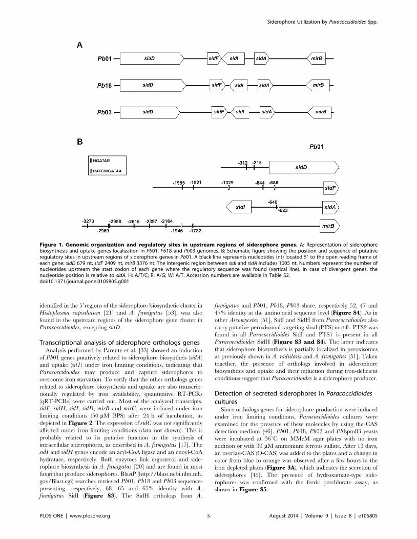
identified in the 59regions of the siderophore biosynthetic cluster in
Histoplasma capsulatum [21] and A. fumigatus [53], was also
found in the upstream regions of the siderophore gene cluster in
Paracoccidioides, excepting sidD.
Transcriptional analysis of siderophore orthologs genesAnalysis performed by Parente et al. [33] showed an induction
of Pb01 genes putatively related to siderophore biosyntheis (sidA)
and uptake (sit1) under iron limiting conditions, indicating that
Paracoccidioides may produce and capture siderophores to
overcome iron starvation. To verify that the other orthologs genes
related to siderophore biosynthesis and uptake are also transcrip-
tionally regulated by iron availability, quantitative RT-PCRs
(qRT-PCRs) were carried out. Most of the analyzed transcripts,
sidF, sidH, sidI, sidD, mirB and mirC, were induced under iron
limiting conditions (50 mM BPS) after 24 h of incubation, as
depicted in Figure 2. The expression of sidC was not significantly
affected under iron limiting conditions (data not shown). This is
probably related to its putative function in the synthesis of
intracellular siderophores, as described in A. fumigatus [17]. The
sidI and sidH genes encode an acyl-CoA ligase and an enoyl-CoA
hydratase, respectively. Both enzymes link ergosterol and side-
rophore biosynthesis in A. fumigatus [20] and are found in most
fungi that produce siderophores. BlastP (http://blast.ncbi.nlm.nih.
gov/Blast.cgi) searches retrieved Pb01, Pb18 and Pb03 sequences
presenting, respectively, 68, 65 and 65% identity with A.fumigatus SidI (Figure S3). The SidH orthologs from A.
fumigatus and Pb01, Pb18, Pb03 share, respectively 52, 47 and
47% identity at the amino acid sequence level (Figure S4). As in
other Ascomycetes [51], SidI and SidH from Paracoccidioides also
carry putative peroxisomal targeting sinal (PTS) motifs. PTS2 was
found in all Paracoccidioides SidI and PTS1 is present in all
Paracoccidioides SidH (Figure S3 and S4). The latter indicates
that siderophore biosynthesis is partially localized in peroxisomes
as previously shown in A. nidulans and A. fumigatus [51]. Taken
together, the presence of orthologs involved in siderophore
biosynthesis and uptake and their induction during iron-deficient
conditions suggest that Paracoccidioides is a siderophore producer.
Detection of secreted siderophores in Paracoccidioidescultures
Since orthologs genes for siderophore production were induced
under iron limiting conditions, Paracoccidioides cultures were
examined for the presence of these molecules by using the CAS
detection medium [46]. Pb01, Pb18, Pb02 and PbEpm83 yeasts
were incubated at 36uC on MMcM agar plates with no iron
addition or with 30 mM ammonium ferrous sulfate. After 13 days,
an overlay-CAS (O-CAS) was added to the plates and a change in
color from blue to orange was observed after a few hours in the
iron depleted plates (Figure 3A), which indicates the secretion of
siderophores [45]. The presence of hydroxamate-type side-
rophores was confirmed with the ferric perchlorate assay, as
shown in Figure S5.
Figure 1. Genomic organization and regulatory sites in upstream regions of siderophore genes. A: Representation of siderophorebiosynthesis and uptake genes localization in Pb01, Pb18 and Pb03 genomes. B: Schematic figure showing the position and sequence of putativeregulatory sites in upstream regions of siderophore genes in Pb01. A black line represents nucleotides (nt) located 59 to the open reading frame ofeach gene: sidD 679 nt, sidF 2409 nt, mirB 3376 nt. The intergenic region between sidI and sidA includes 1005 nt. Numbers represent the number ofnucleotides upstream the start codon of each gene where the regulatory sequence was found (vertical line). In case of divergent genes, thenucleotide position is relative to sidA. H: A/T/C; R: A/G; W: A/T. Accession numbers are available in Table S2.doi:10.1371/journal.pone.0105805.g001
Siderophore Utilization by Paracoccidioides Spp.
PLOS ONE | www.plosone.org 5 August 2014 | Volume 9 | Issue 8 | e105805
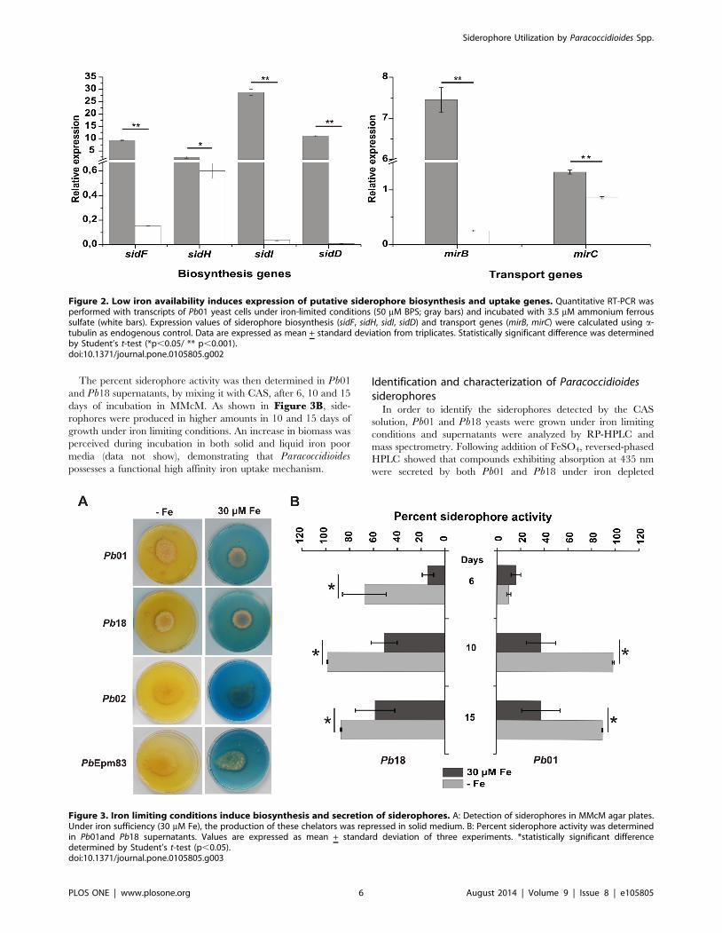
The percent siderophore activity was then determined in Pb01
and Pb18 supernatants, by mixing it with CAS, after 6, 10 and 15
days of incubation in MMcM. As shown in Figure 3B, side-
rophores were produced in higher amounts in 10 and 15 days of
growth under iron limiting conditions. An increase in biomass was
perceived during incubation in both solid and liquid iron poor
media (data not show), demonstrating that Paracoccidioidespossesses a functional high affinity iron uptake mechanism.
Identification and characterization of Paracoccidioidessiderophores
In order to identify the siderophores detected by the CAS
solution, Pb01 and Pb18 yeasts were grown under iron limiting
conditions and supernatants were analyzed by RP-HPLC and
mass spectrometry. Following addition of FeSO4, reversed-phased
HPLC showed that compounds exhibiting absorption at 435 nm
were secreted by both Pb01 and Pb18 under iron depleted
Figure 2. Low iron availability induces expression of putative siderophore biosynthesis and uptake genes. Quantitative RT-PCR wasperformed with transcripts of Pb01 yeast cells under iron-limited conditions (50 mM BPS; gray bars) and incubated with 3.5 mM ammonium ferroussulfate (white bars). Expression values of siderophore biosynthesis (sidF, sidH, sidI, sidD) and transport genes (mirB, mirC) were calculated using a-tubulin as endogenous control. Data are expressed as mean + standard deviation from triplicates. Statistically significant difference was determinedby Student’s t-test (*p,0.05/ ** p,0.001).doi:10.1371/journal.pone.0105805.g002
Figure 3. Iron limiting conditions induce biosynthesis and secretion of siderophores. A: Detection of siderophores in MMcM agar plates.Under iron sufficiency (30 mM Fe), the production of these chelators was repressed in solid medium. B: Percent siderophore activity was determinedin Pb01and Pb18 supernatants. Values are expressed as mean + standard deviation of three experiments. *statistically significant differencedetermined by Student’s t-test (p,0.05).doi:10.1371/journal.pone.0105805.g003
Siderophore Utilization by Paracoccidioides Spp.
PLOS ONE | www.plosone.org 6 August 2014 | Volume 9 | Issue 8 | e105805
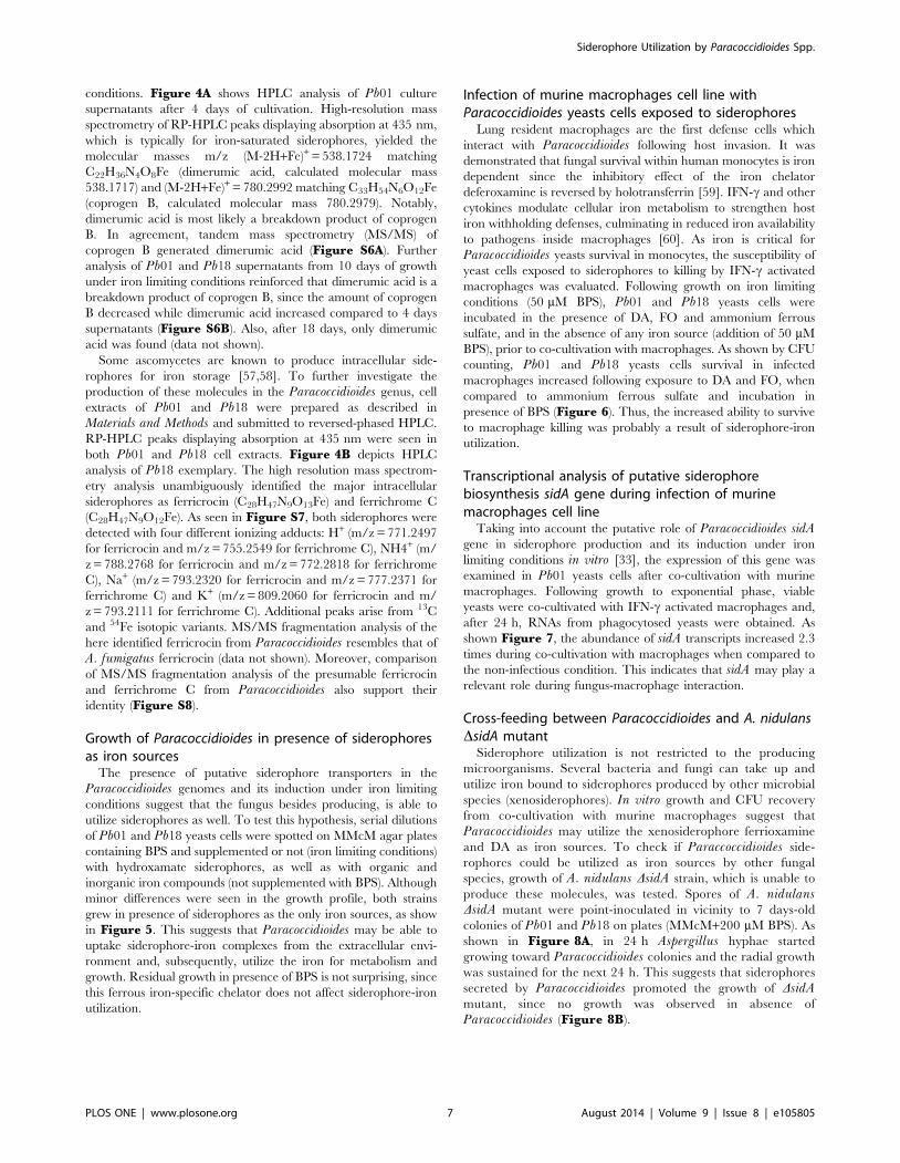
conditions. Figure 4A shows HPLC analysis of Pb01 culture
supernatants after 4 days of cultivation. High-resolution mass
spectrometry of RP-HPLC peaks displaying absorption at 435 nm,
which is typically for iron-saturated siderophores, yielded the
molecular masses m/z (M-2H+Fe)+ = 538.1724 matching
C22H36N4O8Fe (dimerumic acid, calculated molecular mass
538.1717) and (M-2H+Fe)+ = 780.2992 matching C33H54N6O12Fe
(coprogen B, calculated molecular mass 780.2979). Notably,
dimerumic acid is most likely a breakdown product of coprogen
B. In agreement, tandem mass spectrometry (MS/MS) of
coprogen B generated dimerumic acid (Figure S6A). Further
analysis of Pb01 and Pb18 supernatants from 10 days of growth
under iron limiting conditions reinforced that dimerumic acid is a
breakdown product of coprogen B, since the amount of coprogen
B decreased while dimerumic acid increased compared to 4 days
supernatants (Figure S6B). Also, after 18 days, only dimerumic
acid was found (data not shown).
Some ascomycetes are known to produce intracellular side-
rophores for iron storage [57,58]. To further investigate the
production of these molecules in the Paracoccidioides genus, cell
extracts of Pb01 and Pb18 were prepared as described in
Materials and Methods and submitted to reversed-phased HPLC.
RP-HPLC peaks displaying absorption at 435 nm were seen in
both Pb01 and Pb18 cell extracts. Figure 4B depicts HPLC
analysis of Pb18 exemplary. The high resolution mass spectrom-
etry analysis unambiguously identified the major intracellular
siderophores as ferricrocin (C28H47N9O13Fe) and ferrichrome C
(C28H47N9O12Fe). As seen in Figure S7, both siderophores were
detected with four different ionizing adducts: H+ (m/z = 771.2497
for ferricrocin and m/z = 755.2549 for ferrichrome C), NH4+ (m/
z = 788.2768 for ferricrocin and m/z = 772.2818 for ferrichrome
C), Na+ (m/z = 793.2320 for ferricrocin and m/z = 777.2371 for
ferrichrome C) and K+ (m/z = 809.2060 for ferricrocin and m/
z = 793.2111 for ferrichrome C). Additional peaks arise from 13C
and 54Fe isotopic variants. MS/MS fragmentation analysis of the
here identified ferricrocin from Paracoccidioides resembles that of
A. fumigatus ferricrocin (data not shown). Moreover, comparison
of MS/MS fragmentation analysis of the presumable ferricrocin
and ferrichrome C from Paracoccidioides also support their
identity (Figure S8).
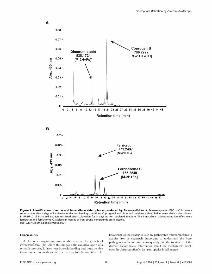
Growth of Paracoccidioides in presence of siderophoresas iron sources
The presence of putative siderophore transporters in the
Paracoccidioides genomes and its induction under iron limiting
conditions suggest that the fungus besides producing, is able to
utilize siderophores as well. To test this hypothesis, serial dilutions
of Pb01 and Pb18 yeasts cells were spotted on MMcM agar plates
containing BPS and supplemented or not (iron limiting conditions)
with hydroxamate siderophores, as well as with organic and
inorganic iron compounds (not supplemented with BPS). Although
minor differences were seen in the growth profile, both strains
grew in presence of siderophores as the only iron sources, as show
in Figure 5. This suggests that Paracoccidioides may be able to
uptake siderophore-iron complexes from the extracellular envi-
ronment and, subsequently, utilize the iron for metabolism and
growth. Residual growth in presence of BPS is not surprising, since
this ferrous iron-specific chelator does not affect siderophore-iron
utilization.
Infection of murine macrophages cell line withParacoccidioides yeasts cells exposed to siderophores
Lung resident macrophages are the first defense cells which
interact with Paracoccidioides following host invasion. It was
demonstrated that fungal survival within human monocytes is iron
dependent since the inhibitory effect of the iron chelator
deferoxamine is reversed by holotransferrin [59]. IFN-c and other
cytokines modulate cellular iron metabolism to strengthen host
iron withholding defenses, culminating in reduced iron availability
to pathogens inside macrophages [60]. As iron is critical for
Paracoccidioides yeasts survival in monocytes, the susceptibility of
yeast cells exposed to siderophores to killing by IFN-c activated
macrophages was evaluated. Following growth on iron limiting
conditions (50 mM BPS), Pb01 and Pb18 yeasts cells were
incubated in the presence of DA, FO and ammonium ferrous
sulfate, and in the absence of any iron source (addition of 50 mM
BPS), prior to co-cultivation with macrophages. As shown by CFU
counting, Pb01 and Pb18 yeasts cells survival in infected
macrophages increased following exposure to DA and FO, when
compared to ammonium ferrous sulfate and incubation in
presence of BPS (Figure 6). Thus, the increased ability to survive
to macrophage killing was probably a result of siderophore-iron
utilization.
Transcriptional analysis of putative siderophorebiosynthesis sidA gene during infection of murinemacrophages cell line
Taking into account the putative role of Paracoccidioides sidAgene in siderophore production and its induction under iron
limiting conditions in vitro [33], the expression of this gene was
examined in Pb01 yeasts cells after co-cultivation with murine
macrophages. Following growth to exponential phase, viable
yeasts were co-cultivated with IFN-c activated macrophages and,
after 24 h, RNAs from phagocytosed yeasts were obtained. As
shown Figure 7, the abundance of sidA transcripts increased 2.3
times during co-cultivation with macrophages when compared to
the non-infectious condition. This indicates that sidA may play a
relevant role during fungus-macrophage interaction.
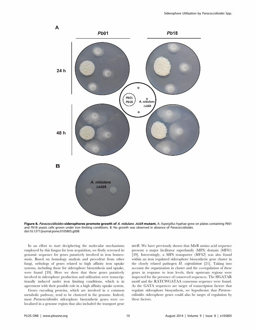
Cross-feeding between Paracoccidioides and A. nidulansDsidA mutant
Siderophore utilization is not restricted to the producing
microorganisms. Several bacteria and fungi can take up and
utilize iron bound to siderophores produced by other microbial
species (xenosiderophores). In vitro growth and CFU recovery
from co-cultivation with murine macrophages suggest that
Paracoccidioides may utilize the xenosiderophore ferrioxamine
and DA as iron sources. To check if Paraccoccidioides side-
rophores could be utilized as iron sources by other fungal
species, growth of A. nidulans DsidA strain, which is unable to
produce these molecules, was tested. Spores of A. nidulansDsidA mutant were point-inoculated in vicinity to 7 days-old
colonies of Pb01 and Pb18 on plates (MMcM+200 mM BPS). As
shown in Figure 8A, in 24 h Aspergillus hyphae started
growing toward Paracoccidioides colonies and the radial growth
was sustained for the next 24 h. This suggests that siderophores
secreted by Paracoccidioides promoted the growth of DsidAmutant, since no growth was observed in absence of
Paracoccidioides (Figure 8B).
Siderophore Utilization by Paracoccidioides Spp.
PLOS ONE | www.plosone.org 7 August 2014 | Volume 9 | Issue 8 | e105805
Discussion
As for other organisms, iron is also essential for growth of
Paracoccidioides [32]. Since this fungus is the causative agent of a
systemic mycosis, it faces host iron-withholding and must be able
to overcome this condition in order to establish the infection. The
knowledge of the strategies used by pathogenic microorganisms to
acquire iron is extremely important to understand the host-
pathogen interaction and, consequently, for the treatment of the
disease. Nevertheless, information about the mechanisms devel-
oped by Paracoccidioides for iron uptake is still scarce.
Figure 4. Identification of extra- and intracellular siderophores produced by Paracoccidioides. A: Reversed-phase HPLC of Pb01culturesupernatants after 4 days of incubation under iron limiting conditions. Coprogen B and dimerumic acid were identified as extracellular siderophores.B: RP-HPLC of Pb18 cell extracts obtained after cultivation for 8 days in iron depleted medium. The intracellular siderophores identified wereferricrocin and ferrichrome C. Molecular masses of iron bound compounds are indicated.doi:10.1371/journal.pone.0105805.g004
Siderophore Utilization by Paracoccidioides Spp.
PLOS ONE | www.plosone.org 8 August 2014 | Volume 9 | Issue 8 | e105805
Figure 5. Effect of siderophores on Paracoccidioides growth. After growth on MMcM + BPS for 24 h, Pb01 and Pb18 yeast cells were washedand serially diluted. 105 to 102 cells were spotted on MMcM agar plates containing 50 mM BPS only, 50 mM BPS plus 10 mM of siderophores and 10 mMof inorganic and organic iron (without BPS). Inorganic iron: ammonium ferrous sulfate; organic iron: ammonium ferric citrate; DA: dimerumic acid; FO:ferrioxamine.doi:10.1371/journal.pone.0105805.g005
Figure 6. Exposure to siderophores increases Paracoccidioidesability to survive macrophage killing. Prior exposure to 10 mM FOand DA enhanced Pb01 and Pb18 resistance to macrophage killing. CFUcounts are expressed as mean + standard error from triplicates,representative of two independent experiments. *statistically significantdifference determined by Student’s t-test (p,0.05).doi:10.1371/journal.pone.0105805.g006
Figure 7. Paracoccidioides sidA expression is induced duringmurine macrophage infection. Quantitative RT-PCR was performedwith transcripts of Pb01 yeast cells phagocytosed by murinemacrophages after 24 h of co-incubation. As control, yeast cells wereincubated for 24 h in RPMI medium. 50 mM BPS was added to the RPMImedium in both conditions. Expression values were calculated using a-tubulin as endogenous control. Data are expressed as mean + standarddeviation from triplicates. *statistically significant difference determinedby Student’s t-test (p,0.001).doi:10.1371/journal.pone.0105805.g007
Siderophore Utilization by Paracoccidioides Spp.
PLOS ONE | www.plosone.org 9 August 2014 | Volume 9 | Issue 8 | e105805
In an effort to start deciphering the molecular mechanisms
employed by this fungus for iron acquisition, we firstly screened its
genomic sequence for genes putatively involved in iron homeo-
stasis. Based on homology analysis and precedent from other
fungi, orthologs of genes related to high affinity iron uptake
systems, including those for siderophore biosynthesis and uptake,
were found [39]. Here we show that these genes putatively
involved in siderophore production and utilization were transcrip-
tionally induced under iron limiting conditions, which is in
agreement with their possible role in a high affinity uptake system.
Genes encoding proteins, which are involved in a common
metabolic pathway, tend to be clustered in the genome. Indeed,
most Paracoccidioides siderophore biosynthetic genes were co-
localized in a genome region that also included the transport gene
mirB. We have previously shown that MirB amino acid sequence
presents a major facilitator superfamily (MFS) domain (MFS1)
[39]. Interestingly, a MFS transporter (MFS2) was also found
within an iron regulated siderophore biosynthetic gene cluster in
the closely related pathogen H. capsulatum [21]. Taking into
account the organization in cluster and the co-regulation of these
genes in response to iron levels, their upstream regions were
inspected for the presence of conserved sequences. The HGATAR
motif and the RATCWGATAA consensus sequence were found.
As the GATA sequences are target of transcription factors that
regulate siderophore biosynthesis, we hypothesize that Paracoc-cidioides siderophore genes could also be target of regulation by
these factors.
Figure 8. Paracoccidioides siderophores promote growth of A. nidulans DsidA mutant. A: Aspergillus hyphae grew on plates containing Pb01and Pb18 yeasts cells grown under iron limiting conditions. B: No growth was observed in absence of Paracoccidioides.doi:10.1371/journal.pone.0105805.g008
Siderophore Utilization by Paracoccidioides Spp.
PLOS ONE | www.plosone.org 10 August 2014 | Volume 9 | Issue 8 | e105805
Taken together, the presence and putative regulatory elements
of siderophore biosynthetic genes strongly suggested that Para-coccidioides could be a siderophore producer. The presence of
these molecules was firstly detected in iron deprived Paracoccid-ioides agar plates with an overlay of CAS solution, which suggested
the presence of hydroxamates, siderophores typically produced by
fungi [14]. Even when iron was omitted from the culture medium,
the production and secretion of desferri-hydroxamates, which
extracellularly chelated traces of iron, and the subsequent uptake
of the iron-siderophore complex allowed fungal growth. Side-
rophore production by Paracoccidioides was more affected by the
iron addition in solid than in liquid medium. Iron at 30 mM
completely abrogated siderophore production in agar plates, but
the same concentration did not impair siderophore secretion in
culture supernatants. Growth rate on liquid medium is higher than
in solid. As the utilization of iron increases according to the
incubation time, due to the consumption by the growing number
of cells, the availability of this nutrient in the culture decreases and
induces the production and secretion of siderophores, even if iron
was added initially. The same differences in siderophore produc-
tion were observed in Aspergillus species [47].
Fungal hydroxamate siderophores can be classified into four
structural families: coprogens, ferrichromes, fusarinines and
rhodotorulic acid. Reversed-phase HPLC and mass spectrometry
analysis allowed the confirmation of hydroxamate production by
Paracoccidioides. Coprogen B and dimerumic acid were identified
as extracellular siderophores while ferrichrome C and ferricrocin
as the intracellular ones. Coprogen-type siderophores contains
anhydromevalonyl residues linked to the hydroxylated ornithine
by the action of the transacylase SidF. This acyl group derives
from mevalonate by CoA ligation and dehydration catalyzed,
respectively, by SidI and SidH [20]. Accordingly, the presence of
orthologs to sidI, sidH and sidF in Paracoccidioides genome is not
surprising. Coprogen B consists of a fusarinine molecule linked to
the dihydroxamate dimerumic acid. In Paracoccidioides young
cultures the amount of coprogen B in the supernatants is higher
than the amount of dimerumic acid while in older cultures this
proportion is reversed. This strongly indicates that the dimerumic
acid found in Paracoccidioides supernatants is a byproduct of
coprogen B. Similarly, dimerumic acid was identified in H.capsulatum cultures as a breakdown product of coprogen B
[61,62]. Both siderophores were also recognized in supernatants of
Blastomyces dermatitidis grown under iron-poor conditions [63].
Ferrichrome C and ferricrocin are cyclic hexapeptides in which
the acyl group bound to the hydroxyornithine is an acetyl [64].
Ferricrocin is produced intracellularly by A. fumigatus and A.nidulans for hyphal iron storage and distribution [17,58,65]. The
basidiomycete Ustilago maydis produces ferrichrome and ferri-
chrome A for iron acquisition [66], while ferrichrome C is
produced by the dermatophyte Trichophyton rubrum, which also
synthesizes ferricrocin [67]. Although both ferrichrome C and
ferrricrocin were identified in Paracoccidioides cellular extracts,
their role in iron storage requires further investigation. Interest-
ingly, the extra- and intracellular siderophores discussed above are
not only produced by human pathogens. The plant pathogen
Magnaporthe grisea and the non-pathogenic model organism
Neurospora crassa secrete siderophores of the coprogen-type for
iron acquisition and use ferricrocin for intracellular iron storage
[68,69]. This demonstrates the broad utilization of siderophores as
a strategy for iron acquisition in fungi.
A previous study demonstrated that P. brasiliensis plating
efficiency is enhanced in presence of coprogen B, dimerumic acid
(both isolated from B. dermatitidis) and ferrichrome, the latter
being the most effective growth factor [70]. Here we showed that
ferricrocin and ferrioxamine can also be used by Paracoccidioidesas iron sources. In fungi, the uptake of siderophore-iron complex is
usually mediated by transporters belonging to the UMF/SIT
subfamily of the major facilitator superfamily (MFS). The three
genes encoding putative siderophore transporter orthologs found
at Paracoccidioides genome present a MFS1 domain indicating
that they belong to the MFS [39]. The presence of more than one
putative siderophore transporter in Paracoccidioides may reflect its
ability to utilize a variety of siderophore as iron sources, including
the xenosiderophore ferrioxamine.
Cross-feeding experiments demonstrated that siderophores
secreted by Paracoccidioides restored the growth of the non-
producer A. nidulans DsidA mutant. This strain is unable to grow
in standard growth media unless siderophores are supplied [16]. A.nidulans encodes 10 putative siderophore transporters [19,71].
This fact, associated with the lack of a reductive iron assimilation
system, is in accordance with the ability of this fungus to utilize
xenosiderophores. Beyond the native siderophores ferricrocin and
TAFC, A. nidulans is also able to utilize iron from enterobactin, a
catecholate-type siderophore produced by bacteria, and from
ferrioxamine B, a less effective iron source [72]. Thereby,
utilization of Paracoccidioides siderophores by the A. nidulansDsidA mutant is in agreement.
Since siderophore production and uptake has been described as
an important virulence attribute for pathogens [13,21], we started
checking the influence of these molecules during fungus interac-
tion with murine macrophages. Following inhalation by the host,
Paracoccidioides propagules bind to macrophages in the lung.
Once phagocytosed, fungal cells are able to survive and multiply in
non-activated macrophages. However, IFN-c activated macro-
phages prevent multiplication of ingested fungus and, consequent-
ly, its survival [73]. Here we demonstrated that exposure of
Paracoccidioides yeasts cells to dimerumic acid and ferrioxamine
before co-cultivation with IFN-c activated macrophages resulted
in an increase in survival when compared to cells treated with
ammonium ferrous sulfate or BPS. We hypothesize that the
siderophore utilization before infection provided the iron require-
ments for fungal metabolism and for defense against oxidative
stress generated by macrophages. Our group previously demon-
strated the induction of a peroxisomal catalase (an iron dependent
enzyme involved in the detoxification of reactive oxygen species) in
Pb01 yeast cells derived from infected macrophages [74].
Paracoccidioides yeasts exposed to ammonium ferrous sulfate
and BPS presented similar survival after co-cultivation with
activated macrophages. BPS is a ferrous iron chelator which
inhibits reductive iron uptake but does not affect siderophore-
mediated iron uptake. Thus, the pre-incubation of yeasts cells in
MMcM containing BPS and the additional treatment for 3 h with
this iron chelator before macrophage infection probably trigged
the induction of siderophore biosynthesis and uptake. This fact
may explain yeasts survival after co-cultivation with macrophages.
The demand for iron for P. brasiliensis survival during interaction
with phagocytes was already investigated. Results demonstrated
that iron is essential for intracellular transformation of ingested
conidia to yeast in murine macrophages [75] and for survival of
yeast cells inside human monocytes [59]. In agreement with the
growth assay in presence of siderophores, we demonstrated that
these molecules play an effective role as iron sources for
Paracoccidioides. It was formerly demonstrated that addition of
FeCl3 to Paracoccidioides minimal medium is not as effective in
the increase of fungus plating efficiency, as supplementation with
siderophores [70]. The exposure to ferrichrome also enhanced the
survival of the opportunistic fungal pathogen Candida glabrata to
macrophage killing [76]. Also, A. fumigatus employs mainly a
Siderophore Utilization by Paracoccidioides Spp.
PLOS ONE | www.plosone.org 11 August 2014 | Volume 9 | Issue 8 | e105805

siderophore-based iron acquisition system in vivo instead of the
reductive iron assimilation pathway [13].
Quantification of transcripts level of the putative siderophore
biosynthetic gene sidA revealed that this gene was induced during
co-cultivation of Paracoccidioides with macrophages. This suggests
that the fungus probably produce siderophores to overcome low
iron availability imposed by these activated phagocytic cells. Such
strategy is employed by other fugal pathogens. Extra- and
intracellular siderophores were shown to be crucial for intracel-
lular growth of A. fumigatus within alveolar murine macrophages
[77] and expression of siderophore biosynthetic genes was detected
during murine infection with conidia [78]. In H. capsulatum the
expression of the sidA ortholog sid1 was also induced after
phagocytosis and required for adequate cellular growth in human
macrophages [79].
Altogether, our results revealed the ability of Paracoccidioides to
synthesize and utilize siderophore as iron sources. Although the
production of these iron chelators had been formerly reported, we
demonstrated here, for the first time, the identity of the produced
siderophores including the intracellular ones, whose production
was not mentioned before. Additionally, infection experiments
carried out with a murine macrophage cell line revealed that
siderophore utilization plays an important role during the
interaction of Paracoccidioides with mammalian cells. Despite
some studies had demonstrated the importance of iron in the
scenario of Paracoccidioides infection, evaluation of the impact of
iron metabolism on fungus pathogenicity was not deeply
investigated. This study was the first step of upcoming molecular
and functional analysis of siderophore biosynthetic and uptake
genes in Paracoccidioides. Indeed, studies are being carried out in
order to investigate the role of these genes as possible virulence
factors in this pathogenic fungus. The added knowledge is
clinically important since siderophore biosynthesis and uptake
represent possible targets for an antifungal chemotherapy due the
absence of these pathways in human cells.
Supporting Information
Figure S1 Biosynthetic pathway for fungal hydroxa-mates. All the expected genes for siderophore biosynthesis are
present in Paracoccidioides genomes. NRPSs: non-ribosomal
peptide synthetases. Adapted from [51].
(TIF)
Figure S2 Usptream regions of siderophore genes inPb01. Sequences are shown in 59R 39 sense.
(PDF)
Figure S3 Similarity of A. fumigatus SidI with putativeacyl-CoA ligase from Pb01, Pb18 and Pb03. The amino acid
sequences of the orthologs were aligned using the software
ClustalX2. Asterisks: amino acid identity. Dots: conserved
substitutions. Grey box: PTS2 motif.
(TIF)
Figure S4 Similarity of A. fumigatus SidH with putativeenoyl-CoA hydratase from Pb01, Pb18 and Pb03. The
amino acid sequences of the orthologs were aligned using the
software ClustalX2. Asterisks: amino acid identity. Dots: con-
served substitutions. Grey box: PTS1 motif. PTS1 scores: Pb01
(8.8), Pb18 (10.4) and Pb03 (10.4).
(TIF)
Figure S5 Detection of hydroxamate-type siderophoresin Pb01 and Pb18 supernatants by the ferric perchlorateassay. Pb01 and Pb18 supernatants from three independent
cultures in no iron MMcM (Fe -) presented an orange-red color
after addition of Fe(ClO4)3, revealing the presence of hydro-
xamates. Cultures of both Paracoccidioides isolates in the presence
of 30 mM ammonium ferrous sulfate (Fe +) were also tested and
the change in color was not observed. Sterile MMcM was used as
reference (Ref + and Ref -).
(TIF)
Figure S6 High-resolution mass spectrometry of Para-coccidioides extracellular siderophores. A: RP-HPLC peak
corresponding to coprogen B in Figure 4A was submitted to MS
and MS/MS analysis, demonstrating that dimerumic acid is as a
breakdown product of coprogen B. B: Longer periods of
cultivation result in an increase in the amount of dimerumic acid
over coprogen B, as demonstrated by RP-HPLC peaks from Pb18
supernatants obtained after 10 days of incubation.
(TIF)
Figure S7 High-resolution mass spectrometry of Para-coccidioides intracellular siderophores. RP-HPLC peaks
displayed at Figure 4B were submitted to mass spectrometry
analysis for molecular masses definition of ferricrocin (A) and
ferrichrome C (B). The four different ionizing adducts are shown.
(TIF)
Figure S8 MS/MS fragmentation analysis of ferricrocinand ferrichrome C. Fragments with identical molecular masses
(m/z = 370.094, m/z = 398.089 and m/z = 455.111) are framed in
red. Fragments that show a molecular mass difference of 15.99,
which corresponds to the mass difference of the two siderophores,
matching the mass difference of serine (in ferricrocin) and alanine
(in ferrichrome C) are framed in blue (m/z = 498.153 for
ferrichrome C plus m/z = 15.99 is m/z = 514.145 for ferricrocin;
m/z = 583.169 for ferrichrome C plus m/z = 15.99 is m/
z = 599.159 for ferricrocin; m/z = 737.244 for ferrichrome C plus
m/z = 15.99 is m/z = 753.234 for ferricrocin).
(TIF)
Table S1 Oligonucleotides primers used in quantitativeRT-PCR.(DOCX)
Table S2 Accession numbers of Paracoccidioides side-rophore genes available at http://www.broadinstitute.org/annotation/genome/paracoccidioides_brasiliensis/MultiHome.html.(DOCX)
Author Contributions
Conceived and designed the experiments: MGSB AMB HH CMAS.
Performed the experiments: MGSB EFLCB BEL GMG AMB. Analyzed
the data: MGSB HL HH CMAS. Contributed reagents/materials/analysis
tools: HH HL CMAS. Wrote the paper: MGSB CMAS.
References
1. Cairo G, Bernuzzi F, Recalcati S (2006) A precious metal: Iron, an essential
nutrient for all cells. Genes Nutr 1: 25–39.
2. Pandey A, Bringel F, Meyer JM (1994) Iron requirement and search for
siderophores in lacti acid bacteria. Appl Microbiol Biotechnol 40: 735–739.
3. Haber F, Weiss J (1934) The catalytic decomposition of hydrogen peroxide by
iron salts. Proc R Soc Lond 147: 332–351.
4. Halliwell B, Gutteridge JM (1984) Oxygen toxicity, oxygen radicals, transition
metals and disease. Biochem J 219: 1–14.
5. Kosman DJ (2003) Molecular mechanisms of iron uptake in fungi. Mol
Microbiol 47: 1185–1197.
6. Ganz T (2009) Iron in innate immunity: starve the invaders. Curr Opin
Immunol 21: 63–67.
Siderophore Utilization by Paracoccidioides Spp.
PLOS ONE | www.plosone.org 12 August 2014 | Volume 9 | Issue 8 | e105805
7. Nemeth E, Rivera S, Gabayan V, Keller C, Taudorf S, et al. (2004) IL-6
mediates hypoferremia of inflammation by inducing the synthesis of the iron
regulatory hormone hepcidin. J Clin Invest 113: 1271–1276.
8. Cassat JE, Skaar EP (2013) Iron in infection and immunity. Cell Host Microbe
13: 509–519.
9. Recalcati S, Locati M, Cairo G (2012) Systemic and cellular consequences of
macrophage control of iron metabolism. Semin Immunol 24: 393–398.
10. Raymond KN, Dertz EA, Kim SS (2003) Enterobactin: an archetype for
microbial iron transport. Proc Natl Acad Sci U S A 100: 3584–3588.
11. Neilands JB (1993) Siderophores. Arch Biochem Biophys 302: 1–3.
12. Hissen AH, Chow JM, Pinto LJ, Moore MM (2004) Survival of Aspergillusfumigatus in serum involves removal of iron from transferrin: the role of
siderophores. Infect Immun 72: 1402–1408.
13. Schrettl M, Bignell E, Kragl C, Joechl C, Rogers T, et al. (2004) Siderophore
biosynthesis but not reductive iron assimilation is essential for Aspergillusfumigatus virulence. J Exp Med 200: 1213–1219.
14. Van der Helm D, Winkelmann G (1994) Hydroxamates and polycarbonates as
iron transport agents (siderophores) in fungi. In: Winkelmann G, Winge DR,
editors. Metal ions in fungi. New York. pp. 39–148.
15. Plattner HJ, Diekmann H (1994) Enzymology of siderophore biosynthesis in
fungi. In: Winge DR, editor. Metal ions in fungi. New York: Marcel Decker. pp.
99–117.
16. Eisendle M, Oberegger H, Zadra I, Haas H (2003) The siderophore system is
essential for viability of Aspergillus nidulans: functional analysis of two genes
encoding l-ornithine N 5-monooxygenase (sidA) and a non-ribosomal peptide
synthetase (sidC). Mol Microbiol 49: 359–375.
17. Schrettl M, Bignell E, Kragl C, Sabiha Y, Loss O, et al. (2007) Distinct roles for
intra- and extracellular siderophores during Aspergillus fumigatus infection.
PLoS Pathog 3: 1195–1207.
18. Blatzer M, Schrettl M, Sarg B, Lindner HH, Pfaller K, et al. (2011) SidL, an
Aspergillus fumigatus transacetylase involved in biosynthesis of the siderophores
ferricrocin and hydroxyferricrocin. Appl Environ Microbiol 77: 4959–4966.
19. Haas H, Eisendle M, Turgeon BG (2008) Siderophores in fungal physiology and
virulence. Annu Rev Phytopathol 46: 149–187.
20. Yasmin S, Alcazar-Fuoli L, Grundlinger M, Puempel T, Cairns T, et al. (2012)
Mevalonate governs interdependency of ergosterol and siderophore biosyntheses
in the fungal pathogen Aspergillus fumigatus. Proc Natl Acad Sci U S A 109:
E497–504.
21. Hwang LH, Mayfield JA, Rine J, Sil A (2008) Histoplasma requires SID1, a
member of an iron-regulated siderophore gene cluster, for host colonization.
PLoS Pathog 4: e1000044.
22. Greenshields DL, Liu G, Feng J, Selvaraj G, Wei Y (2007) The siderophore
biosynthetic gene SID1, but not the ferroxidase gene FET3, is required for full
Fusarium graminearum virulence. Mol Plant Pathol 8: 411–421.
23. Hof C, Eisfeld K, Welzel K, Antelo L, Foster AJ, et al. (2007) Ferricrocin
synthesis in Magnaporthe grisea and its role in pathogenicity in rice. Mol Plant
Pathol 8: 163–172.
24. Hissen AH, Wan AN, Warwas ML, Pinto LJ, Moore MM (2005) The
Aspergillus fumigatus siderophore biosynthetic gene sidA, encoding L-ornithine
N5-oxygenase, is required for virulence. Infect Immun 73: 5493–5503.
25. Oide S, Moeder W, Krasnoff S, Gibson D, Haas H, et al. (2006) NPS6, encoding
a nonribosomal peptide synthetase involved in siderophore-mediated iron
metabolism, is a conserved virulence determinant of plant pathogenic
ascomycetes. Plant Cell 18: 2836–2853.
26. Matute DR, McEwen JG, Puccia R, Montes BA, San-Blas G, et al. (2006)
Cryptic speciation and recombination in the fungus Paracoccidioides brasiliensisas revealed by gene genealogies. Mol Biol Evol 23: 65–73.
27. Theodoro RC, Bagagli E, Oliveira C (2008) Phylogenetic analysis of PRP8
intein in Paracoccidioides brasiliensis species complex. Fungal Genet Biol 45:
1284–1291.
28. Bocca AL, Amaral AC, Teixeira MM, Sato PK, Shikanai-Yasuda MA, et al.
(2013) Paracoccidioidomycosis: eco-epidemiology, taxonomy and clinical and
therapeutic issues. Future Microbiol 8: 1177–1191.
29. Salgado-Salazar C, Jones LR, Restrepo A, McEwen JG (2010) The human
fungal pathogen Paracoccidioides brasiliensis (Onygenales: Ajellomycetaceae) is
a complex of two species: phylogenetic evidence from five mitochondrial
markers. Cladistics 26: 613–624.
30. Teixeira MM, Theodoro RC, de Carvalho MJ, Fernandes L, Paes HC, et al.
(2009) Phylogenetic analysis reveals a high level of speciation in the
Paracoccidioides genus. Mol Phylogenet Evol 52: 273–283.
31. Teixeira MM, Theodoro RC, Oliveira FF, Machado GC, Hahn RC, et al.
(2014) Paracoccidioides lutzii sp. nov.: biological and clinical implications. Med
Mycol 52: 19–28.
32. Arango R, Restrepo A (1988) Growth and production of iron chelants by
Paracoccidioides brasiliensis mycelial and yeast forms. J Med Vet Mycol 26:
113–118.
33. Parente AF, Bailao AM, Borges CL, Parente JA, Magalhaes AD, et al. (2011)
Proteomic analysis reveals that iron availability alters the metabolic status of the
pathogenic fungus Paracoccidioides brasiliensis. PLoS One 6: e22810.
34. Martinez R, Fiorillo AM (1978) Iron metabolism in patients with South
American blastomycosis (paracoccidioidomycosis). Rev Inst Med Trop Sao
Paulo 20: 195–201.
35. Martinez R, Fiorillo AM (1978) Effect of treatment of South American
blastomycosis (paracoccidioidomycosis) on parameters of iron metabolism. Rev
Inst Med Trop Sao Paulo 20: 190–194.
36. Bailao AM, Schrank A, Borges CL, Dutra V, Molinari-Madlum EEWI, et al.
(2006) Differential gene expression by Paracoccidioides brasiliensis in host
interaction conditions: representational difference analysis identifies candidate
genes associated with fungal pathogenesis. Microbes Infect 8: 2686–2697.
37. Bailao AM, Shrank A, Borges CL, Parente JA, Dutra V, et al. (2007) The
transcriptional profile of Paracoccidioides brasiliensis yeast cells is influenced by
human plasma. FEMS Immunol Med Microbiol 51: 43–57.
38. Longo LV, Nakayasu ES, Matsuo AL, Peres da Silva R, Sobreira TJ, et al.
(2013) Identification of human plasma proteins associated with the cell wall of
the pathogenic fungus Paracoccidioides brasiliensis. FEMS Microbiol Lett 341:
87–95.
39. Silva MG, Schrank A, Bailao EF, Bailao AM, Borges CL, et al. (2011) The
homeostasis of iron, copper, and zinc in Paracoccidioides brasiliensis,Cryptococcus neoformans var. grubii, and Cryptococcus gattii: a comparative
analysis. Front Microbiol 2: 49.
40. Restrepo A, Jimenez BE (1980) Growth of Paracoccidioides brasiliensis yeast
phase in a chemically defined culture medium. J Clin Microbiol 12: 279–281.
41. Pontecorvo G, Roper JA, Hemmons LM, Macdonald KD, Bufton AW (1953)
The genetics of Aspergillus nidulans. Adv Genet 5: 141–238.
42. Bookout AL, Cummins CL, Mangelsdorf DJ, Pesola JM, Kramer MF (2006)
High-throughput real-time quantitative reverse transcription PCR. Curr Protoc
Mol Biol Chapter 15: Unit 15 18.
43. Larkin MA, Blackshields G, Brown NP, Chenna R, McGettigan PA, et al. (2007)
Clustal W and Clustal X version 2.0. Bioinformatics 23: 2947–2948.
44. Neuberger G, Maurer-Stroh S, Eisenhaber B, Hartig A, Eisenhaber F (2003)
Prediction of peroxisomal targeting signal 1 containing proteins from amino acid
sequence. J Mol Biol 328: 581–592.
45. Perez-Miranda S, Cabirol N, George-Tellez R, Zamudio-Rivera LS, Fernandez
FJ (2007) O-CAS, a fast and universal method for siderophore detection.
J Microbiol Methods 70: 127–131.
46. Schwyn B, Neilands JB (1987) Universal chemical assay for the detection and
determination of siderophores. Anal Biochem 160: 47–56.
47. Machuca A, Milagres AM (2003) Use of CAS-agar plate modified to study the
effect of different variables on the siderophore production by Aspergillus. Lett
Appl Microbiol 36: 177–181.
48. Cox CD (1994) Deferration of laboratory media and assays for ferric and ferrous
ions. Methods Enzymol 235: 315–329.
49. Atkin CL, Neilands JB, Phaff HJ (1970) Rhodotorulic acid from species of
Leucosporidium, Rhodosporidium, Rhodotorula, Sporidiobolus, and Sporobolo-myces, and a new alanine-containing ferrichrome from Cryptococcus melibiosum.
J Bacteriol 103: 722–733.
50. Youseff BH, Holbrook ED, Smolnycki KA, Rappleye CA (2012) Extracellular
superoxide dismutase protects Histoplasma yeast cells from host-derived
oxidative stress. PLoS Pathog 8: e1002713.
51. Grundlinger M, Yasmin S, Lechner BE, Geley S, Schrettl M, et al. (2013) Fungal
siderophore biosynthesis is partially localized in peroxisomes. Mol Microbiol 88:
862–875.
52. Haas H, Zadra I, Stoffler G, Angermayr K (1999) The Aspergillus nidulansGATA factor SREA is involved in regulation of siderophore biosynthesis and
control of iron uptake. J Biol Chem 274: 4613–4619.
53. Schrettl M, Kim HS, Eisendle M, Kragl C, Nierman WC, et al. (2008) SreA-
mediated iron regulation in Aspergillus fumigatus. Mol Microbiol 70: 27–43.
54. Chao LY, Marletta MA, Rine J (2008) Sre1, an iron-modulated GATA DNA-
binding protein of iron-uptake genes in the fungal pathogen Histoplasmacapsulatum. Biochemistry 47: 7274–7283.
55. Harrison KA, Marzluf GA (2002) Characterization of DNA binding and the
cysteine rich region of SRE, a GATA factor in Neurospora crassa involved in
siderophore synthesis. Biochemistry 41: 15288–15295.
56. Scazzocchio C (2000) The fungal GATA factors. Curr Opin Microbiol 3: 126–
131.
57. Charlang G, Ng B, Horowitz NH, Horowitz RM (1981) Cellular and
extracellular siderophores of Aspergillus nidulans and Penicillium chrysogenum.
Mol Cell Biol 1: 94–100.
58. Wallner A, Blatzer M, Schrettl M, Sarg B, Lindner H, et al. (2009) Ferricrocin, a
siderophore involved in intra- and transcellular iron distribution in Aspergillusfumigatus. Appl Environ Microbiol 75: 4194–4196.
59. Dias-Melicio LA, Calvi SA, Peracoli MT, Soares AM (2005) Inhibitory effect of
deferoxamine on Paracoccidioides brasiliensis survival in human monocytes:
reversal by holotransferrin not by apotransferrin. Rev Inst Med Trop Sao Paulo
47: 263–266.
60. Weiss G (2005) Modification of iron regulation by the inflammatory response.
Best Pract Res Clin Haematol 18: 183–201.
61. Burt WR (1982) Identification of coprogen B and its breakdown products from
Histoplasma capsulatum. Infect Immun 35: 990–996.
62. Howard DH, Rafie R, Tiwari A, Faull KF (2000) Hydroxamate siderophores of
Histoplasma capsulatum. Infect Immun 68: 2338–2343.
63. Gauthier GM, Sullivan TD, Gallardo SS, Brandhorst TT, Vanden Wymelen-
berg AJ, et al. (2010) SREB, a GATA transcription factor that directs disparate
fates in Blastomyces dermatitidis including morphogenesis and siderophore
biosynthesis. PLoS Pathog 6: e1000846.
Siderophore Utilization by Paracoccidioides Spp.
PLOS ONE | www.plosone.org 13 August 2014 | Volume 9 | Issue 8 | e105805

64. Renshaw JC, Robson GD, Trinci APJ, Wiebe MG, Livens FR, et al. (2002)
Fungal siderophores: structures, functions and applications. Mycol Res 106:
1123–1142.
65. Eisendle M, Schrettl M, Kragl C, Muller D, Illmer P, et al. (2006) The
intracellular siderophore ferricrocin is involved in iron storage, oxidative-stress
resistance, germination, and sexual development in Aspergillus nidulans.Eukaryot Cell 5: 1596–1603.
66. Winterberg B, Uhlmann S, Linne U, Lessing F, Marahiel MA, et al. (2010)
Elucidation of the complete ferrichrome A biosynthetic pathway in Ustilagomaydis. Mol Microbiol 75: 1260–1271.
67. Mor H, Kashman Y, Winkelmann G, Barash I (1992) Characterization of
siderophores produced by different species of the dermatophytic fungi
Microsporum and Trichophyton. Biometals 5: 213–216.
68. Antelo L, Hof C, Welzel K, Eisfeld K, Sterner O, et al. (2006) Siderophores
produced by Magnaporthe grisea in the presence and absence of iron.
Z Naturforsch C 61: 461–464.
69. Matzanke BF, Bill E, Trautwein AX, Winkelmann G (1987) Role of
siderophores in iron storage in spores of Neurospora crassa and Aspergillusochraceus. J Bacteriol 169: 5873–5876.
70. Castaneda E, Brummer E, Perlman AM, McEwen JG, Stevens DA (1988) A
culture medium for Paracoccidioides brasiliensis with high plating efficiency, and
the effect of siderophores. J Med Vet Mycol 26: 351–358.
71. Haas H, Schoeser M, Lesuisse E, Ernst JF, Parson W, et al. (2003)
Characterization of the Aspergillus nidulans transporters for the siderophores
enterobactin and triacetylfusarinine C. Biochem J 371: 505–513.
72. Oberegger H, Schoeser M, Zadra I, Abt B, Haas H (2001) SREA is involved in
regulation of siderophore biosynthesis, utilization and uptake in Aspergillusnidulans. Mol Microbiol 41: 1077–1089.
73. Brummer E, Hanson LH, Restrepo A, Stevens DA (1989) Intracellular
multiplication of Paracoccidioides brasiliensis in macrophages: killing andrestriction of multiplication by activated macrophages. Infect Immun 57:
2289–2294.74. Grossklaus DA, Bailao AM, Rezende TCV, Borges CL, Oliveira MA, et al.
(2013) Response to oxidative stress in Paracoccidioides yeast cells as determined
by proteomic analysis. Microbes Infect 15: 347–364.75. Cano LE, Gomez B, Brummer E, Restrepo A, Stevens DA (1994) Inhibitory
effect of deferoxamine or macrophage activation on transformation ofParacoccidioides brasiliensis conidia ingested by macrophages: reversal by
holotransferrin. Infect Immun 62: 1494–1496.76. Nevitt T, Thiele DJ (2011) Host iron withholding demands siderophore
utilization for Candida glabrata to survive macrophage killing. PLoS Pathog 7:
e1001322.77. Schrettl M, Ibrahim-Granet O, Droin S, Huerre M, Latge JP, et al. (2010) The
crucial role of the Aspergillus fumigatus siderophore system in interaction withalveolar macrophages. Microbes Infect 12: 1035–1041.
78. McDonagh A, Fedorova ND, Crabtree J, Yu Y, Kim S, et al. (2008) Sub-
telomere directed gene expression during initiation of invasive aspergillosis.PLoS Pathog 4: e1000154.
79. Hilty J, George Smulian A, Newman SL (2011) Histoplasma capsulatum utilizessiderophores for intracellular iron acquisition in macrophages. Med Mycol 49:
633–642.
Siderophore Utilization by Paracoccidioides Spp.
PLOS ONE | www.plosone.org 14 August 2014 | Volume 9 | Issue 8 | e105805
Copyright © 2022 FDOKUMEN