Vocal individuality in contact calls of giant otters, Pteronura brasiliensis
Upload
independentCategory
view
1download
0
Microbes and Infection 10 (2008) 12e20www.elsevier.com/locate/micinf
Original article
Transcriptional response of murine macrophages upon infection withopsonized Paracoccidioides brasiliensis yeast cells
Simoneide S. Silva a,1, Aldo Henrique F.P. Tavares a,1, Danielle G. Passos-Silva b,Ana L. Fachin f, Santuza M.R. Teixeira b, Celia M.A. Soares e,
Maria Jose A. Carvalho a, Anamelia L. Bocca a, Ildinete Silva-Pereira a,Geraldo A.S. Passos c,d, Maria Sueli Soares Felipe a,*
a Departamento de Biologia Celular, Universidade de Brasılia, Brasılia, DF 70910-900, Brazilb Departamento de Bioquımica e Imunologia, Universidade Federal de Minas Gerais, Belo Horizonte, MG 31270-901, Brazil
c Departamento de Genetica, Universidade de S~ao Paulo, Ribeir~ao Preto, SP 14040-900, Brazild Faculdade de Odontologia, Universidade de S~ao Paulo, Ribeir~ao Preto 14040-900, SP, Brazile Departamento de Bioquımica, Universidade Federal de Goias, Goiania, GO 74001-970, Brazil
f Unidade de Biotecnologia, Universidade de Ribeir~ao Preto, Ribeir~ao Preto, SP 14096-380, Brazil
Received 15 May 2007; accepted 25 September 2007
Available online 2 October 2007
Abstract
Paracoccidioides brasiliensis is the etiologic agent of the Paracoccidioidomycosis the most common systemic mycosis in Latin America.Little is known about the regulation of genes involved in the innate immune host response to P. brasiliensis. We therefore examined thekinetic profile of gene expression of peritoneal macrophage infected with P. brasiliensis. Total RNA from macrophages at 6, 24 and48 h was extracted, hybridized onto nylon membranes and analyzed. An increase in the transcription of a number of pro-inflammatory mol-ecules encoding membrane proteins, metalloproteases, involved in adhesion and phagocytosis, are described. We observed also the differen-tial expression of genes whose products may cause apoptotic events induced at 24 h. In addition, considering the simultaneous analyses ofdifferential gene expression for the pathogen reported before by our group, at six hours post infection, we propose a model at molecular levelfor the P. brasiliensis-macrophage early interaction. In this regard, P. brasiliensis regulates genes specially related to stress and macrophages,at the same time point, up-regulate genes related to inflammation and phagocytosis, probably as an effort to counteract infection by thefungus.� 2007 Elsevier Masson SAS. All rights reserved.
Keywords: Paracoccidioides brasiliensis; Macrophage; cDNA microarray; Host-pathogen interaction
1. Introduction [1]. Animal models suggest that the natural infection route is
Paracoccidioides brasiliensis is a facultative intracellularfungus that causes Paracoccidioidomycosis (PCM), an impor-tant endemic disease in Latin America, especially in Brazil
* Corresponding author. Laboratorio de Biologia Molecular, Departamento
de Biologia Celular, Universidade de Brasılia-Unb Brasılia, Campus Darcy Ri-
beiro-Asa Norte, Brasılia, DF 70910-900, Brazil. Tel.: þ55 61 307 2423; fax:
þ55 61 349 8411.
E-mail address: [email protected] (M.S.S. Felipe).1 S.S.S and A.H.F.P.T contributed equally to this work.
1286-4579/$ - see front matter � 2007 Elsevier Masson SAS. All rights reserved.
doi:10.1016/j.micinf.2007.09.018
by inhalation of airborne conidia derived from the mycelialsaprophytic form of the fungus [2]. Once in the lungs itundergoes a thermally controlled dimorphic transition to theyeast form, which is an essential step for the establishmentof the infection [3].
As observed in other systemic mycoses, the most relevantdefence of the host against PCM is the cellular immunity medi-ated mainly by g-interferon activation of macrophages [4]. Con-versely, the observation of susceptible hosts has revealed thatnon-activated macrophages serve as a protected environment

13S.S. Silva et al. / Microbes and Infection 10 (2008) 12e20
wherein the fungus can survive and replicate, thus leading tofungal dissemination from the lungs to other organs [5].
Despite their important role in PCM, no high-throughputanalysis of host cell genes that be modulated upon infectionhas been reported for P. brasiliensis pathogen. Recently, ourgroup analysed by cDNA microarray technology the differen-tial gene expression of P. brasiliensis recovered from macro-phages upon six hours of infection [6]. However, whole lungor macrophage gene expression profiles in response to infectionby P. brasiliensis were limited to some genes coding for cyto-kines and host adhesion molecules [7,8]. This is in contrast toother models of microbial infection, in which numerous studiesemploying high-throughput genomics have been carried out toassess large-scale changes in the transcriptional profiles of bothhost and pathogen in their complex interaction. As shown forbacteria and in fungus-phagocyte interactions, microarray tech-nology enables the measurement of expression levels for hun-dreds to thousands of genes, providing a global understandingof key effectors related to pathogen virulence and host defencecan emerge [9e13]. In the present study, we have evaluated thekinetic profile of macrophage gene expression after exposure toP. brasiliensis yeast cells for 6, 24 and 48 h. We have been ableto identify a total of 118 genes that are differentially expressedat least at one time point. Those genes are related to severalimmune processes such as inflammation, cell membrane, tran-scriptional regulation, signal transduction and apoptosis. Inaddition we discuss the host-pathogen interaction in the lightof recent results on P. brasiliensis gene expression patternsupon phagocytosis by peritoneal macrophages [6]. Thus ourdata add to the current understanding of early host gene expres-sion upon infection by P. brasiliensis and suggest an interactionmodel for P. brasiliensis-infected macrophages.
2. Materials and methods
2.1. Isolation, cultivation and infection of murineperitoneal macrophages with P. brasiliensis yeast cellsand total RNA extraction of host cells
The clinical isolate of P. brasiliensis-Pb01 (ATCC-MYA-826) was used for macrophages infection. Isolation, cultiva-tion and infection of murine peritoneal macrophages protocolwere carried out as previously described [6]. Cultures wereincubated at 37 �C under a humidified, 5% CO2 atmosphere.At 6, 24 and 48 h upon infection, extracellular fungi wereremoved by exhaustive washing with RPMI pre-warmed to37 �C. Macrophages were then lysed and total RNA was ex-tracted with the Trizol� reagent (Invitrogen, USA). TotalRNA from control peritoneal macrophages was also extractedusing the same protocol. The infection of macrophages withyeast P. brasiliensis was repeated three times for each pointof co-cultivation, the macrophage RNA was extracted individ-ually and pooled in order to use it in the cDNA microarraysand Real Time-PCR analysis. For cytokine dosage, peritonealmacrophage cells (5 � 105) and serum-opsonized fungal cells(1 � 105) were prepared and incubated as above. At 6, 24 or
48 h after infection, supernatants were collected and storedat �20 �C.
2.2. Construction and hybridization of cDNAmicroarrays and data analysis
Mouse expressed sequence tags (ESTs) cDNA clones wereobtained from thymus 2NbMT normalized library and availablein the I.M.A.G.E. Consortium (http://image.llnl.gov/image/html/iresources.shtml). cDNA inserts were cloned in three vec-tors (pT7T3D, pBluescript and Lafmid) and were amplified in384- or 96-well plates using vector-PCR amplification with thefollowing primers, which recognize the three vectors, LBP 1S50-GTGGAATTGTGAGCGGATACC-30 forward and LBP1AS 50-GCAAGGCGATTAAGTTTGG-30 reverse. Distilledwater and a poly (A)80 DNA segment were used as negativecontrols. The products were then spotted in duplicate onto2.5 � 7.5-cm Hybond� Nþ nylon membranes (GE HealthCare, USA) with a Generation III Array Spotter (GE Health-care, USA). A complete file providing all genes present inthe microarrays used in this study is available online at(http://rge.fmrp.usp.br/passos/nylon_array/mtb1). Each micro-array contained 624 murine genes known to be implicated inimmune processes, with a predominance of those involved inthe innate response. The membranes were first hybridizedwith the LBP 1AS [g-33P]dCTP labeled oligonucleotide.Upon vector probe stripping membranes were used for hybrid-ization against [a-33P]dCTP-labelled (GE Health Care, USA)cDNA complex probes. Microarray experiments were carriedout in triplicate for each time and for infected and non-infectedconditions. After normalization, the Significance Analysis ofMicroarrays software (SAM- http://www-stat.standord.edu/wtibs/SAM) were used to assess the significant variations ingene expression between experimental and control conditions.Significantly modulated genes were identified by passing botha statistical (q value <5%; FDR <5%) and a fold-variation(�1.5-fold up or down) cutoff. Microarray data have been de-posited in ArrayExpress under accession numbers A-MEXP-744and E-MEXP-1093.
2.3. Real-time RT-PCR validation of differentiallymodulated genes
The same RNA samples used for microarray experimentswere used for Real-time RT-PCR. The comparative CT (cross-ing threshold) method, employing the constitutive ribosomalRps9 macrophage gene was used in order to normalization(amplified by the primer pair forward 50-CGCCAGAAGCTGGGTTTGT-30 and reverse 30-CGAGACGCGACTTCTCGAA-50) of the expression value (fold-change) of each gene of in-terest in the macrophage infected sample in comparison to thenon-infected control. Other primers used included those forClec1b (forward 50- CTCTTCTTGGTGGCGTGTGA-30, re-verse 30- AACAACCAGCCCCATGGA-50), Nfkb (forward 50
AGCCAGCTTCCGTGTTTGTT-30, reverse 30-AGGGTTTCGGTTCACTAGTTTCC-50), Nkrf (forward 50 ACCTTTCAACCTACGATGGTCAGA-30, reverse 30- GAGCTCTCACATGG
14 S.S. Silva et al. / Microbes and Infection 10 (2008) 12e20
AATTTGGAA-50), and Tnf-a (50-GTACCTTGTCTACTCCCAGGTTCTCT-30, reverse 30-GTGGGTGAGGAGCACGTAGTC-50). Real time RT-PCR experiments were done in dupli-cate for two times for all analyzed genes. All primer pairswere based on the sequences obtained from the mouse tran-scriptome database (http://www.informatics.jax.org) and designedwith the Primer Express software (Applied Biosystems).
2.4. Cytokine quantification
Supernatants from 6, 24 and 48 h of incubation were frozenand stored at �20 �C and thawed immediately prior to each as-say. Secreted TNF-a and IL-12 was assayed by a commercialELISA kit (Pharmigen). Statistical analysis were done usingone-way ANOVA with Dunnett’s post test and performed us-ing GraphPad Prism version 4.00 for Windows, GraphPadSoftware, San Diego California USA.
3. Results
3.1. Expression modulation of macrophages genes uponinfection with P. brasiliensis
Differential gene expression was assessed on peritonealmacrophages after 6, 24 and 48 h of infection with P. brasiliensis.
Table 1
Up-regulated macrophage genes after 6 h of infection with P. brasiliensis fungus
Category Description
Inflammation Similar to interleukin-1 receptor-associated
Interleukin 7 receptor
Chemokine (GC motif ligand 21a (leucine)
Chemokine (C-C motif ligand22)
Chemokine (C-X-C motif ligand 1)
Histocompatibility 2, class II antigen E bet
CD28 antigen
Membrane proteins C-type lectin-like receptor 1, member b
CD8 antigen, alpha chain
Matrix metalloproteinase 17
Lymphocyte antigen 6 complex, locus A
Transcriptional regulation Immediate early response 5
CCR4-NOT transcription complex, subunit
Signal transducer and activator of transcript
Signal transduction Growth factor receptor bound protein 2
CD37 antigen
RAS p21 protein activator 3
T-complex protein 1
Apoptose Granzyme A
Transferase Serine hydroxymethyl transferase 1 (soluble
Protein O-fucosyltransferase 2
Other functions Gamma-glutamyl carboxylase
X-ray repair
RNA binding motif protein 4B
Arsenate reductase activity thioredoxin
DNA segment, Chr2
Transcribed sequences
Transcribed sequences
a Gene identification in SOURCE genome database (http://source.stanford.edu/).
Three independent cDNA microarrays experiments for eachtime were performed, and the transcript levels of 624 genes in-volved in the immune response were investigated. Analysis ofmacrophage gene expression data showed significant changeson expression of 273 genes upon infection with P. brasiliensis,as evaluated by the SAM program, but only those genes pass-ing a fold-variation cut-off (�1.5-fold up or down) were con-sidered for further analysis. Such criteria reduced the numberof differentially expressed genes to 118 (105 induced and 13repressed), when comparing infected to non-infected macro-phage at the aforementioned time points. The majority of dif-ferentially expressed genes were found at 24 h of infectionwith P. brasiliensis.
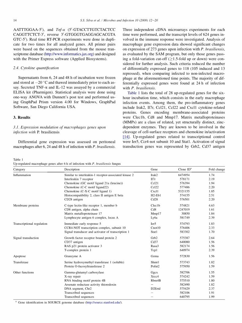
Table 1 lists the total of 28 up-regulated genes for the six-hour incubation time, which consists in the early macrophageinfection events. Among them, the pro-inflammatory genesinclude Irak2, Il7r, Ccl21, Ccl22 and Cxcl1 cytokine-relatedproteins. Genes encoding membrane-associated proteinswere Clec1b, Cd8 and Mmp17. Matrix metalloproteinases(MMPs) are a class of related, yet structurally distinct, zinc-dependent enzymes. They are known to be involved in thecleavage of cell-surface receptors and chemokine in/activation[14]. Up-regulated genes related to transcriptional controlwere Ier5, Ccr4-not subunit 10 and Stat1. Activation of signaltransduction genes was represented by Grb2, Cd37 antigen
Gene Clone IDa Fold change
kinase 2 Irak2 6474954 1.74
Il7r 578171 2.19
) Ccl21 576394 1.94
Ccl22 577486 2.20
Cxcl1 5321155 1.85
a H2-Eb1 574155 2.51
Cd28 576501 2.20
Clec1b 574821 4.63
Cd8 1247019 1.61
Mmp17 30850 1.84
Ly6a 581749 2.39
Ier5 574793 1.83
10 Cnot10 576406 2.33
ion 1 Stat1 583302 1.70
Grb2 575387 2.64
Cd37 640080 1.56
Rasa3 582174 1.56
Tcp1 640974 2.39
Gzma 572830 1.56
) Shmt1 573743 1.82
Pofut2 575950 1.59
Ggcx 582706 1.55
Xrcc4 574242 1.59
Rbm4B 575510 1.80
e 582490 1.82
D2Ertd 575429 2.37
e 582106 1.91
e 640795 1.99

15S.S. Silva et al. / Microbes and Infection 10 (2008) 12e20
and Rasa3. An induced expression has been observed for theapoptosis-related Granzyme A and the redox activation-relatedarsenate reductase thioredoxin. Three induced genes had noknown functions. No down-regulation of genes was observedat this time point by our microarray analysis under the estab-lished statistical significance criteria.
At 24 h of macrophage infection, 59 genes were found to bedifferentially expressed being 54 induced and 5 repressed(Table 2). The gene encoding the cytokine Ccl21 and other in-flammation-related genes such as Irak2, Pparbp and Ly86 wereinduced. Up-regulated genes involved in transcriptional regu-lation were the Stat1 inhibitor, Ccr4-not, Tcf12, Foxo1, andNfkb. Some of the pro-inflammatory genes regulated byNFk-B include several chemokines, all of which can enhanceinflammation by attracting additional inflammatory cells to thesite of injury [15]. Nfk-B protein also regulates the transcrip-tion of its inhibitor Nkrf, which is also induced at 24 h accord-ing to our experiment. Genes encoding membrane-relatedproteins Clec1b and Ddr1, which are related to the effective-ness of phagocytosis, were induced as well as the complementcomponents C2 and C3. The most important events in macro-phage activation are mediated by signal transduction pathwaysand several genes implicated in this process were induced, in-cluding Mapk1, Mapk8ip3, Mapk8, Txk and Rab3d. Apoptosisis an innate host defence mechanism used to prevent prolifer-ation of internalized microorganisms. At this time point fiveapoptosis-related genes were up-regulated, including thosethat encode pro-apoptotic caspases 2, 3 and 8 and the apopto-tic inhibitors Faim2 and Casp8r.
At 48 h of infection, 31 genes were found to be differentiallyexpressed, of whom 23 induced, 8 repressed (Table 3). The pro-inflammatory up-regulated genes included Irak2, Cxcl14 andCxcl4. Among the induced genes related to membrane wereClec1b, Cd14, H2eb, Ly6a and Adam8. Signal transduction-related genes induced were Mapkk11, Txk and SH3-domainGRB2-like1 (Sh3gl1). At this time point we observed a highernumber of down regulated genes from the following functionalcategories: inflammation (Ccl2), transcription regulation(Pias1i, solute carrier family 7, member 11 and RlKEN full-length enriched library) and signal transduction (Rab2 andmicrotubule affinity-regulatin kinase 2).
3.2. Validation of genes involved in immune response
In order to validate our microarray data, we performed Realtime RT-PCR with the same pooled RNA samples from threeindependent experiments of macrophage infection that wereused for microarray experiments. We chose to focus on thevalidation of three key genes that are involved in the immuneresponse (Nfkb, Nkrf, and Clec1b) that were differentially ex-pressed following infection with P. brasiliensis. Nfkb and itsrepressor Nkrf are regulators of inflammatory process. Clec1bis important for a more effectiveness phagocytosis by macro-phages. Table 4 shows that expression profile of all these geneswhen analysed by Real-time RT-PCR, correlated with theprofile observed in the microarray experiments. The magni-tude of fold-changes differed between the methodologies,
a result expected due to their technical differences regardingnormalization protocol, kinetics and sensitivity.
3.3. TNF-a quantification during macrophage infectionby P. brasiliensis
TNF-a itself is associated with the antimicrobial activity ofmacrophages and it is important cytokine in mycosis control-ling [16e18]. TNF-a expression profile was analysed by Realtime RT-PCR associated to ELISA-dosing, since it was notincluded in the cDNA microarrays membranes. Nevertheless,this did not eliminate the possibility of assessing the expres-sion profile of this relevant cytokine. In this regard, it hasbeen found that TNF-a has a fold up-regulation of 3 timesat 24 h of infection. In addition we have confirmed by ELISAthat mRNA induction resulted also in an increase of protein se-cretion. We observed at 6 h discrete increase of TNF-a whileat 24 and 48 h of infection this cytokine was strongly secretedby infected macrophages when compared to non-infected ones(Fig. 1).
4. Discussion
In order to better understand the complex interaction be-tween host cells and P. brasiliensis we have analysed thetranscriptional profile of 624 previously selected murine im-muno-regulatory genes following fungal infection of perito-neal macrophages for 6, 24 and 48 h. To our knowledge, thisstudy is the first to assess the coordinated gene expressionchanges that occur in this innate host defence cell in responseto P. brasiliensis. This time-lapse characterization providesa more encompassing view of events than would an analysisfrom a sole time point. A major concern in setting the exper-imental framework was the choice between opsonised andnon-opsonised yeast cells. When we set out to establish theco-cultivation protocol of ex vivo infection of macrophageswith non-opsonised cells, we observed that both adhesionand internalization of P. brasiliensis occurred at low frequencyand few numbers of macrophages containing adhered and/orinternalized yeast cells. Then we decided to use opsonisedcells that yielded rates of phagocytosis that allowed perform-ing the microarrays experiments.
Our microarray data have identified many genes that aremodulated in particular those related to inflammatory mecha-nisms, membrane proteins, transcription regulation and apo-ptosis. Inflammation is a powerful protective mechanismcoordinated and controlled by cytokines and chemokines.Increased chemokine gene expression has been detected bymicroarray analysis in macrophages infected with, Candida al-bicans, Aspergillus fumigatus and Mycobacterium tuberculosis[12,13,19]. Accordingly, our results show that genes encodingthe chemokines Ccl21, Ccl22 and Cxcl1, all of which are as-sociated with pro-inflammatory response, were significantlyincreased in peritoneal macrophages in response to P. brasi-liensis-Pb01, at the early stage of infection. It is relevant tomention that different gene expression patterns of cytokinesIL-6, IL-10, IL-8 and TNF-a have also been reported by
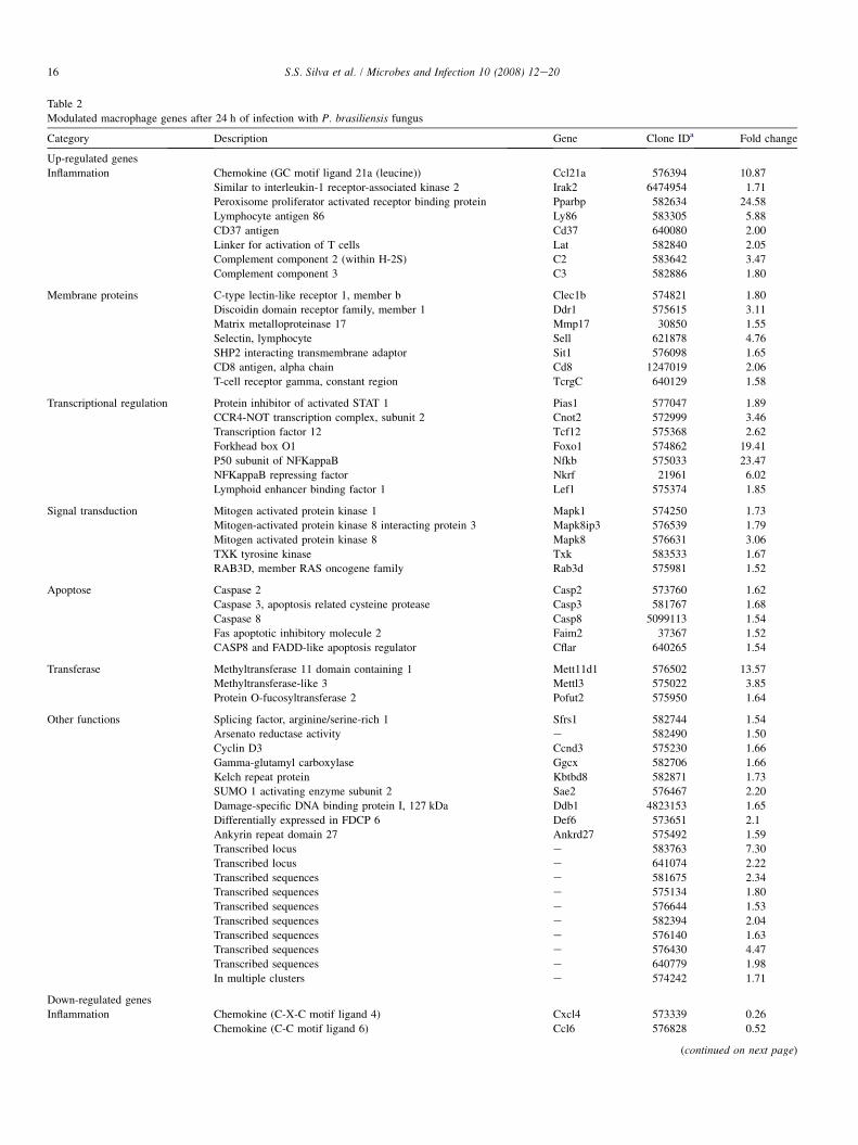
Table 2
Modulated macrophage genes after 24 h of infection with P. brasiliensis fungus
Category Description Gene Clone IDa Fold change
Up-regulated genes
Inflammation Chemokine (GC motif ligand 21a (leucine)) Ccl21a 576394 10.87
Similar to interleukin-1 receptor-associated kinase 2 Irak2 6474954 1.71
Peroxisome proliferator activated receptor binding protein Pparbp 582634 24.58
Lymphocyte antigen 86 Ly86 583305 5.88
CD37 antigen Cd37 640080 2.00
Linker for activation of T cells Lat 582840 2.05
Complement component 2 (within H-2S) C2 583642 3.47
Complement component 3 C3 582886 1.80
Membrane proteins C-type lectin-like receptor 1, member b Clec1b 574821 1.80
Discoidin domain receptor family, member 1 Ddr1 575615 3.11
Matrix metalloproteinase 17 Mmp17 30850 1.55
Selectin, lymphocyte Sell 621878 4.76
SHP2 interacting transmembrane adaptor Sit1 576098 1.65
CD8 antigen, alpha chain Cd8 1247019 2.06
T-cell receptor gamma, constant region TcrgC 640129 1.58
Transcriptional regulation Protein inhibitor of activated STAT 1 Pias1 577047 1.89
CCR4-NOT transcription complex, subunit 2 Cnot2 572999 3.46
Transcription factor 12 Tcf12 575368 2.62
Forkhead box O1 Foxo1 574862 19.41
P50 subunit of NFKappaB Nfkb 575033 23.47
NFKappaB repressing factor Nkrf 21961 6.02
Lymphoid enhancer binding factor 1 Lef1 575374 1.85
Signal transduction Mitogen activated protein kinase 1 Mapk1 574250 1.73
Mitogen-activated protein kinase 8 interacting protein 3 Mapk8ip3 576539 1.79
Mitogen activated protein kinase 8 Mapk8 576631 3.06
TXK tyrosine kinase Txk 583533 1.67
RAB3D, member RAS oncogene family Rab3d 575981 1.52
Apoptose Caspase 2 Casp2 573760 1.62
Caspase 3, apoptosis related cysteine protease Casp3 581767 1.68
Caspase 8 Casp8 5099113 1.54
Fas apoptotic inhibitory molecule 2 Faim2 37367 1.52
CASP8 and FADD-like apoptosis regulator Cflar 640265 1.54
Transferase Methyltransferase 11 domain containing 1 Mett11d1 576502 13.57
Methyltransferase-like 3 Mettl3 575022 3.85
Protein O-fucosyltransferase 2 Pofut2 575950 1.64
Other functions Splicing factor, arginine/serine-rich 1 Sfrs1 582744 1.54
Arsenato reductase activity e 582490 1.50
Cyclin D3 Ccnd3 575230 1.66
Gamma-glutamyl carboxylase Ggcx 582706 1.66
Kelch repeat protein Kbtbd8 582871 1.73
SUMO 1 activating enzyme subunit 2 Sae2 576467 2.20
Damage-specific DNA binding protein I, 127 kDa Ddb1 4823153 1.65
Differentially expressed in FDCP 6 Def6 573651 2.1
Ankyrin repeat domain 27 Ankrd27 575492 1.59
Transcribed locus e 583763 7.30
Transcribed locus e 641074 2.22
Transcribed sequences e 581675 2.34
Transcribed sequences e 575134 1.80
Transcribed sequences e 576644 1.53
Transcribed sequences e 582394 2.04
Transcribed sequences e 576140 1.63
Transcribed sequences e 576430 4.47
Transcribed sequences e 640779 1.98
In multiple clusters e 574242 1.71
Down-regulated genes
Inflammation Chemokine (C-X-C motif ligand 4) Cxcl4 573339 0.26
Chemokine (C-C motif ligand 6) Ccl6 576828 0.52
(continued on next page)
16 S.S. Silva et al. / Microbes and Infection 10 (2008) 12e20

Table 2 (continued)
Category Description Gene Clone IDa Fold change
Other functions Nuclear casein kinase and cyclin-dependent kinase substrate 1 Nucks1 573338 0.19
F.box and leucine-rich repeat protein 5 Fbox5 575998 0.46
Lymphocyte antigen 6 complex, locus D Ly6d 581909 0.46
a Gene identification in SOURCE genome database (http://source.stanford.edu/).
17S.S. Silva et al. / Microbes and Infection 10 (2008) 12e20
different strains of P. brasiliensis such as Pb18 and Pb265[20,21]. Cxcl1 and Ccl22 are neutrophil and monocyte chemo-attractants, respectively. Induction of genes that code for otherneutrophil (KC and CCL3) and monocyte (CCL5, CCL2,CXCL10 and CXCL9) attractants were reported in the lungof mice infected with P. brasiliensis; this observation wascorrelated with an intense recruitment of phagocytes to thesite of infection [22]. Despite being included in the microarraymembrane, IL-12 was found not to be differentially expressedat any of the time points, which led us to confirm this findingby ELISA. Corroborating this, it was also not detected in thesupernatant of infected macrophages. P. brasiliensis is likelyto inhibit IL-12 production as a means to evade the innate
Table 3
Modulated macrophage genes after 48 h of infection with P. brasiliensis fungus
Category Description
Up-regulated genes
Inflammation Similar to interleukin-1 receptor-associated kin
Chemokine (C-X-C motif ligand 14)
Chemokine (C-X-C motif ligand 4)
Membrane proteins C-type lectin-like receptor 1, member b
CD14 antigen
Histocompatibility 2, class II antigen E beta
Lymphocyte antigen 6 complex, locus A
A disintegrin and metalloprotease domain 8
Beta-2 microglobulin
Beta-2 microglobulin
Signal transduction Mitogen activated protein kinase kinase kinase
TXK tyrosine kinase
SH3- domain GRB2-like1
Twin filin, actin-binding protein, homolog 2
Other functions Serine (or cysteine) proteinase inhibitor, clade
13 days embryo head cDNA
Aryl hydrocarbon receptor nuclear translocato
B-cell translocation gene I, anti-proliferative
Arsenate reductase (thioredoxin) activity
Cytochrome b-245, beta polypeptide
Transcribed locus similar heat shock 27 kDa p
Transcribed sequences
Transcribed sequences
Down-regulated genes
Inflammation Chemokine (GC motif ligand 2)
Transcriptional regulation Protein inhibitor of activated STAT 1
Solute carrier family, member 11
RlKEN full-length enriched library
Signal transduction RAB2, member RAS oncogene family
MAP/microtubule affinity-regulating kinase 2
Other functions STT3, subunit of oligosaccharyltransferase com
Insulin degrading enzyme
a Gene identification in SOURCE genome database (http://source.stanford.edu/).
and adaptive responses set up by the host. These data are ingood agreement with similar analyses on other pathogenssuch as Mycobacterium tuberculosis [9] and Histoplasma cap-sulatum [23], which also suppress IL-12. This is presumablyan instance of convergence of pathogen survival strategy in-side macrophages. The expression of genes encoding cytokinereceptor-associated proteins has also been evaluated at thepresent work. In this regard, we have detected an up-regulationof IL-7 receptor in early infection (6 h), which participates inthe pro-inflammatory cytokines in monocytes and macro-phages [24]. It follows that the induction of IL-7r is probablyrelated to macrophage activation in response to early infectionwith P. brasiliensis, presumably in order to enhance the
Gene Clone IDa Fold change
ase 2 Irak2 6474954 1.64
Cxcl14 583442 1.58
Cxcl4 573339 1.52
Clec1b 574821 2.66
Cd14 5120996 1.91
H2-Eb 574155 1.63
Ly6a 581749 1.67
Adam8 582054 1.51
B2m 576472 2.73
B2m 576493 3.11
11 Mapkkk11 575211 3.00
Txk 583533 1.61
Sh3gl1 582991 1.69
Twf2 640339 1.80
H, member 1 Serpinh1 582567 1.98
e 575324 1.73
r-like Arntl 582931 1.93
Btg1 574654 1.95
e 582490 2.75
Cybb 583187 2.05
rotein 1 e 582517 2.45
e 582338 2.12
e 575870 1.69
Ccl2 573898 0.33
Pias1 577047 0.47
S7a11lc 575418 0.50
e 576643 0.29
Rab2 573835 0.50
Mark2 574316 0.36
plex Stt3b 573897 0.36
Ide 577628 0.37
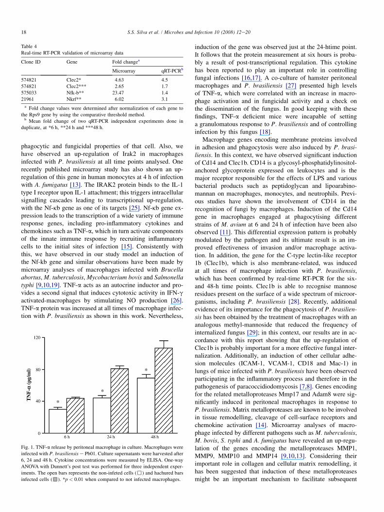
Table 4
Real-time RT-PCR validation of microarray data
Clone ID Gene Fold changea
Microarray qRT-PCRb
574821 Clec2* 4.63 4.5
574821 Clec2*** 2.65 1.7
575033 Nfk-b** 23.47 1.4
21961 Nkrf** 6.02 3.1
a Fold change values were determined after normalization of each gene to
the Rps9 gene by using the comparative threshold method.b Mean fold change of two qRT-PCR independent experiments done in
duplicate, at *6 h, **24 h and ***48 h.
18 S.S. Silva et al. / Microbes and Infection 10 (2008) 12e20
phagocytic and fungicidal properties of that cell. Also, wehave observed an up-regulation of Irak2 in macrophagesinfected with P. brasiliensis at all time points analysed. Onerecently published microarray study has also shown an up-regulation of this gene in human monocytes at 4 h of infectionwith A. fumigatus [13]. The IRAK2 protein binds to the IL-1type I receptor upon IL-1 attachment; this triggers intracellularsignalling cascades leading to transcriptional up-regulation,with the Nf-kb gene as one of its targets [25]. Nf-kb gene ex-pression leads to the transcription of a wide variety of immuneresponse genes, including pro-inflammatory cytokines andchemokines such as TNF-a, which in turn activate componentsof the innate immune response by recruiting inflammatorycells to the initial sites of infection [15]. Consistently withthis, we have observed in our study model an induction ofthe Nf-kb gene and similar observations have been made bymicroarray analyses of macrophages infected with Brucellaabortus, M. tuberculosis, Mycobacterium bovis and Salmonellatyphi [9,10,19]. TNF-a acts as an autocrine inductor and pro-vides a second signal that induces cytotoxic activity in IFN-gactivated-macrophages by stimulating NO production [26].TNF-a protein was increased at all times of macrophage infec-tion with P. brasiliensis as shown in this work. Nevertheless,
Fig. 1. TNF-a release by peritoneal macrophage in culture. Macrophages were
infected with P. brasiliensis e Pb01. Culture supernatants were harvested after
6, 24 and 48 h. Cytokine concentrations were measured by ELISA. One-way
ANOVA with Dunnett’s post test was performed for three independent exper-
iments. The open bars represents the non-infeted cells (,) and hachured bars
infected cells (G). *p < 0.01 when compared to not infected macrophages.
induction of the gene was observed just at the 24-htime point.It follows that the protein measurement at six hours is proba-bly a result of post-transcriptional regulation. This cytokinehas been reported to play an important role in controllingfungal infections [16,17]. A co-culture of hamster peritonealmacrophages and P. brasiliensis [27] presented high levelsof TNF-a, which were correlated with an increase in macro-phage activation and in fungicidal activity and a check onthe dissemination of the fungus. In good keeping with thesefindings, TNF-a deficient mice were incapable of settinga granulomatous response to P. brasiliensis and of controllinginfection by this fungus [18].
Macrophage genes encoding membrane proteins involvedin adhesion and phagocytosis were also induced by P. brasi-liensis. In this context, we have observed significant inductionof Cd14 and Clec1b. CD14 is a glycosyl-phosphatidylinositol-anchored glycoprotein expressed on leukocytes and is themajor receptor responsible for the effects of LPS and variousbacterial products such as peptidoglycan and lipoarabino-mannan on macrophages, monocytes, and neutrophils. Previ-ous studies have shown the involvement of CD14 in therecognition of fungi by macrophages. Induction of the Cd14gene in macrophages engaged at phagocytising differentstrains of M. avium at 6 and 24 h of infection have been alsoobserved [11]. This differential expression pattern is probablymodulated by the pathogen and its ultimate result is an im-proved effectiveness of invasion and/or macrophage activa-tion. In addition, the gene for the C-type lectin-like receptor1b (Clec1b), which is also membrane-related, was inducedat all times of macrophage infection with P. brasiliensis,which has been confirmed by real-time RT-PCR for the six-and 48-h time points. Clec1b is able to recognise mannoseresidues present on the surface of a wide spectrum of microor-ganisms, including P. brasiliensis [28]. Recently, additionalevidence of its importance for the phagocytosis of P. brasilien-sis has been obtained by the treatment of macrophages with ananalogous methyl-mannoside that reduced the frequency ofinternalized fungus [29]; in this context, our results are in ac-cordance with this report showing that the up-regulation ofClec1b is probably important for a more effective fungal inter-nalization. Additionally, an induction of other cellular adhe-sion molecules (ICAM-1, VCAM-1, CD18 and Mac-1) inlungs of mice infected with P. brasiliensis have been observedparticipating in the inflammatory process and therefore in thepathogenesis of paracoccidiodomycosis [7,8]. Genes encodingfor the related metalloproteases Mmp17 and Adam8 were sig-nificantly induced in peritoneal macrophages in response toP. brasiliensis. Matrix metalloproteases are known to be involvedin tissue remodelling, cleavage of cell-surface receptors andchemokine activation [14]. Microarray analyses of macro-phage infected by different pathogens such as M. tuberculosis,M. bovis, S. typhi and A. fumigatus have revealed an up-regu-lation of the genes encoding the metalloproteases MMP1,MMP9, MMP10 and MMP14 [9,10,13]. Considering theirimportant role in collagen and cellular matrix remodelling, ithas been suggested that induction of these metalloproteasesmight be an important mechanism to facilitate subsequent
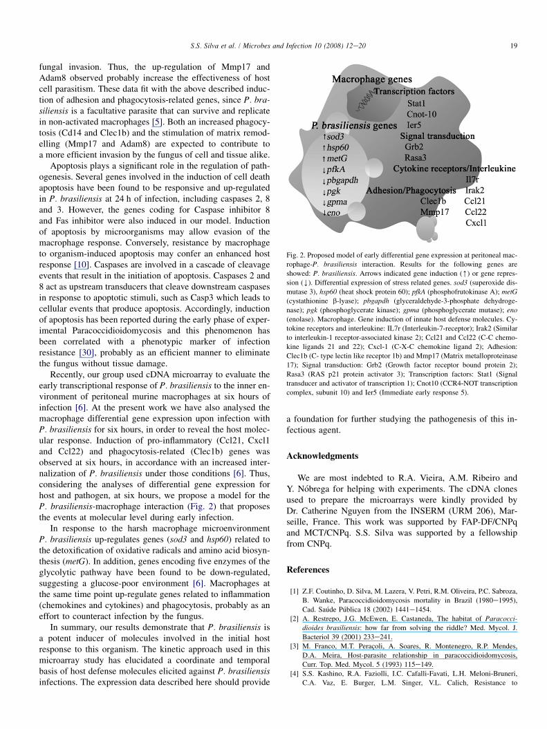
Fig. 2. Proposed model of early differential gene expression at peritoneal mac-
rophage-P. brasiliensis interaction. Results for the following genes are
showed: P. brasiliensis. Arrows indicated gene induction ([) or gene repres-
sion (Y). Differential expression of stress related genes. sod3 (superoxide dis-
mutase 3), hsp60 (heat shock protein 60); pfkA (phosphofrutokinase A); metG(cystathionine b-lyase); pbgapdh (glyceraldehyde-3-phosphate dehydroge-
nase); pgk (phosphoglycerate kinase); gpma (phosphoglycerate mutase); eno
(enolase). Macrophage. Gene induction of innate host defense molecules. Cy-
tokine receptors and interleukine: IL7r (Interleukin-7-receptor); Irak2 (Similar
to interleukin-1 receptor-associated kinase 2); Ccl21 and Ccl22 (C-C chemo-
kine ligands 21 and 22); Cxcl-1 (C-X-C chemokine ligand 2); Adhesion:
Clec1b (C- type lectin like receptor 1b) and Mmp17 (Matrix metalloproteinase
17); Signal transduction: Grb2 (Growth factor receptor bound protein 2);
Rasa3 (RAS p21 protein activator 3); Transcription factors: Stat1 (Signal
transducer and activator of transcription 1); Cnot10 (CCR4-NOT transcription
complex, subunit 10) and Ier5 (Immediate early response 5).
19S.S. Silva et al. / Microbes and Infection 10 (2008) 12e20
fungal invasion. Thus, the up-regulation of Mmp17 andAdam8 observed probably increase the effectiveness of hostcell parasitism. These data fit with the above described induc-tion of adhesion and phagocytosis-related genes, since P. bra-siliensis is a facultative parasite that can survive and replicatein non-activated macrophages [5]. Both an increased phagocy-tosis (Cd14 and Clec1b) and the stimulation of matrix remod-elling (Mmp17 and Adam8) are expected to contribute toa more efficient invasion by the fungus of cell and tissue alike.
Apoptosis plays a significant role in the regulation of path-ogenesis. Several genes involved in the induction of cell deathapoptosis have been found to be responsive and up-regulatedin P. brasiliensis at 24 h of infection, including caspases 2, 8and 3. However, the genes coding for Caspase inhibitor 8and Fas inhibitor were also induced in our model. Inductionof apoptosis by microorganisms may allow evasion of themacrophage response. Conversely, resistance by macrophageto organism-induced apoptosis may confer an enhanced hostresponse [10]. Caspases are involved in a cascade of cleavageevents that result in the initiation of apoptosis. Caspases 2 and8 act as upstream transducers that cleave downstream caspasesin response to apoptotic stimuli, such as Casp3 which leads tocellular events that produce apoptosis. Accordingly, inductionof apoptosis has been reported during the early phase of exper-imental Paracoccidioidomycosis and this phenomenon hasbeen correlated with a phenotypic marker of infectionresistance [30], probably as an efficient manner to eliminatethe fungus without tissue damage.
Recently, our group used cDNA microarray to evaluate theearly transcriptional response of P. brasiliensis to the inner en-vironment of peritoneal murine macrophages at six hours ofinfection [6]. At the present work we have also analysed themacrophage differential gene expression upon infection withP. brasiliensis for six hours, in order to reveal the host molec-ular response. Induction of pro-inflammatory (Ccl21, Cxcl1and Ccl22) and phagocytosis-related (Clec1b) genes wasobserved at six hours, in accordance with an increased inter-nalization of P. brasiliensis under those conditions [6]. Thus,considering the analyses of differential gene expression forhost and pathogen, at six hours, we propose a model for theP. brasiliensis-macrophage interaction (Fig. 2) that proposesthe events at molecular level during early infection.
In response to the harsh macrophage microenvironmentP. brasiliensis up-regulates genes (sod3 and hsp60) related tothe detoxification of oxidative radicals and amino acid biosyn-thesis (metG). In addition, genes encoding five enzymes of theglycolytic pathway have been found to be down-regulated,suggesting a glucose-poor environment [6]. Macrophages atthe same time point up-regulate genes related to inflammation(chemokines and cytokines) and phagocytosis, probably as aneffort to counteract infection by the fungus.
In summary, our results demonstrate that P. brasiliensis isa potent inducer of molecules involved in the initial hostresponse to this organism. The kinetic approach used in thismicroarray study has elucidated a coordinate and temporalbasis of host defense molecules elicited against P. brasiliensisinfections. The expression data described here should provide
a foundation for further studying the pathogenesis of this in-fectious agent.
Acknowledgments
We are most indebted to R.A. Vieira, A.M. Ribeiro andY. Nobrega for helping with experiments. The cDNA clonesused to prepare the microarrays were kindly provided byDr. Catherine Nguyen from the INSERM (URM 206), Mar-seille, France. This work was supported by FAP-DF/CNPqand MCT/CNPq. S.S. Silva was supported by a fellowshipfrom CNPq.
References
[1] Z.F. Coutinho, D. Silva, M. Lazera, V. Petri, R.M. Oliveira, P.C. Sabroza,
B. Wanke, Paracoccidioidomycosis mortality in Brazil (1980e1995),
Cad. Saude Publica 18 (2002) 1441e1454.
[2] A. Restrepo, J.G. McEwen, E. Castaneda, The habitat of Paracocci-
dioides brasiliensis: how far from solving the riddle? Med. Mycol. J.
Bacteriol 39 (2001) 233e241.
[3] M. Franco, M.T. Peracoli, A. Soares, R. Montenegro, R.P. Mendes,
D.A. Meira, Host-parasite relationship in paracoccidioidomycosis,
Curr. Top. Med. Mycol. 5 (1993) 115e149.
[4] S.S. Kashino, R.A. Faziolli, I.C. Cafalli-Favati, L.H. Meloni-Bruneri,
C.A. Vaz, E. Burger, L.M. Singer, V.L. Calich, Resistance to
20 S.S. Silva et al. / Microbes and Infection 10 (2008) 12e20
Paracoccidioides brasiliensis infection is linked to a preferential Th1 im-
mune response, whereas susceptibility is associated with absence of IFN-
gamma production, J. Interferon Cytokine Res. 20 (2000) 89e97.
[5] E. Brummer, L.H. Hanson, A. Restrepo, D.A. Stevens, Intracellular mul-
tiplication of Paracoccidioides brasiliensis in macrophages: killing and
restriction of multiplication by activated macrophages, Infect. Immun.
57 (1989) 2289e2294.
[6] A.H. Tavares, S.S. Silva, A. Dantas, E.G. Campos, R.V. Andrade,
A.Q. Maranh~ao, M.M. Brıgido, D.G. Passos-Silva, A.L. Facchin,
S.M. Teixeira, G.A. Passos, C.M. Soares, A.L. Bocca, M.J. Carvalho,
I. Silva-Pereira, M.S. Felipe, Early transcriptional response of Paracoc-
cidioides brasiliensis upon internalization by murine macrophages, Mi-
crobes Infect 9 (2007) 583e590.
[7] A. Gonzalez, H.L. Lenzi, E.M. Motta, L. Caputo, J.H. Sahaza,
A.M. Cock, A.C. Ruiz, A. Restrepo, L.E. Cano, Expression of adhesion
molecules in lungs of mice infected with Paracoccidioides brasiliensisconidia, Microbes Infect 7 (2005) 666e673.
[8] A.P. Moreira, A.P. Campanelli, K.A. Cavassani, J.T. Souto, B.R. Ferreira,
R. Martinez, M.A. Rossi, J.S. Silva, Intercellular adhesion molecule-1 is
required for the early formation of granulomas and participates in the
resistance of mice to the infection with the fungus Paracoccidioides bra-
siliensis, Am. J. Pathol. 169 (2006) 1270e1281.
[9] G.J. Nau, J.F.L. Richmond, A. Schlesinger, E.G. Jennings, E.S. Lander,
Human macrophage activation programs induced by bacterial pathogens,
PNAS 99 (2002) 1503e1508.
[10] L. Eskra, A. Mathison, G. Splitter, Microarray analysis of mRNA levels
from RAW264.7 macrophages infected with Brucella abortus, Infect. Im-
mun. 71 (2003) 1125e1133.
[11] D.J. Weiss, O.A. Evanson, M. Deng, M.S. Abrahamsen, Sequential
patterns of gene expression by bovine monocyte-derived macrophages
associated with ingestion of mycobacterial organisms, Microb. Pat. 37
(2004) 215e224.
[12] H.S. Kim, E.H. Choi, J. Khan, E. Roilides, A. Francesconi, M. Kasai,
T. Sein, R.L. Schaufele, K. Sakurai, C.G. Son, B.T. Greer, S. Chanock,
C.A. Lyman, T.J. Walsh, Expression of genes encoding innate host
defense molecules in normal human monocytes in response to Candida
albicans, Infect. Immun. 73 (2005) 3714e3724.
[13] K.J. Cortez, C.A. Lyman, S. Kottilil, H.S. Kim, E. Roilides, J. Yang,
B. Fullmer, R. Lempicki, T.J. Walsh, Functional genomics of innate
host defense molecules in normal human monocytes in response to
Aspergillus fumigatus, Infect. Immun. 74 (2006) 2353e2365.
[14] C. Chang, Z. Werb, The many faces of metalloproteases: cell growth,
invasion, angiogenesis and metastasis, Trends Cell Biol. 11 (2001)
S37eS43.
[15] M. Karin, Y. Ben-Neriah, Phosphorylation meets ubiquitination: the con-
trol of NF-[kappa]B activity, Ann. Rev. Immunol. 18 (2000) 621e663.
[16] A. Louie, A.L. Baltch, R.P. Smith, M.A. Franke, W.J. Ritz, J.K. Singh,
M.A. Gordon, Tumor necrosis factor alpha has a protective role in mu-
rine model of systemic candidiasis, Infect. Immun. 62 (1994) 2761e2772.
[17] J.G. Smith, D.M. Magee, D.M. Williams, J.R. Graybill, Tumor necrosis
factor alpha plays a role in host defense against Histoplasma capsulatum,
J. Infect. Dis. 162 (1990) 1349e1353.
[18] J.T. Souto, F. Figueiredo, A. Furlanetto, K. Pfeffer, M.A. Rossi,
J.S. Silva, Interferon-g and Tumor Necrosis Factor a determine resis-
tance to P. brasiliensis infection in mice, Am. J. Pat. 156 (2000)
1811e1820.
[19] J.P. Wang, S.E. Rought, C. Jacques, D.G. Guiney, Gene expression
profiling detects patterns of human macrophage responses following
Mycobacterium tuberculosis infection, FEMS Immun. Med. Microb.
39 (2003) 163e172.
[20] C.S. Kurokawa, J.P. Araujo, A.M.V.C. Soares, M.F. Sugizaki,
M.T.S. Peracoli, Pro-and anti-inflammatory cytokines produced by
human monocytes challenged in vitro with Paracoccidioides brasiliensis,
Microbiol. Immunol. 51 (2007) 421e428.
[21] F. Figueiredo, L.M. Alves, C.L. Silva, Tumour necrosis factor production
in vivo and in vitro in response to Paracoccidioides brasiliensis and the
cell wall fractions thereof, Clin. Exp. Immunol. 93 (1993) 189e194.
[22] J.T. Souto, J.C. Aliberti, A.P. Campanelli, M.C. Livonesi,
C.M.L. Maffei, B.R. Ferreira, L.R. Travassos, R. Martinez,
M.A. Rossi, J.S. Silva, Chemokine production and leukocyte recruitment
to the lungs of Paracoccidioides brasiliensis-infected mice is modulated
by interferon-g, Am. J. Pat. 163 (2003) 583e590.
[23] T. Marth, B.L. Kelsall, Regulation of interleukin-12 by complement
receptor 3 signaling, J. Exp. Med. 185 (1997) 1987e1995.
[24] M.R. Alderson, T.W. Tough, S.F. Ziegler, K.H. Grabstein, Interleukin 7
induces cytokine secretion and tumoricidal activity by human peripheral
blood monocytes, J. Exp. Med. 173 (1991) 923e930.
[25] M. Muzio, J. Ni, P. Feng, V.M. Dixit, IRAK (Pelle) family member
IRAK-2 and Myd88 as proximal mediators of IL-1 signaling, Science
278 (1997) 1612e1615.
[26] S.J. Green, M.S. Meltzer, J.B. Hibbs Jr., C.A. Nacy, Activated mac-
rophages destroy intracellular Leishmania major amastigottes by an
L-arginine-dependent killing mechanism, J. Immunol. 144 (1990)
278e283.
[27] M.R. Parise-Fortes, M.F. Da Silva, M.F. Sugizaki, J. Defaveri,
M.R. Montenegro, A.M. Soares, Experimental paracoccidioidomycosis
of the Syrian hamster: fungicidal activity and production of inflammatory
cytokines by macrophages, Med. Mycol. 38 (2000) 51e60.
[28] F. Kanetsuna, L.M. Carbonell, R.E. Moreno, J. Rodriguez, Cell wall
composition of the yeast and mycelia forms of Paracoccidioides brasi-
liensis, J. Bacteriol. 97 (1969) 1036e1041.
[29] M.P. Jimenez, A. Restrepo, D. Radzioch, L.E. Cano, L.F. Garcia, Impor-
tance of complement 3 and mannose receptors in phagocytosis of Para-
coccidioides brasiliensis conidia by Nramp1 congenic macrophages
lines, FEMS Immun. Med. Microb. 47 (2006) 56e66.
[30] M.A. Verıcimo, K.M. Franca, A.C.V. Arnholdt, T.L. Kipnis, Increased
apoptosis during the early phase of experimental paracoccidioidomyco-
sis as a phenotypic marker of resistance, Microbes Infect. 8 (2006)
2811e2820.
Copyright © 2022 FDOKUMEN