Cell-Free Antigens from Paracoccidioides brasiliensis Drive IL4 Production and Increase the Severity...
9
Cell-Free Antigens from Paracoccidioides brasiliensis Drive IL-4 Production and Increase the Severity of Paracoccidioidomycosis Karen A. Cavassani 1. , Fabrine S. M. Tristao 1. , Leandro L. Oliveira 2 , Fernanda A. Rocha 1 , Jaqueline O. Vancim 1 , Ana Paula Moreira 1 , Ana Paula Campanelli 3 , Luciano A. Panagio 1 , Cristiane M. Milanezi 1 , Roberto Martinez 1 , Marcos A. Rossi 1 , Joao S. Silva 1 * 1 School of Medicine of Ribeirao Preto, Sa ˜o Paulo University, Ribeirao Preto, Sa ˜o Paulo, Brazil, 2 School of Pharmaceutical Sciences of Ribeirao Preto, Sa ˜o Paulo University, Ribeirao Preto, Sa ˜o Paulo, Brazil, 3 School of Dentistry of Bauru, Sa ˜o Paulo University, Bauru, Sa ˜o Paulo, Brazil Abstract The thermally dimorphic fungus Paracoccidioides brasiliensis (Pb) is the causative agent of paracoccidioidomycosis (PCM), one of the most frequent systemic mycosis that affects the rural population in Latin America. PCM is characterized by a chronic inflammatory granulomatous reaction, which is consequence of a Th1-mediated adaptive immune response. In the present study we investigated the mechanisms involved in the immunoregulation triggered after a prior contact with cell- free antigens (CFA) during a murine model of PCM. The results showed that the inoculation of CFA prior to the infection resulted in disorganized granulomatous lesions and increased fungal replication in the lungs, liver and spleen, that paralleled with the higher levels of IL-4 when compared with the control group. The role of IL-4 in facilitating the fungal growth was demonstrated in IL-4-deficient- and neutralizing anti-IL-4 mAb-treated mice. The injection of CFA did not affect the fungal growth in these mice, which, in fact, exhibited a significant diminished amount of fungus in the tissues and smaller granulomas. Considering that in vivo anti-IL-4-application started one week after the CFA-inoculum, it implicates that IL-4-CFA-induced is responsible by the mediation of the observed unresponsiveness. Further, the characterization of CFA indicated that a proteic fraction is required for triggering the immunosuppressive mechanisms, while glycosylation or glycosphingolipids moieties are not. Taken together, our data suggest that the prior contact with soluble Pb antigens leads to severe PCM in an IL-4 dependent manner. Citation: Cavassani KA, Tristao FSM, Oliveira LL, Rocha FA, Vancim JO, et al. (2011) Cell-Free Antigens from Paracoccidioides brasiliensis Drive IL-4 Production and Increase the Severity of Paracoccidioidomycosis. PLoS ONE 6(6): e21423. doi:10.1371/journal.pone.0021423 Editor: Niels Olsen Saraiva Ca ˆmara, Universidade de Sao Paulo, Brazil Received February 2, 2011; Accepted May 30, 2011; Published June 22, 2011 Copyright: ß 2011 Cavassani et al. This is an open-access article distributed under the terms of the Creative Commons Attribution License, which permits unrestricted use, distribution, and reproduction in any medium, provided the original author and source are credited. Funding: This work was supported by grants from Fundac ¸a ˜ o de Amparo a ` Pesquisa do Estado de Sa ˜o Paulo (FAPESP) and National Counsel of Technological and Scientific Development (CNPq). The funders had no role in study design, data collection and analysis, decision to publish, or preparation of the manuscript. Competing Interests: The authors have declared that no competing interests exist. * E-mail: [email protected] . These authors contributed equally to this work. Introduction Paracoccidioides brasiliensis (Pb) is a thermally dimorphic fungus that causes paracoccidioidomycosis (PCM), the most prevalent systemic mycosis in several countries of Latin America including Brazil, Argentina, Venezuela and Colombia. PCM represents the major cause of disability and death among young adult rural workers during their most productive stage of life [1]. In 2001 it was estimated that approximately 10 million people were infected [2], and it seems that the number of new cases have been diminished every year, even in areas with high endemicity. Infection occurs by inhalation of fungal spores or particles, which transform into the pathogenic yeast form after reaching the pulmonary alveolar epithelium [3,4]. Yeast can either be eliminated by immune- competent cells or disseminate to other tissues through lymphatic and hematogenous routes, resulting in a wide range of clinical and immunological manifestations, that vary from asymptomatic, benign and localized to severe and disseminated forms [5]. The broad spectrum of clinical and pathological manifestations is dependent on the patient’s immune response. It is known that patients with PCM often present a depressed cellular immune response [3,6,7,8]. Classical studies demonstrated that benign forms of the disease are associated with low levels of specific antibodies and positive delayed-type hypersensitivity (DTH) reactions [9]. In this context, the resistance to fungal infections are related to T helper 1 (Th1)-type cytokines such as IL-12 and IFN-c, while susceptibility has been linked to the preferential production of the Th2 type responses, including IL-4, IL-5 and IL- 10 [10,11,12,13,14]. Furthermore, several factors may contribute to this process, such as host and pathogen genetic background, fungal load and virulence [15,16,17]. After Pb infection, the host can be exposed to complex fungal antigens, including proteins, glycoproteins, and glycosphingolipids [18]. Although some of these antigens have been identified [19,20,21], the characterization and purification of others as well as their effects on the pathology during an in vivo infection are not well defined. The main P. brasiliensis antigenic component is the exocellular glycoprotein gp43 [20,22,23], an immunodominant antigen for cellular immunity in humans and experimentally infected mice [24,25]. In addition, gp43 is implicated in PLoS ONE | www.plosone.org 1 June 2011 | Volume 6 | Issue 6 | e21423
-
Upload
independent -
Category
Documents
-
view
1 -
download
0
Transcript of Cell-Free Antigens from Paracoccidioides brasiliensis Drive IL4 Production and Increase the Severity...
Cell-Free Antigens from Paracoccidioides brasiliensisDrive IL-4 Production and Increase the Severity ofParacoccidioidomycosisKaren A. Cavassani1., Fabrine S. M. Tristao1., Leandro L. Oliveira2, Fernanda A. Rocha1, Jaqueline O.
Vancim1, Ana Paula Moreira1, Ana Paula Campanelli3, Luciano A. Panagio1, Cristiane M. Milanezi1,
Roberto Martinez1, Marcos A. Rossi1, Joao S. Silva1*
1 School of Medicine of Ribeirao Preto, Sao Paulo University, Ribeirao Preto, Sao Paulo, Brazil, 2 School of Pharmaceutical Sciences of Ribeirao Preto, Sao Paulo University,
Ribeirao Preto, Sao Paulo, Brazil, 3 School of Dentistry of Bauru, Sao Paulo University, Bauru, Sao Paulo, Brazil
Abstract
The thermally dimorphic fungus Paracoccidioides brasiliensis (Pb) is the causative agent of paracoccidioidomycosis (PCM),one of the most frequent systemic mycosis that affects the rural population in Latin America. PCM is characterized by achronic inflammatory granulomatous reaction, which is consequence of a Th1-mediated adaptive immune response. In thepresent study we investigated the mechanisms involved in the immunoregulation triggered after a prior contact with cell-free antigens (CFA) during a murine model of PCM. The results showed that the inoculation of CFA prior to the infectionresulted in disorganized granulomatous lesions and increased fungal replication in the lungs, liver and spleen, thatparalleled with the higher levels of IL-4 when compared with the control group. The role of IL-4 in facilitating the fungalgrowth was demonstrated in IL-4-deficient- and neutralizing anti-IL-4 mAb-treated mice. The injection of CFA did not affectthe fungal growth in these mice, which, in fact, exhibited a significant diminished amount of fungus in the tissues andsmaller granulomas. Considering that in vivo anti-IL-4-application started one week after the CFA-inoculum, it implicatesthat IL-4-CFA-induced is responsible by the mediation of the observed unresponsiveness. Further, the characterization ofCFA indicated that a proteic fraction is required for triggering the immunosuppressive mechanisms, while glycosylation orglycosphingolipids moieties are not. Taken together, our data suggest that the prior contact with soluble Pb antigens leadsto severe PCM in an IL-4 dependent manner.
Citation: Cavassani KA, Tristao FSM, Oliveira LL, Rocha FA, Vancim JO, et al. (2011) Cell-Free Antigens from Paracoccidioides brasiliensis Drive IL-4 Production andIncrease the Severity of Paracoccidioidomycosis. PLoS ONE 6(6): e21423. doi:10.1371/journal.pone.0021423
Editor: Niels Olsen Saraiva Camara, Universidade de Sao Paulo, Brazil
Received February 2, 2011; Accepted May 30, 2011; Published June 22, 2011
Copyright: � 2011 Cavassani et al. This is an open-access article distributed under the terms of the Creative Commons Attribution License, which permitsunrestricted use, distribution, and reproduction in any medium, provided the original author and source are credited.
Funding: This work was supported by grants from Fundacao de Amparo a Pesquisa do Estado de Sao Paulo (FAPESP) and National Counsel of Technological andScientific Development (CNPq). The funders had no role in study design, data collection and analysis, decision to publish, or preparation of the manuscript.
Competing Interests: The authors have declared that no competing interests exist.
* E-mail: [email protected]
. These authors contributed equally to this work.
Introduction
Paracoccidioides brasiliensis (Pb) is a thermally dimorphic fungus
that causes paracoccidioidomycosis (PCM), the most prevalent
systemic mycosis in several countries of Latin America including
Brazil, Argentina, Venezuela and Colombia. PCM represents the
major cause of disability and death among young adult rural
workers during their most productive stage of life [1]. In 2001 it was
estimated that approximately 10 million people were infected [2],
and it seems that the number of new cases have been diminished
every year, even in areas with high endemicity. Infection occurs by
inhalation of fungal spores or particles, which transform into the
pathogenic yeast form after reaching the pulmonary alveolar
epithelium [3,4]. Yeast can either be eliminated by immune-
competent cells or disseminate to other tissues through lymphatic
and hematogenous routes, resulting in a wide range of clinical and
immunological manifestations, that vary from asymptomatic,
benign and localized to severe and disseminated forms [5].
The broad spectrum of clinical and pathological manifestations
is dependent on the patient’s immune response. It is known that
patients with PCM often present a depressed cellular immune
response [3,6,7,8]. Classical studies demonstrated that benign
forms of the disease are associated with low levels of specific
antibodies and positive delayed-type hypersensitivity (DTH)
reactions [9]. In this context, the resistance to fungal infections
are related to T helper 1 (Th1)-type cytokines such as IL-12 and
IFN-c, while susceptibility has been linked to the preferential
production of the Th2 type responses, including IL-4, IL-5 and IL-
10 [10,11,12,13,14]. Furthermore, several factors may contribute
to this process, such as host and pathogen genetic background,
fungal load and virulence [15,16,17].
After Pb infection, the host can be exposed to complex fungal
antigens, including proteins, glycoproteins, and glycosphingolipids
[18]. Although some of these antigens have been identified
[19,20,21], the characterization and purification of others as well
as their effects on the pathology during an in vivo infection are not
well defined. The main P. brasiliensis antigenic component is the
exocellular glycoprotein gp43 [20,22,23], an immunodominant
antigen for cellular immunity in humans and experimentally
infected mice [24,25]. In addition, gp43 is implicated in
PLoS ONE | www.plosone.org 1 June 2011 | Volume 6 | Issue 6 | e21423
suppression processes, participating in evasion mechanisms during
the installation of primary infection, inducing inhibition of
phagocytosis, NO and H2O2 production by macrophages
[23,26]. Likewise, the antigens released from the surface of P.
brasiliensis (CFA) possibly have immunomodulatory effects during
the course of disease. Previous studies showed that CFA injection
induced suppressor T cells, which repress cell-mediated immune
responses against P. brasiliensis [27]. However, the immunosup-
pressive mechanism and the identification of the responsible
soluble immune factors remain unknown.
In the present study, we explored the role of cell-free antigens
from P. brasiliensis in the modulation of immune response. Our
results showed that the inoculation of CFA prior to the infection
with P. brasiliensis resulted in disorganized granulomatous lesions
and diminished control of fungal growth. The severity of the disease
was attributed, in part, to IL-4 production, in an IL-10 independent
manner. Taken together, our data indicated that CFA play an
important role in the modulation of the immune response which
leads to the progression of primary infection in hosts.
Materials and Methods
Ethics statementAll animal protocols used in this study were approved by the
Institutional Animal Care and Use Committee at the Sao Paulo
University, Ribeirao Preto, Brazil (protocol 086/2006). Accord-
ingly, experiments were conducted adhering to both institutional
and national guidelines for animal research, and all possible steps
were taken to minimize animal suffering in these experiments.
P. brasiliensis isolatesYeast cells of a highly virulent strain of P. brasiliensis, Pb18, were
maintained in the yeast-form in semi-solid Fava Netto’s culture
medium at 37uC and used at the 7th day in culture. The fungal
cells were washed in phosphate-buffered saline (PBS, pH 7.2), and
counted in hemocytometer. The viability of fungal suspensions was
determined by fluorescein diacetate-ethidium bromide staining
[28]. High viability suspensions ($90%) were used in this study.
Cell-free antigens (CFA) preparationP. brasiliensis CFA was prepared as described previously [29]. To
isolate different antigenic CFA fractions, three treatments were
used: (a) 1 ml of CFA was boiled for 10 min in order to destroy its
biological activity [30]; (b) 3 ml was deglycosylated through
affinity chromatography using a Con-A column (0.762.5 cm,
1 ml, Sigma), equilibrated with PBS, and the unbound fraction
collected; (c) 3 ml was delipidated using three wash cycles in water-
saturated n-butanol solution (1:1). The aqueous phase was
collected after centrifugation at 10,000 g for 1 min. Protein
concentration of all preparations was assessed with bicinchoninic
acid assay (BCA, Pierce Scientific, Rockford, IL) and stored at
270uC until use.
Mice and experimental PCM protocolWe used pathogen-free inbred mice obtained from our Isogenic
Breeding Unit. Groups of five mice, 6 to 8-weeks-old, were used
for each period of infection. Male C57BL/6 wild type (WT) –
which shows an intermediate resistance during PCM, IL-4- (IL-
42/2), and IL-10-deficient (IL-102/2) mice were supplied with
sterilized food and water ad libitum, and maintained under
specific pathogen-free conditions in micro-isolator cages in the
animal housing facility of the Department of Biochemistry and
Immunology, Ribeirao Preto School of Medicine-USP. Mice were
injected once per week (three times) by subcutaneous route with
CFA (5 mg per inoculation) or with PBS (control). Two weeks after
the last injection, mice were challenged intravenously with
5.06105 viable yeast forms of P. brasiliensis diluted in 100 ml of
PBS (Figure S1A). For the in vivo anti-IL-4 treatment, mice were
intraperitoneally inoculated with 0.5 mg/ml of rat IgG or purified
IgG1 mAb against murine IL-4 (11B11, 100 ml) one week after the
last CFA inoculation, and at days 21, 7 and 10 of Pb-infection
(Figure S1B).
Assay for organ CFUTo determine the growth of P. brasiliensis yeast cells in the lungs,
livers, and spleens, the number of viable microorganisms was
determined by quantitative counts of colony forming units (CFU).
The organs from five mice per group were harvested at days 15
and 30 post-infection (p.i), weighed and homogenized mechani-
cally (Ultra Turrax, Ika-Werke, Germany) in 1 ml of sterile PBS.
The homogenate was diluted 10 times in PBS and 100 ml of this
suspension was placed on brain heart infusion agar (Oxoid
Basingstok, Hampshire, UK) supplemented with 5% (v/v) of
inactivated fetal calf serum plus 5% of P. brasiliensis 265 culture
filtrate. The plates were then incubated at 37uC and colonies
counted 7–14 days later.
C. albicans culture and infectionThe isolate was grown in Sabouraud dextrose agar medium at
37uC for 18 h. Suspensions of 1.06106 yeast cells (100 ml) were
injected intravenously via the lateral tail vein in CFA-treated- and
control mice (as described above). Kidneys, spleens and livers were
removed at day 14 p.i. and CFU was analyzed.
Organ homogenate and cytokine quantificationAt days 15 and 30 p.i. the lungs were harvested and
homogenized in 1 ml of PBS with phenylmethylsulfonil fluoride
(PMSF; Sigma, St Louis, USA) using a tissue homogenizer (Ultra
Turrax T8, Ika-Werke Germany). The samples were centrifuged
at 10,000 g for 10 minutes. Supernatants were collected and stored
at 270uC until analysis by sandwich enzyme-linked immunosor-
bent assay (ELISA). The levels of IFN-c and IL-4 were measured
using commercial antibodies (Pharmingen, San Diego, CA, USA)
according with previous assays performed in our laboratory [31].
Optical densities were measured at 450 nm, using a microplate
ELISA reader (EMAX; Molecular Devices).
HistopathologyThe lungs were excised at days 15 and 30 p.i., fixed in PBS 10%
formalin for 24 hours, dehydrated and embedded in paraffin.
Five-micrometer sections of tissue were stained with hematoxylin
and eosin (H&E) for analysis of lung lesions.
Statistical analysisStatistical analysis was performed using ANOVA comparing
multiple groups or by the parametric Tukey-Kramer test for two
group comparison (GraphPad software). p,0.05 was considered
to indicate statistical significance. The results are expressed as the
mean 6 SEM.
Results
Prior CFA inoculation resulted in diminished control offungal growth
We first addressed the question if prior contact with CFA
provided protection against a subsequent P. brasiliensis infection.
Our results showed the CFA-inoculated mice showed significantly
CFA Induces Susceptibility During Pb-Infection
PLoS ONE | www.plosone.org 2 June 2011 | Volume 6 | Issue 6 | e21423
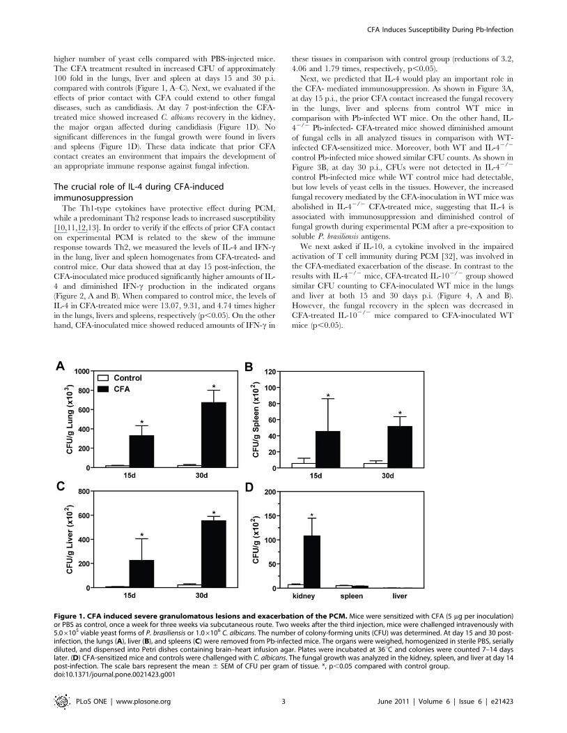
higher number of yeast cells compared with PBS-injected mice.
The CFA treatment resulted in increased CFU of approximately
100 fold in the lungs, liver and spleen at days 15 and 30 p.i.
compared with controls (Figure 1, A–C). Next, we evaluated if the
effects of prior contact with CFA could extend to other fungal
diseases, such as candidiasis. At day 7 post-infection the CFA-
treated mice showed increased C. albicans recovery in the kidney,
the major organ affected during candidiasis (Figure 1D). No
significant differences in the fungal growth were found in livers
and spleens (Figure 1D). These data indicate that prior CFA
contact creates an environment that impairs the development of
an appropriate immune response against fungal infection.
The crucial role of IL-4 during CFA-inducedimmunosuppression
The Th1-type cytokines have protective effect during PCM,
while a predominant Th2 response leads to increased susceptibility
[10,11,12,13]. In order to verify if the effects of prior CFA contact
on experimental PCM is related to the skew of the immune
response towards Th2, we measured the levels of IL-4 and IFN-cin the lung, liver and spleen homogenates from CFA-treated- and
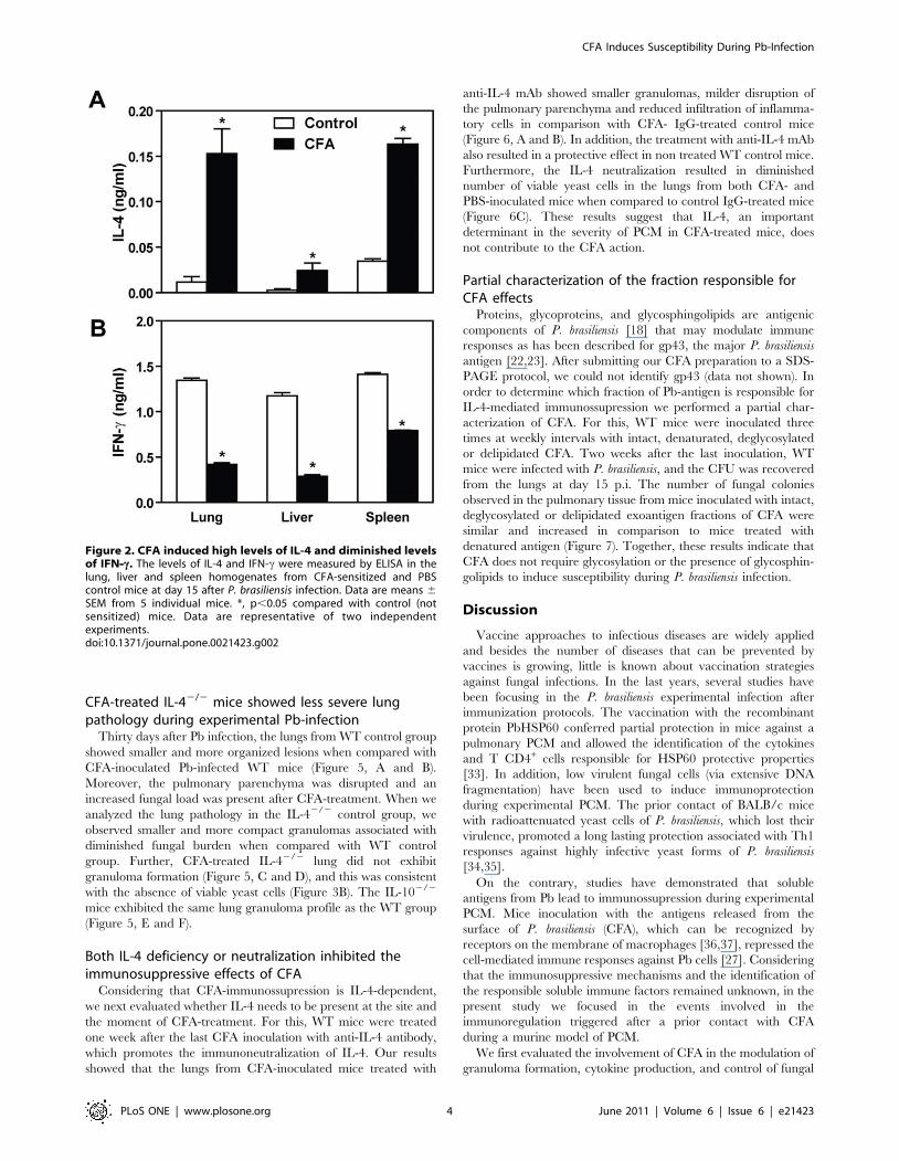
control mice. Our data showed that at day 15 post-infection, the
CFA-inoculated mice produced significantly higher amounts of IL-
4 and diminished IFN-c production in the indicated organs
(Figure 2, A and B). When compared to control mice, the levels of
IL-4 in CFA-treated mice were 13.07, 9.31, and 4.74 times higher
in the lungs, livers and spleens, respectively (p,0.05). On the other
hand, CFA-inoculated mice showed reduced amounts of IFN-c in
these tissues in comparison with control group (reductions of 3.2,
4.06 and 1.79 times, respectively, p,0.05).
Next, we predicted that IL-4 would play an important role in
the CFA- mediated immunosuppression. As shown in Figure 3A,
at day 15 p.i., the prior CFA contact increased the fungal recovery
in the lungs, liver and spleens from control WT mice in
comparison with Pb-infected WT mice. On the other hand, IL-
42/2 Pb-infected- CFA-treated mice showed diminished amount
of fungal cells in all analyzed tissues in comparison with WT-
infected CFA-sensitized mice. Moreover, both WT and IL-42/2
control Pb-infected mice showed similar CFU counts. As shown in
Figure 3B, at day 30 p.i., CFUs were not detected in IL-42/2
control Pb-infected mice while WT control mice had detectable,
but low levels of yeast cells in the tissues. However, the increased
fungal recovery mediated by the CFA-inoculation in WT mice was
abolished in IL-42/2 CFA-treated mice, suggesting that IL-4 is
associated with immunosuppression and diminished control of
fungal growth during experimental PCM after a pre-exposition to
soluble P. brasiliensis antigens.
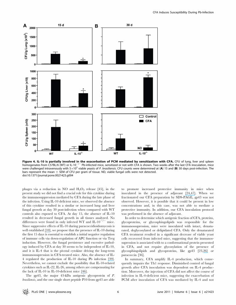
We next asked if IL-10, a cytokine involved in the impaired
activation of T cell immunity during PCM [32], was involved in
the CFA-mediated exacerbation of the disease. In contrast to the
results with IL-42/2 mice, CFA-treated IL-102/2 group showed
similar CFU counting to CFA-inoculated WT mice in the lungs
and liver at both 15 and 30 days p.i. (Figure 4, A and B).
However, the fungal recovery in the spleen was decreased in
CFA-treated IL-102/2 mice compared to CFA-inoculated WT
mice (p,0.05).
Figure 1. CFA induced severe granulomatous lesions and exacerbation of the PCM. Mice were sensitized with CFA (5 mg per inoculation)or PBS as control, once a week for three weeks via subcutaneous route. Two weeks after the third injection, mice were challenged intravenously with5.06105 viable yeast forms of P. brasiliensis or 1.06106 C. albicans. The number of colony-forming units (CFU) was determined. At day 15 and 30 post-infection, the lungs (A), liver (B), and spleens (C) were removed from Pb-infected mice. The organs were weighed, homogenized in sterile PBS, seriallydiluted, and dispensed into Petri dishes containing brain–heart infusion agar. Plates were incubated at 36uC and colonies were counted 7–14 dayslater. (D) CFA-sensitized mice and controls were challenged with C. albicans. The fungal growth was analyzed in the kidney, spleen, and liver at day 14post-infection. The scale bars represent the mean 6 SEM of CFU per gram of tissue. *, p,0.05 compared with control group.doi:10.1371/journal.pone.0021423.g001
CFA Induces Susceptibility During Pb-Infection
PLoS ONE | www.plosone.org 3 June 2011 | Volume 6 | Issue 6 | e21423
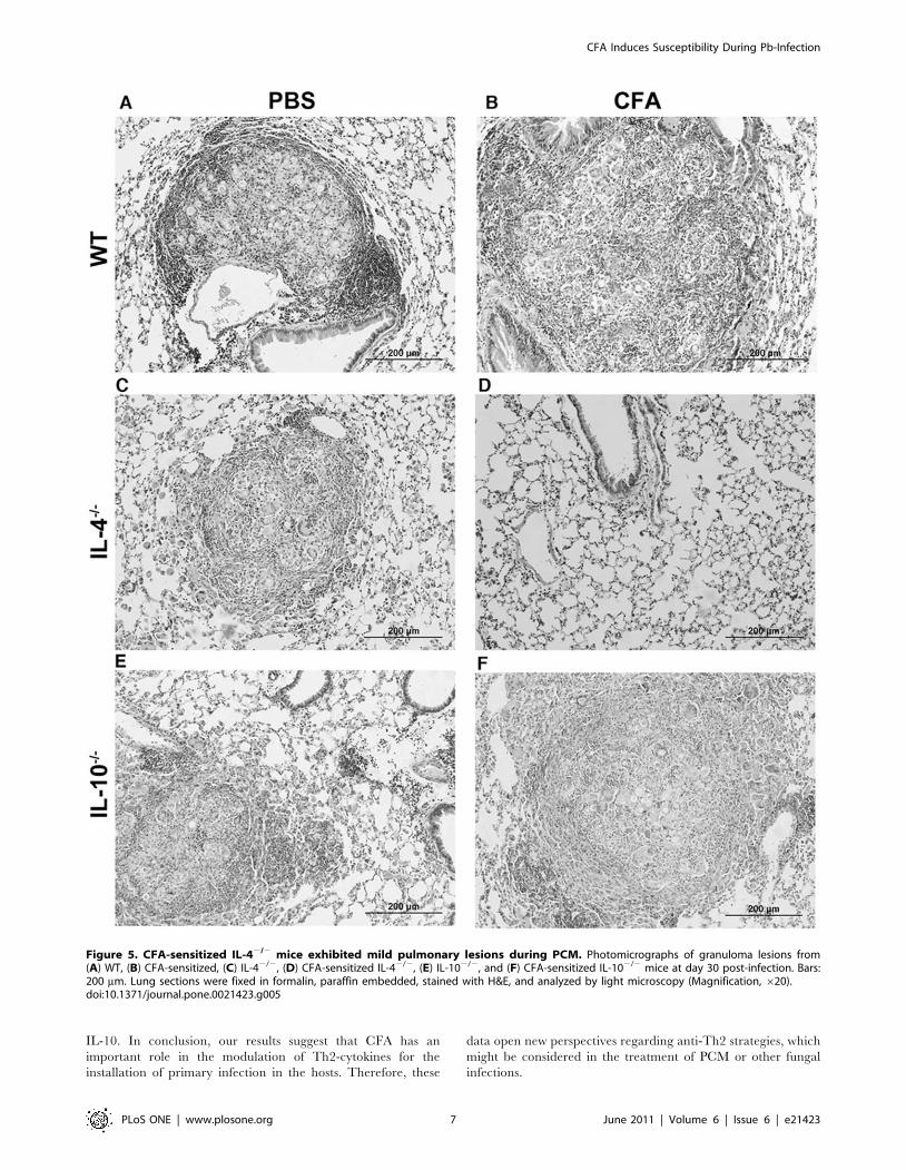
CFA-treated IL-42/2 mice showed less severe lungpathology during experimental Pb-infection
Thirty days after Pb infection, the lungs from WT control group
showed smaller and more organized lesions when compared with
CFA-inoculated Pb-infected WT mice (Figure 5, A and B).
Moreover, the pulmonary parenchyma was disrupted and an
increased fungal load was present after CFA-treatment. When we
analyzed the lung pathology in the IL-42/2 control group, we
observed smaller and more compact granulomas associated with
diminished fungal burden when compared with WT control
group. Further, CFA-treated IL-42/2 lung did not exhibit
granuloma formation (Figure 5, C and D), and this was consistent
with the absence of viable yeast cells (Figure 3B). The IL-102/2
mice exhibited the same lung granuloma profile as the WT group
(Figure 5, E and F).
Both IL-4 deficiency or neutralization inhibited theimmunosuppressive effects of CFA
Considering that CFA-immunossupression is IL-4-dependent,
we next evaluated whether IL-4 needs to be present at the site and
the moment of CFA-treatment. For this, WT mice were treated
one week after the last CFA inoculation with anti-IL-4 antibody,
which promotes the immunoneutralization of IL-4. Our results
showed that the lungs from CFA-inoculated mice treated with
anti-IL-4 mAb showed smaller granulomas, milder disruption of
the pulmonary parenchyma and reduced infiltration of inflamma-
tory cells in comparison with CFA- IgG-treated control mice
(Figure 6, A and B). In addition, the treatment with anti-IL-4 mAb
also resulted in a protective effect in non treated WT control mice.
Furthermore, the IL-4 neutralization resulted in diminished
number of viable yeast cells in the lungs from both CFA- and
PBS-inoculated mice when compared to control IgG-treated mice
(Figure 6C). These results suggest that IL-4, an important
determinant in the severity of PCM in CFA-treated mice, does
not contribute to the CFA action.
Partial characterization of the fraction responsible forCFA effects
Proteins, glycoproteins, and glycosphingolipids are antigenic
components of P. brasiliensis [18] that may modulate immune
responses as has been described for gp43, the major P. brasiliensis
antigen [22,23]. After submitting our CFA preparation to a SDS-
PAGE protocol, we could not identify gp43 (data not shown). In
order to determine which fraction of Pb-antigen is responsible for
IL-4-mediated immunossupression we performed a partial char-
acterization of CFA. For this, WT mice were inoculated three
times at weekly intervals with intact, denaturated, deglycosylated
or delipidated CFA. Two weeks after the last inoculation, WT
mice were infected with P. brasiliensis, and the CFU was recovered
from the lungs at day 15 p.i. The number of fungal colonies
observed in the pulmonary tissue from mice inoculated with intact,
deglycosylated or delipidated exoantigen fractions of CFA were
similar and increased in comparison to mice treated with
denatured antigen (Figure 7). Together, these results indicate that
CFA does not require glycosylation or the presence of glycosphin-
golipids to induce susceptibility during P. brasiliensis infection.
Discussion
Vaccine approaches to infectious diseases are widely applied
and besides the number of diseases that can be prevented by
vaccines is growing, little is known about vaccination strategies
against fungal infections. In the last years, several studies have
been focusing in the P. brasiliensis experimental infection after
immunization protocols. The vaccination with the recombinant
protein PbHSP60 conferred partial protection in mice against a
pulmonary PCM and allowed the identification of the cytokines
and T CD4+ cells responsible for HSP60 protective properties
[33]. In addition, low virulent fungal cells (via extensive DNA
fragmentation) have been used to induce immunoprotection
during experimental PCM. The prior contact of BALB/c mice
with radioattenuated yeast cells of P. brasiliensis, which lost their
virulence, promoted a long lasting protection associated with Th1
responses against highly infective yeast forms of P. brasiliensis
[34,35].
On the contrary, studies have demonstrated that soluble
antigens from Pb lead to immunossupression during experimental
PCM. Mice inoculation with the antigens released from the
surface of P. brasiliensis (CFA), which can be recognized by
receptors on the membrane of macrophages [36,37], repressed the
cell-mediated immune responses against Pb cells [27]. Considering
that the immunosuppressive mechanisms and the identification of
the responsible soluble immune factors remained unknown, in the
present study we focused in the events involved in the
immunoregulation triggered after a prior contact with CFA
during a murine model of PCM.
We first evaluated the involvement of CFA in the modulation of
granuloma formation, cytokine production, and control of fungal
Figure 2. CFA induced high levels of IL-4 and diminished levelsof IFN--c. The levels of IL-4 and IFN-c were measured by ELISA in thelung, liver and spleen homogenates from CFA-sensitized and PBScontrol mice at day 15 after P. brasiliensis infection. Data are means 6
SEM from 5 individual mice. *, p,0.05 compared with control (notsensitized) mice. Data are representative of two independentexperiments.doi:10.1371/journal.pone.0021423.g002
CFA Induces Susceptibility During Pb-Infection
PLoS ONE | www.plosone.org 4 June 2011 | Volume 6 | Issue 6 | e21423

growth in Pb-infected mice. We showed that inoculation of CFA
prior to infection resulted in severe lung pathology. The increased
pulmonary CFU recovered from CFA-treated mice was accom-
panied by intense inflammatory infiltrate and severe granuloma-
tous lesions. Moreover, CFA-treated mice showed granulomas
containing central necrotic areas with diffuse cell distribution and
a massive number of yeasts, which could be contributing to the
intensity and the persistence of the inflammatory response in these
mice.
The Th1/Th2 paradigm provides a basis for understanding T
cell responses and is applicable to explain the resistant/susceptible
pattern observed during experimental paracoccidioidomycosis.
Cytokine studies, mainly in a pulmonary model of infection, have
confirmed that Th1-biased immune response characterized by
IFN-c, TNF-a and IL-12 production, are linked with asymptom-
atic and mild forms of PCM [13,14]. In contrast, a Th2 pattern
has been associated with severe disease. Patients presenting the
disseminated infection produce higher levels of type 2 cytokines,
like IL-4, IL-5 and IL-10 [7,38,39].
In the present study, we described that the injection of CFA
prior to infection induced a Th2 profile, increasing IL-4 levels in
the lung, liver and spleen homogenates, and decreased the IFN-cproduction when compared with only Pb-infected mice. Using IL-
42/2 mice and their IL-4-sufficient counterparts, we observed that
the absence of IL-4 resulted in increased inflammatory reaction in
the lungs as showed before [39,40]. These data corroborates with
elevated numbers of PMN cells in the early phase of infection (data
not shown) and lower CFU counts in the pulmonary tissue. In
agreement, previous studies demonstrated that the severity of
PCM is mild in IL-42/2 mice compared with WT group, with
increased IFN-c release in lung homogenates and enhanced
fungicidal activity of alveolar phagocytes [39,41,42].
When we pre-treated mice with anti-IL-4 monoclonal antibody
one week after the last CFA-inoculation, we observed that the in
vivo IL-4-depletion impaired the suppressive effects of CFA. It
implicates that IL-4-CFA-induced is responsible by the mediation
of the observed unresponsiveness. Moreover, IL-4 favors the
development of a Th1 rather than Th2 immune response. Because
M2-macrophage activation is mediated by IL-4 and/or IL-13
(Th2-type cytokines), these macrophages are normally associated
with immune responses that possess a Th2-skewed cytokine
environment, as observed in parasite infections and allergic
inflammation [43,44]. Future studies should explore the role of
these cells during P. brasiliensis infection.
Although IL-10 have an inhibitory effect during PCM by
preventing fungal killing by IFN-c or TNF-a-activated macro-
Figure 3. The immunosuppressive effects of CFA are mediated by IL-4. CFA-sensitized C57BL/6 (WT) mice, CFA-sensitized IL-42/2 mice,control (not sensitized) WT and control IL-42/2 mice were infected with P. brasiliensis and analyzed at 15 (A) and 30 (B) days post-infection asdescribed in the legend to Figure 1. The scale bars represent the mean 6 SEM of colony-forming units (CFU) per gram of tissue. *, p,0.05 incomparison with CFA-sensitized WT mice (n = 5–7). ND, viable yeast cells were not detected.doi:10.1371/journal.pone.0021423.g003
CFA Induces Susceptibility During Pb-Infection
PLoS ONE | www.plosone.org 5 June 2011 | Volume 6 | Issue 6 | e21423
phages via a reduction in NO and H2O2 release [45], in the
present study we did not find a crucial role for this cytokine during
the immunosuppression mediated by CFA during the late phase of
the infection. Using IL-10 deficient mice, we observed the absence
of this cytokine resulted in a similar or increased lung and liver
fungal growth at day 30 post-infection when compared with WT
controls also exposed to CFA. At day 15, the absence of IL-10
resulted in decreased fungal growth in all tissues analyzed. No
differences were found in only infected WT and IL-102/2 mice.
Since suppressive effects of IL-10 during paracoccidioidomycosis is
well established [10], we propose that the presence of IL-10 during
the first 15 days is essential to establish a initial negative regulation
of immune cells via down-regulation of APC function or via Treg
induction. However, the fungal persistence and excessive pathol-
ogy induced by CFA at day 30 seems to be independent of IL-10,
and it is IL-4 that is the pivotal cytokine driving the long-term
immunossupression in CFA-treated mice. Also, the absence of IL-
4 regulated the production of IL-10 during Pb infection [39].
Nevertheless, we cannot exclude the possibility that IL-10-related
cytokines such as IL-22, IL-26, among others are compensating for
the lack of IL-10 in IL-10-deficient mice [46].
The gp43, the major 43-kDa antigenic glycoprotein of P.
brasiliensis, and the one single short peptide P10 from gp43 are able
to promote increased protective immunity in mice when
inoculated in the presence of adjuvant [24,47]. When we
fractionated our CFA preparation by SDS-PAGE, gp43 was not
observed. However, it is possible that it could be present in low
concentrations and, in this case, was not able to mediate a
protective immunity. In addition, our CFA inoculation protocol
was performed in the absence of adjuvant.
In order to determine which antigenic fraction of CFA, proteins,
glycoproteins, or glycosphingolipids was responsible for the
immunossupression, mice were inoculated with intact, denatu-
rated, deglycosylated or delipidated CFA. Only the denaturated
CFA treatment resulted in a significant decrease of viable yeast
cells recovered from infected mice, suggesting that the immunos-
supression is associated with to a conformational protein presented
in CFA, and not require glycosylation of the presence of
glycosphingolipids and glycoproteins, like gp43 [23,26] or
paracoccin [36].
In summary, CFA amplify IL-4 production, which conse-
quently impairs the Th1 response. Diminished control of fungal
growth after CFA inoculation was dependent on IL-4 produc-
tion. Moreover, the injection of CFA did not affect the course of
infection in IL-4-deficient mice, suggesting the exacerbation of
PCM after inoculation of CFA was mediated by IL-4 and not
Figure 4. IL-10 is partially involved in the exacerbation of PCM mediated by sensitization with CFA. CFU of lung, liver and spleenhomogenates from C57BL/6 (WT) or IL-102/2 Pb-infected mice, sensitized or not with CFA is shown. Two weeks after the last CFA inoculation, micewere challenged intravenously with 56105 viable yeasts of P. brasiliensis. CFU counts were determined at (A) 15 and (B) 30 days post-infection. Thebars represent the mean 6 SEM of CFU per gram of tissue. ND, viable fungal cells were not detected.doi:10.1371/journal.pone.0021423.g004
CFA Induces Susceptibility During Pb-Infection
PLoS ONE | www.plosone.org 6 June 2011 | Volume 6 | Issue 6 | e21423
IL-10. In conclusion, our results suggest that CFA has an
important role in the modulation of Th2-cytokines for the
installation of primary infection in the hosts. Therefore, these
data open new perspectives regarding anti-Th2 strategies, which
might be considered in the treatment of PCM or other fungal
infections.
Figure 5. CFA-sensitized IL-42/2 mice exhibited mild pulmonary lesions during PCM. Photomicrographs of granuloma lesions from(A) WT, (B) CFA-sensitized, (C) IL-42/2, (D) CFA-sensitized IL-42/2, (E) IL-102/2, and (F) CFA-sensitized IL-102/2 mice at day 30 post-infection. Bars:200 mm. Lung sections were fixed in formalin, paraffin embedded, stained with H&E, and analyzed by light microscopy (Magnification, 620).doi:10.1371/journal.pone.0021423.g005
CFA Induces Susceptibility During Pb-Infection
PLoS ONE | www.plosone.org 7 June 2011 | Volume 6 | Issue 6 | e21423

Supporting Information
Figure S1 Experimental PCM protocol. (A) Male C57BL/6
wild type (WT), IL-4- (IL-42/2), and IL-10-deficient (IL-102/2)
mice injected once per week (three times) by subcutaneous route
with CFA (5 mg per inoculation) or with PBS (control). Two weeks
after the last injection, mice were challenged intravenously with
56105 viable yeast forms of P. brasiliensis diluted in 100 ml of PBS.
(B) For the in vivo anti-IL-4 treatment, mice were intraperitoneally
inoculated with 0.5 mg/ml (100 ml) of rat IgG or purified IgG1
mAb against murine IL-4 (11B11) one week after the last CFA
inoculation, and at days 21, 7 and 10 of Pb-infection.
(TIF)
Acknowledgments
The authors thank Dr. Tiago W. Mineo for comments on early drafts of
the manuscript and Dr. Judith Connett (University of Michigan) for
editorial assistance.
Author Contributions
Conceived and designed the experiments: KAC FSMT LLO JSS.
Performed the experiments: KAC FSMT LLO FAR JOV APM APC
LAP. Analyzed the data: KAC FSMT LLO. Contributed reagents/
materials/analysis tools: CMM RM MAR JSS. Wrote the paper: KAC
FSMT JSS.
Figure 6. Anti-IL-4 markedly reduced pulmonary lesions and fungal growth in CFA-sensitized mice. Controls and CFA-sensitized micewere treated with anti-IL-4 mAb or rat IgG one week after the last CFA inoculation, and at days 21, 7, and 10 after P. brasiliensis infection. (A) Lungsections from CFA-sensitized mice treated with anti-IL-4 show smaller and more organized granulomas compared to granulomas in IgG treated mice.Hematoxylin-eosin stain was used; magnification, 620. Bars: 200 mm. (B) Fungal growth recovery in the lungs from CFA-sensitized or control micetreated with anti-IL-4 or IgG is shown. The bars depict mean 6 SEM of log10 CFU obtained from groups of 3–4 mice at day 15 after infection. Data isrepresentative of two independent experiments. *, p,0.05 compared with rat IgG-treated mice. #, p,0.05 compared with unsensitized mice. ND,viable fungal cells were not detected.doi:10.1371/journal.pone.0021423.g006
Figure 7. Protein fraction of CFA is required for immunosup-pression during PCM. WT mice were sensitized once a week for threeweeks with intact, denaturated, deglycosylated or delipidated CFA(5 mg per inoculation, subcutaneously). Two weeks after the lastinoculation, the mice were challenged intravenously with 56105 ofviable P. brasiliensis yeast cells. At day 15 post-infection, lungs wereremoved, weighed, homogenized in sterile PBS, serially diluted, anddispensed into Petri dishes containing brain–heart infusion agar. Plateswere incubated at 37uC and colonies were counted 7–14 days later. Thebars represent the mean 6 SEM of CFU per gram of tissue. *, p,0.05 incomparison with PBS treated mice.doi:10.1371/journal.pone.0021423.g007
CFA Induces Susceptibility During Pb-Infection
PLoS ONE | www.plosone.org 8 June 2011 | Volume 6 | Issue 6 | e21423
References
1. Coutinho ZF, Silva D, Lazera M, Petri V, Oliveira RM, et al. (2002)
Paracoccidioidomycosis mortality in Brazil (1980–1995). Cad Saude Publica 18:
1441–1454.
2. Restrepo A, McEwen JG, Castaneda E (2001) The habitat of Paracoccidioides
brasiliensis: how far from solving the riddle? Med Mycol 39: 233–241.
3. Brummer E, Castaneda E, Restrepo A (1993) Paracoccidioidomycosis: an
update. Clin Microbiol Rev 6: 89–117.
4. Restrepo-Moreno A (1993) Paracoccidioidomycosis. In: Juneann WM,
Herman F, Mauro B, eds. Infections, Agents and Pathogenesis – Fungal
Infections and Immune Responses. pp 251–276.
5. Borges-Walmsley MI, Chen D, Shu X, Walmsley AR (2002) The pathobiology
of Paracoccidioides brasiliensis. Trends Microbiol 10: 80–87.
6. Benard G, Mendes-Giannini MJ, Juvenale M, Miranda ET, Duarte AJ (1997)
Immunosuppression in paracoccidioidomycosis: T cell hyporesponsiveness to
two Paracoccidioides brasiliensis glycoproteins that elicit strong humoral
immune response. J Infect Dis 175: 1263–1267.
7. Benard G, Romano CC, Cacere CR, Juvenale M, Mendes-Giannini MJ, et al.
(2001) Imbalance of IL-2, IFN-gamma and IL-10 secretion in the immunosup-
pression associated with human paracoccidioidomycosis. Cytokine 13: 248–252.
8. Campanelli AP, Martins GA, Souto JT, Pereira MS, Livonesi MC, et al. (2003)
Fas-Fas ligand (CD95-CD95L) and cytotoxic T lymphocyte antigen-4
engagement mediate T cell unresponsiveness in patients with paracoccidioido-
mycosis. J Infect Dis 187: 1496–1505.
9. Franco M, Mendes R, Moscardi-Bacchi M, Rezkallah-Iwasso M, Montenegro M
(1989) Paracoccidioidomycosis. Bailliere’s Clin Trop Med Comm Dis 4:
185–220.
10. Calich VL, Kashino SS (1998) Cytokines produced by susceptible and resistant
mice in the course of Paracoccidioides brasiliensis infection. Braz J Med Biol Res
31: 615–623.
11. Cano LE, Kashino SS, Arruda C, Andre D, Xidieh CF, et al. (1998) Protective
role of gamma interferon in experimental pulmonary paracoccidioidomycosis.
Infect Immun 66: 800–806.
12. Peracoli MT, Kurokawa CS, Calvi SA, Mendes RP, Pereira PC, et al. (2003)
Production of pro- and anti-inflammatory cytokines by monocytes from patients
with paracoccidioidomycosis. Microbes Infect 5: 413–418.
13. Souto JT, Figueiredo F, Furlanetto A, Pfeffer K, Rossi MA, et al. (2000)
Interferon-gamma and tumor necrosis factor-alpha determine resistance to
Paracoccidioides brasiliensis infection in mice. Am J Pathol 156: 1811–1820.
14. Livonesi MC, Souto JT, Campanelli AP, Maffei CM, Martinez R, et al. (2008)
Deficiency of IL-12p40 subunit determines severe paracoccidioidomycosis in
mice. Med Mycol 46: 637–646.
15. McEwen JG, Bedoya V, Patino MM, Salazar ME, Restrepo A (1987)
Experimental murine paracoccidiodomycosis induced by the inhalation of
conidia. J Med Vet Mycol 25: 165–175.
16. Ferreira KS, Lopes JD, Almeida SR (2003) Regulation of T helper cell
differentiation in vivo by GP43 from Paracoccidioides brasiliensis provided by
different antigen-presenting cells. Scand J Immunol 58: 290–297.
17. Restrepo A (1985) The ecology of Paracoccidioides brasiliensis: a puzzle still
unsolved. Sabouraudia 23: 323–334.
18. Bertini S, Colombo AL, Takahashi HK, Straus AH (2007) Expression of
antibodies directed to Paracoccidioides brasiliensis glycosphingolipids during the
course of paracoccidioidomycosis treatment. Clin Vaccine Immunol 14:
150–156.
19. Casotto M (1990) Characterization of the cellular antigens of Paracoccidioides
brasiliensis yeast form. J Clin Microbiol 28: 1188–1193.
20. Puccia R, Schenkman S, Gorin PA, Travassos LR (1986) Exocellular
components of Paracoccidioides brasiliensis: identification of a specific antigen.
Infect Immun 53: 199–206.
21. Figueroa JI, Hamilton A, Allen M, Hay R (1994) Immunohistochemical
detection of a novel 22- to 25-kilodalton glycoprotein of Paracoccidioides
brasiliensis in biopsy material and partial characterization by using species-
specific monoclonal antibodies. J Clin Microbiol 32: 1566–1574.
22. Ferreira KS, Almeida SR (2006) Immunization of susceptible mice with gp43-
pulsed dendritic cells induce an increase of pulmonary Paracoccidioidomycosis.
Immunol Lett 103: 121–126.
23. Popi AF, Lopes JD, Mariano M (2002) Gp43 from Paracoccidioides brasiliensis
inhibits macrophages functions. An evasion mechanism of the fungus. Cel
Immunol 218: 87–94.
24. Rodrigues EG, Travassos LR (1994) Nature of the reactive epitopes in
Paracoccidioides brasiliensis polysaccharide antigen. J Med Vet Mycol 32:
77–81.
25. Saraiva EC, Altemani A, Franco MF, Unterkircher CS, Camargo ZP (1996)
Paracoccidioides brasiliensis-gp43 used as paracoccidioidin. J Med Vet Mycol
34: 155–161.
26. Almeida SR, Unterkircher CS, Camargo ZP (1998) Involvement of the majorglycoprotein (gp43) of Paracoccidioides brasiliensis in attachment to macro-
phages. Med Mycol 36: 405–411.27. Jimenez-Finkel BE, Murphy JW (1988) Induction of antigen-specific T
suppressor cells by soluble Paracoccidioides brasiliensis antigen. Infect Immun56: 734–743.
28. Calich VL, Kipnis TL, Mariano M, Neto CF, Dias da Silva WD (1979) The
activation of the complement system by Paracoccidioides brasiliensis in vitro: itsopsonic effect and possible significance for an in vivo model of infection. Clin
Immunol Immunopathol 12: 21–30.29. Camargo ZP, Taborda CP, Rodrigues EG, Travassos LR (1991) The use of cell-
free antigens of Paracoccidioides brasiliensis in serological tests. J Med Vet
Mycol 29: 31–38.30. Wang WX, Pelah D, Alergand T, Shoseyov O, Altman A (2002) Character-
ization of SP1, a stress-responsive, boiling-soluble, homo-oligomeric proteinfrom aspen. Plant Physiol 130: 865–875.
31. Livonesi MC, Rossi MA, de Souto JT, Campanelli AP, de Sousa RL, et al.
(2009) Inducible nitric oxide synthase-deficient mice show exacerbatedinflammatory process and high production of both Th1 and Th2 cytokines
during paracoccidioidomycosis. Microbes Infect 11: 123–132.32. Costa TA, Calich VL (2005) Pulmonary paracoccidioidomycosis (PCM) in IL-10
knockout (KO) mice. In: IX International Meeting on Paracoccidioidomycosis,Aguas de Lindoia. pp 29.
33. de Bastos Ascenco Soares R, Gomez FJ, de Almeida Soares CM, Deepe GS, Jr.
(2008) Vaccination with heat shock protein 60 induces a protective immuneresponse against experimental Paracoccidioides brasiliensis pulmonary infection.
Infect Immun 76: 4214–4221.34. do Nascimento Martins EM, Reis BS, Fernandes VC, Costa MM, Goes AM, et
al. (2007) Immunization with radioattenuated yeast cells of Paracoccidioides
brasiliensis induces a long lasting protection in BALB/c mice. Vaccine 25:7893–7899.
35. do Nascimento Martins EM, Reis BS, de Resende MA, de Andrade AS,Goes AM (2009) Mice immunization with radioattenuated yeast cells of
Paracoccidiodes brasiliensis: influence of the number of immunizations.Mycopathologia 168: 51–58.
36. Coltri KC, Casabona-Fortunato AS, Gennari-Cardoso ML, Pinzan CF,
Ruas LP, et al. (2006) Paracoccin, a GlcNAc-binding lectin from Paracoccid-ioides brasiliensis, binds to laminin and induces TNF-alpha production by
macrophages. Microbes Infect 8: 704–713.37. Moscardi-Bacchi M, Brummer E, Stevens DA (1994) Support of Paracoccid-
ioides brasiliensis multiplication by human monocytes or macrophages:
inhibition by activated phagocytes. J Med Microbiol 40: 159–164.38. Karhawi AS, Colombo AL, Salomao R (2000) Production of IFN-gamma is
impaired in patients with paracoccidioidomycosis during active disease and isrestored after clinical remission. Med Mycol 38: 225–229.
39. Pina A, Valente-Ferreira RC, Molinari-Madlum EE, Vaz CA, Keller AC, et al.(2004) Absence of interleukin-4 determines less severe pulmonary paracoccid-
ioidomycosis associated with impaired Th2 response. Infect Immun 72:
2369–2378.40. Arruda C, Valente-Ferreira RC, Pina A, Kashino SS, Fazioli RA, et al. (2004)
Dual role of interleukin-4 (IL-4) in pulmonary paracoccidioidomycosis:endogenous IL-4 can induce protection or exacerbation of disease depending
on the host genetic pattern. Infect Immun 72: 3932–3940.
41. Cenci E, Mencacci A, Del Sero G, Bacci A, Montagnoli C, et al. (1999)Interleukin-4 causes susceptibility to invasive pulmonary aspergillosis through
suppression of protective type I responses. J Infect Dis 180: 1957–1968.42. Kopf M, Brombacher F, Kohler G, Kienzle G, Widmann KH, et al. (1996) IL-4-
deficient Balb/c mice resist infection with Leishmania major. J Exp Med 184:1127–1136.
43. Noel W, Raes G, Hassanzadeh Ghassabeh G, De Baetselier P, Beschin A (2004)
Alternatively activated macrophages during parasite infections. Trends Parasitol20: 126–133.
44. Zhu Z, Zheng T, Homer RJ, Kim YK, Chen NY, et al. (2004) Acidicmammalian chitinase in asthmatic Th2 inflammation and IL-13 pathway
activation. Science 304: 1678–1682.
45. Moreira AP, Dias-Melicio LA, Soares AM (2010) Interleukin-10 but notTransforming Growth Factor beta inhibits murine activated macrophages
Paracoccidioides brasiliensis killing: effect on H2O2 and NO production. CellImmunol 263: 196–203.
46. Commins S, Steinke JW, Borish L (2008) The extended IL-10 superfamily: IL-
10, IL-19, IL-20, IL-22, IL-24, IL-26, IL-28, and IL-29. J Allergy Clin Immunol121: 1108–1111.
47. Taborda CP, Juliano MA, Puccia R, Franco M, Travassos LR (1998) Mappingof the T-cell epitope in the major 43-kilodalton glycoprotein of Paracoccidioides
brasiliensis which induces a Th-1 response protective against fungal infection inBALB/c mice. Infect Immun 66: 786–793.
CFA Induces Susceptibility During Pb-Infection
PLoS ONE | www.plosone.org 9 June 2011 | Volume 6 | Issue 6 | e21423