The anti-inflammatory effects of omalizumab confirm the central role of IgE in allergic inflammation

IL-3 is required for increases in blood basophils during nematodeinfection in mice and can influence IgE-dependent intra-cellularIL-4 production by basophils in vitro
Chris S. Lantz1, Booki Min2, Mindy Tsai3, Devavani Chatterjea4, Glenn Dranoff5, and StephenJ. Galli31Department of Biology, James Madison University, Harrisonburg, VA2Department of Immunology, Lerner Research Institute, Cleveland Clinic Foundation, Cleveland,OH3Department of Pathology, Stanford University School of Medicine, Stanford, CA4Department of Biology, Macalester College, Saint Paul, MN5Department of Medical Oncology, Dana-Farber Cancer Institute, Harvard Medical School, Boston,MA
AbstractBasophils represent potential effector and immunoregulatory cells, as well as a potential source ofIL-4, during the immune response elicited by infection with the nematode Nippostrongylusbrasiliensis (N.b.), and in other settings. However, the factors which regulate the numbers of bloodbasophils in mice, or the ability of these cells to produce IL-4, are not fully understood. We foundthat infection of mice with the nematodes N.b. or Strongyloides venezuelensis (S.v.) inducedsubstantial increases in the numbers of blood basophils (to as high as 18 % of circulating bloodleukocytes). Experiments in IL-3−/− vs IL-3+/+ mice, and in IL-3-treated IL-3−/− mice, showed thatessentially all of the increases in blood or bone marrow basophils during N.b. or S.v. infection wereIL-3-dependent. Many of the blood, bone marrow or liver-derived basophils from IL-3−/− orIL-3+/+ mice expressed intra-cellular IL-4 upon stimulation with anti-IgE in vitro. However, afterincubation of the cells with exogenous IgE in vitro, blood- or liver-derived basophils from IL-3+/+
mice exhibited higher levels of intra-cellular IL-4 after stimulation with anti-IgE than did basophilsderived from IL-3−/− mice. Thus, IL-3 is a major regulator of the marked increases in blood basophillevels observed during infection of mice with N.b. or S.v. and also can enhance levels of intra-cellularIL-4 upon activation of basophils with anti-IgE in vitro.
KeywordsAllergy; Cytokines; Immune response; Inflammation; Mast cells; Parasites
Basophils have been characterized in the mouse, as well as in several other species, as bonemarrow-derived granulocytes that circulate in the blood and can be recruited into tissues atsites of inflammation 1–6. Recent work has identified potential immunoregulatory functionsof basophils as well 7–10. However, the factors that regulate the development and function ofmouse basophils are not fully understood.
Corresponding author: Stephen J. Galli, MD, Department of Pathology, Stanford University School of Medicine, 300 Pasteur Drive,Stanford, CA 94305-5324, USA, Tel.: 650-723-7975; Fax: 650-725-6902, E-mail: [email protected].
NIH Public AccessAuthor ManuscriptLab Invest. Author manuscript; available in PMC 2009 December 3.
Published in final edited form as:Lab Invest. 2008 November ; 88(11): 1134–1142.
NIH
-PA Author Manuscript
NIH
-PA Author Manuscript
NIH
-PA Author Manuscript
Both basophils and mast cells express the high affinity IgE receptor, FcεRI, and can be activatedto release IL-4 and other mediators upon aggregation of their FcεRI by IgE and specific antigen11–14. Moreover, we previously reported that nematode infections can substantially enhancethe differentiation and the production of basophils in mice, as assessed by counting numbersof basophils in the bone marrow, as well as increase certain populations of tissue mast cells,by mechanisms that are largely or fully IL-3-dependent (for basophils) or partially IL-3dependent (for mast cells) 4. Nevertheless, several lines of evidence indicate that mousebasophils and mast cells represent distinct lineages 4, 5, 12 and can have distinct roles in parasiteinfections, allergic disorders and other settings 4, 5, 7–10, 15–18.
During nematode infections 2, 5 and in models of allergic inflammation 16, 19, mouse basophilsrepresent a potentially significant source of IL-4, a cytokine that may help to sustain the Th2responses in these models, as well as mediate other functions 15. Basophils also recently havebeen shown to be important for the development of hapten-specific chronic skin inflammationin transgenic mice which express high amounts of hapten-specific IgE 7, 8. Notably, increasedamounts of IL-4 mRNA were detected in the skin lesions associated with such IgE-inducedchronic inflammation 7. However, it is not clear to what extent this reflected IL-4 mRNA inthe basophils, as opposed to other cell types, present at these sites.
We previously showed that IL-3 is not necessary for the development of baseline levels ofbone marrow basophils (or tissue mast cells) in the mouse, but that during nematode infectionsIL-3 is required for the expansion of numbers of bone marrow basophils, and contributes tothe expansion of spleen and small intestinal mast cell populations 4. In vitro, IL-3 cansignificantly enhance FcεRI-dependent production of IL-4 by cell populations in whichbasophils represent the most likely source of IL-4 20, as well as FcεRI-dependent activationand mediator secretion by mouse peritoneal mast cells 21 and human blood basophils 22–24.Thus, IL-3 produced during nematode infections may both regulate the size of two populationsof effector cells that express FcεRI (i.e., basophils and mast cells), and also regulate theFcεRI-dependent secretory function of these cells.
However, past studies of mouse basophils primarily quantified these cells in the spleen 25,26, bone marrow 4, 26, lung 5, 25, 26 or liver 25, 26, and used exogenous IL-3 to investigatewhether this cytokine influenced basophil IL-4 production in vitro 20. This work indicated theinfection with N.b. can result in increases in populations of basophils 4, 5, 25, 26, including inthe blood 5, 25, 26, and that exogenous IL-3 can increase levels of basophils in wild typemice25, 27. In the present study, we used IL-3−/− and IL-3+/+ mice, and IL-3-treated IL-3−/−
mice, to investigate the potential role of IL-3 in regulating blood basophil levels in mice andassessed whether basophils from IL-3−/− mice exhibited any impairment in their ability tosecrete IL-4 upon stimulation via the FcεRI in vitro.
MATERIALS AND METHODSMice and nematode infection
BALB/c and C57BL/6J mice, 8–13 weeks of age, were obtained from Charles RiverLaboratory, Wilmington, MA, and the Jackson Laboratory, Bar Harbor, ME for experimentsconducted at the Beth Israel Deaconess Medical Center, from the Laboratory Animal BreedingColony at Stanford University for experiments conducted at Stanford, and from the Jacksonlaboratory for the experiments conducted at the Cleveland Clinic. IL-3−/− and their wild-typelittermates (IL-3+/+) were back-crossed for at least nine generations onto C57BL/6 or BALB/c mice 28. Some mice were infected by subcutaneous inoculation with 500 or 800Nippostrongylus brasiliensis (N.b.) larvae 29 or 2000 Strongyloides venezuelensis (S.v.) larvae4. The degree of infection of individual mice was monitored by counting the numbers of eggsexcreted daily (eggs/g feces) and/or, at the time of sacrifice, the numbers of adult intestinal
Lantz et al. Page 2
Lab Invest. Author manuscript; available in PMC 2009 December 3.
NIH
-PA Author Manuscript
NIH
-PA Author Manuscript
NIH
-PA Author Manuscript
worms. S.v. and N.b. were maintained by serial passage in male Wistar rats or female BALB/c mice, respectively. All animal care and experimentation were conducted according toguidelines of the National Institutes of Health and the Beth Israel Deaconess Medical Center,Stanford University, or Cleveland Clinic Institutional Animal Care and Use Committees.
Identification and quantification of mouse basophilsMouse blood and bone marrow cells were isolated from the orbital sinus and femurs, and RBCswere lysed with Tris-buffered ammonium chloride. Cells were incubated with B3B4 and 2.4G2mAbs for 15 min. to block low affinity binding of IgE or other subsequent Abs to CD23 orFcγRII/III, respectively, followed by sensitization with mouse IgE mAb (Sigma ChemicalCompany, St. Louis, MO; clone SPE-7) for 50 min. Cells were washed and stained with FITCanti-IgE (clone R35-72) and PE anti-CD45R/B220 (clone RA3-6B2) mAbs for 25 min. Fordetection of c-Kit, cells were incubated for 25 min. with biotinylated anti-c-kit mAb (clone2B8) followed by PE-streptavidin. Except for the SPE-7 IgE mAb, all Abs listed above,including appropriate isotype-matched controls, were obtained from Pharmingen, San Diego,CA, and used at 10 µg/ml on ice. In some experiments, we sought to identify basophils withoutusing antibodies to IgE or FcεRI. For these experiments, blood or liver cells were obtained asdescribed in detail in 26 and basophils in those populations were identified as described in 26,30, by staining with FITC-labeled anti-CD16/32 (clone 93) and APC-labeled anti-CD45 (30-F11) (these Abs, and isotype-matched controls, were obtained from eBioscience, San Diego,CA, and used at 10 µg/ml on ice). In such experiments, some cells were incubated ex vivo withan anti-TNP IgE mAb (IgE-3) and/or stimulated with anti-IgE (R35-72). Stained cells wereanalyzed using a FACSCalibur® with CellQuest® (Becton Dickinson, San Jose, CA) orFlowJo (Ashland, OR) software. The number of basophils/mm3 blood was calculated bymultiplying the percentage of basophils (as determined by flow cytometry) by the total WBCcount. For light microscopic examination, cells were sorted using a FACSvantage® (BectonDickinson), cytocentrifuged onto slides, and stained 6 h with 1.0 % alcian blue at pH 1.0followed by safranin for 10 min.
Treatment with IL-3 in vivoMale S.v.-infected IL-3+/+ and IL-3−/− mice received twice daily i.p. injections of IL-3 (140ng/day, from supernatants conditioned by B16-F10 murine melanoma cells that weregenetically engineered to produce murine IL-3 31) or vehicle beginning on d 5 of infection.IL-3 concentration was determined by ELISA (Endogen, Cambridge, MA). Mice weresacrificed the day each cleared their individual infection (Figure 3b, c) or, in a separateexperiment, on d 13 of infection (Figure 3a). To quantify mast cells, 4 µm paraffin sections ofCarnoy’s-fixed tissues were stained with safranin and 1.0 % alcian blue at pH 0.3 (or pH 1.0for spleen) and mast cells were counted as the number/mm2 of spleen, or for jejunum and ileum,as the number/villus crypt unit.
Basophil activation and staining of intra-cellular IL-4 for flow cytometric analysisTwo approaches were used for these experiments. In the first method, blood and bone marrowcells from IL-3+/+ and IL-3−/− mice were depleted of RBCs, pooled according to genotype,and sensitized with mouse IgE mAb for 50 min. on ice (without B3B4 and 2.4G2 mAbs). Cellswere washed, and incubated for 5 h at 37° C in complete DMEM containing Brefeldin A(Sigma; 10 µg/ml) and FITC anti-IgE (10 µg/ml). This step served simultaneously to activateand fluorescently stain FcεRI+ basophils, and to allow IL-4 to accumulate within the ER and/or Golgi complex. Cells were fixed for 10 min. with 4 % paraformaldehyde in PBS at 37° C,permeabilized on ice for 15 min. with PBS containing 0.1 % saponin, 2.4G2 mAb (10 µg/ml),and 2 % FCS, and stained for 25 min. on ice with PE anti-IL-4 mAb (clone 11B11, at 2 µg/ml) in PBS containing 2 % FCS. Many negative controls, all of which yielded similar results,
Lantz et al. Page 3
Lab Invest. Author manuscript; available in PMC 2009 December 3.
NIH
-PA Author Manuscript
NIH
-PA Author Manuscript
NIH
-PA Author Manuscript

were used to establish flow cytometry gates. As one control, IgE-sensitized cells wereincubated with FITC IgG1 (clone R3-34, as an FITC-labeled antibody of irrelevant antigenspecificity) for 5 h at 37° C with Brefeldin A followed by staining with FITC anti-IgE (10 µg/ml) mAb on ice for 25 min. Specificity of IL-4 staining was demonstrated both by pre-incubating cells with excess unlabeled anti-IL-4 mAb (60 µg/ml), and by staining cells with aPE isotype-matched control mAb (clone R3-34). Except for IgE, all Abs were purchased fromPharmingen.
A second approach was used to analyze basophils without using FITC anti-IgE to identify thebasophils by FACS. This permitted us to avoid activating the basophils via the FcεRI exceptat the time we wished to stimulate some of these cells in vitro. Groups of wild type BALB/c(IL-3+/+) and BALB/c IL-3−/− mice were subcutaneously infected with 500 N. b. larvae andmice were sacrificed 10 days post infection. Blood and liver cells were incubated with orwithout 5 µg/ml of a mouse IgE mAb (IgE-3, anti-TNP) for 50 min on ice and subsequentlyincubated with or without 1 µg/ml anti-IgE (R35–72) for an additional 4 h at 37° C. 2 µMMonensin was added during the last 2 hours of incubation. Cells were harvested, fixed, andstained for surface expression of FITC labeled anti-CD16/32 (clone 93), APC labeled anti-CD45 (30-F11), and PE labeled anti-IL-4 (11B11) for 45 min. Cells were acquired using aFACSCalilbur and analyzed using FlowJo software.
StatisticsUnless otherwise specified, all data are expressed as the mean ± SEM, and all differencesbetween values were compared using the unpaired two-tailed Student’s t test.
RESULTSMouse blood basophil levels are markedly increased following infection with N.b
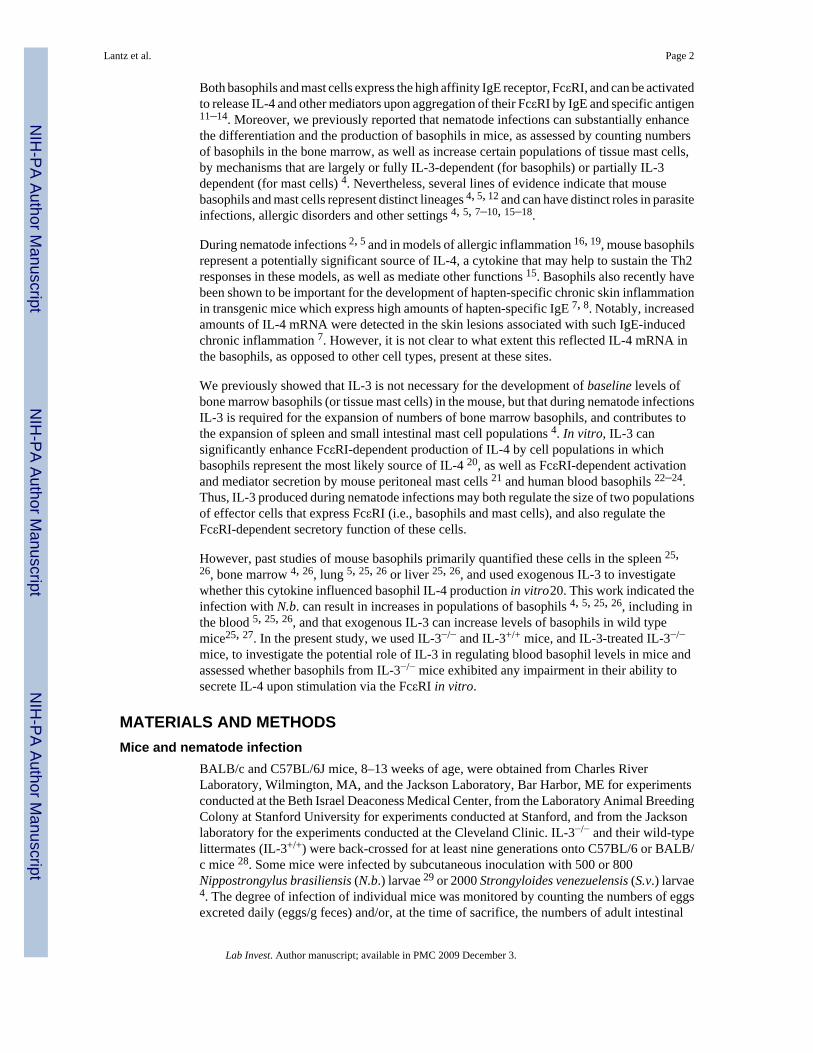
Blood cells obtained from 8–13 week old BALB/c mice were stained for the expression ofB220 and FcεRI. The percentage of B220−, FcεRI+ cells identified as basophils by flowcytometric analysis, as well as alcian blue/safranin staining in the blood, showed a markedincrease above baseline (Figure 1a) in mice infected with N. b.; a 7-fold increase 9 d afterinfection with 800 parasites (Figure 1b) and an 18-fold increase 12 d after infection (Figure1c). Few or no c-kit-expressing mast cells were detected in the blood at any time, including at12 d after infection (Figure 1d). The B220−, FcεRI+ cell population was sorted and stainedwith alcian blue/safranin. 95 % of these cells had a poly-lobed nucleus and 97 % containedsmall granules that stained lightly with alcian blue (Figure 1e). By contrast, only a single cellwith the morphological characteristics of a mast cell was found in all of the slides of bloodexamined (* in Figure 1e). Similar results were obtained in 2 other experiments using BALB/c mice (data not shown).
IL-3 is required for the development of increased numbers of blood and bone marrowbasophils in parasite-infected mice
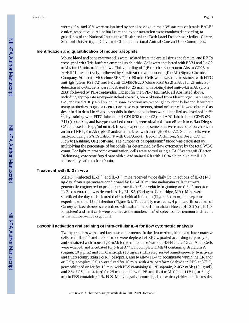
Basophils/mm3 of blood were ~ 3-fold higher than baseline levels 9 d after S.v. infection and> 5-fold higher 11 d after N.b. infection in C57BL/6-IL-3+/+ mice, whereas C57BL/6-IL-3−/− mice had levels of blood basophils that were unchanged or even lower than baselinefollowing parasite infection (Figure 2a). The % of bone marrow basophils in mice of the twogenotypes showed an identical pattern of change following infection with N.b. and S.v. larvae(Figure 2b); levels of bone marrow basophils increased to 2 and 3 fold baseline levels 9 and11 d after S.v. and N.b. infection, respectively, in IL-3+/+ mice while they remained unchangedor at lower than baseline levels in the bone marrow of IL-3−/− mice at those time points. BothIL-3+/+ and IL-3−/− N.b.-infected mice cleared their infection by d 11 and there was nostatistically significant difference between the numbers of adult S.v. worms 9 d after infection
Lantz et al. Page 4
Lab Invest. Author manuscript; available in PMC 2009 December 3.
NIH
-PA Author Manuscript
NIH
-PA Author Manuscript
NIH
-PA Author Manuscript
in IL-3+/+ and IL-3−/− mice (adult worms/small intestine: IL-3+/+, 145 ± 15 versus IL-3−/−, 230± 68; p = 0.31). Thus, the IL-3 deficiency did not affect parasite clearance in N.b.-infected miceor numbers of adult worms or egg production at d 9 in S.v.-infected mice, but IL-3 was requiredfor the expansion of blood basophils following parasite infection. By contrast, IL-3 was notrequired for the maintenance of baseline levels of bone marrow or blood basophils in naïvemice.
Treatment of S.v.-infected IL-3−/− mice with exogenous IL-3 restores basophil and tissue mastcell numbers to levels that are similar to those observed in S.v.-infected IL-3+/+ mice
Intra-peritoneal injection twice daily of IL-3 (total of 140 ng/d) into male IL-3−/− mice duringinfection with S.v. restored blood and bone marrow basophil levels, as well as mast cell numbersin the spleen and ileum, to values that were statistically indistinguishable from those observedin S.v.–infected IL-3+/+ mice (Figure 3). IL-3 treatment also markedly increased numbers ofmast cells in the jejunum of S.v.–infected IL-3−/− mice (Figure 3c). Although the effects werenot statistically significant, injection of IL-3 in S.v.–infected IL-3+/+ animals may have resultedin slight increases in levels of blood and bone marrow basophils, and perhaps in some mastcell populations (Figure 3).
IL-3 is not required for, but can enhance, IL-4 production by blood or bone marrow basophilsfollowing FcεRI cross-linkage in vitro
Although IL-3−/− mice did not exhibit increases in blood or bone marrow basophils followinginfection with N.b. or S.v., we found that IL-3 was not required for production of intra-cellularIL-4 following FcεRI cross-linkage in vitro in blood or bone marrow basophils derived fromeither normal or N.b.-infected mice. As assessed by intra-cellular cytokine staining, freshlyisolated blood and bone marrow basophils produced IL-4 after activation by FITC anti-IgEmAb in vitro, whether the cells were derived from C57BL/6 IL-3+/+ or IL-3−/− mice (Figure4). In uninfected IL-3−/− and IL-3+/+ mice, basophils represented ~ 1.5 % of total bloodleukocytes and ~ 1.0 % of total bone marrow cells. After incubation with mouse IgE followedby challenge with FITC anti-IgE in vitro, ~ 45–52 % of the blood basophils and ~ 10 % of thebone marrow basophils were IL-4+. The differences in the levels of IL-4 producing cells withinthe bone marrow and blood basophil populations may reflect, at least in part, the fact thatcirculating basophils in the periphery are, as a population, more mature developmentally thanis the bone marrow population, which includes multiple developmental stages.
In blood leukocytes that were obtained from N.b.-infected C57BL/6 mice and then wereincubated with IgE and FITC anti-IgE in vitro, 80 % of IL-3−/− and 95 % of IL-3+/+ bloodbasophils were IL-4+ and 47 % of IL-3−/− or IL-3+/+ bone marrow basophils were IL-4+ (Figure4). As shown in Figure 2 & Figure 4, IL-3−/− mice had a significant defect in nematode-inducedbasophil hyperplasia. In Figure 4, 10.7 % of total blood leukocytes in IL-3+/+ mice werebasophils vs. only 1.3 % in the IL-3-deficient counterparts. For bone marrow basophils, thecorresponding values were 2.6 % in IL-3+/+ mice vs. 0.8 % in IL-3−/− mice (Figure 4). Suchexperiments showed that, under the conditions tested, a lack of IL-3 does not eliminate theIL-4 production capacity of mouse basophils after anti-IgE stimulation in vitro.
We also examined intra-cellular IL-4 production in basophils that were analyzed by FACSwithout using antibodies to IgE or FcεRI to identify the cells 26. Blood cells were pooled fromBALB/c IL-3−/− and IL-3+/+ mice (n = 3–4 in each group) on d 10 of N.b. infection. Duplicatealiquots of these blood leukocytes were then incubated for 50 min on ice ± IgE and thenstimulated for 4 h with anti-IgE, with Monensin added for the last 2 h; negative control cellswere incubated under the same conditions but without exogenous IgE or anti-IgE.
Lantz et al. Page 5
Lab Invest. Author manuscript; available in PMC 2009 December 3.
NIH
-PA Author Manuscript
NIH
-PA Author Manuscript
NIH
-PA Author Manuscript

Little intracellular IL-4 was detectable in basophils incubated in vitro with neither exogenousIgE nor anti-IgE (Figure 5). However, anti-IgE stimulation of cells which had not beenincubated with additional IgE in vitro induced similar percentages of IL-3+/+ or IL-3−/−
basophils (41–54 %) to exhibit intra-cellular staining for IL-4 (Figure 5). By contrast, afterincubation with exogenous IgE in vitro prior to activation with anti-IgE, blood basophils fromIL-3+/+ mice exhibited substantially higher levels of intra-cellular staining for IL-4 than didthe corresponding cells from IL-3−/− mice (Figure 5). Similar results were obtained in a separateexperiment analyzing IL-3+/+ vs. IL-3−/− mouse blood basophils that had been incubated withexogenous IgE prior to stimulation with anti-IgE in vitro. Using data pooled from the latterexperiment and the experiment shown in Figure 5, we analyzed the percentage of IL-3+/+ vs.IL-3−/− blood basophils that stained for intra-cellular IL-4, and the MFI of IL-4 staining inblood basophils (Figure 6a). Incubation with exogenous IgE before stimulation with anti-IgEresulted in a significantly higher percentage of basophils that exhibited intracellular stainingwith IL-4, and a significantly higher MFI of such staining, in blood basophils derived fromIL-3+/+ vs. IL-3−/− mice.
We also analyzed intracellular IL-4 production following ex vivo stimulation in basophilsisolated from the liver of N.b. infected mice. Since more basophils can be obtained from thelivers than the blood of such mice, we individually tested the liver basophils obtained fromIL-3+/+ (n = 4) mice and from IL-3−/− mice (n = 3). As shown in Figure 6b, the results obtainedwith liver-derived basophils were very similar to those obtained in tests of blood basophils(Figure 6a). Only low levels of intra-cellular IL-4 were detected in basophils incubated withoutexogenous IgE or anti-IgE (MFI = 16.2 ± 1.1 vs. 18.8 ± 1.6 for IL-4+ basophils derived fromIL-3+/+ vs. IL-3−/− mice [p > 0.05]), higher levels of intra-cellular IL-4 were detected in anti-IgE stimulated basophils which had not been incubated with exogenous IgE in vitro (MFI =35.9 ± 1.2 vs. 30.9 ± 2.5 for IL-4+ IL-3+/+ vs. IL-3−/− cells [p > 0.05]), and, after incubationwith exogenous IgE in vitro, these responses were even stronger in anti-IgE stimulatedbasophils from IL-3+/+ mice but not in the identically-treated basophils from IL-3−/− mice (MFI= 47.9 ± 1.9 vs. 26.7 ± 1.7 for IL-3+/+ vs. IL-3−/− cells [p < 0.005]).
Taken together, the data in Figure 4–Figure 6, obtained using basophils derived from IL-3+/+
vs. IL-3−/− mice on either the C57BL/6 (Figure 4) or BALB/c (Figure 5 and Figure 6)backgrounds, indicate that basophils derived from IL-3−/− mice can produce intra-cellular IL-4upon activation via IgE and FcεRI in vitro, but that such responses can be enhanced in basophilsderived from IL-3+/+ mice.
DISCUSSIONOur findings show that IL-3 is required for the striking increases in blood basophils that occurin C57BL/6 or BALB/c mice during infection with either Nippostrongylus brasiliensis (N.b.)or Strongyloides venezuelensis (S.v.) in vivo. By contrast, IL-3 appears to be largely dispensablefor maintaining the low levels of blood basophils observed in the circulation of uninfectedmice. These finding are in accord with our observation that IL-3 is required for the increasesin bone marrow basophils associated with S.v. or N.b. infection in mice, but is not required formaintaining low levels of bone marrow basophils in uninfected animals 4. While thismanuscript was under review, Shen et al. also found, using BALB/c IL-3−/− vs. IL-3+/+ mice,that IL-3 was required for the increases in numbers of basophils in the blood, bone marrow,and liver in BALB/c mice infected with N.b., and provided evidence that T cells represent animportant source of such IL-3 in this setting 26.
Khodoun et al. 27 showed that the administration of IL-3 to IL-3 sufficient mice can result inexpanded basophil populations in the spleen of naïve wild type mice 27. We found thatadministration of recombinant IL-3 beginning on d 5 of S.v. infection permitted IL-3−/− mice
Lantz et al. Page 6
Lab Invest. Author manuscript; available in PMC 2009 December 3.
NIH
-PA Author Manuscript
NIH
-PA Author Manuscript
NIH
-PA Author Manuscript

to exhibit numbers of blood and bone marrow basophils, as well as mast cells in the spleen andileum, that were statistically indistinguishable from those observed in S.v.– infected IL-3+/+
mice. These findings support the conclusion that the striking abnormalities in basophil andmast cell numbers that occur during nematode infection in IL-3−/− mice reflect the lack of IL-3in the adult animals, rather than the consequences of a developmental problem, related to thegenetic deficiency in IL-3, that irreversibly alters the responsiveness of the basophil or mastcell lineages to IL-3.
When stimulated with anti-IgE in vitro in the presence of Brefeldin A (Figure 4) or Monensin(Figure 5), blood or liver-derived basophils from IL-3−/− or IL-3+/+ mice exhibited substantialIL-4 production, as assessed by flow cytometric analysis of intra-cellular IL-4. By contrast,few blood or liver basophils derived from IL-3−/− or IL-3+/+ mice exhibited intra-cellular IL-4after in vitro incubation for 4 h in the absence of anti-IgE stimulation (Figure 5 and Figure 6).Similarly, blood or liver basophils examined by flow cytometry in cells freshly isolated fromN.b.-infected IL-3−/− or IL-3+/+ mice exhibited little or no evidence of intra-cellular IL-4, nordid such mice exhibit differences in levels of serum IgE (data not shown).
These findings indicate that blood or liver basophils are capable of upregulating IL-4production upon stimulation via IgE and anti-IgE, whether such basophils are derived fromwild type mice or mice that lack IL-3. It should be emphasized that our experiments used flowcytometry to examine intra-cellular IL-4 in basophils that had been stimulated via the FcεRIex vivo, and this approach may not be sensitive enough to detect what might be significantdifferences in the amounts of cytokines produced by various basophil populations that exhibitsimilar levels of intra-cellular IL-4 (e.g., as in blood basophils derived from IL-3−/− vs.IL-3+/+ mice that had been stimulated by anti-IgE in vitro in the absence of prior incubationwith exogenous IgE).
However, when populations of blood or liver cells were incubated in vitro with exogenous IgEbefore their stimulation with anti-IgE, we found that blood or liver-derived basophils fromIL-3+/+ mice exhibited substantially greater intracellular IL-4 responses than did identically-treated basophils from IL-3−/− mice (Figure 5 and Figure 6). These data are consistent with theconclusions of prior in vitro work indicating that IL-3 can significantly enhance FcεRI-dependent production of IL-4 by cell populations in which basophils represent the most likelysource of IL-4 20, as well as FcεRI-dependent activation and mediator secretion by mouseperitoneal mast cells 21 and human blood basophils 22–24. Thus, even though our studies showthat IL-3 is not required for basophil IL-4 production in response to challenge via the FcεRIex vivo, they also suggest that, at least under some circumstances, basophils derived fromIL-3+/+ mice can exhibit higher levels of IgE- and FcεRI-dependent IL-4 production that dobasophils from IL-3−/− mice.
AcknowledgmentsWe are grateful to Fred Finkelman and William Paul and their colleagues for helpful comments.
Supported by NIH grants AI23990, CA72074 and HL67674 to SJG, NIH grant AI049932 and Jeffress Memorial TrustGrant J-782 to CSL, and startup funds from the Cleveland Clinic Foundation to BM; BM is the recipient of the careerdevelopment grant from the American Heart Association.
References1. Dvorak AM, Nabel G, Pyne K, et al. Ultrastructural identification of the mouse basophil. Blood
1982;59:1279–1285. [PubMed: 7082829]
Lantz et al. Page 7
Lab Invest. Author manuscript; available in PMC 2009 December 3.
NIH
-PA Author Manuscript
NIH
-PA Author Manuscript
NIH
-PA Author Manuscript

2. Seder RA, Paul WE, Dvorak AM, et al. Mouse splenic and bone marrow cell populations that expresshigh-affinity Fc epsilon receptors and produce interleukin 4 are highly enriched in basophils. Proc NatlAcad Sci U S A 1991;88:2835–2839. [PubMed: 1826367]
3. Dvorak AM, Seder RA, Paul WE, et al. Effects of interleukin-3 with or without the c-kit ligand, stemcell factor, on the survival and cytoplasmic granule formation of mouse basophils and mast cells invitro. Am J Pathol 1994;144:160–170. [PubMed: 7507298]
4. Lantz CS, Boesiger J, Song CH, et al. Role for interleukin-3 in mast-cell and basophil developmentand in immunity to parasites. Nature 1998;392:90–93. [PubMed: 9510253]
5. Voehringer D, Shinkai K, Locksley RM. Type 2 immunity reflects orchestrated recruitment of cellscommitted to IL-4 production. Immunity 2004;20:267–277. [PubMed: 15030771]
6. Galli, SJ.; Metcalfe, DD.; Arber, DA., et al. Basophils and mast cells and their disorders. In: Lichtman,MA.; Beutler, E.; Kipps, TJ.; Seligsohn, U.; Kaushansky, K.; Prchal, JT., editors. WilliamsHematology. Vol. 7th ed.. New York: McGraw-Hill; 2005. p. 879-897.
7. Mukai K, Matsuoka K, Taya C, et al. Basophils play a critical role in the development of IgE-mediatedchronic allergic inflammation independently of T cells and mast cells. Immunity 2005;23:191–202.[PubMed: 16111637]
8. Obata K, Mukai K, Tsujimura Y, et al. Basophils are essential initiators of a novel type of chronicallergic inflammation. Blood 2007;110:913–920. [PubMed: 17409268]
9. Sokol CL, Barton GM, Farr AG, et al. A mechanism for the initiation of allergen-induced T helpertype 2 responses. Nat Immunol 2008;9:310–318. [PubMed: 18300366]
10. Denzel A, Maus UA, Gomez MR, et al. Basophils enhance immunological memory responses. NatImmunol 2008;9:733–742. [PubMed: 18516038]
11. Jacoby W, Cammarata PV, Findlay S, et al. Anaphylaxis in mast cell-deficient mice. J Invest Dermatol1984;83:302–304. [PubMed: 6481181]
12. Kinet JP. The high-affinity IgE receptor (Fc epsilon RI): from physiology to pathology. Annu RevImmunol 1999;17:931–972. [PubMed: 10358778]
13. Marone G, Florio G, Petraroli A, et al. Human mast cells and basophils in HIV-1 infection. TrendsImmunol 2001;22:229–232. [PubMed: 11323269]
14. Marone G, Triggiani M, de Paulis A. Mast cells and basophils: friends as well as foes in bronchialasthma? Trends Immunol 2005;26:25–31. [PubMed: 15629406]
15. Paul WE, Seder RA, Plaut M. Lymphokine and cytokine production by Fc epsilon RI+ cells. AdvImmunol 1993;53:1–29. [PubMed: 8512033]
16. Luccioli S, Brody DT, Hasan S, et al. IgE(+), Kit(−), I-A/I-E(−) myeloid cells are the initial sourceof Il-4 after antigen challenge in a mouse model of allergic pulmonary inflammation. J Allergy ClinImmunol 2002;110:117–124. [PubMed: 12110830]
17. Galli SJ, Franco CB. Basophils are back! Immunity 2008;28:495–497. [PubMed: 18400194]18. Tsujimura Y, Obata K, Mukai K, et al. Basophils play a pivotal role in immunoglobulin-G-mediated
but not immunoglobulin-E-mediated systemic anaphylaxis. Immunity 2008;28:581–589. [PubMed:18342553]
19. Prussin C, Metcalfe DD. 4. IgE, mast cells, basophils, and eosinophils. J Allergy Clin Immunol2003;111:S486–S494. [PubMed: 12592295]
20. Le Gros G, Ben-Sasson SZ, Conrad DH, et al. IL-3 promotes production of IL-4 by splenic non-B,non-T cells in response to Fc receptor cross-linkage. J Immunol 1990;145:2500–2506. [PubMed:2145362]
21. Coleman JW, Holliday MR, Kimber I, et al. Regulation of mouse peritoneal mast cell secretoryfunction by stem cell factor, IL-3 or IL-4. J Immunol 1993;150:556–562. [PubMed: 7678275]
22. Hirai K, Morita Y, Misaki Y, et al. Modulation of human basophil histamine release by hemopoieticgrowth factors. J Immunol 1988;141:3958–3964. [PubMed: 2460553]
23. Miura K, MacGlashan DW Jr. Dual phase priming by IL-3 for leukotriene C4 generation in humanbasophils: difference in characteristics between acute and late priming effects. J Immunol2000;164:3026–3034. [PubMed: 10706691]
Lantz et al. Page 8
Lab Invest. Author manuscript; available in PMC 2009 December 3.
NIH
-PA Author Manuscript
NIH
-PA Author Manuscript
NIH
-PA Author Manuscript

24. Vilarino N, Miura K, MacGlashan DW Jr. Acute IL-3 priming up-regulates the stimulus-inducedRaf-1-Mek-Erk cascade independently of IL-3-induced activation of Erk. J Immunol2005;175:3006–3014. [PubMed: 16116188]
25. Min B, Prout M, Hu-Li J, et al. Basophils produce IL-4 and accumulate in tissues after infection witha Th2-inducing parasite. J Exp Med 2004;200:507–517. [PubMed: 15314076]
26. Shen T, Kim S, Do JS, et al. T cell-derived IL-3 plays key role in parasite infection-induced basophilproduction but is dispensable for in vivo basophil survival. Int Immunol. 2008
27. Khodoun MV, Orekhova T, Potter C, et al. Basophils initiate IL-4 production during a memory T-dependent response. J Exp Med 2004;200:857–870. [PubMed: 15466620]
28. Mach N, Lantz CS, Galli SJ, et al. Involvement of interleukin-3 in delayed-type hypersensitivity.Blood 1998;91:778–783. [PubMed: 9446636]
29. Lantz CS, Huff TF. Differential responsiveness of purified mouse c-kit+ mast cells and theirprogenitors to IL-3 and stem cell factor. J Immunol 1995;155:4024–4029. [PubMed: 7561112]
30. Mack M, Schneider MA, Moll C, et al. Identification of antigen-capturing cells as basophils. JImmunol 2005;174:735–741. [PubMed: 15634893]
31. Sampson JH, Archer GE, Ashley DM, et al. Subcutaneous vaccination with irradiated, cytokine-producing tumor cells stimulates CD8+ cell-mediated immunity against tumors located in the"immunologically privileged" central nervous system. Proc Natl Acad Sci U S A 1996;93:10399–10404. [PubMed: 8816812]
Lantz et al. Page 9
Lab Invest. Author manuscript; available in PMC 2009 December 3.
NIH
-PA Author Manuscript
NIH
-PA Author Manuscript
NIH
-PA Author Manuscript

Figure 1.Blood basophils are markedly increased following infection with N.b. (800 larvae,subcutaneously). Blood cells from uninfected (a) and N.b.-infected (b-e) female BALB/c micewere stained for FcεRI and CD45R/B220 (a-c) or c-kit (d). Representative FACS plots fromindividual mice indicate the percentage of blood basophils at baseline (a; basophils/mm3 blood= 47 ± 3; n = 4), and at d 9 (b; basophils/mm3 blood = 284 ± 35; n = 5) and 12 (c, d; basophils/mm3 blood = 787 ± 89; n = 4) after infection. Few c-kit+ cells were detectable above backgroundat d 12 (d) or at other time periods examined (a-c, data not shown). The FcεRI+, CD45R/B220− cells in (c) were sorted, stained with alcian blue and safranin, and found microscopicallyto consist predominantly (95%) of cells with a polylobed nucleus (e). 97% of these cellscontained small numbers of granules that stained lightly with alcian blue (arrowheads). Onemast cell (*) was observed among multiple slides examined. Similar results were obtained intwo other experiments using BALB/c mice.
Lantz et al. Page 10
Lab Invest. Author manuscript; available in PMC 2009 December 3.
NIH
-PA Author Manuscript
NIH
-PA Author Manuscript
NIH
-PA Author Manuscript
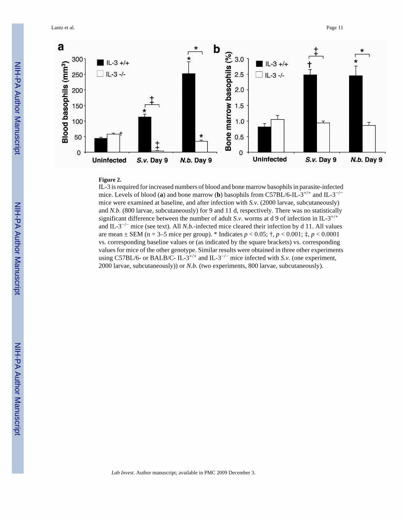
Figure 2.IL-3 is required for increased numbers of blood and bone marrow basophils in parasite-infectedmice. Levels of blood (a) and bone marrow (b) basophils from C57BL/6-IL-3+/+ and IL-3−/−
mice were examined at baseline, and after infection with S.v. (2000 larvae, subcutaneously)and N.b. (800 larvae, subcutaneously) for 9 and 11 d, respectively. There was no statisticallysignificant difference between the number of adult S.v. worms at d 9 of infection in IL-3+/+
and IL-3−/− mice (see text). All N.b.-infected mice cleared their infection by d 11. All valuesare mean ± SEM (n = 3–5 mice per group). * Indicates p < 0.05; †, p < 0.001; ‡, p < 0.0001vs. corresponding baseline values or (as indicated by the square brackets) vs. correspondingvalues for mice of the other genotype. Similar results were obtained in three other experimentsusing C57BL/6- or BALB/C- IL-3+/+ and IL-3−/− mice infected with S.v. (one experiment,2000 larvae, subcutaneously)) or N.b. (two experiments, 800 larvae, subcutaneously).
Lantz et al. Page 11
Lab Invest. Author manuscript; available in PMC 2009 December 3.
NIH
-PA Author Manuscript
NIH
-PA Author Manuscript
NIH
-PA Author Manuscript
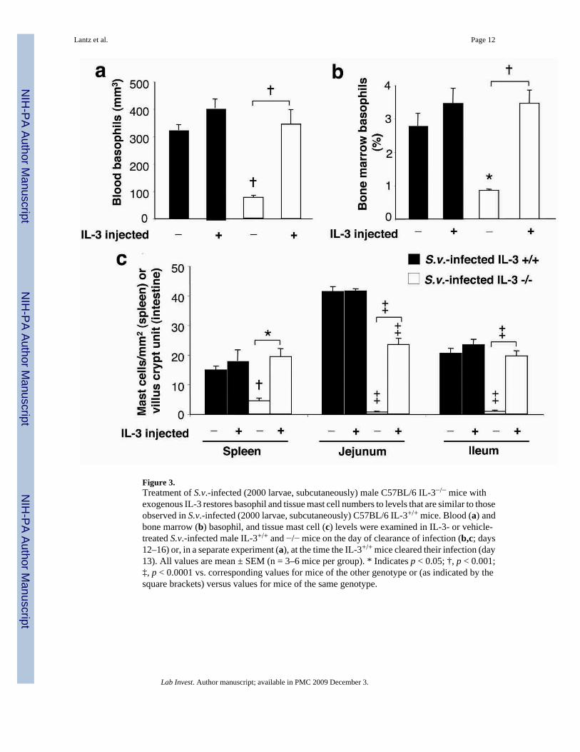
Figure 3.Treatment of S.v.-infected (2000 larvae, subcutaneously) male C57BL/6 IL-3−/− mice withexogenous IL-3 restores basophil and tissue mast cell numbers to levels that are similar to thoseobserved in S.v.-infected (2000 larvae, subcutaneously) C57BL/6 IL-3+/+ mice. Blood (a) andbone marrow (b) basophil, and tissue mast cell (c) levels were examined in IL-3- or vehicle-treated S.v.-infected male IL-3+/+ and −/− mice on the day of clearance of infection (b,c; days12–16) or, in a separate experiment (a), at the time the IL-3+/+ mice cleared their infection (day13). All values are mean ± SEM (n = 3–6 mice per group). * Indicates p < 0.05; †, p < 0.001;‡, p < 0.0001 vs. corresponding values for mice of the other genotype or (as indicated by thesquare brackets) versus values for mice of the same genotype.
Lantz et al. Page 12
Lab Invest. Author manuscript; available in PMC 2009 December 3.
NIH
-PA Author Manuscript
NIH
-PA Author Manuscript
NIH
-PA Author Manuscript
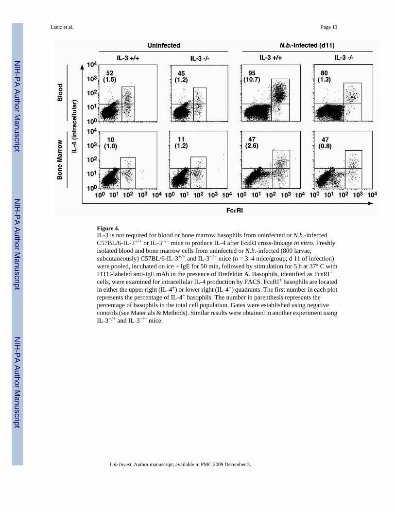
Figure 4.IL-3 is not required for blood or bone marrow basophils from uninfected or N.b.-infectedC57BL/6-IL-3+/+ or IL-3−/− mice to produce IL-4 after FcεRI cross-linkage in vitro. Freshlyisolated blood and bone marrow cells from uninfected or N.b.-infected (800 larvae,subcutaneously) C57BL/6-IL-3+/+ and IL-3−/− mice (n = 3–4 mice/group; d 11 of infection)were pooled, incubated on ice + IgE for 50 min, followed by stimulation for 5 h at 37° C withFITC-labeled anti-IgE mAb in the presence of Brefeldin A. Basophils, identified as FcεRI+
cells, were examined for intracellular IL-4 production by FACS. FcεRI+ basophils are locatedin either the upper right (IL-4+) or lower right (IL-4−) quadrants. The first number in each plotrepresents the percentage of IL-4+ basophils. The number in parenthesis represents thepercentage of basophils in the total cell population. Gates were established using negativecontrols (see Materials & Methods). Similar results were obtained in another experiment usingIL-3+/+ and IL-3−/− mice.
Lantz et al. Page 13
Lab Invest. Author manuscript; available in PMC 2009 December 3.
NIH
-PA Author Manuscript
NIH
-PA Author Manuscript
NIH
-PA Author Manuscript

Figure 5.IL-3 is not required for blood basophils from N.b.-infected BALB/c-IL-3+/+ (a) or IL-3−/−
(b) mice to produce IL-4 after FcεRI cross-linkage in vitro. Freshly isolated blood leukocytesfrom N.b.-infected (500 larvae, subcutaneously) BALB/c IL-3+/+ and IL-3−/− mice (n = 3–4mice/group) were obtained on d 10 of infection, pooled, and divided into two aliquots. Thecells were then incubated on ice ± IgE for 50 min, followed by stimulation for 4 h at 37° Cwith anti-IgE mAb, the last 2 h in the presence of Monensin. Basophils, identified as FcγR+,CD45+ cells, were examined for intracellular IL-4 production by FACS. The number in eachplot represents the percentage of IL-4+ basophils.
Lantz et al. Page 14
Lab Invest. Author manuscript; available in PMC 2009 December 3.
NIH
-PA Author Manuscript
NIH
-PA Author Manuscript
NIH
-PA Author Manuscript
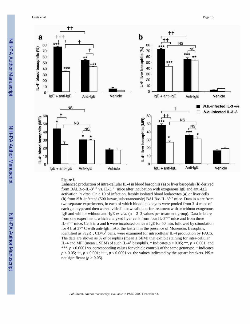
Figure 6.Enhanced production of intra-cellular IL-4 in blood basophils (a) or liver basophils (b) derivedfrom BALB/c-IL-3+/+ vs. IL-3−/− mice after incubation with exogenous IgE and anti-IgEactivation in vitro. On d 10 of infection, freshly isolated blood leukocytes (a) or liver cells(b) from N.b.-infected (500 larvae, subcutaneously) BALB/c-IL-3+/+ mice. Data in a are fromtwo separate experiments, in each of which blood leukocytes were pooled from 3–4 mice ofeach genotype and then were divided into two aliquots for treatment with or without exogenousIgE and with or without anti-IgE ex vivo (n = 2–3 values per treatment group). Data in b arefrom one experiment, which analyzed liver cells from four IL-3+/+ mice and from threeIL-3−/− mice. Cells in a and b were incubated on ice ± IgE for 50 min, followed by stimulationfor 4 h at 37° C with anti-IgE mAb, the last 2 h in the presence of Monensin. Basophils,identified as FcγR+, CD45+ cells, were examined for intracellular IL-4 production by FACS.The data are shown as % of basophils (mean ± SEM) that exhibit staining for intra-cellularIL-4 and MFI (mean ± SEM) of such IL-4+ basophils. * Indicates p < 0.05; **, p < 0.001; and***, p < 0.0001 vs. corresponding values for vehicle controls of the same genotype. † Indicatesp < 0.05; ††, p < 0.001; †††, p < 0.0001 vs. the values indicated by the square brackets. NS =not significant (p > 0.05).
Lantz et al. Page 15
Lab Invest. Author manuscript; available in PMC 2009 December 3.
NIH
-PA Author Manuscript
NIH
-PA Author Manuscript
NIH
-PA Author Manuscript
Copyright © 2022 FDOKUMEN