Tolerogenic dendritic cells derived from donors with natural rubber latex allergy modulate...
10
Tolerogenic Dendritic Cells Derived from Donors with Natural Rubber Latex Allergy Modulate Allergen-Specific T-Cell Responses and IgE Production Alejandro Escobar 1 *, Adam Aguirre 2 , Marı´a Antonieta Guzma ´n 3 , Rodrigo Gonza ´ lez 4 , Diego Catala ´n 5 , Claudio Acun ˜ a-Castillo 6 , Milton Larrondo 4 , Mercedes Lo ´ pez 5 , Barbara Pesce 5 , Jennifer Rolland 7 , Robyn O’Hehir 7 , Juan Carlos Aguillo ´n 5 * 1 Research Institute of Dental Science, Faculty of Dentistry, University of Chile, Santiago, Chile, 2 Department of Pharmacy, Faculty of Chemistry, Catholic University of Chile, Santiago, Chile, 3 Allergy Center, Clinical Hospital of University of Chile, University of Chile, Santiago, Chile, 4 Blood bank Clinical Hospital of University of Chile, University of Chile, Santiago, Chile, 5 Immunology Program, Faculty of Medicine, University of Chile Millennium Institute on Immunology and Immunotherapy, Santiago, Chile, 6 Department of Biology, Faculty of Chemistry and Biology, University of Santiago, Santiago, Chile, 7 Department of Immunology, AMREP, Monash University, Melbourne, Australia Abstract Natural rubber latex (NRL; Hevea brasiliensis) allergy is an IgE-mediated reaction to latex proteins. When latex glove exposure is the main sensitizing agent, Hev b 5 is one of the major allergens. Dendritic cells (DC), the main antigen presenting cells, modulated with pharmacological agents can restore tolerance in several experimental models, including allergy. In the current study, we aimed to generate DC with tolerogenic properties from NRL-allergic patients and evaluate their ability to modulate allergen-specific T and B cell responses. Here we show that dexamethasone-treated DC (dxDC) differentiated into a subset of DC, characterized by low expression of MHC class II, CD40, CD80, CD86 and CD83 molecules. Compared with LPS-matured DC, dxDC secreted lower IL-12 and higher IL-10 after CD40L activation, and induced lower alloantigenic T cell proliferation. We also show that dxDC pulsed with the dominant Hev b 5 T-cell epitope peptide, Hev b 5 46–65 , inhibited both proliferation of Hev b 5-specific T-cell lines and the production of Hev b 5-specific IgE. Additionally, dxDC induced a subpopulation of IL-10-producing regulatory T cells that suppressed proliferation of Hev b 5-primed T cells. In conclusion, dxDC generated from NRL-allergic patients can modulate allergen-specific T-cell responses and IgE production, supporting their potential use in allergen-specific immunotherapy. Citation: Escobar A, Aguirre A, Guzma ´n MA, Gonza ´lez R, Catala ´ n D, et al. (2014) Tolerogenic Dendritic Cells Derived from Donors with Natural Rubber Latex Allergy Modulate Allergen-Specific T-Cell Responses and IgE Production. PLoS ONE 9(1): e85930. doi:10.1371/journal.pone.0085930 Editor: Jacques Zimmer, Centre de Recherche Public de la Sante ´ (CRP-Sante ´ ), Luxembourg Received August 23, 2013; Accepted December 3, 2013; Published January 22, 2014 Copyright: ß 2014 Escobar et al. This is an open-access article distributed under the terms of the Creative Commons Attribution License, which permits unrestricted use, distribution, and reproduction in any medium, provided the original author and source are credited. Funding: This work was supported by grants from the Millennium Institute on Immunology and Immunotherapy P09-016-F, Research Support Office, Clinical Hospital of the University of Chile, Santiago and the National Fund for Scientific and Technological Development, Chile (FONDECYT 1100102). All authors have no conflict of interest. The funders had no role in study design, data collection and analysis, decision to publish, or preparation of the manuscript. Competing Interests: The authors have declared that no competing interests exist. * E-mail: [email protected] (AE); [email protected] (JCA) Introduction Natural rubber latex (NRL; Hevea brasiliensis) allergy is an IgE- mediated reaction to latex proteins, first described in 1979 and increasingly recognized in subsequent years [1,2,3]. Several NRL allergenic proteins are described, Hev b 1–13. Of these, Hev b 2, Hev b 5, Hev b 6 and Hev b 13 are the major allergens for health care workers, typically sensitized by exposure to NRL gloves [4]. Hev b 5 is a strong antigen and one of the most important latex allergens, with a high prevalence of immunoglobulin (Ig)-E reactivity in health care workers [5,6]. Pharmacotherapy for allergic diseases, such as antihistamines or corticosteroids, ameliorates symptoms but does not stop progres- sion; the only therapy that modifies progression of allergic diseases is allergen specific immunotherapy (SIT). However, application of this potentially curative treatment is restricted, largely due to the risk of serious adverse events, especially in asthmatics and for potent allergens such as peanut, seafood and latex [7]. Mechanisms described for regulation of the immune response to allergens include those driven by naturally occurring CD4+CD25+ Treg and inducible populations of allergen-specific, interleukin (IL)-10-secreting Type 1 regulatory T cells (Tr1) [8,9]. Dendritic cells (DC) play a pivotal role in the immunoregulatory mechanisms underlying successful SIT, dampening allergic immune responses by absence of co-stimulation [10] or increasing IL-10 production to expand allergen-specific Tr1 cells [11,12]. These features can be observed at an immature stage of DC differentiation [13] and in a specialized DC subset, called tolerogenic DC (t-DC) [14]. At present, allergen avoidance and symptomatic treatment for adverse reactions are the only therapeutic strategies available for NRL allergy [15]. Therefore, t-DC could provide a novel treatment strategy for inducing allergen-specific desensitization in NRL-allergic patients. Several agents have been used to generate t-DC, including IL-10 [13], transforming growth factor- b1 (TGF-b1), the active form of vitamin D3 [16], neuropeptides [17], corticosteroids and cyclosporin, amongst others [18,19]. PLOS ONE | www.plosone.org 1 January 2014 | Volume 9 | Issue 1 | e85930
-
Upload
independent -
Category
Documents
-
view
0 -
download
0
Transcript of Tolerogenic dendritic cells derived from donors with natural rubber latex allergy modulate...

Tolerogenic Dendritic Cells Derived from Donors withNatural Rubber Latex Allergy Modulate Allergen-SpecificT-Cell Responses and IgE ProductionAlejandro Escobar1*, Adam Aguirre2, Marıa Antonieta Guzman3, Rodrigo Gonzalez4, Diego Catalan5,
Claudio Acuna-Castillo6, Milton Larrondo4, Mercedes Lopez5, Barbara Pesce5, Jennifer Rolland7,
Robyn O’Hehir7, Juan Carlos Aguillon5*
1 Research Institute of Dental Science, Faculty of Dentistry, University of Chile, Santiago, Chile, 2 Department of Pharmacy, Faculty of Chemistry, Catholic University of
Chile, Santiago, Chile, 3 Allergy Center, Clinical Hospital of University of Chile, University of Chile, Santiago, Chile, 4 Blood bank Clinical Hospital of University of Chile,
University of Chile, Santiago, Chile, 5 Immunology Program, Faculty of Medicine, University of Chile Millennium Institute on Immunology and Immunotherapy, Santiago,
Chile, 6 Department of Biology, Faculty of Chemistry and Biology, University of Santiago, Santiago, Chile, 7 Department of Immunology, AMREP, Monash University,
Melbourne, Australia
Abstract
Natural rubber latex (NRL; Hevea brasiliensis) allergy is an IgE-mediated reaction to latex proteins. When latex glove exposureis the main sensitizing agent, Hev b 5 is one of the major allergens. Dendritic cells (DC), the main antigen presenting cells,modulated with pharmacological agents can restore tolerance in several experimental models, including allergy. In thecurrent study, we aimed to generate DC with tolerogenic properties from NRL-allergic patients and evaluate their ability tomodulate allergen-specific T and B cell responses. Here we show that dexamethasone-treated DC (dxDC) differentiated intoa subset of DC, characterized by low expression of MHC class II, CD40, CD80, CD86 and CD83 molecules. Compared withLPS-matured DC, dxDC secreted lower IL-12 and higher IL-10 after CD40L activation, and induced lower alloantigenic T cellproliferation. We also show that dxDC pulsed with the dominant Hev b 5 T-cell epitope peptide, Hev b 546–65
, inhibited bothproliferation of Hev b 5-specific T-cell lines and the production of Hev b 5-specific IgE. Additionally, dxDC induced asubpopulation of IL-10-producing regulatory T cells that suppressed proliferation of Hev b 5-primed T cells. In conclusion,dxDC generated from NRL-allergic patients can modulate allergen-specific T-cell responses and IgE production, supportingtheir potential use in allergen-specific immunotherapy.
Citation: Escobar A, Aguirre A, Guzman MA, Gonzalez R, Catalan D, et al. (2014) Tolerogenic Dendritic Cells Derived from Donors with Natural Rubber LatexAllergy Modulate Allergen-Specific T-Cell Responses and IgE Production. PLoS ONE 9(1): e85930. doi:10.1371/journal.pone.0085930
Editor: Jacques Zimmer, Centre de Recherche Public de la Sante (CRP-Sante), Luxembourg
Received August 23, 2013; Accepted December 3, 2013; Published January 22, 2014
Copyright: � 2014 Escobar et al. This is an open-access article distributed under the terms of the Creative Commons Attribution License, which permitsunrestricted use, distribution, and reproduction in any medium, provided the original author and source are credited.
Funding: This work was supported by grants from the Millennium Institute on Immunology and Immunotherapy P09-016-F, Research Support Office, ClinicalHospital of the University of Chile, Santiago and the National Fund for Scientific and Technological Development, Chile (FONDECYT 1100102). All authors have noconflict of interest. The funders had no role in study design, data collection and analysis, decision to publish, or preparation of the manuscript.
Competing Interests: The authors have declared that no competing interests exist.
* E-mail: [email protected] (AE); [email protected] (JCA)
Introduction
Natural rubber latex (NRL; Hevea brasiliensis) allergy is an IgE-
mediated reaction to latex proteins, first described in 1979 and
increasingly recognized in subsequent years [1,2,3]. Several NRL
allergenic proteins are described, Hev b 1–13. Of these, Hev b 2,
Hev b 5, Hev b 6 and Hev b 13 are the major allergens for health
care workers, typically sensitized by exposure to NRL gloves [4].
Hev b 5 is a strong antigen and one of the most important latex
allergens, with a high prevalence of immunoglobulin (Ig)-E
reactivity in health care workers [5,6].
Pharmacotherapy for allergic diseases, such as antihistamines or
corticosteroids, ameliorates symptoms but does not stop progres-
sion; the only therapy that modifies progression of allergic diseases
is allergen specific immunotherapy (SIT). However, application of
this potentially curative treatment is restricted, largely due to the
risk of serious adverse events, especially in asthmatics and for
potent allergens such as peanut, seafood and latex [7].
Mechanisms described for regulation of the immune response to
allergens include those driven by naturally occurring
CD4+CD25+ Treg and inducible populations of allergen-specific,
interleukin (IL)-10-secreting Type 1 regulatory T cells (Tr1) [8,9].
Dendritic cells (DC) play a pivotal role in the immunoregulatory
mechanisms underlying successful SIT, dampening allergic
immune responses by absence of co-stimulation [10] or increasing
IL-10 production to expand allergen-specific Tr1 cells [11,12].
These features can be observed at an immature stage of DC
differentiation [13] and in a specialized DC subset, called
tolerogenic DC (t-DC) [14].
At present, allergen avoidance and symptomatic treatment for
adverse reactions are the only therapeutic strategies available for
NRL allergy [15]. Therefore, t-DC could provide a novel
treatment strategy for inducing allergen-specific desensitization
in NRL-allergic patients. Several agents have been used to
generate t-DC, including IL-10 [13], transforming growth factor-
b1 (TGF-b1), the active form of vitamin D3 [16], neuropeptides
[17], corticosteroids and cyclosporin, amongst others [18,19].
PLOS ONE | www.plosone.org 1 January 2014 | Volume 9 | Issue 1 | e85930

Glucocorticoids (GC) are potent immunosuppressive and anti-
inflammatory agents used to treat autoimmune diseases and
prevent graft rejection [20]. Studies have indicated that GC affects
the phenotype of monocyte-derived DC, modulating their
differentiation, maturation and function [21,22].
In the current study, we aimed to generate t-DC from NRL-
allergic patients using dexamethasone (dx), and determine whether
dx-treated DC (dxDC) can modulate NLR-specific T-cell
responses and prime T cells to become allergen-specific IL-10-
producing cells. We demonstrate that dxDC have reduced
alloantigen and allergen-specific T-cell stimulatory capacity and
can inhibit Th2 differentiation of naıve CD4+ T cells. In addition,
dxDC induced the expansion of an IL-10-producing regulatory T-
cell subset with suppressive activity. These findings indicate that
dxDC may be useful for antigen-specific tolerization of allergic
immune responses in NRL-allergic patients.
Materials and Methods
Ethics StatementsThe study was approved by the local medical ethics committee
(Hospital Clınico Universidad de Chile Mm Nu 304). All patients
provided written informed consent. After the samples had been
collected, each patient was allocated a trial number, demographic
data collected, and the database anonymised.
Study SubjectsPeripheral venous blood from 8 NRL-allergic patients (1–8) was
collected. All patients had positive skin prick tests ($3 mm above
saline negative control) to latex extract (Stallergenes, Paris, France)
and increased levels of IgE specific to an extract of NRL (Immulite
2000 analyzer; Siemens Healthcare, Germany). All patients were
no atopic or sensitized to other allergens unrelated to latex. Three
of eight tested patients (NRL-1, NRL-5, NRL-7) were positive for
specific IgE against recombinant Hev b 5 protein, determined by
ELISA. The demographic and clinical characteristics of the
patients are reported in Table 1.
Preparation of T and B CellsPBMC were obtained from peripheral blood by density gradient
separation with Ficoll-Hypaque (Axis-Shield, Oslo, Norway).
CD45RA+ CD4+ T cells and CD27- B cells were isolated from
PBMC using antibody-coated magnetic Microbeads (MACS,
MiltenyiBiotec Inc, CA, USA), according to the manufacturer’s
protocol. Separation was assessed by flow cytometry (purity 90%
and 97%, respectively).
Generation of DCDC were differentiated from monocytes using established
methods [23]. Briefly PBMCs were obtained from buffy coats.
Cells (36107/well) were incubated in serum-free AIM-V thera-
peutic medium (Gibco BLR, Paisley, UK) at 37uC, 5% CO2 for
2 h in a six-well plate (Falcon Becton Dickinson, Hershey, PA,
USA). Non-adherent cells were removed, and the remaining cells
were incubated for 7 days in the presence of 500 U/ml
recombinant human IL-4 (rhIL-4) (US Biological, Swampscott,
MA, USA) and 800 U/ml of GM-CSF (Shering Plough, Brinny
Co., Ireland). The cultures were maintained for 7 days, replacing
the medium every 2 days.
DC were left unstimulated (iDC) or matured (mDC) with 1 mg/
mL of LPS from Escherichia coli (Sigma-Aldrich, St Louis, MO,
USA) at day 6. To generate dxDC, at day 5 of the culture, dx
(1026 M; Sigma Aldrich) was added [24]. In some experiments
dxDC were activated with 1 mg/mL LPS (LPS-dxDC). DC were
routinely checked by flow-cytometric analysis (FACScanto,
Beckton-Dickinson, San Diego, CA, USA) to determine expression
of CD11c, HLA II (DR/DP/DQ), CD40, CD80, CD86 and
CD83 (eBioscience, San Diego, CA, USA). Data analysis was
performed using WinMDI 2.8 (freeware http://facs.scripps.edu/
software.html).
FITC-dextran Endocytosis AssayDextran uptake activity was assessed by incubating 0.56106 DC
with FITC-conjugated dextran (Molecular Probes, Eugene, OR)
(0.2 mg/mL) for 2 h at 37uC in the dark. Cells were washed
carefully with PBS and FITC-dextran uptake was quantified by
flow cytometry. Background of dextran incorporation was assessed
by incubating DC on ice.
Measurement of DC and T-cell Cytokine ProductionDC (26104) were incubated with an irradiated CD40L-
expressing 3T3 fibroblast cell line (cell ratio 10:1) at 37uC and
5% CO2 overnight. IL-10 and IL-12 producing cells were
enumerated using an ELISPOT Ready-SET-Go!H according to
the manufacturer’s instructions (eBioscience). Spots were counted
using A.EL.VIS ELISPOT Analysis Software (Hannover, Ger-
many). T-cell production of IL-4 and IFN-c was also evaluated by
ELISPOT Ready-SET-Go!H (eBioscience). Tumoral necrosis
factor (TNF)-a production was measured by intracellular cytokine
staining and samples were analyzed by flow cytometry (FACS-
canto; Becton Dickinson).
T-cell DifferentiationThe Hev b 546–65 peptide (TPEKEEPTAAPAEPEAPAPE), an
immunodominant T-cell epitope not associated with a particular
MHC II haplotype [25], was synthesized at GenScript (NJ, USA).
To induce T-cell differentiation, autologous-naıve T cells were
primed with 36104 Hev b 546–65-pulsed DC (THev b 5-DC) (10:1)
for 6 days and rested for 4 days with 10 IU/ml IL-2 (ProleukinH,
Novartis Pharmaceuticals Corporation, East Hanover, NJ, USA)
in round-bottomed 96-well plates. Finally, THev b 5-DC were
harvested after 10 days and re-stimulated for 16 h with Phorbol
12-Myristate 13 Acetate (PMA)/ionomycin (Sigma-Aldrich) to
Table 1. Demographic and clinical description of studysubjects.
Descriptors Patients
Age (years) 36.6±9.3 (range 22–54)
Sex (male/female) 2/6
Main symptoms of NRL allergy
Rhinoconjunctivitis 1/8
Urticaria 8/8
Lipoedema 3/8
Anaphylaxis 2/8
Allergy Medication
Use of antihistamine 7/8
Atopy 0/8
All patients were defined NRL-allergic on the basis of presence of both historyof allergic symptoms after allergen exposure and specific IgE and skin prick testpositive to NRL.doi:10.1371/journal.pone.0085930.t001
Tolerogenic Dendritic Cells Modulate Latex Allergy
PLOS ONE | www.plosone.org 2 January 2014 | Volume 9 | Issue 1 | e85930

Figure 1. Dexamethasone-treated dendritic cells (dxDC) from natural rubber latex (NRL)-allergic patients display a stable immaturephenotype. (A) Representative histograms of 1 out of 6 patients for HLA II, CD80, CD86, CD40, CD83 and CCR7 expression on immature DCs (iDC),LPS-modulated DCs (mDC) and dexamethasone-modulated DCs (dxDC). (B) Representative histograms show the uptake of FITC-dextran by iDC, dxDCand mDC. Results from 1 representative NRL-allergic patient are shown. (C) iDC and dxDC were exposed to LPS (1 ug/ml) for 18 hours. Graphsrepresent the mean+SD of 3 separate experiments performed in duplicate. *p,0.05; **p,0.01. Histograms show results from 1 representative NRL-allergic patient.doi:10.1371/journal.pone.0085930.g001
Tolerogenic Dendritic Cells Modulate Latex Allergy
PLOS ONE | www.plosone.org 3 January 2014 | Volume 9 | Issue 1 | e85930

assess IL-10 production by ELISPOT Ready-SET-Go!H(eBioscience) as before.
Proliferation AssaysAllogeneic PBMC or Hev b 5-specific T-cell lines, generated
using established methods [26], were labeled with CFSE (5 mM
per 16107 cells) (Renovar, USA) for 15 min at 37uC. Cells were
washed extensively and 26105 cells/well were cultured with Hev b
546–65 peptide-pulsed DC in round-bottomed 96-well plates in
serum-free AIM-V medium (Gibco BLR) for 5 days. Type II
human collagen (CII)259–263 peptide (GIAGFKGEQGPKGET)
(GenScript) was used as a control. CD4+ T-cell proliferation was
determined by CFSE dilution analysis by flow cytometry
(FACScanto; Becton Dickinson). Apoptosis of T cells was
measured using an Annexin V Apoptosis Detection Kit APC
(eBioscience).
IgE ProductionAutologous naıve B cells (16105), naıve T cells (2.56105), Hev b
546–65 peptide-pulsed DC (2.56104) and CD40L-expressing
fibroblasts (2.56103) were co-cultured in round-bottomed 96-well
plates in the presence of rhIL-4 (1000 IU/ml) (eBioscience). After
10 days, supernatants were harvested and assessed for total and
Hev b 5-specific IgE levels by Serum samples were tested for
specific IgE using our standard ELISA protocol. In brief, ELISA
plates (Falcon Becton Dickinson) were coated with rhev b 5
(2.5 mg/ml) [27] in 0.1 M bicarbonate buffer (pH 9.6). After
blocked, diluted plasma (1/10) were added. IgE were quantified
with biotinylated anti-human IgE mAb (BD Pharmingen, USA)
diluted 1/1000. Development was gone with substrate solution
(ATBS/H2O2). Plates were read at 460 nm using an ELISA plate
reader. Background values obtained for sera and mAb on wells
uncoated with Ag were subtracted from values obtained on wells
coated with Ag. Values were considered positive when they
differed from control supernatant values .2 times the SD.
CD4 T-cell Suppression AssayCFSE-labeled THev b 5-mDC cells (36105) were boosted with
mDC (36104) in the presence of increasing numbers of THev b 5-
dxDC at different ratios, in round-bottomed 96-well plates. After 7
days, THev b 5 cell proliferation was determined by CFSE dilution
analysis on a FACScanto flow cytometer.
Statistical AnalysisResults are presented as mean 6 SD. The Kruskal-Wallis test
with Dunn’s Multiple Comparison post-test was used to compare
the mean values of cell surface marker expression, cytokine and
IgE production between different cell culture conditions. Prolifer-
Figure 2. Dexamethasone-treated dendritic cells (dxDC) from natural rubber latex (NRL)-allergic patients display a stable lowproduction of the pro-inflammatory cytokine TNF. iDC and dxDC were exposed to LPS (1 ug/ml) for 18 hours. TNF was analyzed byintracellular cytokine staining of the CD11c+ cell population. (A) Dot plots show results for LPS-stimulated dxDC (left plot) and LPS-stimulated iDC(right plot) from 1 representative NRL allergic patient. (B) The bar graph represents the mean+SD of 3 separate experiments performed in duplicate.*p,0.05.doi:10.1371/journal.pone.0085930.g002
Figure 3. Dexamethasone-treated dendritic cells (dxDC) from natural rubber latex (NRL)-allergic patients display an anti-inflammatory cytokine profile. IL-12 and IL-10 production was analyzed by ELISPOT after DC were activated with CD40L overnight. Results arethe mean+SD of 5 different patients performed in duplicate. ***p,0.001.doi:10.1371/journal.pone.0085930.g003
Tolerogenic Dendritic Cells Modulate Latex Allergy
PLOS ONE | www.plosone.org 4 January 2014 | Volume 9 | Issue 1 | e85930
ative responses were compared using the Mann-Whitney test.
Analyses were performed using GraphPad Prism version 5.0 for
Windows, GraphPad Software (San Diego, CA, USA, www.
graphpad.com). A p value ,0.05 was considered statistically
significant.
Results
Characterization of Tolerogenic dxDCAnalysis of the phenotype and function of dxDC showed a lower
level of HLA II (p = 0.0058), CD80 (p = 0.0117), CD86
(p = 0.0058), CD40 (p = 0.0055) and CD83 (p = 0.0058) expression
compared with mDC, while the expression of the chemokine
receptor CCR7 was similar to mDC (Figure 1A). In contrast,
dxDC expressed equivalent levels of surface markers to untreated
iDC that were used as an immature control. To further
characterize dxDC, their ability to take up FITC-dextran was
examined. dxDC displayed similar uptake to iDC, and higher than
mDC, which showed characteristically reduced activity (Figure 1B).
The suppressive effect of dx on DC was not affected by LPS,
indicating that dx can induce durable immaturity of DC in terms
of HLA II, CD86 and CD83 expression and production of the
pivotal pro-inflammatory cytokine, TNF-a (Figure 2).
As cytokines secreted by DC are important in determining the
differentiation fate of T cells, we assessed the cytokine profile of
our DC populations. dxDC secreted high levels of IL-10 and low
levels of IL-12 upon activation via CD40L. iDC and mDC
produced significantly greater levels of IL-12 and lower levels of
IL-10 compared with dxDC (Figure 3). These results indicate that
dxDC have an anti-inflammatory profile characterized by high
production of IL-10.
Modulation of T-cell Response by Tolerogenic dxDCStimulation of allogeneic CD4+ T cells by dxDC at different
stimulator:effector ratios induced a lower proliferative CD4+percentage compared with allogeneic CD4+ T cells stimulated
with mDC or iDC (p,0.05) (Figure 4A). To discard that the
inhibitory effect of dxDC was mediated by a mechanism of T cell
death, we examined the level of apoptosis and necrosis in CD4+ T
cells by Annexin V staining and propidium iodide dye. No
significant differences were observed in the fraction of apoptotic
cells to T cells co-cultured with DC subjected to different
treatments (Figure 4B).
To determine the modulation of dxDC in NRL-specific T-cell
response, oligoclonal Hev b 5-specific T-cell lines were generated
from 3 patients with Hev b 5-specific IgE reactivity (patients 1, 5
and 7). These T-cell lines were stimulated with Hev b 546–65-
pulsed dxDC or with Hev b 546–65-pulsed mDC. We observed that
while peptide-pulsed mDC induced an intense proliferative
response, stimulation with peptide-pulsed dxDC induced hypore-
sponsiveness of specific T-cell lines to their cognate antigen
(p = 0.013) (Figure 5). T-cell lines stimulated with mDC pulsed
with the irrelevant peptide CII259–263 did not show proliferation at
all (Figure S1).
dxDC Modulate T Cell Differentiation HinderingSubsequent B-cell Help for IgE Secretion
In order to evaluate the T-helper cytokine profile driven by
dxDC, naıve CD4+ T cells from 3 patients (1, 5 and 7) were
primed with autologous Hev b 546–65 pulsed dxDC (THev b 5-dxDC)
or mDC (THev b 5-mDC), and IL-4 and IFN-c secreting T cells were
measured (Table 2).
dxDC stimulated greater IL-4 and IFN-c production by CD4+T cells than mDC, but with a more pronounced effect on IL-4
production. As IL-4 favours the differentiation of naıve B cells into
IgE antibody-secreting cells, we assessed the ability of THev b 5-
dxDC to induce total and allergen-specific IgE production. For this
purpose, we co-cultured Hev b 546–65 peptide-pulsed dxDC with
autologous naive CD4+T cells, naıve B cells and CD40L-
transfected fibroblasts in the presence of IL-4 for 10 days.
Interestingly, total IgE (Figure 6A) and Hev b 5-specific IgE
(Figure 6B) production decreased in the presence of dxDC
compared to stimulation with mDC.
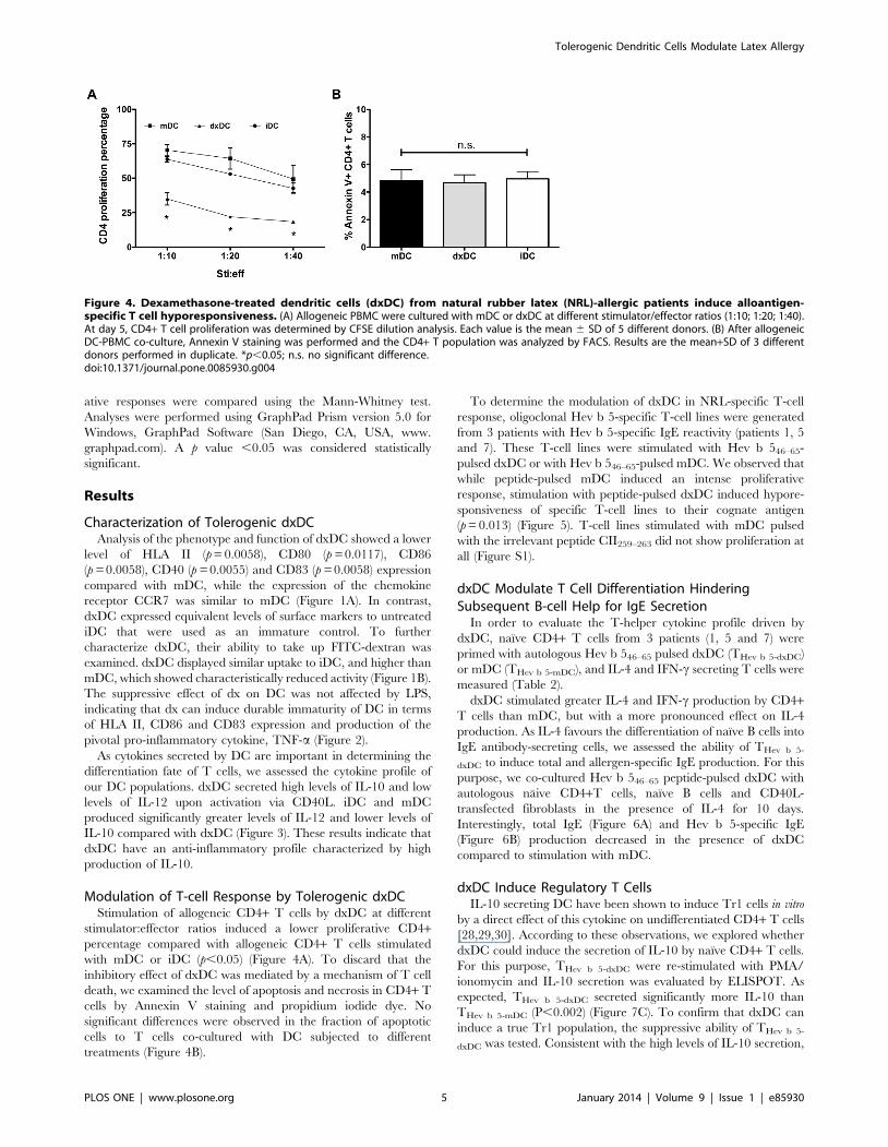
dxDC Induce Regulatory T CellsIL-10 secreting DC have been shown to induce Tr1 cells in vitro
by a direct effect of this cytokine on undifferentiated CD4+ T cells
[28,29,30]. According to these observations, we explored whether
dxDC could induce the secretion of IL-10 by naıve CD4+ T cells.
For this purpose, THev b 5-dxDC were re-stimulated with PMA/
ionomycin and IL-10 secretion was evaluated by ELISPOT. As
expected, THev b 5-dxDC secreted significantly more IL-10 than
THev b 5-mDC (P,0.002) (Figure 7C). To confirm that dxDC can
induce a true Tr1 population, the suppressive ability of THev b 5-
dxDC was tested. Consistent with the high levels of IL-10 secretion,
Figure 4. Dexamethasone-treated dendritic cells (dxDC) from natural rubber latex (NRL)-allergic patients induce alloantigen-specific T cell hyporesponsiveness. (A) Allogeneic PBMC were cultured with mDC or dxDC at different stimulator/effector ratios (1:10; 1:20; 1:40).At day 5, CD4+ T cell proliferation was determined by CFSE dilution analysis. Each value is the mean 6 SD of 5 different donors. (B) After allogeneicDC-PBMC co-culture, Annexin V staining was performed and the CD4+ T population was analyzed by FACS. Results are the mean+SD of 3 differentdonors performed in duplicate. *p,0.05; n.s. no significant difference.doi:10.1371/journal.pone.0085930.g004
Tolerogenic Dendritic Cells Modulate Latex Allergy
PLOS ONE | www.plosone.org 5 January 2014 | Volume 9 | Issue 1 | e85930

Figure 5. Dexamethasone-treated dendritic cells (dxDC) from natural rubber latex (NRL)-allergic patients induce allergen-specific Tcell hyporesponsiveness. NRL-specific T cell lines were cultured with dxDC or mDC loaded with 10 ug/ml of NRL Hevb 546–64 peptide at 1:20stimulator/effector ratio. At day 5, CD4+ T cell proliferation was determined by CFSE dilution analysis. (A) Representative dot plots for each of thethree NRL-allergic patients. (B) The bar graph represents the mean+SD of experiments performed in triplicate on 3 patients. *p,0.05.doi:10.1371/journal.pone.0085930.g005
Tolerogenic Dendritic Cells Modulate Latex Allergy
PLOS ONE | www.plosone.org 6 January 2014 | Volume 9 | Issue 1 | e85930

THev b 5-dxDC suppressed Hev b 5-specific T-cell proliferation
(Figure 7A, B).
Discussion
iDC resident in peripheral tissues in a steady-state can induce T-
cell tolerance [31]. Similarly, DC prepared ex vivo and exposed to
antigens in the absence of full-maturation stimuli down-regulate
immunity and induce tolerance. Similarly, the use of immuno-
suppressants such as corticosteroids can induce human DC to
acquire tolerogenic properties [18,19]. In this study, we demon-
strate the feasibility of generating t-DC from NRL-allergy patients
using dexamethasone. We demonstrate that dexamethasone
induces an immature phenotype on DC, with low expression
levels of HLA II and co-stimulatory molecules such as CD40,
CD80, CD86 and CD83 and high particle uptake ability.
However, the expression of the homing receptor CCR7 was not
affected by dexamethasone, showing similar levels to mDC. This
would enable dxDC to migrate to lymph nodes, as would be
required if they were to be used in immunotherapy. Moreover, if
tolerance restitution is the goal of immunotherapy, as in NRL
allergy, it is essential that DC maintain an immature state in the
presence of inflammatory factors. Therefore, we evaluated the
resistance of the dxDC phenotype to LPS exposure. Our results
demonstrate that dxDC, unlike iDC, were unaffected by LPS
stimulation in terms of HLA II, CD86 and CD83 expression and
production of the pivotal pro-inflammatory cytokine, TNF-a.
Low antigen presenting and co-stimulatory molecule expression
levels, strongly suggest that one of the most likely mechanisms of
T-cell tolerization by dxDC is through anergy induction, due to
the lack of sufficient first and second activation signals [32].
Consequently, we showed that dxDC have weak allogeneic T-
cell stimulatory activity. This effect was not due to deletion of
alloreactive T cells, because an increase in apoptosis was ruled out.
Since the aim of immunotherapy is to induce antigen-specific
tolerance without impairing other immune functions, we evaluated
the ability of dxDC to present the immunodominant T-cell
epitope for the NRL allergen Hev b 5 [26]. Our results
demonstrate that dxDC have a reduced ability to present antigen
to Hev b 5 specific oligoclonal T-cell lines compared with mDC.
This is consistent with the results obtained from our allogeneic
experiments, and with previous data from Steinbrink et al. showing
that clonal anergy was induced in an influenza hemagglutinin-
specific CD4+ T-cell clone via stimulation with IL-10 treated DC
[29].
As noted earlier, dxDC display low levels of activation signal 1
(HLA II) and 2 (co-stimulatory molecules), however, a third signal,
established by secreted cytokines, is also relevant in guiding T-cell
polarization [33,34,35]. IL-10-secreting DC have been associated
with tolerance induction against common antigens [36,37] and
with poor T-cell stimulatory function [38,39]. Furthermore, IL-10
expression has been observed in DC located in lung tissue and the
intestine, suggesting an important role in maintaining local T-cell
tolerance to common environmental antigens [36]. Our study
shows that dxDC produce large amounts of IL-10 and low levels of
IL-12 upon CD40L stimulation; these findings are in agreement
with previous work using dexamethasone as a tolerogenic agent
[40,41].
As demonstrated previously, IL-10-secreting DC induce regu-
latory T cells upon activation [42,43]. In line with this evidence,
our study shows that naıve CD4+ T cells that were primed by
dxDC (THev b 5-dxDC) differentiated into an IL-10+ T-cell
population, compatible with a Tr1 cell phenotype [44]. These
findings agree with those described by Bosma et al. for myeloid DC
isolated from peripheral blood and tolerized with dexamethasone
and LPS [45]. When we assessed the regulatory properties of THev
Table 2. IFN-c and IL-4 production in primed CD4+ T cells.
NRL Patient N6
THev b5-mDC IL-4/IFNcratio1
THev b5-dxDC IL-4/IFNcratio1
1 30/117 = 0.26 217/295 = 0.73
5 17/132 = 0.13 160/205 = 0.78
7 4/204 = 0.01 59/313 = 0.18
Naive CD4+ (36105) cells from selected NRL-allergic patient were coculturedwith Hev b 546–65 pulsed mature or dexamethasone-treated DC (36104) for 6days. Then CD4+ cells were re-stimulated for 16 h with PMA/ionomycin toassess cytokine production by ELISPOT.1Spots per 56104 cells.doi:10.1371/journal.pone.0085930.t002
Figure 6. Dexamethasone-treated dendritic cells (dxDC) decrease total and allergen-specific IgE production by human autologousnaıve B cells from natural rubber latex (NRL)-allergic patients. Naıve T cells and B cells were stimulated with Hev b 5-pulsed mDC or dxDC inthe presence of CD40L+3T3 fibroblasts and IL-4 for 10 days. B cells stimulated in presence of IL-4 and CD40L were used as positive control. Total IgE(A) and Hev b 5-specific IgE (B) production was measured by ELISA. Each group of bars represents the mean+SD of experiments performed induplicate on 3 different donors (NLR 1, 5 and 7). *p,0.05.doi:10.1371/journal.pone.0085930.g006
Tolerogenic Dendritic Cells Modulate Latex Allergy
PLOS ONE | www.plosone.org 7 January 2014 | Volume 9 | Issue 1 | e85930

Tolerogenic Dendritic Cells Modulate Latex Allergy
PLOS ONE | www.plosone.org 8 January 2014 | Volume 9 | Issue 1 | e85930
b5-dxDC cells on Hev b 5-specific-CD4+ memory T cells, we
observed inhibition of T-cell proliferation, where the suppressor
effect was independent of THev b 5-dxDC cell number. This
observation is compatible with an immunomodulatory mechanism
of T-cell response, mediated by cytokines such as IL-10 [46],
however, we do not exclude regulatory activity by other molecules.
The determination of the full range of cytokines and/or molecules
involved in the suppressor activity of THev b 5-dx-DC needs further
investigation.
In this study, we analyzed the influence of dxDC on T cell help
for total and allergen-specific IgE production by autologous B cells
from NRL-allergic patients. We observed that the presence of
dxDC inhibits the secretion of total and Hev b 5-specific IgE by B
cells. The reduction of IgE production could be explained by the
induction of a Tr1 population. Kanjarawi et al. reported that
CD4+CD25+ regulatory T cells were able to modulate total and
b-lactoglobulin-specific serum IgE production in a food allergy
murine model [47]. IL-10 appears to be an important cytokine in
the success of specific immunotherapy in allergy since it has been
described to be increased in blood and affected tissues of treated
patients [48,49,50]. Akdis et al. reported that Tr1 cells consistently
represent the dominant T cell subset specific for common
environmental allergens in healthy individuals [8]. They suggested
that Tr1 cells might control allergen-specific Th2 responses in
allergic and healthy individuals. Pacciani et al. demonstrated that
IL-10-producing DC can induce specific T cells with suppressive
activity on allergen-specific Th2 cells from house dust mite-allergic
patients [30]. In addition, experiments in murine models showed
that DC transduced with IL-10 negatively regulate allergic
inflammation of airways by inducing IL-10-producing T cells
[51]. T cells transduced to express IL-10 have also been shown to
inhibit airway hyper-responsiveness induced by Th2 cells [52].
Taken together, these studies suggest that the DC-induced
deviation in T cell cytokine production towards IL-10 could be
an important factor in preventing allergic responses.
In summary, our results demonstrate that Hev b 5-pulsed
dxDC-primed naive T cells become Tr1 cells, which modulate
allergen-specific T-cell responses. These findings indicate that
dxDC may be a useful immunotherapy tool for antigen-specific
down-regulation of allergic immune responses in NRL-allergic
patients.
Supporting Information
Figure S1 T-cell lines stimulated with mDC pulsed withthe irrelevant peptide CII259–263. NRL-specific T cell lines
were cultured with mDC loaded with 10 ug/ml of NRL Hev b
546–64 or CII259–263 peptide at 1:20 stimulator/effector ratio. At
day 5, CD4+ T cell proliferation was determined by CFSE
dilution analysis. (A) Representative dot plots for stimulation with
Hev b 546–64 (left), CII259–263 (centre) and no antigen (right). (B)
The bar graph represents the mean+SD of experiments performed
in triplicate on 3 patients.
(TIFF)
Author Contributions
Conceived and designed the experiments: AE JCA. Performed the
experiments: AE AA RG. Analyzed the data: AE MAG BP CAC M.
Lopez. Contributed reagents/materials/analysis tools: JCA JR RO M.
Larrondo. Wrote the paper: AE JCA DC.
References
1. Charous BL, Blanco C, Tarlo S, Hamilton RG, Baur X, et al. (2002) Natural
rubber latex allergy after 12 years: recommendations and perspectives. J Allergy
Clin Immunol 109: 31–34.
2. Ranta PM, Ownby DR (2004) A review of natural-rubber latex allergy in health
care workers. Clin Infect Dis 38: 252–256.
3. Reunala T, Alenius H, Turjanmaa K, Palosuo T (2004) Latex allergy and skin.
Curr Opin Allergy Clin Immunol 4: 397–401.
4. Bernstein DI, Biagini RE, Karnani R, Hamilton R, Murphy K, et al. (2003) In
vivo sensitization to purified Hevea brasiliensis proteins in health care workers
sensitized to natural rubber latex. J Allergy Clin Immunol 111: 610–616.
5. Beezhold DH, Hickey VL, Sutherland MF, O’Hehir RE (2004) The latex
allergen hev B 5 is an antigen with repetitive murine B-cell epitopes. Int Arch
Allergy Immunol 134: 334–340.
6. Slater JE, Vedvick T, Arthur-Smith A, Trybul DE, Kekwick RGO (1996)
Identification, Cloning, and Sequence of a Major Allergen (Hev b 5) from
Natural Rubber Latex (Hevea brasiliensis)10.1074/jbc.271.41.25394. J Biol
Chem 271: 25394–25399.
7. Rolland JM, Gardner LM, O’Hehir RE (2009) Allergen-related approaches to
immunotherapy. Pharmacol Ther 121: 273–284.
8. Akdis M, Verhagen J, Taylor A, Karamloo F, Karagiannidis C, et al. (2004)
Immune responses in healthy and allergic individuals are characterized by a fine
balance between allergen-specific T regulatory 1 and T helper 2 cells. J Exp Med
199: 1567–1575.
9. Gardner LM, Thien FC, Douglass JA, Rolland JM, O’Hehir RE (2004)
Induction of T ‘regulatory’ cells by standardized house dust mite immunother-
apy: an increase in CD4+ CD25+ interleukin-10+ T cells expressing peripheral
tissue trafficking markers. Clin Exp Allergy 34: 1209–1219.
10. Mueller DL, Jenkins MK, Schwartz RH (1989) Clonal expansion versus
functional clonal inactivation: a costimulatory signalling pathway determines the
outcome of T cell antigen receptor occupancy. Annu Rev Immunol 7: 445–480.
11. Francis JN, Till SJ, Durham SR (2003) Induction of IL-10+CD4+CD25+ T cells
by grass pollen immunotherapy. J Allergy Clin Immunol 111: 1255–1261.
12. Mahnke K, Ring S, Bedke T, Karakhanova S, Enk AH (2008) Interaction of
regulatory T cells with antigen-presenting cells in health and disease. Chem
Immunol Allergy 94: 29–39.
13. Jonuleit H, Schmitt E, Schuler G, Knop J, Enk AH (2000) Induction of
interleukin 10-producing, nonproliferating CD4(+) T cells with regulatory
properties by repetitive stimulation with allogeneic immature human dendritic
cells. J Exp Med 192: 1213–1222.
14. Rutella S, Danese S, Leone G (2006) Tolerogenic dendritic cells: cytokine
modulation comes of age. Blood 108: 1435–1440.
15. Drew AC, Eusebius NP, Kenins L, de Silva HD, Suphioglu C, et al. (2004)
Hypoallergenic variants of the major latex allergen Hev b 6.01 retaining human
T lymphocyte reactivity. J Immunol 173: 5872–5879.
16. Lyakh LA, Sanford M, Chekol S, Young HA, Roberts AB (2005) TGF-beta and
vitamin D3 utilize distinct pathways to suppress IL-12 production and modulate
rapid differentiation of human monocytes into CD83+ dendritic cells. J Immunol
174: 2061–2070.
17. Delgado M, Chorny A, Ganea D, Gonzalez-Rey E (2006) Vasoactive intestinal
polypeptide induces regulatory dendritic cells that prevent acute graft versus host
disease and leukemia relapse after bone marrow transplantation. Ann N Y Acad
Sci 1070: 226–232.
18. Ciesek S, Ringe BP, Strassburg CP, Klempnauer J, Manns MP, et al. (2005)
Effects of cyclosporine on human dendritic cell subsets. Transplant Proc 37: 20–
24.
19. de Jong EC, Vieira PL, Kalinski P, Kapsenberg ML (1999) Corticosteroids
inhibit the production of inflammatory mediators in immature monocyte-
derived DC and induce the development of tolerogenic DC3. J Leukoc Biol 66:
201–204.
20. Abe M, Thomson AW (2003) Influence of immunosuppressive drugs on
dendritic cells. Transpl Immunol 11: 357–365.
Figure 7. Dexamethasone-treated dendritic cell (dxDC)-primed naıve T cells (THev b 5-dxDC) exhibit a Tr1 phenotype. CFSE-labeled THev
b 5-mDC were harvested and boosted with Hev b 5 46–65 pulsed mDC in the presence of an increasing number of THev b 5-dxDC or THev b 5-mDC. At day 7,THev b 5-mDC proliferation was determined by CFSE dilution analysis of CD4+ T cells. (A) Representative dot plots. Percentages of proliferating CD4+ Tcells are indicated in the upper left quadrant. (B) The graph shows the mean +/2 SD of 3 different patients performed in duplicate. (C) IL-10production by THev b 5-dxDC and THev b 5-mDC after PMA/ionomycin re-stimulation. The graph (mean+SD) summarizes experiments performed induplicate on 3 different patients. *p,0.05; **p,0.01.doi:10.1371/journal.pone.0085930.g007
Tolerogenic Dendritic Cells Modulate Latex Allergy
PLOS ONE | www.plosone.org 9 January 2014 | Volume 9 | Issue 1 | e85930

21. Piemonti L, Monti P, Allavena P, Sironi M, Soldini L, et al. (1999)
Glucocorticoids affect human dendritic cell differentiation and maturation.
J Immunol 162: 6473–6481.
22. Moser M, De Smedt T, Sornasse T, Tielemans F, Chentoufi AA, et al. (1995)
Glucocorticoids down-regulate dendritic cell function in vitro and in vivo.
Eur J Immunol 25: 2818–2824.
23. Escobar A, Lopez M, Serrano A, Ramirez M, Perez C, et al. (2005) Dendritic
cell immunizations alone or combined with low doses of interleukin-2 induce
specific immune responses in melanoma patients. Clin Exp Immunol 142: 555–
568.
24. Rozkova D, Horvath R, Bartunkova J, Spisek R (2006) Glucocorticoids severely
impair differentiation and antigen presenting function of dendritic cells despite
upregulation of Toll-like receptors. Clin Immunol 120: 260–271.
25. Rihs H-P, Chen Z, Rueff F, Cremer R, Raulf-Heimsoth M, et al. (2002) HLA-
DQ8 and the HLA-DQ8-DR4 haplotype are positively associated with the
hevein-specific IgE immune response in health care workers with latex allergy.
Journal of Allergy and Clinical Immunology 110: 507–514.
26. de Silva HD, Sutherland MF, Suphioglu C, McLellan SC, Slater JE, et al. (2000)
Human T-cell epitopes of the latex allergen Hev b 5 in health care workers.
Journal of Allergy and Clinical Immunology 105: 1017–1024.
27. Sutherland MF, Drew A, Rolland JM, Slater JE, Suphioglu C, et al. (2002)
Specific monoclonal antibodies and human immunoglobulin E show that Hev b
5 is an abundant allergen in high protein powdered latex gloves. Clinical &
Experimental Allergy 32: 583–589.
28. Levings MK, Gregori S, Tresoldi E, Cazzaniga S, Bonini C, et al. (2005)
Differentiation of Tr1 cells by immature dendritic cells requires IL-10 but not
CD25+CD4+ Tr cells. Blood 105: 1162–1169.
29. Steinbrink K, Graulich E, Kubsch S, Knop J, Enk AH (2002) CD4(+) and
CD8(+) anergic T cells induced by interleukin-10-treated human dendritic cells
display antigen-specific suppressor activity. Blood 99: 2468–2476.
30. Pacciani V, Gregori S, Chini L, Corrente S, Chianca M, et al. (2010) Induction
of anergic allergen-specific suppressor T cells using tolerogenic dendritic cells
derived from children with allergies to house dust mites. J Allergy Clin Immunol
125: 727–736.
31. Steinman RM, Hawiger D, Nussenzweig MC (2003) Tolerogenic dendritic cells.
Annu Rev Immunol 21: 685–711.
32. Xia CQ, Kao KJ (2003) Suppression of interleukin-12 production through
endogenously secreted interleukin-10 in activated dendritic cells: involvement of
activation of extracellular signal-regulated protein kinase. Scand J Immunol 58:
23–32.
33. Kalinski P, Schuitemaker JH, Hilkens CM, Wierenga EA, Kapsenberg ML
(1999) Final maturation of dendritic cells is associated with impaired
responsiveness to IFN-gamma and to bacterial IL-12 inducers: decreased ability
of mature dendritic cells to produce IL-12 during the interaction with Th cells.
J Immunol 162: 3231–3236.
34. Morelli AE, Zahorchak AF, Larregina AT, Colvin BL, Logar AJ, et al. (2001)
Cytokine production by mouse myeloid dendritic cells in relation to
differentiation and terminal maturation induced by lipopolysaccharide or
CD40 ligation. Blood 98: 1512–1523.
35. van der Merwe PA (2000) Modeling costimulation. Nat Immunol 1: 194–195.
36. Akbari O, DeKruyff RH, Umetsu DT (2001) Pulmonary dendritic cells
producing IL-10 mediate tolerance induced by respiratory exposure to antigen.Nat Immunol 2: 725–731.
37. Iwasaki A, Kelsall BL (1999) Freshly isolated Peyer’s patch, but not spleen,
dendritic cells produce interleukin 10 and induce the differentiation of T helpertype 2 cells. J Exp Med 190: 229–239.
38. Akdis CA, Blaser K (2001) Role of IL-10 in allergen-specific immunotherapyand normal response to allergens. Microbes Infect 3: 891–898.
39. Akdis CA, Blaser K (2001) Mechanisms of interleukin-10-mediated immune
suppression. Immunology 103: 131–136.40. Jugde F, Boissier C, Rougier-Larzat N, Corlu A, Chesne C, et al. (2005)
Regulation by allergens of chemokine receptor expression on in vitro-generateddendritic cells. Toxicology 212: 227–238.
41. Xia CQ, Peng R, Beato F, Clare-Salzler MJ (2005) Dexamethasone induces IL-10-producing monocyte-derived dendritic cells with durable immaturity.
Scand J Immunol 62: 45–54.
42. Groux H, Bigler M, de Vries JE, Roncarolo MG (1996) Interleukin-10 induces along-term antigen-specific anergic state in human CD4+ T cells. J Exp Med 184:
19–29.43. Groux H, O’Garra A, Bigler M, Rouleau M, Antonenko S, et al. (1997) A CD4+
T-cell subset inhibits antigen-specific T-cell responses and prevents colitis.
Nature 389: 737–742.44. Allan SE, Broady R, Gregori S, Himmel ME, Locke N, et al. (2008) CD4+ T-
regulatory cells: toward therapy for human diseases. Immunol Rev 223: 391–421.
45. Bosma BM, Metselaar HJ, Nagtzaam NM, de Haan R, Mancham S, et al.(2008) Dexamethasone transforms lipopolysaccharide-stimulated human blood
myeloid dendritic cells into myeloid dendritic cells that prime interleukin-10
production in T cells. Immunology 125: 91–100.46. Roncarolo MG, Gregori S, Battaglia M, Bacchetta R, Fleischhauer K, et al.
(2006) Interleukin-10-secreting type 1 regulatory T cells in rodents and humans.Immunol Rev 212: 28–50.
47. Kanjarawi R, Dercamp C, Etchart N, Adel-Patient K, Nicolas JF, et al. (2011)
Regulatory T cells control type I food allergy to Beta-lactoglobulin in mice. IntArch Allergy Immunol 156: 387–396.
48. Bellinghausen I, Metz G, Enk AH, Christmann S, Knop J, et al. (1997) Insectvenom immunotherapy induces interleukin-10 production and a Th2-to-Th1
shift, and changes surface marker expression in venom-allergic subjects.Eur J Immunol 27: 1131–1139.
49. Nasser SM, Ying S, Meng Q, Kay AB, Ewan PW (2001) Interleukin-10 levels
increase in cutaneous biopsies of patients undergoing wasp venom immuno-therapy. Eur J Immunol 31: 3704–3713.
50. Nouri-Aria KT, Wachholz PA, Francis JN, Jacobson MR, Walker SM, et al.(2004) Grass pollen immunotherapy induces mucosal and peripheral IL-10
responses and blocking IgG activity. J Immunol 172: 3252–3259.
51. Henry E, Desmet CJ, Garze V, Fievez L, Bedoret D, et al. (2008) Dendritic cellsgenetically engineered to express IL-10 induce long-lasting antigen-specific
tolerance in experimental asthma. J Immunol 181: 7230–7242.52. Oh JW, Seroogy CM, Meyer EH, Akbari O, Berry G, et al. (2002) CD4 T-
helper cells engineered to produce IL-10 prevent allergen-induced airwayhyperreactivity and inflammation. J Allergy Clin Immunol 110: 460–468.
Tolerogenic Dendritic Cells Modulate Latex Allergy
PLOS ONE | www.plosone.org 10 January 2014 | Volume 9 | Issue 1 | e85930