Bronchodilatory effect of ethanolic extract of the leaves of Nyctanthes arbortristis
Upload
khangminh22Category
view
0download
0
Ahmed et al. SpringerPlus (2016) 5:1644 DOI 10.1186/s40064-016-3326-7
RESEARCH
Cardiac and testicular toxicity effects of the latex and ethanolic leaf extract of Calotropis procera on male albino rats in comparison to abamectinOsama M. Ahmed1*, Hanaa I. Fahim1, Magdy W. Boules2 and Heba Y. Ahmed2
Abstract
The present study aims to assess the toxic effect of latex and ethanolic leaf extract of Calotropis procera (C. procera), in comparison to abamectin, on serum biomarkers of function and histological integrity of heart and testis in male albino rats. To achieve this aim, the albino rats were separately administered 1/20 and 1/10 of LD50 of C. procera latex, ethanolic C. procera leaf extract and abamectin respectively by oral gavage for 4 and 8 weeks. C. procera latex and leaf extract as well as abamectin markedly elevated the activities of serum CK-MB, AST and LDH at the two tested peri-ods in a dose dependent manner. Lipid peroxidation was significantly increased while GSH level and GPx, GST and SOD activities were significantly depleted in heart and testis of all treated rats. All treatments also induced a marked increase in serum TNF-α and decrease in serum IL-4, testosterone, FSH and LH levels in a dose dependent manner. The latex seemed to be more effective in deteriorating the testicular function and sex hormones’ levels while the etha-nolic leaf extract produced more deleterious effects on oxidative stress and antioxidant defense system in both heart and testis. The normal histological architecture and integrity of the heart and testis were perturbed after treatments and the severity of lesions, which include odema, inflammatory cell infiltration, necrosis and degeneration, is dose and time dependent. In conclusion, the findings of this study indicated that C. procera latex and ethanolic extract of leaves could induce marked toxicity in heart and testis and these toxic effects may be more or less similar to those of abamectin. The cardiotoxicity and testicular toxicity may be mediated via stimulation of inflammation, increased oxidative stress and suppression of antioxidant defense system.
Keywords: Calotropis procera, Abamectin, Heart, Testis, Toxicity
© 2016 The Author(s). This article is distributed under the terms of the Creative Commons Attribution 4.0 International License (http://creativecommons.org/licenses/by/4.0/), which permits unrestricted use, distribution, and reproduction in any medium, provided you give appropriate credit to the original author(s) and the source, provide a link to the Creative Commons license, and indicate if changes were made.
BackgroundDevelopment of new rodent control methodologies and strategies continues to be an exciting subject for researchers. In the last two decades, there has been a shift in rodenticide use, with researchers and pest control practitioners taking a renewed interest in alternatives to anticoagulants; this stems from increased resistance of
pests to 1st generation anticoagulants (Quy et al. 1995), as well as concerns over the secondary-poisoning risks and wildlife contamination associated with field use of 2nd generation anticoagulants (Eason and Turck 2002; Thomas et al. 2011). It was found by Carvalho et al. (2003) that many natural compounds have been sug-gested as alternatives to conventional chemicals used for pest control. It was reported that plant extracts have been used as pesticides by humans (Abou-Hashem 2013). The use of toxic plants is especially prevalent in the develop-ing and underdeveloped countries, where plants grown locally are cheaper than the synthetic chemical pesticides (EL-Gengaihi et al. 1997; Paul and Kumar 2009; Million et al. 2010).
Open Access
*Correspondence: [email protected]; [email protected] 1 Physiology Division, Zoology Department, Faculty of Science, Beni-Suef University, Beni Suef, EgyptFull list of author information is available at the end of the article
Page 2 of 21Ahmed et al. SpringerPlus (2016) 5:1644
Calotropis is a small genus having six species of shrubs or small trees, distributed in tropical and subtropical Africa, Asia and America. Two species namely Calotro-pis procera (C. procera) and Calotropis gigantea (C. gigantea) are found in India which closely resembled to each other in structure and in functional uses (Bhat-nagar 1950). It was revealed that C. procera includes various chemicals which are useful for various activities (Sheth 2011; Begum et al. 2013). The entire plant has been reported to contain alkaloids, sterols, flavonoids, cardiac glycosides, triterpenoids and usharin (Suresh Kumar et al. 2013). In an earlier study, various medici-nal properties such as a laxative, anthelmintic, purgative, anti-inflammatory and diuretic have been documented (Iqbal et al. 2005). Different parts of C. procera and its latex have shown analgesic, antibacterial and wound healing properties in traditional medicine (Laitiff et al. 2010; Lima-Filho et al. 2010). The previous pharmaco-logical studies on C. procera include reports of its anti-cancer, antifungal and insecticidal activity (Ahmed et al. 2006; Hassan et al. 2006).
Despite these uses, C. procera poses varying toxic effects in animals through air borne allergies, touch and consumption in livestock. Vadlapudi and Naidu (2010) revealed that the plant is also known for its toxic proper-ties that include iridocyclites, dermatitis and acts like a poison and produces lethal effects. Toxicity of C. procera is reported in sheep in the form of anorexia and diarrhea. Consumption of this plant leads to severe poisoning to livestock as well as man. Incidental ingestion of fresh C. procera leaves has been suggested as toxic to many rumi-nants by several farmers from the Brazilian semi-arid region. These observations are supported by some stud-ies that have reported toxic effects promoted by C. pro-cera latex and leaves (Mahmoud et al. 1979a, b; Singhal and Kumar 2009). The latex of C. procera contains several cardenolides such as calotropin, catotoxin, calcilin and gigantin which are caustic and considered poisonous in nature (Kuriachen and Dave 1989).
Biocides are widely used in agriculture and can con-taminate rivers and other water bodies due to transport from cultivated areas (Cerejeira et al. 2003; Maloschik et al. 2007). Abamectin, the non-proprietary name assigned to avermectin B1, is a mixture of two compo-nents, with the major component avermectin B1a 80 % of the mixture, and the minor component avermectin B1b, 20 % of the mixture, differing by a single methylene group (Agarwal 1998). The two components, B1a and B1b, have similar biological and toxicological properties (Lankas and Gordon 1989; Gallo and Lawryk 1991). As indicated by Kolar et al. (2008), abamectin has been used in several countries as a pest control agent in livestock
and as an active substance of nematicides and insecti-cides for agricultural use. ABM may be valuable in agri-culture; it may be highly toxic to mammals (Moline et al. 2000).
Therefore, this study aims to verify the toxic effect of latex and ethanolic extract of leaves of C. procera on heart and testis compared with the biocide abamectin.
MethodsPlant materialsThe leaves and latex of C. procera were obtained from East desert of Beni-Suef Governorate. The plant was authenticated by Dr. Walaa Azmy Hasan, lecturer of plant taxonomy, Department of Botany, Faculty of Sci-ence, Beni Suef University.
Collection of leaves and extract preparationOnly mature leaves without sign of lesion were used. The leaves of C. procera were extracted by ethanol according to Freedman et al. (1979). Leaves were washed with dis-tilled water, air dried at room temperature and ground into fine powder using electrical mixer. Five hindered grams of the powder were suspended in 1 l of ethanol 95 % for 72 h then filtered and the filtrate was evaporated by rotary evaporator at high pressure and temperature 40–50 °C at Faculty of pharmacy, Beni-Suef University, Egypt. The extract was kept in a refrigerator at −30 °C until use.
Latex collectionFresh latex was obtained by breaking the leaf stock and allowing the latex to flow into a glass beaker. It was freshly prepared before injection.
Pesticide and chemicalsAbamectin (1.8 % EC) is a mixture of 80 % avermec-tin B1a and maximum of avermectin B1b used as an acaricide. It was obtained from Synganta Agro. Co. (Switzerland).
Reagent kits used for determination of creatinine kinase-MB (CK-MB) activity was purchased from Spinre-act Company (Spain). Aspartate aminotransferase (AST) and lactate dehydrogenase (LDH) reagent kits were pur-chased from Biosystems Company (Spain). Testosterone reagent kits were purchased from BioSource Company (Belgium). Follicle stimulating hormone (FSH) and lute-inizing hormone (LH) reagent kits were purchased from Monobind, INC. (USA). Tumor necrosis factor-alpha (TNF-α) and Interleukin-4 (IL-4) kits were purchased from R&D Systems, Inc. (USA). All other used chemicals are of analytical grade and were obtained from Sigma-Aldrich Chemical Company (USA).
Page 3 of 21Ahmed et al. SpringerPlus (2016) 5:1644
Experimental animalsMale albino rats weighing 120–150 g (8–10 weeks of age) were used as experimental animals in this investigation. They were obtained from the Animal House of Research Institute of Ophthalmology, Giza, Egypt. Animals were supplied daily standard pellet diet and were given water ad libitum. The animals were housed in polypropylene cages with good aerated stainless steel in Animal House of Zoology Department, Faculty of Science, Beni-Suef University, Egypt at temperature 20–25 °C and 12-h daily light dark cycles. The animals were kept for 2 weeks under observation before the onset of the experiment to exclude any intercurrent infection. All animal proce-dures are in accordance with the recommendation of the Experimental Animals Ethics Committee of Faculty of Science, Beni-Suef University. All efforts were done to decrease the suffering of animals to a minimum.
Experimental designExperimental animals were divided into seven groups as follow:
Group 1: Rats of this group are regarded as control group and were administered 1 % carboxy methyl cel-lulose (CMC) by oral gavage for 4 and 8 weeks.Group 2: Rats of this group were orally administered 1/20 of LD50 (50 % lethal dose) of C. procera latex (66 µl/kg b. wt), dissolved in 1 % CMC, for 4 and 8 weeks. LD50 of C. procera latex is 1.316 ml/kg b. wt as detected by Fahim et al. (2016).Group 3: Rats of this group were orally administered 1/10 of LD50 of C. procera latex (132 µl/kg b. wt), dis-solved in 1 % CMC, for 4 and 8 weeks.Group 4: Rats of this group were orally administered 1/20 of LD50 of ethanolic extract of C. procera leaves (4.78 mg/kg b. wt), dissolved in 1 % CMC, for 4 and 8 weeks. LD50 of ethanolic extract of C. procera leaves is 95.52 mg/kg b. wt (El-Shafey et al. 2011).Group 5: Rats of this group were orally administered 1/10 of LD50 of ethanolic extract of C. procera leaves (9.56 mg/kg b. wt), dissolved in 1 % CMC, for 4 and 8 weeks.Group 6: Rats of this group were orally adminis-tered 1/20 of LD50 of abamectin (vertimec 1.8 % EC) (0.44 mg/kg b. wt), dissolved in 1 % CMC, for 4 and 8 weeks. LD50 of abamectin vertimec (1.8 % EC) is 8.7 mg/kg body weight (Lankas and Gordon 1989; El-Shafey et al. 2011).Group 7: Rats of this group were orally adminis-tered 1/10 of LD50 of abamectin (vertimec 1.8 % EC) (0.87 mg/kg b. wt), dissolved in 1 % CMC, for 4 and 8 weeks.
Samples preparationAt the end of the 4th and 10th weeks, six animals of each group were sacrificed under diethyl ether anesthesia. Blood samples were obtained from cervical vein, left to coagulate at room temperature and then they were cen-trifuged at 3000 rpm for 30 min. The clear non-haemo-lysed supernatant sera were quickly removed and were divided into 3 portions. The obtained samples were kept in deep freezer at −30 °C till used. Half of heart and testis were excised and removed quickly, homogenized by using in isotonic solution (0.9 % NaCl) and kept in deep freezer at −30 °C till used. The other half of heart and other tes-tis were immediately excised and fixed in 10 % neutral buffered formalin for histopathological processing.
Biochemical investigationsDetection of serum parameters related to heart functionSerum CK-MB activity was determined according to the method of Gerhart and Waldenström (1979). Serum AST and LDH activities were detected according to the meth-ods of Gella et al. (1985) and Young (2000) respectively.
Assay of male sex hormones levelsConcentrations of FSH and LH in serum were detected according to the method of Odell et al. (1968) and Braun-stein et al. (1976). Serum testosterone concentration was determined according to the method of Andreyko et al. (1986).
Assays TNF‑α and IL‑4 levelsSerum TNF-α and IL-4 levels were determined by the quantitative sandwich enzyme immunoassay technique according to the methods of Howard and Harada (1994) and Croft et al. (2012) respectively.
Assay of oxidative stress and antioxidant defense markersHeart and testis glutathione content and lipid peroxi-dation were determined according to the methods of Beutler et al. (1963) and Preuss et al. (1998) respectively. Glutathione peroxidase (GPx), glutathione-S-transferase (GST) and superoxide dismutase (SOD) activities in heart and testis were assayed according to the methods of Matkovics et al. (1997), Mannervik and Gutenberg (1981) and Marklund and Marklund (1974) respectively.
Statistical analysisThe data obtained from the experiment were ana-lyzed using the one-way analysis of variance (ANOVA) (Roa et al. 1985) followed by LSD test to compare vari-ous groups with each other. Results were expressed as mean ± standard error (SE) and values of P > 0.05 were considered non-significantly different while those of
Page 4 of 21Ahmed et al. SpringerPlus (2016) 5:1644
P < 0.05 and P < 0.01 were considered significant and highly significant respectively. F-probability expresses the general effect between groups. Multi-factor analysis of variance (MANOVA) was also performed to evaluate the effect of time, dose and time–dose interaction.
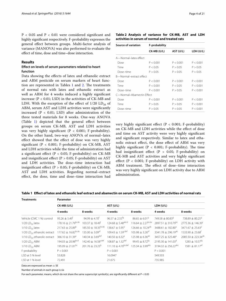
ResultsEffect on levels of serum parameters related to heart functionData showing the effects of latex and ethanolic extract and ABM pesticide on serum markers of heart func-tion are represented in Tables 1 and 2. The treatments of normal rats with latex and ethanolic extract as well as ABM for 4 weeks induced a highly significant increase (P < 0.01; LSD) in the activities of CK-MB and LDH. With the exception of the effect of 1/20 LD50 of ABM, serum AST and LDH activities were significantly increased (P < 0.01; LSD) after administration of the three tested materials for 8 weeks. One-way ANOVA (Table 1) depicted that the general effect between groups on serum CK-MB, AST and LDH activities was very highly significant (P < 0.001; F-probability). On the other hand, two-way ANOVA of normal–latex effect showed that the effect of dose was very highly significant (P < 0.001; F-probability) on CK-MB, AST and LDH activities while the time of administration had a significant effect (P < 0.05; F-probability) on CK-MB and insignificant effect (P > 0.05; F-probability) on AST and LDH activities. The dose–time interaction had insignificant effect (P > 0.05; F-probability) on CK-MB, AST and LDH activities. Regarding normal–extract effect, the dose, time and dose–time interaction had
very highly significant effect (P < 0.001; F-probability) on CK-MB and LDH activities while the effect of dose and time on AST activity were very highly significant and significant respectively. Similar to latex and etha-nolic extract effect, the dose effect of ABM was very highly significant (P < 0.001; F-probability). The time had insignificant effect (P > 0.05; F-probability) on CK-MB and AST activities and very highly significant effect (P < 0.001; F-probability) on LDH activity with ABM treatment. The effect of dose–time interaction was very highly significant on LDH activity due to ABM administration.
Table 1 Effect of latex and ethanolic leaf extract and abamectin on serum CK-MB, AST and LDH activities of normal rats
Data are expressed as mean ± SE
Number of animals in each group is six
For each parameter, means, which do not share the same superscript symbol(s), are significantly different at P < 0.05
Treatments Parameter
CK-MB (U/L) AST (U/L) LDH (U/L)
4 weeks 8 weeks 4 weeks 8 weeks 4 weeks 8 weeks
Vehicle (CMC 1 %) control 93.26 ± 5.49f 94.99 ± 4.70f 96.57 ± 2.55fg 86.65 ± 6.01g 749.58 ± 80.83g 738.89 ± 80.25g
1/20 LD50 latex 178.16 ± 21.79bcde 103.37 ± 18.49f 124.68 ± 5.48abcd 116.64 ± 2.27bcde 2897.51 ± 310.79bc 2775.36 ± 146.39c
1/10 LD50 latex 217.63 ± 25.89b 185.50 ± 18.35bcde 138.67 ± 5.95a 126.66 ± 10.24ab 3488.61 ± 182.80a 3471.67 ± 25.63a
1/20 LD50 ethanolic extract 177.62 ± 16.82bcde 135.00 ± 3.09ef 109.43 ± 1.59cdef 105.98 ± 3.26ef 3341.78 ± 296.19ab 1533.90 ± 23.68f
1/10 LD50 ethanolic extract 366.10 ± 31.39a 140.34 ± 3.69def 140.50 ± 6.32a 125.98 ± 6.36ab 3457.25 ± 325.48a 2085.50 ± 223.34de
1/20 LD50 ABM 194.03 ± 20.98bcd 142.40 ± 16.58cdef 108.87 ± 1.32def 99.45 ± 8.72fg 2195.30 ± 141.03d 1283 ± 10.57fg
1/10 LD50 ABM 195.09 ± 21.01bc 201.76 ± 23.23b 111.10 ± 4.70bcdef 125.34 ± 3.99abc 3194.32 ± 256.22abc 1581 ± 81.17ef
F-probability P < 0.001 P < 0.001 P < 0.001
LSD at 5 % level 53.828 16.0947 549.503
LSD at 1 % level 72.491 21.675 735.985
Table 2 Analysis of variance for CK-MB, AST and LDH activities in serum of normal and treated rats
Source of variation F-probability
CK-MB (U/L) AST (U/L) LDH (U/L)
A—Normal–latex effect
Dose P < 0.001 P < 0.001 P < 0.001
Time P < 0.05 P > 0.05 P > 0.05
Dose–time P > 0.05 P > 0.05 P > 0.05
B—Normal–extract effect
Dose P < 0.001 P < 0.001 P < 0.001
Time P < 0.001 P < 0.05 P < 0.001
Dose–time P < 0.001 P > 0.05 P < 0.001
C—Normal–Abamectin Effect
Dose P < 0.001 P < 0.001 P < 0.001
Time P > 0.05 P > 0.05 P < 0.001
Dose–time P > 0.05 P < 0.05 P < 0.001

Page 5 of 21Ahmed et al. SpringerPlus (2016) 5:1644
Effect on male sex hormones levelsSerum testosterone level was highly significantly reduced (P < 0.01; LSD) in male rats ingested latex, ethanolic extract and ABM for 4 and 8 weeks; the effect seemed to dose dependent. Treatments with 1/10 LD50 of the tested materials for 4 weeks induced a highly significant decrease (P < 0.01) in LH level while 1/20 extract only produced a significant effect at the same experimental period. With regard to FSH, the administration of the high dose of latex and extract significantly (P < 0.01; LSD) decreased serum FSH level after 4 weeks whereas the high dose of the three tested treatments induced a significant deple-tion (P < 0.01; LSD) of FSH level at the 8th week (Table 3). Concerning one way ANOVA, it was found that the gen-eral effect between groups on serum testosterone, FSH and LH concentrations was very highly significant (P < 0.001; F-probability) throughout the experiment (Table 3). Two-way ANOVA (Table 4) stated that the dose effect of latex was very highly significant (P < 0.001; LSD) on testoster-one, FSH and LH while the time had very highly signifi-cant effect (P < 0.001; F-probability) on FSH level, highly significant effect (P < 0.01; F-probability) on LH level and insignificant effect (P > 0.05; F-probability) on testosterone level. The dose–time interaction had insignificant effect (P > 0.05; F-probability) on the three tested hormones. Concerning extract effect, the dose had very highly signif-icant effect (P < 0.001; F-probability) on testosterone and FSH levels and highly significant effect (P < 0.01; F-prob-ability) on LH level. Time had significant effect (P < 0.05; F-probability) on FSH level and insignificant effect signifi-cant effect (P > 0.05; F-probability) on testosterone and LH levels. The interaction between dose and time had insig-nificant effect (P > 0.05; F-probability) on the three tested
hormones. Regarding ABM effect, dose had very highly sig-nificant effect (P < 0.001; F-probability) on testosterone and LH levels and highly significant effect (P < 0.01; F-proba-bility) on FSH level. Time had significant effect (P < 0.05; F-probability) on testosterone and LH levels and very highly significant effect (P < 0.001; F-probability) on FSH level. The dose–time interaction had insignificant effect (P > 0.05; F-probability) on the three tested hormones.
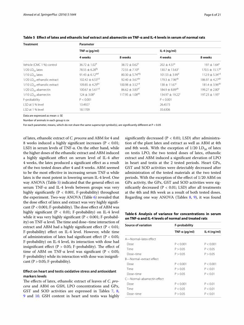
Effect on serum TNF-α and IL-4 levelsData represented in Tables 5 and 6 depicted that all treat-ments induced strong adverse effects on the normal levels of serum TNF-α and IL-4 of normal rats. Administrations
Table 3 Effect of latex and ethanolic leaf extract and abamectin on serum testosterone, FSH and LH levels in normal rats
Data are expressed as mean ± SE
Number of animals in each group is six
For each parameter, means, which do not share the same superscript symbol(s), are significantly different at P < 0.05
Treatments Parameter
Testosterone (ng/ml) FSH (mlU/ml) LH (mlU/ml)
4 weeks 8 weeks 4 weeks 8 weeks 4 weeks 8 weeks
Vehicle (CMC1 %) control 2.71 ± 0.28a 2.22 ± 0.35a 0.26 ± 0.01a 0.23 ± 0.01abc 0.19 ± 0.01a 0.17 ± 0.01abcd
1/20 LD50 latex 1.21 ± 0.11bcd 1.1 ± 0.16bcd 0.25 ± 0.01a 0.20 ± 0.01defg 0.18 ± 0.01ab 0.14 ± 0.01fg
1/10 LD50 latex 0.75 ± 0.13d 0.84 ± 0.04cd 0.19 ± 0.01efg 0.18 ± 0.01fg 0.15 ± 0.01efg 0.14 ± 0.01g
1/20 LD50 ethanolic extract 1.63 ± 0.21b 1.38 ± 0.36bc 0.23 ± 0.01abcd 0.21 ± 0.01cdef 0.16 ± 0.01cdef 0.16 ± 0.01bcde
1/10 LD50 ethanolic extract 1.19 ± 0.06bcd 0.88 ± 0.16cd 0.20 ± 0.01efg 0.19 ± 0.02efg 0.16 ± 0.01def 0.14 ± 0.01efg
1/20 LD50 ABM 1.02 ± 0.15cd 0.81 ± 0.11d 0.25 ± 0.01abc 0.22 ± 0.01bcde 0.18 ± 0.01ab 0.18 ± 0.01abc
1/10 LD50 ABM 0.88 ± 0.07cd 0.68 ± 0.06d 0.24 ± 0.02ab 0.18 ± 0.02g 0.16 ± 0.01cdef 0.15 ± 0.01efg
F-probability P < 0.001 P < 0.001 P < 0.001
LSD at 5 % level 0.5479 0.0311 0.0199
LSD at 1 % level 0.7379 0.0419 0.0269
Table 4 Analysis of variance for testosterone, FSH and LH concentrations in serum of normal and treated rats
Source of variation F-probability
Testosterone (ng/ml)
FSH (mlU/ml) LH (mlU/ml)
A—Normal–latex effect
Dose P < 0.001 P < 0.001 P < 0.001
Time P > 0.05 P < 0.001 P < 0.01
Dose–time P > 0.05 P > 0.05 P > 0.05
B—Normal–extract effect
Dose P < 0.001 P < 0.001 P < 0.01
Time P > 0.05 P < 0.05 P > 0.05
Dose–time P > 0.05 P > 0.05 P > 0.05
C—Normal–abamectin effect
Dose P < 0.001 P < 0.01 P < 0.001
Time P < 0.05 P < 0.001 P < 0.05
Dose–time P > 0.05 P > 0.05 P > 0.05
Page 6 of 21Ahmed et al. SpringerPlus (2016) 5:1644
of latex, ethanolic extract of C. procera and ABM for 4 and 8 weeks induced a highly significant increases (P < 0.01; LSD) in serum levels of TNF-α. On the other hand, while the higher doses of the ethanolic extract and ABM induced a highly significant effect on serum level of IL-4 after 4 weeks, the latex produced a significant effect as a result of the two tested doses after 4 and 8 weeks. ABM seemed to be the most effective in increasing serum TNF-α while latex is the most potent in lowering serum IL-4 level. One way ANOVA (Table 5) indicated that the general effect on serum TNF-α and IL-4 levels between groups was very highly significantly (P < 0.001, F-probability) throughout the experiment. Two-way ANOVA (Table 6) revealed that the dose effect of latex and extract was very highly signifi-cant (P < 0.001; F-probability). The dose effect of ABM was highly significant (P < 0.01; F-probability) on IL-4 level while it was very highly significant (P < 0.001; F-probabil-ity) on TNF-α level. The time and dose–time interaction of extract and ABM had a highly significant effect (P < 0.01; F-probability) effect on IL-4 level. However, while time of administration of latex had significant effect (P < 0.05; F-probability) on IL-4 level, its interaction with dose had insignificant effect (P > 0.05; F-probability). The effect of time of ABM on TNF-α level was significant (P < 0.05; F-probability) while its interaction with dose was insignifi-cant (P > 0.05; F-probability).
Effect on heart and testis oxidative stress and antioxidant markers levelsThe effects of latex, ethanolic extract of leaves of C. pro-cera and ABM on GSH, LPO concentrations and GPx, GST and SOD activities are expressed in Tables 7, 8, 9 and 10. GSH content in heart and testis was highly
significantly decreased (P < 0.01; LSD) after administra-tion of the plant latex and extract as well as ABM at 4th and 8th week. With the exception of 1/20 LD50 of latex on testis LPO, the two tested doses of latex, ethanolic extract and ABM induced a significant elevation of LPO in heart and testis at the 2 tested periods. Heart GPx, GST and SOD activities were detectably decreased after administration of the tested materials at the two tested periods. With the exception of the effect of 1/20 ABM on GPx activity, the GPx, GST and SOD activities were sig-nificantly decreased (P < 0.01; LSD) after all treatments at the 4th and 8th week as a result of both tested doses. Regarding one way ANOVA (Tables 8, 9), it was found
Table 5 Effect of latex and ethanolic leaf extract and abamectin on TNF-α and IL-4 levels in serum of normal rats
Data are expressed as mean ± SE
Number of animals in each group is six
For each parameter, means, which do not share the same superscript symbol(s), are significantly different at P < 0.05
Treatment Parameter
TNF-α (pg/ml) IL-4 (ng/ml)
4 weeks 8 weeks 4 weeks 8 weeks
Vehicle (CMC 1 %) control 36.72 ± 1.02h 38.72 ± 0.62h 202 ± 4.37a 197 ± 1.64a
1/20 LD50 latex 78.55 ± 6.28fg 72.55 ± 7.10g 130.7 ± 13.63c 170.5 ± 15.17b
1/10 LD50 latex 91.43 ± 6.12def 80.30 ± 5.74efg 101.53 ± 3.99d 112.9 ± 5.34cd
1/20 LD50 ethanolic extract 102.42 ± 6.53cd 92.40 ± 3.67de 179.3 ± 7.96ab 186.97 ± 4.27ab
1/10 LD50 ethanolic extract 109.85 ± 4.29bc 100.98 ± 3.52cd 138 ± 11.67c 181.4 ± 3.96ab
1/20 LD50 abamectin 100.67 ± 5.61cd 86.62 ± 3.83ef 184.9 ± 8.89ab 199.27 ± 2.80a
1/10 LD50 abamectin 124 ± 3.08a 117.95 ± 1.08ab 134.97 ± 19.22c 197.23 ± 1.95a
F-probability P < 0.001 P < 0.001
LSD at 5 % level 13.4927 26.4573
LSD at 1 % level 18.1709 35.6306
Table 6 Analysis of variance for concentrations in serum on TNF-α and IL-4 levels of normal and treated rats
Source of variation F-probability
TNF-α (pg/ml) IL-4 (ng/ml)
A—Normal–latex effect
Dose P < 0.001 P < 0.001
Time P > 0.05 P < 0.05
Dose–time P > 0.05 P > 0.05
B—Normal–extract effect
Dose P < 0.001 P < 0.001
Time P > 0.05 P < 0.01
Dose–time P > 0.05 P < 0.01
C—Normal–abamectin effect
Dose P < 0.001 P < 0.01
Time P < 0.05 P < 0.01
Dose–time P > 0.05 P < 0.01
Page 7 of 21Ahmed et al. SpringerPlus (2016) 5:1644
Tabl
e 7
Effec
t of l
atex
and
eth
anol
ic le
af e
xtra
ct a
nd a
bam
ecti
n on
car
diac
GSH
con
tent
, LPO
and
GPx
, GST
and
SO
D a
ctiv
itie
s in
nor
mal
rats
Dat
a ar
e ex
pres
sed
as m
ean ±
SE
Num
ber o
f ani
mal
s in
eac
h gr
oup
is s
ix
For e
ach
para
met
er, m
eans
, whi
ch d
o no
t sha
re th
e sa
me
supe
rscr
ipt s
ymbo
l(s),
are
sign
ifica
ntly
diff
eren
t at P
< 0
.05
Trea
tmen
tsPa
ram
eter
GSH
(nm
ol/1
00 m
g tis
sue)
LPO
(nm
ol/1
00 m
g tis
sue)
GPx
(U/g
tiss
ue)
GST
(U/g
tiss
ue)
SOD
(U/g
tiss
ue)
4 w
eeks
8 w
eeks
4 w
eeks
8 w
eeks
4 w
eeks
8 w
eeks
4 w
eeks
8 w
eeks
4 w
eek
8 w
eeks
Vehi
cle
(CM
C 1
%)
cont
rol
163.
34 ±
1.4
4a14
3.42
± 0
.93b
46.3
1 ±
0.7
1f47
.85 ±
2.1
1f84
.85 ±
0.9
9a85
.50 ±
2.2
5a48
9.93
± 1
4.33
ab50
6.25
± 3
.74a
14.3
5 ±
0.1
8bc15
.31 ±
0.7
1ab
1/20
LD
50 la
tex
109.
87 ±
7.5
2cd11
9.65
± 8
.74c
64.5
0 ±
3.6
8de61
.84 ±
1.1
3e77
.39 ±
2.0
4bc69
.00 ±
1.2
4defg
447.
92 ±
2.6
9ef48
1.80
± 2
.60bc
9.07
± 0
.23e
11.9
5 ±
0.2
9d
1/10
LD
50 la
tex
69.6
9 ±
6.2
9e97
.71 ±
6.1
0d72
.82 ±
2.1
7cd67
.16 ±
2.6
5cde
69.9
3 ±
2.7
0defg
63.9
6 ±
0.4
3g42
9.86
± 1
2.39
f46
0.07
± 6
.63cd
e1.
93 ±
0.2
3h4.
62 ±
0.3
0g
1/20
LD
50 e
than
olic
ex
trac
t68
.39 ±
6.2
3e10
8.40
± 1
.00cd
65.1
9 ±
3.7
5de59
.78 ±
4.3
3e68
.06 ±
1.6
9efg
79.6
3 ±
1.3
ab48
0.49
± 1
3.70
bc45
4.51
± 8
.65de
15.7
1 ±
0.1
4a6.
84 ±
0.7
8f
1/10
LD
50 e
than
olic
ex
trac
t45
.35 ±
5.2
8f10
2.50
± 8
.26cd
84.5
3 ±
4.6
6a72
.71 ±
2.5
6cd63
.96 ±
2.9
6g77
.11 ±
0.7
4bc47
2.31
± 5
.63bc
d38
6.06
± 8
.17h
2.11
± 0
.25h
4.04
± 0
.06g
1/20
LD
50 A
BM11
8.73
± 1
1.35
c11
6.51
± 4
.20c
76.5
5 ±
6.4
6abc
72.5
6 ±
2.0
2cd74
.22 ±
2.5
9bcd
72.3
5 ±
3.3
3cdef
470.
49 ±
3.3
9bcde
487.
85 ±
0.7
7ab13
.39 ±
0.1
1c7.
12 ±
0.1
5f
1/10
LD
50 A
BM77
.61 ±
0.7
0e77
.43 ±
1.3
1e84
.30 ±
5.4
4ab74
.19 ±
3.1
2bcd
72.5
4 ±
2.3
5cde
84.8
5 ±
0.9
9a44
7.92
± 7
.12ef
395.
83 ±
3.3
6g12
.26 ±
0.2
2d4.
84 ±
0.3
8g
F-pr
obab
ility
P <
0.0
01P
< 0
.001
P <
0.0
01P
< 0
.001
P <
0.0
01
LSD
at 5
% le
vel
17.2
065
10.2
926
6.01
0222
.709
1.01
3
LSD
at 1
% le
vel
23.1
723
13.8
613
8.09
4130
.582
71.
3642

Page 8 of 21Ahmed et al. SpringerPlus (2016) 5:1644
Tabl
e 8
Effec
t of l
atex
and
eth
anol
ic le
af e
xtra
ct a
nd a
bam
ecti
n on
test
icul
ar G
SH c
onte
nt, L
PO a
nd G
Px, G
ST a
nd S
OD
act
ivit
ies
in n
orm
al ra
ts
Dat
a ar
e ex
pres
sed
as m
ean ±
SE
Num
ber o
f ani
mal
s in
eac
h gr
oup
is s
ix
For e
ach
para
met
er, m
eans
, whi
ch d
o no
t sha
re th
e sa
me
supe
rscr
ipt s
ymbo
l(s),
are
sign
ifica
ntly
diff
eren
t at P
< 0
.05
Trea
tmen
tsPa
ram
eter
GSH
(nm
ol/1
00 m
g tis
sue)
LPO
(nm
ol/1
00 m
g tis
sue)
GPx
(U/g
tiss
ue)
GST
(U/g
tiss
ue)
SOD
(U/g
tiss
ue)
4 w
eeks
8 w
eeks
4 w
eeks
8 w
eeks
4 w
eeks
8 w
eeks
4 w
eeks
8 w
eeks
4 w
eek
8 w
eeks
Vehi
cle
(CM
C 1
%) c
ontr
ol86
.78 ±
9.1
1a69
.60 ±
6.1
2b13
.68 ±
1.7
2e14
.46 ±
2.4
0e71
.89 ±
3.2
5a73
.20 ±
6.6
8a97
6.18
± 2
3.75
a10
11.1
8 ±
27.
05a
10.2
4 ±
1.7
7a9.
33 ±
0.9
7ab
1/20
LD
50 la
tex
68.9
7 ±
3.9
4b33
.41 ±
5.6
8e16
.30 ±
0.5
6de23
.33 ±
1.9
7bc50
.85 ±
6.5
2cd44
.53 ±
3.4
2cde
683.
16 ±
37.
74bc
d78
7.50
± 5
2.34
b6.
45 ±
0.3
8cde
5.12
± 0
.44de
fg
1/10
LD
50 la
tex
54.2
4 ±
1.8
5cd28
.32 ±
3.0
2ef29
.76 ±
1.9
8a30
.06 ±
3.0
2a48
.50 ±
4.3
8cde
46.9
9 ±
4.2
9cde
637.
50 ±
42.
81cd
e76
9.38
± 2
9.28
b5.
53 ±
0.5
4def
4.40
± 0
.24ef
g
1/20
LD
50 e
than
olic
ex
trac
t64
.06 ±
2.1
8bc19
.03 ±
0.5
5fg25
.37 ±
2.6
7abc
21.3
5 ±
1.7
3cd55
.13 ±
3.6
9bc49
.06 ±
1.3
7cd75
3.96
± 3
3.91
bc67
3.09
± 7
6.22
bcd
7.93
± 0
.98bc
6.36
± 0
.56cd
e
1/10
LD
50 e
than
olic
ex
trac
t48
.05 ±
1.7
7d14
.87 ±
0.5
9g28
.10 ±
2.4
2ab22
.56 ±
1.6
5bc49
.44 ±
2.1
5cd37
.27 ±
3.2
8e48
9.58
± 6
.99f
584.
21 ±
47.
88de
f7.
13 ±
0.4
6cd3.
07 ±
0.7
6g
1/20
LD
50 A
BM53
.45 ±
4.5
8cd31
.06 ±
3.7
4e21
.72 ±
1.6
1cd21
.80 ±
1.5
0cd64
.25 ±
3.0
5ab51
.47 ±
2.6
7cd67
2.29
± 2
1.79
bcd
596.
88 ±
66.
45de
f6.
99 ±
0.8
0cd4.
05 ±
0.3
7fg
1/10
LD
50 A
BM48
.87 ±
3.7
9d18
.42 ±
0.9
6fg23
.33 ±
0.8
7bc27
.92 ±
1.9
9ab52
.10 ±
3.1
8cd41
.90 ±
3.2
2de56
9.17
± 2
9.95
def
546.
87 ±
51.
57ef
5.06
± 0
.48d
efg
3.59
± 0
.40fg
F-pr
obab
ility
P <
0.0
01P
< 0
.001
P <
0.0
01P
< 0
.001
P <
0.0
01
LSD
at 5
% le
vel
11.9
465.
6821
11.3
121
124.
2059
2.17
84
LSD
at 1
% le
vel
16.0
879
7.65
2215
.234
216
7.27
042.
9337

Page 9 of 21Ahmed et al. SpringerPlus (2016) 5:1644
that the general effect on heart and testis GSH content, LPO and the activities of GPx, GST and SOD between groups was very highly significantly (P < 0.001, F-proba-bility) throughout the experiment.
Concerning two-way ANOVA (Tables 9, 10), the dose effect of latex, ethanolic extract and ABM on GSH con-tent and GPx, GST and SOD activities in heart and tes-tis was very highly significant (P < 0.001, F-probability) throughout the experiment. Regarding latex effect, the time had insignificant effect (P > 0.05, F-probability) on heart GSH content and LPO, testis LPO, testis GPx and SOD activities and highly significantly effect (P < 0.01, F-probability) on heart GPx and testis GST activities and very highly significantly effect (P < 0.001, F-probability)
on testis GSH level and activities of heart SOD and GST. The effect of interaction between dose and time was highly significant (P < 0.01, F-probability) on heart GSH content and only significant (P < 0.05, F-probability) on heart GPx and SOD activities. Concerning extract effect, the time had very highly significant effect (P < 0.001, F-probability) on cardiac GPx, GST and SOD activities and cardiac and testicular GSH levels and only significant effect on testicular SOD activity. Dose–time interaction had a very highly significant effect (P < 0.001, F-probabil-ity) on cardiac GSH content and GST and SOD activities and a highly significant effect on GPx activity. Concern-ing to ABM, the time had a significant effect (P < 0.05, F-probability) on testicular GPx and SOD activities and a very highly significant effect (P < 0.001, F-probability) on cardiac SOD activity and testicular GSH level. Dose–time interaction had a very highly significant effect (P < 0.001, F-probability) on cardiac GST and SOD activities.
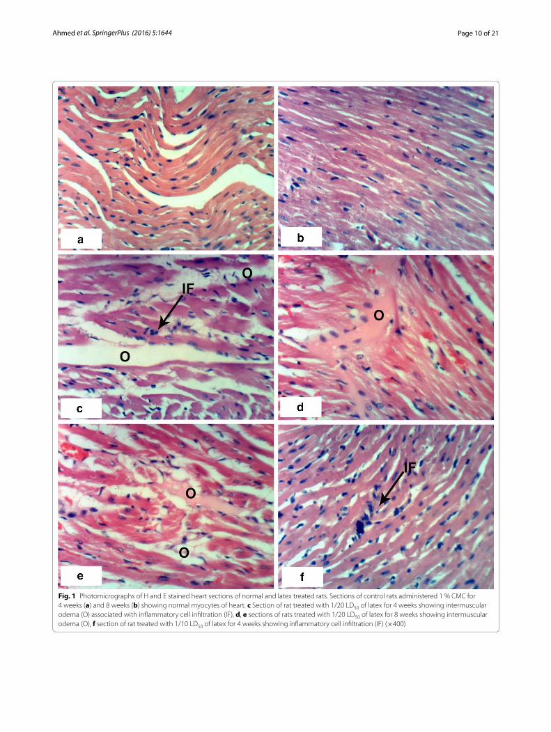
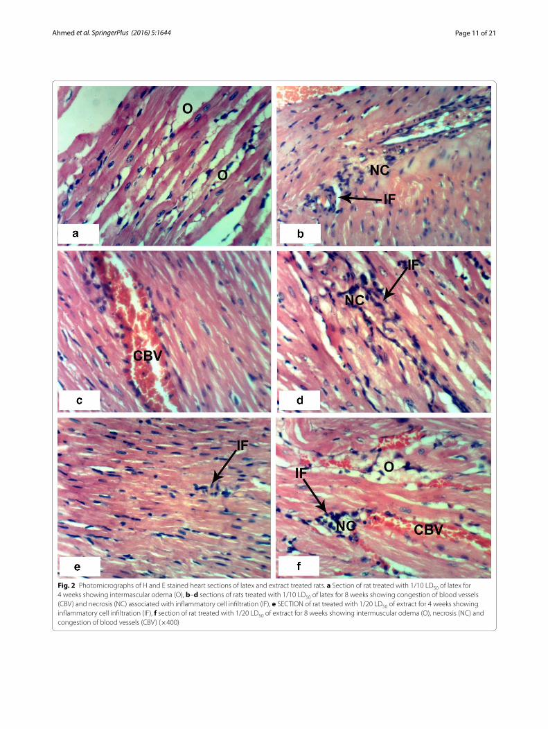
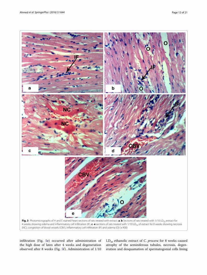
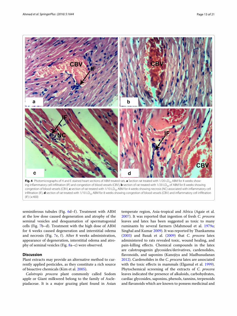
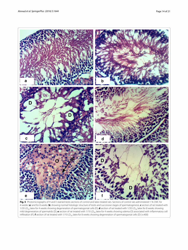
Histopathological effectsNormal myocytes architecture of normal rat heart in control animals were observed (Fig. 1a, b). Treatments of normal rats with 1/20 of LD50 of C. procera latex caused intermuscular odema (Fig. 1c–e) associated with inflam-matory cell infiltration (Fig. 1c). Administration of 1/10 of LD50 of latex caused marked alterations of normal structure of heart by affecting on cardiac myocytes caus-ing odema and inflammatory cell infiltration (Figs. 1f, 2a) in short time (4 weeks) while after 8 weeks, it caused necrosis associated with inflammatory cells infiltration and congestion of blood vessels (Fig. 2b–d). The treat-ment of rats with 1/20 of LD50 of ethanolic extract caused inflammatory cells infiltration at the end of the 4th week (Fig. 2e) and it caused intermuscular odema, necrosis of cardiac myocytes associated with inflammatory cells infiltration and congestion of blood vessels at the end of the 8th week (Fig. 2f ). Increasing the concentration of the ethanolic extract (1/10 of LD50) caused odema associ-ated with inflammatory cell infiltration (Fig. 3a, b) after 4 week. Prolongation of period of administration of this dose caused congestion of blood vessels, necrosis of car-diac myocytes associated with inflammatory cell infil-tration (Fig. 3c–e). Regarding ABM administration, low dose caused congestion of blood vessels and inflamma-tory cells infiltration (Fig. 4a, b) and high dose caused necrosis of cardiac myocytes associated with inflamma-tory cell infiltration (Fig. 4c, d).
Control groups demonstrated normal testicular histol-ogy with all successive stages of spermatogenesis (Fig. 5a, b). Administration of 1/20 LD50 latex for 4 weeks altered the normal testis structure by causing degeneration of spermatogonial cells lining seminiferous tubules (Fig. 5c). Interstitial oedema associated with inflammatory cells
Table 9 Analysis of variance for oxidative stress and anti-oxidant enzymes in heart of normal and treated rats
Source of vari-ation
F-probability
GSH LPO GPx GST SOD
A—Normal–latex effect
Dose P < 0.001 P < 0.001 P < 0.001 P < 0.001 P < 0.001
Time P > 0.05 P > 0.05 P < 0.01 P < 0.001 P < 0.001
Dose–time P < 0.01 P > 0.05 P < 0.05 P > 0.05 P < 0.05
B—Normal–extract effect
Dose P < 0.001 P < 0.001 P < 0.001 P < 0.001 P < 0.001
Time P < 0.001 P > 0.05 P < 0.001 P < 0.001 P < 0.001
Dose–time P < 0.001 P > 0.05 P < 0.01 P < 0.001 P < 0.001
C—Normal–abamectin effect
Dose P < 0.001 P < 0.001 P < 0.001 P < 0.001 P < 0.001
Time P > 0.05 P > 0.05 P > 0.05 P > 0.05 P < 0.001
Dose–time P > 0.05 P > 0.05 P > 0.05 P < 0.001 P < 0.001
Table 10 Analysis of variance for oxidative stress and anti-oxidant markers in testis of normal and treated rats
Source of variation
F-probability
GSH LPO GPx GST SOD
A—Normal–latex effect
Dose P < 0.001 P < 0.001 P < 0.001 P < 0.001 P < 0.001
Time P < 0.001 P > 0.05 P > 0.05 P < 0.01 P > 0.05
Dose–time P > 0.05 P > 0.05 P > 0.05 P > 0.05 P > 0.05
B—Normal–extract effect
Dose P < 0.001 P < 0.001 P < 0.001 P < 0.001 P < 0.001
Time P < 0.001 P > 0.05 P > 0.05 P > 0.05 P < 0.05
Dose–time P < 0.05 P > 0.05 P > 0.05 P > 0.05 P > 0.05
C—Normal–abamectin effect
Dose P < 0.001 P < 0.001 P < 0.001 P < 0.001 P < 0.001
Time P < 0.001 P > 0.05 P < 0.05 P > 0.05 P < 0.05
Dose–time P > 0.05 P > 0.05 P > 0.05 P > 0.05 P > 0.05
Page 10 of 21Ahmed et al. SpringerPlus (2016) 5:1644
Fig. 1 Photomicrographs of H and E stained heart sections of normal and latex treated rats. Sections of control rats administered 1 % CMC for 4 weeks (a) and 8 weeks (b) showing normal myocytes of heart. c Section of rat treated with 1/20 LD50 of latex for 4 weeks showing intermuscular odema (O) associated with inflammatory cell infiltration (IF), d, e sections of rats treated with 1/20 LD50 of latex for 8 weeks showing intermuscular odema (O), f section of rat treated with 1/10 LD50 of latex for 4 weeks showing inflammatory cell infiltration (IF) (×400)
Page 11 of 21Ahmed et al. SpringerPlus (2016) 5:1644
Fig. 2 Photomicrographs of H and E stained heart sections of latex and extract treated rats. a Section of rat treated with 1/10 LD50 of latex for 4 weeks showing intermascular odema (O), b–d sections of rats treated with 1/10 LD50 of latex for 8 weeks showing congestion of blood vessels (CBV) and necrosis (NC) associated with inflammatory cell infiltration (IF), e SECTION of rat treated with 1/20 LD50 of extract for 4 weeks showing inflammatory cell infiltration (IF), f section of rat treated with 1/20 LD50 of extract for 8 weeks showing intermuscular odema (O), necrosis (NC) and congestion of blood vessels (CBV) (×400)
Page 12 of 21Ahmed et al. SpringerPlus (2016) 5:1644
infiltration (Fig. 5e) occurred after administration of the high dose of latex after 4 weeks and degeneration observed after 8 weeks (Fig. 5f ). Administration of 1/10
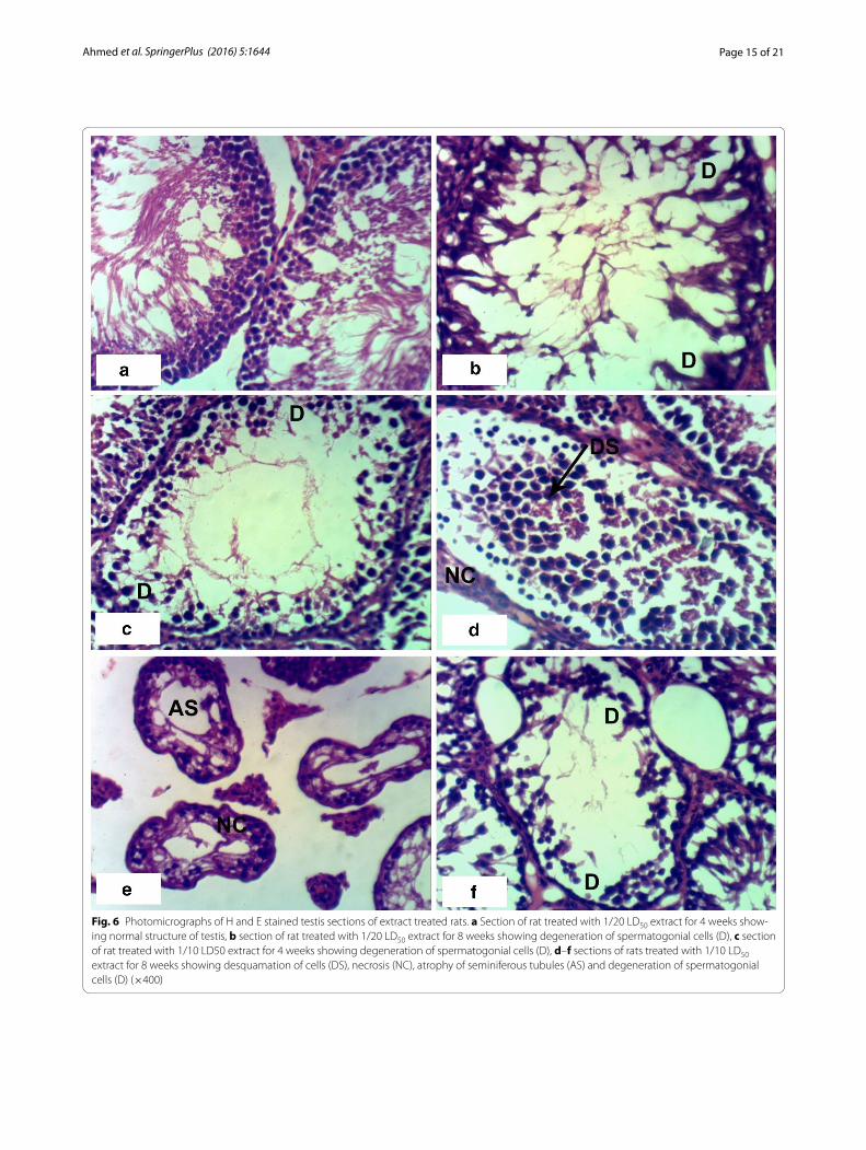
LD50 ethanolic extract of C. procera for 8 weeks caused atrophy of the seminiferous tubules, necrosis, degen-eration and desquamation of spermatogonial cells lining
Fig. 3 Photomicrographs of H and E stained heart sections of rats treated with extract. a, b Sections of rats treated with 1/10 LD50 extract for 4 weeks showing odema and inflammatory cell infiltration (IF), c–e sections of rats treated with 1/10 LD50 of extract for 8 weeks showing necrosis (NC), congestion of blood vessels (CBV), inflammatory cell infiltration (IF) and odema (O) (×400)
Page 13 of 21Ahmed et al. SpringerPlus (2016) 5:1644
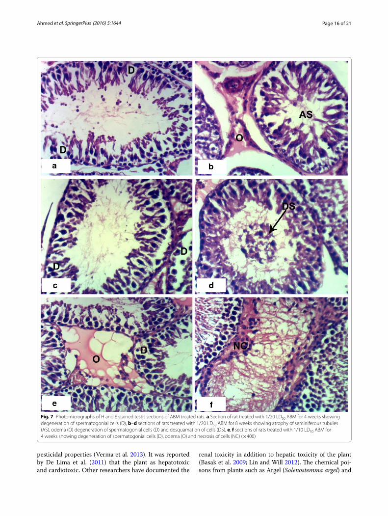
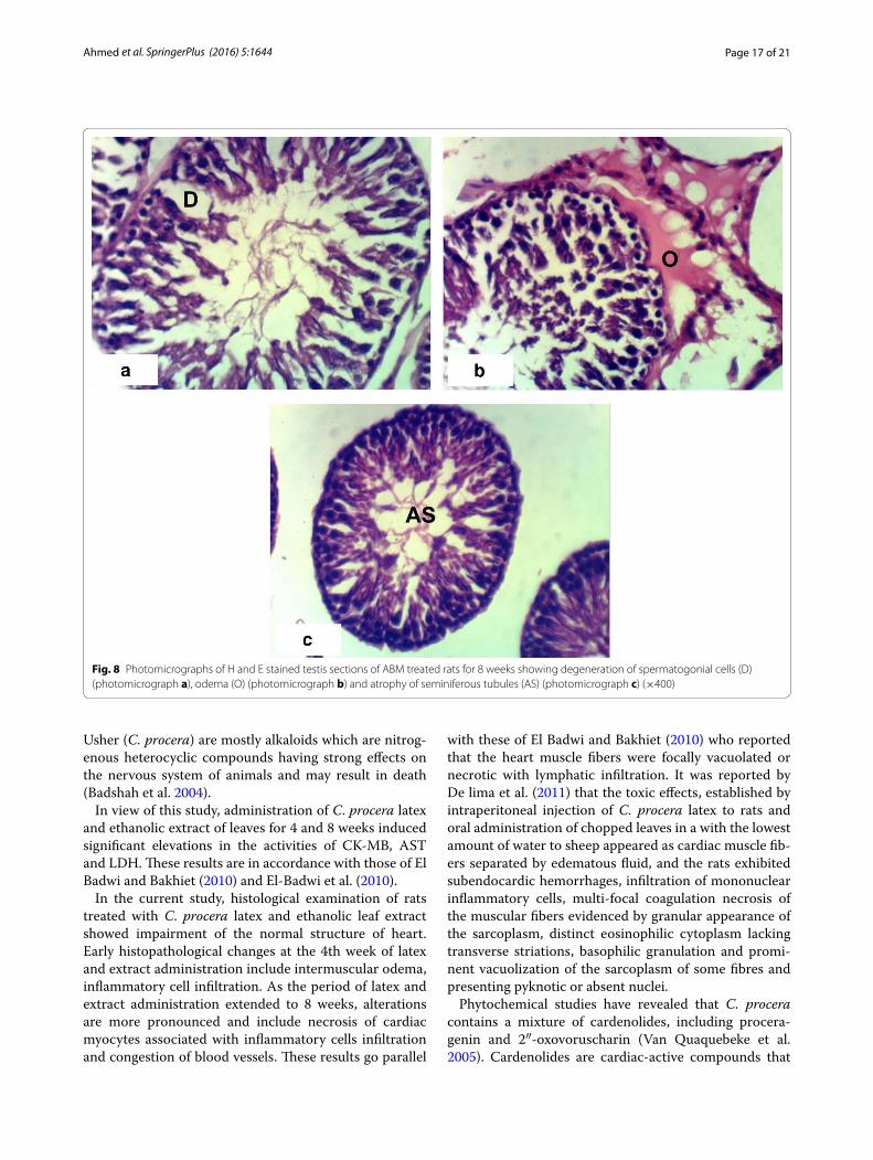
seminiferous tubules (Fig. 6d–f). Treatment with ABM at the low dose caused degeneration and atrophy of the seminal vesicles and desquamation of spermatogonial cells (Fig. 7b–d). Treatment with the high dose of ABM for 4 weeks caused degeneration and interstitial odema and necrosis (Fig. 7e, f ). After 8 weeks administration, appearance of degeneration, interstitial odema and atro-phy of seminal vesicles (Fig. 8a–c) were observed.
DiscussionPlant extracts may provide an alternative method to cur-rently applied pesticides, as they constitute a rich source of bioactive chemicals (Kim et al. 2005).
Calotropis procera plant commonly called Sodom apple or Giant milkweed belong to the family of Ascle-piadaceae. It is a major grazing plant found in Asian
temperate region, Asia-tropical and Africa (Agaie et al. 2007). It was reported that ingestion of fresh C. procera leaves and latex has been suggested as toxic to many ruminants by several farmers (Mahmoud et al. 1979a; Singhal and Kumar 2009). It was reported by Thankamma (2003) and Basak et al. (2009) that C. procera latex administered to rats revealed toxic, wound healing, and pain-killing effects. Chemical compounds in the latex are calotropagenin glycosides/derivatives, cardenolides, flavonoids, and saponins (Kanojiya and Madhusudanan 2012). Cardenolides in the C. procera latex are associated with the toxic effects in mammals (Elgamal et al. 1999). Phytochemical screening of the extracts of C. procera leaves indicated the presence of alkaloids, carbohydrates, cardiac glycosides, saponins, phenols, tannins, terpenoids and flavanoids which are known to possess medicinal and
Fig. 4 Photomicrographs of H and E stained heart sections of ABM treated rats. a Section rat treated with 1/20 LD50 ABM for 4 weeks show-ing inflammatory cell infiltration (IF) and congestion of blood vessels (CBV), b section of rat treated with 1/20 LD50 of ABM for 8 weeks showing congestion of blood vessels (CBV), c section of rat treated with 1/10 LD50 ABM for 4 weeks showing necrosis (NC) associated with inflammatory cell infiltration (IF), d section of rat treated with 1/10 LD50 ABM for 8 weeks showing congestion of blood vessels (CBV) and inflammatory cell infiltration (IF) (×400)
Page 14 of 21Ahmed et al. SpringerPlus (2016) 5:1644
Fig. 5 Photomicrographs of H and E stained testis sections of control and latex treated rats. Sections of control rats administered 1 % CMC for 4 weeks (a) and for 8 weeks (b) showing normal histologic structure of testis and successive stages of spermatogenesis, c section of rat treated with 1/20 LD50 latex for 4 weeks showing degeneration of spermatogonial cells (D), d section of rat treated with 1/20 LD50 latex for 8 weeks showing mild degeneration of spermatids (D), e section of rat treated with 1/10 LD50 latex for 4 weeks showing odema (O) associated with inflammatory cell infiltration (IF), f section of rat treated with 1/10 LD50 latex for 8 weeks showing degeneration of spermatogonial cells (D) (×400)
Page 15 of 21Ahmed et al. SpringerPlus (2016) 5:1644
Fig. 6 Photomicrographs of H and E stained testis sections of extract treated rats. a Section of rat treated with 1/20 LD50 extract for 4 weeks show-ing normal structure of testis, b section of rat treated with 1/20 LD50 extract for 8 weeks showing degeneration of spermatogonial cells (D), c section of rat treated with 1/10 LD50 extract for 4 weeks showing degeneration of spermatogonial cells (D), d–f sections of rats treated with 1/10 LD50 extract for 8 weeks showing desquamation of cells (DS), necrosis (NC), atrophy of seminiferous tubules (AS) and degeneration of spermatogonial cells (D) (×400)
Page 16 of 21Ahmed et al. SpringerPlus (2016) 5:1644
pesticidal properties (Verma et al. 2013). It was reported by De Lima et al. (2011) that the plant as hepatotoxic and cardiotoxic. Other researchers have documented the
renal toxicity in addition to hepatic toxicity of the plant (Basak et al. 2009; Lin and Will 2012). The chemical poi-sons from plants such as Argel (Solenostemma argel) and
Fig. 7 Photomicrographs of H and E stained testis sections of ABM treated rats. a Section of rat treated with 1/20 LD50 ABM for 4 weeks showing degeneration of spermatogonial cells (D), b–d sections of rats treated with 1/20 LD50 ABM for 8 weeks showing atrophy of seminiferous tubules (AS), odema (O) degeneration of spermatogonial cells (D) and desquamation of cells (DS), e, f sections of rats treated with 1/10 LD50 ABM for 4 weeks showing degeneration of spermatogonial cells (D), odema (O) and necrosis of cells (NC) (×400)
Page 17 of 21Ahmed et al. SpringerPlus (2016) 5:1644
Usher (C. procera) are mostly alkaloids which are nitrog-enous heterocyclic compounds having strong effects on the nervous system of animals and may result in death (Badshah et al. 2004).
In view of this study, administration of C. procera latex and ethanolic extract of leaves for 4 and 8 weeks induced significant elevations in the activities of CK-MB, AST and LDH. These results are in accordance with those of El Badwi and Bakhiet (2010) and El-Badwi et al. (2010).
In the current study, histological examination of rats treated with C. procera latex and ethanolic leaf extract showed impairment of the normal structure of heart. Early histopathological changes at the 4th week of latex and extract administration include intermuscular odema, inflammatory cell infiltration. As the period of latex and extract administration extended to 8 weeks, alterations are more pronounced and include necrosis of cardiac myocytes associated with inflammatory cells infiltration and congestion of blood vessels. These results go parallel
with these of El Badwi and Bakhiet (2010) who reported that the heart muscle fibers were focally vacuolated or necrotic with lymphatic infiltration. It was reported by De lima et al. (2011) that the toxic effects, established by intraperitoneal injection of C. procera latex to rats and oral administration of chopped leaves in a with the lowest amount of water to sheep appeared as cardiac muscle fib-ers separated by edematous fluid, and the rats exhibited subendocardic hemorrhages, infiltration of mononuclear inflammatory cells, multi-focal coagulation necrosis of the muscular fibers evidenced by granular appearance of the sarcoplasm, distinct eosinophilic cytoplasm lacking transverse striations, basophilic granulation and promi-nent vacuolization of the sarcoplasm of some fibres and presenting pyknotic or absent nuclei.
Phytochemical studies have revealed that C. procera contains a mixture of cardenolides, including procera-genin and 2″-oxovoruscharin (Van Quaquebeke et al. 2005). Cardenolides are cardiac-active compounds that
Fig. 8 Photomicrographs of H and E stained testis sections of ABM treated rats for 8 weeks showing degeneration of spermatogonial cells (D) (photomicrograph a), odema (O) (photomicrograph b) and atrophy of seminiferous tubules (AS) (photomicrograph c) (×400)
Page 18 of 21Ahmed et al. SpringerPlus (2016) 5:1644
inhibit the cellular membrane Na+/K+ ATPase, result-ing in an electrolytic disturbance that affects the elec-trical conductivity of the heart (Poindexter et al. 2007). Thus, the heart dysfunction and the elevations in heart enzymes (CK-MB, AST and LDH) in serum and cardiac toxicity induced by the plant, in this study, may attrib-uted to its constituting cardenolides. The milky latex contains a powerful bacteriolytic enzyme, toxic glycoside calactin, calotropin D1, calotropin D2 calotropin F11, and a nontoxic powerful proteolytic enzyme and it exhib-ited local anesthetic activity (Samar et al. 2009). In this study, administration of ABM showed marked elevations in LDH, AST and CK-MB. These results reflected the toxic effect of ABM and impairment of heart function. The elevated of serum enzymes related to heart func-tion was associated with cardiac histopathological lesions which include oedema, inflammatory cells infiltration and necrosis observed in the present investigation.
In view of this study, treatments with C. procera latex and ethanolic extract of leaves induced marked decrease in the levels of male sex hormones testosterone, FSH and LH. In conduction with the present study, Sharma and Jacob (2001) found that intermuscular administration of aqueous and ethanolic extracts of flowers of C. procera has been shown to induce functional sterility and has a potent antispermatogenic activity in the albino mouse, but at the doses and experimental regimen employed, had no apparent effect on sexual behavior or libido. In the same way, Akinloye et al. (2002) reported that fresh leaves extract has depicted potential deleterious effects on the rat testes and accessory sex organs represented by degeneration of seminiferous epithelium of varying degrees as well as presence of large-sized multimate cells in the tubules and empty interstitial spaces. Calotropis is extensively used in both male and female rats for under-standing its role in fertility (Akinloye et al. 2002; Circosta et al. 2001; Ahirwar et al. 2007). It was indicated previ-ously that, an active principle of flower extract of C. pro-cera showed spermicidal effect on testicular functions in Indian desert male Gerbil (Garg 1979).
Histologic profile of testes in the present study revealed extensive deleterious changes in the germinal tubules which contained mainly necrotic and degenerating germ cells. Further, the epididymal lumina appeared devoid of spermatozoa and exhibited mainly cell debris. Also, the seminiferous tubules were atrophied and necrosis and desquamation of spermatogonial cells after adminis-tration of the high dose of the extract. The interstitium was observed to be devoid of leydig cells in this study. This change may be due to decreased production of testosterone known to be responsible for normal tes-ticular architecture. Histological changes observed in the testes of treated rats in this study may be due to the
cardiac glycosides found in the plant which was incrimi-nated to be responsible also for pathological and ultra-structural changes in the kidney tubules of Wistar rats. These changes are in concordance with Akinloye et al. (2002). In the current study, the plant showed toxic effect on the testis through effect on the germ cell and this is conducted by Akinloye et al. (2002) who said C. procera extract has destructive effect on the germ cells which are actively dividing. In addition, it was reported that testos-terone maintained the viability of spermatozoa (Bhar-gava 1989). The toxic effect of the plant in this study to decrease the serum level of testosterone may be mediated via affecting on leydig cells and impairing their functions and structures.
Previous reports have indicated a strong link between male infertility and exposure to more than 50 pesticides (Victor-Costa et al. 2010; Manfo et al. 2010; Tiwari et al. 2011). The adverse effects of ABM on the fertility of adult male rats have been demonstrated in the present study. The serum levels of testosterone, FSH and LH was signifi-cantly reduced in rats treated with 1/20 and 1/10 LD50 of ABM for 4 and 8 weeks. These results are in agreement with those of Elbetieha and Daas (2003) and Abd-Elhady and Abou-Elghar (2013) who found reduction in testos-terone level at dose of 2.13 mg ABM/animal/day. Elbe-tieha and Daas (2003) indicated that ingestion of ABM for 6 weeks induced adverse effects on male rat fertility and reproduction. The decrease in male fertility via decrease in male sex hormones in rats treated with ABM in the present study is explained by Abd-Elhady and Abou-Elghar (2013) who suggested that ABM may have acted directly on the testes and affected the androgen bio-synthesis pathway. These are strongly supported by the wide array of abnormalities seen when histopathological sections of the testes were examined. These abnormali-ties include necrotic changes in the tissues, interstitial odema, and degeneration and atrophy of seminal vesicles as well as desquamation of spermatogonial cells. In our opinion, it can be suggested that ABM may act directly on the testes and affected the androgen biosynthesis pathway or may directly act on the brain, hypothalamus or anterior pituitary gland which will indirectly affect the testes and possibly affect sexual activity. Both attributions are supported by the present study which revealed direct histological effect of ABM on testis and direct effects on pituitary hormones FSH and LH that respectively control spermatogenesis and testosterone secretion from leydig cells.
The shift in balance between oxidant/antioxidant in favor of oxidants is termed oxidative stress. Oxidative stress contributes to many pathological conditions. When oxidative stress occurs, cells attempt to coun-teract the oxidant effects and restore the redox balance
Page 19 of 21Ahmed et al. SpringerPlus (2016) 5:1644
by activation or silencing of genes encoding defensive enzymes, transcription factors, and structural proteins Scandalios (2004). Glutathione (GSH) is highly abun-dant in all cell compartments and is the major soluble antioxidant. It detoxifies hydrogen peroxide and lipid peroxides via action of glutathione peroxidase. GSH donates its electron to H2O2 to reduce it into H2O and O2. GSH protects cells against apoptosis by interacting with proapoptotic and antiapoptotic signaling pathways (Masella et al. 2005). In this study, it is observed that administration of latex and ethanolic extract induced marked decrease in heart and testis GSH levels and GPx, GST and SOD activities and on the other hand, it induced significant increase in lipid peroxidation. This increase in lipid peroxidation and suppression of anti-oxidant defense system as a result of latex and ethanolic extract administration lead to excess production and less scavenging of reactive oxygen species which in turn result in cardiac and testicular oxidative damage. In the current study, the deterioration of non-enzymatic and enzymatic antioxidants and exacerbated production of lipid peroxidation was associated with the elevation in the levels of various serum biochemical markers of car-diac and testicular damage including CK-MB, AST and LDH.
Administration of different doses of ABM caused deple-tion in GSH content, GPx, GST and SOD activities in this study. These results are in line with those of El-Shenawy (2010) who studied the toxic effect of ABM on isolated rat hepatocytes and found ABM decreased GSH concen-tration and GPx and SOD activities. Since superoxide is the primary ROS produced from the toxic substances, its dismutation by SOD is the primary importance for each cell. So, the depletion of the activity of SOD in this study caused accumulations of ROS in tissues causing distur-bance of cell membrane and damage of cells.
Lipid peroxidation is the oxidative deterioration of polyunsaturated lipids to form radical intermediates that bring about cellular damage. Malondialdehyde (MDA), a major end product of this reaction, is an index of lipid peroxidation and has been estimated as thiobarbituric acid (TBARS) (Kohen and Nyska 2002). The increase in MDA level after the latex and ethanolic extract admin-istration reflects that the plant induced increase in ROS and lipid peroxides. The elevation of lipid peroxides caused disturbance in cell membrane structure, dam-age of cell and cell death. Degree of toxicity induced by the plant is dose dependent. Significant increases in lipid peroxidation in heart and testis after ingestion of ABM in this study were resulted. Increasing dose progres-sively increased the toxic effect on the normal oxidant/antioxidant state in tissues. These results are in line with those of El-Shenawy (2010) who studied the toxic effect
of ABM on isolated rat hepatocytes and found that ABM increased LPO.
Another substantiation of the impairment of the oxi-dant/antioxidant status in cells occurred with the plant and ABM administration is the depletion of the GPx, GST and SOD activities in tissues in a dose dependent manner. These results are in line with those of El-Shafey et al. (2011) and in contrast with those of El-Shenawy (2010). Based on the findings of the present study, it can be concluded that the cardiotoxic and testicular toxicity of C. procera leaf extract and latex as well as ABM is due to the elevation of lipid peroxidation and ROS and deple-tion of antioxidant levels.
The milky sap is a mixture of various chemicals includ-ing calotropis glycosides such as calotropin, calotoxin, calactin, uscharidin, voruscharin which are caustic in nature and are considered poisonous. The irritant and pro-inflammatory property of latex of C. procera has been well established (Alencar et al. 2006). Acciden-tal exposure to the latex has been reported to cause inflammation of the skin and eyes (Shivkar and Kumar 2003; Al-Mezaine et al. 2005). In the present study, it was resulted that the latex and ethanolic extract as well as ABM induced marked inflammations as observed in photomicrographs of heart and testis histological sec-tions. This heart and testis inflammatory status was concomitant with the marked elevation of serum levels of pro-inflammatory cytokine, TNF-α, and depletion of anti-inflammatory cytokine, IL-4, in a dose and time dependent manner.
Overall, C. procera latex and ethanolic extract of leaves as well as ABM induced cardiotoxic and testicular toxic effects which was evidenced by increases in the activi-ties of heart enzymes in serum and decreases in male sex hormones in serum in addition to heart and testis histo-logical perturbances. However the ethanolic leaf extract seemed to be more effective in deteriorating oxidative stress and antioxidant defense system in both heart and testis, the latex produced more deleterious effects on the testicular function and sex hormones’ levels. So, the plant latex and ethanolic extract are considered toxic and they may be suggested as rodenticides at 1/10 and 1/20 of LD50 which are more or less similar to the reference pesticide, ABM.
Authors’ contributionsOMA, HIA and MWB planned and designed the experiment. HYA measured the detected parameters. OMA and HYA performed statistical analysis. All authors drafted the manuscript. All authors read and approved the final manuscript.
Author details1 Physiology Division, Zoology Department, Faculty of Science, Beni-Suef University, Beni Suef, Egypt. 2 Rodents Division, Harmful Animals Depart-ment, Plant Protection Research Institute, Agriculture Research Center, Giza, Egypt.
Page 20 of 21Ahmed et al. SpringerPlus (2016) 5:1644
AcknowledgementsThe authors would like to express their sincere appreciation to Dr. Walaa Azmy Hasan for her assistance in recognition and classification of the plant. The authors also acknowledged Prof. Dr. Kawkab Abd El Aziz Ahmed, Professor of Pathology, Pathology Department, Faculty of Veterinary Medicine, Cairo Uni-versity for her great help in the examination of liver sections and description of histopathological changes.
Competing interestsThe authors declare that they have no competing interests.
Received: 12 May 2016 Accepted: 15 September 2016
ReferencesAbd-Elhady HK, Abou-Elghar GE (2013) Abamectin induced biochemical and
histopathological changes in the albino rat, Rattus norvegicus. Plant Prot Res 53(3):263–270
Abou-Hashem AAM (2013) Rodenticidal effect of argel (Gomphocarpus sinaicus Boiss) leaves on the Norway rat (Albino), Rattus norvegicus, Berk-enhout under laboratory conditions. J Appl Sci Res 9(3):690–1695
Agaie BM, Salisu A, Ebobo AA (2007) A survey of common toxic plants of livestock in Sokoto State, Nigeria. Sci Res Essay 2(2):40–42
Agarwal AK (1998) Avermectin. In: Wexler P (ed) Encyclopedia of toxicology, 1st edn. Academic Press, San Diego, pp 89–90
Ahirwar D, Ahirwar B, Khary MD (2007) Influence of Calotropis procera roots on biochemistry of reproductive organs of ovariectomized rats. Indian J Pharm Sci 69:459–461
Ahmed UAM, Zuhua S, Bashier NHH, Muafi H, Zhongping H, Yuling G (2006) Evaluation of insecticidal potentialities of aqueous extracts from Calotropis procera (Ait) against Henosepilachna elaterii rossi. J Appl Sci 6:2466–2470
Akinloye AK, Abatan MA, Akaka OO, Oke BA (2002) Histomorphometric and histopathological studies on the effects of Calotropis procera (Giant milkwood) on the male reproductive organs of Wistar rats. Afr J Biomed Res 5:57–61
Alencar NM, Oliveira JS, Mesquita RO, Lima MW, Vale MR, Etchells JP et al (2006) Pro- and anti-inflammatory activities of the latex from Calotropis procera (Ait.) R.Br. are triggered by compounds fractionated by dialysis. Inflamm Res 55(12):559–564
Al-Mezaine HS, Al-Rajhi AA, Al-Assiri A, Wagoner MD (2005) Calotropis procera (ushaar) keratitis. Am J Ophthalmol 139(1):199–202
Andreyko JL, Bhavnani BR, Nisker JA, Walker WH, Woolever CA (1986) Role of serum androgens and sex hormones binding globulin capacity in the evaluation of hirsutism in women. Clin Biochem 19(1):58–61
Badshah H, Farmanullah Salihah Z, Saljoqi A, Shakur M (2004) Toxic effects of AK (Calotropis procera) plant extracts against termites (Heterotermes indicola and Coptotermes heimi) Isoptera: Rhinotermitidae. Pak J Biol Sci 7(9):1603–1606
Basak SK, Bhaumik A, Mohanta A, Singhal P (2009) Ocular toxicity by latex of Calotropis procera (Sodom apple). Indian J Opthalmol 57(3):232–234
Begum N, Sharma B, Pandey RS (2013) Calotropis procera and Annona squa-mosa: potential alternatives to chemical pesticides. Br J Appl Sci Technol 3(2):254–267
Beutler E, Duron O, Kelly BM (1963) Improved method for determination of blood glutathione. J Lab Clin Med 61:882–888
Bhargava SK (1989) Antiandrogenic effects of a flavonoid rich fraction of Vitex negundo seeds: a histological and biochemical study in dogs. J Ethnop-harmacol 27:327–339
Bhatnagar SS (1950) The wealth of India, vol 2. CSIR, New Delhi, p 23Braunstein GD, Rasor J, Alder D, Danzer H, Wade ME (1976) Serum human
luteinizing hormone levels through normal pregnancy. Am J Obstet Gynecol 126:678–681
Carvalho AFU, Melo VMM, Craveiro AA, Machado MIL, Bantim MB, Rabelo EF (2003) Larvicidal activity of the essential oil from Lippia sidoides Cham. against Aedes aegypti Linn. Mem Inst Oswaldo Cruz 98:569–571
Cerejeira MJ, Viana P, Batista S, Pereira T, Silva E, Valério MJ et al (2003) Pesticides in Portuguese surface and ground waters. Water Res 37(5):1055–1063
Circosta C, Sanogo R, Occhiuto F (2001) Effects of Calotropis procera on oes-trous cycle and oestrogentic functionality in rats. IL Farm 56:373–378
Croft M, Duan M, Choi H, Eun S, Madireddi S, Mehta A (2012) TNF superfam-ily in inflammatory disease: translating basic insights. Trends Immunol 33:144–152
De Lima JM, De Freitas FJ, Amorim RN, Camara AC, Batista JS, Soto-Blanco B (2011) Clinical and pathological effects of Calotropis procera exposure in sheep and rats. Toxicon 57(1):183–185
Eason CT, Turck P (2002) A 90-day toxicological evaluation of compound 1080 (sodium monofluoroacetate) in Sprague Dawley rats. Toxicol Sci 69:439–447
El Badwi SMA, Bakhiet AO (2010) Toxicity of Calotropis procera latex in pregnant and non-pregnant goats. Sci Res Essays 5(17):2404–2408
El-Badwi SMA, Bakhiet AO, Medani AB, Shamseldin ZY (2010) Influence of phe-nobarbital pretreatment on toxicity of Calotropis procera latex in Nubian goats. Res J Vet Sci 5(1):25–31
Elbetieha A, Daas SI (2003) Assessment of antifertility activities of ABM pesti-cide in male rats. Ecotoxicol Environ Saf 55(3):307–313
Elgamal MHA, Hanna AG, Morsy NAM, Duddeck H, Simon A, Toth G (1999) Complete 1H and 13C signal assignments of 5α cardenolides isolated from Calotropis procera R. Br. J Mol Struct 477:201–208
EL-Gengaihi SE, Dimetry NZ, Mohamed SM (1997) Chemical and biological investigation of harmal plant. 2-Alkaloidal investigation. J Appl Entomol 12(3):165–167
El-Shafey AAM, Seliem MME, El-Mahrouky F, Gabr WM, Kandil RA (2011) Some physiological and biochemical effects of oshar extract and abamectin biocide on male albino rats. J Am Sci 7(12):254–261
El-Shenawy NS (2010) Effects of insecticides fenitrothion, endosulfan and abamectin on antioxidant parameters of isolated rat hepatocytes. Toxicol In Vitro 24:1148–1157
Fahim HE, Ahmed OM, Boules MW, Ahmed HY (2016) Nephrotoxic effects of abamectin and Calotropis procera latex and leaf extract in male albino rats. Am J Med Med Sci 6(3):73–86
Freedman B, Nowak J, Kwolek WF (1979) Abiossay for plant derived pest con-trol agent using the European comborer. Econ Entomol 72:45–54
Gallo MA, Lawryk NJ (1991) Organic phosphorus pesticides. In: Hayes WJ Jr, Laws ER Jr (eds) Handbook of Pesticide Toxicology. Classes of pesticides, vol 2. Academic Press, San Diego, pp 917–1123
Garg A (1979) Effect of Aak Calotropis procera (Ait.) R. Br. flower extract on tes-ticular function of the Indian desert male gerbil Meriones hurrianae Jer-don: a biochemical & histological study. Indian J Exp Biol 17(9):859–862
Gella FJ, Olivella T, Cruz Pastor M, Arenas J, Moreno R, Durban R et al (1985) A simple procedure for routine determination of aspartate aminotrans-ferase and alanine aminotransferase with pyridoxal phosphate. Clin Chim Acta 153:241–247
Gerhart W, Waldenström J (1979) Creatine kinase B-Subunit activity in serum after immunoinhibition of M-Subunit activity. Clin Chem 25(7):1274–1280
Hassan SW, Bilbis FL, Ladan MJ, Umar RA, Dangoggo SM, Saidu Y, Abubakar MK, Faruk UZ (2006) Evaluation of antifungal activity and phytochemi-cal analysis of leaves, roots and stem bark extracts of Calotropis procera (Asclepiadaceae). Pak J Biol Sci 9(14):2624–2629
Howard M, Harada N (1994) Guidebook to cytokines and their receptors. In: Nicola NA (ed) Oxford University Press, New York, p 44
Iqbal Z, Lateef M, Jabbar A, Muhammad G, Khan MN (2005) Anthelmintic activ-ity of Calotropis procera (Ait.) Ait F. flowers in sheep. J Ethnopharmacol 102(2):256–261
Kanojiya S, Madhusudanan KP (2012) Rapid identification of calotropagenin glycosides using high-performance liquid chromatography electrospray ionisation tandem mass spectrometry. Phytochem Anal 23(2):117–125
Kim HG, Jeon JH, Kim MK, Lee HS (2005) Pharmacological effects of asaronal-dehyde isolated from Acorus gramineus rhizome. Food Sci Biotechnol 14:685–688
Kohen R, Nyska A (2002) Oxidation of biological systems: oxidative stress phe-nomena, antioxidants, redox reactions, and methods for their quantifica-tion. Toxicol Pathol 30(6):620–650
Kolar L, Erzen NK, Hogerwerf L, Van Gestel CAM (2008) Toxicity of abamectin and doramectin to soil invertebrates. Environ Pollut 151(1):182–189
Kuriachen PM, Dave Y (1989) Structural, developmental and histochemical studies in the collectors of Calotropis procera (Asclepiadaceae). J Phyto-logic Res 2:7–14

Page 21 of 21Ahmed et al. SpringerPlus (2016) 5:1644
Laitiff AA, Teoh SL, Das S (2010) Wound healing in diabetes mellitus: traditional treatment modalities. Clin Ter 161(4):359–364
Lankas GR, Gordon LR (1989) Toxicology. In: Campbell WC (ed) Ivermectin and abamectin. Springer, New York, p 363
Lima-Filho JV, Patriota JM, Silva AF, Filho NT, Oliveira RS, Alencar NM et al (2010) Proteins from latex of Calotropis procera prevent septic shock due to lethal infection by Salmonella enterica serovar Typhimurium. J Ethnophar-macol 129:327–334
Lin Z, Will Y (2012) Evaluation of drugs with specific organ toxicities in organ-specific cell lines. Toxicol Sci 126(1):114–127
Mahmoud OM, Adam SEI, Tartour G (1979a) The effects of Calotropis procera on small ruminants. I. Effects of feeding sheep with the plant. J Comp Pathol 89:241–250
Mahmoud OM, Adam SEI, Tartour G (1979b) The effects of Calotropis procera on small ruminants. II. Effects of administration of the latex to sheep and goats. J Comp Pathol 89:251–263
Maloschik E, Ernst A, Hegedus G, Darvas B, Székács A (2007) Monitoring water-polluting pesticides in Hungary. Microchem J 85(1):88–97
Manfo FP, Moundipa PF, Déchaud H, Tchana AL, Nantia EA, Zabot MT et al (2010) Effect of agropesticides use on male reproductive function: a study on farmers in Djutitsa (Cameroon). Environ Toxicol 27(7):423–432
Mannervik B, Gutenberg C (1981) Glutathione transferase (human placenta). Methods Enzymol 77:231–235
Marklund S, Marklund G (1974) Involvement of superoxide anion radical in the autoxidation of pyrogallol and convenient assay for superoxide dismutase. Eur J Biochem 47:469–474
Masella R, Di Benedetto R, Vari R, Filesi C, Giovannini C (2005) Novel mecha-nisms of natural antioxidant compounds in biological systems: involve-ment of glutathione and glutathione-related enzymes. J Nutr Biochem 16:577–586
Matkovics B, Kotorman M, Varga IS, Hai DQ, Varga C (1997) Oxidative stress in experimental diabetes induced by streptozotocin. Acta Physiol Hung 85(1):29–38
Million T, Kassa H, Charles K (2010) The toxicity of plant material, Drimia altissima (Urginea altissima), against the field rat, Arvicanthis abys-sinicus: a potential non synthetic rodenticide. Ethiop J Health Dev 24(3):175–179
Moline JM, Golden AL, Bar-Chama N, Smith E, Rauch ME, Chapin RE et al (2000) Exposure to hazardous substances and male reproductive health: a research framework. Environ Health Perspect 108:803–813
Odell WD, Parlow AF, Cargille CM, Ross GI (1968) Radioimmunioassay for human FSH: physiological studies. J Clin Invest 47:2551–2562
Paul TK, Kumar A (2009) Dendrocnide sinuata (Blume) Chew (Urticaceae)—a plant that can be grown to repulse the wild elephants. ENVIS Newsl 14(2):5–6
Poindexter B, Feng W, Dasgupta A, Bick R (2007) Oleandrin produces changes in intracellular calcium levels in isolated cardiomyocytes, a real-time fluo-rescence imaging study comparing adult to neonatal cardiomyocytes. J Toxicol Environ Health 70:568–574
Preuss HG, Jarrel ST, Scheckenbach R, Lieberman S, Anderson RA (1998) Com-parative effects of chromium, vanadium and Gymnema sylvestre on sugar-induced blood pressure elevations in SHR. J Am Coll Nutr 17(2):116–123
Quy RJ, Cowan DP, Prescott CV, Gill JE, Kerins GM, Dunsford G et al (1995) Control of a population of Norway rats resistant to anticoagulant rodenti-cides. Pest Sci 45:247–256
Roa M, Blane K, Zonneberg M (1985) PC-STAT one-way analysis of variance. Version IA (C) copyright. The University of Georgia, University of Georgia, USA
Samar KB, Arup B, Ayan M, Prashant S (2009) Ocular toxicity by latex of Calotro-pis procera. Indian J Ophthalmol 57:232–234
Scandalios JG (2004) Genomic responses to oxidative stress. In: Meyers RA (ed) Encyclopedia of molecular cell biology and molecular medicine, vol 5, 2nd edn. Wiley-VCH, Weinheim, pp 489–512
Sharma N, Jacob D (2001) Inhibition of fertility and functional alteration in the genital organs of male Swiss albino mouse after administration of Calotropis procera flower extract. Pharma Biol 39(6):403–407
Sheth F (2011) Range of seasonal phytochemical variations in Calotropis procera (Ait.) R. Br. Int J Med Arom Plants 1(2):180–183
Shivkar YM, Kumar VL (2003) Histamine mediates the pro-inflammatory effect of latex of Calotropis procera in rats. Mediat Inflamm 12(5):299–302
Singhal A, Kumar VL (2009) Effect of aqueous suspension of dried latex of Calotropis procera on hepatorenal functions in rat. J Ethnopharmacol 122(1):172–174
Suresh Kumar P, Suresh E, Kalavathy S (2013) Review on a potential herb Calotropis gigantea (L.) R. Br. Sch Acad J Pharm 2(2):135–143
Thankamma L (2003) Hevea latex as a wound healer and pain killer. Curr Sci 84(8):971–972
Thomas PJ, Mineau P, Shore RF, Champoux L, Martin PA, Wilson LK, Fitzgerald G, Elliot JE (2011) Second generation anticoagulant rodenticides in predatory birds: probabilistic characterization of toxic liver concentra-tions and implications for predatory bird populations in Canada. Environ Int 37:914–920
Tiwari AK, Pragya P, Ram KR, Chowdhuri DK (2011) Environmental chemical mediated male reproductive toxicity: Drosophila melanogaster as an alternate animal model. Theriogenology 76(2):197–216
Vadlapudi V, Naidu CK (2010) In vitro bioactivity of indian medicinal plant Calotropis procera (Ait). J Glob Pharm Technol 2:43–45
Van Quaquebeke E, Simon G, Andre A, Dewelle J, El Yazidi M, Bruyneel F et al (2005) Identification of a novel cardenolide (200-oxovoruscharin) from Calotropis procera and the hemisynthesis of a novel derivative displaying potent in vitro antitumor activities and high in vivo tolerance: structure–activity relationship analyses. J Med Chem 48:849–856
Verma R, Satsangi GP, Shrivastava JN (2013) Analysis of phytochemical con-stituents of the ethanolic and chloroform extracts of Calotropis procera using gas chromatography-mass spectroscopy (GC-MS) technique. J Med Plants Res 7(40):2986–2991
Victor-Costa AB, Bandeira SM, Oliveira AG, Mahecha GA, Oliveira CA (2010) Changes in testicular morphology and steroidogenesis in adult rats exposed to Atrazine. Reprod Toxicol 29:323–331
Young DS (2000) Effects of drugs on clinical laboratory tests, 5th edn. Ameri-can Association for Clinical Chemistry (AACC) Press, Washington, DC, USA. https://www.aacc.org/store/books/6600/effects-of-drugs-on-clinical-laboratory-tests-5th-edition.aspx
Copyright © 2022 FDOKUMEN