Effects of FSH on testicular mRNA transcript levels in the hypogonadal mouse
13
Effects of FSH on testicular mRNA transcript levels in the hypogonadal mouse M H Abel, D Baban, S Lee, H M Charlton and P J O’Shaughnessy 1 Department of Physiology, Anatomy and Genetics, University of Oxford, Le Gros Clarke Building, Oxford OX1 3QX, UK 1 Institute of Comparative Medicine, University of Glasgow Veterinary School, Bearsden Road, Glasgow G61 1QH, UK (Correspondence should be addressed to P J O’Shaughnessy; Email: [email protected]) (D Baban is now at Genomics Group, The Wellcome Trust Centre for Human Genetics, University of Oxford, Roosevelt Drive, Oxford OX3 7BN, UK) Abstract FSH acts through the Sertoli cell to ensure normal testicular development and function. To identify transcriptional mechanisms through which FSH acts in the testis, we have treated gonadotrophin-deficient hypogonadal (hpg) mice with recombinant FSH and measured changes in testicular transcript levels using microarrays and real-time PCR 12, 24 and 72 h after the start of treatment. Approximately 400 transcripts were significantly altered at each time point by FSH treatment. At 12 h, there was a clear increase in the levels of a number of known Sertoli cell transcripts (e.g. Fabp5, Lgals1, Tesc, Scara5, Aqp5). Additionally, levels of Leydig cell transcripts were also markedly increased (e.g. Ren1, Cyp17a1, Akr1b7, Star, Nr4a1). This was associated with a small but significant rise in testosterone at 24 and 72 h. At 24 h, androgen-dependent Sertoli cell transcripts were up-regulated (e.g. Rhox5, Drd4, Spinlw1, Tubb3 and Tsx) and this trend continued up to 72 h. By contrast with the somatic cells, only five germ cell transcripts (Dkkl1, Hdc, Pou5f1, Zfp541 and 1700021K02Rik) were altered by FSH within the time-course of the experiment. Analysis of canonical pathways showed that FSH induced a general decline in transcripts related to formation and regulation of tight junctions. Results show that FSH acts directly and indirectly to induce rapid changes in Sertoli cell and Leydig cell transcript levels in the hpg mouse but that effects on germ cell development must occur over a longer time-span. Journal of Molecular Endocrinology (2009) 42, 291–303 Introduction Postnatal testicular growth, spermatogenesis and fertility are dependent upon the pituitary gonado- trophins FSH and LH. LH acts directly on Leydig cells to stimulate androgen production, while androgens and FSH stimulate spermatogenesis through direct action on the Sertoli cells (McLachlan et al. 2002). The role of gonadotrophins is clearly seen in the hypogo- nadal (hpg) mouse that lacks GnRH (Mason et al. 1986) and, consequently, has undetectable circulating levels of LH and FSH (Cattanach et al. 1977). The gonads of the hpg mouse remain in a pre-pubertal state throughout life, with spermatogenesis blocked at early meiosis (Cattanach et al. 1977, Myers et al. 2005) although treatment with exogenous gonado- trophins or androgens will increase testicular growth and restore germ cell development (Charlton et al. 1983, Singh & Handelsman 1996a,b, Haywood et al. 2003). In recent years, generation of mice lacking individual hormones or hormone receptors has allowed us to investigate more clearly the roles played by LH, FSH and androgen in the regulation of testicular function. In particular, study of mice lacking androgen receptors (AR) in the Sertoli cells (SCARKO (De Gendt et al. 2004)) has shown that androgens are essential for spermatocyte progression through meio- sis. By contrast, mice lacking FSH (FSHbKO (Kumar et al. 1997)) or the FSH receptor (FSHRKO; Dierich et al. 1998, Abel et al. 2000) are fertile with all stages of spermatogenesis present. Nevertheless, in FSHRKO and FSHbKO mice there is a reduction in sperm number and quality (Krishnamurthy et al. 2001, Wreford et al. 2001) suggesting that FSH action optimises spermatogenesis. In addition, comparison of SCARKO mice with mice lacking both FSHR and AR on the Sertoli cells has shown that FSH acts to increase Sertoli cell number, total germ cell number and the number of germ cells associated with each Sertoli cell (Abel et al. 2008). This is achieved by an increase in the number of spermatogonia and enhanced entry of these cells into meiosis (Abel et al. 2008). Previous studies have identified a number of Sertoli cell products or mRNA transcripts that are FSH- sensitive including, for example, inhibin, AR, transfer- rin, doublesex and mab-3 related transcription factor1 (DMRT), androgen-binding protein and inducible cAMP early repressor (Morris et al. 1988, Verhoeven & Cailleau 1988, Skinner et al. 1989, Monaco et al. 1995, Chen & Heckert 2001). In addition, an earlier study has 291 Journal of Molecular Endocrinology (2009) 42, 291–303 DOI: 10.1677/JME-08-0107 0952–5041/09/042–291 q 2009 Society for Endocrinology Printed in Great Britain Online version via http://www.endocrinology-journals.org This is an Open Access article distributed under the terms of the Society for Endocrinology’s Re-use Licence which permits unrestricted non-commercial use, distribution, and reproduction in any medium, provided the original work is properly cited.
-
Upload
independent -
Category
Documents
-
view
2 -
download
0
Transcript of Effects of FSH on testicular mRNA transcript levels in the hypogonadal mouse

291
Effects of FSH on testicular mR
NA transcript levelsin the hypogonadal mouseM H Abel, D Baban, S Lee, H M Charlton and P J O’Shaughnessy1
Department of Physiology, Anatomy and Genetics, University of Oxford, Le Gros Clarke Building, Oxford OX1 3QX, UK
1Institute of Comparative Medicine, University of Glasgow Veterinary School, Bearsden Road, Glasgow G61 1QH, UK
(Correspondence should be addressed to P J O’Shaughnessy; Email: [email protected])
(D Baban is now at Genomics Group, The Wellcome Trust Centre for Human Genetics, University of Oxford, Roosevelt Drive, Oxford OX3 7BN, UK)
Abstract
FSH acts through the Sertoli cell to ensure normal testicular development and function. To identify transcriptional
mechanisms through which FSH acts in the testis, we have treated gonadotrophin-deficient hypogonadal (hpg) mice with
recombinant FSH and measured changes in testicular transcript levels using microarrays and real-time PCR 12, 24 and
72 h after the start of treatment. Approximately 400 transcripts were significantly altered at each time point by FSH
treatment. At 12 h, there was a clear increase in the levels of a number of known Sertoli cell transcripts (e.g. Fabp5,
Lgals1, Tesc, Scara5, Aqp5). Additionally, levels of Leydig cell transcripts were also markedly increased (e.g. Ren1,
Cyp17a1, Akr1b7, Star, Nr4a1). This was associated with a small but significant rise in testosterone at 24 and 72 h. At
24 h, androgen-dependent Sertoli cell transcripts were up-regulated (e.g. Rhox5, Drd4, Spinlw1, Tubb3 and Tsx) and this
trend continued up to 72 h. By contrast with the somatic cells, only five germ cell transcripts (Dkkl1, Hdc, Pou5f1, Zfp541
and 1700021K02Rik) were altered by FSH within the time-course of the experiment. Analysis of canonical pathways
showed that FSH induced a general decline in transcripts related to formation and regulation of tight junctions. Results
show that FSH acts directly and indirectly to induce rapid changes in Sertoli cell and Leydig cell transcript levels in the hpg
mouse but that effects on germ cell development must occur over a longer time-span.
Journal of Molecular Endocrinology (2009) 42, 291–303
Introduction
Postnatal testicular growth, spermatogenesis andfertility are dependent upon the pituitary gonado-trophins FSH and LH. LH acts directly on Leydig cellsto stimulate androgen production, while androgensand FSH stimulate spermatogenesis through directaction on the Sertoli cells (McLachlan et al. 2002). Therole of gonadotrophins is clearly seen in the hypogo-nadal (hpg) mouse that lacks GnRH (Mason et al. 1986)and, consequently, has undetectable circulating levelsof LH and FSH (Cattanach et al. 1977). The gonads ofthe hpg mouse remain in a pre-pubertal statethroughout life, with spermatogenesis blocked atearly meiosis (Cattanach et al. 1977, Myers et al.2005) although treatment with exogenous gonado-trophins or androgens will increase testicular growthand restore germ cell development (Charlton et al.1983, Singh & Handelsman 1996a,b, Haywood et al.2003). In recent years, generation of mice lackingindividual hormones or hormone receptors hasallowed us to investigate more clearly the roles playedby LH, FSH and androgen in the regulation oftesticular function. In particular, study of mice lackingandrogen receptors (AR) in the Sertoli cells (SCARKO
Journal of Molecular Endocrinology (2009) 42, 291–3030952–5041/09/042–291 q 2009 Society for Endocrinology Printed in Great Britain
This is an Open Access article distributed under the terms of the Socienon-commercial use, distribution, and reproduction in any medium, p
(De Gendt et al. 2004)) has shown that androgens areessential for spermatocyte progression through meio-sis. By contrast, mice lacking FSH (FSHbKO (Kumaret al. 1997)) or the FSH receptor (FSHRKO; Dierichet al. 1998, Abel et al. 2000) are fertile with all stagesof spermatogenesis present. Nevertheless, in FSHRKOand FSHbKO mice there is a reduction in spermnumber and quality (Krishnamurthy et al. 2001,Wreford et al. 2001) suggesting that FSH actionoptimises spermatogenesis. In addition, comparisonof SCARKO mice with mice lacking both FSHR and ARon the Sertoli cells has shown that FSH acts to increaseSertoli cell number, total germ cell number and thenumber of germ cells associated with each Sertoli cell(Abel et al. 2008). This is achieved by an increase in thenumber of spermatogonia and enhanced entry ofthese cells into meiosis (Abel et al. 2008).
Previous studies have identified a number of Sertolicell products or mRNA transcripts that are FSH-sensitive including, for example, inhibin, AR, transfer-rin, doublesex and mab-3 related transcription factor1(DMRT), androgen-binding protein and induciblecAMP early repressor (Morris et al. 1988, Verhoeven &Cailleau 1988, Skinner et al. 1989, Monaco et al. 1995,Chen & Heckert 2001). In addition, an earlier study has
DOI: 10.1677/JME-08-0107Online version via http://www.endocrinology-journals.org
ty for Endocrinology’s Re-use Licence which permits unrestrictedrovided the original work is properly cited.

M H ABEL and others . Effects of FSH on testicular transcript levels292
used arrays to examine the short-term effects (up to24 h) of a single injection of FSH on testicular geneexpression in vivo (Sadate-Ngatchou et al. 2004). Fromthese studies, we can now identify a number oftranscripts acutely regulated by FSH but we continueto lack a clear understanding of how FSH acts toregulate testicular development and function over thelonger term. To address this issue, we have carried out acomprehensive review of the effects of more prolongedFSH treatment (multiple injections up to 72 h) ontranscript levels in the testis of the hpg mouse.
Materials and methods
Animals and treatments
hpg mice from the original colony first identified at theMRC Laboratories, Harwell, Oxford (Cattanach et al.1977) were bred at Oxford. The hpg mutation wasidentified by PCR analysis of tail DNA as previouslyreported (Lang 1995). All procedures were carried outin accordance with the UK Animals (ScientificProcedures) Act 1986 and with the approval of a localethical review committee.
Male hpg mice, 10 weeks of age and in group sizes of3–4, were injected subcutaneously with 8 IU recombi-nant human FSH (rhFSH) (Serono Ltd) in 0.2 ml PBS(PBS, pH 7.4, Sigma Aldrich) at the start of theexperiment and every 12 h thereafter for 12, 24or 72 h. This dose of recombinant hormone hadpreviously been shown to induce a significant increasein testis weight in hpgmice when given for 1 week (Abeland Charlton unpublished). Mice were killed 1 h afterthe last injection, testes removed, snap frozen in liquidnitrogen and stored at K70 8C.
Testicular histology
Three hpgmice treated as above were killed at each timepoint. The testes were weighed and one testis fromeach animal was fixed in 1% glutaraldehyde, 4%paraformaldehyde, in phosphate buffer, 0.1 M, pH 7.2for 24 h at 4 8C, and embedded in araldite. Semi-thin,1 mm sections were cut and stained with toluidine blue.
DNA microarray
Three or four animals from FSH-treated or control hpggroups were killed at each time point and the RNA fromtestes of individual animals extracted on RNeasycolumns (Qiagen). RNA was quantified using aNanoDrop ND-1000 (NanoDrop, Wilmington, DE,USA) and RNA quality was checked using the Agilentbioanalyzer 2100 (Agilent, Santa Clara, CA, USA).Samples of total RNA (8 mg) from individual animals
Journal of Molecular Endocrinology (2009) 42, 291–303
were reverse transcribed and then in vitro transcribedand hybridised to mouse MOE430A arrays (Affymetrix,Santa Clara, CA, USA) (nZ3 or 4 for each group)according to the GeneChip expression technicalmanual (Affymetrix) as previously reported (Baban &Davies 2008). All the experiments were designed andinformation compiled in compliance with MIAMEguide lines. Gene transcript levels were determinedfrom data image files using algorithms in Gene ChipOperating Software (GCOS1.2, Affymetrix).
The array data were generated in two batches. In thefirst experiment control, 12 and 72 h FSH groups wereextracted and hybridised to the arrays and in asubsequent experiment control and 24 h FSH groupswere processed in the same way. Each treatment groupwas analysed against its own control. Differentiallyexpressed genes were identified using the Welcht-test, variance not assumed equal, P!0.05. Analysis ofcanonical pathways was carried out using IngenuityPathways Analysis (www.ingenuity.com).
The data discussed in this publication have beendeposited in NCBIs Gene Expression Omnibus (GEO,http://www.ncbi.nlm.nih.gov/geo/) and are accessiblethrough GEO Series accession GSE8924.
Real-time PCR
Total RNA was extracted from individual testes ofcontrol or FSH-treated hpg mice using Trizol (Lifetechnologies) and residual genomic DNA was removedby DNAse treatment (DNA-free, Ambion Inc., Austin,TX, USA, supplied by AMS Biotechnology, Abingdon,UK). RNA (1 mg) was reverse transcribed using randomhexamers (Ambion) and Moloney murine leukaemiavirus reverse transcriptase (Life Technologies) aspreviously described (Hirst et al. 2004).
Quantitative real-time PCR was used to confirmchanges in selected mRNA transcripts identified fromthe microarray analysis or to examine other transcriptsof potential interest. The real-time PCR used either theTaqman (Inha, Inhba, Inhbb andHdc) or the SYBR green(all other transcripts) method in a 96-well plate format.For Taqman, Universal Taqman master mix, andoptimised primer and probe sets were purchased fromApplied Biosystems (Warrington, UK) and used accord-ing to the manufacturer’s recommendations in a 25 mlvolume. For SYBR green, each reaction contained 5 ml2!SYBR mastermix (Stratagene, Amsterdam, Nether-lands), primer (100 nM) and template in a total volumeof 10 ml. The thermal profile used for amplification was95 8C for 8 min followed by 40 cycles of 95 8C for 25 s,63 8C for 25 s and 72 8C for 30 s. At the end of theamplification phase a melting curve analysis was carriedout on the products formed. All primers were designedby Primer Express 2.0 (Applied Biosystems) using para-meters previously described (Czechowski et al. 2004).
www.endocrinology-journals.org

Effects of FSH on testicular transcript levels . M H ABEL and others 293
No-RTcontrols for each sample were screened to checkfor the presence of residual genomic DNA. The primersand probes used for real-time SYBR PCR are shown inSupplementary Table 1, see supplementary data in theonline version of the Journal of Molecular Endo-crinology at http://jme.endocrinology-journals.org/content/vol42/issue4/. Different animals were usedto provide RNA for real-time PCR and microarraystudies.
Hormone assay
In a separate study adult hpg mice were treated withrhFSH as above and intratesticular levels of testosteronemeasured by RIA following ethanol extraction aspreviously described (O’Shaughnessy & Sheffield1990). The limit of detection of the assay was25 fmol/testis.
Statistical analysis
With the exception of the array studies described above,the effects of FSH treatment were analysed initially bysingle-factor ANOVA followed by post hoc analysis usingFisher’s test.
Figure 1 Effect of rhFSH on testis weight and morphology in hpgmice. A) Testis weights of control adult hpg mice and mice treatedevery 12 h with FSH (n for each group is shown in the histogram).B) Semi-thin sections of testes from control adult hpg mice andmice treated with FSH for 12, 24 and 72 h. Note the appearance ofvacuoles within the cytoplasm of the Sertoli cell at 24 and 72 hpost-treatment. C) Electron micrographs at 24 and 72 h, arrowsindicate the progression from multiple small vacuoles to fewerlarge vacuoles.
Results
Testicular weight and histology after rhFSH treatment
There was a significant increase in testis weight within12 h of the start of FSH treatment and weightcontinued to increase up to 24 h (Fig. 1A). Thisweight increase was accompanied by an apparentincrease in tubular diameter with clear formation ofa tubular lumen (Fig. 1B). On the semi-thin lightmicrographs, there was also an apparent increase invacuolation of the Sertoli cell cytoplasm by 24 h whichbecame more marked by 72 h (Fig. 1B). This wasconfirmed on electron micrographs with several smallvacuoles apparent within the cytoplasm at 24 h andlarger vacuoles present at 72 h (Fig. 1C). There was noclear advancement of spermatogenesis within thetimescale of the experiment.
Hormone levels
Intratesticular testosterone levels were undetectable incontrol hpg mice (!25 fmol/testis (!12 fmol/mgtissue), nZ8) and increased to low but consistentlydetectable levels 24 h after the start of treatment withFSH (65.0G12.4 fmol/testis (19.1G3.6 fmol/mg),nZ4) and remained detectable up to 72 h (76.2G45.0 fmol/testis (19.5G11.5 fmol/mg), nZ4).
www.endocrinology-journals.org
Microarray data
Analysis of the array data showed that there were 182,164 and 203 transcripts significantly (O2-fold)increased in the hpg testis 12, 24 and 72 h after thestart of FSH treatment and 162, 411 and 215significantly decreased at the same times. Transcriptswith the highest fold changes in expression at each timeduring treatment are listed in Table 1 and the completelist of significantly altered transcripts (O2-fold) isshown in Supplementary Table 2, see supplementarydata in the online version of the Journal of MolecularEndocrinology at http://jme.endocrinology-journals.org/content/vol42/issue4/. At 12 h after the start ofFSH treatment, there was a clear increase in the levelsof a number of transcripts known to be expressed inthe Sertoli cells (e.g. Fabp5, Lgals1, Tesc and Scara5;
Journal of Molecular Endocrinology (2009) 42, 291–303

Table 1 Effects of FSH treatment on testicular transcript levels – highest-regulated transcripts from microarray studiesa
Genesymbol Gene title
Foldchange
Genesymbol Gene title
Foldchange
Transcripts up-regulated 12 h after start of treatment Transcripts down-regulated 12 h after start of treatment20.7 Ren1 Renin 1 structural 7.27 Myh8 Myosin, heavy polypeptide 818.2 Dmkn Dermokine 6.65 Rin2 Ras and Rab interactor 213.3 Aqp5 Aquaporin 5 5.03 Pdgfc Platelet-derived growth factor C12.1 Wif1 Wnt inhibitory factor 1 4.55 Transcribed locusb
11.0 Fabp5 Fatty acid binding protein 5, epidermal 4.53 Bcan Brevican6.6 Tubb3 Tubulin, b 3 4.46 Rgs11 Regulator of G-protein signalling 116.5 Cyp17a1 Cytochrome P450, family 17a 1 4.45 Tmem37 Transmembrane protein 376.3 Akr1b7 Aldo-keto reductase family 1 B7 4.43 Fhod3 Formin homology 2 domain containing 36.0 Lgals1 Lectin, galactose binding, soluble 1 4.38 Derl3 Der1-like domain family, member 35.9 Col4a1 Procollagen, type IV, a1 4.01 Ddit4l DNA-damage-inducible transcript 4-like5.7 Pappa Pregnancy-associated plasma protein A 3.98 BC013672 cDNA sequence BC0136725.2 Star Steroidogenic acute regulatory protein 3.97 Scin Scinderin4.6 Ldlr Low density lipoprotein receptor 3.96 Ddit4l DNA-damage-inducible transcript 4-like4.6 Tesc Tescalcin 3.89 Cabc1 Chaperone, ABC1 complex-like4.6 Rps6ka2 Ribosomal protein S6 kinase 2 3.86 Krt20 Keratin 204.6 Scara5 Scavenger receptor class A 5 3.74 Tmem140 Transmembrane protein 1404.4 Hgsnat Heparan N-acetyltransferase 3.71 Dbp D site albumin promoter binding protein4.4 Hs3st1 Heparan sulphate 3-O-sulphotransferase 1 3.71 Rnasel Ribonuclease L4.2 Syne1 Synaptic nuclear envelope 1 3.70 Spsb1 splA receptor domain and SOCS box 14.1 Slc38a5 Solute carrier family 38, member 5 3.67 Tnni3 Troponin I, cardiac4.0 Gpd1 Glycerol-3-phosphate dehydrogenase 1 3.65 Cdo1 Cysteine dioxygenase 1, cytosolic3.9 Dos Downstream of Stk11 3.57 Stard8 START domain containing 83.8 Svs5 Seminal vesicle secretory protein 5 3.55 Slc40a1 Solute carrier family 40, member 13.8 Bhmt Betaine-homocysteine methyltransferase 3.52 Hdac5 Histone deacetylase 53.7 D9Ertd280e Chr 9, ERATO Doi 280 3.32 Dbp D site albumin promoter binding protein3.7 Tnfrsf12a Tumour necrosis factor receptor 12a 3.23 Chdh Choline dehydrogenase3.7 1200016-
E24RikRIKEN cDNA 1200016E24 3.23 8030411-
F24RikRIKEN cDNA 8030411F24 gene
3.7 Nr4a1 Nuclear receptor subfamily 4,group A1
3.23 Per3 Period homolog 3 (Drosophila)
3.6 Dkk3 Dickkopf homolog 3 3.22 Ctnna2 Catenin, a 23.6 Cyp51 Cytochrome P450, family 51 3.19 Trim47 Tripartite motif protein 47
Transcripts up-regulated 24 h after start of treatment Transcripts down-regulated 24 h after start of treatment28.8 Lin7c Lin-7 homolog C (C. elegans) 9.9 Ddit4l DNA-damage-inducible transcript
4-like18.0 Cyp17a1 Cytochrome P450, family 17a1 9.9 Rin2 Ras and Rab interactor 213.6 Cyp11a1 Cytochrome P450, family 11a1 9.1 Slc40a1 Solute carrier family 40, member 111.0 Fabp5 Fatty acid binding protein 5, epidermal 8.0 Rgs11 Regulator of G-protein signalling 1110.1 Rhox5 Reproductive homeobox 5 7.6 Igfbp3 Insulin-like growth factor binding protein 39.4 Star Steroidogenic acute regulatory protein 7.4 Myh6 Myosin, heavy polypeptide 6 a8.7 Slc38a5 Solute carrier family 38, member 5 7.2 Apbb2 Amyloid precursor protein-binding B27.9 Aqp5 Aquaporin 5 6.7 Rassf5 Ras association domain family 57.4 Tubb3 Tubulin, b 3 6.3 Tmem37 Transmembrane protein 377.0 Drd4 Dopamine receptor 4 6.3 Chdh Choline dehydrogenase6.3 Tesc Tescalcin 6.2 Itga9 Integrin a 95.5 Lgals1 Lectin, galactose binding, soluble 1 6.2 Thbd Thrombomodulin5.0 Spinlw1 Eppin 5.9 Fcgr2b Fc receptor, IgG, low affinity IIb4.6 Osr1 Odd-skipped related 1 5.8 Trim47 Tripartite motif protein 474.5 Fads2 Fatty acid desaturase 2 5.7 Spsb1 splA receptor domain and SOCS box 14.2 Pappa Pregnancy-associated plasma protein A 5.6 Ddit4l DNA-damage-inducible transcript 4-like4.2 Scara5 Scavenger receptor class A5 5.5 Vnn1 Vanin 14.2 Pscdbp Pleckstrin homology binding protein 5.5 Fcgr2b Fc receptor, IgG, low affinity IIb4.1 Plac8 Placenta-specific 8 5.4 Ptprd Protein tyrosine phosphatase, receptor D4.0 Gpt2 Glutamic pyruvate transaminase 2 5.4 Nkx3-1 NK-3 transcription factor, locus 14.0 Rps6ka2 Ribosomal protein S6 kinase 2 5.3 Fcgr2b Fc receptor, IgG, low affinity IIb4.0 Gpd1 Glycerol-3-phosphate dehydrogenase 1 5.1 Ctgf Connective tissue growth factor4.0 Hdc Histidine decarboxylase 5.0 H19 H19 fetal liver mRNA3.8 Igf1 Insulin-like growth factor 1 5.0 Pdgfc Platelet-derived growth factor C
(continued)
M H ABEL and others . Effects of FSH on testicular transcript levels294
Journal of Molecular Endocrinology (2009) 42, 291–303 www.endocrinology-journals.org
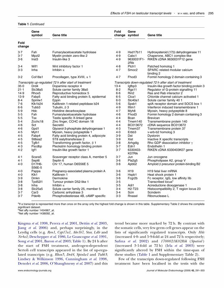
Table 1 Continued
Genesymbol Gene title
Foldchange
Genesymbol Gene title
Foldchange
3.7 Fah Fumarylacetoacetate hydrolase 4.9 Hsd17b11 Hydroxysteroid (17b) dehydrogenase 113.7 Mpzl2 Myelin protein zero-like 2 4.9 Cabc1 Chaperone, ABC1 complex-like3.6 Insl3 Insulin-like 3 4.9 9630031F1-
2RikRIKEN cDNA 9630031F12 gene
3.4 Wif1 Wnt inhibitory factor 1 4.8 Ptch1 Patched homolog 13.3 Inha Inhibin a 4.7 Smoc2 SPARC related modular calcium
binding 23.2 Col18a1 Procollagen, type XVIII, a 1 4.7 Fhod3 Formin homology 2 domain containing 3
Transcripts up-regulated 72 h after start of treatment Transcripts down-regulated 72 h after start of treatment35.0 Drd4 Dopamine receptor 4 13.4 Igfbp3 Insulin-like growth factor binding protein 321.1 Slc38a5 Solute carrier family 38a5 8.2 Rgs11 Regulator of G-protein signalling 1114.9 Rhox5 Reproductive homeobox 5 6.6 Rin2 Ras and Rab interactor 213.9 Fabp5 Fatty acid binding protein 5, epidermal 6.5 Clca1 Chloride channel calcium activated 18.4 Spinlw1 Eppin 6.0 Slc40a1 Solute carrier family 40 17.6 Klk1b24 Kallikrein 1-related peptidase b24 5.6 Spsb1 splA receptor domain and SOCS box 16.6 Tubb3 Tubulin, b 3 4.9 Ifitm1 Interferon induced transmembrane 15.9 Hdc Histidine decarboxylase 4.7 Myh8 Myosin, heavy polypeptide 85.5 Fah Fumarylacetoacetate hydrolase 4.6 Fhod3 Formin homology 2 domain containing 35.5 Tsx Testis specific X-linked gene 4.4 Bcan Brevican5.4 Zcchc18 Zinc finger, CCHC domain 18 4.4 Tmem140 Transmembrane protein 1404.6 Sct Secretin 4.4 BC013672 cDNA sequence BC0136724.6 Gpd1 Glycerol-3-phosphate dehydrogenase 1 4.3 Tmem37 Transmembrane protein 374.5 Myh1 Myosin, heavy polypeptide 1 4.0 Erbb3 v-erb-b2 homolog 34.5 Fabp4 Fatty acid binding protein 4, adipocyte 3.9 Dst Dystonin4.5 St8sia2 ST8 sialyltransferase 2 3.9 Xist Inactive X specific transcripts4.5 Tgfb1 Transforming growth factor, b 1 3.8 Arhgdig Rho GDP dissociation inhibitor g4.3 Pscdbp Pleckstrin homology binding protein 3.7 Edn1 Endothelin 14.2 Igf1 Insulin-like growth factor 1 3.7 6330403-
K07RikRIKEN cDNA 6330403K07 gene
4.1 Scara5 Scavenger receptor class A, member 5 3.7 Jun Jun oncogene4.1 Sept6 Septin 6 3.6 Pla2g5 Phospholipase A2, group V4.0 D17H6-
S56E-5Chr 17, human D6S56E 5 3.6 Apbb2 Amyloid b precursor protein-binding B2
4.0 Pappa Pregnancy-associated plasma protein A 3.6 H19 H19 fetal liver mRNA4.0 Klk1 Kallikrein 1 3.6 Hspb1 Heat shock protein 13.9 Dmkn Dermokine 3.6 Fcgr2b Fc receptor, IgG, low affinity IIb3.8 Tpd52l1 Tumour protein D52-like 1 3.5 Transcribed locusc
3.8 Inha Inhibin a 3.5 Adi1 Acireductone dioxygenase 13.8 Slc25a5 Solute carrier family 25, member 5 3.4 H2-T23 Histocompatibility 2, T region locus 233.7 Car3 Carbonic anhydrase 3 3.4 Scin Scinderin3.7 Pde4b Phosphodiesterase 4B, cAMP specific 3.3 Rnasel Ribonuclease L
aIf a transcript is represented more than once on the array only the highest fold change is shown in this table. Supplementary Table 2 shows the completesignificant dataset.bNet affy number 1454967_at.cNet affy number 1436092_at.
Effects of FSH on testicular transcript levels . M H ABEL and others 295
Kingma et al. 1998, Perera et al. 2001, Dettin et al. 2003,Jiang et al. 2006) and, perhaps surprisingly, in theLeydig cells (e.g. Ren1, Cyp17a1, Akr1b7, Star, Ldlr andNr4a1; Deschepper et al. 1986, Le Goascogne et al. 1991,Song et al. 2001, Baron et al. 2003; Table 1). By 24 h afterthe start of FSH treatment, androgen-dependentSertoli cell transcripts appeared in the list of up-regu-lated transcripts (e.g. Rhox5, Drd4, Spinlw1 and Tubb3;Lindsey & Wilkinson 1996, Cunningham et al. 1998,Denolet et al. 2006, O’Shaughnessy et al. 2007) and this
www.endocrinology-journals.org
trend became more marked by 72 h. By contrast withthe somatic cells, very few germ cell genes appear on thelists of significantly regulated transcripts. Only Hdc(increased 4.0- and 5.9-fold at 24 and 72 h respectively;Safina et al. 2002) and 1700021K02Rik (Spatial )(increased 3.1-fold at 72 h) (Irla et al. 2003) weresignificantly altered by FSH within the time-span ofthese studies (Table 1 and Supplementary Table 2).
Few of the transcripts down-regulated following FSHtreatment have been localised in the testis with the
Journal of Molecular Endocrinology (2009) 42, 291–303

M H ABEL and others . Effects of FSH on testicular transcript levels296
exception of Igfbp3 which has been shown to be of Sertolicell origin (Smith et al. 1990). In order to identifymore ofthe differentially expressed transcripts on the arrays,which may be of a Sertoli cell origin, up- and down-regulated transcripts were compared with those ident-ified as being of likely Sertoli cell origin by Chalmel et al.(2007) using a cell isolation, GeneChip and clusteringapproach (Supplementary Table 2A and B). A degree ofcaution is required as some known Sertoli cell transcripts(e.g. Rhox5) are missing from the list generated byChalmel et al. (2007) probably due to Chip sensitivity orthe subsequent filtering process. Nevertheless, of thetranscripts up-regulated at 12 h, 44%matched to the datafrom Chalmel et al. (2007). Interestingly, this numberdeclined to 35% at 24 h and 27% at 72 h after the start ofFSH treatment (Supplementary Table 2A). The numberof down-regulated transcripts that matched the Sertolicell list was 27, 21 and 28% at 12, 24 and 72 h respectively(Supplementary Table 2B).
Real-time PCR
Leydig cell genes
To confirm results from the array studies, real-time PCRwas used to measure the effect of FSH treatment ontesticular expression of selected transcripts which areknown to be expressed exclusively in the Leydig cells(O’Shaughnessy et al. 2002). Eight mRNA species weretested which had shown an increase in transcript levelson the arrays after FSH (Star, Cyp17a1, Hsd17b3, Akr1b7,Lhr, Cyp11a1, Insl3 and Ren1) (Fig. 2A). Results from thereal-time PCR studies confirmed that seven of thesetranscripts are regulated by FSH in the hpg testis,although no change in Lhr was seen. Two other Leydigcell mRNA species (Hsd3b6 and Sult1e1) that had notshown any response to FSH on the arrays were alsotested by real-time PCR (Fig. 2A). Levels of Hsd3b6 didnot show a response to FSH but there was a significant,if variable, increase in Sult1e1 after 72 h.
Androgen-dependent genes
The array studies showed clearly that a number ofandrogen-dependent Sertoli cell transcripts werealtered after FSH treatment. Real-time PCR was usedto confirm changes in selected transcripts (Rhox5, Tsx,Drd4, Spinlw1 (Eppin) and Igfbp3) shown previously tobe androgen-regulated (Lindsey & Wilkinson 1996,Denolet et al. 2006, O’Shaughnessy et al. 2007; Fig. 2B).In agreement with results from the array studies fourtranscripts (Rhox5, Tsx, Drd4 and Spinlw1) showedincreased expression 24–72 h after FSH treatmentwhile one transcript (Igfbp3) showed a significantdecrease in expression (Fig. 2B).
Journal of Molecular Endocrinology (2009) 42, 291–303
Sertoli cell genes
In addition to androgen-dependent Sertoli cell genesdescribed above, 16 other Sertoli cell transcripts weremeasured by real-time PCR following FSH treatment ofadult hpg mice (Fig. 3). Of these transcripts, seven hadshown significantly increased expression on the arrays(Tesc,Lgals1,Aqp5,Dhh,Pappa,Wnt4and Shbg), sixhadnotshown any significant change (Trf,Wt1, Amh, Spata2, Tjp1andGdnf), twohad shown a significant decrease (Fshr andRgs11) after FSH treatment and one transcript (Defb19)was not on the array. Results from real-time studiesconfirmed increased transcript levels for six out of theseven mRNA species identified on the array (theexception was Shbg) and for the one transcript (Defb19)not on the array (Fig. 3A). Both transcripts decreased onthe arrays after FSH treatment also showed a significantdecrease by real-time PCR (Fig. 3A). Interestingly,however, the real-time PCR data showed there was asignificant increase in levels of three out of the sixtranscripts that were not significantly changed on thearrays (Trf,Wt1 andAmh; Fig. 3A).Twoof these transcripts(Trf and Amh) had shown a greater than twofold increaseon the arrays but had not reached significance.Differences between results from real-time PCR andarrays may be a matter of sensitivity and variability of thetwo techniques or may be due to the choice of primers,away from the 30 region targeted by these arrays.
The inhibin subunits are expressed in a number ofcell types in the testis (Barakat et al. 2008) although itmight be expected that initial responsiveness to FSHwould be predominantly localised in the Sertoli cells.On the arrays, both Inha and Inhbb were significantlyincreased by FSH (Table 1 and Supplementary Table 2)while there was no effect on Inhba. Results from the real-time PCR studies reflected the same pattern of results(Fig. 3B).
Germ cell genes
The number of known germ cell transcripts on thearray affected by FSH, within the time-span of the study,was not high. This was, therefore, investigated furtherusing real-time PCR. Expression of nine germ celltranscripts known to be expressed predominantly inspermatogonia (Stra8, Pou5f1, Dkkl1 and Spo11),spermatocytes (Mybl1, Zfp541) or spermatids(1700021K02Rik, Hdc and Tp1) was measured followingFSH treatment (Fig. 4). Expression levels of Stra8,Spo11, Mybl1 and Tp1 were unaffected by treatment butthere was a transient increase in Pou5f1 at 12 h whileDkkl1 and Zfp541 were significantly increased at 72 h.Expression of Hdc and 1700021K02Rik (shown pre-viously to be increased on the array) was increased at alltimes after treatment.
www.endocrinology-journals.org

Figure 2 Real-time PCR measurements of mRNA transcript levels in testes from adult hpgmice treated for 0(control), 12, 24 or 72 h with FSH. Data show results from Leydig cell-specific transcripts (A) and from Sertolicell-specific, androgen-dependent transcripts (B). The meanGS.E.M. of three or four animals per group isshown. Groups with different letter superscripts are significantly different.
Effects of FSH on testicular transcript levels . M H ABEL and others 297
Other transcripts
Results from the array studies identified a number oftranscripts regulated by FSH but without knownfunction and/or known expression pattern in the testis.Levels of three of these transcripts (Wif1, Dmkn, Dkk3)weremeasured in hpg testes after FSH treatment (Fig. 5).In all cases, FSH caused a significant increase intranscript levels confirming the results of the array study.
Canonical pathway analysis
Analysis of canonical pathways showed that com-ponents of the cholesterol biosynthetic pathway weresignificantly increased at 12 h but not at other times
www.endocrinology-journals.org
(Supplementary Table 3A, see supplementary data inthe online version of the Journal of MolecularEndocrinology at http://jme.endocrinology-journals.org/content/vol42/issue4/) and that there was ageneral decline in transcripts encoding factors involvedin formation and regulation of tight junctions (Supple-mentary Table 3B).
Discussion
FSH is essential for optimum fertility in the adult malebut uncertainty remains about how it acts to regulateSertoli cell activity and spermatogenesis. The hpgmouse
Journal of Molecular Endocrinology (2009) 42, 291–303

Figure 3 Real-time PCR measurements of Sertoli cell-specific mRNA transcript levels in testes from adulthpg mice treated for 0 (control), 12, 24 or 72 h with FSH. Results show the meanGS.E.M. of three or fouranimals per group. Groups with different letter superscripts are significantly different; where no superscriptsare shown there was no difference between groups.
M H ABEL and others . Effects of FSH on testicular transcript levels298
is an excellent model system with which to test theeffects of FSH since the Sertoli cells have not beenexposed to the hormone but express FSHR and aresensitive to FSH action. This study is an extension ofearlier work by Sadate-Ngatchou et al. (2004) using alonger treatment period, different array chips with alarger characterised gene set (MOE430A chips (14 000characterised genes) versus MG U74Av2 chips (6000characterised genes, 6500 ESTs)), a significantly largeranimal cohort and recombinant FSH. In addition, thepurpose of this study was to follow changes in testiculartranscript levels in the hpg in response to maintainedlevels of FSH rather than the acute response to a singleadministration. Together, the two studies complement
Journal of Molecular Endocrinology (2009) 42, 291–303
each other and serve to identify transcripts regulated byFSH over the short and medium term. Interestingly, at12 h after the start of FSH administration, when bothstudies can be directly compared with a degree ofcaution, there were only 44 differentially transcriptscommon to both studies, 25 up-regulated (e.g. Cyp17,Ren1, Fos, Hdc, Col4a1) and 19 down-regulated (e.g.Rgs11, Cdo1, Erbb3, Ptk2b, Vnn1). This low number oftranscripts in common may be due to a combinationof the chips used, the age of the animals, the number ofanimals used and the treatment regime.
In this study, the total number of transcripts alteredat each time point did not vary markedly across thetreatment period but only 39 transcripts were
www.endocrinology-journals.org

Figure 4 Real-time PCR measurements of germ cell-specific mRNA transcript levels in testes from adulthpg mice treated for 0 (control), 12, 24 or 72 h with FSH. Results show the meanGS.E.M. of three or fouranimals per group. Groups with different letter superscripts are significantly different; where no superscriptsare shown there was no difference between groups.
Effects of FSH on testicular transcript levels . M H ABEL and others 299
up-regulated more than twofold at all times indicatingthat there was a changing pattern of expression asthe exposure to FSH was maintained. There were also63 transcripts down-regulated more than twofold at alltimes suggesting that the inhibitory effects of FSH aremore consistent. Results from the arrays and from real-time PCR showed that FSH treatment caused a generalincrease in many transcripts encoding known Sertolicell-specific products such as Tesc, Lgals1, Fabp5 andAqp5 (Kingma et al. 1998, Perera et al. 2001, Dettin et al.2003) although some transcripts (e.g. Shbg, Tjp1 (Wanget al. 1989, Byers et al. 1991)) were unaffected whileothers were decreased (see below) indicating that theeffect of FSH was not simply to increase the overallactivity of the cells. Comparison of the up-regulated
Figure 5 Real-time PCR measurements of mRNA traorigin. Levels were measured in testes from adult hpg mResults show the meanGS.E.M. of three or four animalsare significantly different.
www.endocrinology-journals.org
transcripts in this study with the list of Sertoli celltranscripts generated by Chalmel et al. (2007) confirmsthat, at least initially, a high proportion of the affectedtranscripts are likely to be of Sertoli cell origin. Thedeclining proportion of Sertoli cell transcripts at latertimes is likely to be due to increasing activity in othercells such as the Leydig cell. The lower proportion ofdown-regulated transcripts that match to the Sertoli celllist of Chalmel et al. (2007) may reflect the GeneChipsensitivity involved in generating that list since many ofthese down-regulated transcripts might be expected tohave a low level of expression in the normal animal.
Among the transcripts that showed decreased levelsin response to FSH were a number encoding tightjunction components. This is consistent with a recent
nscript levels of species with unknown testicularice treated for 0 (control), 12, 24 or 72 h with FSH.per group. Groups with different letter superscripts
Journal of Molecular Endocrinology (2009) 42, 291–303

M H ABEL and others . Effects of FSH on testicular transcript levels300
study which reported that gonadotrophins reducetranscript levels of Sertoli cell barrier components butthat FSH may act at the level of protein organisationto induce barrier functionality (Tarulli et al. 2008).Other transcripts that showed a significant decreasein levels after FSH treatment included Fshr and Rgs11.It is well established that FSH will cause down-regulation of its receptor by decreasing transcript levels(O’Shaughnessy 1980, Themmen et al. 1991) and areduction in Fshr is to be expected. RGS11, in contrast,belongs to the regulator of G protein-signalling familywhich are GTPase-activating proteins that act to inhibitsignal transduction and thus play a role in desensitisa-tion (Chasse & Dohlman 2003). The role of RGSs innormal hormonal signalling is not well established butthe declining levels of Rgs11 after FSH treatment mayact to enhance signal transduction despite a reductionin receptor levels.
In addition to changes in Sertoli cell transcriptsinduced by FSH, it was clear from the rise in testicularandrogen and the array and real-time PCR data thatFSH was also acting to induce Leydig cell function. Thiseffect was marked and rapid with a Leydig cell transcript(Ren1) showing the greatest fold change at 12 h(Deschepper et al. 1986). Results from the arrays andreal-time PCR show that all components of theandrogen biosynthetic pathway were induced at 12 hapart from Hsd3b6. Since Hsd3b1 is already highlyexpressed in the adult hpg testis (Baker et al. 2003) lackof HSD3B6 is unlikely to affect the steroidogenicpotential of the cells. In addition to the steroidogenicenzymes, pathway analysis showed that most com-ponents of the cholesterol biosynthetic pathway wereinduced 12 h after FSH treatment while Ldlr levels areincreased. This shows that FSH is acting to increase thecapacity of the Leydig cells to produce and sequestercholesterol and to convert cholesterol to androgen.Interestingly, one of the critical components of thecholesterol biosynthetic pathways (Mvk) also acts toinhibit Lhr translation (Nair & Menon 2004) and thismay serve to regulate further Leydig cell sensitivity toLH. The hpg mouse testis is likely to contain both adultand fetal-type Leydig cells (Baker et al. 2003) andHsd3b6 is a marker of adult Leydig cell differentiation(Baker et al. 1999). This might imply that FSH is actingto induce activity in the fetal Leydig cell population butSult1e1 is a marker of adult Leydig cells (Song et al.1997) and is increased 72 h after FSH suggesting thatthe effects of FSH are probably being mediated throughthe adult Leydig cells.
In the testis, receptors for FSH are only found in theSertoli cells (Heckert & Griswold 2002) and the effectsof FSH must be mediated by a factor or factors releasedby the Sertoli cells which act on the Leydig cells. In theshort-term, the effects of FSH on Leydig cell function inthe hpg appear to be more marked than effects of hCG
Journal of Molecular Endocrinology (2009) 42, 291–303
(Baker et al. 2003) and the effects are also very rapidsince Sadate-Ngatchou et al. (2004) saw a markedincrease in Cyp17a1 after only 4 h of FSH treatment.FSH appears, therefore, to be able to induce a powerfuland rapid response in Leydig cells presumably throughstimulation of release of potent trophic factors by theSertoli cells. The presence of such factors has beenpostulated for a number of years since early studies onperfused testes or hypophysectomised animals treatedwith FSH (Johnson & Ewing 1971, Chen et al. 1976,Vihko et al. 1991). One report has suggested that theactive factors are TIMP1 and Procathepsin L (Boujradet al. 1995) but this has not been confirmed and we sawno evidence of changes in these factors in our study.Following FSH treatment, our array data showed thatthere was an increase in Igf1 levels and IGF1 has beensuggested to play a role in Leydig cell differentiation(Morera et al. 1987). Interestingly, there was a markeddecline in Igfbp3 and an increase in Pappa levels afterFSH treatment. Increased Pappa would be expectedto increase the bioavailability and activity of IGF1(Conover et al. 2004) although the effect of alteredIgfbp3 may be more complex (Modric et al. 2001). Thetime-course of changes in expression of Igf1 levels doesnot appear to fit well with a role in the stimulation ofLeydig cell function after FSH treatment although it ispossible that early changes in Pappa and Igfbp3may alterearly IGF1 bioavailability. Other secreted moleculesshowing a marked increase in transcript levels after FSHinclude Wif1, Dkk3 and Dmkn. Both WIF1 and DKK3 actto regulate WNT signalling and the WNT/CTNNB1pathway is critical for normal Sertoli cell development(Boyer et al. 2008) although its function in the Leydigcell remains uncertain.
Following the increase in Leydig cell activity afterFSH treatment, there was a significant change in thelevels of known androgen-dependent Sertoli cell-specific transcripts. It is possible that changes in thesetranscripts are due to direct effects of FSH treatmentbut the known androgen-dependence of the transcriptsmakes it more likely that changes are related toincreased Leydig cell androgen production inducedby FSH. The rise in intratesticular androgen after FSHtreatment was significant but levels remained very low,probably because FSH stimulates synthesis of thecomponents of the steroidogenic pathway withoutbeing able to stimulate the pathway itself. The apparenteffect of these low levels of androgen on Sertoli celltranscript levels suggests that the Sertoli cells areextremely sensitive to androgen stimulation.
Treatment of hpg mice with FSH increased vacuo-lation in the Sertoli cells and induced formation of alumen within the seminiferous tubules but had littleapparent effect on germ cell morphology or pro-gression up to 72 h. Changes in the Sertoli cell andtubule diameter correlate with a marked rise in Aqp5 at
www.endocrinology-journals.org

Effects of FSH on testicular transcript levels . M H ABEL and others 301
12 h suggesting that increased water movement acrossthe Sertoli cell membrane may contribute to increasedtubular diameter and testis weight. The absence ofspermatogenic progression over the time-coursestudied is likely to reflect the inactive state of theSertoli cell in the adult hpg testis and the time requiredfor the Sertoli cell to become active enough to supportgerm cell maturation. Real-time PCR studies of a smallnumber of known germ cell genes showed that therewas a variable response of germ cell transcripts to FSHstimulation. POU5F1 has been shown to be necessaryfor primordial germ cell survival (Kehler et al. 2004)and the increase in response to FSH, albeit small, mayfacilitate an increase in spermatogonial number withinthe testis. DKKL1 and ZFP541 are expressed in bothspermatocytes and spermatids (Kohn et al. 2005, Choiet al. 2008) while HDC and SPATIAL are associatedwith round spermatids and the later stages of sperma-togenesis (Safina et al. 2002, Irla et al. 2003). Lack of ageneral increase in germ cell transcripts (data fromboth arrays and real-time PCR) would indicate that theeffects seen are not due to an overall increase in germcell number but are more likely to be part of an earlyspecific response to FSH stimulation. It has beenshown previously that more prolonged treatment ofhpg mice with FSH will stimulate an increase in germcell number and development (O’Shaughnessy et al.1992, Singh & Handelsman 1996b, Baines et al. 2008)but the stimulatory effect of FSH on the Leydig cellsmakes interpretation of the FSH effects on the germcells difficult because of the known stimulatory effectof testosterone on germ cell development in the hpgmouse (O’Shaughnessy & Sheffield 1990, Singh et al.1995).
In this study, FSH treatment of hpg mice for up to72 h induced significant changes in Sertoli celltranscript levels and led to indirect stimulation ofLeydig cell function. The changes in Leydig cell activityprobably induced further changes in androgen-depen-dent Sertoli cell transcripts. While FSH is known to berequired for optimal germ cell development (Abel et al.2000, 2008), treatment of hpgmice for 72 h did not havea marked effect on germ cell differentiation suggestingthat longer-term action of FSH is required to inducegerm cell proliferation and progression.
Declaration of interest
The authors declare that there is no conflict of interest that could beperceived as prejudicing the impartiality of the research reported.
Funding
This study was supported by the Wellcome Trust (grant number078137).
www.endocrinology-journals.org
Acknowledgements
We would like to thank Ana Monteiro for technical assistance, MohanMasih for histology and animal care staff for assistance with thisproject. Sheena Lee is supported by the Wellcome Trust fundedOXION initiative.
References
Abel MH, Wootton AN, Wilkins V, Huhtaniemi I, Knight PG &Charlton HM 2000 The effect of a null mutation in the follicle-stimulating hormone receptor gene on mouse reproduction.Endocrinology 141 1795–1803.
Abel MH, Baker PJ, Charlton HM, Monteiro A, Verhoeven G, DeGendt K, Guillou F & O’Shaughnessy PJ 2008 Spermatogenesisand Sertoli cell activity in mice lacking Sertoli cell receptors forfollicle stimulating hormone and androgen. Endocrinology 1493279–3285.
Baban D & Davies KE 2008 Microarray analysis of mdx mice expressinghigh levels of utrophin: therapeutic implications for dystrophindeficiency. Neuromuscular Disorders 18 239–247.
Baines H, Nwagwu MO, Hastie GR, Wiles RA, Mayhew TM & EblingFJ 2008 Effects of estradiol and FSH on maturation of the testisin the hypogonadal (hpg) mouse. Reproductive Biology andEndocrinology 6 4.
Baker PJ, Sha JA, McBride MW, Peng L, Payne AH&O’Shaughnessy PJ1999 Expression of 3beta-hydroxysteroid dehydrogenase type I andVI isoforms in the mouse testis during development. EuropeanJournal of Biochemistry 260 911–916.
Baker PJ, Johnston H, Abel MH, Charlton HM & O’Shaughnessy PJ2003 Differentiation of adult-type Leydig cells occurs in gonado-trophin-deficient mice. Reproductive Biology and Endocrinology 1 4.
Barakat B, O’Connor A, Gold E, de Kretser D & Loveland K 2008Inhibin, activin, follistatin and follicle stimulating hormone serumlevels and testicular production are highly modulated during thefirst spermatogenic wave in mice. Reproduction 136 345–359.
Baron S, Manin M, Aigueperse C, Berger M, Jean C, Veyssiere G &Morel L 2003 Hormonal and developmental regulation of themouse aldose reductase-like gene akr1b7 expression in Leydig cells.Journal of Molecular Endocrinology 31 71–81.
Boujrad N, Ogwuegbu SO, Garnier M, Lee CH, Martin BM &Papadopoulos V 1995 Identification of a stimulator of steroid-hormone synthesis isolated from testis. Science 268 1609–1612.
Boyer A, Hermo L, Paquet M, Robaire B & Boerboom D 2008Seminiferous tubule degeneration and infertility in mice withsustained activation of WNT/CTNNB1 signaling in Sertoli cells.Biology of Reproduction 79 475–485.
Byers S, Graham R, Dai HN & Hoxter B 1991 Development of Sertolicell junctional specializations and the distribution of the tight-junction-associated protein ZO-1 in the mouse testis. AmericanJournal of Anatomy 191 35–47.
Cattanach BM, Iddon CA, Charlton HM, Chiappa SA & Fink G 1977Gonadtrophin releasing hormone deficiency in a mutant mousewith hypogonadism. Nature 269 338–340.
Chalmel F, Rolland AD, Niederhauser-Wiederkehr C, Chung SS,Demougin P, Gattiker A, Moore J, Patard JJ, Wolgemuth DJ, Jegou Bet al. 2007 The conserved transcriptome in human and rodent malegametogenesis. PNAS 104 8346–8351.
Charlton HM, Halpin DMG, Iddon CA, Rosie R, Levy G, McDowellIFW, Megson A, Morris JF, Bramwell A, Speight A et al. 1983 Theeffects of daily administration of single and multiple injections ofgonadotrophin-releasing hormone on pituitary and gonadalfunction in the hypogonadal (hpg) mouse. Endocrinology 113535–544.
Journal of Molecular Endocrinology (2009) 42, 291–303

M H ABEL and others . Effects of FSH on testicular transcript levels302
Chasse SA & Dohlman HG 2003 RGS proteins: G protein-coupledreceptors meet their match. Assay and Drug Development Technologies 1357–364.
Chen JK & Heckert LL 2001 Dmrt1 expression is regulated by follicle-stimulating hormone and phorbol esters in postnatal Sertoli cells.Endocrinology 142 1167–1178.
Chen YI, Payne AH & Kelch RP 1976 FSH stimulation of Leydig cellfunction in the hypophysectomized immature rat. Proceedings of theSociety for Experimental Biology and Medicine 153 473–475.
Choi E, Han C, Park I, Lee B, Jin S, Choi H, Kim DH, Park ZY, Eddy EM& Cho C 2008 A novel germ cell-specific protein, SHIP1, forms acomplex with chromatin remodeling activity during spermatogen-esis. Journal of Biological Chemistry 283 35283–35294.
Conover CA, Bale LK, Overgaard MT, Johnstone EW, Laursen UH,Fuchtbauer EM, Oxvig C & van Deursen J 2004 Metalloproteinasepregnancy-associated plasma protein A is a critical growthregulatory factor during fetal development. Development 1311187–1194.
Cunningham DB, Segretain D, Arnaud D, Rogner UC & Avner P 1998The mouse Tsx gene is expressed in Sertoli cells of the adult testisand transiently in premeiotic germ cells during puberty. Develop-mental Biology 204 345–360.
Czechowski T, Bari RP, Stitt M, Scheible WR & Udvardi MK 2004 Real-time RT-PCR profiling of over 1400 Arabidopsis transcriptionfactors: unprecedented sensitivity reveals novel root- and shoot-specific genes. Plant Journal 38 366–379.
Denolet E, De Gendt K, Allemeersch J, Engelen K, Marchal K, VanHummelen P, Tan KA, Sharpe RM, Saunders PT, Swinnen JV et al.2006 The effect of a Sertoli cell-selective knockout of the androgenreceptor on testicular gene expression in prepubertal mice.Molecular Endocrinology 20 321–334.
Deschepper CF, Mellon SH, Cumin F, Baxter JD & Ganong WF 1986Analysis by immunocytochemistry and in situ hybridization of reninand its mRNA in kidney, testis, adrenal, and pituitary of the rat.PNAS 83 7552–7556.
Dettin L, Rubinstein N, Aoki A, Rabinovich GA &Maldonado CA 2003Regulated expression and ultrastructural localization of galectin-1,a proapoptotic beta-galactoside-binding lectin, during spermato-genesis in rat testis. Biology of Reproduction 68 51–59.
Dierich A, SairamMR, Monaco L, Fimia GM, Gansmuller A, LeMeur M& Sassone-Corsi P 1998 Impairing follicle-stimulating hormone(FSH) signaling in vivo: targeted disruption of the FSH receptorleads to aberrant gametogenesis and hormonal imbalance. PNAS 9513612–13617.
De Gendt K, Swinnen JV, Saunders PT, Schoonjans L, Dewerchin M,Devos A, Tan K, Atanassova N, Claessens F, Lecureuil C et al. 2004 ASertoli cell-selective knockout of the androgen receptor causesspermatogenic arrest in meiosis. PNAS 101 1327–1332.
Le Goascogne C, Sananes N, Gouezou M, Takemori S, Kominami S,Baulieu EE & Robel P 1991 Immunoreactive cytochrome P450(17alpha) in rat and guinea-pig gonads, adrenal-glands and brain.Journal of Reproduction and Fertility 93 609–622.
HaywoodM, Spaliviero J, Jimemez M, King NJ, Handelsman DJ & AllanCM 2003 Sertoli and germ cell development in hypogonadal (hpg)mice expressing transgenic follicle-stimulating hormone alone or incombination with testosterone. Endocrinology 144 509–517.
Heckert LL & Griswold MD 2002 The expression of the follicle-stimulating hormone receptor in spermatogenesis. Recent Progress inHormone Research 57 129–148.
Hirst RC, Abel MH, Wilkins V, Simpson C, Knight PG, Zhang FP,Huhtaniemi I, Kumar TR & Charlton HM 2004 Influence ofmutations affecting gonadotropin production or responsiveness onexpression of inhibin subunit mRNA and protein in the mouseovary. Reproduction 128 43–52.
Irla M, Puthier D, Le Goffic R, Victorero G, Freeman T, Naquet P,Samson M & Nguyen C 2003 Spatial, a new nuclear factor tightlyregulated during mouse spermatogenesis. Gene Expression Patterns 3135–138.
Journal of Molecular Endocrinology (2009) 42, 291–303
Jiang Y, Oliver P, Davies KE & Platt N 2006 Identification andcharacterization of murine SCARA5, a novel class A scavengerreceptor that is expressed by populations of epithelial cells.Journal of Biological Chemistry 281 11834–11845.
Johnson BH & Ewing LL 1971 Follicle-stimulating hormone and theregulation of testosterone secretion in rabbit testes. Science 173635–637.
Kehler J, Tolkunova E, Koschorz B, Pesce M, Gentile L, Boiani M,Lomeli H, Nagy A, McLaughlin KJ, Scholer HR et al. 2004 Oct4 isrequired for primordial germ cell survival. EMBO Reports 51078–1083.
Kingma PB, Bok D & Ong DE 1998 Bovine epidermal fattyacid-binding protein: determination of ligand specificity andcellular localization in retina and testis. Biochemistry 373250–3257.
Kohn MJ, Kaneko KJ & DePamphilis ML 2005 DkkL1 (Soggy), aDickkopf family member, localizes to the acrosome duringmammalian spermatogenesis. Molecular Reproduction and Develop-ment 71 516–522.
Krishnamurthy H, Babu PS, Morales CR & Sairam MR 2001 Delay insexual maturity of the follicle-stimulating hormone receptorknockout male mouse. Biology of Reproduction 65 522–531.
Kumar TR, Wang Y, Lu N & Matzuk MM 1997 Follicle stimulatinghormone is required for ovarian follicle maturation but not malefertility. Nature Genetics 15 201–204.
Lang J 1995 Assay for deletion in GnRH (hpg) locus using PCR.MouseGenome 89 857.
Lindsey JS & Wilkinson MF 1996 Pem: a testosterone-regulated andLH-regulated homeobox gene expressed in mouse sertoli cells andepididymis. Developmental Biology 179 471–484.
Mason AJ, Hayflick JS, Zoeller RT, Young WS, Phillips HS, Nikolics K &Seeburg TA 1986 A deletion truncating the GnRH gene isresponsible for hypogonadism in the hpg mouse. Science 2341366–1371.
McLachlan RI, O’Donnell L, Meachem SJ, Stanton PG, de Kretser DM,Pratis K & Robertson DM 2002 Identification of specific sites ofhormonal regulation in spermatogenesis in rats, monkeys, andman. Recent Progress in Hormone Research 57 149–179.
Modric T, Silha JV, Shi Z, Gui Y, Suwanichkul A, Durham SK, Powell DR& Murphy LJ 2001 Phenotypic manifestations of insulin-like growthfactor-binding protein-3 overexpression in transgenic mice. Endo-crinology 142 1958–1967.
Monaco L, Foulkes NS & Sassone-Corsi P 1995 Pituitary follicle-stimulating hormone (FSH) induces CREM gene expression inSertoli cells: involvement in long-term desensitization of the FSHreceptor. PNAS 92 10673–10677.
Morera AM, Chauvin MA, De Peretti E, Binoux M & Benahmed M1987 Somatomedin C/insulin-like growth factor 1: an intratestic-ular differentiative factor of Leydig cells? Hormone Research 2850–57.
Morris PL, Vale WW, Cappel S & Bardin CW 1988 Inhibin productionby primary Sertoli cell-enriched cultures: regulation by follicle-stimulating hormone, androgens, and epidermal growth factor.Endocrinology 122 717–725.
Myers M, Ebling FJ, NwagwuM, Boulton R, Wadhwa K, Stewart J & KerrJB 2005 Atypical development of Sertoli cells and impairment ofspermatogenesis in the hypogonadal (hpg) mouse. Journal ofAnatomy 207 797–811.
Nair AK & Menon KMJ 2004 Isolation and characterization of a noveltrans-factor for luteinizing hormone receptor mRNA from ovary.Journal of Biological Chemistry 279 14937–14944.
O’Shaughnessy PJ 1980 FSH receptor autoregulation and cyclic AMPproduction in the immature rat testis. Biology of Reproduction 23810–814.
O’Shaughnessy PJ & Sheffield JW 1990 Effect of testosterone ontesticular steroidogenesis in the hypogonadal (hpg) mouse.Journal of Steroid Biochemistry 35 729–734.
www.endocrinology-journals.org

Effects of FSH on testicular transcript levels . M H ABEL and others 303
O’Shaughnessy PJ, Bennett MK, Scott IS & Charlton HM 1992 Effectsof FSH on Leydig cell morphology and function in the hypogonadalmouse. Journal of Endocrinology 135 517–525.
O’Shaughnessy PJ, Willerton L & Baker PJ 2002 Changes in Leydig cellgene expression during development in the mouse. Biology ofReproduction 66 966–975.
O’Shaughnessy PJ, Abel M, Charlton HM, Hu B, Johnston H & BakerPJ 2007 Altered expression of genes involved in regulation ofvitamin A metabolism, solute transportation, and cytoskeletalfunction in the androgen-insensitive tfm mouse testis. Endocrinology148 2914–2924.
Perera EM, Martin H, Seeherunvong T, Kos L, Hughes IA, Hawkins JR& Berkovitz GD 2001 Tescalcin, a novel gene encoding a putativeEF-hand Ca(2C)-binding protein, Col9a3, and renin are expressedin the mouse testis during the early stages of gonadal differen-tiation. Endocrinology 142 455–463.
Sadate-Ngatchou PI, Pouchnik DJ & Griswold MD 2004 Follicle-stimulating hormone induced changes in gene expression ofmurine testis. Molecular Endocrinology 18 2805–2816.
Safina F, Tanaka S, Inagaki M, Tsuboi K, Sugimoto Y & Ichikawa A 2002Expression of L-histidine decarboxylase in mouse male germ cells.Journal of Biological Chemistry 277 14211–14215.
Singh J & Handelsman DJ 1996a Neonatal administration ofFSH increases Sertoli cell numbers and spermatogenesis ingonadotropin-deficient (hpg) mice. Journal of Endocrinology 15137–48.
Singh J & Handelsman DJ 1996b The effects of recombinant FSH ontestosterone-induced spermatogenesis in gonadotropin-deficient(Hpg) mice. Journal of Andrology 17 382–393.
Singh J, O’Neill C & Handelsman DJ 1995 Induction of spermato-genesis by androgens in gonadotropin-deficient (Hpg) mice.Endocrinology 136 5311–5321.
Skinner MK, Schlitz SM & Anthony CT 1989 Regulation of Sertoli celldifferentiated function: testicular transferrin and androgen-bind-ing protein expression. Endocrinology 124 3015–3024.
Smith EP, Dickson BA & Chernausek SD 1990 Insulin-like growthfactor binding protein-3 secretion from cultured rat sertoli cells:
www.endocrinology-journals.org
dual regulation by follicle stimulating hormone and insulin-likegrowth factor-I. Endocrinology 127 2744–2751.
Song WC, Qian Y, Sun X & Negishi M 1997 Cellular localization andregulation of expression of testicular estrogen sulfotransferase.Endocrinology 138 5006–5012.
Song KH, Park JI, Lee MO, Soh J, Lee K & Choi HS 2001 LH inducesorphan nuclear receptor Nur77 gene expression in testicularLeydig cells. Endocrinology 142 5116–5123.
Tarulli GA, Meachem SJ, Schlatt S & Stanton PG 2008 Regulation oftesticular tight junctions by gonadotrophins in the adult Djungar-ian hamster in vivo. Reproduction 135 867–877.
Themmen AP, Blok LJ, Post M, Baarends WM, Hoogerbrugge JW,Parmentier M, Vassart G & Grootegoed JA 1991 Follitropin receptordown-regulation involves a cAMP-dependent post-transcriptionaldecrease of receptor mRNA expression. Molecular and CellularEndocrinology 78 R7–R13.
Verhoeven G & Cailleau J 1988 Follicle-stimulating hormone andandrogens increase the concentration of the androgen receptor inSertoli cells. Endocrinology 122 1541–1550.
Vihko KK, Lapolt PS, Nishimori K & Hsueh AJ 1991 Stimulatory effectsof recombinant follicle-stimulating hormone on Leydig cellfunction and spermatogenesis in immature hypophysectomizedrats. Endocrinology 129 1926–1932.
Wang Y-M, Sullivan PM, Petrusz P, Yarbrough W& Joseph DR 1989 Theandrogen-binding protein gene is expressed in CD1 mouse testis.Molecular and Cellular Endocrinology 63 85–92.
Wreford NG, Rajendra Kumar T, Matzuk MM & de Kretser DM 2001Analysis of the testicular phenotype of the follicle-stimulatinghormone beta-subunit knockout and the activin type II receptorknockout mice by stereological analysis. Endocrinology 1422916–2920.
Received in final form 5 January 2009Accepted 9 January 2009Made available online as an Accepted Preprint 9 January 2009
Journal of Molecular Endocrinology (2009) 42, 291–303