Analgesic Effect Of Dioclenol And Dioflorin Isolated From Dioclea Grandiflora
Upload
khangminh22Category
view
1download
0
Anti-ulcer and Analgesic activity of Ethanolic extract of
Annona squamosa Leaves
Dissertation Submitted to
THE TAMIL NADU Dr. M.G.R. MEDICAL UNIVERSITY
Chennai-32
In partial fulfillment for the award of the degree of
MASTER OF PHARMACY
IN
PHARMACOLOGY
Submitted by
Reg.No: 26103093
Under the guidance of
Dr. P. ASHOKKUMAR, M. Pharm Ph. D.,
Professor
DEPARTMENT OF PHARMACOLOGY
J.K.K.NATTRAJA COLLEGE OF PHARMACY
KOMARAPALAYAM-638 183
TAMIL NADU
MAY-2012
EVALUATION CERTIFICATE
This is to certify that the dissertation work entitled “Anti-ulcer and
Analgesic activity of Ethanolic extract of Annona squamosa leaves” submitted
by the student bearing [Reg.No. 26103093] to “The Tamil Nadu Dr. M.G.R.
Medical University”, Chennai, in partial fulfillment for the award of degree of
MASTER OF PHARMACY in PHARMACOLOGY was evaluated by us during
the examination held on……………………….
Internal Examiner External Examiner
CERTIFICATE
This is to certify that the work embodied in this dissertation “Anti-ulcer and
Analgesic activity of Ethanolic extract of Annona squamosa leaves”, submitted
to “The Tamil Nadu Dr.M.G.R.Medical University”, Chennai, was carried out by
Mr.BillaVamsi [Reg.No. 26103093], in the Partial fulfillment of award of degree of
MASTER OF PHARMACY in Pharmacology under direct supervision of
Dr. P. ASHOKKUMAR, M. Pharm, Ph. D., Professor, Department of
Pharmacology, J.K.K. Nattraja College of Pharmacy, Komarapalayam, during the
academic year 2011-2012.
PLACE: Komarapalayam Dr. P. PERUMAL, M. Pharm., Ph. D., A.I.C.,
DATE: Principal,
J.K.K.Nattraja College of Pharmacy,
Komarapalayam – 638 183,
Tamil Nadu.
CERTIFICATE
This is to certify that the work embodied in this dissertation “Anti-ulcer
and Analgesic activity of Ethanolic extract of Annona squamosa leaves”,
submitted to“The Tamil Nadu Dr.M.G.R. Medical University”, Chennai, in partial
fulfillment for the award of degree of MASTER OF PHARMACY in
Pharmacology, is a bonafide work carried out by Mr. BILLA VAMSI,
[Reg. No. 26103093] during the academic year 2011-2012, under my guidance and
direct supervision in the Department of Pharmacology, J.K.K.Nattraja College of
Pharmacy, Komarapalayam.
DR. P. ASHOKKUMAR, M. Pharm, Ph. D.,
Professor,
Department of Pharmacology,
J.K.K.Nattraja College of Pharmacy,
Komarapalayam-638 183,
Tamil Nadu.
Mr. V. RAJESH, M. Pharm.,
Head of the Department,
Department of Pharmacology,
J.K.K.Nattraja College of Pharmacy,
Komarapalayam- 638 183,
Tamil Nadu.
DECLARATION
The work presented in this dissertation entitled “Anti-ulcer and Analgesic
activity of Ethanolic extract of Annona squamosa leaves”, was carried out by me,
under the direct supervision of Dr. P. ASHOKKUMAR, M. Pharm,
Ph. D., Professor, Department of Pharmacology, J.K.K. Nattraja College of
Pharmacy, Komarapalayam.
I further declare that, this work is original and has not been submitted in part
or full for the award of any other degree or diploma in any other university.
PLACE:Komarapalayam BILLA VAMSI
DATE: [Reg.No. 26103093]
Department of Pharmacology,
J.K.K. Nattraja College of Pharmacy,
Komarapalayam – 638 183,
Tamil Nadu.
ACKNOWLEDGEMENT
I acknowledge first of all, the almighty, for his goodness and grace, which
have brought me this far successfully. Without his kindness and blessings, I could
not have made this far.
I take this opportunity with pride and enormous gratification to express the
deeply embedded feeling of thanks and gratefulness to all the persons who backed
me directly or indirectly throughout the materialization of this research work.
I am swollen with pride to dedicate my humblest regards and deep sense of
gratitude and heart felt thanks to late Thiru. J.K.K. NATARAJAH CHETTIAR,
founder of our college. I wish to express my sincere thanks to our respectful
correspondent Smt. N. SENDAMARAAI and our Managing Director
Mr. S. OMM SHARRAVANA, B.Com., LLB., and Executive director
Mr. S. OMM SINGARAVEL, B.E., M.S., for enabling us to do the project work.
I take this opportunity in expressing my deep sense of gratitude to my
respectable and beloved guide Dr. P. ASHOKKUMAR, M. Pharm, Ph. D.,
Professor, Department of Pharmacology, J.K.K. Nattraja College of
Pharmacy,whose active guidance, innovative ideas, constant inspiration, untiring
efforts help encouragement and continuous supervision has made the presentation of
dissertation a grand and glaring success to complete this research work successfully.
I express my heartful thanks to our beloved Dr. P. PERUMAL, M. Pharm.,
Ph. D.,A.I.C., Principal, J.K.K. Nattraja College of Pharmacy, Komarapalayam
for his indispensable support which enabled us to complete this task with success.
My glorious acknowledgement to Dr. K. SENGODAN, M.B.B.S.,
administrative officer for encouraging us in a kind and generous manner to complete
this work.
My sincere thanks to Mr. V. RAJESH, M. Pharm., Professor and Head.,
Mrs. M. SUDHA, M. Pharm., Asst. Professor, Mr. B. SABARISENTHIL,
M. Pharm., Lecturer, Department of Pharmacology, for their valuable help during
my project.
My sincere thanks to Mr. V. SEKAR, M. Pharm., Asst. Professor and
Head., Mr. S. JAYASEELAN, M. Pharm., Asst.Professor, Mr. D. BOOPATHY,
M.Pharm., Asst. Professor, Mr. M. SENTHILRAJA, M. Pharm., Asst.Professor,
Department of Pharmaceutical Analysis for their valuable suggestions.
I expresses my sincere thanks to Dr. R. SAMBATHKUMAR, M. Pharm.,
Ph.D, Professor and Head, Mrs. S. BHAMA, M. Pharm., Lecturer,
Mr. K. JAGANATHAN, M. Pharm., Lecturer, Mr. R.KANAGASABAI,
B. Pharm., M. Tech., Asst.Professor, Department of Pharmaceutics, for their
valuable help during my project.
I expresses my sincere thanks to Dr. P. SIVAKUMAR, M. Pharm.,
Ph.D, Vice Principal, Mr. M. VIJAYABASKARAN, M. Pharm., Asst.Professor,
Mrs. P. VIJAYANTHIMALA, M. Pharm, Lecturer, Mrs. K. MAHALAKSHMI,
M. Pharm., Lecturer, Department of Pharmaceutical Chemistry, for their valuable
suggestion and inspiration.
My sincere thanks to Dr. S. SURESHKUMAR, M. Pharm., Ph. D.,
Professor and Head, Mr. M. K. SENTHILKUMAR, M. Pharm., Asst.Professor,
Department of Pharmacognosy for their valuable suggestions.
I express my sincere thanks to Mr. N. VENKATESWARA MURTHY,
M. Pharm., Asst Professor & Head, Mr. RAJARAJAN, M. Pharm., Lecturer,
Ms. S. Thangamani, M. Pharm., Lecturer, Department of Pharmacy practice for
their Valuable suggestions.
My sincere thanks to Mrs. K. RANI, office Superintendent,
Mr. K. SKATHIVEL, Clerical Assistant, Ms. V. V. PRABHA, Typist,
Mr. K. EASWARAMOORTHY, M.C.A., for there help during the project. I am
delighted to thank Mr. RAMESH, Lab Assistant, Mrs. V. GANDHIMATHI,
M.A., M.L.I.S., Librarian., Mr. E. R. MAHALINGAM, Mrs. S. JAYAKALA,
B.A., Asst.Librarians, for providing necessary facilities from Library at the time of
Work.
I extend my thanks to Mr. S. VENKATESAN, Mr. P. RAMADAS, Office
assistant, Mr. G. SENTHIL KUMAR, Storekeeper, Mr. B. MANIKANDAN,
Computer lab Assistant, for their help during the project.
I am thankful to all my classmates , friends and juniors .
I pay tribute to My Lovable Parents Mr. B. RAMANAREDDY and
Mr. B. SUBHASHINI, for the love and encouragement they showered upon me.
Itis very difficult task to acknowledge the services to thank all those gentle
people. So I would like to thank all those people who have helped me directly or
indirectly to complete this project work successfully.
BILLA VAMSI
[Reg. No. 26103093]

DEDICATED TO
ALMIGHTY
MY BELOVED PARENTS,MY GUIDE
&FRIENDS
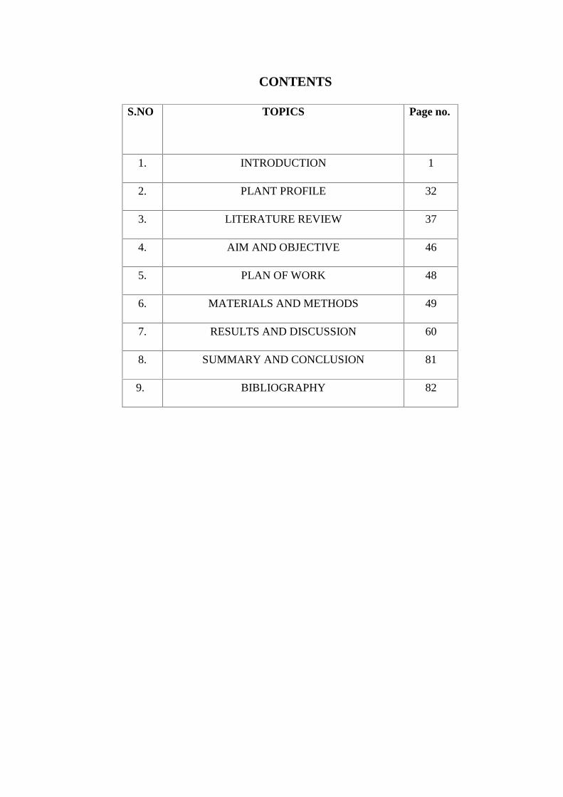
CONTENTS
S.NO TOPICS Page no.
1. INTRODUCTION 1
2. PLANT PROFILE 32
3. LITERATURE REVIEW 37
4. AIM AND OBJECTIVE 46
5. PLAN OF WORK 48
6. MATERIALS AND METHODS 49
7. RESULTS AND DISCUSSION 60
8. SUMMARY AND CONCLUSION 81
9. BIBLIOGRAPHY 82
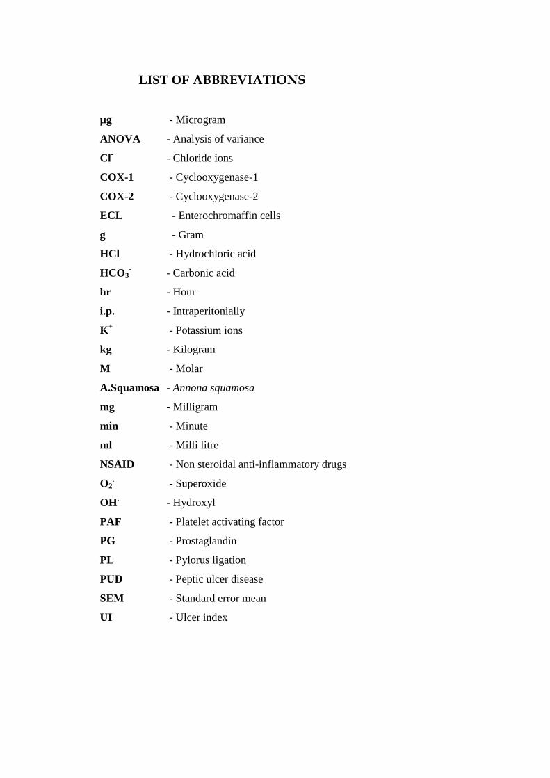
LIST OF ABBREVIATIONS
µg - Microgram
ANOVA - Analysis of variance
Cl- - Chloride ions
COX-1 - Cyclooxygenase-1
COX-2 - Cyclooxygenase-2
ECL - Enterochromaffin cells
g - Gram
HCl - Hydrochloric acid
HCO3- - Carbonic acid
hr - Hour
i.p. - Intraperitonially
K+ - Potassium ions
kg - Kilogram
M - Molar
A.Squamosa - Annona squamosa
mg - Milligram
min - Minute
ml - Milli litre
NSAID - Non steroidal anti-inflammatory drugs
O2. - Superoxide
OH. - Hydroxyl
PAF - Platelet activating factor
PG - Prostaglandin
PL - Pylorus ligation
PUD - Peptic ulcer disease
SEM - Standard error mean
UI - Ulcer index
Chapter 1 Introduction
Dept. of Pharmacology 1 J.K.K. Nattraja College of Pharmacy
1. INTRODUCTION
Herbal Medicine sometimes referred to as Herbalism or Botanical Medicine, is
the use of herbs for their therapeutic or medicinal value. An herb is a plant or plant
part valued for its medicinal, aromatic or savoury qualities. Botanists define an herb
as being a soft stemmed plant, which dies after flowering, while “herbalists define
an herb as any part of a plant which can be used for medicine”, cooking, and
cosmetic uses and as a scent or dye. Herb plants produce and contain a variety of
chemical substances that act upon the body.
The leaves, flowers, stems, berries, and roots of plants are used to prevent,
relieve, and treat illness. From a "scientific" perspective, many herbal treatments are
considered experimental. The reality is, however, that herbal medicine has a long and
respected history. Many familiar medications of the twentieth century were
developed from ancient healing traditions that treated health problems with specific
plants. Today, science has isolated the medicinal properties of a large number of
botanicals, and their healing components have been extracted and analyzed. Many
plant components are now synthesized in large laboratories for use in pharmaceutical
preparations. For example, vincristine (an antitumor), digitalis (a heart regulator),
and ephedrine (a bronchodilator used to decrease respiratory congestion) were all
originally discovered through research on plants.(Kokate C.K., 1995)
IMPORTANCE OF HERBAL
Herbs are staging a comeback and herbal ‘renaissance’ is happening all over
the globe. The herbal products today symbolise safety in contrast to the synthetics
drug that are regarded as unsafe to human and environment. Although herbs had
been priced for their medicinal, flavouring and aromatic qualities for centuries, the
synthetic products of the modern age surpassed their importance, for a while.
However, the blind dependence on synthetics is over and people are returning to the
naturals with hope of safety and security.
Our ancestors used trial and error to discover the most effective local
plants for the treatment of illnesses. Advances in science have enabled a better
understanding of the physiological effects of herbs on the human body and therefore
Chapter 1 Introduction
Dept. of Pharmacology 2 J.K.K. Nattraja College of Pharmacy
their role in restoring health. Herbal medicines support the body’s natural healing
process, and aim to treat the person as well as the disease. This means it can bring
about a deep and lasting change.
PROSPECTS OF HERBAL RESEARCH
There is a worldwide ‘green revolution’, (Mukherjee, P.K., 2002)
which is reflected in the belief that herbal remedies are safer and less damaging to
the human body than synthetic drugs. Furthermore, underlying this upsurge of
interest in plants is the fact that many important drugs in use today were derived
from plants or from starting molecules of plant origin.
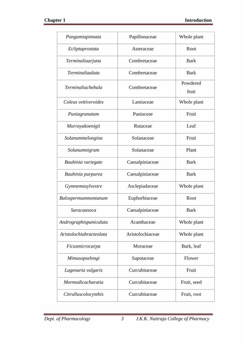
Table No. 1. List of Anti-ulcer plants
Plant FamilyPlant part
used
Alpiniaallughas Zingiberaceae Rhizome
Alpinia galangal Zingiberaceae Rhizome
Alpiniacalcarata Zingiberaceae Rhizome
Glycyrrhizaglabra Liquorice Stem, root
Azadirachtaindica MeliaceaeBark, leaves,
flower
Acacia catechu Mimosaceae Bark
Sophorasubprostrata Leguminaceae Root
Magnolia bark Magnoliaceae Bark
Gloriosa superb LiliaceaeRoots,
rhizome
Phyllanthusemblica Euphorbiaceae Root, bark
Aeglemarmelos Rutaceae Leaf
Indigoferatinctoria Papillonaceae Leaf, fruit
Chapter 1 Introduction
Dept. of Pharmacology 3 J.K.K. Nattraja College of Pharmacy
Pongamiapinnata Papillonaceae Whole plant
Ecliptaprostata Asteraceae Root
Terminaliaarjuna Combretaceae Bark
Terminaliaalata Combretaceae Bark
Terminaliachebula CombretaceaePowdered
fruit
Coleus vettiveroides Lamiaceae Whole plant
Puniagranatum Puniaceae Fruit
Murrayakoenigii Rutaceae Leaf
Solanummelongina Solanaceae Fruit
Solanumnigram Solanaceae Plant
Bauhinia variegate Caesalpiniaceae Bark
Bauhinia purpurea Caesalpiniaceae Bark
Gymnemasylvestre Asclepiadaceae Whole plant
Balospermummontanum Euphorbiaceae Root
Saracaasoca Caesalpiniaceae Bark
Andrographispaniculata Acanthaceae Whole plant
Aristolochiabracteolata Aristolochiaceae Whole plant
Ficusmicrocarpa Moraceae Bark, leaf
Mimusopselengi Sapotaceae Flower
Lagenaria vulgaris Curcubitaceae Fruit
Mormodicacharatia Curcubitaceae Fruit, seed
Citrulluscolocynthis Curcubitaceae Fruit, root
Chapter 1 Introduction
Dept. of Pharmacology 4 J.K.K. Nattraja College of Pharmacy
Since modern drugs are not able to completely cure chronic diseases but,
rather, to prevent further deterioration associated with them, patients must take drugs
for extended periods of time. Due to the ineffectiveness as well as the potential side
effects, patients are often led to explore complementary/ alternative medicines such
as herb, and medicinal botanicals in particular. However, simultaneous
administration of herbs and drugs may mimic, magnify or oppose the
pharmacological effects of each other. It is widely believed that although herbs hold
promise as therapeutically effective medicaments, in-depth and appropriate studies
should be carried out to confirm their efficacy in the presence of modern medicines.
PHYSIOLOGY OF STOMACH
The stomach is a muscular, hollow, dilated part of the alimentary canal which
functions as an important organ of the digestive tract. It is involved in the second
phase of digestion, following mastication (chewing). The stomach is located between
the oesophagus and the small intestine. The stomach is the most dilated part of the
digestive tube, and is situated between the end of the oesophagus and the beginning
of the small intestine. It lies in the epigastric, umbilical, and left hypochondriac
regions of the abdomen, and occupies a recess bounded by the upper abdominal
viscera, and completed in front and on the left side by the anterior abdominal wall
and the diaphragm. It secretes protein-digesting enzymes and strong acids to aid in
food digestion, (sent to it via oesophagealperistalsis) through smooth muscular
contortions (called segmentation) before sending partially digested food (chyme) to
the small intestines.
The word stomach is derived from the Latinstomachus which is derived from
the Greek word stomachos, ultimately from stoma, "mouth". The words gastro- and
gastric (meaning related to the stomach) are both derived from the Greek word
gaster. The stomach is an organ between the oesophagus and the small intestine. It is
where digestion of protein begins. The stomach has three tasks. It stores swallowed
food. It mixes the food with stomach acids. Then it sends the mixture on to the small
intestine.
Chapter 1 Introduction
Dept. of Pharmacology 5 J.K.K. Nattraja College of Pharmacy
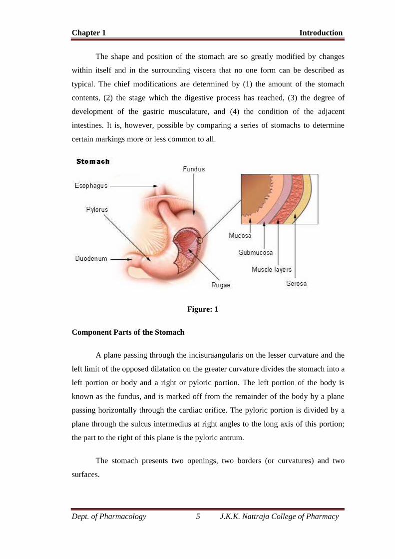
The shape and position of the stomach are so greatly modified by changes
within itself and in the surrounding viscera that no one form can be described as
typical. The chief modifications are determined by (1) the amount of the stomach
contents, (2) the stage which the digestive process has reached, (3) the degree of
development of the gastric musculature, and (4) the condition of the adjacent
intestines. It is, however, possible by comparing a series of stomachs to determine
certain markings more or less common to all.
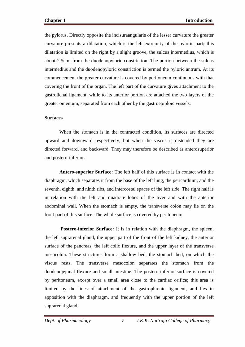
Figure: 1
Component Parts of the Stomach
A plane passing through the incisuraangularis on the lesser curvature and the
left limit of the opposed dilatation on the greater curvature divides the stomach into a
left portion or body and a right or pyloric portion. The left portion of the body is
known as the fundus, and is marked off from the remainder of the body by a plane
passing horizontally through the cardiac orifice. The pyloric portion is divided by a
plane through the sulcus intermedius at right angles to the long axis of this portion;
the part to the right of this plane is the pyloric antrum.
The stomach presents two openings, two borders (or curvatures) and two
surfaces.
Chapter 1 Introduction
Dept. of Pharmacology 6 J.K.K. Nattraja College of Pharmacy
Openings
The opening by which the esophagus communicates with the stomach is
known as the cardiac orifice, and is situated on the left of the middle line at the level
of the tenth thoracic vertebra. The short abdominal portion of the esophagus
(antrumcardiacum) is conical in shape and curved sharply to the left, the base of the
cone being continuous with the cardiac orifice of the stomach. The right margin of
the esophagus is continuous with the lesser curvature of the stomach, while the left
margin joins the greater curvature at an acute angle, termed the incisuracardiaca.
The pyloric orifice communicates with the duodenum, and its position is
usually indicated on the surface of the stomach by a circular groove, the
duodenopyloric constriction. This orifice lies to the right of the middle line at the
level of the upper border of the first lumbar vertebra.
Curvatures
The lesser curvature (curvaturaventriculi minor), extending between the
cardiac and pyloric orifices, forms the right or posterior border of the stomach. It
descends as a continuation of the right margin of the esophagus in front of the fibers
of the right crus of the diaphragm, and then, turning to the right, it crosses the first
lumbar vertebra and ends at the pylorus. Nearer its pyloric than its cardiac end is a
well-marked notch, the incisuraangularis, which varies somewhat in position with the
state of distension of the viscus; it serves to separate the stomach into a right and a
left portion. The lesser curvature gives attachment to the two layers of the
hepatogastric ligament, and between these two layers are the left gastric artery and
the right gastric branch of the hepatic artery.
The greater curvature (curvaturaventriculi major) is directed mainly
forward, and is four or five times as long as the lesser curvature. Starting from the
cardiac orifice at the incisuracardiaca, it forms an arch backward, upward, and to the
left; the highest point of the convexity is on a level with the sixth left costal cartilage.
From this level it may be followed downward and forward, with a slight convexity to
the left as low as the cartilage of the ninth rib, it then turns to the right, to the end of
Chapter 1 Introduction
Dept. of Pharmacology 7 J.K.K. Nattraja College of Pharmacy
the pylorus. Directly opposite the incisuraangularis of the lesser curvature the greater
curvature presents a dilatation, which is the left extremity of the pyloric part; this
dilatation is limited on the right by a slight groove, the sulcus intermedius, which is
about 2.5cm, from the duodenopyloric constriction. The portion between the sulcus
intermedius and the duodenopyloric constriction is termed the pyloric antrum. At its
commencement the greater curvature is covered by peritoneum continuous with that
covering the front of the organ. The left part of the curvature gives attachment to the
gastrolienal ligament, while to its anterior portion are attached the two layers of the
greater omentum, separated from each other by the gastroepiploic vessels.
Surfaces
When the stomach is in the contracted condition, its surfaces are directed
upward and downward respectively, but when the viscus is distended they are
directed forward, and backward. They may therefore be described as anterosuperior
and postero-inferior.
Antero-superior Surface: The left half of this surface is in contact with the
diaphragm, which separates it from the base of the left lung, the pericardium, and the
seventh, eighth, and ninth ribs, and intercostal spaces of the left side. The right half is
in relation with the left and quadrate lobes of the liver and with the anterior
abdominal wall. When the stomach is empty, the transverse colon may lie on the
front part of this surface. The whole surface is covered by peritoneum.
Postero-inferior Surface: It is in relation with the diaphragm, the spleen,
the left suprarenal gland, the upper part of the front of the left kidney, the anterior
surface of the pancreas, the left colic flexure, and the upper layer of the transverse
mesocolon. These structures form a shallow bed, the stomach bed, on which the
viscus rests. The transverse mesocolon separates the stomach from the
duodenojejunal flexure and small intestine. The postero-inferior surface is covered
by peritoneum, except over a small area close to the cardiac orifice; this area is
limited by the lines of attachment of the gastrophrenic ligament, and lies in
apposition with the diaphragm, and frequently with the upper portion of the left
suprarenal gland.
Chapter 1 Introduction
Dept. of Pharmacology 8 J.K.K. Nattraja College of Pharmacy
Role in Digestion
Bolus (masticated food) enters the stomach through the oesophagus via the
oesophageal sphincter. The stomach releases proteases (protein-digesting enzymes
such as pepsin) and hydrochloric acid, which kills or inhibits bacteria and provides
the acidic pH of 2 for the proteases to work. Food is churned by the stomach through
muscular contractions of the wall - reducing the volume of the fundus, before
looping around the fundus and the body of stomach as the boluses is converted into
chyme (partially digested food). Chyme slowly passes through the pyloric sphincter
and into the duodenum, where the extraction of nutrients begins. Depending on the
quantity and contents of the meal, the stomach will digest the food into chyme
anywhere between 40 minutes and a few hours.
Acid Secretion
Parietal cells in the stomach secrete roughly two liters of acid a day in the
form of hydrochloric acid. When stimulated, these parietal cells secrete HCl at a
concentration of roughly 160 mM (equivalent to a pH of 0.8). Acid in the stomach
functions to kill bacteria, and to aid digestion by solubilizing food. The acid is also
important to establish the optimal pH (1.8-3.5) for the function of the digestive
enzyme pepsin.
The acid is secreted into large cannaliculi, deep invaginations of the plasma
membrane which are continuous with the lumen of the stomach. When acid secretion
is stimulated there is a dramatic change in the morphology of the membranes of the
parietal cell. Cytoplasmic tubulovesicular membranes which are abundant in the
resting cell virtually disappear in concert with a large increase in the cannalicular
membrane
The epithelium of the stomach is intrinsically resistant to the damaging
effects of gastric acid and other consequences. Nonetheless, excessive secretion of
gastric acid is a major problem in humans leading to gastritis, gastric ulcers and
peptic acid disease.

Chapter 1 Introduction
Dept. of Pharmacology 9 J.K.K. Nattraja College of Pharmacy
Mechanism
The ability of the parietal cell to secrete acid is dependent on active transport.
The key player in acid secretion is H+/K+ ATPase or "proton pump" located in the
cannalicular membrane. This ATPase is magnesium-dependent, and not inhibited by
ouabain. The current model for explaining acid secretion is as follows
Hydrogen ions are generated within the parietal cell from dissociation of
water. The hydroxyl ions formed in this process rapidly combine with carbon
dioxide to form bicarbonate ion, a reaction catalyzed by carbonic anhydrase.
Bicarbonate is transported out of the basolateral membrane in exchange for
chloride. The outflow of bicarbonate into blood results in a slight elevation of
blood pH known as the "alkaline tide". This process serves to maintain
intracellular pH in the parietal cell.
Chloride and potassium ions are transported into the lumen of the
cannaliculus by conductance channels, and such is necessary for secretion of
acid.
Hydrogen ion is pumped out of the cell, into the lumen, in exchange for
potassium through the action of the proton pump; potassium is thus
effectively recycled.
Accumulation of osmotically-active hydrogen ion in the cannaliculus
generates an osmotic gradient across the membrane that results in outward
diffusion of water - the resulting gastric juice is 155 mM HCl and 15 mM
HCl with a small amount of NaCl.
Regulation of Acid secretion
Parietal cells bear receptors for three stimulators (positive regulators) of acid
secretion, reflecting a triumverate of neural, endocrine and paracrine control:
I. Acetylcholine (muscarinic type receptor)
II. Gastrin
III. Histamine (H2 type receptor)
Chapter 1 Introduction
Dept. of Pharmacology 10 J.K.K. Nattraja College of Pharmacy
Acetylcholine is a neurotransmitter that is released by enteric neurons. Gastrin
is a hormone that is released by G cells, endocrine cells that are located in the gastric
epithelium. Histamine is a paracrine that is released from ECL (enterochromaffin-
like) cells.
Enterochromaffin-like or ECL cells are a distinctive type of neuroendocrine cell
in the gastric mucosa underlying the epithelium. They are most prevalent in the acid-
secreting regions of the stomach. ECL cells synthesize and secrete histamine in
response to stimulation by the hormones gastrin and pituitary adenylyl cyclase-
activating peptide. Gastrin itself is secreted by cells in the epithelium of the stomach,
but travels to ECL cells via the blood. Together, histamine and gastrin are primary
positive regulators of acid secretion from the parietal cell. ECL cells also secrete
pancreastatin and probably are the source of one or more other peptide hormones and
growth factors. ECL cells are readily identified in histologic sections stained by
silver impregnation.
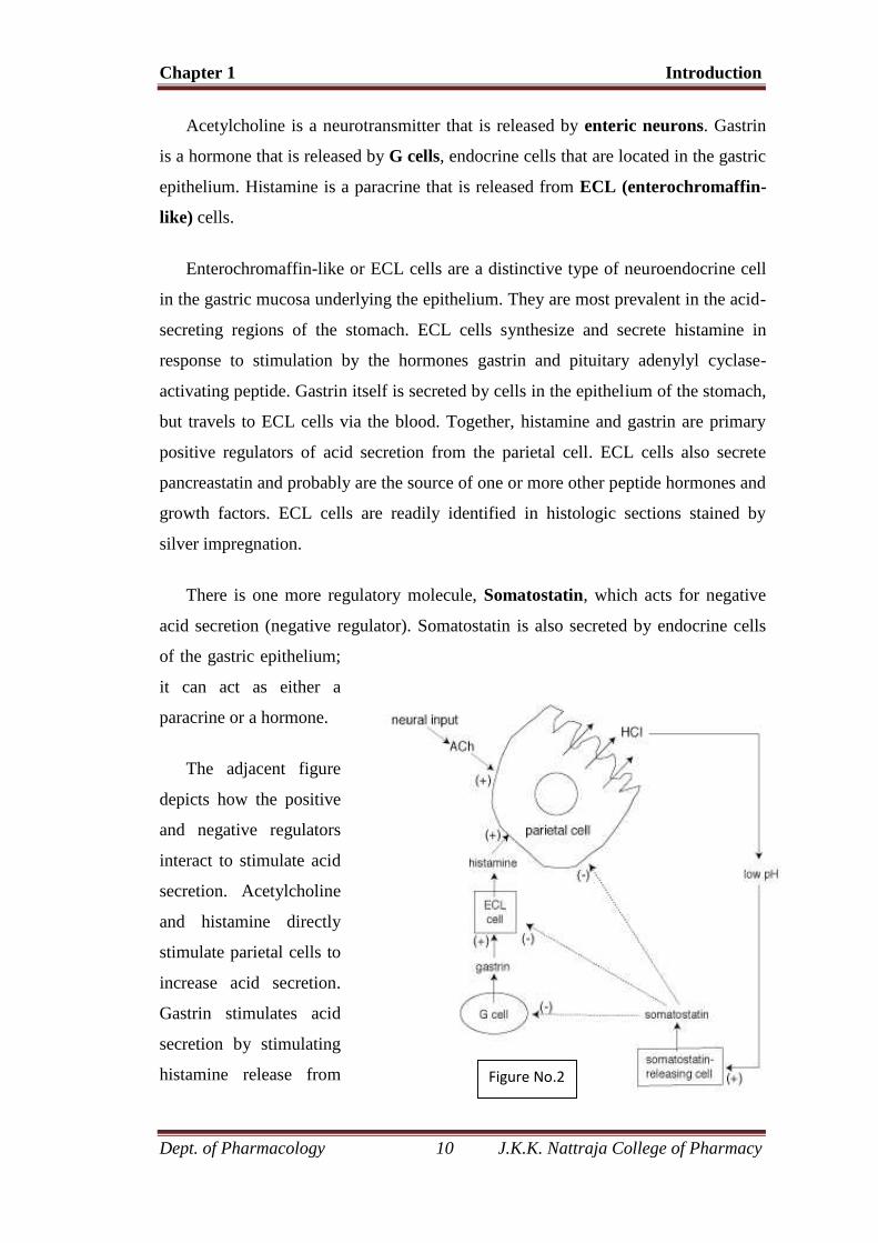
There is one more regulatory molecule, Somatostatin, which acts for negative
acid secretion (negative regulator). Somatostatin is also secreted by endocrine cells
of the gastric epithelium;
it can act as either a
paracrine or a hormone.
The adjacent figure
depicts how the positive
and negative regulators
interact to stimulate acid
secretion. Acetylcholine
and histamine directly
stimulate parietal cells to
increase acid secretion.
Gastrin stimulates acid
secretion by stimulating
histamine release from Figure No.22
Chapter 1 Introduction
Dept. of Pharmacology 11 J.K.K. Nattraja College of Pharmacy
ECL cells. (Gastrin also has a direct effect on parietal cells, which is to stimulate
their proliferation). When the pH of the stomach gets too low, that stimulates
somatostatin secretion. Somatostatin inhibits acid secretion by direct effects on
parietal cells, and also by inhibiting release of the positive regulators histamine and
gastrin. The balance of activity of the different regulators changes as food is
consumed and passes through different segments of the upper GI
tract.(http://courses.washington.edu)
PEPTIC ULCER DISEASE
A peptic ulcer is a sore or hole in the lining of the stomach or duodenum and
is a term used to describe a lesion in the oesophagus, stomach, or duodenum. More
specific names are used to describe an ulcer located at a specific site. Duodenal
ulcers (the first part of the small intestine) are more common than other types of
peptic ulcers. Peptic ulcers are caused by hyper secretion of hydrochloric acid and
pepsin that erode the GI mucosal lining.
BASIC CAUSE OF PEPTIC ULCERATION
The usual cause of peptic ulceration is an imbalance between the rate of
secretion of gastric juice and the degree of protection afforded by (1) the gastro
duodenal mucosal barrier and (2) the neutralization of the gastric acid by duodenal
juices. It will be recalled that all areas normally exposed to gastric juice are well
supplied with mucous glands, beginning with compound mucous glands in the lower
oesophagus plus the mucous cell coating of the stomach mucosa, the mucous neck
cells of the gastric glands, the deep pyloric glands that secrete mainly mucus, and,
finally, the glands of Brunner of the upper duodenum, which secrete a highly alkaline
mucus. In addition to the mucus protection of the mucosa, the duodenum is protected
by the alkalinity of the small intestinal secretions. Especially important is pancreatic
secretion, which contains large quantities of sodium bicarbonate that neutralize the
hydrochloric acid of the gastric juice, thus also inactivating pepsin and preventing
digestion of the mucosa. In addition, large amounts of bicarbonate ions are provided
in (1) the secretions of the large Brunner’s glands in the first few centimetres of the
duodenal wall and (2) in bile coming from the liver.
Chapter 1 Introduction
Dept. of Pharmacology 12 J.K.K. Nattraja College of Pharmacy
Causes
1. High acid and peptic content
2. Irritation
3. Poor blood supply
4. Poor secretion of mucus
5. Infection, H. Pylori
PATHOPHYSIOLOGY
A physiologic imbalance between aggressive factors (gastric acid and pepsin)
and protective factors (mucosal defense and repair) remain important issues in the
pathophysiology of gastric and duodenal ulcers. Gastric acid is secreted by the
parietal cells, which contain receptors for histamine, gastrin, and acetylcholine. Acid
(as well as H. pylori infection and NSAID use) is an independent factor that
contributes to the disruption of mucosal integrity. Increased acid secretion has been
observed in patients with duodenal ulcers and may be a consequence of H. pylori
infection. Patients with ZES have profound gastric acid hyper secretion resulting
from a gastrin producing tumour. Patients with gastric ulcer usually have normal or
reduced rates of acid secretion (hypochlorhydria). Acid secretion is expressed as the
amount of acid secreted under basal or fasting conditions, basal acid output (BAO);
after maximal stimulation, maximal acid output (MAO); or in response to a meal.
Basal, maximal, and meal-stimulated acid secretion varies according to time of day
and the individual’s psychological state, age, gender, and health status. The BAO
follows a circadian rhythm, with the highest acid secretion occurring at night and the
lowest in the morning. An increase in the BAO: MAO ratio suggests a basal hyper
secretory state such as ZES. Pepsin is an important cofactor that plays a role in the
proteolytic activity involved in ulcer formation. Pepsinogen, the inactive precursor of
pepsin, is secreted by the chief cells located in the gastric fundus. Pepsin is activated
by acid pH (optimal pH of 1.8 to 3.5), inactivated reversibly at pH 4, and irreversibly
destroyed at pH 7. Mucosal defence and repair mechanisms (mucus and bicarbonate
secretion, intrinsic epithelial cell defence, and mucosal blood flow) protect the gastro
duodenal mucosa from noxious endogenous and exogenous substances. The viscous
nature and near-neutral pH of the mucus-bicarbonate barrier protect the stomach
from the acidic contents in the gastric lumen. Mucosal repair after injury is related to
Chapter 1 Introduction
Dept. of Pharmacology 13 J.K.K. Nattraja College of Pharmacy
epithelial cell restitution, growth, and regeneration. The maintenance of mucosal
integrity and repair is mediated by the production of endogenous prostaglandins. The
term cytoprotectionis often used to describe this process, but mucosal defense and
mucosal protection are more accurate terms, as prostaglandins prevent deep mucosal
injury and not superficial damage to individual cells. Gastric hyperemia and
increased prostaglandin synthesis characterize adaptive cytoprotection, the short-
term adaptation of mucosal cells to mild topical irritants. This phenomenon enables
the stomach to initially withstand the damaging effects of irritants. Alterations in
mucosal defence that are induced by H. pylori or NSAIDs are the most important
cofactors in the formation of peptic ulcers.
Chronic Gastritis
Chronic gastritis is defined as the presence of chronic mucosal inflammatory
changes leading eventually to mucosal atrophy and epithelial metaplasia.It is notable
for distinct causal subgroups and for patterns of histologic alterations that vary in
different parts of the world. In the Western world, the prevalence of histologic
changes indicative of chronic gastritis exceeds 50% in the later decades of adult life.
Acute Gastritis
Acute gastritis is an acute mucosal inflammatory process, usually of a
transient nature. The inflammation may be accompanied by haemorrhage into the
mucosa and, in more severe circumstances, by sloughing of the superficial mucosal
epithelium (erosion). This severe erosive form of the disease is an important cause of
acute gastrointestinal bleeding.
Peptic ulcers are remitting, relapsing lesions that are most often diagnosed in
middle-aged to older adults, but they may first become evident in young adult life.
They often appear without obvious precipitating influences and may then heal after a
period of weeks to months of active disease. Even with healing, however, the
propensity to develop peptic ulcers remains. Thus, it is difficult to obtain accurate
data on the prevalence of active disease. Best estimates suggest that in the American
population, about 2.5% of males and 1.5% of females have peptic ulcers. For both
men and women in the United States, the lifetime risk of developing peptic ulcer
disease is about 10%.
Chapter 1 Introduction
Dept. of Pharmacology 14 J.K.K. Nattraja College of Pharmacy
EPIDEMIOLOGY
Genetic or racial influences appear to play little or no role in the causation of
peptic ulcers. Duodenal ulcers are more frequent in patients with alcoholic cirrhosis,
chronic obstructive pulmonary disease, chronic renal failure, and
hyperparathyroidism.
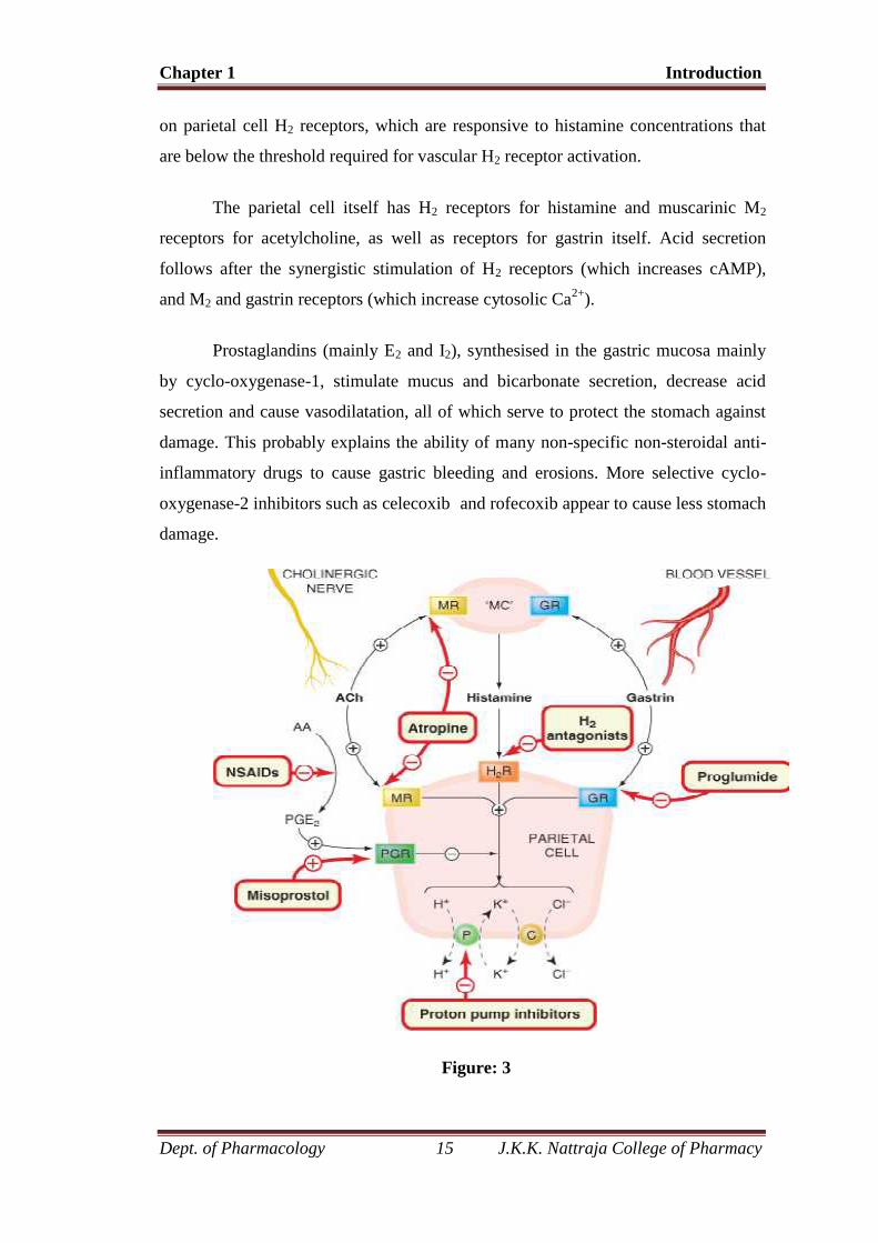
THE REGULATION OF ACID SECRETION
The regulation of acid secretion by parietal cells is especially important in the
pathogenesis of peptic ulcer, and constitutes a particular target for drug action. The
secretion of the parietal cells is an isotonic solution of HCl (150 mmol /l) with a pH
less than 1, the concentration of hydrogen ions being more than a million times
higher than that of the plasma. The Cl- is actively transported into canaliculated in
the cells that communicate with the lumen of the gastric glands and thus with the
stomach itself. This Cl- secretion is accompanied by K+, which is then exchanged for
H+ from within the cell by a K+/H+ ATPase and bicarbonate ions. The latter
exchanges across the basal membrane of the parietal cell for Cl-. The principal
stimuli acting on the parietal cells are
Gastrin (a stimulatory hormone)
Acetylcholine (a stimulatory neurotransmitter)
Histamine (a stimulatory local hormone)
Prostaglandins E2 and I2 (local hormones that inhibit acid secretion).
Gastrin is a peptide hormone synthesised in the mucosa of the gastric antrum
and duodenum, and secreted into portal blood. Its main action is stimulation of the
secretion of acid by the parietal cells. These receptors are blocked by proglumide
which inhibits gastrin action.
Acetylcholine is released from neurons and stimulates specific muscarinic
receptors on the surface of the parietal cells and on the surface of histamine-
containing cells.
Histamine: Within the stomach, mast cells (or histamine-containing cells
similar to mast cells) lying close to the parietal cell release a steady basal release of
histamine, which is further increased by gastrin and acetylcholine. The hormone acts
Chapter 1 Introduction
Dept. of Pharmacology 15 J.K.K. Nattraja College of Pharmacy
on parietal cell H2 receptors, which are responsive to histamine concentrations that
are below the threshold required for vascular H2 receptor activation.
The parietal cell itself has H2 receptors for histamine and muscarinic M2
receptors for acetylcholine, as well as receptors for gastrin itself. Acid secretion
follows after the synergistic stimulation of H2 receptors (which increases cAMP),
and M2 and gastrin receptors (which increase cytosolic Ca2+).
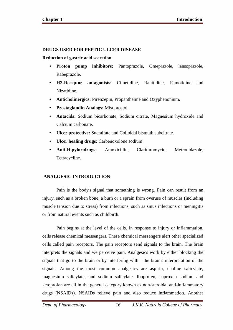
Prostaglandins (mainly E2 and I2), synthesised in the gastric mucosa mainly
by cyclo-oxygenase-1, stimulate mucus and bicarbonate secretion, decrease acid
secretion and cause vasodilatation, all of which serve to protect the stomach against
damage. This probably explains the ability of many non-specific non-steroidal anti-
inflammatory drugs to cause gastric bleeding and erosions. More selective cyclo-
oxygenase-2 inhibitors such as celecoxib and rofecoxib appear to cause less stomach
damage.
Figure: 3
Chapter 1 Introduction
Dept. of Pharmacology 16 J.K.K. Nattraja College of Pharmacy
DRUGS USED FOR PEPTIC ULCER DISEASE
Reduction of gastric acid secretion
Proton pump inhibitors: Pantoprazole, Omeprazole, lansoprazole,
Rabeprazole.
H2-Receptor antagonists: Cimetidine, Ranitidine, Famotidine and
Nizatidine.
Anticholinergics: Pirenzepin, Propantheline and Oxyphenonium.
Prostaglandin Analogs: Misoprostol
Antacids: Sodium bicarbonate, Sodium citrate, Magnesium hydroxide and
Calcium carbonate.
Ulcer protective: Sucralfate and Colloidal bismuth subcitrate.
Ulcer healing drugs: Carbenoxolone sodium
Anti-H.pyloridrugs: Amoxicillin, Clarithromycin, Metronidazole,
Tetracycline.
ANALGESIC INTRODUCTION
Pain is the body's signal that something is wrong. Pain can result from an
injury, such as a broken bone, a burn or a sprain from overuse of muscles (including
muscle tension due to stress) from infections, such as sinus infections or meningitis
or from natural events such as childbirth.
Pain begins at the level of the cells. In response to injury or inflammation,
cells release chemical messengers. These chemical messengers alert other specialized
cells called pain receptors. The pain receptors send signals to the brain. The brain
interprets the signals and we perceive pain. Analgesics work by either blocking the
signals that go to the brain or by interfering with the brain's interpretation of the
signals. Among the most common analgesics are aspirin, choline salicylate,
magnesium salicylate, and sodium salicylate. Ibuprofen, naproxen sodium and
ketoprofen are all in the general category known as non-steroidal anti-inflammatory
drugs (NSAIDs). NSAIDs relieve pain and also reduce inflammation. Another
Chapter 1 Introduction
Dept. of Pharmacology 17 J.K.K. Nattraja College of Pharmacy
common analgesic, acetaminophen, provides pain relief but does not reduce
inflammation.
ammation.
Figure: 4
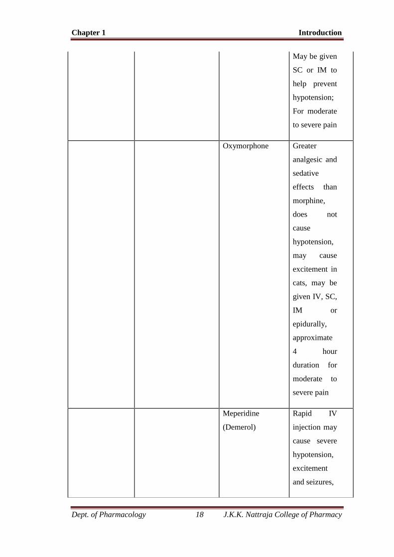
Table: 2
Analgesic classification
Drug Class Characteristics Examples Comments
Opioids* Acts on pain
receptors in both
spinal cord and
brain; May be used
with tranquilizers to
induce a state of
potent sedation
(neuroleptanalgesia)
Morphine Stimulates
vomiting and
vagal CNS
centers; Can
cause
excitement in
cats, horses
and food
animals; 4-6
hours
duration;
Chapter 1 Introduction
Dept. of Pharmacology 18 J.K.K. Nattraja College of Pharmacy
May be given
SC or IM to
help prevent
hypotension;
For moderate
to severe pain
Oxymorphone Greater
analgesic and
sedative
effects than
morphine,
does not
cause
hypotension,
may cause
excitement in
cats, may be
given IV, SC,
IM or
epidurally,
approximate
4 hour
duration for
moderate to
severe pain
Meperidine
(Demerol)
Rapid IV
injection may
cause severe
hypotension,
excitement
and seizures,
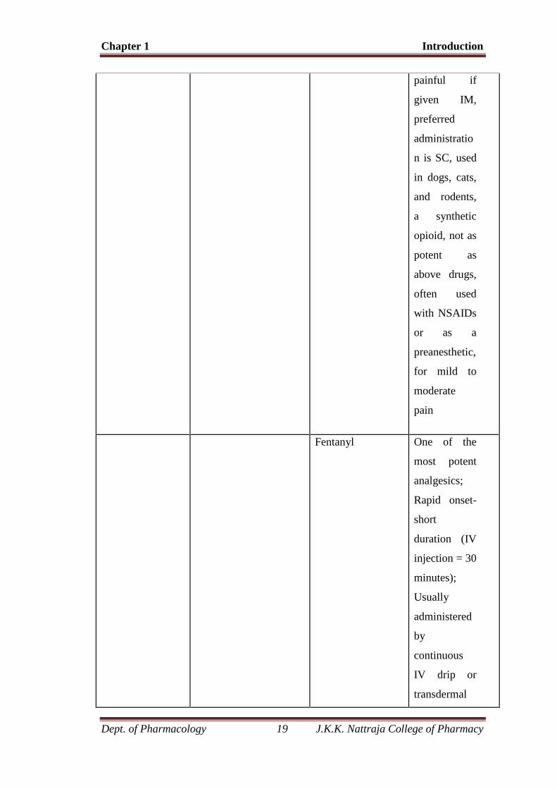
Chapter 1 Introduction
Dept. of Pharmacology 19 J.K.K. Nattraja College of Pharmacy
painful if
given IM,
preferred
administratio
n is SC, used
in dogs, cats,
and rodents,
a synthetic
opioid, not as
potent as
above drugs,
often used
with NSAIDs
or as a
preanesthetic,
for mild to
moderate
pain
Fentanyl One of the
most potent
analgesics;
Rapid onset-
short
duration (IV
injection = 30
minutes);
Usually
administered
by
continuous
IV drip or
transdermal
Chapter 1 Introduction
Dept. of Pharmacology 20 J.K.K. Nattraja College of Pharmacy
patch; Can
cause panting
and increased
sensitivity to
sound; For
severe to
moderate
pain
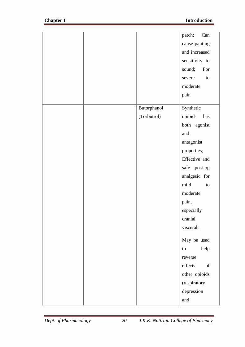
Butorphanol
(Torbutrol)
Synthetic
opioid- has
both agonist
and
antagonist
properties;
Effective and
safe post-op
analgesic for
mild to
moderate
pain,
especially
cranial
visceral;
May be used
to help
reverse
effects of
other opioids
(respiratory
depression
and
Chapter 1 Introduction
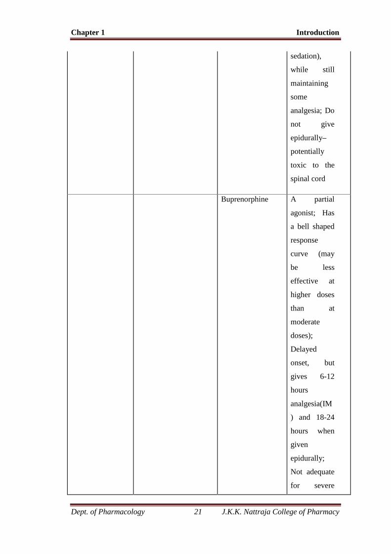
Dept. of Pharmacology 21 J.K.K. Nattraja College of Pharmacy
sedation),
while still
maintaining
some
analgesia; Do
not give
epidurally–
potentially
toxic to the
spinal cord
Buprenorphine A partial
agonist; Has
a bell shaped
response
curve (may
be less
effective at
higher doses
than at
moderate
doses);
Delayed
onset, but
gives 6-12
hours
analgesia(IM
) and 18-24
hours when
given
epidurally;
Not adequate
for severe
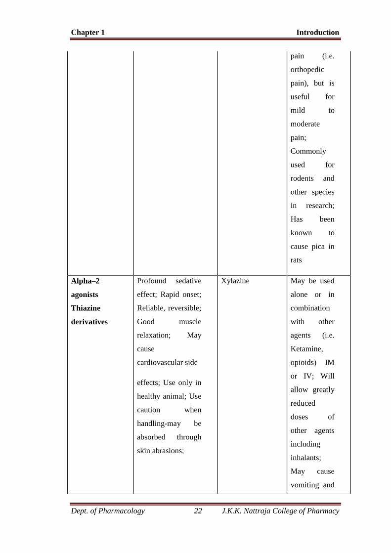
Chapter 1 Introduction
Dept. of Pharmacology 22 J.K.K. Nattraja College of Pharmacy
pain (i.e.
orthopedic
pain), but is
useful for
mild to
moderate
pain;
Commonly
used for
rodents and
other species
in research;
Has been
known to
cause pica in
rats
Alpha–2
agonists
Thiazine
derivatives
Profound sedative
effect; Rapid onset;
Reliable, reversible;
Good muscle
relaxation; May
cause
cardiovascular side
effects; Use only in
healthy animal; Use
caution when
handling-may be
absorbed through
skin abrasions;
Xylazine May be used
alone or in
combination
with other
agents (i.e.
Ketamine,
opioids) IM
or IV; Will
allow greatly
reduced
doses of
other agents
including
inhalants;
May cause
vomiting and
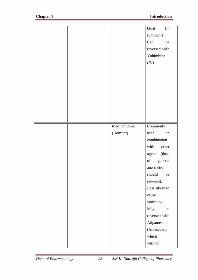
Chapter 1 Introduction
Dept. of Pharmacology 23 J.K.K. Nattraja College of Pharmacy
bloat (in
ruminants);
Can be
reversed with
Yohimbine
(IV)
Meditomidine
(Domitor)
Commonly
used in
combination
with other
agents (dose
of general
anesthetic
should be
reduced);
Less likely to
cause
vomiting;
May be
reversed with
Atipamesole
(Antesedan)
which
will not
Chapter 1 Introduction
Dept. of Pharmacology 24 J.K.K. Nattraja College of Pharmacy
reverse the
effects of
other drugs
given
concurrently
with
Meditomidi-
ne; *High
doses of
Atipamesole
can cause
panting,
excitement,
muscle
tremors,
hypotension,
and
tachycardia-
especially if
given IV
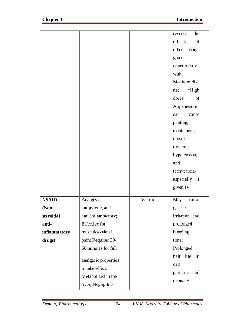
NSAID
(Non-
steroidal
anti-
inflammatory
drugs)
Analgesic,
antipyretic, and
anti-inflammatory;
Effective for
musculoskeletal
pain; Requires 30-
60 minutes for full
analgesic properties
to take effect;
Metabolized in the
liver; Negligible
Aspirin May cause
gastric
irritation and
prolonged
bleeding
time;
Prolonged
half life in
cats,
geriatrics and
neonates
Chapter 1 Introduction
Dept. of Pharmacology 25 J.K.K. Nattraja College of Pharmacy
effect on
cardiovascular and
respiratory systems
Acetaminophen Toxic to cats
and
hepatotoxic
to dogs; Less
gastric
irritation than
Aspirin
Ibuprofen Renal, gastric
effects;
Narrow
safety margin
in cats
Flunixin (Banamine) Significant
renal toxicity
is possible in
hypotensive
Chapter 1 Introduction
Dept. of Pharmacology 26 J.K.K. Nattraja College of Pharmacy
patients; Do
not use with
Methoxyflur-
ane
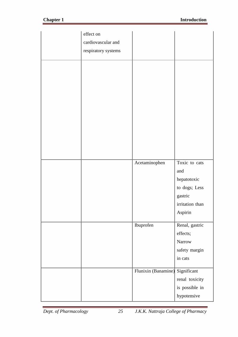
Ketoprofen Potent
analgesic,
especially for
orthopedic
patients;
Gastric
irritation and
ulceration
may occur at
therapeutic
doses
Carprofen
(Rimadyl)
Less
potential for
gastric
ulceration
than some
NSAID’s;
Renal
toxicity seen
in dogs with
prolonged
use
(especially
Labrador
Retrievers)
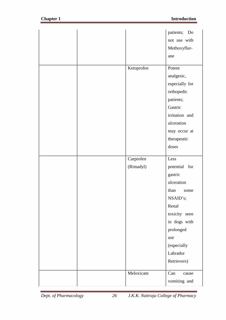
Meloxicam Can cause
vomiting and
Chapter 1 Introduction
Dept. of Pharmacology 27 J.K.K. Nattraja College of Pharmacy
diarrhea;
Less
potential for
gastric
ulceration
than some
NSAID’s; 5-
day limit for
treatment of
cats
Local
analgesia
Can be sprayed,
injected at surgical
site, or infiltrated
around a nerve
supplying the
affected area; May
be used to
desensitize an entire
area by using an
Lidocaine
(Xylocaine)
Immediate
onset; Lasts
about 1-2
hours (with
epinephrine)
or 1 hour
without
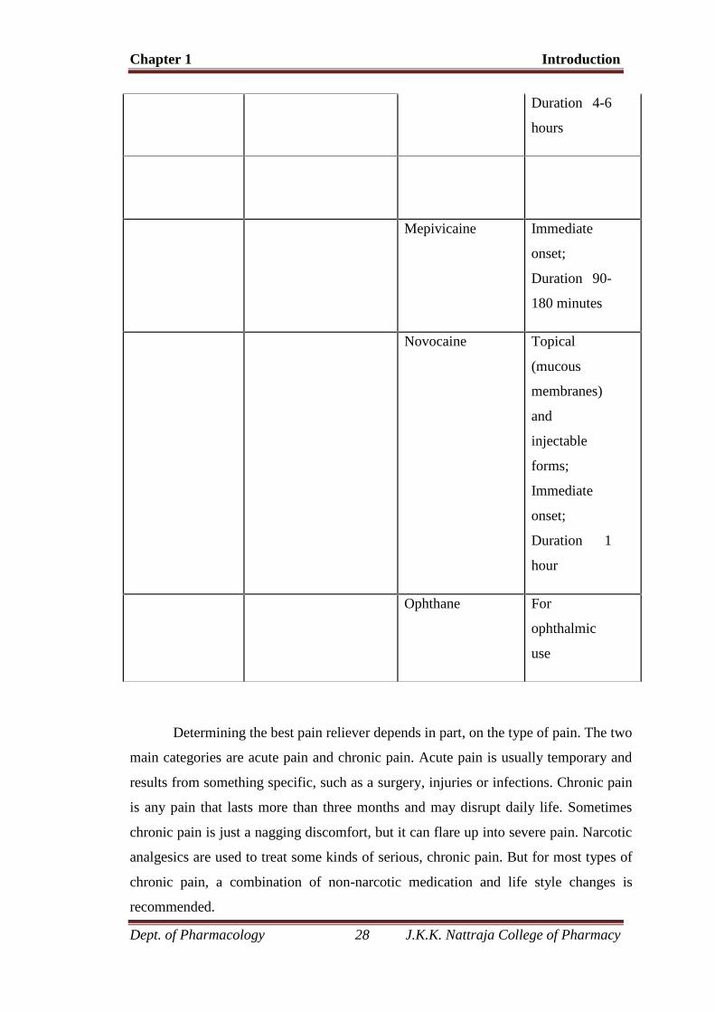
Bupivicaine Onset 20
min;
Chapter 1 Introduction
Dept. of Pharmacology 28 J.K.K. Nattraja College of Pharmacy
Determining the best pain reliever depends in part, on the type of pain. The two
main categories are acute pain and chronic pain. Acute pain is usually temporary and
results from something specific, such as a surgery, injuries or infections. Chronic pain
is any pain that lasts more than three months and may disrupt daily life. Sometimes
chronic pain is just a nagging discomfort, but it can flare up into severe pain. Narcotic
analgesics are used to treat some kinds of serious, chronic pain. But for most types of
chronic pain, a combination of non-narcotic medication and life style changes is
recommended.
Duration 4-6
hours
Mepivicaine Immediate
onset;
Duration 90-
180 minutes
Novocaine Topical
(mucous
membranes)
and
injectable
forms;
Immediate
onset;
Duration 1
hour
Ophthane For
ophthalmic
use
Chapter 1 Introduction
Dept. of Pharmacology 29 J.K.K. Nattraja College of Pharmacy
Side effects are one reason to be careful about frequent or long-term use of pain
relievers.Narcotic analgesics are very effective, but can cause addiction. Aspirin and
ibuprofen can irritate the stomach. Acetaminophen (Tylenol, Panadol) does not
produce the side effects that aspirin, ibuprofen do, but high doses can cause liver
damage, especially in people who drink alcohol regularly. Some pain relievers
contain caffeine, which enhances their effectiveness. Taking these drugs near
bedtime can interfere with sleep. Anyone who gets edgy or jittery from caffeine
should also be careful about using them during the day.
Pain carrying pathway
When there is tissue damage in the periphery, afferent fibers from the
damaged site carry the pain impulses via dorsal root ganglion and terminate in the
SubstanciaGelatinosaRolandi (SGR) situated at the tip of the dorsal horn. The first
neuron ends here.
The second neuron arises from the SGR, crosses to the opposite side and
constitutes anterior and lateral spinothalamic tract (STT).
There are 2 groups of fibres in the STT
Direct spinothalamic system—these fibers terminates in the ventral
posterolateral nucleus of the thalamus. Next order neuron arises from there and
terminates in the post-central gyrus of the cerebral cortex.
i. Spino-reticular thalamic system—these fibers arising from SGR terminate in
the intermediate relaying center of the brain stem. Next order neuron arises from
here and to terminate in the thalamus, then to the cortex.
Mechanism of action of narcotic analgesic
All the opioid analgesics bind with selective endogenous opioid receptors and
produces action. They are all G-protein linked (2ndmessenger system). They produce
action by 2 well known mechanisms.
1. They either close voltage gated Ca+2 channel presynaptically and decreases the
release of excitatory neurotransmitters, such as substance –P, Ach, Glutamate etc.
2. They may hyperpolarize or inhibit post-synaptic membrane by opening of
K+ channel.
Chapter 1 Introduction
Dept. of Pharmacology 30 J.K.K. Nattraja College of Pharmacy
Mechanism of opioid analgesic
Pain consists of both sensory and affective (emotional) components. Opioid can
change both components by,
i. Opioid agonists inhibit the release of excitatory neurotransmitter from the
primary afferents of the spinal cord and directly inhibit dorsal horn pain
transmission neuron of the spinal cord.
ii. It also directly inhibits the sites concerned with pain modulation. Eg:-clostrum.
iii. ↑ Pain threshold.
iv. It also releases endogenous opioid peptide.
Description
Pain has been classified as "productive" and "non-productive." While this
distinction has no physiologic meaning, it may serve as a guide to treatment.
Productive pain has been described as a warning of injury and so may be both an
indication of need for treatment and a guide to diagnosis. Non-productive pain by
definition serves no purpose either as a warning or diagnostic tool.
Although pain syndromes may be dissimilar, the common factor is a sensory
pathway from the affected organ to the brain. Analgesics work at the level of the
nerves, either by blocking the signal from the peripheral nervous system or by
distorting the interpretation by the central nervous system. Selection of an
appropriate analgesic is based on consideration of the risk-benefit factors of each
class of drugs, based on type of pain, severity of pain, and risk of adverse effects.
Traditionally, pain has been divided into two classes, acute and chronic, although
severity and projected patient survival are other factors that must be considered in
drug selection.
Acute pain
Acute pain is self-limiting in duration and includes post-operative pain, pain
of injury and childbirth. Because pain of these types is expected to be short term, the
long-term side effects of analgesic therapy may routinely be ignored. Thus, these
patients may safely be treated with narcotic analgesics without concern for their
addictive potential, or NSAIDs with only limited concern for their ulcerogenic
Chapter 1 Introduction
Dept. of Pharmacology 31 J.K.K. Nattraja College of Pharmacy
(ulcer-causing) risks. Drugs and doses should be adjusted based on observation of
healing rate, switching patients from high to low doses and from narcotic analgesics
to non-narcotics when circumstances permit.
An important consideration of pain management in severe pain is that patients
should not be subject to the return of pain. Analgesics should be dosed adequately to
assure that the pain is at least tolerable and frequently enough to avoid the anxiety
that accompanies the anticipated return of pain. Generally analgesics should not be
dosed on an as-needed basis but should be administered often enough to assure
constant blood levels of analgesic. This applies to both the narcotic and non-narcotic
analgesics.
Chronic pain
Chronic pain, pain lasting over three months and severe enough to impair
function, is more difficult to treat, since the anticipated side effects of the analgesics
are more difficult to manage. In the case of narcotic analgesics this means the
addiction potential, as well as respiratory depression and constipation. For the
NSAIDs, the risk of gastric ulcers may be dose limiting. While some classes of
drugs, such as the narcotic agonist / antagonist drugs buprenorphine, nalbuphine and
pentazocineand the selective COX-2 inhibitors celecoxib and rofecoxib represent
advances in reduction of adverse effects, they are still not fully suitable for long-
term management of severe pain.
Chapter - 2 Plant profile
Dept. of Pharmacology 32 J.K.K. Nattraja College of Pharmacy
2. PLANT PROFILE
2.1 DESCRIPTION OF THE PLANT
Chapter - 2 Plant profile
Dept. of Pharmacology 33 J.K.K. Nattraja College of Pharmacy
Kingdom - Plantae
Subkingdom - Angiosperms
Division - Magnoliophyta
Order - Magnoliales
Family - Annonaceae
Subfamily - Maloideae
Genus - Annona
Species - A. squamosa L.
Synonyms - Annona asiatica L.
- Annona cinerea Dunal
- Guanabanus squamosus( L)
VERNACULAR NAMES
English : Custard-apple, sugar-apple, sweetsop
Hindi : Shareefa
Malayalam : Aathappazham
Tamil : Seetha pazham
Telugu : Seetha phalam
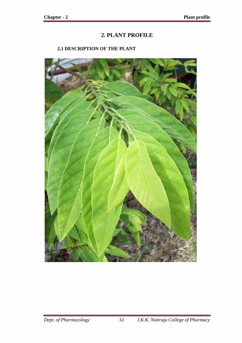
DESCRIPTION
Annona squamosa is a small, semi-deciduous tree, 3-7 m in height, with a
broad, open crown or irregularly spreading branches; bark light brown with visible
leaf scars and smoothish to slightly fissured into plates; inner bark light yellow and
slightly bitter; twigs become brown with light brown dots(lenticels).

Chapter - 2 Plant profile
Dept. of Pharmacology 34 J.K.K. Nattraja College of Pharmacy
LEAVES
Leaves occur singly, 6-17 x 3-6 cm, lanceolate or oblong lanceolate, pale
green on both surfaces and glabrate or nearly so; sides sometimes slightly unequal;
edges without teeth, inconspicuously hairy, at least when young, minutely dotted on
examination with a lens; thin, dull green to dark green on top surface, and pale blue-
green and covered with bloom on underside; apex short or long pointed; base short
pointed or rounded; petioles 0.6-1.3 cm long, green, sparsely pubescent.
FLOWERS
Flowers greenish-yellow, fragrant, on slender hairy stalks, produced singly
or in short lateral clusters about 2.5 cm long, 2-4 flowers but not at the base of the
leaves; sepals pointed, hairy, green, about 16 mm long; 3 outer petals oblong, thick
and rounded at the tips, fleshy, 1.6-2.5 cm long,0.6 cm wide, yellow-green, slightly
hairy, inside light yellow and keeled with a purplish or reddish spot at the thin,
enlarged base; inner petals 3 minute, ovate, pointed scales; stamens very numerous,
crowded, white and less than 16 mm long; ovary light green, styles white, crowded
on the raised axis.
FRUIT
The aggregate fruit formed from the numerous pistils of a flower, which are
loosely united, is soft and distinct from other species of the genus.Each pistil forms a
separate tubercle, mostly 1.3-1.9 cm long and 0.6-1.3cm wide. Fruit is round, heart
shaped, ovate or conical, 5-10 cm in diameter, with many round protuberances;
greenish-yellow when ripe, witha white, powdery bloom; the pulp is white, edible
and sweetly aromatic; ineach carpel is embedded a seed, oblong, shiny and smooth,
blackish ordark brown, 1.3-1.6 cm long, numerous.
GEOGRAPHIC SPREAD
Annona squamosa native range extends from Africa to Asia including India,
Philippines, China, Indonesia, Malaysia, Thailand, and Vietnam, eastern Papua,
New Guinea and Northern Territories (Australia) (PIER, 2003). Introduction of it in

Chapter - 2 Plant profile
Dept. of Pharmacology 35 J.K.K. Nattraja College of Pharmacy
the Federated States of Micronesia, Fiji, Guam, Saipan, Hawaii islands and Marshall
Islands has been documented.
ECOLOGY
A. squamosa is distributed throughout the tropics and is preeminently a
desert fruit. Trees do well in hot and relatively dry climates such as those of the low-
lying interior plains of many tropical countries.A. squamosa has the reputation,
particularly in India, of being a hardy, rought-resistant crop. This is only partly
correct. Although the rest period and leaf fall enable the tree to bridge a severe dry
season, it requires adequate moisture during the growing season, responding well to
supplementary irrigation. The importance of moisture is borne out by the fact that in
India as well as Southeast Asia, fruit set is largely limited to the onset of the rains,
notwithstanding the prolonged flowering season. Trees are common on the dry coast
of Puerto Rico, and also grow in Vieques, St Croix, St Thomas, StJohn, Tortola, and
Virgin Gorda.
PARTS USED
Annona seeds, Annona leaves, Annona bark, twigs.
ACTIVE INGREDIENTS
The active ingredients in Annona squamosa includeglycosides,alkaloids,
saponins, flavonoids, tannins,carbohydrates, proteins, phenolic compounds,
phytosterols and amino acids.
PHARMACOLOGICAL EFFECTS
Folkloric record reports its use as an insecticide and an anti-tumor agent,
anti-diabetic, anti-oxidant and anti-lipidimic activity, anti-inflammatory activities
due to presence of cyclic peptides. In addition, the crushed leaves are sniffed to
overcome hysteria and fainting spells, they are also applied on ulcers and wounds,
and a leaf decoction is taken in case of dysentery.
Chapter - 2 Plant profile
Dept. of Pharmacology 36 J.K.K. Nattraja College of Pharmacy
MEDICINAL USES
The bark of custard apple tree can be used to stop diarrhea in children and
adults. In addition, the plant is effective to treat diabetes.
Its fruit is used to make a hair tonic in some parts of India.
The plant bears some amazing medicinal qualities, like serving as an
insecticide, anti-ovulatory and abortificient.
The grounded seeds can be applied on hair, to get rid of lice. However, make
sure that it does not come in contact with ice or else, it can irritate the eye,
leading to blindness.
Custard apple can treat burning sensation, as it is an effective coolant.
It is used to produce sugar wine apple and is the perfect plant for indoors.
The crushed leaves of the tree are used to treat hysteria (fearful state of
mind) and fainting spells.
The treatment of ulcer, wound, dysentery and other ailments is also done by
its concentrated leaf extract (in which the leaf is boiled and its essence is
extracted).
The good part of custard apple tree is that its remains disease free most of
the time; however, it is susceptible to fungus and wilt.
Ants can create problems for the fruit, by producing mealy bugs on it.
The roots of the tree are quite powerful and can cause abortions; hence,
expecting mothers should take care while eating the herb.
Chapter 3 Literature review
Dept. of Pharmacology 37 J.K.K. Nattraja College of Pharmacy
3. LITERATURE REVIEW
Mohamed Saleem et al., 2008 investigated the hepatoprotective effect of alcoholic
and water extract of Annonasquamosa (custard apple) hepatotoxic animals with a
view to explore its use for the treatment of hepatotoxicity in human. These extracts
were used to study the Hepatoprotective effect in isoniazid + rifampicin induced
hepatotoxic model. There was a significant decrease in total bilirubin accompanied
by significant increase in the level of total protein and also significant decrease in
ALP, AST, ALT and γ-GT in treatment group as compared to the hepatotoxic group.
In the histopathological study the hepatotoxic group showed hepatocyticnecrosis and
inflammation in the centrilobular region with portal triaditis. The treatment group
showed minimal inflammation with moderate portal triaditis and their lobular
architecture was normal. It should be concluded that the extracts of
Annonasquamosa were not able to revert completely hepatic injury induced by
isoniazid + rifampicin, but it could limit the effect of these drugs in liver. The effect
of extracts compared with standard drug silymarin.
SharmaAbhishek et al., 2009 investigated the macroscopically, microscopically&
preliminary phytochemical studies on the leaf of AnnonasquamosaLinn., All the
parameters were studied according to the WHO &Pharmacopoeial guidelines. The
qualitative phytochemical fingerprint of the methonalic extract revealed the presence
of alkaloids, terpenoids, phenolics, fats and waxes. The aqueous leaf slurry was
found to be safe at the dose level of 2g/kg body weight of mice.
Aditya V et al., 2010 investigatedthe separation and identification of the active
compounds against head lice from the ethyl acetate extract of Annonasquamosa
seed. Chromatographic and spectroscopic techniques revealed that two major
compounds of the hexane seed extract were oleic acid and triglyceride with one
oleate ester. The yields of these compounds were 13.88% and 7.70% dry weight,
respectively. The compounds were tested in vitro against head lice, comparing to the
crude ethyl acetate extract of the seed. The triglyceride with one oleate ester and the
crude ethyl acetate extract diluted with coconut oil 1:1. These compounds were
found to kill all tested head lice in 10 and 31 minutes, respectively. The triglyceride
Chapter 3 Literature review
Dept. of Pharmacology 38 J.K.K. Nattraja College of Pharmacy
ester can be used as a marker for quantitative analysis of the active compound for
quality control of the raw material A. squamosa seed and its extract. This finding
will be useful for quality assessment and the chemical stability of the anti-head lice
preparation from this plant.
Pardhasaradhi et al., 2005 investigatedthepesticidal,parasiticidal,anti-microbial,
cell growth inhibitory activities. In this study, organic and aqueous extracts from
thedefatted seeds of Annonasquamosa(custard apple) were tested on different human
tumor cell lines for antitumouralactivity. While organic and aqueous extracts
induced apoptosis in MCF-7 and K-562 cells, they failed todo so in COLO-205
cells. Treatment of MCF-7 and K-562 cells with organic and aqueous extracts
resulted in nuclear condensation, DNA fragmentation, induction of reactive oxygen
species (ROS) generation and reduced intracellular glutathione levels. In addition
down regulation of Bcl-2 and PS externalization by Annexin-V stainingsuggested
induction of apoptosis in MCF-7 and K-562 cells by both the extracts through
oxidative stress. Onthe contrary, COLO-205 cells showed only PS externalization
but no change in ROS and glutathione levels. Theseobservations suggest that the
induction of apoptosis by A. squamosaextracts can be selective for certain types of
cancerous cells.
RakeshRanjan et al., 2008 investigatedthephytochemical investigations of
Annonasquamosaseeds have led to the isolation of three lignans consisting of
coumarin moiety, Cleomiscosin A, Cleomiscosin B and Cleomiscosin C. Their
structures were arrived at by detailedspectroscopic analysis. Cleomiscosin A and
Cleomiscosin B are position isomer.
Nighat Begum et al., 2010 investigated the crude ethanol extracts of
Calotropisproceraand Annonasquamosaleaves have beenscreened for their against
Muscadomestica. The third instar larvae of housefly were treated with thedifferent
concentrations of both the extracts by dipping method for 48 h. The LC50 values of
the extracts of C. proceraandA. squamosaleaves were found to be 282.5 and 550
mgl-1, respectively. The phytochemical analysis of these extracts suggested
presence of alkaloids as the major component. The larvae were exposed to 5 and
Chapter 3 Literature review
Dept. of Pharmacology 39 J.K.K. Nattraja College of Pharmacy
10% concentrations of the LC50 value of each extract along with their control sets to
evaluate their effect on metamorphosis, nucleic acid and protein content in different
developmental stages. The leaf extract of C. procerawas found to be more active in
terms of insecticidal potential. The data indicate that the leaf extracts of these plants
may be utilized as the probable candidates for the development of bio insecticides to
control the population of Muscadomesticaas safer and economic alternativesto the
synthetic insecticides.
Kaleem M et al., 2008 investigated the possible therapeutic effects of
Annonasquamosa (A.squamosa) extract on certain biochemicalmarkers in
streptozotocin (STZ) –induceddiabetes mellitus in rats.
M. Lebrini et al., 2010 investigated the Annonasquamosaplant have been studied as
possible corrosioninhibitor for C38 steel in molar hydrochloric acid (1 M HCl).
Potentiodynamic polarization and ACimpedance methods have been used. The
corrosion inhibition efficiency increases on increasing plantextract concentration.
Polarization studies showed that Annonasquamosa extract was mixed-typeinhibitor
in 1 M HCl. The inhibition efficiency of Annonasquamosa extract was
temperaturedependentand its addition led to an increase of the activation corrosion
energy revealing a physicaladsorption between the extract and the metal surface.
The adsorption of the Annonasquamosaextract followed Langmuir’s adsorption
isotherm. The inhibitive effect of Annonasquamosa is ascribed to thepresence of
organic compounds in the extract. The examined extract is considered as non-
cytotoxicsubstance.
Marta Souza et al., 2007 investigated the anthelmintic activity against
Haemonchuscontortus, the main nematodeof sheep and goat in Northeastern Brazil.
A compound 1 was isolated from ethyl acetate extract and inhibited theegg hatching
of H. contortus at 25 mg ml−1. The structure of 1 was determined as a C37 trihydroxy
adjacent bis-tetrahydrofuranacetogenin based on spectroscopic analysis.
Pardhasaradhi et al., 2004 investigated the rat histiocytictumor cell line,AK-5.Both
the extracts caused significantApoptotictumor cell death with enhanced capase-3
Chapter 3 Literature review
Dept. of Pharmacology 40 J.K.K. Nattraja College of Pharmacy
activity, down regulation of anti-apoptotic genes BclXL.In addition, DNA
fragmentation and annexine-v staining,confirmed that the extracts induced apoptosis
in tumor cell through the oxidativestress.Aqueous extracts of Annona squamosa
seeds possessed significant anti-tumor activity in vivo against AK-5 tumor.
.
Audrey Leatemia et al., 2004 investigated the inhibition of larval growth against
thepolyphagouslepidopteranSpodopteralitura (Noctuidae). Extracts of A.squamosa
weresignificantly more active (20-fold) than those of A.muricata.A. squamosa
collected fromNamlea yielded the extracts with the greatest inhibitory activity.
There were significantdifferences among locations for both A. squamosa and A.
muricata but not for L. domesticumand S. koetjape. Extracts of A. squamosa,
collected from Namlea, inhibited larval growth in a dose-dependent manner, with a
dietary EC50(effective concentration to inhibit growth by 50% relative to controls) of
191.7 ppm fresh weight. Extracts of A. squamosa collectedfrom individual trees in
Namlea also varied in growth inhibitory effect against S. litura andTrichoplusiani
larvae. This species is a candidate for development of a botanical insecticidefor local
use in Indonesia.
Mohamed Saleem et al., 2011 investigated the protective effect of methonalic
extract of Annonasquamosa onisoniazid-rifampicin-induced hepatotoxicity in rats.
Rats were divided into five different groups (n=6), group 1served as a control,
Group 2 received isoniazid (100 mg/kg, i.p.) and co-administered with rifampicin
(100mg/kg, i.p.), in sterile water, group 3 and 4 served as extract treatment groups
and received 250 & 500 mg/kgbw, p.omethonalic extract of Annonasquamosa and
group 5 served as standard group and received Silymarin 2.5 mg/kg bw, p.o. All the
treatment protocols followed 21 days and after rats were sacrificed blood and liver
were used for biochemical and histological studies, respectively. Administration of
isoniazid and rifampicincaused a significant elevation in the levels of liver marker
enzymes and thiobarbituric acid reactive substances (TBARS, oxidative stress
markers) in experimental rats. Administration of methonalic extracts of
Annonasquamosa significantly prevented isoniazid-rifampicin-induced elevation in
the levels of serum diagnostic livermarker enzymes (alanine amino transferase
(ALT), aspartate amino transferase (AST), alkaline phosphatase(ALP) and gamma

Chapter 3 Literature review
Dept. of Pharmacology 41 J.K.K. Nattraja College of Pharmacy
glutamate trans peptidase (γ-GT)), serum bilirubin, and TBARS level in
experimental groups of rats. Moreover, total protein and reduced glutathione (GSH)
levels were significantly increased in treatment group. The effect of extract was
compared with a standard drug, silymarin. The changes in biochemical parameters
were supported by histological profile. It is to be concluded that the methonalic
extract of Annonasquamosa protects against isoniazid and rifampicin-induced
oxidative liver injury in rats.
NehaPandey et al., 2011 investigated the antioxidant, anti diabetic,
hepatoprotective, cytotoxic activity, gene toxicity, antitumor activity, antilice
activities of aqueous and methonalic extracts of Annonasquamosa leaves and roots.
It is related to contain alkaloids,carbohydrates, fixed oils, tannins & phenolic
compounds.
Tylor Johns et al., 2011 investigated the antimalarial activity of ethonalic extract of
Annonasquamosa bark, which is traditionally used in diseases including infections
associated with malarial parasites. N-Nitrosoxylopine, roemerolidine and
Duguevalline were isolated from the extract of bark. All compounds showed
moderate activity against chloroquine sensitive strain (D10) and a chloroquine
resistant strain (Dd2) of Plasmodium falciparum with IC50 values ranging between
7.8 and 34.2μM/mL. N-Nitrosoxylopine also showed cytotoxicity in MTT assay
while no cytotoxicity was observed for other two compounds.
Dinesh K. Yadav et al., 2011 investigated anticancer activity of ethonalic,
chloroform, butanalic and aqueous extracts of Annonasquamosa twigs, resulted in
isolation and identification of twelve known (1-12) compounds among them one 1-
(4-β-D-glucopyranosyloxyphenyl)-2-(β-D-glucopyranosyloxy)-ethane(11) is
synthetically known but first time isolated from natural sources. Their structures
were elucidated using 1D and 2D NMR spectroscopic analysis. The isolated
compounds (2-8, 11) were evaluated for H+ K+-ATPase activity. Three of these
compounds (+)-O-methylarmepavine, N-methylcorydaldine,isocorydine showed
promisinganti-secretory activity. Activity of these compounds, comparable to
standard drug omeprazole is novel to our finding. Moreover, there is no information
Chapter 3 Literature review
Dept. of Pharmacology 42 J.K.K. Nattraja College of Pharmacy
accessible regarding the pharmacological effect of Annonasquamosa on the
gastrointestinal system. This study is the first of its kind to show significant anti-
ulcer effect of Annonasquamosa. Present study aimed to evaluate the gastro
protective effect of Annonasquamosa (AS) and to identify its active constituents.
Anti-ulcer activity was evaluated against cold restraint (CRU), pyloric ligation (PL),
aspirin (ASP), alcohol (AL) induced gastric ulcer and histamine (HA) induced
duodenal ulcer model and further confirmed through in vitro assay of H+ K+-
ATPase activity and plasma gastrin level. AS and its chloroform and hexane fraction
attenuated ulcer formation in CRU, PL, HA model and displayed anti-secretory
activity in vivo through reduced free, total acidity and pepsin in PL, confirmed by in
vitro inhibition of H+ K+-ATPase activity with corresponding decrease in plasma
gastrin level. Cytoprotection of AS was apparent with protection in AL, ASP models
and enhanced mucin level in PL.
L. G. Matos et al., 2002 investigated acetic acid-induced abdominal writhing, the
tail flick test and carrageenan-induced peritonitis were used to study the analgesic
and anti-inflammatory activity of the crude ethanolic extract from
Spirantheraodoratissimaroots. Pentobarbital-induced sleeping time was used to
study the central depressant effect of the extract. Theethanolic extract caused a dose
dependent inhibition of acetic acid-induced abdominal writhing and leukocyte
migration, and produced a significant, dose-related increase in the duration of sleep.
The results suggest that Spirantheraodoratissima roots contain compounds with anti-
inflammatory and central depressant actions.
Paul V Tan et al., 2002 evaluated the anti-ulcerogenic effects of the leaf methanol
extract of Ocimum suave using four ulcer models in Wistar rats. Administration of
extract to the rats showed dose dependent reduction in gastric ulcer in all four
models, accompanied by significant increase in the gastric mucus production. The
extract dose 500mg/kg in pylorus ligation model inhibited the gastric lesion
formation and less effect on gastric secretion as observed with the control. In ethanol
induced model, the dose 250mg/kg showed complete inhibition of gastric lesion.
During high gastric acidic environment the effect of extract on mucus secretion was
not much significant.
Chapter 3 Literature review
Dept. of Pharmacology 43 J.K.K. Nattraja College of Pharmacy
Akilandeswari et al., 2010 evaluated the screening of gastric antiulcer activity of
sida acute using aspirin plus pylorus ligation, aspirin induced and ethanol induced
ulcer model. The normal control exhibited very severe ulceration in aspirin plus
pylorus and aspirin. Hence aspirin proved to be most potent in gastric ulcer
induction. In aspirin (300mg/kg) plus pylorus model the statistical data 100mg/kg
shows significant when compared to famotidine group. The group 200mg/kg showed
significant activity with famotidine. In ethanol model the statistical data 100mgkg
shows significant when compared to famotidine group. The flavonoids have been
reported to possess significant antiulcer activity in various experimental model.
Jaikumar et al., investigated the antiulcer activity of methonalic extract of
Jatrophacurcus on aspirin plus pylorus ligation induced gastric lesion in Wistar
strain rats. In the present study, methonalic extract of jc have been shown to posses
antiulcer activity against experimentally induced ulcer model, methonalic extract
significantly reduced (p<o.o5) the acid secretory parameters i.e free and total acidity,
gastric volume and ulcer index. Aspirin plus pylorus group show the ulcerated
mucosa with hemorrhage and discontinuity of lining epithelium while cut showed the
normal mucosa with mild hyperplasia and mild edematous sub mucosa, compared to
ranitidinetreated group which show the normal mucosa with no ulcer.
Kannappan et al., 2008 Studied the effect of methonalic extract of leaves of
Jatrophacurcas Linn on pylorus ligation and aspirin induced ulcer in Wistar rats.
Administration of extract dose (100mg/kg) and ranitidine (50mg/kg) for 5 days to the
aspirin plus pylorus ligation rats and its parameter were observed. The extract
showed the markedly reduction in the acid parameter like gastric volume, pH , free
acidity and total acidity. Treatment with extract 100 and 200mg/kg showed dose
dependent reduction in ulcer index. Histopathological studies revealed that the
stomach mucosa showed the protective action of extract against mucosal damage
caused by aspirin.
Malairajan et al., 2006 evaluated the anti-ulcer activity of Toonaciliata
roemeragainst aspirin plus pylorus ligation (ant secretory), ethanol-induced ulcer

Chapter 3 Literature review
Dept. of Pharmacology 44 J.K.K. Nattraja College of Pharmacy
and stress induced ulcer in rats. The plant extract markedly showed gastroprotective
activity when compared with that of standard drug sucralfate. The extract showed
decrease in the ulcer index and significant reduction in gastric volume, free acidity,
total acidity.
Sunita M et al.., investigated the anti-ulcer activity of Benincasahispada(Thunb)
fruit. The anti-ulcer activity was evaluated in rats against ethanol-induced gastric
mucosal damage, pylorus-ligated gastric models and cold restraint-stress induced
gastric ulcers. Many parameters were studied for the models employed in ulcer
studies of which ulcer index was common one for all the models. Vascular
permeability was evaluated in the ethanol model. Effect of peroxidation,
vizmelondialdehyde (MDA) content, superoxide desmutase (SOD) and catalase
(CAT) were studied in Cold Restraint Stress model. The results showed significant
reduction in ulcer index in all the models and the results were comparable with the
Omeprazole-treated group.
B Rajkapoor et al., investigated the effect of alcoholic extract of dried fruits of
Carica papaya in rats to evaluate the antiulcer activity by using pyloric ligation and
aspirin-induced gastric ulcer. The parameters taken to assess antiulcer activity were
volume of gastric secretion, free acidity, total acidity and ulcer index. The results
indicate that the alcoholic extract significantly (P<0.001) decreases the volume of
gastric acid secretion, free acidity, total acidity and ulcer index with respect to
control.
S. Narayan et al., investigated the protective effect of ambrex, a poly-herbal drug,
in ethanol induce gastric mucosal lesions in experimental rats. The response to
ambrex was assessed from ulcer index, cell proliferation, histopathological changes
and alkaline phosphatase (ALP) activity. Ambrex pre-treatment showed protection
against ethanol-induced gastric mucosal damage, a significant reduction in the ulcer
index and ALP activity and an increase in DNA content.
Madhu et al., investigated the anti-ulcer activity of Wrightiatinctora(Roxb). The
antiulcer activity of the Wrightiatinctoriamethonalic extract and Wrightiatinctoria
Chapter 3 Literature review
Dept. of Pharmacology 45 J.K.K. Nattraja College of Pharmacy
70% ethanolic extract were compared with carboxy methyl cellulose (CMC),
pylorus control, Aspirin and standard famotidine which was evaluated by
employing aspirin plus pylorus ligation induced ulcer model. The Biochemical
parameters like volume of gastric juice secretion, pH, free acidity, total acidity, ulcer
index and percentage inhibition were studied at the concentration of 200
mg/kg body weight. The results showed significant gastro-protective effect when
compared to the standard drug Famotidine.
Chapter 4 Aim and Objective
Dept. of Pharmacology 46 J.K.K. Nattraja College of Pharmacy
4. AIM AND OBJECTIVE
Medicinal plants have been in India for centuries as a therapeutic source for
treating wide variety of ailments and have been found to be of immense global
importance. It is estimated by World Health Organization that, 80% of the world
population must rely on traditional medicines for health care: these traditional
medicines are mainly plant based. Most of the studies demonstrate the importance of
natural products in drug discovery. The use of phytoconstituents as drug therapy to
treat major ailments has proved to be clinically effective and less relatively toxic
than the existing drugs. The selection of plant was made on the basis of its
availability, therapeutic value and degree of research work which is not done.
Peptic ulcer disease(PUD) is a serious gastrointestinal disorder that requires
a well targeted therapeutic strategy. It includes number of drugs such as proton
pump inhibitors and H2 receptor antagonists are available for the treatment of peptic
ulcer, but clinical evaluation of these drugs has shown incidence of relapse, side
effects, and drug interaction. This has been the rational for the development of new
antiulcer drugs and the search for novel molecules has been extended to herbal drugs
that offer better protection and decreased relapse. Drug of plant origin are gaining
popularity and are being investigated for a number of disorders, including peptic
ulcer.
Peptic ulcer occurs due to an imbalance between aggressive (acid, pepsin)
and defensive (gastric mucosal barrier) factors of gastric mucosa. Local mechanism
implicated in mucosal hydrophobicity, rapid epithelial cell renewal and rich
mucosal blood flow. PGE2 and PGI2 are predominant prostaglandins synthesized by
the gastric mucosa and are known to inhibit the secretion of gastric acid and
stimulate the secretion of mucus and bicarbonate. The treatment of peptic ulcer is
directed against either reduction of aggressive factors or enhancement of mucosal
defense of stomach and duodenum with cytoprotective agents.
In India, PUD is common. In the Indian Pharmaceutical industry, antacids
and antiulcer drugs share 6.2 billion rupees and occupy 4.3% of the market share.
Today, there are two main approaches for treating peptic ulcer. The first deals with
Chapter 4 Aim and Objective
Dept. of Pharmacology 47 J.K.K. Nattraja College of Pharmacy
reducing the production of gastric acid and second with re-enforcing gastric mucosal
barrier. (Dharmani P et al., 2006)
Analgesia may be loss of sensation of pain that result from an interruption in
the nervous system pathway between sense organ and brain. Different form of
sensation (touch, temperature and pain) stimulating an area of skin travels to the
spinal cord by different nerves fibres in the same nerve bundle.
According to the current unifying concept of NSAIDS action, during pain,
inflammation, fever, arachidonic acid is liberated from phospholipase fraction of the
cell membrane; arachidonic acid is then converted to prostaglandins via cyclo-
oxygenase (COX 1 & 2) pathway. COX-1 is constitutively present in stomach,
kidney and blood vessels whereas COX-2 is inducible in activated leucocytes and
other inflammatory cells. It is postulated that PGs sensitize blood vessels to the
effects of mediators such as 5-HT, bradykinin and histamine that increase the
permeability. PGs particularly PGE and PGI produce hyperalgesia associated with
inflammation. Pain can be constant (chronic) or fleeting and come and go (acute).
There are many herbs that are useful and effective for pain relief many are safe for
everyone but some should be avoided.
OBJECTIVES OF THE STUDY
The objectives of the present study are-
Exploring the traditional medicines with proper chemical and
pharmacological profiles.
Extraction of Annona squamosa leaf by different solvents.
To conduct systematic phytochemical investigation of Annona squamosa.
To evaluate the antiulcer activity in rats.
To evaluate the analgesic activity in mice.
Chapter- 5 Plan of Work
Dept. of Pharmacology 48 J.K.K. Nattraja College of Pharmacy
5. PLAN OF WORK
PHYTOCHEMICAL STUDIES
1. Collection of plant ( Annona squamosa ) leaves and shade dried.
2. Extraction of Annona squamosa leaves powder using ethanol in soxhlet
apparatus for 72 hrs.
3. Phytochemical investigation of ethanolic extract of Annona squamosa leaf.
PHARMACOLOGICAL STUDIES
1. Evaluation of anti-ulcer activity of Annona squamosa by
Aspirin induced ulcer model
Pylorus ligation model
1.1 Evaluation of analgesic activity of Annona squamosa by
Acetic acid induced writhing in mice
1.2 Determination of following parameters
Ulcer index
Percentage inhibition
Gastric volume
pH of gastric juice
Total acidity
Free acidity
1.3 Histopathological studies
Chapter-6 Materials and Methods
Dept. of Pharmacology 49 J.K.K. Nattraja College of Pharmacy
6. MATERIALS AND METHODS
PLANT MATERIAL
The fresh leaves of Annona Squamosa L were collected from local area at
komarapalayam, Tamilnadu. The material was taxonomically identified, confirmed
and authenticated by Botanical survey of India (BSI) at TN agricultural
university, Coimbatore. With authentication no BSI/SRC/5/23/2011-12/Tech.-
1201 and the voucher specimen was retained in our laboratory for further reference.
The collected leaves were shade dried and the dried material was crushed to coarse
powder with mechanical grinder. The powder was stored in air-tight container which
was used for extraction.
6.1. PHYTOCHEMICAL STUDIES
6.1.1 EXTRACTION OF PLANT MATERIAL
Ethanolic extraction
About 300 gm of air dried powdered material was taken in 1000ml soxhlet
apparatus and extracted with petroleum ether for 18 hours till the solvent became
colourless. At the end of the extraction process the marc was taken out and it was
dried. After drying, the powdered marc was weighed & again packed and extracted
with ethanol for another 72 hours till it became colourless. After that extract was
concentrated by distillation. The final solution was evaporated, to obtain a syrupy
greenish mass.
6.1.2. PRELIMINARY PHYTOCHEMICAL ANALYSIS
The plant may be subjected to preliminary phytochemical screening for
detection of various bioactive chemical constituents. The important steps involved
in the phytochemical screening of a plant include extraction of constituents using
suitable solvents followed by screening with various chemical tests.
In the process of phytochemical screening, the crude extracts or isolated
constituents are subjected to qualitative and quantitative chemical analyses.
Qualitative chemical analysis includes the determination of nature of the
Chapter-6 Materials and Methods
Dept. of Pharmacology 50 J.K.K. Nattraja College of Pharmacy
constituents in an extract or its fractions which lead to the isolation of the active lead
compound. Quantitative chemical analysis includes the determination of the purity
of isolated substances or group of substances in a mixture by finger printing and
different analytical techniques.
Qualitative tests
A) Test for carbohydrates
1. Molisch Test
It consists of treating the compounds of -naphthol and concentrated
sulphuric acid along the sides of the test tube. Purple color or reddish violet color at
the junction between two liquids.
2. Fehling’s Test
Equal quantity of Fehling’s solution A and B is added. Heat gently, brick
red precipitate is obtained.
3. Benedict’s test
To the 5ml of Benedict’s reagent, add 8 drops of solution under examination.
Mix well, boiling the mixture vigorously for two minutes and then cool. Red
precipitate is obtained.
4. Barfoed’s test
To the 5ml of the Barfoed’s solution add 0.5ml of solution under
examination, heat to boiling, formation of red precipitate of copper oxide is
obtained.
B) Test for Alkaloids
1. Dragendroff’s Test
To the extract, add 1ml of Dragendroff’s reagent Orange red precipitate is
produced.
2. Wagner’s test
To the extract add Wagner reagent. Reddish brown precipitate is produced.
Chapter-6 Materials and Methods
Dept. of Pharmacology 51 J.K.K. Nattraja College of Pharmacy
3. Mayer’s Test
To the extract add 1ml or 2ml of Mayer’s reagent. Dull white precipitate is
produced.
4. Hager’s Test
To the extract add 3ml of Hager’s reagent, yellow precipitate is produced.
C) Test for Steroids and Sterols
1. Libermann Burchard test
Dissolve the test sample in 2ml of chloroform in a dry test tube. Now
add 10 drops of acetic anhydride and 2 drops of concentrated sulphuric acid. The
solution becomes red, then blue and finally bluish green in color.
2. Salkowski test
Dissolve the sample of test solution in chloroform and add equal volume of
conc. sulphuric acid. Bluish red cherry red and purple color is noted in chloroform
layer, whereas acid assumes marked green fluorescence.
D) Test for Glycosides
1. Legal’s test
Sample is dissolved in pyridine; sodium nitropruside solution is added to
it and made alkaline. Pink red color is produced.
2. Baljet test
To the drug sample, sodium picrate solution is added. Yellow to orange
color is produced.
3. Borntrager test
Add a few ml of dilute sulphuric acid to the test solution. Boil, filter and
extract the filtrate with ether or chloroform. Then organic layer is separated to which
ammonia is added, pink, red or violet colour is produced in organic layer.
Chapter-6 Materials and Methods
Dept. of Pharmacology 52 J.K.K. Nattraja College of Pharmacy
4. Killer Killani test
Sample is dissolved in acetic acid containing trace of ferric chloride and
transferred to the surface of concentrated sulphuric acid. At the junction of liquid
reddish brown color is produced which gradually becomes blue.
E) Test for Saponins
Foam test
About 1ml of alcoholic sample is diluted separately with distilled water to
20ml, and shaken in graduated cylinder for 15 minutes.1 cm layer of foam indicates
the presence of Saponins.
F) Test for Flavonoids
Shinoda test
To the sample, magnesium turnings and then concentrated hydrochloric acid
is added. Red color is produced.
Lead acetate test
The extracts were treated with few drops of lead acetate solution, formation
of yellow
Precipitate indicates the presence of flavonoids.
Alkaline reagent test
The extracts were treated with few drops of sodium hydroxide separately.
Formation of intense Yellow color, which becomes colorless on addition of few
drops of dilute acid, indicates the presence of flavonoids.
G) Test for Triterpenoid
In the test tube, 2 or 3 granules of tin was added, and dissolved in a 2ml of
thionyl chloride solution and test solution is added. Pink colour is produced which
indicates the presence of triterpenoids.
Chapter-6 Materials and Methods
Dept. of Pharmacology 53 J.K.K. Nattraja College of Pharmacy
H) Test for Protein and Amino acid
1. Biuret test
Add 1 ml of 40% sodium hydroxide and 2 drops of 1% copper sulphate to
the extract, a violet colour indicates the presence of proteins.
2. Ninhydrin test
Add 2 drops of freshly prepared 0.2% ninhydrin reagent to the extract and
heat. A blue colour develops indicating the presence of proteins, peptides or amino
acids.
3. Xanthoprotein test
To the extract, add 20% of sodium hydroxide or ammonia. Orange colour
indicates presence of aromatic amino acid. (Evans W.C 1996, Kandelwal, 2004.)
6.2 ACUTE ORAL TOXICITY STUDIES
Animals
Swiss albino mice of female sex weighing 20-25gms were used for the study.
The animals were obtained from Agricultural University, Manuthy, Trissur and were
housed in polypropylene cages. The animals were maintained under standard
laboratory conditions (250 + 20C; 12hr light and dark cycle). The animals were fed
with standard diet and water ad libitum. Ethical clearance (for handling of animals
and the procedures used in study) was obtained from the Institutional Animal Ethical
Committee before performing the study on animals.
Acute toxicity Test
Acute oral toxicity study for ethanol extract of Annona squamosa leaves was
carried out as per OECD guideline 425 (Up and Down procedure). The test
procedure minimizes the number of animals required to estimate the acute oral
toxicity. The test allows the observation of signs of toxicity and can also be used to
identify chemicals that are likely to have low toxicity.
Animals were fasted (food but not water was with held overnight) prior to
dosing. The fasted body weight of each animal was determined and the dose was
calculated according to the body weight.
Chapter-6 Materials and Methods
Dept. of Pharmacology 54 J.K.K. Nattraja College of Pharmacy
6.3 PHARMACOLOGICAL EVALUATION
6.3.1 EVALUATION OF ANTI-ULCER ACTIVITY
a) Animal study
Male albino-Wistar rats, weighing 150-200g were used in the present study.
Housing and feeding condition
All the rats were kept at room temperature (22±30◦C) in the animal house.
All the animals were housed and treated as per the internationally accepted ethical
guidelines for the care of laboratory animals. Prior to the experiments, rats were fed
with standard food and were acclimatized to standard laboratory conditions of
temperature (22±30˚) and maintained an 12:12 hr light: dark cycle . They were
provided with regular rat chow and distilled water ad libitum. All the experimental
procedures were performed on animal after approval from the ethics committee and
in accordance with the recommendations for the proper care and use of laboratory
animals.
Extract
Ethanolic extract of Annona squamosa leaves.
Standard drug used
Omeprazole (20mg/kg)
Acetic acid (1%v/v)
Pentazocine (30mg/kg)
Drug omeprazole and the extract of Annona squamosa were suspended in
0.5% CMC.
b) Anti-ulcer activity
The anti-ulcerogenic potential of the leaf ethanol extract of Annona
squamosa was investigated by three different models in experimental Wistar rats.
1. Aspirin induced ulcer (Anti-secretary mechanism).
2. Pylorus ligation. (Anti-secretary mechanism).
Two models were used to elucidate the possible mechanism of action of the
ethanolic extract of Annona squamosa. The respective standard drug (Omeprazole
20mg/kg,) was used for the particular experimental models.
Chapter-6 Materials and Methods
Dept. of Pharmacology 55 J.K.K. Nattraja College of Pharmacy
2. EXPERIMENTAL PROCEDURES
a) Aspirin induced ulcer
Male albino-Wistar rats were divided in to four groups as mentioned above
of six animals per group and animals were fasted for 24 hrs prior to the experiment
in perforated steel cages to avoid coprophagy.
Group I - control.
Group II - received 50mg/kg, p.o ethanol extract of Annona squamosa leaves.
Group III - received 100mg/kg, p.o ethanol extract of Annona squamosa leaves.
Group IV - received 20mg/kg, p.o Omeprazole as standard.
Group was kept as control without any treatment. One hour after the drug
treatment, the animals were treated with aspirin 200mg/kg by p.o, to induce ulcers.
The animals were sacrificed after 4hrs and stomach was opened and percentage
inhibition of ulcer was determined. (Kannappan et al., 2008, Panda et al., 1993,
Parmar NS et al., 1991, Pati K.S. et al., 2008).
b) Pylorus ligation model
The animals were divided in to four groups of six animals each as mentioned
above.
Group I - received 1% CMC (1.0ml/kg p.o) as vehicle control
Group II - received 20mg/kg, p.o Omeprazole as standard
Group III - received 50mg/kg, p.o ethanol extract of Annona squamosa leaves.
Group IV - received 100mg/kg, p.o ethanol extract of Annona squamosa leaves.
Animals in all the groups were fasted for 36 h after the respective assigned
treatment and were anaesthetized with anaesthetic ether. The abdomen was
opened by a small midline incision below the xiphoid process and pylorus
portion of stomach was lifted out and ligated. Precaution was taken to avoid
traction to the blood supply. The stomach was sutured with interrupted sutures.
Animals were allowed to recover and stabilize in individual cages and were deprived
of water during post-operative period. Four hours after the pyloric ligation, the
animals were sacrificed by an excess dose of ether. The stomach was carefully
removed and the gastric contents were collected. The gastric juice was centrifuged at
Chapter-6 Materials and Methods
Dept. of Pharmacology 56 J.K.K. Nattraja College of Pharmacy
1000rpm and gastric volume was measured. Free and total acidities of the
supernatant were determined by titration with 0.1 N NaOH and expressed as mEq/ L
/100 g. The stomach was cut open along the greater curvature and pinned onto a soft
board for evaluating the gastric ulcers and to calculate ulcer index. Ulcer scoring is
done according to the scale mentioned below.
BIOCHEMICAL PARAMETERS
The stomach was carefully excised keeping oesophagus closed and opened
along greater curvature and luminal contents were removed. The gastric contents
were collected in a test tube and centrifuged. The gastric contents were analyzed for
gastric juice volume, pH, free and total acidity.
MEASUREMENT OF GASTRIC JUICE VOLUME AND PH
Gastric juice was collected from pylorus ligation induced ulcer rats. The
gastric juice thus collected was centrifuged at 3000 rpm for 10 min. The volume of
supernatant was measured and expressed as ml/100g body weight. The pH of the
supernatant was measured using digital pH meter. (Canmon DC., et al., 1969,
Kannappan et al., 2008, Patil K.S. et al., 2008, Paul V. et al., 2000)
DETERMINATION OF FREE AND TOTAL ACIDITY
An aliquot of 1.0 ml of gastric juice was pipette out in to a 50 ml conical flask
and 2/3 drops of Topfers reagent was added to it and titrated with 0.01N NaOH until
all traces of the red color disappeared and the color of the solution turned yellowish
orange. The volume of 0.01N NaOH was noted which corresponds to free acidity.
Then 2/3 drops of phenolphthalein was added and titration was continued until a
permanent pink color was developed. The volume of total alkali consumed was
noted which corresponds to total acidity. The free acidity and total acidity was
determined using the formula and values are expressed as mEq/l 100g. (Kannappan
et al., 2008, Rajkapoor et al., 2002)

Chapter-6 Materials and Methods
Dept. of Pharmacology 57 J.K.K. Nattraja College of Pharmacy
Acidity =Volume of NaOH X Normality of NaOH X 100
(mEq/L per 100g)
0.01
ULCER INDEX (UI)
The mucosa was flushed with saline and stomach was pinned on frog board.
The lesion in glandular portion was examined under a 10x magnifying glass and
length was measured using a divider and scale and gastric ulcer was scored. Ulcer
index of each animal was calculated by adding the values and their mean values
were determined. (Malairajan et al., 2007)
0 – Normal colored stomach
0.5 – Red coloration
1 – Spot ulceration
1.5 – Hemorrhagic streak
2 – Ulcers
3 – Perforations
PERCENTAGE INHIBITION
Percentage inhibition was calculated using the following formula.
(Malairajan et al., 2007)
UI ulcer control — UI ulcer treated
% inhibition = X 100
UI ulcer control
Chapter-6 Materials and Methods
Dept. of Pharmacology 58 J.K.K. Nattraja College of Pharmacy
HISTOPATHOLOGICAL STUDIES
The stomach tissue were removed from the rats and fixed in10% formaline
saline for at least 48 hrs. These were then processed routinely and the tissue was
embedded in paraffin wax. Histological section were cut at 5-6µm and stained with
routine haemotoxylin and eosin. These were examined by a consultant
histopathologist.
6.3.9 STATISTICAL STUDIES
The data obtained by the various parameters was statistically evaluated by
one way analysis of variance (ANOVA) followed by Dunnetts‘t’ test using Graph
Pad Prism software. The mean values ± SEM were calculated for each parameter.
6.4 EVALUATION OF ANALGESIC ACTIVITY
6.4.1 ANALGESIC ACTIVITY
The leaf of ethanolic extract of Annona squamosa were undertaken for the
present study of analgesic activity by
Acetic acid induced writhing method
6.4.2 EXPERIMENTAL PROCEDURE
The analgesic activity was performed on mice, fasted for 12 hrs prior the
experiment, but provided water. The animals were divided into four groups of four
in each.
Group I - Received acetic acid 1%w/v.
Group II - Received extract 50mg/kg.
Group III - Received extract 100mg/kg.
Group IV - Standard 30mg/kg.
The activity was performed by acetic acid induced writhing method. The
ethanolic extract in dose 50 and 100 mg/kg body weight, an acetic acid (1%w/v) is
suspended in 1% suspension of carboxy methyl cellulose were injected
Chapter-6 Materials and Methods
Dept. of Pharmacology 59 J.K.K. Nattraja College of Pharmacy
intraperitoneally. An aliquot of 0.25 ml of this suspension is injected
intraperitoneally in each animal. The animal reacts with a characteristic stretching
behavior i.e., a series of constrictions occur that travel along the abdominal wall,
sometimes accompanied by turning movements of the body and extension of the
hind limbs.
A group of animals (n=4) is used as test group in which prior to acetic acid
administration, test drugs are administered orally. The mice are placed individually
into glass chambers and number of writhes is recorded for 10 min in each animal.
For scoring, a writhe is indicated by stretching of the abdomen with
simultaneous stretching of at least one hind limb.
Formula for computing percent inhibition is
Average writhes in control group-writhes in test group ×100
=
Writhes in the control group
The time period with the greatest percent of inhibition is considered the peak time
% decrease in activity= 100-(test / standard × 100)
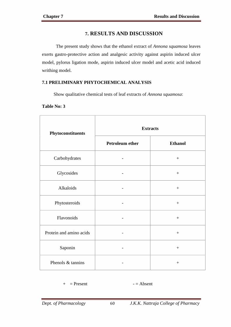
Chapter 7 Results and Discussion
Dept. of Pharmacology 60 J.K.K. Nattraja College of Pharmacy
7. RESULTS AND DISCUSSION
The present study shows that the ethanol extract of Annona squamosa leaves
exerts gastro-protective action and analgesic activity against aspirin induced ulcer
model, pylorus ligation mode, aspirin induced ulcer model and acetic acid induced
writhing model.
7.1 PRELIMINARY PHYTOCHEMICAL ANALYSIS
Show qualitative chemical tests of leaf extracts of Annona squamosa:
Table No: 3
PhytoconstituentsExtracts
Petroleum ether Ethanol
Carbohydrates - +
Glycosides - +
Alkaloids - +
Phytosteroids - +
Flavonoids - +
Protein and amino acids - +
Saponin - +
Phenols & tannins - +
+ = Present - = Absent
Chapter 7 Results and Discussion
Dept. of Pharmacology 61 J.K.K. Nattraja College of Pharmacy
The preliminary Phytochemical screening of the extract of Annona
squamosa leaves showed the presence of carbohydrates, alkaloids, sterols,
flavonoids, saponins, tannins and phenolic compounds, Protein and amino acids.
The various phytoconstituents present in the extract is shown in the TABLE 3.
The phytoconstituents like flavonoids, tannins, terpenoids, phenols and
saponins and sterols have been reported in several anti-ulcer literatures as possible
gastroprotective agents. Flavonoids, tannins and triterpenes are among the
cytoprotective active materials for which antiulcerogenic efficacy has been
extensively confirmed. (Borelli F. et al., 2000)
It is suggested that these compounds will be able to stimulate mucus,
bicarbonate and prostaglandin secretion, and counteract with the deteriorating
effects of reactive oxidants in gastrointestinal lumen. (suja pandian et.al., 2002)
Tannins are known to “tan” the outermost layer of the mucosa and render it less
permeable and more resistant to chemical and mechanical injuries. Tannins may
prevent ulcer development due to their protein precipitating and vasoconstriction
effects. Their astringent action can help precipitating micro proteins on the ulcer
site, thereby forming an impervious layer over the lining that hinders gut secretions
and protects the underlying mucosa from toxins and other irritants.(Berenguer B et
al .,2005)
Similarly, the ethanol extract of Annona squamosa leaf, showed the presence
flavonoids and their alkaloids, phenols and tannins, triterpenoids and saponins.
These phytoconstituents present in the extract could be the possible agents involved
in the prevention of gastric lesions induced by aspirin, pylorus ligation and ethanol
induced models.
Flavonoids are also capable of scavenging the free radicals.
Phenols and Phenolic compounds have potent antioxidant properties
(Berenguer et al., 2005),
The analgesic activity of Annona squamosa may be attributed to the presence
of various phytoconstituents such as alkaloids, tannins, carbohydrates, flavonoids.
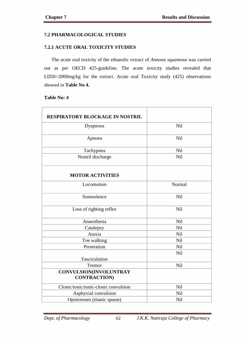
Chapter 7 Results and Discussion
Dept. of Pharmacology 62 J.K.K. Nattraja College of Pharmacy
7.2 PHARMACOLOGICAL STUDIES
7.2.1 ACUTE ORAL TOXICITY STUDIES
The acute oral toxicity of the ethanolic extract of Annona squamosa was carried
out as per OECD 425-guideline. The acute toxicity studies revealed that
LD50>2000mg/kg for the extract. Acute oral Toxicity study (425) observations
showed in Table No 4.
Table No: 4
RESPIRATORY BLOCKAGE IN NOSTRIL
Dyspnoea Nil
Apnoea Nil
Tachypnea NilNostril discharge Nil
MOTOR ACTIVITIES
Locomotion Normal
Somnolence Nil
Loss of righting reflex Nil
Anaesthesia NilCatalepsy Nil
Ataxia NilToe walking NilProstration Nil
FasciculationNil
Tremor NilCONVULSION(INVOLUNTRAY
CONTRACTION)
Clonic/tonic/tonic-clonic convulsion NilAsphyxial convulsion Nil
Opistotones (titanic spasm) Nil
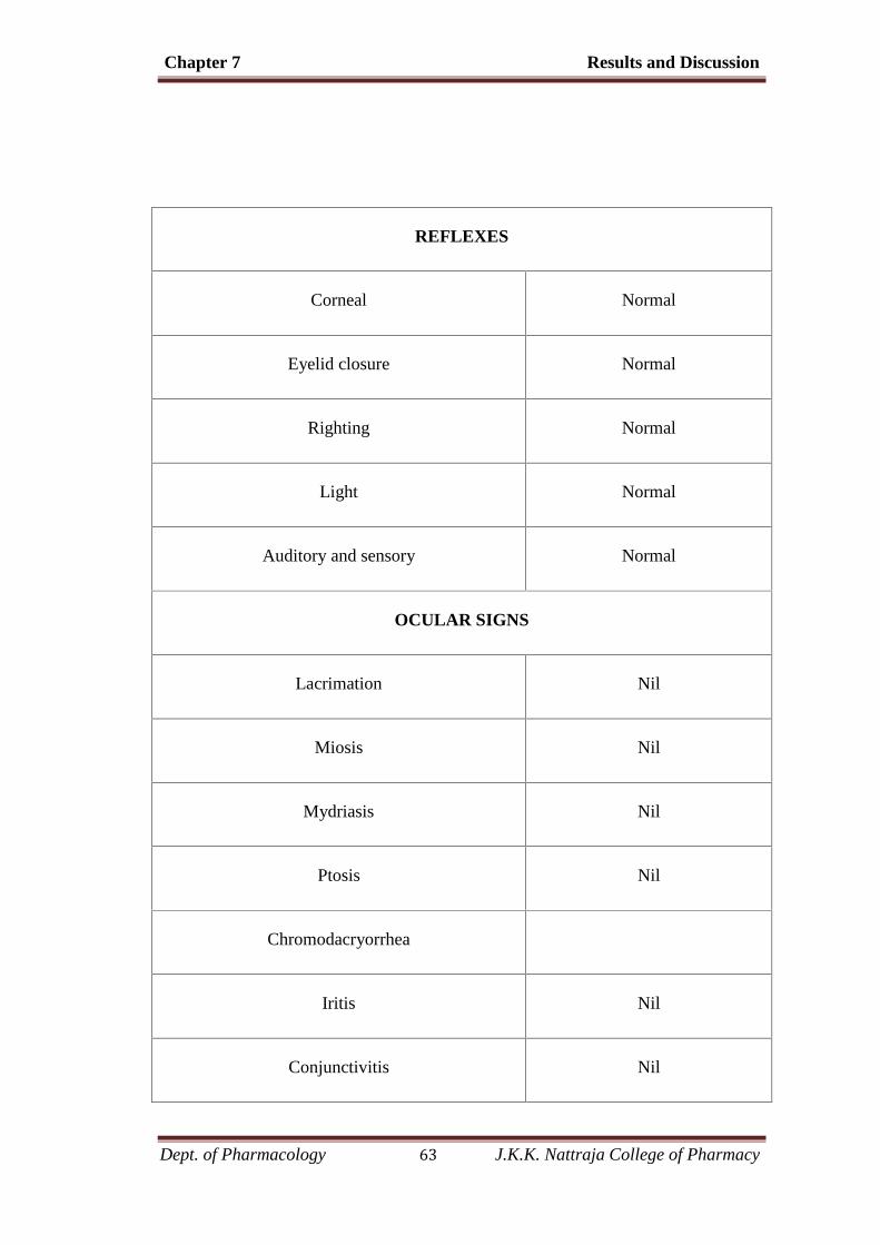
Chapter 7 Results and Discussion
Dept. of Pharmacology 63 J.K.K. Nattraja College of Pharmacy
REFLEXES
Corneal Normal
Eyelid closure Normal
Righting Normal
Light Normal
Auditory and sensory Normal
OCULAR SIGNS
Lacrimation Nil
Miosis Nil
Mydriasis Nil
Ptosis Nil
Chromodacryorrhea
Iritis Nil
Conjunctivitis Nil
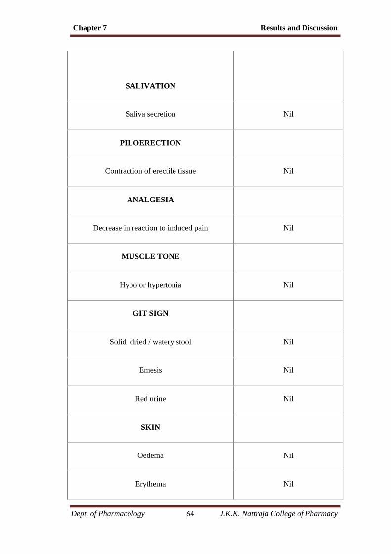
Chapter 7 Results and Discussion
Dept. of Pharmacology 64 J.K.K. Nattraja College of Pharmacy
SALIVATION
Saliva secretion Nil
PILOERECTION
Contraction of erectile tissue Nil
ANALGESIA
Decrease in reaction to induced pain Nil
MUSCLE TONE
Hypo or hypertonia Nil
GIT SIGN
Solid dried / watery stool Nil
Emesis Nil
Red urine Nil
SKIN
Oedema Nil
Erythema Nil
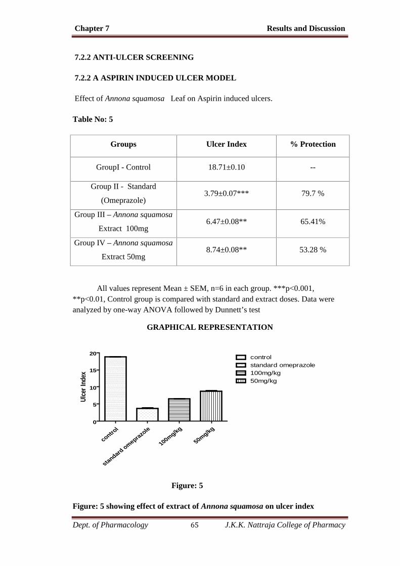
Chapter 7 Results and Discussion
Dept. of Pharmacology 65 J.K.K. Nattraja College of Pharmacy
7.2.2 ANTI-ULCER SCREENING
7.2.2 A ASPIRIN INDUCED ULCER MODEL
Effect of Annona squamosa Leaf on Aspirin induced ulcers.
Table No: 5
Groups Ulcer Index % Protection
GroupI - Control 18.71±0.10 --
Group II - Standard
(Omeprazole)3.79±0.07*** 79.7 %
Group III – Annona squamosa
Extract 100mg6.47±0.08** 65.41%
Group IV – Annona squamosa
Extract 50mg8.74±0.08** 53.28 %
All values represent Mean ± SEM, n=6 in each group. ***p<0.001,**p<0.01, Control group is compared with standard and extract doses. Data wereanalyzed by one-way ANOVA followed by Dunnett’s test
GRAPHICAL REPRESENTATION
control
standard omeprazole
100mg/kg
50mg/kg0
5
10
15
20 controlstandard omeprazole100mg/kg50mg/kg
Ulce
r Ind
ex
Figure: 5
Figure: 5 showing effect of extract of Annona squamosa on ulcer index
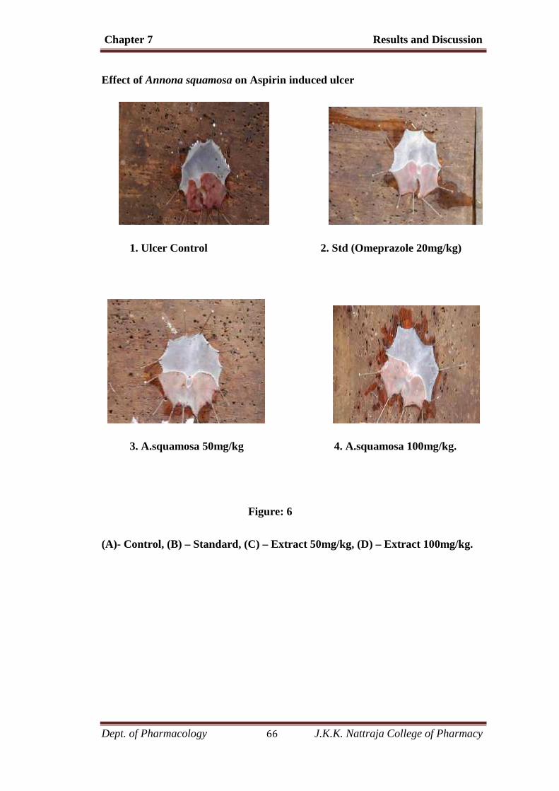
Chapter 7 Results and Discussion
Dept. of Pharmacology 66 J.K.K. Nattraja College of Pharmacy
Effect of Annona squamosa on Aspirin induced ulcer
1. Ulcer Control 2. Std (Omeprazole 20mg/kg)
3. A.squamosa 50mg/kg 4. A.squamosa 100mg/kg.
Figure: 6
(A)- Control, (B) – Standard, (C) – Extract 50mg/kg, (D) – Extract 100mg/kg.
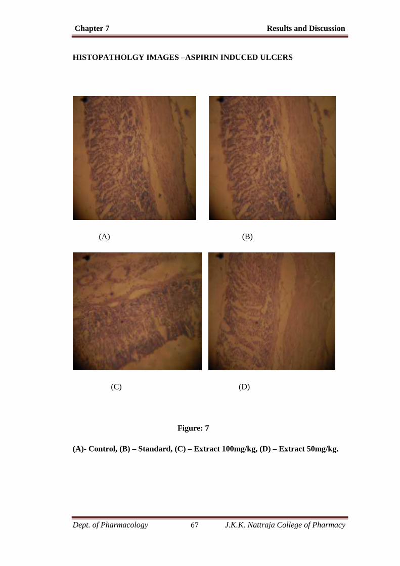
Chapter 7 Results and Discussion
Dept. of Pharmacology 67 J.K.K. Nattraja College of Pharmacy
HISTOPATHOLGY IMAGES –ASPIRIN INDUCED ULCERS
(A) (B)
(C) (D)
Figure: 7
(A)- Control, (B) – Standard, (C) – Extract 100mg/kg, (D) – Extract 50mg/kg.
Chapter 7 Results and Discussion
Dept. of Pharmacology 68 J.K.K. Nattraja College of Pharmacy
Histopathology Examination
The images and the reports demonstrate a prominent damage, loss of mucus
and chief cells as well as marked infiltration of the leucocytes to the stomach surface
of the rats in group-I treated only with aspirin. In Group-II, the standard group
showed no damage to the gastric mucosa. The histopathological sections of group-
III, treated with ethanolic extract of A.squamosa 100 mg/kg, shows a reduction in
the ulcer focus and a hyperplastic gastric mucosa with regenerating mucosal
epithelium. The section of IV- group rats shows mucosal erosion and ulceration to
the stomach surface illustrating a less protection to the mucosa and the gastric
epithelium.
Ulcer index (UI)
Oral administration of ethanol extract of Annona squamosa at doses of 50
and 100mg/kg exhibited dose dependent inhibition percentage of 53.28 and 65.41
(p<0.001) respectively compared to the ulcer control, proving the anti ulcer activity.
The standard drug omeprazole (20mg/kg) exhibited percentage inhibition of 79.7
when compared with ulcer control. Extract treated and ulcer control group was
compared with standard group.
Aspirin is a cyclooxygenase inhibitor which suppresses gastroduodenal
bicarbonate secretion, reduces endogenous prostaglandin biosynthesis and disrupts
the mucosal barrier as well as mucosal blood flow in animals. It is also well known
that prostaglandins synthesized in large quantities by the gastrointestinal mucosa can
prevent experimentally induced ulcers by ulcerogens. (Telesphore et al., 2008)
The effects of ethanolic extract of Annona squamosa on acid parameters
were less significant at 50mg/kg dose. But ethanol extract of Annona squamosa
showed significant (p<0.001) effect at 100mg/kg dose compared to ulcer control
animals. But, in this gastric environment also able to induce ulcer, so it can be
thought that the anti-secretory activity might not be the main mechanism of action of
these extracts.

Chapter 7 Results and Discussion
Dept. of Pharmacology 69 J.K.K. Nattraja College of Pharmacy
7.2.3.B. PYLORUS LIGATION INDUCED ULCERS
Table 6: Effect of Annona squamosa on pylorus ligation induced ulcers
Groups Ulcer Index % Protection
Group I - Control14.64±0.10
--
GroupII - Standard
(Omeprazole)5.63±0.10*** 61.54 %
Group III – Annona squamosa
Extract 100mg8.67±0.07** 40.77%
Group IV – Annona squamosa
Extract 50mg10.65±0.10** 27.25 %
All values represent Mean ± SEM, n=6 in each group. ***p<0.001,**p<0.01, Control group (Group I) is compared with standard and extract doses.Data were analyzed by one-way ANOVA followed by Dunnett’s test.
GRAPHICAL REPRESENTATION
control
standard omepraz
ole
100mg/kg
50mg/kg
0
5
10
15
20control
100mg/kgstandard omeprazole
50mg/kg
Ulce
r Ind
ex
Figure: 7
Figure: 7 showing effects of extract of Annona squamosa on ulcer index.
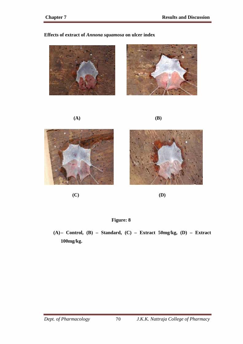
Chapter 7 Results and Discussion
Dept. of Pharmacology 70 J.K.K. Nattraja College of Pharmacy
Effects of extract of Annona squamosa on ulcer index
(A) (B)
(C) (D)
Figure: 8
(A)– Control, (B) – Standard, (C) – Extract 50mg/kg, (D) – Extract
100mg/kg.
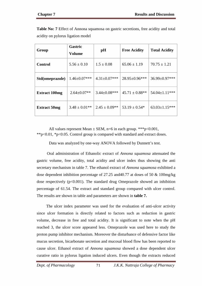
Chapter 7 Results and Discussion
Dept. of Pharmacology 71 J.K.K. Nattraja College of Pharmacy
Table No: 7 Effect of Annona squamosa on gastric secretions, free acidity and total
acidity on pylorus ligation model
GroupGastric
VolumepH Free Acidity Total Acidity
Control 5.56 ± 0.10 1.5 ± 0.08 65.06 ± 1.19 70.75 ± 1.21
Std(omeprazole) 1.46±0.07*** 4.31±0.07*** 28.95±0.96*** 36.99±0.97***
Extract 100mg 2.64±0.07** 3.44±0.08*** 45.71 ± 0.88** 54.04±1.11***
Extract 50mg 3.48 ± 0.01** 2.45 ± 0.09** 53.19 ± 0.54* 63.03±1.15***
All values represent Mean ± SEM, n=6 in each group. ***p<0.001,**p<0.01, *p<0.05. Control group is compared with standard and extract doses.
Data was analyzed by one-way ANOVA followed by Dunnett’s test.
Oral administration of Ethanolic extract of Annona squamosa attenuated the
gastric volume, free acidity, total acidity and ulcer index thus showing the anti
secretary mechanism in table 7. The ethanol extract of Annona squamosa exhibited a
dose dependent inhibition percentage of 27.25 and40.77 at doses of 50 & 100mg/kg
dose respectively (p<0.001). The standard drug Omeprazole showed an inhibition
percentage of 61.54. The extract and standard group compared with ulcer control.
The results are shown in table and parameters are shown in table 7.
The ulcer index parameter was used for the evaluation of anti-ulcer activity
since ulcer formation is directly related to factors such as reduction in gastric
volume, decrease in free and total acidity. It is significant to note when the pH
reached 3, the ulcer score appeared less. Omeprazole was used here to study the
proton pump inhibitor mechanism. Moreover the disturbance of defensive factor like
mucus secretion, bicarbonate secretion and mucosal blood flow has been reported to
cause ulcer. Ethanol extract of Annona squamosa showed a dose dependent ulcer
curative ratio in pylorus ligation induced ulcers. Even though the extracts reduced

Chapter 7 Results and Discussion
Dept. of Pharmacology 72 J.K.K. Nattraja College of Pharmacy
the incidence of ulcers when compared to ulcer controls, the inhibition percentage of
extracts were not closer to the standard drug omeprazole,
Graph Comparing the amount of Gastric volume collected from each of the
Control, Standard and the Extract (100 and 50mg/kg) groups.
contrlo
standard omeprozole
100mg/kg
50mg/kg0
2
4
6contrlostandard omeprozole100mg/kg50mg/kg
Gastr
ic vo
l
Figure: 9
Graph Comparing the pH of Gastric contents collected from each of the
Control, Standard and the Extract (100 and 50mg/kg) groups
control
standard
100mg/kg
50mg/kg0
1
2
3
4
5controlstandard100mg/kg50mg/kg
pH
Figure: 10

Chapter 7 Results and Discussion
Dept. of Pharmacology 73 J.K.K. Nattraja College of Pharmacy
Comparison of Free Acidity of the Control, Standard and the
Extract (100 and 50mg/kg) groups.
control
standard omeprazole
100mg/kg
50mg/kg0
20
40
60
80controlstandard omeprazole100mg/kg50mg/kg
Free
Acid
ity
Figure: 11
Comparison of Total Acidity of the Control, Standard and the
Extract (100 and 50mg/kg) groups
control
standardomerazole
100mg/kg
50mg/kg0
20
40
60
80controlstandardomerazole100mg/kg50mg/kg
mEq
/ l
Figure: 12

Chapter 7 Results and Discussion
Dept. of Pharmacology 74 J.K.K. Nattraja College of Pharmacy
HISTOPATHOLOGY IMAGES – PYLORUS LIGATED ULCERS
( A ) ( B)
( C ) (D)
Figure: 13
(A) – Control, (B) – Standard, (C) – Extract 100mg, (D) – Extract 50mg.
Chapter 7 Results and Discussion
Dept. of Pharmacology 75 J.K.K. Nattraja College of Pharmacy
Histopathology examination
The histopathological examinations of stomachs of the rats show us a better
picture of the gastric lesions and the damage occured to the stomach mucosa.
Significant acute ulceration of the stomach was observed in group-I which was
Pylorus ligation. This is revealed from Figure (A) (control group rats). The pylorus
ligation induced extensive macroscopic mucosal damage which can be known by the
injury to the epithelia of the mucosa. Also, elongated hemorrhagic lesions were
observed running perpendicular to the axis of the stomach. Histology of the rat
stomach of the standard group pertaining to the figure (B) showed normal
architecture of the gastric mucosa. The histopathological reports of the group three
rats shows appearance of gastric epithelium being showed in (C). Figure (D) shows
almost normal appearance of the epithelium. These observations matched well with
the ulcer index parameter.
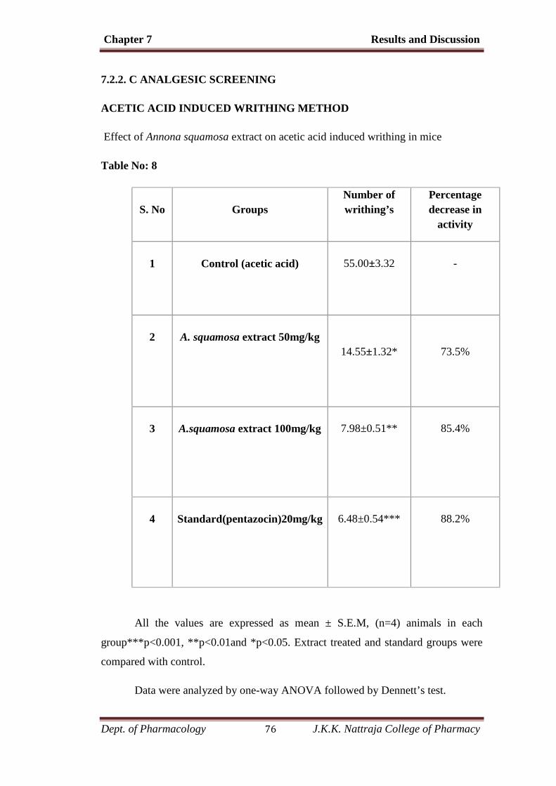
Chapter 7 Results and Discussion
Dept. of Pharmacology 76 J.K.K. Nattraja College of Pharmacy
7.2.2. C ANALGESIC SCREENING
ACETIC ACID INDUCED WRITHING METHOD
Effect of Annona squamosa extract on acetic acid induced writhing in mice
Table No: 8
S. No GroupsNumber ofwrithing’s
Percentagedecrease in
activity
1 Control (acetic acid) 55.00±3.32 -
2 A. squamosa extract 50mg/kg14.55±1.32* 73.5%
3 A.squamosa extract 100mg/kg 7.98±0.51** 85.4%
4 Standard(pentazocin)20mg/kg 6.48±0.54*** 88.2%
All the values are expressed as mean ± S.E.M, (n=4) animals in each
group***p<0.001, **p<0.01and *p<0.05. Extract treated and standard groups were
compared with control.
Data were analyzed by one-way ANOVA followed by Dennett’s test.
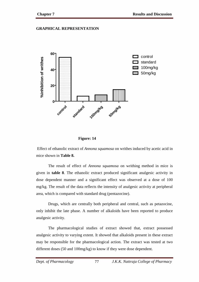
Chapter 7 Results and Discussion
Dept. of Pharmacology 77 J.K.K. Nattraja College of Pharmacy
GRAPHICAL REPRESENTATION
contro
l
standard
100m
g/kg
50mg/kg
0
20
40
60controlstandard100mg/kg50mg/kg
%in
hibi
tion
of w
rithe
s
Figure: 14
Effect of ethanolic extract of Annona squamosa on writhes induced by acetic acid in
mice shown in Table 8.
The result of effect of Annona squamosa on writhing method in mice is
given in table 8. The ethanolic extract produced significant analgesic activity in
dose dependent manner and a significant effect was observed at a dose of 100
mg/kg. The result of the data reflects the intensity of analgesic activity at peripheral
area, which is compared with standard drug (pentazocine).
Drugs, which are centrally both peripheral and central, such as petazocine,
only inhibit the late phase. A number of alkaloids have been reported to produce
analgesic activity.
The pharmacological studies of extract showed that, extract possessed
analgesic activity to varying extent. It showed that alkaloids present in these extract
may be responsible for the pharmacological action. The extract was tested at two
different doses (50 and 100mg/kg) to know if they were dose dependent.
Chapter 7 Results and Discussion
Dept. of Pharmacology 78 J.K.K. Nattraja College of Pharmacy
First the control group of animal were tested with the acetic acid, and then
observed the number of writhes produced by the animal. The extract exhibited an
percentage inhibition of 73.5 and 85.49 at doses of 50 and 100 mg/kg respectively.
These shows that ethanolic extract of Annona squamosa shows high significantly
decrease in the writhing response induced by acetic acid when compared with
control group.
.
Chapter 7 Results and Discussion
Dept. of Pharmacology 79 J.K.K. Nattraja College of Pharmacy
DISCUSSION
Most of the studies demonstrate the importance of natural products in drug
discovery. In these study antiulcer and analgesic activities of ethanolic extract of
Annona squamosa has been studied. The antiulcer study was evaluated using aspirin
and pylorus ligation models whereas analgesic activity was evaluated by acetic acid
induced writhing model.
Most of the studies demonstrate the importance of natural products in drug
discovery. The use of phytoconstituents as drug therapy to treat major ailments has
proved to be clinically effective and less relatively toxic than the existing drugs. The
acute oral toxicity study result showed that the plant leaf is safe.
Peptic ulcer describes a condition in which there is a discontinuity in the
entire thickness of the gastric and duodenal mucosa that persists as a result of acid
and pepsin in gastric juice. Peptic ulcer disease (PUD) is a serious gastrointestinal
disorder that requires a well targeted therapeutic strategy. It includes number of
drugs such as proton pump inhibitors and H2 receptor antagonists are available for
the treatment of peptic ulcer, Peptic ulcer occurs due to an imbalance between
aggressive (acid, pepsin) and defensive (gastric mucosal barrier) factors of gastric
mucosa.
Aspirin induced model shows significant percentage inhibition when
compared with standard. As aspirin is COX inhibitors suppress gastro duodenal
bicarbonates secretion and endogenous prostaglandin biosynthesis, disrupts mucosal
barrier. The ulcer index parameter was used for the evaluation of ulcer activity.
Moreover the disturbance of defensive factor like mucus secretion, bicarbonate
secretion and mucosal blood flow has been reported to cause ulcer.
Pylorus ligation model is usually employed to observe the potential of anti
ulcer drugs for their anti-secretory activity by checking the gastric volume and its
effect on gastric pH, total acidity and free acidity. Pylorus ligation induced ulcers are
due to auto digestion of the gastric mucosa and breakdown of the gastric mucosal
barrier and also because of an increase in acid-pepsin accumulation due to pylorus
obstruction and subsequent mucosal digestion.

Chapter 7 Results and Discussion
Dept. of Pharmacology 80 J.K.K. Nattraja College of Pharmacy
. The pharmacological studies of extract showed that, extract possessed
analgesic activity to varying extent. It showed that alkaloids present in these extract
may be responsible for the pharmacological action. The analgesic activity may be
attributed by the presence of various phytoconsitiuents such as alkaloids, tannins,
carbohydrates, flavonoids. Antinociceptive analgesic activity of Annona squamosa
was evaluated using chemical model of nociception inice. Acetic acid induced
writhing test is used for detecting both central and peripheral analgesics as these
method is very sensitive and able to detect antinociceptive effect of compounds at
dose levels. The intraperitoneal administration of acetic acid produces abdominal
writhing response due to sensitization of chemo sensitive nociceptors by PGs. It is
therefore possible that the extract exerts their analgesic effect probably by inhibiting
the synthesis or action of PGs.

Chapter 8 Summary and Conclusion
Dept. of Pharmacology 81 J.K.K. Nattraja College of Pharmacy
8. SUMMARY AND CONCLUSION
Summary
In the present work medicinally important active constituents of dried leaves
of Annona squamosa were studied with special emphasis on biological activities.
Pharmacognostical, Preliminary phytochemical and Pharmacological activities
were studied and reported.
Histopathological observation for antiulcer activity was done on abdominal
section. Antiulcer and analgesic activities were studied.
Conclusion
It was found that antiulcer activity exhibited was due to mucosal defensive
factor. Hence it can be used for management of peptic ulcer. Analgesic activity was
exhibited to the level of clinical importance. It was found that the activity was due to
the presence of flavonoids.
Chemical substances derived from plant have got a very long history in
treatment of human diseases. Nearly 50% of new chemical entities introduced during
the past two decades were from natural products.
Further research is required to isolate the active phytoconstituents present in
the extract and experimentation on the healing action of drug on chronic ulcer as well
as on the possible side effects. The investigation on mode of action may pave way for
establishment of new anti-ulcer therapy regimen.
Chapter 9 Bibliography
Dept. of Pharmacology 82 J.K.K. Nattraja College of Pharmacy
9. BIBLIOGRAPHY
1. Astin J. Why patients use alternative medicine? Results of a national survey. J
Amer Med Associat. 1998; 279: 1548-1553.
2. Berenguer B Sanchez LM Qulilez A Lopez-barreiro Galvez J Martin M.J;
Protective and antioxidant effects of Rhizophora mangle L. against NSAID-
induced gastric ulcers. Journal of Ethnopharmacology,2005; 103: 104-200.
3. Borelli F Izzo AA., “The plant kingdom as a source of anti-ulcer remedies”
Phytotheraphy research, 2000; 14: 581-591.
4. BVV Pardhasaradhi, Madhurima Reddy, A Mubarak Ali, A Leela Kumari and
Ashok Khar, Differential cytotoxic effects of Annona squamosa seed extracts
on human tumour cell lines: Role of reactive oxygen species and glutathione,
J. Biosci, 2005, 30(2), 237–244.
5. Chevallier A. The Encyclopedia of Medicinal Plants, Dorling Kindersly Ltd,
London., 1996;10.
6. Dharmani P and Palit G. “Exploring Indian medicinal plants for anti-ulcer
activity” Indian journal of pharmacology 2006; 38(2): 95-99.
7. Dinesh K. Yadava, Neetu Singhb, Rolee Sharmac, Mahendra Sahaid, Gautam
Palitb, Rakesh Mauryaa, Anti-ulcer constituents of Annona squamosa Twigs,
Fitoterapia, 2011, 82(4), 666-675.
8. Fugh-Berman A. Herb-drug interaction. Lancet. 2000; 355: 134-138.
9. Gupta AK, Chitme HR. Herbal Medicine for Health, The Eastern Pharmacist
2000: 41.
10. J. Audrey Leatemia and Murray B. Isman, Insecticidal Activity of Crude Seed
Extracts of Annonaspp., Lansium domesticum and Sandoricum
koetjapeAgainst Lepidopteran Larvae, Phytoparasitica, 2004, 32(1),30-37.

Chapter 9 Bibliography
Dept. of Pharmacology 83 J.K.K. Nattraja College of Pharmacy
11. Jeane silva,woru Abebeb,S.M.sousa,V.G.Duarte,M..I.L.Machadoc,F.J.A.
Matos;Analgesic and anti inflammatory effects of essential oils of
Eucalyptus.Journal of Ethnopharmacology,2003, 89, 277-283.
12. Kaleem M, Medha P, Ahmed Q U, Asif M, Bano B, Beneficial effects of
Annona squamosaextract in streptozotocin-induceddiabetic rats, Singapore
Med J, 2008, 49(10), 800-804.
13. Kannappan N S jaikumar manavalan R Kottai Muthu A: “ Anti-ulcer
activity of methanolic extract of Jatropha curcas on Aspirin-induced gastric
lesions in Wistar rats”. Pharmacologyonline. 2008; 1:279-293.
14. Kokate CK Purohit AP Gokhale SB Pharmacognosy.7th Edition, Nirali
Prakashan, Pune, 1997:105-144.
15. Kulkarni SK. Hand book of experimental pharmacology, 3rd edition, New
Delhi, vallabh prakasham, 199,142-147.
16. M Lebrini, F Robert, C Roos, Inhibition Effect of Alkaloids Extract from
Annona Squamosa Plant on the Corrosion of C38 Steel in Normal
HydrochloricAcid Medium, Int. J. Electrochem. Sci., 2010, 5, 1698 – 1712.
17. Malairajan P Gopalakrishnan G Narasimhan S Veni KJK and Kavimani S
“Antiulcer activity of crude alcoholic extract of Toona ciliata Roemer
(heartwood)”Journal of Ethnopharmacology. 2007; 110: 348-351.
18. Marta M.C. Souza, Claudia M.L. Bevilaqua, Selene M. Morais, Cícero T.C.
Costa, Ana R.A. Silva and Raimundo Braz-Filho, Anthelmintic acetogenin
from Annona squamosa L. Seeds, An Acad Bras Cienc, 2008, 80 (2), 271-277.
19. Mate, G.S. Naikwade, N.S.Magdum, C.S. Chowki, A.A Patil, S.B; Evaluation
of anti-nociceptive activity of cissus quadrangularies on albino mice.
International journal of green pharmacy, 2008, 118-121.
20. Mohamed Saleem Thattakudian Sheik Uduman, Ramkanth Sundarapandian,
Azagusundharam Muthumanikkam, Gnanaprakash Kalimuthu, Angala
Parameswari S, Thiruvengada rajan Vasanthi Srinivas and Gauthaman
Karunakaran, Protective Effect of methanolic extract of Annona squamosa
Chapter 9 Bibliography
Dept. of Pharmacology 84 J.K.K. Nattraja College of Pharmacy
linn in Isoniazid-Rifampicin induced Hepatotoxicity in Rats, Pak. J. Pharm.
Sci., 2011, 24(2), 129-134.
21. Mohamed Saleem TS, Christina AJM, Chidambaranathan N, Ravi V,
Gauthaman K, Hepatoprotective activity of Annona squamosa Linn. on
experimental animal model, International Journal of Applied Research in
Natural Products, 2008, 1(3), 1-7.
22. Mozafar Khazaei Hossein salehi, “ Protective effect of Falcaria Vulgaris on
ethanol induced gastric ulcer in rats” Indian journal of Pharmacology and
Therapeutics 2006, 5, 43-46.
23. Narayan Das Prajapati, SS Purohit and Tarun Kumar. “A Hand Book of
medicinal plants”.
24. Neha Pandey, Dushyant Barve, Phytochemical and Pharmacological Review
on Annona squamosa Linn, International Journal of Research in
Pharmaceutical and Biomedical Sciences, 2011, 2(4), 1404-1412.
25. Nighat Begum, Bechan Sharma and Ravi S. Pandey, Evaluation of Insecticidal
Efficacy of Calotropis Procera and Annona Squamosa Ethanol Extracts
Against Musca Domestica, J Biofertil Biopestici, 2010, 1(1), 1-6.
26. Parmar NS. “Effect of nalaxone and morphine on the experimentally induced
Gastric ulers in rats” Indian Drugs 1991, 29(7), 299-302.
27. Rakesh Ranjan and Mahendra Sahai, Coumarinolignans from the Seeds of
Annona squamosa Linn, E-Journal of Chemistry, 2009, 6(2), 518-522.
28. Rao,C.V., S.K. Ojha, K.Radhakrishnan, R. Govindarajan, S.Rastogi, S.
Mehrotra and P. Pushpangadan, 2004. Antiulcer activity of Utleria salicifolia
rhizome extract. J.Ethnopharmacol., 91, 243-249.
29. Sherwood: Human physiology. Thomson books., 6th edition, 591-593.
30. Telesphore B Nguelefack Catherine B Feumebo Gilbert Ateufack Pierre
Watcho Simplice tatsimo Albert D Atsamo Pierre Tane Albert Kamanyi.
“Anti-ulcerogenic properties of the aqueous and methanol extracts of leaves
Chapter 9 Bibliography
Dept. of Pharmacology 85 J.K.K. Nattraja College of Pharmacy
of Solanum torvum Swartz (Solanaceae) in rats” Journal of
Ethnopharmacology 2008, 119, 135-140.
31. Tylor Johns, Arnold Windust, Thomas Jurgens, Saleh Muhammad Mansor,
Antimalarial alkaloids isolated from Annona squamosa, Phytopharmacology,
2011, 1(3), 49-53. Astin J. Why patients use alternative medicine? Results of a
national survey. J Amer Med Associat. 1998, 279, 1548-1553.
32. Vogel, W.H., B.A Scholkens, J. Sandow, G.Muller and W.F. Vogel, 2002.
Drug Discovery and Evaluation, 2nd Edition., Springer, New York, 978-
3540423966, 670-725.
Copyright © 2022 FDOKUMEN