AntIoxIdAtIve propertIes of bee pollen extrActs exAmIned by ...
Upload
khangminh22Category
view
0download
0
RESEARCH ARTICLE Am. J. PharmTech Res. 2018; 8(1) ISSN: 2249-3387
Please cite this article as: Alphonse S et al., Evaluation of the toxicity of Annona muricata leaf extracts
on liver and kidney function and investigation of acute and subacute toxicity in Wistar rats. American
Journal of PharmTech Research 2018.
Evaluation of the toxicity of Annona muricata leaf extracts on liver
and kidney function and investigation of acute and subacute toxicity
in Wistar rats
Aivodji Natacha1, Ahokpe Melanie
1, Justin Behanzin
1, Sezan Alphonse
1*
1.Laboratory of Biomembranes and Cell Signaling University of AbomeyCalavi, 06 BP 3041
Cotonou, Benin
ABSTRACT
The present work carried out in the laboratory of Biomembranes and Cell Signaling of the
University of Abomey-Calavi in Benin aims to conduct a toxicological study on the ethyl extract
of the leaves of Annona muricata, with a view to verifying the toxic effects in general, and in
particular on the functioning of the kidneys and the liver in the wistar rat. In the present study, the
extraction yield of this plant by ethanol was 9.6 ± 1.89. Then, on the one hand, the acute and
subacute toxicities were induced in the rats of the experiment by gavage to our extracts at an
interval of 48 hours for 10 days. These rats were distributed in 05 batches. Each batch receiving or
not receiving a different dose of the Annona muricata extract. On the other hand, the toxic activity
on the liver and kidneys was evaluated by daily gavage of 14 rats of 3 rats each using the Annona
muricata ethyl extract at different doses (0.50 100 and 200 mg / kg). Measurement of biochemical
parameters (Urea, Creatinine, ASAT, and ALAT), weight-loss records, carried out during the first
experiment showed that there was no significant increase urea, creatinine, and a significant
decrease in ALT levels. On the other hand, a significant increase was observed in the ASAT.
Following the death of the rat of lot 5 receiving 5000mg / kg, the LD50 was determined (LD 50 =
3750 mg / kg). This LD50 indicates that Annona muricata extract is weakly toxic. Sub-chronic
administration of the extract confirmed that ASAT was progressively increasing in rats. The
decrease in body mass was a good indicator of toxicity. In the second experiment, the
measurement of biochemical parameters (Urea, Creatinine, Uric Acid, ASAT, ALAT) revealed an
increase in the concentration of urea and creatinine and decreased concentration of uric acid at
doses of extracts greater than or equal to 100 mg / kg PC. These results may suggest renal damage
that has not been confirmed by the histological study of the organs taken (Liver and kidneys) from
experimental rats. However, this study showed an early onset of hepatic involvement at the dose of
100mg / kg, which increased at a dose of 200mg / kg.
Keywords: Annona muricata, liver, kidneys, LD50, toxicity, Benin
*Corresponding Author Email: [email protected] Received 4 October 2017, Accepted 30 October 2017
Journal home page: http://www.ajptr.com/

Alphonse et. al., Am. J. PharmTech Res. 2018;8(1) ISSN: 2249-3387
www.ajptr.com 190
INTRODUCTION
Plants, which are vital elements of biological diversity, are used primarily for human well-being
(Adjanohoun et al., 2000). The African populations are confronted with the emergence of chronic
diseases whose treatment and follow-up often constitute a major economic problem; they use
traditional medicine. Diabetes, high blood pressure and cancer are among the so-called new
diseases for this medicine. Once unknown, these diseases have become increasingly important
until they become a real public health problem. In response to the expansion of these high-burden
diseases, WHO, in its resolution AFR / RC50 / R3 of 31 August 2000, encouraged African
countries to develop regional strategies on traditional medicine; to undertake research on medicinal
plants and to promote their optimal use in health care delivery systems. Benin is no exception to
the practice of this form of medicine. According to the World Health Organization (WHO), 80 to
85% of Beninese people use traditional medicine in the event of illness. One of the plants used in
this context in Benin is Annona muricata.
The leaves of Annona muricata are used as a diuretic, depurative. They are also used for liver
diseases. Their use is also found in the treatment of blisters, edema, edema, hygroma, brucellosis,
dropsy and undulating fever. (Adjanohoun, E, 2000)
But very often, plants are consumed without taking into consideration their toxicity, their
synergism with other substances or even the original medicinal proposal established by medicine.
It is recommended to use a plant only on the advice of a specialist because badly dosed the latter
can become highly toxic. It is in this context that the present work is carried out, whose main
objective is to study the toxicity caused by the ethanolic extract of the leaves of Annona muricata
on the liver and the kidney.
Generalities on Annona muricata
Figure 1: Leaf and fruit of Annona muricata

Alphonse et. al., Am. J. PharmTech Res. 2018; 8(1) ISSN: 2249-3387
191 www.ajptr.com
Biology of Annona muricata
Annona muricata also named guanábana, guabano or soursop is a type of tree of the Annunciated
family (Annunceae), and originally grew in the West Indian islands. Today the tree grows in all the
Caribbean and Amazonia. The tree is still green and under normal conditions it reaches 6-10
meters in height and has bright oblong-lanceolate leaves that resemble laurel. The yellow-green
flowers with 6 petals appear on the branch opposite the leaves. They spread an Asian fragrance and
thus attract flies for pollination. The fruits are armored with large black seeds, the size of a small
peanut. The fruit is the size of a big apple, green, soft when it is ripe, it is covered with areolas
(small growths often present on the cacti) curved soft. The black seeds contain neurotoxin
annunacin. Amazonian Indians use the leaves as a medicine. Its fruits are appreciated by the
natives for their refreshing virtues and its leaves are traditionally used to cure many diseases.
Classification
Kingdom: Plantae
Under the reign of: Tracheobionta
Division: Magnoliophyta
Class: Magnoliopsida
Subclass: Magnoliidae
Super-order: Magnolianae
Order: Magnoliales
Family: Annonaceae
Genre: Annona
Species: muricata
Acute toxicity
It represents the most spectacular manifestation of the harmfulness of a poison. This fact, which
leads us to regard as poisonous any substance which kills violently, results in the rapid death of the
individual or of the contaminated populations. Acute toxicity can therefore be defined as that
which causes death or very serious physiological disorders after a short period of time following
transtectional, pulmonary or buccal absorption. (Ramade, 1979)
The study of acute toxicity is also qualitative and quantitative of the toxic phenomena that can be
encountered after administration of the active substance. This study describes the observed
symptoms, including local phenomena. It allows:
Indication of the maximum dose without toxic effect (DME), ie the highest dose for which no
toxic effect is observed compared to the control batch;

Alphonse et. al., Am. J. PharmTech Res. 2018;8(1) ISSN: 2249-3387
www.ajptr.com 192
Notation
determination of the LD50 with its 95% confidence limits (Ruckebusch, 1981).
Determination of the lethal dose (LD50)
The LD50 is in its simplest form the dose of a compound that causes a mortality of 50% in a
population of experimental animals. That is, having received a single administration of a product
under well-defined experimental conditions. This determination is based on the assessment of all-
or-nothing responses: death or survival of animals. The experimental protocol consists of
experimenting on 5 to 6 batches of rats to which increasing doses of the test substance are
administered in such a way that the percentage of mortality varies between 0 and 100%. This is
because it is impossible to immediately obtain 50% of deaths from a single group. The
construction of a curve giving the percentage of mortality as a function of the logarithm of the dose
leads to determine the dose that would be the LD50 (Wallace Hayes, 2008)
Different methods for determining the LD50
The LD50 can be determined by two calculation methods, the Dragstedt and Lang Method (1957)
and the method of Karber and Behrens (1935). As can be determined by two graphical methods,
which are the method of Miller and Tainter, (1944), and the method of Litchfield and Wilcoxon,
(1949).
Subacute toxicity
It differs from the previous one (acute toxicity) in that a significant proportion of the population
can survive intoxication, although all individuals showed short-term clinical signs on target organs,
sometimes reversible and resulting from repeated intake of the toxicant, but at a lower dose than
that of acute toxicity (Ramade, 1979).Materiels et Methodes
Extraction
The ethanolic extract of the dried leaves powder of Annona muricata was prepared from 134.58 g
of powder. The powder was macerated in ethanol (95 °). The maceration with duration 72h during
which filtrations were carried out every 24h. The solvent was recovered from the filtrate by
evaporation with Rotavapor. The filtrate was placed in an oven at a temperature of 40 ° C. The
extract obtained was stored in the freezer until it was used.

Alphonse et. al., Am. J. PharmTech Res. 2018; 8(1) ISSN: 2249-3387
193 www.ajptr.com
a. b. c.
Figure 2: a. Powder of leaves of Annona muricata, b. Evaporation with ROTA VAPOR, c.
Extract obtained
Evaluation of the acute toxicity of the ethanolic extract of Annona muricata
For the determination of acute toxicity, the experimental protocol used is that described by Rasekh
et al., (2008). The rats were randomly assigned to five batches. One batch is used as a control and
the other batches are each treated with a single dose of the ethanolic extract of Annona muricata.
At the beginning of the experiment the rats had an average weight of 200 ± 30.
o Administration of extracts
The gavages are done every 48 hours and this for 10 days. The doses administered were 0, 100,
1000, 2500, 5000 milligrams of extract per kilogram of rat body weight respectively to the 5
batches.
o Observations
After administration of the extract, the rats are monitored continuously each day. We took notes on
apparent signs of toxicity. The purpose of this monitoring was to report changes in diet and water
consumption, or behavioral or clinical signs of toxicity.
o Expression of results
The LD50 expressed in mg / kg of body weight is determined by the calculation method of
Dragstedt et lang, (1957).
This method is based on the following assumption:
Any animal that has survived a dose administered to it will survive at any lower dose. Any animal
which has succumbed to a dose administered to it will succumb to any higher dose than the latter.
Thus, the percentage of mortality (M [%]) can be calculated for each dose by cumulating all
observed deaths at lower doses and all survivors observed at higher doses.
( ) ( )
( ) X100
The LD50 is calculated by interpolation:

Alphonse et. al., Am. J. PharmTech Res. 2018;8(1) ISSN: 2249-3387
www.ajptr.com 194
DL50 = ( ) ( )
X2: Upper dose framing the LD50; X1: Lower dose framing the LD50; Y2: Percentage of
mortality corresponding to X2; Y1: Percentage of mortality corresponding to X1
Evaluation of the subacute toxicity of the ethanolic extracts of Annona muricata
For the evaluation of subacute toxicity, four batches of rats were used. The treatments were
administered orally once every 48 hours to the rats for 10 days. The first group of animals serving
as controls received water, while the other batches received the extracts in respective doses. Lot 2
received 100 mg / kg, lot 3 received 1000 mg / kg, lot 4 received 2500 mg / kg of the extract.
Animals are given free tap water and food throughout the test period; and do not receive any other
drug treatment.
o Method of observation and examination
Several methods have been used to produce the toxicity texts. These include observation of weight
evolution and biochemical tests.
Chronology of weight change
As weight evolution is a good indicator of toxicity, we have followed the weight evolution of rats
during our work. Thus, at each 48 hour, the rats of each batch were weighed and their body mass
measured in order to see the effect of the extract on the weight evolution.
Blood Collection
Rats are taken before each gavage. The principle of the sample is as follows: the rats are
anesthetized with diethyl ether and using a hematocrit tube. the samples are taken from the vein in
the angle of the eye. Two tubes were used in EDTA tubes and dry tubes. These tubes were
centrifuged at 3000t / 15min and the serum obtained was stored at -15 ° C until use (Ben-
Romdhane et al., 2003).
Biochemical examinations
The biochemical analyzes were carried out in the Laboratory of Biochemistry of the Faculty of
Health Sciences using a spectrophotometer: Kenza Max distributed by Biolabo. The assay
parameters are ALAT (Alanine amino transferase), ASAT (Aspartate amino transferase), CREA
(Creatine) and Urea.
Procedure used for the measurement of biochemical parameters
Creatinine

Alphonse et. al., Am. J. PharmTech Res. 2018; 8(1) ISSN: 2249-3387
195 www.ajptr.com
Table 1: Mode of measurement for creatinine
Measure in a vessel of 1cm optical path. White
(optional)
Standard Sample
Work reagent (R1+R2) 1ml 1ml 1ml
Demineralized Water 100µl
Standard 100µl
Specimen 100µl
Mix thoroughly after 30 seconds, record absorbance A1 at 490nm (480-510) against
reagent blank or distilled water. Read the absorbance.
UREA
More than 90% of the urea is eliminated by the kidneys in the urine. Measurement of plasma or
serum concentration of urea is often considered an indicator of renal function. However, certain
non-renal factors also influence the concentration of urea: urea is increased, among other things, in
cases of accelerated protein catabolism (burns, trauma, myocardial infarction ...). The urea level is
lowered to the terminal stage of severe hepatic insufficiency and is then accompanied by an
increase in ammonia. The urea level is generally studied in conjunction with the creatinine ratio
(urea / creatine ratio) to refine the diagnosis of post-renal or pre-renal azotemia. (Pirro, 2014)
Table 2: Mode of measurement of urea
Measure in a thermostatically controlled tank (30 ° C
or 37 ° C)
Standard Sample
Reagent 1ml 1ml
Standard 5µl
Specimen 5µl
Mix. Read the concentrations at 340nm. 1st reading A1 in 30 seconds. 2nd reading A2 at
90 seconds.
ALAT
Table 3: Mode Used for ALT Measurement
Introduce into a reading tank of 1cm optical path:
Reagents 1 ml
Allow the temperature to equilibrate to 37 ° C and then add:
Samples 100 µl
Mix. After 1 minute, read the absorbance at 340 nm every minute for 3 minutes.
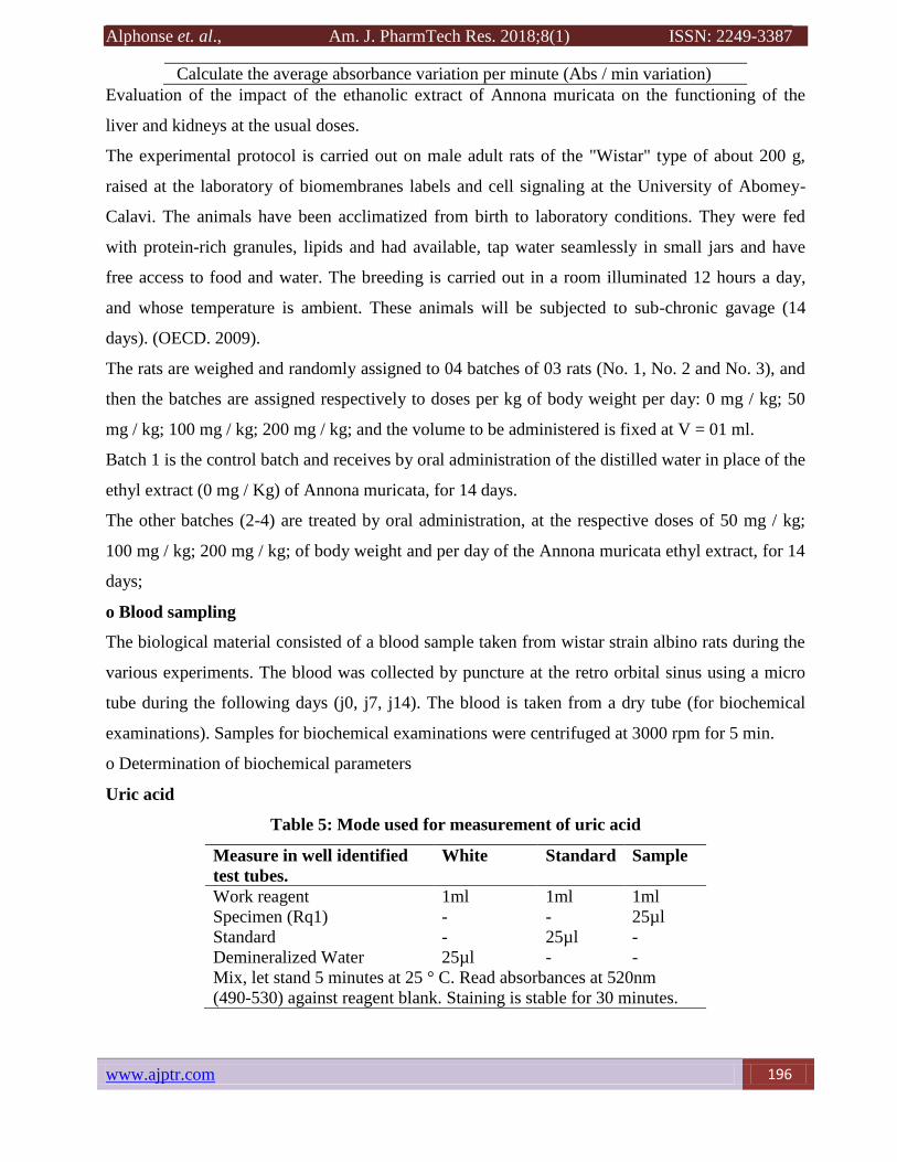
Calculate the average absorbance variation per minute (Abs / min variation)
ASAT
Table 4: Mode Used for ASAT Measurement
Introduce into a reading tank of 1cm optical path:
Reagents 1 ml
Allow the temperature to equilibrate to 37 ° C then add:
Samples 100 µl
Mix. After 1 minute, read the absorbance at 340 nm every minute for 3 minutes.
Alphonse et. al., Am. J. PharmTech Res. 2018;8(1) ISSN: 2249-3387
www.ajptr.com 196
Calculate the average absorbance variation per minute (Abs / min variation)
Evaluation of the impact of the ethanolic extract of Annona muricata on the functioning of the
liver and kidneys at the usual doses.
The experimental protocol is carried out on male adult rats of the "Wistar" type of about 200 g,
raised at the laboratory of biomembranes labels and cell signaling at the University of Abomey-
Calavi. The animals have been acclimatized from birth to laboratory conditions. They were fed
with protein-rich granules, lipids and had available, tap water seamlessly in small jars and have
free access to food and water. The breeding is carried out in a room illuminated 12 hours a day,
and whose temperature is ambient. These animals will be subjected to sub-chronic gavage (14
days). (OECD. 2009).
The rats are weighed and randomly assigned to 04 batches of 03 rats (No. 1, No. 2 and No. 3), and
then the batches are assigned respectively to doses per kg of body weight per day: 0 mg / kg; 50
mg / kg; 100 mg / kg; 200 mg / kg; and the volume to be administered is fixed at V = 01 ml.
Batch 1 is the control batch and receives by oral administration of the distilled water in place of the
ethyl extract (0 mg / Kg) of Annona muricata, for 14 days.
The other batches (2-4) are treated by oral administration, at the respective doses of 50 mg / kg;
100 mg / kg; 200 mg / kg; of body weight and per day of the Annona muricata ethyl extract, for 14
days;
o Blood sampling
The biological material consisted of a blood sample taken from wistar strain albino rats during the
various experiments. The blood was collected by puncture at the retro orbital sinus using a micro
tube during the following days (j0, j7, j14). The blood is taken from a dry tube (for biochemical
examinations). Samples for biochemical examinations were centrifuged at 3000 rpm for 5 min.
o Determination of biochemical parameters
Uric acid
Table 5: Mode used for measurement of uric acid
Measure in well identified
test tubes.
White Standard Sample
Work reagent 1ml 1ml 1ml
Specimen (Rq1) - - 25µl
Standard - 25µl -
Demineralized Water 25µl - -
Mix, let stand 5 minutes at 25 ° C. Read absorbances at 520nm
(490-530) against reagent blank. Staining is stable for 30 minutes.

Alphonse et. al., Am. J. PharmTech Res. 2018; 8(1) ISSN: 2249-3387
197 www.ajptr.com
For the other parameters: ALAT (Alanine amino transferase), ASAT (Aspartate amino
transferase), CREA (Creatine) and Urea; the method used is the same as that described above in
the first experiment.
o Histological study of the organs taken (kidneys and liver)
After the last sampling on the 14th day, all surviving animals are sacrificed and dissected in order
to observe any signs of tissue toxicity (kidneys and liver).
Procedure of the histological study
Preparation of casettes
The organs resulting from the dissection were cut into small pieces and then placed in cassettes for
fixation. Sharp instruments are used in order not to crush the tissues and thus avoid the formation
of artifacts (the scalpel). The sampling is done as quickly as possible.
Fastening
Its purpose is to preserve structures and harden parts. Indeed, the tissue collection causes them to
die: the cells discharge their enzymes, which causes a self-regulation of the tissue. Moreover, in
the ambient air, the samples can be contaminated by bacteria, which leads to putrefaction of the
tissues. It must be done immediately after sampling, by immersing the material in a large volume
of fixing liquid. The most frequently used fixing fluids are 4% formalin or Bouin liquid (picric
acid + formol + acetic acid water).
The choice of fixer was made on 10% buffered formalin for morphological studies because it is
easy to prepare and stable. Moreover, this fixator penetrates the tissues well. The preservation can
be carried out in this solution for a relatively long time without the tissues deteriorating. The
duration of the fixation is on average of 3 days for the cuts of small sizes.
Interest of fixation:
Fixation is the primary step even in the histological study because it has the advantage:
immobilization of tissue / cellular constituents; prevents cellular autolysis; prevents post-mortem
bacterial putrefaction;allows the histological technique and subsequent colorations
Its principle is based on the fact that it reacts with amino groups of proteins. The fixation time is
variable and the amount of fixer used must be at least ten times greater than the volume of tissue to
be fixed: a few hours are sufficient to fix the small fragments. It has the disadvantage of causing a
phenomenon of cellular auto-fluorescence which can hamper observations under the fluorescence
photon microscope. It also rapidly degrades the nucleic acids.

Alphonse et. al., Am. J. PharmTech Res. 2018;8(1) ISSN: 2249-3387
www.ajptr.com 198
Role of the fixer:
The role of the fixer rests on actions such as:precipitation, polymerization, coagulation of
proteins;establishment covalent bonds;kills the cells;blocking of enzymatic reactions.
Inclusion or circulation
The inclusion allows fine and regular cuts. In order for the light to pass through the tissue to be
examined, it must be very thin. Now the tissues are soft, so they must be given a solid consistency.
This is the principle of inclusion: paraffin inclusion involves infiltrating and coating the tissues to
be examined with paraffin. Inclusion is preceded by two essential steps. First of all,
dehydration: the tissues are passed into alcohol baths of increasing degree (70 °, 80 °, 90 °, 95 °, 99
°, and finally 100 °). The interest of dehydration is to eliminate the fixative.We therefore used as a
dehydrating agent ethanol because it has a great miscibility with water and allows a good
hardening of the tissues. Dehydration was progressive, by passing the tissue pieces into alcohol
baths of increasing concentrations, to avoid distortion of the tissue structures. We used seven
ethanol baths of increasing concentration. The rooms stayed for an hour in each of these baths.
The alcohol (ethanol) is then replaced by a solvent which is miscible with paraffin: it is either
xylene or toluene (hydrocarbons). These substances eliminate ethanol. As they are infiltrated by
the solvent, the tissues tend to become lighter: this stage is sometimes called clarification or
clarification.
The choice of our solvent is based on xylene, we have carried out three (03) baths there. Our
manipulations were done under a chemical hood. As soon as the first xylene bath begins to become
cloudy (which translates to incomplete dehydration of the room), we immediately remove the
tissue and plunge it into a new bath of absolute ethanol. The rooms have stayed about an hour in
each of the baths.
The impregnation, the fabric is placed in molten paraffin (raised to 56/58 ° C); the heat causes the
solvent to evaporate (and its dissolution in the paraffin): the spaces thus released are filled with
paraffin. We have here used three paraffin baths, the rooms have stayed about an hour in each of
the baths.
Embedding
The paraffin is placed in small molds at ambient temperature, which causes it to harden and thus to
stiffen the tissue fragments taken. The demoulding is then carried out: tissue fragments included in
a paraffin block are obtained. We have deposited and oriented tissues in metal molds to facilitate
further study. The molten paraffin is poured into the molds which support the blocks to be formed.

Alphonse et. al., Am. J. PharmTech Res. 2018; 8(1) ISSN: 2249-3387
199 www.ajptr.com
Finally the mold block assembly is set to cool in a refrigerator. Once the block of paraffin has
solidified, it is separated from its mold and is then ready to be cut to the microtome.
Microtomy
We then isolate cuts in the paraffin block. We use a microtome, which advances the block on a
razor: the block advances about 2 to 3 μm each time (in classic MO, the cuts measure about 5 μm).
The whole of the slices will form a ribbon in which there are serial sections for tissue sampling.
To do this we have inserted the block into the object clamp and then immobilized by means of the
locking screw and oriented with the adjustment screws, making sure that the bottom and top faces
of the block are parallel to the cutting edge of the razor. After checking the parallelism between the
block and the razor, the motor wheel of the microtome is turned by means of the crank.
We heat the paraffin on a hot plate and so the paraffin sticks to the blade.We cover blades clean
and engraved with glycerine water. Successful ribbon segments are then selected using lancets and
placed on slides. The whole is deposited on the heating plate and the ribbons are allowed to stretch.
With two lancets we pull on the edges of the tissues to eliminate the persistent folds; the excess
liquid is absorbed with a blotting paper and the ribbons are allowed to adhere to the slides. These
are then placed in an oven at 40 ° C. for one hour after drying; finally they are allowed to cool for
at least 15 minutes. The preparation is then ready for coloring.
Made on slides, the coloring accentuates the contrasts to better recognize the different elements of
the preparation. The tissues of the organism are not spontaneously colored, which makes the
observations difficult. The dyes used in histology are more or less selective; most are acidic or
basic components in an aqueous medium which form salts with the ionized radicals of the tissues.
Acidic components are used for basophilic tissue areas, and basic components are used for
acidophilic tissue areas. The most commonly used staining is HES: hematein / eosin / saffron.
Hemathein is a rather basic substance, which colors the nuclei in violet thus coloring the nucleic
acids. Eosin is a rather acidic substance, which rather colored the cytoplasms (in pink) thus colored
the proteins. Finally, the saffron colors the collagen fibers in yellow.
Howeverin order for a stain to be used, paraffin must be removed. Dewaxing is then performed,
which consists of passing the slides in toluene or xylene baths in order to dissolve the paraffin. The
mixture is then rehydrated: the alcohol is mixed with water and toluene, and the slides are passed
through alcohol baths of decreasing degree (from 100 ° to 70 ° or even 50/40 °). You can also use

Alphonse et. al., Am. J. PharmTech Res. 2018;8(1) ISSN: 2249-3387
www.ajptr.com 200
histochemical staining, called PAS (Periodic Acid Schiff), which highlights glycogen,
glycoproteins and just about anything that contains sugars.
The coloring chosen in the case of our study is that with haemotein eosin. The haematin staining
lasted 15 min, the running water bath → 5 min, the acidic alcohol bath → 2 to 3 dives, the running
water bath → 5 min, the ammonia water bath → 30sec to 1 min, the running water bath → 5 min,
the eosin stain: 15 sec → 2 min, the running water bath → 5 min.
Blade assembly
The assembly is carried out by affixing the object slides to the histological slide using Eukitt glue.
Reading
Microscopic observation is here coupled with photomicrography. Our observations were made on
a light microscope equipped with a camera. The images were transferred to a processing software
for color to be used, the paraffin must be removed. Dewaxing is therefore carried out, which
consists in passing the slides in baths of toluene or of xylene in order to dissolve the paraffin. The
alcohol is then mixed with water and toluene, the slides are passed into alcohol baths of decreasing
degree (from 100 ° to 70 °, or even 50/40 °). One can also resort to a histochemical staining, called
PAS (Periodic Acid Schiff), which highlights glycogen, glycoproteins and just about anything that
contains sugars.
The coloration chosen in the case of our study is that to hematein eosin. The hematein staining
lasted 15 minutes, the bath of running water → 5 minutes, the acid alcohol bath → 2 to 3 dives, the
water bath → 5 minutes, the ammonia water bath → 30 seconds to 1 mn, running water bath → 5
mn, staining with eosin: 15 sec → 2 mn, running water bath → 5 mn.
Assembly of the blades
The assembly is made by affixing the object slides on the histological slide with Eukitt glue.
Playback
Microscopic observation is here coupled with photomicrography. Our observations were made on
a photonic microscope equipped with a camera. The images were transferred to image processing
software (Adobe Photoshop Image) using a digitizer that transforms the analog image into a digital
image. The cuts are observed at the appropriate magnifications; the images are captured according
to the desired objective and are transferred in JPG format.
RESULTS AND DISCUSSION
Evaluation of extraction techniques
The ethanolic extract of Annona muricata was obtained from a fine powder of the leaves of the

Alphonse et. al., Am. J. PharmTech Res. 2018; 8(1) ISSN: 2249-3387
201 www.ajptr.com
corossolier. The method consisted in carrying out a maceration of the Annona muricata powder in
ethanol for 72 hours and every 24 hours was carried out the evaporation of the solvent to the
rotavapor. The results of the present study indicate that from 134.58 g of Annona muricata powder
and 1 liter of ethanol we obtained an ethanol extract, considered as the dark green colored extract
which may contain chlorophyll , flavonoids, polyphenols and other compounds. In order to use the
extract for pharmacological and toxicological tests, the conservation of the bioactive state of the
extracted molecules seems important. Complete depletion of the solvent is necessary. The presence
of traces of ethanol in the extract can cause undesirable side effects. Therefore, the positive or
curative effect of the pharmacological substance can be masked by the action of the solvent. The
yield was calculated on the total weight of the powder of the Annona muricta powder and that of
the ethanol extract gave a yield of 9.60 ± 1.89%. This result is close to that found by AÏVODJI N.,
(2014) who used the same extraction protocol for the leaves of the same plant.
Study of Acute Toxicity
o Determination of LD 50
Before attempting to determine the LD50, a preliminary search for LD0: the highest dose at which
all experiencing animals survive, and LD100: the lowest dose at which all animals die, is required.
These two doses delimit the area in which the final test is to be performed. In our work the test is
carried out according to the method described by Rasekh et al., (2008), on male Albino Wistar
white rats. Five (05) doses, including DL0 and DL100, were retained (Table 6), while control rats
(0mg / kg) remained healthy.
Table 6: Doses injected and percentage conversion of the number of dead rats in each batch.
Lot Number of rats (n) Doses (mg/kg) Number of dead rats Percentage (%)
1 1 0 0 0
2 1 100 0 0
3 1 1000 0 0
4 1 2500 0 0
5 1 5000 1 100
After calculating the percentages of mortality in each batch, we can then determine the LD50
o Calculation method or method of Dragestedt and Lang. After applying the relationship:
( ) ( )
( ) X100
The LD50 is calculated by interpolation:
LD50 = ( ) ( )

Alphonse et. al., Am. J. PharmTech Res. 2018;8(1) ISSN: 2249-3387
www.ajptr.com 202
X2: Upper dose framing the LD50; X1: Lower dose framing the LD50; Y2: Percentage of
mortality corresponding to X2; Y1: Percentage of mortality corresponding to X1.This method
revealed an LD50 equal to: 3750 mg / kg.
o Characterization of the form of toxicity of the ethanolic extract of Annona muricata
The highest dose killing all animals or 100% lethal dose (LD100) is 5000 mg / kg and the
maximum tolerated dose (DMT), the maximum dose that does not kill any animal when the extract
is administered, is 2,500 mg / kg /. In the numerical application of the formula of Dragestedt and
Lang (1957), the calculation of the LD 50 of the extract gives the value of 3750 mg / kg.
According to the Diezi (1989) toxicity scale, this LD50 value (3750 mg / kg) indicates that the
ethanolic extract of Annona muricata leaves, administered orally (VO), is weakly toxic in rats
under the conditions of this study.
Table 7:Toxicity class, according to the Diezi toxicity scale (1989)
Index or class of toxicity Common term used Toxicological parameter (LD50)
1 Very toxic substance LD50< 5mg/kg
2 Toxic substance 5mg/kg<LD50<500mg/kg
3 Low toxic substance 500mg/kg<LD50<5000mg/kg
4 Non-toxic substance LD50>500mg/kg
In this study, the LD50 of the ethanolic extract of Annona muricata calculer is included between
500 mg / kg and 5000 mg / kg (500 mg / kg <3750 mg / kg <500 mg / kg). The ethanolic extract of
Annona muricata is considered to be low toxicity in rats by oral route according to the
classification of Diezi, (1989) and therefore this plant deserves to be used with caution in human
Study of Acute Toxicity
After administering the oral ethanol extract of Annona muricata to male rats, there is a steady
increase in signs of poisoning and mortality in relation to the dose injected. The signs are
summarized in the table.
Table 8: Sign of poisoning observed at each dose
Doses
(mg/kg)
Rats Toxicity symptoms
Lots M/T
0 Lot1: witness 0/1 Nothing
100 Lot 2: a blue bar 0/1 No visible signs toxicity
1000 Lot 3: a red bar 0/1 Hair straightening, drowsiness, loss of appetite, weakness
2500 Lot 4: a green bar 0/1 Hair straightening, drowsiness, loss of appetite, weakness
5000 Lot 5: 2 red bars 1/1 Hair straightening, drowsiness, loss of appetite, weakness,
rapid heartbeat, labored breathing, body tremor, loss of
balance, convulsion, total paralysis, coma, death

Alphonse et. al., Am. J. PharmTech Res. 2018; 8(1) ISSN: 2249-3387
203 www.ajptr.com
The first dead rat was recorded at a dose of 5000 mg / kg after 4 injections following severe signs
of intoxication. After the second injection, the rat showed almost cut breathing, loss of balance,
muscle contractions, partial paralysis (Fig 3-c), coma and total paralysis, he died on the 6th day.
All the other rats survived. However, signs of toxicity have been noted. Namely hair straightening,
drowsiness, motor weakness and loss of appetite. The dose that kills the entire rat population is
5000mg / kg, while the 2500mg / kg dose is tolerated. The signs of toxicity increased
proportionally with the doses administered. Thus we observed more severe signs of toxicity in rats
given 2500 mg / kg. Gradually we noticed that the movement of rats as well as their appetite
decreased and that their breathing became difficult. At the 1000mg / kg dose, the percentage of
death is nil however we noted a motor weakness which limited the rat in its movement. As for the
100 mg / kg dose, it did not show signs of tangible toxicity, however, this dose could be useful in
the study of long-term toxicity.
a. b. c.
Figure 3: Acute signs of acute toxicity in rats. (a) Straightening of the hairs; (b) rats are
drowsy and tired; (c) Partial paralysis at dose 5000mg / kg
Subacute toxicity study
o General signs
No deaths were recorded during the experiment. However, from a behavioral point of view, the
rats are calm, showing a decrease in liveliness and appetite compared to the control rat; their sleep
time has become more considerable.

Alphonse et. al., Am. J. PharmTech Res. 2018;8(1) ISSN: 2249-3387
www.ajptr.com 204
o Chronology of weight change
The change in animal body mass during the subacute toxicity experiment with the ethanol extract
of Annona muricata showed that there was a decrease in weight throughout the 2 weeks, compared
to witness (Figure. 7).
Figure 5: Changes in body weight of rats as a function of time under subacute toxicity
conditions by the ethanolic extract of Annona muricata.
The percentage decrease was minimal during the first six days of the experiment. This percentage
was 4.08% for lot 2 receiving 100 mg / kg; there was no decrease in lot 3; at Lot 4 a decrease of
2.43% was observed. These decreases are greater on the eleventh day of the experiment and were
18.36% for lot 2; 8.10% for lot 3 and 14.63% for lot 4. (Tab 10).
0
50
100
150
200
250
300
Day0 Day6 Day11
batch with distilled water batch2 batch3 batch 4 batch 5

Alphonse et. al., Am. J. PharmTech Res. 2018; 8(1) ISSN: 2249-3387
205 www.ajptr.com
Table 10: Percentage decrease in body weight of rats during the period of subacute
treatment with ethanolic extract of Annona muricata.
Days Lot2 Lot 3 Lot4
6 4,08% 0% 2,43%
11 18,36% 8,1% 14,63%
The change in body weight is used as an indicator of the adverse effects of chemical compounds
(Hilaly et al., 2004). Weight loss is correlated with the physiological state of the animal. These
decreases in body weight in animals may be due to lower food consumption, we can also relate this
decrease in weight to the possibility of dose / absorption interactions and the decrease in the
amount of food intake. The decrease in body weight during the treatment period suggests that
subchronic and oral administration of the ethanolic extract of Annona muricata has effects on the
growth of Albino Wistar rats. To make this hypothesis more precise, other analyzes deserve to be
carried out.
o Biochemical data
Several toxic plant compounds accumulate in the liver where they are detoxified. The liver is the
first target of toxicity and the first organ exposed to all that is absorbed in the small intestine; it
metabolizes foreign substances to compounds that may be hepatotoxic. (P. Hanover, 1963) The
liver works in combination with the kidneys to remove toxic substances from the blood
(Tulsawani, 2010). A study of renal and hepatic function may therefore be useful for assessing the
toxic effects of medicinal plants. These tests mainly include the determination of ASAT, ALAT,
CREA and others (Tilkian, 1979) and any necrosis of liver cells leads to a significant increase in
ASAT, ALT, and serum enzymes (Adeneye et al., 2006). ). Serum studies in rats treated with the
ethanolic extract of Annona muricata show a significant increase in the ASAT parameter in rats
and a significant decrease in ALT in rats. The other parameters: CREA, Urea do not show any
significant changes.

Alphonse et. al., Am. J. PharmTech Res. 2018;8(1) ISSN: 2249-3387
www.ajptr.com 206
Figure 6: Changes in serum creatinine levels of rats as a function of time under subacute
toxicity conditions by the ethanolic extract of Annona muricata.
Figure 7: Changes in serum urea level of rats as a function of time under the conditions of
subacute toxicity by the ethanolic Annona muricata extract.
0
1
2
3
4
5
6
7
8
9
10
Day 0 Day 2 Day 4 Day 6 Day 8
Bacth 1 Batch 2 Batch 3 Batch 4 Batch 5
0
0.1
0.2
0.3
0.4
0.5
0.6
0.7
0.8
0.9
Day 0 Day 2 Day 4 Day 6 Day 8
Bacth 1 Batch 2 Batch 3 Batch 4 Batch 5

Alphonse et. al., Am. J. PharmTech Res. 2018; 8(1) ISSN: 2249-3387
207 www.ajptr.com
Figure 8: Variations in serum ALT levels of rats as a function of time under subacute toxicity
conditions by the ethanolic extract of Annona muricata.
Figure 9: Changes in serum ASAT levels of rats as a function of time under subacute toxicity
conditions by the ethanolic extract of Annona muricata.
ASAT is an enzyme considered to be a good indicator of liver function (Hilaly et al., 2004) and a
bio marker to predict possible toxicity. In general the elevation of transaminases in the blood is an
0
20
40
60
80
100
120
140
160
180
200
Day 0 Day 2 Day 4 Day 6 Day 8
Bacth 1 Batch 2 Batch 3 Batch 4 Batch 5
0
50
100
150
200
250
300
Day 0 Day 2 Day 4 Day 6 Day 8
Bacth 1 Batch 2 Batch 3 Batch 4 Batch 5

Alphonse et. al., Am. J. PharmTech Res. 2018;8(1) ISSN: 2249-3387
www.ajptr.com 208
index of damage of the parenchymal cells of the liver. In addition, ASAT found in serum is of
mitochondrial and cytoplasmic origin, and any increase can be taken as a first sign of cell damage
that leads to the flow of the enzyme into the serum. Thus, the significant increase in ASAT
strongly suggests that subchronic administration of Annona muricata alters the hepatocytes and
hence the metabolism of the rat. ASAT is an enzyme with a high concentration in hepatocytes, but
it is also widely distributed in the brain, kidneys, skeletal muscles and myocardium. The increase
of this enzyme is an indication of the likely toxicity of these organs and requires further
analysis.Evaluation of the impact of the ethanolic extract of Annona muricata on the functioning of
the liver and kidneys at the usual doses.The results of the analysis of the variance showed a very
highly significant difference between the different doses of the extract with respect to the amount
of creatinine in the blood (Prob. <0.001). Similarly, the time on this parameter is very highly
significant (Prob. <0.001). However, there was no significant difference in the interaction between
doses of the extract versus time on this parameter (Prob. ˃ 0.05).
Figure 10: Variation of creatinine by doses of A.muricata ethyl extract
The Student-Newman-Keuls test (SNK) made it possible to form homogeneous groups of extract
with respect to creatinine. A high creatinine value was observed with the use of the Ext100 mg /
Kg extract followed by the Ext200 mg / Kg. On the other hand, the control treatment gave the
lowest creatinine value.
The SNK test showed that the highest creatinine value was observed on day 14, followed by the
7th day after administration regardless of dose of the extract.
UREA
0
2
4
6
8
10
12
14
16
18
Day 0 Day 7 Day 14
Bacth 50 Batch 100 Batch 200 Batch with distilled water

Alphonse et. al., Am. J. PharmTech Res. 2018; 8(1) ISSN: 2249-3387
209 www.ajptr.com
The results of analysis of the variance showed a very highly significant difference between the
different doses of the extract with respect to the amount of urea in the blood (Prob. <0.001).
Similarly, the effect of time on this parameter is very highly significant (Prob. <0.001). However,
there was no significant difference in the interaction between doses of the extract and time-
dependence on this parameter (Prob. ˃ 0.05).
Figure 11: Variation of the urea according to the doses of the ethyl A extract. muricata
The SNK test made it possible to form homogeneous groups of extract with respect to the amount
of urea in the blood. A high value of urea is observed with the use of the Extra200 mg / Kg extract
followed by the Ext100 mg / Kg. On the other hand, the control treatment gave the lowest value of
the urea in the blood. The SNK test showed that the highest value of urea was observed on the 14th
day and 7th day after administration regardless of the dose of the extract.
Uric Acid
The results of analysis of the variance showed a significant difference between the different doses
of the extract with respect to the amount of uric acid in the blood (Prob <0.05). Similarly, the time
factor on this parameter is significant (Prob <0.05). On the other hand, there is a significant
difference in the interaction between the doses of the extract and the time dependence on this
parameter (Prob <0.05).
0
0.1
0.2
0.3
0.4
0.5
0.6
0.7
Day 0 Day 7 Day 14
Bacth 50 Batch 100 Batch 200 Batch with distilled water

Alphonse et. al., Am. J. PharmTech Res. 2018;8(1) ISSN: 2249-3387
www.ajptr.com 210
Figure 12: Variation of the ASAT according to the doses of the ethyl A extract. muricata
The SNK test showed that the highest value of ASAT on day zero and on the 7th day after
administration whatever the dose of the extract.
ALT
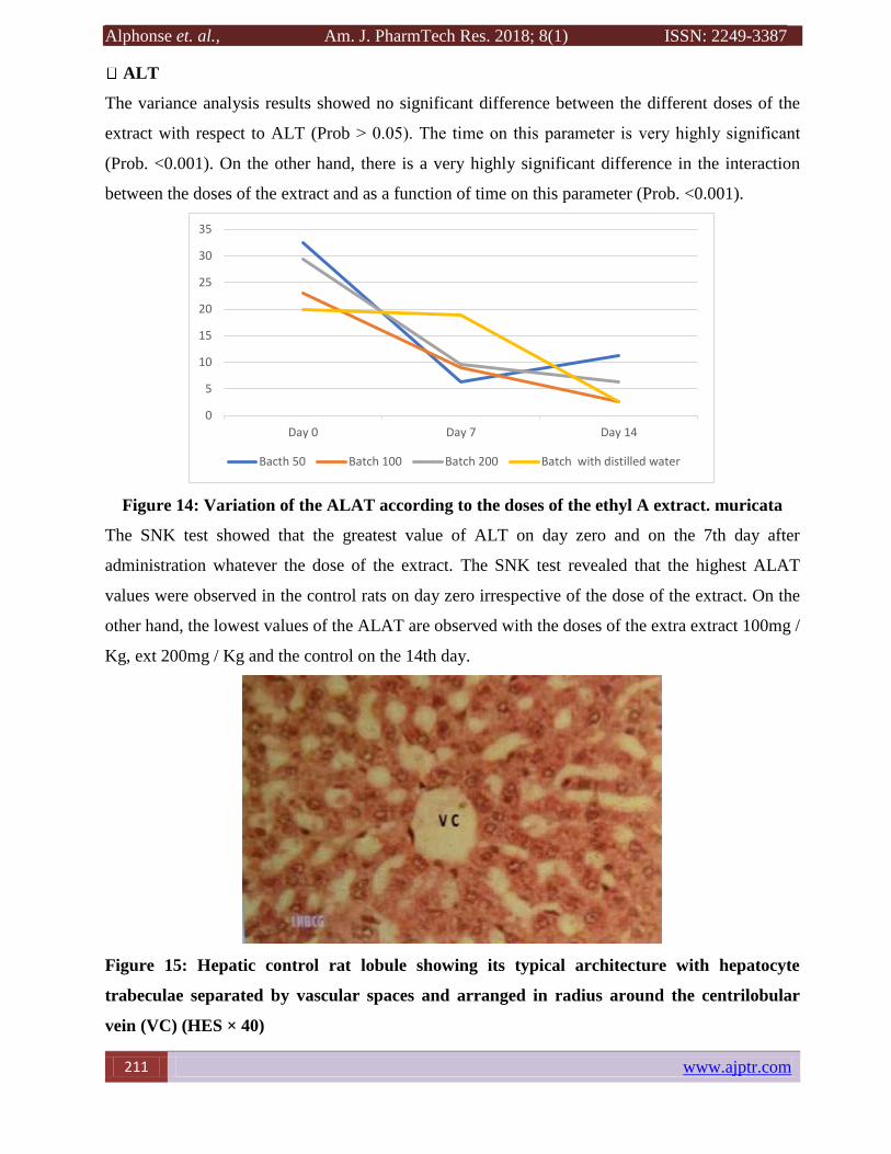
The variance analysis results showed no significant difference between the different doses of the
extract with respect to ALT (Prob ˃ 0.05). The time on this parameter is very highly significant
(Prob. <0.001). On the other hand, there is a very highly significant difference in the interaction
between the doses of the extract and as a function of time on this parameter (Prob. <0.001).
Figure 13: Variation of the ASAT according to the doses of the ethyl A extract. muricata
The SNK test showed that the highest value of ASAT on day zero and on the 7th day after
administration whatever the dose of the extract.
0
10
20
30
40
50
60
70
80
Day 0 Day 7 Day 14
Bacth 50 Batch 100 Batch 200 Batch with distilled water
0
5
10
15
20
25
30
35
Day 0 Day 7 Day 14
Bacth 50 Batch 100 Batch 200 Batch with distilled water
Alphonse et. al., Am. J. PharmTech Res. 2018; 8(1) ISSN: 2249-3387
211 www.ajptr.com
ALT
The variance analysis results showed no significant difference between the different doses of the
extract with respect to ALT (Prob ˃ 0.05). The time on this parameter is very highly significant
(Prob. <0.001). On the other hand, there is a very highly significant difference in the interaction
between the doses of the extract and as a function of time on this parameter (Prob. <0.001).
Figure 14: Variation of the ALAT according to the doses of the ethyl A extract. muricata
The SNK test showed that the greatest value of ALT on day zero and on the 7th day after
administration whatever the dose of the extract. The SNK test revealed that the highest ALAT
values were observed in the control rats on day zero irrespective of the dose of the extract. On the
other hand, the lowest values of the ALAT are observed with the doses of the extra extract 100mg /
Kg, ext 200mg / Kg and the control on the 14th day.
Figure 15: Hepatic control rat lobule showing its typical architecture with hepatocyte
trabeculae separated by vascular spaces and arranged in radius around the centrilobular
vein (VC) (HES × 40)
0
5
10
15
20
25
30
35
Day 0 Day 7 Day 14
Bacth 50 Batch 100 Batch 200 Batch with distilled water

Alphonse et. al., Am. J. PharmTech Res. 2018;8(1) ISSN: 2249-3387
www.ajptr.com 212
Figure 16: Renal cortex of control rats showing a renal glomerulus with flocculi and urinary
tract and urinary tubules (HES × 40)
Figure 17: rat hepatic lobule of lot 1, which received 50 mg / kg of PC of the Annona
muricata ethyl extract: the hepatic architecture is generally conserved (HES × 40)
Figure 18: Batch 1 rat kidney cortex receiving 50 mg / kg PC of Annona muricata ethyl
extract: no significant structural changes (HES × 40)
Alphonse et. al., Am. J. PharmTech Res. 2018; 8(1) ISSN: 2249-3387
213 www.ajptr.com
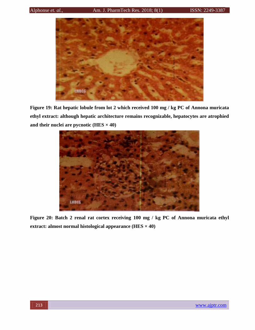
Figure 19: Rat hepatic lobule from lot 2 which received 100 mg / kg PC of Annona muricata
ethyl extract: although hepatic architecture remains recognizable, hepatocytes are atrophied
and their nuclei are pycnotic (HES × 40)
Figure 20: Batch 2 renal rat cortex receiving 100 mg / kg PC of Annona muricata ethyl
extract: almost normal histological appearance (HES × 40)

Alphonse et. al., Am. J. PharmTech Res. 2018;8(1) ISSN: 2249-3387
www.ajptr.com 214
Figure 21: Rat hepatic lobule of lot 3 which received 200 mg / kg of PC of the Annona
muricata ethyl extract: in addition to pycnosis, it should be noted that some hepatocytes are
in hydropic degeneration (HES × 40)
Figure 22: Rat Renal Cortex of Lot 3 receiving 200mg / kg PC of Annona muricata ethyl
extract: there are no morphologically identifiable abnormalities (HES × 40)
The study of the biochemical parameters showed that the extract at various doses had no effect on
the liver cells since this had no influence on the biochemical parameters (ASAT and ALAT)
during the 14 days in the blood (these results were consistent with that of Bridgemohan P., 2013).
On the other hand, the histological study showed that from the dose 100 mg / kg PC of the extract,
hepatic involvement was observed. The latter is accentuated in a hepatic pycnosis of the nuclei at
the dose of 200 mg / kg PC, with an erasure of the cellular limits in the hepatocytes without
modification of the architecture. These results confirm those of our first experiment, in which
hepatic involvement was suspected in acute toxicity. The variance analysis results showed a very

Alphonse et. al., Am. J. PharmTech Res. 2018; 8(1) ISSN: 2249-3387
215 www.ajptr.com
highly significant difference between the different doses of the extract with respect to the amount
of creatinine and urea in the blood. A high value of creatinine and urea was observed with the use
of extract Ext100 mg / Kg followed by Ext200 mg / Kg in blood compared to the time of
administration of the extract. However, there is no change in values or toxic effect on these
parameters with the use of the extract Ext50mg / Kg. In terms of uric acid variance analysis results,
there was a reduction in the time-dependent rate of administration of the Ext50mg / Kg extract,
which would explain the results of Bridgemohan P. ( 2013). Therefore, at a dose greater than or
equal to 100mg / Kg PC, there would be damage to the renal tissues. However, the histological
study showed that at concentrations of 50,100 and 200 there is no negative effect on the kidneys.
The animals in general have supported the usual doses allowed. No deaths have been recorded. All
animals survived at the end of the 14 days of observation, which did not determine the LD50. This
would imply that the LD50 is greater than 200mg / kg bw, and confirm our previous results which
set the LD50 at 3750mg / kg PC extract.
CONCLUSION
Our study is based on the species Annona muricata, a plant widely used in Benin for its
therapeutic properties in the treatment of hypertension, diarrhea, fever, flu and spasms. The
continued use of this plant can lead to side effects that can extend to the death of the individual.
The risk of using this plant would be closely correlated with the doses administered. In this work,
the toxicity of Annona murica has been studied at two levels: acute toxicity and subacute toxicity
of the plant's ethanol extract. Information from Acid toxicity, subacute toxicity and even routine
doses (50,100 and 200) in the Albino Wistar rats suggest that Annona muricata the category of
low-toxic plants orally. The ethanolic extract of Annona muricata may cause symptoms of toxicity
that are dose-dependent, ranging from mild sleepiness, loss of appetite, breathing problems, and
accelerated heart rhythm, leading to seizures , tremors, comas and paralysis of the legs and even
total paralysis leading to death. This toxicity also results in decreased weight, and liver damage.
The results of our work give us an idea of the toxicity of this plant. However, further studies are
desirable.
REFERENCES
1. Adjanohoun E., de Souza S., et Sinsin B. 2000. La biodiversité face au développement des
industries pharmaceutiques africaines. In : Réseau des « espèces ligneuses médicinales »,
EyogMatig O (eds). Compte rendu de la première réunion du réseau tenue 15-17 décembre
1999 à la station IITA Cotonou, Bénin, 88-103

Alphonse et. al., Am. J. PharmTech Res. 2018;8(1) ISSN: 2249-3387
www.ajptr.com 216
2. Ramade, F., 1979. Ecotoxicologie, Ed Masson, Paris, pp. 5.
3. Ruckebusch, Y., 1981. Physiologie, pharmacologie, thérapeutique animale, Ed. Maloine,
Paris, p. 611.
4. Wallace Hayes, A., 2008. Principle and methods of toxicology. Ed Tayler& Francis, New
York, p. 1134.
5. Ben-Romdhane, S., Romdane, M.N., Feki, M., Sanhagi, H., Kaabachi, N., M'Bazaa, A.,
2003. Valeurs usuelles des principaux constituants biochimiques sériques du dromadaire
(Camelusdromedarius). Méd. Vét154(11), 695-702.
6. Rasekh, H.R, Khoshnood-Mansourkhani, M.J., Kamalinejad, M., 2001
7. Dragsted, A., Lang, B., 1957. Etude de la toxicité par administration unique d'un nouveau
médicament. Annales pharmaceutiques Française, p.11.
8. Diezi, J., 1989. Toxicologie : principes de base et répercussions cliniques. Ed Slatkine,
Genève, pp. 33-44.
9. Hilaly et al., 2004. Hypolipidemic effects of Teucriumpoliumin rats. Fitoterapia72, 937-
939.
10. Tulsawani, R., 2010. Ninety day repeated gavage administration of Hipphophaerhamnoides
extract in rats. Food and ChemicalToxicology48, 2483– 2489.
11. Tilkian, S.M., 1979. Clinical Implications of Laboratory Tests. The C.V. Mosby Company,
Missouri, pp. 11–17.
12. Adeneye, A.A., Ajagbonna, O.P., Adeleke, T.I., Bello, S.O., 2006. Preliminary toxicity and
phytochemical studies of the stem bark aqueous extract of Musangacecropioidesin rats.
Journal of Ethnopharmacology105, 374–379.
13. Karber, C., Behrens, B., 1935. WiesindReihenversuche fur biologischeAuswertungen am
ZweckmässigstenAnzuordnen. Arch. Exp. Path. Pharm177, 379-388.
14. Litchfield, J.T., Wilcoxon, F.A., 1949. A simplified method of evaluating dose-effect
experiments. J. Pharmacol. Exp. Ther96, 99-113.
15. Miller, L.C., Tainter, M.L., 1944. Estimation of ED50 and its error by means of
logarithmic. Probitpaper. Proc Soc Exp Viol Med 57, 261-4.
16. OCDE. (2009). PROJET DE LIGNE DIRECTRICE DE L'OCDE POUR LES ESSAIS DE
PRODUITS CHIMIQUES (Section 4 : Effets sur la santé ; Essai N°453 Études combinées
de toxicité chronique et de cancérogenèse, adoptée le 08 Septembre 2009).
17. Pirro, 2014. Reins et voies urinaires-appareil génital masculin : anatomie du rein et
vascularisation

Alphonse et. al., Am. J. PharmTech Res. 2018; 8(1) ISSN: 2249-3387
217 www.ajptr.com
18. P. HANOVER (1963 - 1964), METHODES D'ANALYSES utilisées au Laboratoire des
Glucides c.s.T. BONDY
19. Natacha A. 2014, Action d’extraits éthyliques de Annona muricata dans le traitement
traditionnel du cancer de foie. Screening phytochimique et recherche de l’activité
antiproliférative chez le rat wistar. Mémoire de fin de formation en Master de Physiologie
et Pharmacologie Cellulaires.
20. Bridgemohan P., Bridgemohan R.S.H., 2013; Evaluation of decoction extracts of two types
of soursop (Annona nuricata) for annonaceous acetogenin properties.
AJPTR is
Peer-reviewed
bimonthly
Rapid publication
Submit your manuscript at: [email protected]
Copyright © 2022 FDOKUMEN